The content of dopamine, serotonin, and their metabolites in the neural circuit that mediates...
10
UNCORRECTED PROOF Brain Research Bulletin xxx (2004) xxx–xxx 3 The content of dopamine, serotonin, and their metabolites in the neural circuit that mediates maternal behavior in juvenile and adult rats 4 5 D.E. Olazábal a,∗ , E. Abercrombie a , J.S. Rosenblatt b , J.I. Morrell a 6 a Center for Molecular and Behavioral Neuroscience, Rutgers, The State University of New Jersey, Newark, NJ 07102, USA 7 b Department of Psychology, Rutgers, The State University of New Jersey, Newark, NJ 07102, USA 8 Received 2 September 2003; received in revised form 16 February 2004; accepted 24 February 2004 9 Abstract 10 Continuous exposure of non-parturient rats to pups can induce maternal behavior similar in most aspects to that found in the postpartum rat. Surprisingly, young juvenile rats (20–24 days of age) only require 1–3 days of exposure to pups, while adults require 4–8 days before maternal behavior emerges. Dopamine (DA) and possibly serotonin (5-HT) may mediate the expression of adult maternal behavior. We hypothesize that postnatal changes in DA and 5-HT within the neural circuit that supports maternal behavior including the medial preoptic area (MPOA), medial and cortical amygdala (MCA), and nucleus accumbens (NAC), may underlie these differences in responsiveness across juveniles and adults. We measured DA, 5-HT, and their metabolites in postmortem samples of these regions in maternal and non-maternal juvenile and adult females. 11 12 13 14 15 16 17 The only difference found across behavioral groups was that the MPOA of adults induced into maternal behavior by pup exposure had more DA than did that of isolated adult females or maternal juveniles. However, when adults versus juveniles were compared, the content of DA and 3,4-dihydroxyphenylacetic (DOPAC) was higher in the adult than in the juvenile NAC and MCA; the content of 5-HT and 5-hydroxyindoleacetic acid (5-HIAA) in these structures did not vary across the age groups. In contrast, higher levels of 5-HT and 5-HIAA were found in the MPOA in juveniles compared to adults. We propose that these region-specific age differences in DA and 5HT may underlie differences in juvenile–adult responses to pups. 18 19 20 21 22 23 © 2004 Published by Elsevier Inc. 24 Keywords: Accumbens; Amygdala; MPOA; Neurochemistry; Pups 25 1. Introduction 26 Maternal behavior in the rat consists of maternal nest 27 building, and pup directed behaviors including retrieving, 28 nursing, grooming, and protection from intruders. Parturient 29 females are immediately maternally responsive to their pups. 30 Less generally known is that fact that female, male, or juve- 31 nile rats can be induced to express maternal behavior by con- 32 tinuous exposure to new born pups provided from parturient 33 female foster mothers. The expression of pup-induced ma- 34 ternal behavior has been studied extensively; it is virtually 35 indistinguishable from hormonally-induced maternal behav- 36 ior, except that parturient females nurse their pups. [47,58]. 37 Adult rats (>60 days of age) require 4–8 days of exposure 38 ∗ Corresponding author. Present address: Center for Behavioral Neuro- science, 954 Gatewood Road, N.E., Atlanta, GA 30329, USA. Tel.: +1-404-727-8269; fax: +1-404-727-8070. E-mail address: [email protected] (D.E. Olaz´ abal). to pups before maternal behavior emerges, while surpris- 39 ingly young juvenile rats (20–26 days of age) only require 40 1–3 days of exposure to pups. This required exposure period 41 increases as the juveniles approach puberty [6,7,43–45,65]. 42 These data suggest an intriguing difference in behavioral re- 43 sponsiveness across young juveniles and adults. 44 Key components of the neural circuit that mediate mater- 45 nal behavior include the medial preoptic area (MPOA), the 46 medial and cortical amygdala (MCA), and the nucleus ac- 47 cumbens (NAC), along with other limbic and hypothalamic 48 structures [48]. Most of our knowledge of the neural cir- 49 cuit that mediates maternal behavior has been gathered from 50 studies using adult animals; much less has been done using 51 the juvenile. The MPOA is necessary for both the expres- 52 sion of and motivation to perform maternal behavior [48]. In 53 adults both large and very small lesions in the MPOA dis- 54 rupt maternal behavior while in the juvenile or pubertal rat, 55 only large lesions disrupt the behavior, small lesions do not 56 [36,50,51]. Moreover, increased expression of immunoreac- 57 1 0361-9230/$ – see front matter © 2004 Published by Elsevier Inc. 2 doi:10.1016/j.brainresbull.2004.02.009 BRB 6797 1–10
Transcript of The content of dopamine, serotonin, and their metabolites in the neural circuit that mediates...
UN
CO
RR
EC
TED
PR
OO
F
Brain Research Bulletin xxx (2004) xxx–xxx
3
The content of dopamine, serotonin, and their metabolites in the neuralcircuit that mediates maternal behavior in juvenile and adult rats
4
5
D.E. Olazábala,∗, E. Abercrombiea, J.S. Rosenblattb, J.I. Morrella6
a Center for Molecular and Behavioral Neuroscience, Rutgers, The State University of New Jersey, Newark, NJ 07102, USA7b Department of Psychology, Rutgers, The State University of New Jersey, Newark, NJ 07102, USA8
Received 2 September 2003; received in revised form 16 February 2004; accepted 24 February 20049
Abstract10
Continuous exposure of non-parturient rats to pups can induce maternal behavior similar in most aspects to that found in the postpartum rat.Surprisingly, young juvenile rats (20–24 days of age) only require 1–3 days of exposure to pups, while adults require 4–8 days before maternalbehavior emerges. Dopamine (DA) and possibly serotonin (5-HT) may mediate the expression of adult maternal behavior. We hypothesizethat postnatal changes in DA and 5-HT within the neural circuit that supports maternal behavior including the medial preoptic area (MPOA),medial and cortical amygdala (MCA), and nucleus accumbens (NAC), may underlie these differences in responsiveness across juveniles andadults. We measured DA, 5-HT, and their metabolites in postmortem samples of these regions in maternal and non-maternal juvenile andadult females.
11
12
13
14
15
16
17
The only difference found across behavioral groups was that the MPOA of adults induced into maternal behavior by pup exposure hadmore DA than did that of isolated adult females or maternal juveniles. However, when adults versus juveniles were compared, the contentof DA and 3,4-dihydroxyphenylacetic (DOPAC) was higher in the adult than in the juvenile NAC and MCA; the content of 5-HT and5-hydroxyindoleacetic acid (5-HIAA) in these structures did not vary across the age groups. In contrast, higher levels of 5-HT and 5-HIAAwere found in the MPOA in juveniles compared to adults. We propose that these region-specific age differences in DA and 5HT may underliedifferences in juvenile–adult responses to pups.
18
19
20
21
22
23
© 2004 Published by Elsevier Inc.24
Keywords: Accumbens; Amygdala; MPOA; Neurochemistry; Pups25
1. Introduction26
Maternal behavior in the rat consists of maternal nest27
building, and pup directed behaviors including retrieving,28
nursing, grooming, and protection from intruders. Parturient29
females are immediately maternally responsive to their pups.30
Less generally known is that fact that female, male, or juve-31
nile rats can be induced to express maternal behavior by con-32
tinuous exposure to new born pups provided from parturient33
female foster mothers. The expression of pup-induced ma-34
ternal behavior has been studied extensively; it is virtually35
indistinguishable from hormonally-induced maternal behav-36
ior, except that parturient females nurse their pups.[47,58].37
Adult rats (>60 days of age) require 4–8 days of exposure38
∗ Corresponding author. Present address: Center for Behavioral Neuro-science, 954 Gatewood Road, N.E., Atlanta, GA 30329, USA.Tel.: +1-404-727-8269; fax:+1-404-727-8070.
E-mail address: [email protected] (D.E. Olazabal).
to pups before maternal behavior emerges, while surpris-39
ingly young juvenile rats (20–26 days of age) only require40
1–3 days of exposure to pups. This required exposure period41
increases as the juveniles approach puberty[6,7,43–45,65]. 42
These data suggest an intriguing difference in behavioral re-43
sponsiveness across young juveniles and adults. 44
Key components of the neural circuit that mediate mater-45
nal behavior include the medial preoptic area (MPOA), the46
medial and cortical amygdala (MCA), and the nucleus ac-47
cumbens (NAC), along with other limbic and hypothalamic48
structures[48]. Most of our knowledge of the neural cir- 49
cuit that mediates maternal behavior has been gathered from50
studies using adult animals; much less has been done using51
the juvenile. The MPOA is necessary for both the expres-52
sion of and motivation to perform maternal behavior[48]. In 53
adults both large and very small lesions in the MPOA dis-54
rupt maternal behavior while in the juvenile or pubertal rat,55
only large lesions disrupt the behavior, small lesions do not56
[36,50,51]. Moreover, increased expression of immunoreac-57
1 0361-9230/$ – see front matter © 2004 Published by Elsevier Inc.2 doi:10.1016/j.brainresbull.2004.02.009
BRB 6797 1–10
UN
CO
RR
EC
TED
PR
OO
F
2 D.E. Olazabal et al. / Brain Research Bulletin xxx (2004) xxx–xxx
tive c-fos in the MPOA and other components of this neural58
circuit occurs in adults during pup-induced maternal behav-59
ior, however, in juveniles c-fos induction depends upon the60
age of the juveniles and perhaps on the extent of pup ex-61
posure[24,37,51], suggesting that this is a maturing circuit62
component.63
In the non-maternal adult female, neuronal activity in the64
MCA inhibits maternal behavior by a mechanism that in-65
volves processing of olfactory cues from the initial exposure66
to pups[20,47,48]. The hormones of the peripartum period67
are thought to alter or release this basal inhibition so that68
the expression of maternal behavior can occur. Prolonged69
pup exposure also reduces this basal inhibition allowing the70
onset of pup-induced maternal behavior. Lesions in the me-71
dial amygdala had similar effects in both older juveniles and72
adults, shortening the time of pup exposure needed to in-73
duce maternal behavior[51]. The NAC is necessary for re-74
trieval components of maternal behavior, and for processing75
of pup related stimuli likely to be related to mechanisms of76
motivation in the adult, not yet studied in the juvenile[48].77
The scope of our knowledge concerning the neurotrans-78
mitters that mediate maternal behavior is limited to a few79
transmitters and to the adult model[48]. The literature pro-80
vides the clearest case for a role for dopamine (DA) in the81
mediation of maternal behavior[5,21,22,25,26,28,49,66].82
Extensive lesions of the dopaminergic system[25–27] dis-83
rupt maternal behavior, and dopamine receptor antagonists84
infused locally into the NAC inhibited retrieval and licking85
components of maternal behavior[39]. These studies sug-86
gest that expression of the behavior requires dopamine. In-87
creases in DA and DA metabolites, dihydroxyphenylacetic88
acid (DOPAC) and homovanillic acid (HVA), in the extra-89
cellular space in the ventral striatum after separated mother90
rats and pups were reunited[28] suggest a role in the moti-91
vational components of the behavioral response.92
The exploration of a role for other neurotransmitters in93
the mediation of maternal behavior is very limited; how-94
ever, there is modest evidence that serotonin (5-HT) may95
be involved. Barofsky et al.[2] found that serotonergic le-96
sions in the median raphe nucleus caused short-term disrup-97
tion in maternal behavior but not the prolactin response with98
suckling, however, the study does not establish functional99
specificity since locomotor impairment was not ruled out.100
Ferreira et al.[17] found that buspirone (a partial 5-HT1A101
agonist) inhibited maternal behavior; however, they did find102
reduced motor activity, limiting the specificity of the inter-103
pretation. Microdialysis also showed an increase in the con-104
centration of the metabolite for 5-HT, 5-hydroxyindoleacetic105
(5-HIAA), in the ventral striatum when dams were reunited106
with pups[28]; perhaps motivational components of mater-107
nal behavior are supported by this system as well. Infusions108
of cocaine into the NAC or MPOA disrupt adult maternal109
behavior[74]; suggesting the exact level of DA and possi-110
bly 5-HT neurotransmitters is important. Thus, an excess of111
these transmitters could be as disruptive as their reduction112
or absence.113
Our working hypothesis is that the brain regions medi-114
ating maternal behavior in the adult, also mediate maternal115
behavior in the juvenile, and that postnatal neurochemical116
changes within these regions may underlie the maturing time117
course of pup-induced maternal behavior. While it is for-118
mally possible that maternal behavior in the juvenile might119
be supported by different neural structures or transmitters120
than those that support maternal behavior in the adult, we121
know of no data to support this alternative hypothesis. 122
It has been hypothesized that adult rats, without benefit of123
the hormones of parturition, require longer exposure to pups124
to become maternal because their initial response to pups is125
a typical adult neophobic response to novel stimuli, while126
juveniles are generally less neophobic and affiliate readily127
with pups[18,19,43,45,55,63]. We hypothesize that postna-128
tal changes within the neural circuit may add a level of in-129
hibition to social stimuli, and that this is the fundamental130
difference between the juvenile and adult state. The com-131
parison of juveniles (20–27 days old) to adults (>60 days)132
offers the intriguing view before and after these hypothe-133
sized developmental changes in the inhibitory components134
of the neural circuit occur, and to consider that these two135
neurotransmitters might be part of the mechanistic basis for136
this additional behavioral inhibition. 137
Our goal was to examine the content of dopamine and138
serotonin and their metabolites in juveniles and adults within139
the neural circuit that mediates the expression of maternal140
behavior. We choose postmortem sampling of tissue punches141
as our approach so that we could examine multiple brain142
areas in juveniles and adults. An alternative, microdialysis143
would allow measurements in only one area at a time. First,144
the behavioral state of each animal was assessed. Then post-145
mortem tissue analysis was used to determine, in each in-146
dividual animal, the amount of these neurotransmitters and147
their metabolites in three different brain regions, the NAC,148
MPOA, and MCA. 149
Two specific questions were addressed. Does the amount150
of these neurotransmitters and their metabolites vary with the151
maternal state of the rat? Is the amount of these substances152
in these specific regions different in adults compared with153
juveniles? 154
2. Material and methods 155
2.1. Animal care and subjects 156
The animals were from our colony maintained at the157
Rutgers University Laboratory Animal Facility in Newark158
which is accredited by the Association for the Accredi-159
tation and Assessment of Laboratory Animal Care. They160
were Sprague–Dawley strain rats, bred from animals orig-161
inally purchased from Charles River Laboratories (Wilm-162
ington, DE). Additional breeding animals are systematically163
added to the colony. Animals are maintained under a 12-h164
light/12-h dark cycle at 22◦C with ad libitum access to165
BRB 6797 1–10
UN
CO
RR
EC
TED
PR
OO
F
D.E. Olazabal et al. / Brain Research Bulletin xxx (2004) xxx–xxx 3
food and water. Wood shavings (BETA CHIP®, NEPCOTM,166
North Eastern Products Corp., NY) were used to cover the167
floor of the cages. All procedures followed the standards168
approved by the National Institutes of Health Guide for the169
Care and Use of Laboratory Animals.170
Rats were housed in groups of two until used in the exper-171
iments. Then they were housed individually in cages mea-172
suring 53 cm× 36 cm× 25 cm with transparent Plexiglas173
walls. Females for the parturient adult experimental group174
or as donor mothers to supply foster pups were mated by175
our published procedures[36,37,50].176
2.2. Behavioral paradigm177
2.2.1. Pup-induction of maternal behavior178
Adult virgin or juvenile females were continuously ex-179
posed in their home cages to three newborn pups 2–7 days180
old. Every 24 h these pups were returned to donor mothers181
for care and feeding, while other freshly fed pups were sup-182
plied to the experimental animals. Donor mothers were sep-183
arate from experimental groups[58]. The number of days184
of pup exposure needed to induce maternal behavior is also185
called the latency to express maternal behavior. This was186
calculated as the interval between the first day of pup expo-187
sure and the first of the two consecutive days in which the188
animals exhibited maternal behavior. Further, details of our189
operational definition of maternal behavior and our behavior190
testing paradigm were as previously published[36,37,50].191
2.2.2. Time of pup exposure and maternal behavior192
latencies193
All groups exposed to pups were tested daily for maternal194
behavior. As expected, the average number of days of con-195
tinuous pup exposure required for the emergence of mater-196
nal behavior was much longer in adults (6± 1 days, median197
6 days) than in juveniles (1.5 ± 0.4 days, median 1 day).198
Maternal groups included only animals that showed all com-199
ponents of maternal behavior and were exposed to pups in200
this state for 4 days before sacrifice. Adults induced into the201
maternal state by pup exposure were, therefore, necessarily202
exposed to pups on average for 10±1 day (median 10) total,203
whereas juveniles were exposed for 5.5±0.4 days (median 5)204
total. In order to produce groups of non-maternal juveniles,205
exposure to pups had to be limited to 1 day, whereas some206
adults could be exposed to pups 7± 1 days without being207
induced into the maternal state. For these two non-maternal208
pup exposed groups, this was a close match of pup exposure209
to the comparable maternal groups as was possible for the210
two age groups.211
2.3. Animal groups212
Twenty-eight adult females (virgin adult females or par-213
turient females, 60–100 days old) and twenty seven juvenile214
females (19–21 days old), were used. Adult virgins were not215
classified as to stage of their cycle.216
Three behavioral groups were comparable across the two217
ages. 218
1. Maternal by pup exposure 219
These were adults or juveniles that became maternal220
by pup exposure which were exposed to pups, obtained221
from donor mothers, until they showed maternal behavior222
on four consecutive days, so that the fourth day of full223
maternal behavior was the day of sacrifice. 224
2. Non-maternal pup exposed 225
These were adults or juveniles which did not become226
maternal with pup exposure. These differ from animals227
group 1 only in that they were non-maternal. 228
3. Social isolates 229
These were adults or juveniles isolated 1–8 days, and230
not exposed to either pups or comparable age siblings.231
Two additional groups specific to each age group included232
4. Sibling exposed juveniles 233
Juveniles that were exposed to sibling, age-matched234
females, sacrificed on the fourth day of such housing.235
5. Maternal lactating females 236
These were sacrificed on day 4 postpartum that is 4237
days after the onset of maternal behavior at parturition.238
2.4. Brain tissue sample preparation 239
Rats were decapitated with a guillotine, immediately af-240
ter behavioral testing. Sacrifice was during the mid-portion241
of the light phase of the light/dark cycle. The brains were242
immediately removed from the skull, submerged in cold243
phosphate-buffered saline (PBS, pH 7.4; 4◦C) for 20 s, and 244
placed ventral side up in a chilled brain mold designed for245
cross sectioning the brain. Sections were made at 1 mm in-246
tervals; five razor blades were used to cut the cross sections247
needed for further dissection of the NAC, the MPOA, and248
the MCA. The first blade was placed in the mold’s division249
parallel to the rostral extent of the optic chiasm. A second250
blade was placed 1 mm caudal to it, and the section between251
blades 1 and 2 contained the MPOA sample. A third blade252
was placed 1 mm rostral to the optic chiasm, and the sec-253
tion between blades 1 and 3 was discarded. A fourth blade254
was placed 3 mm rostral to the optic chiasm to obtain a255
2-mm-wide section, and this section between blades 3 and 4256
was sampled for the NAC. The last blade was placed 3 mm257
caudal to the optic chiasm to obtain a 2 mm wide section,258
between blades 2 and 5, which was sampled for the MCA.259
Once all blades were in place, the sections were cut and260
then the blades were lifted out in sequence from rostral to261
caudal, with the sections adhering to blade. 262
The selected brain regions were further dissected on the263
blade in the order NAC, MPOA, and MCA[52]. A bilateral 264
sample of the NAC was taken using a 12-gauge stainless steel265
tube. The tube was pushed into the NAC immediately medial266
and adjacent to the anterior commissure (Fig. 1A), and then 267
withdrawn. When necessary, a thinner tube placed inside the268
BRB 6797 1–10
UN
CO
RR
EC
TED
PR
OO
F
4 D.E. Olazabal et al. / Brain Research Bulletin xxx (2004) xxx–xxx
Bregma 0.70 mm
VP
NAC
LV
ICjM
Bregma 2.70 mmac
NAC
Bregma -1.30 mm
ox
3VMPOA
Bregma -0.26 mmf
ox
LPOA
ac
Nucleus Accumbens
Medial Preoptic Area
MPOA
Bregma -3.30 mm3V
LHCeC
BlA
PirMCA
optrf
Bregma -1.40 mm
f
MPOA
ox
LH
MCA
Medial and Cortical Amygdala
(A)
(B)
(C)
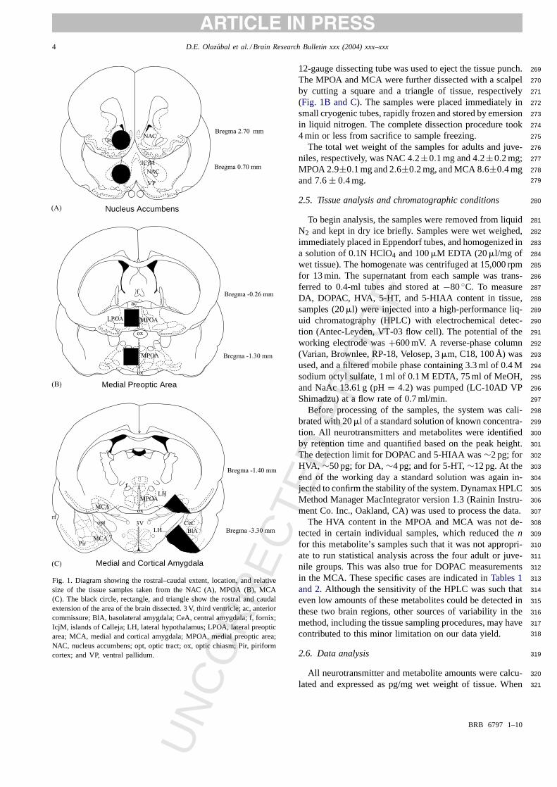
Fig. 1. Diagram showing the rostral–caudal extent, location, and relativesize of the tissue samples taken from the NAC (A), MPOA (B), MCA(C). The black circle, rectangle, and triangle show the rostral and caudalextension of the area of the brain dissected. 3 V, third ventricle; ac, anteriorcommissure; BlA, basolateral amygdala; CeA, central amygdala; f, fornix;IcjM, islands of Calleja; LH, lateral hypothalamus; LPOA, lateral preopticarea; MCA, medial and cortical amygdala; MPOA, medial preoptic area;NAC, nucleus accumbens; opt, optic tract; ox, optic chiasm; Pir, piriformcortex; and VP, ventral pallidum.
12-gauge dissecting tube was used to eject the tissue punch.269
The MPOA and MCA were further dissected with a scalpel270
by cutting a square and a triangle of tissue, respectively271
(Fig. 1B and C). The samples were placed immediately in272
small cryogenic tubes, rapidly frozen and stored by emersion273
in liquid nitrogen. The complete dissection procedure took274
4 min or less from sacrifice to sample freezing. 275
The total wet weight of the samples for adults and juve-276
niles, respectively, was NAC 4.2±0.1 mg and 4.2±0.2 mg; 277
MPOA 2.9±0.1 mg and 2.6±0.2 mg, and MCA 8.6±0.4 mg 278
and 7.6 ± 0.4 mg. 279
2.5. Tissue analysis and chromatographic conditions 280
To begin analysis, the samples were removed from liquid281
N2 and kept in dry ice briefly. Samples were wet weighed,282
immediately placed in Eppendorf tubes, and homogenized in283
a solution of 0.1N HClO4 and 100�M EDTA (20�l/mg of 284
wet tissue). The homogenate was centrifuged at 15,000 rpm285
for 13 min. The supernatant from each sample was trans-286
ferred to 0.4-ml tubes and stored at−80◦C. To measure 287
DA, DOPAC, HVA, 5-HT, and 5-HIAA content in tissue,288
samples (20�l) were injected into a high-performance liq-289
uid chromatography (HPLC) with electrochemical detec-290
tion (Antec-Leyden, VT-03 flow cell). The potential of the291
working electrode was+600 mV. A reverse-phase column292
(Varian, Brownlee, RP-18, Velosep, 3�m, C18, 100 Å) was 293
used, and a filtered mobile phase containing 3.3 ml of 0.4 M294
sodium octyl sulfate, 1 ml of 0.1 M EDTA, 75 ml of MeOH,295
and NaAc 13.61 g (pH= 4.2) was pumped (LC-10AD VP296
Shimadzu) at a flow rate of 0.7 ml/min. 297
Before processing of the samples, the system was cali-298
brated with 20�l of a standard solution of known concentra-299
tion. All neurotransmitters and metabolites were identified300
by retention time and quantified based on the peak height.301
The detection limit for DOPAC and 5-HIAA was∼2 pg; for 302
HVA, ∼50 pg; for DA,∼4 pg; and for 5-HT,∼12 pg. At the 303
end of the working day a standard solution was again in-304
jected to confirm the stability of the system. Dynamax HPLC305
Method Manager MacIntegrator version 1.3 (Rainin Instru-306
ment Co. Inc., Oakland, CA) was used to process the data.307
The HVA content in the MPOA and MCA was not de-308
tected in certain individual samples, which reduced then 309
for this metabolite’s samples such that it was not appropri-310
ate to run statistical analysis across the four adult or juve-311
nile groups. This was also true for DOPAC measurements312
in the MCA. These specific cases are indicated inTables 1 313
and 2. Although the sensitivity of the HPLC was such that314
even low amounts of these metabolites could be detected in315
these two brain regions, other sources of variability in the316
method, including the tissue sampling procedures, may have317
contributed to this minor limitation on our data yield. 318
2.6. Data analysis 319
All neurotransmitter and metabolite amounts were calcu-320
lated and expressed as pg/mg wet weight of tissue. When321
BRB 6797 1–10
UN
CO
RR
EC
TED
PR
OO
F
D.E. Olazabal et al. / Brain Research Bulletin xxx (2004) xxx–xxx 5
Table 1Concentrations of dopamine, serotonin, and metabolites in brain regions of adults
Treatment DA DOPAC HVA 5-HT 5-HIAA
NAC Isolated w/o pups (n = 8) 7419± 936 1328± 187 408± 47 342± 84 153± 34Non-maternal w/pups (n = 7) 6781± 1375 1057± 146 436± 56 344± 120 92± 28Maternal sensitized (n = 6) 8542± 713 1221± 122 449± 39 632± 216 209± 63Maternal lactating (n = 7) 7598± 912 1187± 94 444± 73 375± 125 171± 58
MPOA Isolated w/o pups (n = 5) 184± 26 65± 11 63±11 (1 n.d) 228± 99 93± 36Non-maternal w/pups (n = 5) 248± 34 75± 15 38± 3 (3 n.d) 100± 13 74± 9Maternal sensitized (n = 5) 300± 36∗ 87 ± 12 62± 14 (2 n.d) 245± 150 102± 57Maternal lactating (n = 8) 218± 28 63± 6 66 ± 19 (4 n.d) 105± 22 65± 12
MCA Isolated w/o pups (n = 7) 198± 40 46± 19 (5 n.d) 159 (6 n.d) 320± 93 102± 40Non-maternal w/pups (n = 6) 263± 96 74± 12 (3 n.d) 33± 14 (4 n.d) 408± 91 165± 50Maternal sensitized (n = 5) 247± 68 62± 27 187± 143 (3 n.d) 223± 59 116± 27Maternal lactating (n = 9) 165± 24 66± 18 (1 n.d) 136 (8 n.d) 484± 127 175± 43
Data are expressed in pg/mg wet weight (means± S.E.M.). n.d., non-detected values.∗ P < 0.03 statistically significant with respect to isolated.
Table 2Concentrations of dopamine, serotonin, and metabolites in brain regions of juveniles
Treatment DA DOPAC HVA 5-HT 5-HIAA
NAC Isolated w/o pups (n = 4) 4307± 768 1011± 87 524± 68 105± 56 72± 39Non-maternal w/pups (n = 5) 6453± 1009 1007± 82 637± 112 224± 104 141± 68Maternal sensitized (n = 6) 6234± 657 936± 98 509± 33 438± 110 172± 35Social (n = 4) 6334± 798 991± 99 602± 55 303± 127 109± 31
MPOA Isolated w/o pups (n = 3) 112± 15 32± 17 46 (2 n.d) 248± 79 149± 18Non-maternal w/pups (n = 5) 445± 194 87± 22 81± 20 (1 n.d) 260± 39 124± 12Maternal sensitized (n = 7) 146± 21 77± 17 70± 16 (2 n.d) 266± 34 121± 13Social (n = 6) 117± 8 50 ± 6 57 ± 3 (2 n.d) 239± 39 112± 20
MCA Isolated w/o pups (n = 8) 158± 41 59± 16 (4 n.d) 66± 18 (3 n.d) 358± 33 140± 21Non-maternal w/pups (n = 5) 100± 16 37± 6 (2 n.d) 69± 9 (1 n.d) 332± 89 148± 42Maternal sensitized (n = 8) 125± 25 50± 18 (4 n.d) 53± 3 (6 n.d) 322± 66 115± 13Social (n = 6) 91±20 34±13 (2 n.d) 44± 9 (4 n.d) 297±62 103±19
Data are expressed in pg/mg wet weight (means± S.E.M.). n.d., non-detected values.
the data passed Bartlett’s test of homogeneity of variance,322
the parametric tests including ANOVA and the post hoc323
Fisher protected least significant difference (PLSD) test324
were applied. In only one case, the juvenile MPOA value325
for dopamine, the variance was not homogeneous and no326
correction was possible; these comparisons were made with327
non-parametric methods, specifically the Kruskal–Wallis328
test followed by the Mann–Whitney test. Adult and juvenile329
behavioral groups that were not statistically different from330
each other (P > 0.05) were combined within the age cate-331
gories of adults or juveniles, and subsequently the two age332
groups were compared byt-test, after Bartlett’s test verified333
that parametric testing was appropriate.334
3. Results335
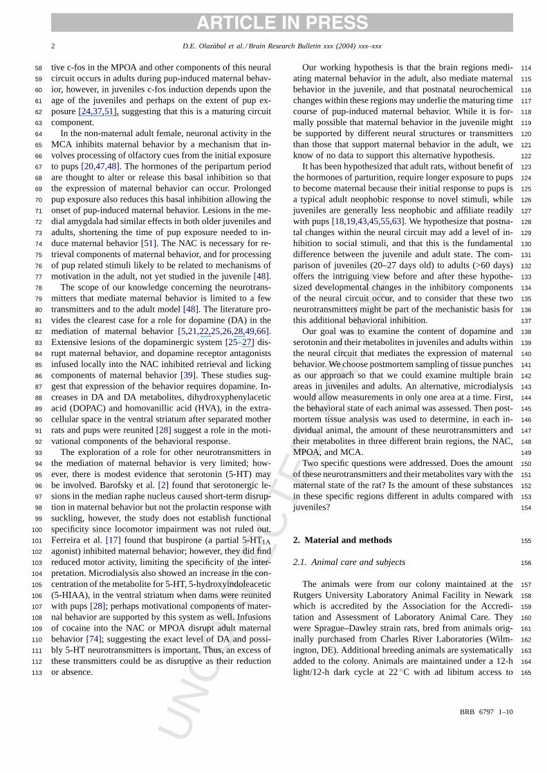
3.1. Nucleus accumbens and medial cortical amygdala336
There were no statistically significant differences across337
behavioral groups within each age group in samples from the338
NAC or the amygdala. Therefore, all the individuals in the339
behavioral groups were averaged for comparisons of adults340
versus juveniles. 341
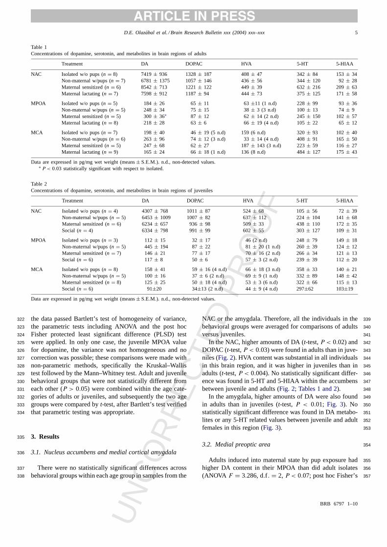
In the NAC, higher amounts of DA (t-test,P < 0.02) and 342
DOPAC (t-test,P < 0.03) were found in adults than in juve-343
niles (Fig. 2). HVA content was substantial in all individuals344
in this brain region, and it was higher in juveniles than in345
adults (t-test,P < 0.004). No statistically significant differ-346
ence was found in 5-HT and 5-HIAA within the accumbens347
between juvenile and adults (Fig. 2; Tables 1 and 2). 348
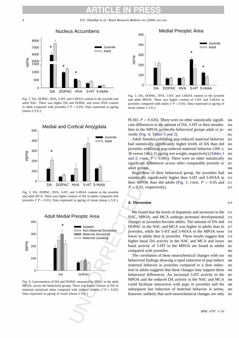
In the amygdala, higher amounts of DA were also found349
in adults than in juveniles (t-test, P < 0.01; Fig. 3). No 350
statistically significant difference was found in DA metabo-351
lites or any 5-HT related values between juvenile and adult352
females in this region (Fig. 3). 353
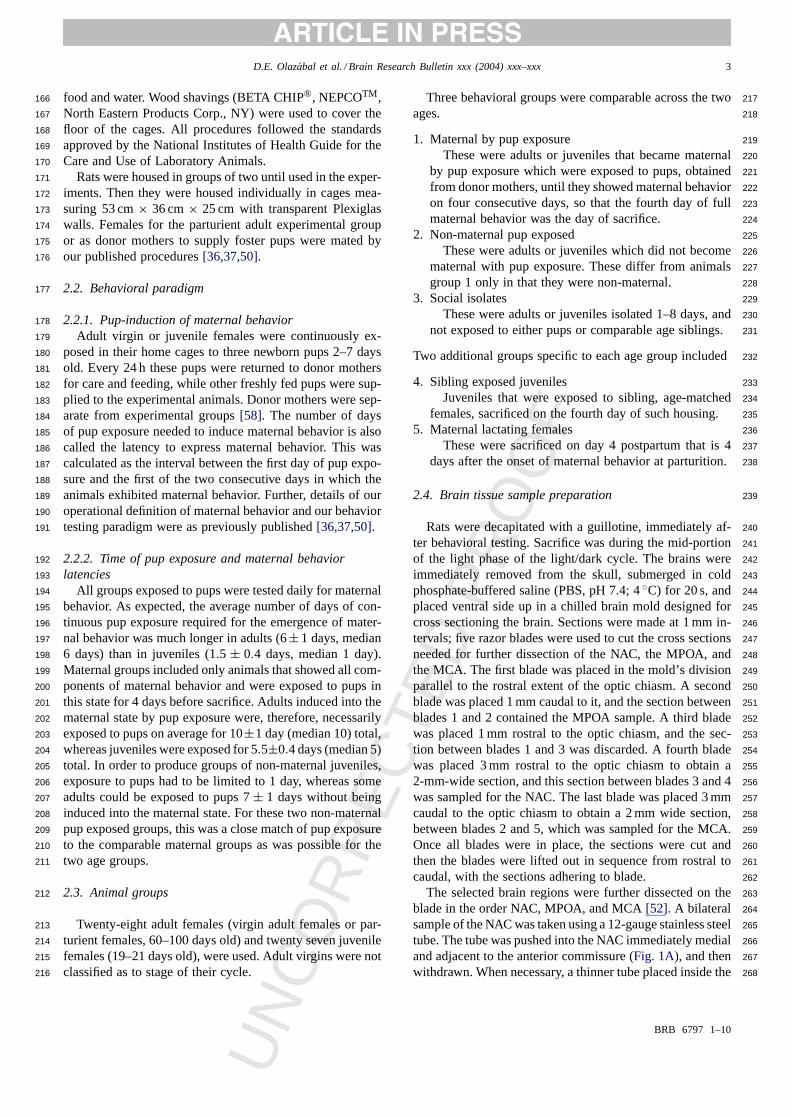
3.2. Medial preoptic area 354
Adults induced into maternal state by pup exposure had355
higher DA content in their MPOA than did adult isolates356
(ANOVA F = 3.286, d.f . = 2, P < 0.07; post hoc Fisher’s357
BRB 6797 1–10
UN
CO
RR
EC
TED
PR
OO
F
6 D.E. Olazabal et al. / Brain Research Bulletin xxx (2004) xxx–xxx
Nucleus Accumbenspg
/mg
0
500
1000
15005000
6000
7000
8000
JuvenileAdult
DA DOPAC HVA 5-HT 5-HIAA
*
**
Fig. 2. DA, DOPAC, HVA, 5-HT, and 5-HIAA content in the juvenile andadult NAC. There was higher DA and DOPAC and lower HVA contentin adult compared with juveniles (∗P < 0.03). Data expressed as pg/mg(mean± S.E.).
Medial and Cortical Amygdala
pg/m
g
0
100
200
300
400
500
JuvenileAdult
DA DOPAC HVA 5-HT 5-HIAA
*
Fig. 3. DA, DOPAC, HVA, 5-HT, and 5-HIAA content in the juvenileand adult MCA. There was higher content of DA in adults compared withjuveniles (∗P < 0.01). Data expressed as pg/mg of tissue (mean± S.E.).
Adult Medial Preoptic Area
pg/m
g
0
100
200
300
400
Isolated Non-Maternal SensitizedMaternal Sensitized Maternal Lactating
*
DA DOPAC
Fig. 4. Concentration of DA and DOPAC measured by HPLC in the adultMPOA, across the behavioral groups. There was higher content of DA inmaternal sensitized when compared with isolated females (∗P < 0.03).Data expressed as pg/mg of tissue (mean± S.E.).
Medial Preoptic Area
pg/m
g
0
100
200
300
400
500
JuvenileAdult
DA DOPAC HVA 5-HT 5-HIAA
*
*
Fig. 5. DA, DOPAC, HVA, 5-HT, and 5-HIAA content in the juvenileand adult MPOA. There was higher content of 5-HT and 5-HIAA injuveniles compared with adults (∗P < 0.05). Data expressed as pg/mg oftissue (mean± S.E.).
PLSD,P < 0.026). There were no other statistically signifi-358
cant differences in the amount of DA, 5-HT or their metabo-359
lites in the MPOA across the behavioral groups adult or ju-360
venile (Fig. 4; Tables 1 and 2). 361
Adult females exhibiting pup-induced maternal behavior362
had statistically significantly higher levels of DA than did363
juveniles exhibiting pup-induced maternal behavior (300± 364
36 versus 146±21 pg/mg wet weight, respectively) (Tables 1 365
and 2; t-test,P < 0.005). There were no other statistically366
significant differences across other comparable juvenile or367
adult groups. 368
Regardless of their behavioral group, the juveniles had369
statistically significantly higher than 5-HT and 5-HIAA in370
their MPOA than did adults (Fig. 5, t-test,P < 0.05 and 371
P < 0.02, respectively). 372
4. Discussion 373
We found that the levels of dopamine and serotonin in the374
NAC, MPOA, and MCA undergo postnatal developmental375
changes as juveniles become adults. The amount of DA and376
DOPAC in the NAC and MCA was higher in adults than in377
juveniles, while the 5-HT and 5-HIAA in the MPOA were378
lower in adults than in juveniles. These results suggest that379
higher basal DA activity in the NAC and MCA and lower380
basal activity of 5-HT in the MPOA are found in adults381
compared with juveniles. 382
The correlation of these neurochemical changes with our383
behavioral findings showing a rapid induction of pup-induce384
maternal behavior in juveniles compared to a slow induc-385
tion in adults suggests that these changes may support these386
behavioral differences. An increased 5-HT activity in the387
MPOA and the reduced DA activity in the NAC and MCA388
could facilitate interaction with pups in juveniles and the389
subsequent fast induction of maternal behavior. It seems,390
however, unlikely that such neurochemical changes are only391
BRB 6797 1–10
UN
CO
RR
EC
TED
PR
OO
F
D.E. Olazabal et al. / Brain Research Bulletin xxx (2004) xxx–xxx 7
related to differences in maternal responsiveness, rather we392
propose that they may underlie more broadly functional dif-393
ferences in the processing of information or salience from394
novel stimuli resulting in the general differences in socia-395
bility, and response to novelty found in juveniles compared396
with adults. Our current behavioral work suggests that juve-397
niles are generally less neophobic to both novel social and398
object stimuli [63]. These interpretations are further sup-399
ported by the fact that DA has been implicated in the me-400
diation of the response to novelty, principally acting in the401
NAC [1,3,8,33,56,57,59], and adult response to novelty in-402
cluding the salience of pup stimuli, has been characterized403
as an inhibition or aversive response[18–20,43,45].404
The data additionally show increased DA content in the405
MPOA of adults induced into maternal behavior compared406
with both isolated adults and juveniles induced into mater-407
nal behavior. This suggests that dopamine has a role more408
complex than simple inhibition of these behavioral compo-409
nents once the system has matured, and that the function410
may vary across different structures in this behaviorally im-411
portant neural circuit.412
4.1. Concentration of dopamine and serotonin and413
metabolites414
Our neurotransmitter and metabolite levels in the adult415
NAC are in good accord with levels reported in prior work416
[15,34]. Less is known about transmitter concentrations in417
the MPOA and MCA and since some of these data were gath-418
ered using specialized microdialysis it is difficult to com-419
pare data directly[29,32,46,54,76], however, our data are in420
agreement with that of others for the amygdala[60]. Our421
dopamine related levels in the MPOA were slightly lower422
than those of Du et al.[14] probably since our samples423
were slight rostral to theirs, and thus rostral to the somas424
of dopaminergic neurons in the caudal MPOA and the ros-425
tral anterior hypothalamus[11,62]. Lonstein, et al.[40], also426
using HPLC on postmortem samples, found that dopamine427
levels in the MPOA change across the peripartum and post-428
partum period in correlation with behavioral and parturtional429
events during a period we did not examine. However, our430
data do accord well with several of their other data points,431
virgins and lactating females. Since their virgin females were432
at diestrous, and our virgin control isolates were taken at433
random cycle points this suggests that the levels of these434
transmitters in the MPOA do not vary substantially across435
the cycle. Possibly the additional temporal precision of mi-436
crodialysis is critical to examine this type of regulation since437
others have found that in the female rat, extracellular DA is438
responsive to hormonal state[42].439
We considered two methodological issues, the wet weight440
basis of our calculations and developmental differences in441
myelination, and believe these issues do not limit the use442
of our data to support our conclusions. In the present study,443
all neurotransmitter and metabolite amounts were calculated444
and expressed as pg/mg wet weight of tissue, and since we445
found that data points from similar conditions were in agree-446
ment with Hohn and Wuttke[31] who used pg/mg protein,447
both calculations are valid. The myelin component of the448
brain is rapidly developing in juveniles during the second to449
fourth postnatal weeks, reaching adult levels much later[9]. 450
By logic, less myelin would result in higher neurotransmitter451
concentration per mg of tissue in the juvenile. We, however,452
found that either the concentration of serotonin was higher453
in juveniles while dopamine was lower, or there were no dif-454
ferences across the ages depending upon the brain region.455
Therefore, differences in myelin cannot be the basis for the456
differences we have documented in juveniles and adults.457
4.2. Dopamine increases with maturation 458
We found that dopamine and serotonin levels had different459
patterns in juveniles compared to adults, and so we will460
discuss each neurotransmitter separately. Adults had higher461
concentrations of DA and metabolites than did juveniles in462
the accumbens and amygdala. Our results accord with lower463
dopamine concentration and turnover rates in the NAC of464
20-day-old rats, compared to older rats reported by Hohn and465
Wuttke[31]. Other components of the dopaminergic system466
also differ since adults have a higher density of D1 receptors 467
and DA transporter in the NAC and a lower density of D2 468
autoreceptors receptors than juveniles[10,67–69,70]. 469
Since stimulation of autoreceptors in dopaminergic nerve470
terminals inhibits DA synthesis and release in those termi-471
nals [64,71,75], it is possible that the reduced DA-related472
activity in these brain regions of the juvenile may be mech-473
anistically related to differences in the levels of DA re-474
ceptors and transporters. Indeed mouse models lacking the475
dopamine transporter have a reduction in DA and an increase476
in HVA in the striatum[35]. 477
Possibly a lower density of presynaptic D2 autorecep- 478
tors in adults could disinhibit DA synthesis, and result479
in higher dopamine levels in adults. A higher density of480
DA transporter in adults would increase the re-uptake of481
DA reducing the extracellular degradation of DA. This482
may contribute to a lower concentration of HVA in the483
adult NAC compared with juveniles that we found. Other484
mechanisms such as adult–juvenile differences in the ac-485
tivity of enzymatic degradation in the extracellular space486
by catechol-O-methyltransferase (COMT) and intracellular487
monoamine oxidase (MAO) may also be involved. 488
4.3. Serotonin decreases with maturation in the MPOA 489
In contrast to higher dopamine levels in two regions in490
adults, we found decreased serotonin levels in adults in a491
single region, the MPOA. These data accord with previous492
reports of higher levels of the 5-HT metabolite 5-HIAA in493
the cerebrospinal fluid in both prepubertal humans and rats494
compared with adults[30,61]. Decreased 5-HT and 5-HIAA495
content in the adult MPOA suggests decreased 5-HT based496
neural activity. 497
BRB 6797 1–10
UN
CO
RR
EC
TED
PR
OO
F
8 D.E. Olazabal et al. / Brain Research Bulletin xxx (2004) xxx–xxx
Key differences between prepubertal juveniles and re-498
productively mature adults are likely to involve changes499
that occur during puberty, and that are hormonally driven500
particularly in this brain region. Pubertal changes are,501
however, complex and beyond the scope of our focus.502
However, since 5-HT has been implicated in reproduc-503
tive behavior and function[2,16,23,72], this juvenile to504
adult decrease suggests there may be a precise hormonally505
driven set point for these transmitters in adult reproduc-506
tive function and such hormonal events may not yet be507
occurring in the juvenile. Whether 5-HT changes in the508
MPOA found in the present experiment were dependent on509
ovarian hormones is not known because all rats had intact510
reproductive organs; however, the MPOA is well known to511
play a critical role in the hormonally dependent maturation512
of reproductive neuroendocrine and behavioral functions513
[4,38,47,73].514
4.4. Comparisons across behavioral groups515
We found no changes in DA or 5-HT associated with516
pup exposure or the expression of maternal behavior in517
the continuous presence of the pups in the NAC or MCA.518
Changes in DA release in the NAC are associated with the519
maternal response to pups upon being reunited with pups520
[25,26,28]. Perhaps only specific behavioral components of521
responses to pups are under the mediation of dopamine in522
these regions, e.g. the motivational components of the be-523
havior or the initial pup directed actions which occur at524
different time points in the behavioral sequence than we525
used.526
We, like Lonstein et al.[40] who was using a different527
paradigm, found no changes in 5-HT in the MPOA associ-528
ated with pup exposure or expression of maternal behavior.529
We, like Lonstein et al.[40], found no simple correlation be-530
tween dopamine levels in the MPOA and maternal behavior531
in the parturient female at day 4 or 7 postpartum. We did find532
an increase in DA in the adults that were induced into mater-533
nal behavior by pup exposure compared to isolated adults.534
The maternal juveniles induced by pup stimuli did not show535
an increase in DA content. This change may not be needed536
in juveniles that may be less inhibited, or alternatively, in-537
creased DA in adults might be related not to maternal state538
but to responses to pup stimuli. Perhaps juveniles exposed539
to pups for the same time would also have resulted in these540
higher dopamine levels. For example, non-maternal adults,541
and lactating females exposed to pups for shorter period of542
time (7± 1 days and 4 days, respectively) than maternal543
adults (10± 1 day), showed intermediate values for DA in544
the MPOA, suggesting that longer pup exposure may con-545
tribute to higher DA levels.546
While this one data point suggests that DA in the adult547
MPOA may be implicated in the expression of maternal be-548
havior or responsiveness to pups as stimuli, the general pat-549
tern of results across the behavioral groups suggests an ad-550
ditional possibility. Perhaps these neurotransmitters do me-551
diate maternal behavior or social responsiveness by acting552
within several of these areas, but the changes in neurotrans-553
mitter content correlated with this naturally occurring spon-554
taneous behavior or exposure to the natural pup stimulus is555
either quantitatively smaller or has a much shorter temporal556
course than we could measure. Perhaps it is not mechanisti-557
cally realistic to expect the large changes in these neurotrans-558
mitter levels found after pharmacological agents are used559
in the central nervous system. Instead, modest and tightly560
temporally regulated levels of neurotransmitters within be-561
haviorally critical regions might mediate such a behavioral562
sequence. 563
The model of DA and 5-HT action in the MPOA in564
the mediation of male sexual behavior may inform future565
studies on maternal behavior[12,13,32,41,53]. Particularly 566
interesting is the work in the male showing that neural567
activity in the medial amygdala enhances levels of extra-568
cellular dopamine in the male MPOA and that an intact569
medial amygdala is necessary for both copulatory ability570
and DA response in the MPOA[12,13]. Our data show- 571
ing that there are differences in the juvenile versus adult572
in the MPOA and amygdala suggest that there may also573
be similar neurotransmitter based co-ordination across these574
two regions for the regulation of maternal behavior. Such575
multi-region data is informative as to how these circuits576
might work as a unit to yield their respective complex be-577
haviors. 578
5. Conclusions 579
The present study demonstrated that developmental580
changes in the DA and 5-HT systems occur between 20 and581
60 days of age in brain regions that participate in the neural582
circuit that supports maternal behavior in rats. Increased583
basal DA activity in the NAC and the MCA, and decreased584
basal activity of 5-HT in the MPOA may affect the ini-585
tial response to pups, delaying the induction of maternal586
behavior in adults. In addition, these findings suggest that587
developmental changes in the level of DA and 5-HT recep-588
tors may also occur in the MCA and MPOA, respectively,589
as previously found in the NAC. Future studies must exam-590
ine the causality of these changes by using region-specific591
administration of neurotransmitter analogues to determine592
whether the juvenile state can be induced prematurely to593
change into the adult behavioral state, or whether the adult594
state can be altered to return to the state in which maternal595
behavior is more easily induced perhaps by greater positive596
responsiveness to salient novel stimuli. 597
Acknowledgements 598
The authors want to thanks Mary Antonuccio, Williams599
Cobb, Andrew Fisher, and James Zackheim for their help600
and support during several stages of the present study. 601
BRB 6797 1–10
UN
CO
RR
EC
TED
PR
OO
F
D.E. Olazabal et al. / Brain Research Bulletin xxx (2004) xxx–xxx 9
References602
[1] M.T. Bardo, R.L. Donohew, N.G. Harrington, Psychobiology of nov-603
elty seeking and drug seeking behavior, Behav. Brain Res. 77 (1/2)604
(1996) 23–43.605
[2] A.L. Barofsky, J. Taylor, Y. Tizabi, R. Kumar, K. Jones-Quartey,606
Specific neurotoxin lesions of median raphe serotonergic neurons607
disrupt maternal behavior in the lactating rat, Endocrinology 113608
(1983) 1884–1893.609
[3] K.C. Berridge, T.E. Robinson, What is the role of dopamine in610
reward: hedonic impact, reward learning, or incentive salience? Brain611
Res. Brain. Res. Rev. 28 (3) (1998) 309–369.612
[4] D.W. Brann, V.B. Mahesh, Excitatory amino acids: function and613
significance in reproduction and neuroendocrine regulation, Front.614
Neuroendocrinol. 15 (1994) 3–49.615
[5] R.S. Bridges, Biochemical basis of parental behavior in the rat, in:616
J.S. Rosenblatt, Ch.T. Snowdon (Eds.), Parental Care: Evolution,617
Mechanisms, and Adaptive Significance, Advances in the Study of618
Behavior, Academic Press, Toronto, 1996, pp. 215–237.619
[6] R.S. Bridges, A developmental study of maternal responsiveness in620
the rat, Physiol. Behav. 12 (1974) 149–151.621
[7] S.A. Brunelli, R.D. Shindledecker, M.A. Hofer, Early experience622
and maternal behavior in rats, Dev. Psychobiol. 22 (3) (1989) 295–623
314.624
[8] L.H. Burns, L. Annett, A.E. Kelley, B.J. Everitt, T.W. Robbins,625
Effects of lesions to amygdala, ventral subiculum, medial prefrontal626
cortex, and nucleus accumbens on the reaction to novelty: implication627
for limbic-striatal interactions, Behav. Neurosci. 110 (1) (1996) 60–628
73.629
[9] E. Constantino-Ceccarini, P. Morell, Biosynthesis of brain sphin-630
golipids and myelin accumulation in the mouse, Lipids 7 (1972)631
656–659.632
[10] C.L. Coulter, H.K. Happe, L.C. Murrin, Postnatal development of the633
dopamine transporter: a quantitative autoradiographic study, Brain634
Res. Dev. Brain Res. 92 (2) (1996) 172–181.635
[11] V. Chan-Palay, L. Zaborszky, Ch. Kohler, M. Goldstein, S.L. Palay,636
Distribution of tyrosine-hydroxylase-immunoreactive neurons in the637
hypothalamus of rats, J. Comp. Neurol. 227 (1984) 467–496.638
[12] J.M. Dominguez, E.M. Hull, Stimulation of the medial amygdala639
enhances medial preoptic dopamine release: implications for male640
rat sexual behavior, Brain Res. 917 (2001) 225–229.641
[13] J.M. Dominguez, J.V. Riolo, Z. Xu, E.M. Hull, Regulation by the642
medial amygdala of copulation and medial preoptic dopamine release,643
J. Neurosci. 21 (2001) 349–355.644
[14] J. Du, D.S. Lorrain, E.M. Hull, Castration decreases extracellular,645
but increases intracellular, dopamine in medial preoptic area of male646
rats, Brain Res. 782 (1/2) (1998) 11–17.647
[15] A.J. Dunn, Stress-related changes in cerebral catecholamine and in-648
doleamine metabolism: lack of effect of adrenalectomy and corti-649
costerone, J. Neurochem. 51 (2) (1998) 406–412.650
[16] A. Fernandez-Guasti, A.L. Escalante, S. Ahlenius, V. Hillegaart, K.651
Larsson, Stimulation of 5HT1A and 5HT1B receptors in brain regions652
and its effects on male rat sexual behaviour, Eur. J. Pharmacol.653
210 (2) (1992) 121–129.654
[17] A. Ferreira, O. Picazo, N. Uriarte, M. Pereira, A. Fernandez-Guasti,655
Inhibitory effect of buspirone and diazepam, but not of 8-OH-DPAT,656
on maternal behavior and aggression, Pharmacol. Biochem. Behav.657
66 (2) (2000) 389–396.658
[18] A.S. Fleming, J.S. Rosenblatt, Olfactory regulation of maternal be-659
havior in rats. I. Effects of olfactory bulb removal in experienced660
and inexperienced lactating and cycling females, J. Comp. Physiol.661
Psychol. 86 (2) (1974a) 221–232.662
[19] A.S. Fleming, J.S. Rosenblatt, Olfactory regulation of maternal be-663
havior in rats. II. Effects of peripherally induced anosmia and lesions664
of the lateral olfactory tract in pup-induced virgins, J. Comp. Phys-665
iol. Psychol. 86 (2) (1974b) 233–246.666
[20] A.S. Fleming, F. Vaccarino, C. Luebke, Amygdaloid inhibition of667
maternal behavior in the nulliparous female rat, Phsyiol. Behav. 25668
(1980) 731–743. 669
[21] O. Gaffori, M. Le Moal, Disruption of maternal behavior and appear-670
ance of cannibalism after ventral mesencephalic tegmentum lesions,671
Physiol. Behav. 23 (1979) 317–323. 672
[22] A.L. Giordano, A.E. Johnson, J.S. Rosenblatt, Haloperidol-induced673
disruption of retrieval behavior and reversal with apomorphine in674
lactating rats, Physiol. Behav. 48 (1990) 211–214. 675
[23] M.J. Gitlin, Psychotropic medications and their effects on sexual676
function: diagnosis, biology, and treatment approaches, J. Clin. Psy-677
chiatry 55 (9) (1994) 406–413. 678
[24] A. Gonzalez, A.S. Fleming, Artificial rearing causes changes in679
maternal behavior and c-fos expression in juvenile female rats, Behav.680
Neurosci. 116 (6) (2002) 999–1013. 681
[25] S. Hansen, C. Harthon, E. Wallin, L. Lofberg, K. Svensson, The682
effects of 6-OHDA-induced dopamine depletions in the ventral or683
dorsal striatum on maternal and sexual behavior in the female rat,684
Pharmacol. Biochem. Behav. 39 (1991a) 71–77. 685
[26] S. Hansen, C. Harthon, E. Wallin, L. Lofberg, K. Svensson, Mesote-686
lencephalic dopamine system and reproductive behavior in the fe-687
male: effects of ventral tegmental 6-hydroxydopamine lesions on ma-688
ternal and sexual responsiveness, Behav. Neurosci. 105 (4) (1991b)689
588–598. 690
[27] S. Hansen, Maternal behavior of female rats with 6-OHDA lesions691
in the ventral striatum: characterization of the pup retrieval deficit,692
Physiol. Behav. 55 (1994) 615–620. 693
[28] S. Hansen, A.H. Bergvall, S. Nyiredi, Interaction with pups enhances694
dopamine release in the ventral striatum of maternal rats: a micro-695
dialysis study, Pharmacol. Biochem. Behav. 45 (1993) 673–676.696
[29] C.J. Harmer, G.D. Phillips, Enhanced dopamine efflux in the amyg-697
dale by a predictive, but not a non-predictive, stimulus: facilitation by698
prior repeatedd-amphetamine, Neuroscience 90 (1) (1999) 119–130.699
[30] J. Hedner, K.H. Lundell, G.R. Breese, R.A. Mueller, T. Hedner,700
Developmental variations in CSF monoamine metabolites during701
childhood, Biol. Neonate 49 (4) (1986) 190–197. 702
[31] K.-G. Hohn, W. Wuttke, Ontogeny of catecholamine turnover rates703
in limbic and hypothalamic structures in relation to serum prolactin704
and gonadotropin levels, Brain Res. 179 (1979) 281–293. 705
[32] E.M. Hull, J. Du, D.S. Lorrain, L. Matuszewich, Extracellular706
dopamine in the medial preoptic area: implications for sexual mo-707
tivation and hormonal control of copulation, J. Neurosci. 15 (11)708
(1995) 7465–7471. 709
[33] S. Ikemoto, J. Panksepp, The role of nucleus accumbens dopamine in710
motivated behavior: a unifying interpretation with special reference711
to reward-seeking, Brain Res. Brain Res. Rev. 31 (1) (1999) 6–41.712
[34] G.H. Jones, T.D. Hernandez, D.A. Kendall, C.A. Marsden, T.W.713
Robbins, Dopaminergic and serotonergic function following isolation714
rearing in rats: study of behavioural responses and postmortem and in715
vivo neurochemistry, Pharmacol. Biochem. Behav. 43 (1992) 17–35.716
[35] S.R. Jones, R.R. Gainetdinov, M. Jaber, B. Giros, R.M. Wightman,717
M.G. Caron, Profound neuronal plasticity in response to inactivation718
of the dopamine transporter, Proc. Natl. Acad. Sci. U.S.A. 95 (7)719
(1998) 4029–4034. 720
[36] M. Kalinichev, J.S. Rosenblatt, J.I. Morrell, The medial preoptic721
area, necessary for adult maternal behavior in rats, is only partially722
established as a component of the neural circuit that supports maternal723
behavior in juvenile rats, Behav. Neurosci. 114 (1) (2000a) 196–210.724
[37] M. Kalinichev, J.S. Rosenblatt, Y. Nakabeppu, J.I. Morrell, Induc-725
tion of c-fos-like and fosB-like immunoreactivity reveals forebrain726
neuronal populations involved differentially in pup-mediated mater-727
nal behavior in juvenile and adult rats, J. Comp. Neurol. 416 (1)728
(2000b) 45–78. 729
[38] S.P. Kalra, Mandatory neuropeptide-steroid signaling for the pre-730
ovulatory luteinizing hormone-releasing hormone discharge, Endocr.731
Rev. 14 (1993) 507–538. 732
BRB 6797 1–10
UN
CO
RR
EC
TED
PR
OO
F
10 D.E. Olazabal et al. / Brain Research Bulletin xxx (2004) xxx–xxx
[39] S.E. Keer, J.M. Stern, Dopamine receptor blockade in the nucleus ac-733
cumbens inhibits maternal retrieval and licking, but enhances nursing734
behavior in lactating rats, Physiol. Behav. 67 (5) (1999) 659–669.735
[40] J.S. Lonstein, J.D. Dominguez, S.K. Putman, G.J. De Vries, E.M.736
Hull, Intracellular preoptic and striatal dopamine and serotonin in737
pregnant and lactating rats: possible role in maternal behavior, Brain738
Res. 970 (2003) 149–158.739
[41] D.S. Lorrain, L. Matuszewich, E.M. Hull, 8-OH-DPAT influences740
extracellular levels of serotonin and dopamine in the medial preoptic741
area of male rats, Brain Res. 790 (1998) 217–223.742
[42] L. Matuszewich, D.S. Lorrain, E.M. Hull, Dopamine release in the743
medial preoptic area of female rats in response to hormonal manipu-744
lation and sexual activity, Behav. Neurosci. 114 (4) (2000) 772–782.745
[43] A.D. Mayer, J.S. Rosenblatt, Olfactory basis for the delayed onset746
of maternal behavior in virgin female rats: experimental basis, J.747
Comp. Physiol. Psychol. 89 (1975) 701–710.748
[44] A.B. Mayer, J.S. Rosenblatt, Ontogeny of maternal behavior in the749
laboratory rat: early origins in 18- to 27-day-old young, Dev. Psy-750
chobiol. 12 (5) (1979) 407–424.751
[45] A.D. Mayer, N.C.G. Freeman, J.S. Rosenblatt, Ontogeny of mater-752
nal behavior in the laboratory rat: factors underlying changes in re-753
sponsiveness from 30 to 90 days, Dev. Psychobiol. 12 (5) (1979)754
425–439.755
[46] K. Nakamura, M. Shirane, N. Koshikawa, Site-specific activation of756
dopamine and serotonin transmission by aniracetam in the meso-757
corticolimbic pathway in rats, Brain Res. 897 (1/2) (2001) 82–758
92.759
[47] M. Numan, Maternal behavior, in: E. Knobil, J.D. Neill (Eds.),760
The Physiology of Reproduction, Raven Press, New York, 1994,761
pp. 221–302.762
[48] M. Numan, T.R. Insel, The Neurobiology of Parental Behavior, New763
York, Springer-Verlag, 2003.764
[49] M. Numan, D.S. Nagle, Preoptic area and substantia nigra interact in765
the control of maternal behavior in the rat, Behav. Neurosci. 97 (1)766
(1983) 120–139.767
[50] D.E. Olazábal, M. Kalinichev, J.I. Morrell, J.S. Rosenblatt, MPOA768
cytotoxic lesions and maternal behavior in the rat: effects of midpu-769
bertal lesions on maternal behavior and the role of ovarian hormones770
in maturation of MPOA. Control of maternal behavior, Horm. Behav.771
41 (2) (2002) 126–138.772
[51] G. Oxley, A.S. Fleming, The effects of medial preoptic area and773
amygdala lesions on maternal behavior in the juvenile rat, Dev.774
Psychobiol. 37 (4) (2000) 253–265.775
[52] G. Paxinos, C. Watson, The Rat Brain in Stereotaxic Coordinates,776
Academic Press, Orlando, FL, 1994.777
[53] S.K. Putnam, J. Du, S. Sato, E.M. Hull, Testosterone restoration of778
copulatory behavior correlates with medial preoptic dopamine release779
in castrated male rats, Horm. Behav. 39 (2001) 216–224.780
[54] A. Python, T. Steimer, Z. de Saint Hilaire, R. Mikolajewski, S.781
Nicolaidis, Extracellular serotonin variations during vigilance states782
in the preoptic area of rats: a microdialysis study, Brain Res. 910 (1/2)783
(2001) 49–54.784
[55] J.I. Reiss, K.S. Smith, J.I. Morrell, What Properties Attract Juveniles785
to Rat Pups? Is it Novelty or is it More Specific? Society for786
Neuroscience, New Orleans, Louisiana, 2003 (abstract).787
[56] T.W. Robbins, M. Cador, J.R. Taylor, B.J. Everitt, Limbic-striatal788
interactions in reward-related processes, Neurosci. Biobehav. Rev.789
13 (2/3) (1989) 155–162.790
[57] T.W. Robbins, B.J. Everitt, Neurobehavioural mechanisms of reward791
and motivation, Curr. Opin. Neurobiol. 6 (2) (1996) 228–236.792
[58] J.S. Rosenblatt, Nonhormonal basis of maternal behavior in the rat,793
Science 158 (1967) 1512–1514. 794
[59] J.D. Salamone, The involvement of nucleus accumbens dopamine in795
appetitive and aversive motivation, Behav. Brain Res. 61 (2) (1994)796
117–133. 797
[60] R. Schwarting, J.P. Huston, Dopamine and serotonin metabolism in798
brain sites ipsi- and contralateral to direction of conditioned turning799
in rats, J. Neurochem. 48 (5) (1987) 1473–1479. 800
[61] W.E. Seifert, J.L. Foxx, I.J. Butler, Age effect on dopamine and801
serotonin metabolite levels in cerebrospinal fluid, Ann. Neurol. 8 (1)802
(1980) 38–42. 803
[62] R.B. Simerly, R.A. Gorski, L.W. Swanson, The neurotransmitter804
specificity of cells and fibers in the medial preoptic nucleus, J.805
Comp. Neurol. 246 (1986) 343–363. 806
[63] K.S. Smith, J.I. Morrell, Distinct Adult–Juvenile Responses to Nov-807
elty Correlate to Postnatal Changes in Glutamatergic Innervation,808
Society for Neuroscience, New Orleans, Louisiana, 2003 (abstract).809
[64] K. Starke, M. Gothert, H. Kilbinger, Modulation of neurotransmitter810
release by presynaptic autoreceptors, Physiol. Rev. 69 (1989) 864–811
989. 812
[65] J.M. Stern, Pubertal decline in maternal responsiveness in Long–813
Evans rats: maturational influences, Physiol. Behav. 41 (1987) 93–98.814
[66] J.M. Stern, L.A. Taylor, Haloperidol inhibits maternal retrieval and815
licking, but enhances nursing behavior and litter weight gains in816
lactating rats, J. Neuroendocrinol. 3 (6) (1991) 591–596. 817
[67] F.I. Tarazi, R.J. Baldessarini, Comparative postnatal development of818
dopamine D(1), D(2) and D(4) receptors in rat forebrain, Int. J. Dev.819
Neurosci. 18 (1) (2000) 29–37. 820
[68] F.I. Tarazi, E.C. Tomasini, R.J. Baldessarini, Postnatal development821
of dopamine and serotonin transporters in rat caudate-putamen and822
nucleus accumbens septi, Neurosci. Lett. 254 (1) (1998a) 21–24.823
[69] F.I. Tarazi, E.C. Tomasini, R.J. Baldessarini, Postnatal development824
of dopamine D4-like receptors in rat forebrain regions: comparison825
with D2-like receptors, Brain Res. Dev. Brain Res. 110 (1998b)826
227–233. 827
[70] F.I. Tarazi, E.C. Tomasini, R.J. Baldessarini, Postnatal development828
of dopamine D1-like receptors in rat cortical and striatolimbic brain829
regions: an autoradiograhic study, Dev. Neurosci. 21 (1999) 43–49.830
[71] J.M. Tepper, R.F. Gariano, P.M. Groves, The neurophysiology of831
dopamine nerve terminal autoreceptors, in: L.A. Chiodo, A.S. Free-832
man (Eds.), Neurophysiology of Dopaminergic Systems: Current Sta-833
tus and Clinical Perspectives, Lakeshore, Grosse Point, MI, 1987,834
pp. 93–127. 835
[72] L. Uphouse, M. Caldarola-Pastuszka, Female sexual behavior fol-836
lowing intracerebral infusion of the 5-HT1A agonist, 8-OH-DPAT,837
into the medial preoptic area, Brain Res. 601 (1/2) (1993) 203–208.838
[73] W.R. van Furth, G. Wolterink, J.M. van Ree, Regulation of masculine839
sexual behavior: involvement of brain opioids and dopamine, Brain840
Res. Brain Res. Rev. 21 (1995) 162–184. 841
[74] E.M. Vernotica, J.S. Rosenblatt, J.I. Morrell, Microinfusion of Co-842
caine into the Medial Preoptic Area or the Nucleus Accumbens843
Transiently Impairs Maternal Behavior in the rat, Behav. Neurosci.844
113 (2) 377–390. 845
[75] M.E. Wolf, R.H. Roth, Autoreceptor regulation of dopamine synthe-846
sis, Ann. N.Y. Acad. Sci. 604 (1990) 323–343. 847
[76] M. Yasumatsu, T. Yazawa, M. Otokawa, K. Kuwasawa, H. Hasegawa,848
Y. Aihara, Monoamines, amino acids and acetylcholine in the pre-849
optic area and anterior hypothalamus of rats: measurements of tissue850
extracts and in vivo microdialysates, Comp. Biochem. Physiol. A851
Mol. Integr. Physiol. 121 (1) (1998) 13–23. 852
BRB 6797 1–10

![Orally active central dopamine and serotonin receptor ligands: 5-, 6-, 7-, and 8-[[(trifluoromethyl)sulfonyl]oxy]-2-(di-n-propylamino)tetralins and the formation of active metabolites](https://static.fdokumen.com/doc/165x107/632316ee61d7e169b00ceb64/orally-active-central-dopamine-and-serotonin-receptor-ligands-5-6-7-and-8-trifluoromethylsulfonyloxy-2-di-n-propylaminotetralins.jpg)