The Combination of Tissue-Engineered Blood Vessel ... - MDPI
17
pharmaceutics Article The Combination of Tissue-Engineered Blood Vessel Constructs and Parallel Flow Chamber Provides a Potential Alternative to In Vivo Drug Testing Models Wanjiku Njoroge 1 , Andrea C. Hernández Hernández 1 , Faiza Idris Musa 1 , Robert Butler 2 , Alan G. S. Harper 3, * and Ying Yang 1, * Citation: Njoroge, W.; Hernández, A.C.H.; Musa, F.I.; Butler, R.; Harper, A.G.S.; Yang, Y. The Combination of Tissue-Engineered Blood Vessel Constructs and Parallel Flow Chamber Provides a Potential Alternative to In Vivo Drug Testing Models. Pharmaceutics 2021, 13, 340. https://doi.org/10.3390/ pharmaceutics13030340 Academic Editor: Giovanna Della Porta Received: 31 December 2020 Accepted: 27 February 2021 Published: 5 March 2021 Publisher’s Note: MDPI stays neutral with regard to jurisdictional claims in published maps and institutional affil- iations. Copyright: © 2021 by the authors. Licensee MDPI, Basel, Switzerland. This article is an open access article distributed under the terms and conditions of the Creative Commons Attribution (CC BY) license (https:// creativecommons.org/licenses/by/ 4.0/). 1 School of Pharmacy and Bioengineering, Keele University, Stoke-on-Trent ST4 7QB, UK; [email protected] (W.N.); [email protected] (A.C.H.H.); [email protected] (F.I.M.) 2 Department of Cardiology, Royal Stoke Hospital, Stoke-on-Trent ST4 6QG, UK; [email protected] 3 School of Medicine, Keele University, Staffs ST5 5BG, UK * Correspondence: [email protected] (A.G.S.H.); [email protected] (Y.Y.); Tel.: +44-17-8273-4654 (A.G.S.H.); +44-17-8267-4386 (Y.Y.) Abstract: Cardiovascular disease is a major cause of death globally. This has led to significant efforts to develop new anti-thrombotic therapies or re-purpose existing drugs to treat cardiovascular diseases. Due to difficulties of obtaining healthy human blood vessel tissues to recreate in vivo conditions, pre-clinical testing of these drugs currently requires significant use of animal experimentation, however, the successful translation of drugs from animal tests to use in humans is poor. Developing humanised drug test models that better replicate the human vasculature will help to develop anti- thrombotic therapies more rapidly. Tissue-engineered human blood vessel (TEBV) models were fabricated with biomimetic matrix and cellular components. The pro- and anti-aggregatory properties of both intact and FeCl 3 -injured TEBVs were assessed under physiological flow conditions using a modified parallel-plate flow chamber. These were perfused with fluorescently labelled human platelets and endothelial progenitor cells (EPCs), and their responses were monitored in real-time using fluorescent imaging. An endothelium-free TEBV exhibited the capacity to trigger platelet activation and aggregation in a shear stress-dependent manner, similar to the responses observed in vivo. Ketamine is commonly used as an anaesthetic in current in vivo models, but this drug significantly inhibited platelet aggregation on the injured TEBV. Atorvastatin was also shown to enhance EPC attachment on the injured TEBV. The TEBV, when perfused with human blood or blood components under physiological conditions, provides a powerful alternative to current in vivo drug testing models to assess their effects on thrombus formation and EPC recruitment. Keywords: cardiovascular disease; ketamine; atorvastatin; platelets; endothelial progenitor cells; test tissue models 1. Introduction Cardiovascular diseases are among the leading causes of mortality and morbid- ity worldwide. These are caused by aberrant platelet activation caused by endothelial dysfunction and exposure of plasma to collagen- and tissue factor-rich atherosclerotic plaques. However, it is not possible to study this practically or ethically in human patients. Most studies assessing the effect of drugs on cardiovascular disease rely on animal models to predict and explain their effects in humans [1]. Different animal species have been used to evaluate certain features of cardiovascular disease, such as zebrafish, pigs, rabbits, and rodents. Mice have become the animal of choice for disease modelling given their genetic similarity to humans, their fast breeding rate, and well-established methods for creating genetic knock-outs [2,3]. Additionally, intravital microscopy allows the real-time examination of thrombus formation on artificial vessel injuries in response to ferric chloride Pharmaceutics 2021, 13, 340. https://doi.org/10.3390/pharmaceutics13030340 https://www.mdpi.com/journal/pharmaceutics
-
Upload
khangminh22 -
Category
Documents
-
view
0 -
download
0
Transcript of The Combination of Tissue-Engineered Blood Vessel ... - MDPI

pharmaceutics
Article
The Combination of Tissue-Engineered Blood VesselConstructs and Parallel Flow Chamber Provides a PotentialAlternative to In Vivo Drug Testing Models
Wanjiku Njoroge 1, Andrea C. Hernández Hernández 1, Faiza Idris Musa 1, Robert Butler 2, Alan G. S. Harper 3,*and Ying Yang 1,*
�����������������
Citation: Njoroge, W.; Hernández,
A.C.H.; Musa, F.I.; Butler, R.; Harper,
A.G.S.; Yang, Y. The Combination of
Tissue-Engineered Blood Vessel
Constructs and Parallel Flow
Chamber Provides a Potential
Alternative to In Vivo Drug Testing
Models. Pharmaceutics 2021, 13, 340.
https://doi.org/10.3390/
pharmaceutics13030340
Academic Editor: Giovanna Della
Porta
Received: 31 December 2020
Accepted: 27 February 2021
Published: 5 March 2021
Publisher’s Note: MDPI stays neutral
with regard to jurisdictional claims in
published maps and institutional affil-
iations.
Copyright: © 2021 by the authors.
Licensee MDPI, Basel, Switzerland.
This article is an open access article
distributed under the terms and
conditions of the Creative Commons
Attribution (CC BY) license (https://
creativecommons.org/licenses/by/
4.0/).
1 School of Pharmacy and Bioengineering, Keele University, Stoke-on-Trent ST4 7QB, UK;[email protected] (W.N.); [email protected] (A.C.H.H.); [email protected] (F.I.M.)
2 Department of Cardiology, Royal Stoke Hospital, Stoke-on-Trent ST4 6QG, UK; [email protected] School of Medicine, Keele University, Staffs ST5 5BG, UK* Correspondence: [email protected] (A.G.S.H.); [email protected] (Y.Y.);
Tel.: +44-17-8273-4654 (A.G.S.H.); +44-17-8267-4386 (Y.Y.)
Abstract: Cardiovascular disease is a major cause of death globally. This has led to significant effortsto develop new anti-thrombotic therapies or re-purpose existing drugs to treat cardiovascular diseases.Due to difficulties of obtaining healthy human blood vessel tissues to recreate in vivo conditions,pre-clinical testing of these drugs currently requires significant use of animal experimentation,however, the successful translation of drugs from animal tests to use in humans is poor. Developinghumanised drug test models that better replicate the human vasculature will help to develop anti-thrombotic therapies more rapidly. Tissue-engineered human blood vessel (TEBV) models werefabricated with biomimetic matrix and cellular components. The pro- and anti-aggregatory propertiesof both intact and FeCl3-injured TEBVs were assessed under physiological flow conditions usinga modified parallel-plate flow chamber. These were perfused with fluorescently labelled humanplatelets and endothelial progenitor cells (EPCs), and their responses were monitored in real-timeusing fluorescent imaging. An endothelium-free TEBV exhibited the capacity to trigger plateletactivation and aggregation in a shear stress-dependent manner, similar to the responses observedin vivo. Ketamine is commonly used as an anaesthetic in current in vivo models, but this drugsignificantly inhibited platelet aggregation on the injured TEBV. Atorvastatin was also shown toenhance EPC attachment on the injured TEBV. The TEBV, when perfused with human blood or bloodcomponents under physiological conditions, provides a powerful alternative to current in vivo drugtesting models to assess their effects on thrombus formation and EPC recruitment.
Keywords: cardiovascular disease; ketamine; atorvastatin; platelets; endothelial progenitor cells;test tissue models
1. Introduction
Cardiovascular diseases are among the leading causes of mortality and morbid-ity worldwide. These are caused by aberrant platelet activation caused by endothelialdysfunction and exposure of plasma to collagen- and tissue factor-rich atheroscleroticplaques. However, it is not possible to study this practically or ethically in human patients.Most studies assessing the effect of drugs on cardiovascular disease rely on animal modelsto predict and explain their effects in humans [1]. Different animal species have beenused to evaluate certain features of cardiovascular disease, such as zebrafish, pigs, rabbits,and rodents. Mice have become the animal of choice for disease modelling given theirgenetic similarity to humans, their fast breeding rate, and well-established methods forcreating genetic knock-outs [2,3]. Additionally, intravital microscopy allows the real-timeexamination of thrombus formation on artificial vessel injuries in response to ferric chloride
Pharmaceutics 2021, 13, 340. https://doi.org/10.3390/pharmaceutics13030340 https://www.mdpi.com/journal/pharmaceutics
Pharmaceutics 2021, 13, 340 2 of 17
(FeCl3) or laser injury [4]. These arterial thrombosis models have become popular forexamining the molecular mechanisms underlying thrombus formation and how these canbe impacted by drug treatments.
The interpretation of the data obtained from murine thrombosis models is complicatedby the use of anaesthetics. A survey of investigators performing intravital microscopyin murine thrombosis models found that ketamine, xylazine, and pentobarbital are themost commonly used anaesthetics [5]. However, previous studies have demonstrated thateach of these anaesthetics can have an inhibitory effect on platelet function [5–7]. For in-stance, ketamine inhibited platelet aggregation through the suppression of IP3 formationand also by inhibiting thromboxane synthase activity [7,8]. Additionally, ketamine canalso interfere with endothelial nitric oxide production, as well as smooth muscle Ca2+
signalling [9,10]. This suggests that the use of ketamine in intravital microscopy studiescould create a baseline inhibition of platelet function as well as modulation of normalhaemostatic properties of the vessel wall, which could overestimate the effect of geneticknockouts or drug treatments on normal haemostatic responses.
These shortcomings provide an opportunity to create alternative thrombosis modelsby recreating normal haemostatic conditions by flowing human blood through humantissue-engineered arterial constructs. Tissue-engineered arteries were initially producedto use as alternatives to autologous vessels for vascular grafting. Vascular tissue en-gineering was pioneered in 1986 by Weinberg and Bell, who generated the first tissue-engineered blood vessels (TEBVs) by culturing vascular cells on a collagen-based scaf-fold [11]. Nearly 40 years later, there are few TEBVs currently being used in clinicalapplication. However, great progress has been made in improving the biomimicry ofTEBVs. A number of previous studies have demonstrated that it is possible to gener-ate tissue-engineered arteries through a variety of methods that can withstand normalarterial blood flow conditions whilst replicating the functional properties of the native arter-ies [12–14]. This has been achieved through using a variety of approaches including the useof a variety of scaffold material both synthetic (e.g., polyvinyl alcohol and gelatin [15]) andnatural extracellular matrix molecules (collagen, elastin [16]). The properties of these scaf-folds can be further modified to increase their mechanical strength through compression orchemical crosslinking or made more porous by freeze-drying [16]. Furthermore, the useof bioreactors has been influential in producing ideal culture conditions for vascular cellsto ensure they assume the cellular phenotypes found in vivo [17]. These approaches havecreated TEBVs that possess a number of desirable properties such as the ability to supportphysiological spiral laminar flow [15], to mechanically withstand physiological arterialblood pressures [15,16], and to support the growth of a healthy endothelial cell lining [17].This is consistent with the key parameters required for a substrate for clinical vasculargrafting. These studies have commonly assessed the ability of the TEBVs to withstandactivation of haemostatic and inflammatory responses of blood cells flowing through them.However, to utilise TEBVs as an animal-free alternative to current in vivo arterial thrombo-sis models requires a demonstration that they are capable of eliciting appropriate cellularreactions upon damage and that drug treatments are able to modulate that response.
Previously, we have utilised tissue engineering approaches to create human arterialmodels that replicate the normal haemostatic properties of the intimal and medial lining ofhuman arteries [18]. This includes the use of an electrospun polylactic acid (PLA) nanofiberscaffold to create an intimal layer construct that provides contact guidance to ensure thatthe endothelial cells can be aligned in the direction of flow, similar to the native artery.The medial layer construct is formed by human coronary artery smooth muscle cells cul-tured within a collagen hydrogel. Musa et al. (2016) showed that their tissue-engineeredblood vessels are able to replicate the anti- and pro-aggregatory properties of native arterieswhen the intimal layer is intact and absent, respectively [18]. Our real-time spectrofluo-rimetry measurements of cytosolic Ca2+ signalling provided a sensitive method to assessplatelet activation upon exposure to the tissue-engineered constructs. These results clearlydemonstrate that the intimal, medial, and full blood vessel constructs replicate the in vivo

Pharmaceutics 2021, 13, 340 3 of 17
ability to modulate platelet function [18]. The thrombus formation upon the surface of theconstruct indicated by aggregated DiOC6-labelled platelets under a fluorescent microscopecan be visualized in the consequent examination. However, these studies were performedunder non-physiological mixing conditions. We have not previously examined the abilityof the constructs to support thrombus formation under physiologically relevant shear stressfrom perfusion of platelets. The flexibility of a layer-by-layer fabrication approach in tissueengineering in conjunction with a perfusion device offers a great opportunity to studyendothelial dysfunction and repair mechanisms. Endothelial function is an important andindependent predictor for the severity of cardiovascular disease. An impaired endotheliumis a key driver in the development of cardiovascular disease [19]. Circulating bone marrow-derived endothelial progenitor cells (EPCs) have been found to correlate to endothelialfunction and to aid in neovascularisation and re-endothelialisation of injured vessels, main-taining vascular function and homoeostasis [19,20]. In models of myocardial infarctionsand arterial injury, EPCs have been shown to localize preferentially to sites of vascularlesions, after which they divide, proliferate, and become incorporated into the endotheliallayer of existing vessels, and promote the outgrowth of new vascular networks. These cellsalso have an effect on surrounding cells by producing angiogenic growth factors [21,22].
The most common drugs used to treat/prevent the development of cardiovasculardisease are statins, with atorvastatin being the most well-known. This drug has been in usefor decades, and its effects have been extensively studied. Some of these include reductionof the accumulation of esterified cholesterol into macrophages, increase of endothelialnitric oxide (NO) synthase, reduction of the inflammatory process, increased stability ofthe atherosclerotic plaques, and restoration of platelets activity and of the coagulationprocess [23,24]. Despite the known pleiotropic actions of atorvastatin, there is currentlylimited data on the impact this drug has on EPC ability to mediate endothelium repair.
In this study, we aimed to examine whether our tissue-engineered (TE) human arterialmodels were able to mimic the pro- and anti-aggregatory properties of the damaged andintact artery under physiological flow conditions. We also aimed to examine whetherour tissue-engineered arterial constructs could support EPC recruitment and whetherthis could be modulated by drug treatments. This was achieved by incorporating theconstructs within a commercially available parallel-plate flow chamber and perfusingthem with washed human platelet suspensions at arterial shear stresses. We examinedwhether this biomimetic test model system could be used as a potential alternative toin vivo drug testing models in thrombosis and EPC homing. This system was used toperfuse platelets and various cell populations over the TE constructs at physiologicallyrelevant or pathological shear stress, allowing the real-time profiling of their interactions,and for the evaluation of changes in both the surface and structure of the blood vessel,as well as changes in the perfusate. The impact of ketamine on platelet activation andthe effect of atorvastatin on EPC homing when EPC being exposed to TEBVs with aFeCl3-induced lesion were investigated. Through these studies, we demonstrated thathuman tissue-engineered arterial constructs, when perfused with human blood or freshlyprepared washed human platelet suspension under physiological conditions, provide ahuman model system that can be used to study the effect of drugs without the potentialconfounding impact of species differences and use of anaesthetics.
2. Materials and Methods2.1. Fabrication of 3D Tissue-Engineered Blood Vessel Constructs
Fabrication of 3D tissue-engineered intimal layers (TEILs), media layers (TEMLs), andthe complete tissue-engineered blood vessel was achieved using human umbilical veinendothelial cells (HUVECs) and human cardiac artery smooth muscle cells (HCASMCs),both obtained from GIBCO, Life Technologies. Cells were cultured with medium 200 and231, respectively, also obtained from GIBCO, Life Technologies, and used between passagenumbers 2 and 5. The construction of the TEIL, TEML, and TEBV constructs was performed

Pharmaceutics 2021, 13, 340 4 of 17
using our previously described methodology [18], as such these protocols are outlinedbriefly below.
2.2. Electrospinning
Aligned nanofibers were made by dissolving Poly-L,D-lactic acid (96% L/4% D, inher-ent viscosity of 5.21 dL/g, Purac BV, Gorinchem, the Netherlands) (PLA) in a 7:3 mixtureof chloroform and dimethylformamide (DMF) (Sigma, Welwyn Garden City, UK) into 2%solution. The operational parameters of nanofiber fabrication followed the establishedprotocol [25]. In brief, this 2% PLA solution was deposited onto detachable metal collectors,comprised of two partially insulated steel blades (30 cm × 10 cm), and connected to apermanent copper plate with a steel wire. The two steel blades had a gap of 5 cm betweenthem where the fibers were deposited. Deposition of the fibers involved connecting thepermanent plate to a negative electrode, and a syringe containing the solution was con-nected to a positive electrode. The PLA was extruded through an 18G needle and deliveredat a rate of 0.025 mL/min. The electrodes were electrified with a power supply chargedat ±6 kV (Spellman HV, Pulborough, UK). Nanofibers were collected and affixed ontoacetate frames and were sterilized by UV irradiation thrice per side before use in culture.The nanofiber diameter was measured as ~500 µm and the mat thickness ~3 µm [25].The porosity of the mat was smaller than 1 µm since no endothelial cells were observed tomigrate through the nanofiber layer [18].
2.3. TEML Assembly
To create TEML constructs, HCASMCs, at a density of 5 × 10⁵ cells /mL, were mixedwith neutralized 3 mg/mL type I collagen (Corning) solution. Two hundred microlitres ofthis solution was loaded onto 0.5 cm × 2.0 cm filter paper frames, which fit the dimensionsof the parallel-plate flow chamber. The formed TEML constructs were used when theHCASMCs attained typical spindle-shaped morphology and reached confluence. TEMLswere cultured in whole medium 231, with media changes every 2 days for up to 10 days.
2.4. TEIL Assembly
To prepare TEIL constructs, a neutralized acellular collagen gel (3 mg/mL), havingthe same dimensions detailed above, was formed first. Once the gel set, aligned PLAnanofibers [18], coated in 10 ng/mL fibronectin, were placed on the surface of the gel.HUVECs were then seeded at a density of 2 × 10⁵ cells/mL on the nanofibers. The TEILsamples were cultured in whole medium 200, with media changes every 2 days, for 10 daysto allow attainment of normal cell morphology and surface area coverage.
2.5. TEBV Assembly
This model was a combination of the TEIL and TEML. TEML was created first andHUVECs seeded, as previously described, on fibronectin-coated PLA nanofibers afterHCASMCs attained desired spindle-shaped morphology. The complete TEBV was returnedto culture with HCASMC and HUVEC whole media mixed 7:3. The schematic for TEML,TEIL, and TEBV assembly is shown in Figure 1.
Figure 1. Schematic representation of a 3D human tissue-engineered blood vessel (TEBV). The di-agram shows the structure of the tissue-engineered (TE) construct and the locations of the cellswithin the intimal (TEIL) and medial layer (TEML). The medial layer adopts an eventual thickness ofapproximately 3 mm while the intimal layer atop the nanofibers is approximately 30 µm.
Pharmaceutics 2021, 13, 340 5 of 17
2.6. Perfusion System2.6.1. Perfusion Chamber
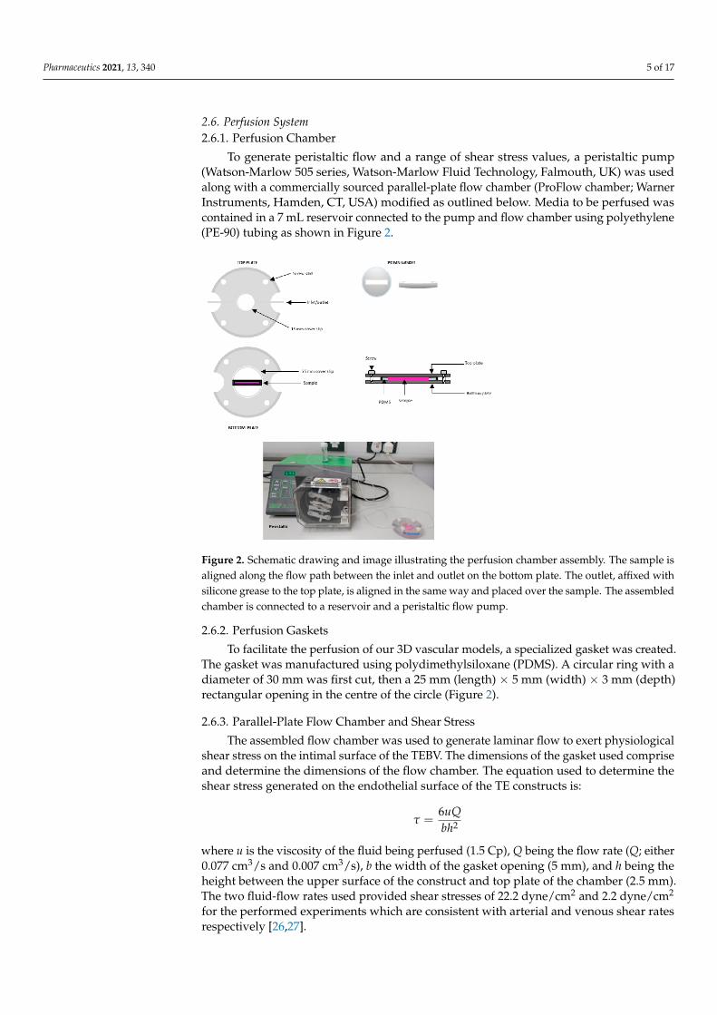
To generate peristaltic flow and a range of shear stress values, a peristaltic pump(Watson-Marlow 505 series, Watson-Marlow Fluid Technology, Falmouth, UK) was usedalong with a commercially sourced parallel-plate flow chamber (ProFlow chamber; WarnerInstruments, Hamden, CT, USA) modified as outlined below. Media to be perfused wascontained in a 7 mL reservoir connected to the pump and flow chamber using polyethylene(PE-90) tubing as shown in Figure 2.
Figure 2. Schematic drawing and image illustrating the perfusion chamber assembly. The sample isaligned along the flow path between the inlet and outlet on the bottom plate. The outlet, affixed withsilicone grease to the top plate, is aligned in the same way and placed over the sample. The assembledchamber is connected to a reservoir and a peristaltic flow pump.
2.6.2. Perfusion Gaskets
To facilitate the perfusion of our 3D vascular models, a specialized gasket was created.The gasket was manufactured using polydimethylsiloxane (PDMS). A circular ring with adiameter of 30 mm was first cut, then a 25 mm (length) × 5 mm (width) × 3 mm (depth)rectangular opening in the centre of the circle (Figure 2).
2.6.3. Parallel-Plate Flow Chamber and Shear Stress
The assembled flow chamber was used to generate laminar flow to exert physiologicalshear stress on the intimal surface of the TEBV. The dimensions of the gasket used compriseand determine the dimensions of the flow chamber. The equation used to determine theshear stress generated on the endothelial surface of the TE constructs is:
τ =6uQbh2
where u is the viscosity of the fluid being perfused (1.5 Cp), Q being the flow rate (Q; either0.077 cm3/s and 0.007 cm3/s), b the width of the gasket opening (5 mm), and h being theheight between the upper surface of the construct and top plate of the chamber (2.5 mm).The two fluid-flow rates used provided shear stresses of 22.2 dyne/cm2 and 2.2 dyne/cm2
for the performed experiments which are consistent with arterial and venous shear ratesrespectively [26,27].
Pharmaceutics 2021, 13, 340 6 of 17
2.7. Lesion Models
To mimic vascular injury, a FeCl3 lesion was created on the TEBV by dipping a 1 mm2
square of filter paper in 10% FeCl3 and placing this onto the upper surface of the TEconstructs for 1 min. After this, the TE constructs were washed with PBS/HBS to eliminateexcess FeCl3, then topped up with fresh media. This mode of lesioning was also applied toTE constructs for EPC perfusion.
2.8. Platelet Preparation
This study was approved by the Keele University (UK) Research Ethics Committee(MH-200155, 1 May 2018). Blood was donated by healthy, drug-free volunteers whogave written informed consent. Blood was obtained by venepuncture. The blood wasmixed with acid citrate dextrose (ACD; 85 mM sodium citrate, 78 mM citric acid, and111 mM D-glucose) at a ratio of 5:1. Platelet-rich plasma (PRP) was obtained by softdescent centrifugation at 725 g for 8 min. After centrifugation, the PRP was collectedand treated with aspirin (50 mM) and apyrase (0.1 U/mL). PRP was again centrifuged at450 g for 20 min, then resuspended in supplemented HEPES-buffered saline (HBS; pH 7.4,145 mM NaOH, 10 mM HEPES, 10 mM D-glucose, 5 mM KCl, 1 mM MgSO4) to reach aplatelet density of 2 × 10⁸ cells/mL. The HBS was supplemented with 1 mg/mL bovineserum albumin (BSA), 1.8 mg/mL glucose, 0.1 U/mL apyrase, and 200 µM CaCl2. Prior toperfusion, the assembled perfusion system without the TE construct was perfused with1% BSA solution for 1 h to prevent unwanted platelet adhesion to the components of theperfusion system. The gasket and the spacers were incubated overnight with 1% BSAsolution at room temperature.
2.9. EPC Isolation and Culture
EPCs were isolated by collecting 60 mL of whole blood from healthy volunteers.To prevent coagulation, the blood was split into 2 falcon tubes with 5 mL of ACD in each.The blood-anticoagulant mix was then split into 15 mL falcon tubes and centrifuged at 700 gfor 8 min. After centrifugation, the sequence of layers occurred as follows (seen from top tobottom): plasma, enriched cell fraction (interphase consisting of lymphocytes/peripheralbone marrow cells (PBMCs), erythrocytes, and granulocytes. The plasma fraction wascarefully discarded, leaving approximately 0.5–1 mL above the interface. The enriched cellfraction was pooled into one tube and diluted 1:1 with PBS. To further separate the cell, i.e.,eliminate residual plasma and Red Blood Cells (RBCs), the pooled fraction was carefullylayered over 8 mL of Ficoll-Paque, ensuring no mixing of the layers, and centrifugedagain for 20 min at 400 g. After centrifugation, any residual red blood cells are below theseparation medium, and the enriched cell fraction should be immediately above it withdiluted plasma and platelets above this. After isolation, the cell-rich fraction was dilutedwith PBS, then centrifuged again at 400 g for 10 min. After centrifugation, the supernatantwas discarded, and the resultant pellet was resuspended in 2 mL of complete EPC media.The cell suspension was then split into 2 wells of a 12-well plate that had been coated with2.5 µg/cm2 fibronectin. On day 1, the contents of the wells were agitated and transferredto new wells. This was repeated for the next 3 days. Media was changed daily for the first7 days then every 2–3 days for up to 20 days.
2.10. Cell Labelling2.10.1. CFSE
Carboxyfluorescein succinimidyl ester (CFSE) dye was used to label EPC cells at aconcentration of 2 µL/mL of cell suspension. Cells were incubated for 15 min at 37 ◦C,then centrifuged for 3 min at 300 g. The pellet was re-suspended in 5 mL of fresh supple-mented media. The cell suspension was allowed to rest for 30 min at 37 ◦C before loadinginto the perfusion system.

Pharmaceutics 2021, 13, 340 7 of 17
2.10.2. DiOC6
To facilitate visualization of platelet adhesion and aggregation upon the TE constructsunder flow conditions, platelets were labelled with DiOC6, a fluorescent membrane dye.Blood was mixed 5:1 with ACD (anticoagulant). The anticoagulant was mixed with themembrane dye to make a final concentration of 1µM, prior to the addition of whole blood.This mixture was then incubated for 10 min at room temperature, then centrifuged toobtain PRP. Centrifugation was done at 1500 g for 8 min. The resultant PRP was thentreated with 100 µM aspirin and 0.1 U/mL apyrase. This was followed by a centrifugationwash at 350 g for 20 min. The platelet pellet was then re-suspended with supplementedHBS, creating a final cell density of 2 × 10⁸ cells/mL.
2.11. Ketamine Treatment
To assess if the platelet responses might be affected by ketamine treatment, exper-iments were performed in which both platelets and the TE constructs were pre-treatedwith ketamine before the perfusion of platelets on TE constructs. TEMLs were incubatedwith 1 mM ketamine (Narketan) or HBS for 1 h at 37 ◦C, after which they were perfusedwith washed DiOC6-labelled platelets incubated with either 300 µM ketamine or an equiv-alent volume of the vehicle, or HBS, under the same conditions stated above. Plateletaggregation on the perfused TE constructs was evaluated by fluorescence microscopy(Leica MSV269) under excitation wavelength of 485 nm and emission of 501 nm. As astatic comparative, TE constructs were treated with 1 mM ketamine and placed in a cuvettewith DiOC6-labelled platelets treated with ketamine or HBS for 15 min at 37 ◦C. Whilstuntreated TEML constructs were placed atop untreated platelet samples as the control.
2.12. Platelet Aggregometry
Platelet aggregometry was performed using a modification of the previously publishedtechnique [28]. Following platelet incubation with TE constructs, 200 µL of the plateletsuspension was transferred into a 96-well plate and then placed into a plate reader pre-warmed to 37 ◦C (BioTek Synergy 2 microplate, Winooski, VT, USA). Baseline absorbancereadings were taken once at a wavelength of 600 nm, obtaining an absolute absorbancereading post TE construct incubation. In the present assay, the plate reader was set up touse a fast-shaking mode between absorbance readings to aid in sample mixing.
2.13. Atorvastatin Treatment
Following FeCl3 lesioning, the TE constructs were incubated with 60 µg/mL ator-vastatin calcium trihydrate (Sanofi) for 3 and 5 h at 37 ◦C. This was followed by theperfusion of CFSE-labelled EPCs (without atorvastatin in EPC perfusate as control) for45 min in the parallel-plate chamber. Images were taken using a Leica inverted microscope(Leica MSV269) under an excitation wavelength of 485 nm and emission of 501 nm. Cellattachment was quantified with ImageJ.
2.14. Statistics and Data Analysis
Values stated are mean ± SEM of the number of observations (n) indicated. Analysisof statistical significance was performed using a two-tailed Student’s t-test as well as two-way analysis of variance (ANOVA), confirmed using the Brown–Forsythe test. p < 0.05 wasconsidered statistically significant.
3. Results
The adhesion and aggregation of platelets by exposure to the TE constructs underdynamic flow conditions were assessed by monitoring fluorescence from DiOC6-labelledhuman platelets upon the surface of the TE constructs (solid phase activation), as well asmonitoring activation of platelets remaining within the solution by platelet aggregome-try (liquid phase activation). Three types of TE constructs were evaluated, TEIL, TEML,and TEBV, with the acellular collagen hydrogel as a negative control as we have previ-

Pharmaceutics 2021, 13, 340 8 of 17
ously demonstrated this to not elicit platelet activation [18]. The homing effect of drugatorvastatin on EPC attachment on these TE constructs under dynamic flow conditionswas assessed.
3.1. Cells in TE Constructs Attained Typical Morphology and Organisation
Consistent with our previous findings, it was possible to generate TEIL, TEML, andTEBV constructs with HUVECs and HCASMCs showing typical normal cellular morphol-ogy when grown atop (HUVECs) or within (HCASMCs) the collagen hydrogel scaffoldusing our previously published layer-by-layer fabrication technique [18]. Figure 3 demon-strates the effect of aligned nanofibers on HUVEC alignment, with cells showing moreorganised growth/orientation compared to culture flasks. Meanwhile, smooth musclecells maintain spindle-shaped morphology while embedded in the collagen gel (data notshown [18]).
Figure 3. Cellular morphology for TEIL constructs. (A) Human umbilical vein endothelial cells (HUVECs) cultured for4 days in a culture flask. (B) HUVECs cultured on the nanofibers for 10 days, (C) SEM picture of aligned PLA nanofibers.
3.2. TEML Supports Shear-Dependent Platelet Aggregation under Physiological Flow Conditions
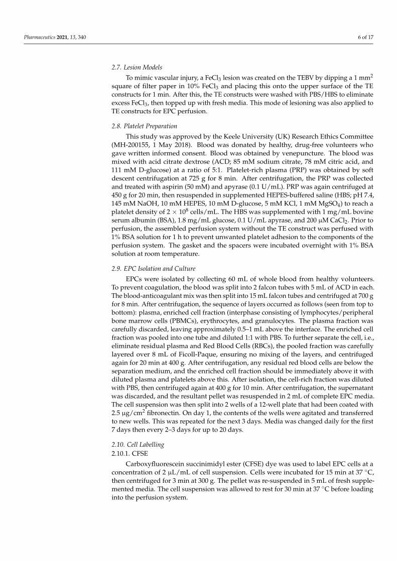
We first assessed whether the TEML is able to support platelet aggregation undertwo different shear stresses indicative of those found in the arterial (22.2 dyne/cm2) andin the venous circulation (2.2 dynes/cm2; [29]). The TEML construct is a model for anendothelium-denuded blood vessel and therefore can be used to assess pro-aggregatoryproperties of the medial layer. Figure 4A,B show that acellular collagen gels did not triggerplatelets’ activation under both low and high shear stress rate, consistent with our previousfindings under static conditions [18]. When the TEML constructs were exposed to plateletsperfused at venous shear stresses, sporadic platelet adhesion was observed (Figure 4C).The strong platelet aggregation observed under arterial shear stresses demonstrates thatshear stress and fluid flow influence platelet adhesion (Figure 4D). These studies areconsistent with a number of previous studies that have demonstrated a role for shear-dependent platelet aggregation in driving thrombus formation [30,31].
Pharmaceutics 2021, 13, 340 9 of 17
Figure 4. Fluorescence images of tissue-engineered media layer (TEML) constructs after exposure toplatelets perfusion at different shear stress rates. DiOC6-labelled human platelets suspension wasperfused through a parallel-plate flow chamber over the surface of acellular collagen gels (A,B) andTEML (C,D) for 15 min at venous (A,C) (2.2 dyne/cm2) and arterial (B,D) (22.2 dyne/cm2) shearstresses. White dotted line denotes the central hydrogel area. Area on either side of the dotted line isthe filter paper frame. Arrows highlight aggregated platelets.
The platelets adhered and significantly aggregated when exposing to the media layerof the constructs (Figure 4D). Since no aggregation was observed on the acellular hydrogels(Figure 4A,B), platelet aggregation should be triggered by neo-collagen produced by theembedded HCASMCs. This corresponded well with previous studies that demonstratedthat the pro-aggregatory properties of the TEML were mainly attributed to the nativecollagen secretion of the SMCs [32] and is consistent with our previous work under stirredconditions [18]. Additional synthesis and secretion of thrombogenic molecules may alsocontribute to platelet activation and aggregation.
3.3. TEBVs Prevent Platelet Aggregation under Physiological Flow Conditions
The endothelial lining of the native human artery produces platelet inhibitors suchas nitric oxide (NO) and prostaglandins to prevent thrombosis. Upon vascular damage,the loss of the antithrombotic endothelial lining, and the exposure of the prothromboticproperties of the medial layer, triggers thrombus formation. To test whether the TEBV isable to replicate this endothelium-dependent modulation of the haemostatic properties ofthe construct under physiological flow conditions, TEBVs with differences in the integrityof the endothelial cell layer were exposed to human platelet suspension under arterialshear stresses. In these experiments, we used (i) a TEBV with a fully confluent endotheliallayer, (ii) a TEBV with a partially confluent endothelial layer, and (iii) a TEBV in which anintimal injury was triggered with FeCl3, a common injury model used in murine thrombosismodels [33].
Real-time fluorescence imaging revealed that TEBVs with an intact, confluent en-dothelial layer did not show platelet aggregation upon their surfaces over a 15 min periodof perfusion (Figure 5I (A)). In contrast, the TEBV with a partially confluent endotheliallayer exhibited limited platelet aggregation. Platelets adhered only in areas that were notcovered with endothelial cells, allowing their direct contact with the media layer. However,not all the areas lacking endothelial coverage showed platelet aggregation, suggesting that

Pharmaceutics 2021, 13, 340 10 of 17
the endothelial cells on the TEIL may be capable of secreting sufficient anti-thromboticmolecules to prevent platelet aggregation (Figure 5I (B,C)). Dramatic contrast observa-tions of platelets’ aggregation behaviours on FeCl3-lesioned TEBV samples were revealedas shown in Figure 5I (D), in which massive platelet aggregates are attached to the con-structs. The insert picture shows the dense morphology of the aggregated platelets, andthe low magnification image shows the overall localisation of the aggregates. The strip-likeaggregate location was larger than the lesion area (1 mm2), implying that cytokines pro-duced by lesioned endothelial layer and exposed subendothelial proteins (collagens in ourmodel) could be circulated away from the lesion site, triggering aggregation away fromthe lesion site. FeCl3-mediated endothelial cell injury allowed exposure of platelets to thepro-aggregatory medial layer in the construct, providing a reliable FeCl3-triggered arterialinjury model.
Figure 5. Characterisation of tissue-engineered blood vessel (TEBV) constructs with full, partial,and impaired intimal layer after exposure to platelets perfusion for 15 min at a shear stress of22.2 dynes/cm2. (I) The fluorescence images of DiOC6-labelled human platelet suspension whichwas perfused through a parallel flow chamber over the surface of the TEBV. (A) TEBV constructcultured for 10 days with a confluent endothelial cell layer. (B,C) TEBV with a partial endotheliallayer coverage, 4-day culture, and (D) with impairment of the endothelial layer by FeCl3 incubation.The green clumps over the construct surface indicate platelet aggregation and single platelet adhesion.Higher magnification images of the platelet aggregates are shown in the inserts. (II) The aggregometeranalysis of the aggregation state of the platelet solution before and after exposure to tissue-engineeredblood vessels (TEBVs). The data represent mean ± SEM; n = 3. Significance is indicated by (*),p ≤ 0.01.
These qualitative results correspond well with the comparison of the quantitativeaggregation state of the platelets before and after perfusion, in which FeCl3 injury couldbe seen to trigger aggregation of platelets within the platelet suspension (Figure 5II).In regards to the integrity of the endothelial layer, it can be observed that measurements ofthe absorbance, before and after perfusion, do not present a significant difference in theTEBV with a full endothelial layer. This indicates that platelet aggregation is preventedby the presence of an intact endothelial layer. On the contrary, significant differences inplatelet aggregation were observed in FeCl3-treated TEBVs. This is consistent with plateletsproducing and releasing autocrine activators to recruit platelets to the growing thrombi,thus triggering platelet aggregation within the platelet suspension.
3.4. Ketamine Inhibited Platelet Aggregation at Arterial Shear Stress
The data above demonstrate that the TEML, and the FeCl3-treated TEBV, triggerplatelet aggregation through the exposure of the pro-aggregatory medial layer in theendothelial denuded section of these constructs. Thus, our TEBV perfusion system canbe used in conjunction with the FeCl3 injury model, which is commonly used in murinethrombosis models [33]. As our human TEBV model does not require the addition of
Pharmaceutics 2021, 13, 340 11 of 17
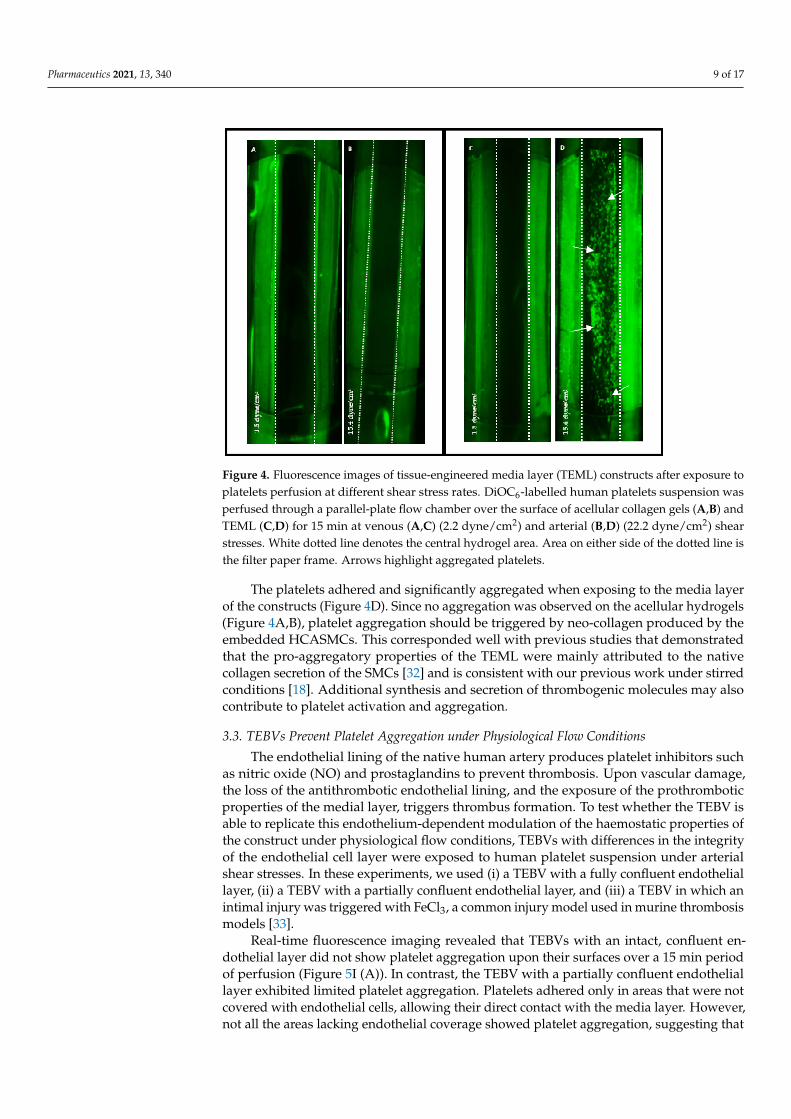
anaesthetics to our blood samples, experiments were performed to assess if the additionof ketamine, the most commonly used anaesthetic in murine thrombosis models, couldartificially alter the platelet aggregatory responses seen. As the TEML provides a morepronounced aggregatory response due to the absence of an endothelial lining, we usedthis system to investigate whether ketamine could impact thrombus formation by humanplatelets under physiological flow conditions. DiOC6-labelled platelets were treated with300 µM ketamine prior to perfusion over the TEML surface under arterial shear stress(22.2 dynes/cm2). Significant platelet aggregation was found in the TEML perfused at highshear stress without ketamine treatment (Figure 6). The platelets treated with ketaminewere found to lose their ability to adhere to the TEML surface. These results indicatethat ketamine-treated platelets (Figure 6I (B)) were less reactive than untreated platelets(Figure 6I (A)), which showed significant aggregation on the surface. Ketamine not onlysignificantly inhibited platelet aggregation on the construct surface but also inhibited theiractivation within the surrounding platelet suspension, as demonstrated by the aggregationstate of platelets before and after exposure to the TEML (Figure 6II). This is likely due to theknown effect of ketamine inhibiting platelet Ca2+ signalling [34,35]. This would preventdense granule secretion, which would in turn prevent the activation of these cells in theperfused platelet suspension.
Figure 6. Characterisation of perfused platelet solution to the surface TEML incubated with andwithout ketamine under physiological shear strain. (I) The fluorescence images of DiOC6-labelledhuman platelet suspension which was perfused through a parallel flow chamber over the surface ofthe TEML. Human platelet suspensions were treated with 300 µM ketamine or an equivalent volumeof its vehicle, HBS, and perfused over the TEML for 15 min using a parallel-plate flow chamber at22.2 dynes/cm2. Images were recorded in real-time over the course of 15 min. (A) Untreated sample;(B) Ketamine-treated sample. (T0) Beginning of perfusion; (T7) 7 min into perfusion; (T15) 15 mininto perfusion. (II) Aggregometer analysis of the aggregation state of the perfused platelet solution.Aggregation states of platelets untreated (left) and pre-treated (right) with ketamine before and afterperfusion over TEML surface. Error bars represent SEM. Significance is indicated by (*), p ≤ 0.05.
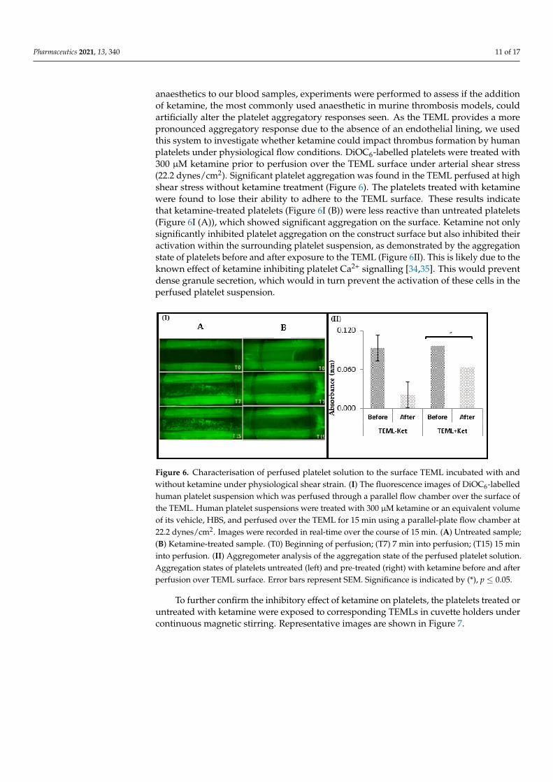
To further confirm the inhibitory effect of ketamine on platelets, the platelets treated oruntreated with ketamine were exposed to corresponding TEMLs in cuvette holders undercontinuous magnetic stirring. Representative images are shown in Figure 7.
Pharmaceutics 2021, 13, 340 12 of 17
Figure 7. Visible and fluorescence images illustrating the state of platelet aggregation after TEMLincubation either with or without ketamine. Constructs were first incubated with 1 mM ketamine,at 37 ◦C, for 1 h then a 15 min incubation, also at 37 ◦C, with 1 mL of washed platelets that werepre-treated with 1 mM ketamine. Following the second incubation, platelets were washed, andaggregates on the surface were imaged with a fluorescent microscope. Images (A–C): untreatedsamples. Images (D–F): ketamine-treated samples. Results are representative of 4 experiments, n = 6.Scale bar = 100 µm.
Fluorescent imaging of the TEML surface exposed to DiOC6-labelled platelets showedthat samples treated with ketamine had fewer platelet aggregates appearing on the TEMLsurface (Figure 7D–F). The untreated samples (Figure 7A–C) displayed greater adhesion,as well as formation of multiple platelet aggregates.
3.5. Atorvastatin Increases EPC Attachment
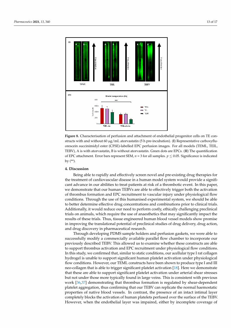
Atorvastatin has been reported to increase circulating numbers of EPCs [23]. To evalu-ate whether atorvastatin also has an effect on the recruitment and attachment of perfusedEPCs, our three TE construct variants were lesioned with FeCl3, then incubated with60 µg/mL atorvastatin for 5 h. Constructs were then perfused with EPCs for 45 min at22.2 dynes/cm2. EPCs were perfused at a density of 1 × 10⁴ cells/mL. After perfusion,constructs were imaged and attached cells quantified (Figure 8).
Across all the models shown here, it was evident that atorvastatin incubation increasedthe number of cells that attached to the lesioned surfaces of the constructs (Figure 8II).We have previously demonstrated the pro aggregatory properties of the TEML, and Figure 8suggests that atorvastatin increases these pro-aggregatory properties. The data presentedhere suggest that without either the endothelial (TEML) or medial (TEIL) layers, theresponse is stronger than when both layers are present, suggesting a synergistic effectbetween the medial and intimal layers in terms of modulating cell recruitment. The al-most steady state of attachment of EPCs on the TEML suggests that the FeCl3 lesion isnot the driving factor for cell attachment but rather time-dependent signalling/cytokineproduction by the representative cells and the presence of atorvastatin.
Pharmaceutics 2021, 13, 340 13 of 17
Figure 8. Characterisation of perfusion and attachment of endothelial progenitor cells on TE con-structs with and without 60 µg/mL atorvastatin (5 h pre-incubation). (I) Representative carboxyflu-orescein succinimidyl ester (CFSE)-labelled EPC perfusion images. For all models (TEML, TEIL,TEBV), A is with atorvastatin, B is without atorvastatin. Green dots are EPCs. (II) The quantificationof EPC attachment. Error bars represent SEM, n = 3 for all samples. p ≤ 0.05. Significance is indicatedby (**).
4. Discussion
Being able to rapidly and effectively screen novel and pre-existing drug therapies forthe treatment of cardiovascular disease in a human model system would provide a signifi-cant advance in our abilities to treat patients at risk of a thrombotic event. In this paper,we demonstrate that our human TEBVs are able to effectively trigger both the activationof thrombus formation and EPC recruitment to vascular injury under physiological flowconditions. Through the use of this humanised experimental system, we should be ableto better determine effective drug concentrations and combinations prior to clinical trials.Additionally, it would reduce our need to perform costly, ethically challenging preclinicaltrials on animals, which require the use of anaesthetics that may significantly impact theresults of these trials. Thus, tissue engineered human blood vessel models show promisein improving the translational potential of preclinical studies of drug delivery, drug action,and drug discovery in pharmaceutical research.
Through developing PDMS sample holders and perfusion gaskets, we were able tosuccessfully modify a commercially available parallel flow chamber to incorporate ourpreviously described TEBV. This allowed us to examine whether these constructs are ableto support thrombus activation and EPC recruitment under physiological flow conditions.In this study, we confirmed that, similar to static conditions, our acellular type I rat collagenhydrogel is unable to support significant human platelet activation under physiologicalflow conditions. However, our TEML constructs have been shown to produce type I and IIIneo-collagen that is able to trigger significant platelet activation [18]. Here we demonstratethat these are able to support significant platelet activation under arterial shear stressesbut not under those more typically found in large veins. This is consistent with previouswork [36,37] demonstrating that thrombus formation is regulated by shear-dependentplatelet aggregation, thus confirming that our TEBV can replicate the normal haemostaticproperties of native blood vessels. In contrast, the presence of an intact intimal layercompletely blocks the activation of human platelets perfused over the surface of the TEBV.However, when the endothelial layer was impaired, either by incomplete coverage of
Pharmaceutics 2021, 13, 340 14 of 17
endothelial cells (via a shortened culture period) or via FeCl3-induced injury, the TEBVconstructs were able to trigger an effective haemostatic reaction consistent with that seenin the native artery (Figure 5). FeCl3-injured TEML also displayed platelet aggregationunder flow, an observation that was absent on uninjured constructs. Location of FeCl3application had no impact on this observation, demonstrating that the observed effect isdue to endothelial damage caused by FeCl3 and not via the presence of FeCl3 itself [33,38].This can be concluded as no enhancement of platelet activation and aggregation observedupon FeCl3 treatment of the endothelial-free TEML constructs (data not shown). The datapresented here support the traditional explanation that FeCl3-induced injury results inthrombus formation in murine arteries through endothelial denudation, facilitating plateletactivation upon the exposure of sub-endothelial collagen. These findings demonstratethat this test model can be used to study platelet inhibition and activation in a convenient,operator-friendly, and dynamic manner. The image analysis of the adhered platelets on theTE constructs, and assessment of platelet aggregation in liquid phase using aggregometry,allows the extraction of both qualitative and quantitative datasets to assess ex vivo humanthrombus formation.
This study demonstrated that ketamine exerted a strong negative effect on plateletaggregation and activation. This corresponds well with previous in vitro and in vivo stud-ies that have investigated the underlying mechanisms of ketamine’s inhibition of plateletfunction [6]. In our experiments, we observed that ketamine almost completely inhibitsthrombus formation upon the TEML construct. It is possible that this effect is caused bythe inhibition of dense granule secretion by ketamine, as autocrine signalling moleculesreleased from here are known to be crucial to the recruitment of circulating platelets ontothe surface of forming thrombi [7,39]. This inhibitory effect could artefactually alter the sizeand structure of thrombi seen in current animal thrombosis models, potentially leading toan overestimation of the effectiveness of putative anti-platelet therapies. This is consistentwith previous findings that show that the use of different anaesthetics differentially impactsthe efficacy of integrin αIIbβ3 blockers in murine thrombosis models [39], thus providinginitial evidence that our model system is a potential alternative to current in vivo studies.A more detailed side-by-side comparison examining the impact of different anaestheticson the thrombotic response in current in vivo models, and in the TEBV model presentedhere, will be required to fully validate the model system.
These findings highlight the value of using tissue-engineered human blood vessels fordrug testing. By using human cells and eliminating the need for anaesthesia, we shouldbe able to accurately model the processes of haemostasis and vascular repair to improvethe translational potential of any findings. Additional advantages of our model includea reduction in the use of animal thrombosis models, elimination of the need for costlyintravital microscopy equipment, and a lower cost of TE construct production compared tohousing mouse colonies.
We also used the TEBV to demonstrate that atorvastatin enhances recruitment andattachment of perfused EPCs. Although previous studies have demonstrated that atorvas-tatin increases circulating numbers of EPCs [19], there has been limited study on its abilityto modulate EPC recruitment to the damaged vascular wall, which may partly underlie thebeneficial effects of this drug in preventing acute cardiovascular events. Possible mech-anisms by which atorvastatin might enhance EPC recruitment to the damaged TEBV isvia increased bioavailability of NO [40] or activation of matrix metalloproteinase-2 and9 (MMP2 and MMP9) [41]. It is also possible that the SDF-1 CXCR4 axis is also involvedin recruiting EPCs to sites of vascular injury. This theory is supported by the findingsby Luo et al., 2018 [42]. They found that NO production was induced by SDF-1, whichtriggers multiple signalling pathways, resulting in chemokine-induced changes of theEPC cytoskeleton leading to enhanced cell migration [42]. The value of our models isthat various doses/concentrations of different drugs, as well as combinations of drugs,can be tested faster than conventional models and also allows for real-time monitoringwithout measures such as anaesthesia being needed. The ease of assembly also makes it

Pharmaceutics 2021, 13, 340 15 of 17
possible to combine multiple cell types and can be adapted for any species. In comparisonto other in vitro models [43,44], our models also allow for real-time visualisation of cellattachment due to the nature of the perfusion chamber used, containing both a top andbottom window, as well as the open-faced nature of the models themselves.
5. Conclusions
The layer-by-layer assembly of human blood vessel models provides a convenientand reliable research tool to investigate the interaction of blood components, such asplatelets and circulated progenitor cells, with a blood vessel. This provides a simplemethod to assess the impact of a variety of drug interactions on haemostasis and vascularrepair. The parallel-plate flow chamber, plus the adjustable dimensions of the PDMSgasket, enabled incorporation of 3D tissues into the chamber, permitting their exposure toperfusion with blood components at different physiological shear stresses. The labelledcells allow the monitoring of cellular activation and adhesion in real-time. The intact,confluent intimal layer can inhibit platelet activation, whilst the partially formed intimadid not. The medial layer with newly formed collagen triggered platelet aggregation,whilst collagen gel made from rat skin collagen type I did not. The presence of a similarconcentration of ketamine used in current in vivo thrombosis models inhibited the abilityof human platelets to adhere to the TEML surface. The tissue-engineered vessels couldalso be used to demonstrate that atorvastatin is able to enhance the homing capabilitiesof EPCs by improving their ability to adhere to the damaged tissue-engineered bloodvessel under flow conditions. Here we show that the combination of tissue-engineeredarterial constructs and a parallel-plate flow chamber can be used to effectively simulate thehaemostatic and vascular repair processes ex vivo. Therefore, these results indicate that thismodel system is able to provide a potential alternative to in vivo testing models. This willalso permit us to test the predicted mechanisms of action of a selected anti-thromboticdrug without the need for animal models and may be modified further to simulate otherclinical conditions.
Author Contributions: Conceptualization and supervision, Y.Y., A.G.S.H., and R.B.; methodology,W.N., A.C.H.H., and F.I.M.; writing—original draft preparation, W.N.; writing—review and editing,Y.Y., A.G.S.H., and R.B. All authors have read and agreed to the published version of the manuscript.
Funding: This work is financially supported by British Heart Foundation (FS/12/48/29719) and anNC3R and British Hearth Foundation co-funded Ph.D. studentship (NC/R001642/1).
Institutional Review Board Statement: The study was conducted according to the guidelines of theDeclaration of Helsinki, and approved by Keele University Research Ethics Committee (MH-200155on the 1 May 2018).
Informed Consent Statement: Informed written consent was collected from all volunteers involvedin this study.
Data Availability Statement: All data in this study have been included in this manuscript.
Conflicts of Interest: The authors declare no conflict of interest.
References1. Benam, K.H.; Dauth, S.; Hassell, B.; Herland, A.; Jain, A.; Jang, K.-J.; Karalis, K.; Kim, H.J.; MacQueen, L.; Mahmoodian, R.; et al.
Engineered In Vitro Disease Models. Annu. Rev. Pathol. Mech. Dis. 2015, 10, 195–262. [CrossRef]2. Cesarovic, N.; Lipiski, M.; Falk, V.; Emmert, M.Y. Animals in cardiovascular research. Eur. Hear. J. 2020, 41, 200–203. [CrossRef]
[PubMed]3. Camacho, P.; Fan, H.; Liu, Z.; He, J.-Q. Small mammalian animal models of heart disease. Am. J. Cardiovasc. Dis. 2016, 6, 70–80.
[PubMed]4. Furie, B.C. Thrombus formation in vivo. J. Clin. Investig. 2005, 115, 3355–3362. [CrossRef] [PubMed]5. Janssen, B.J.A.; De Celle, T.; Debets, J.J.M.; Brouns, A.E.; Callahan, M.F.; Smith, T.L. Effects of anesthetics on systemic hemody-
namics in mice. Am. J. Physiol. Circ. Physiol. 2004, 287, H1618–H1624. [CrossRef] [PubMed]6. Chang, Y.; Chen, T.-L.; Wu, G.-J.; Hsiao, G.; Shen, M.-Y.; Lin, K.-H.; Chou, D.-S.; Lin, C.-H.; Sheu, J.-R. Mechanisms Involved in
the Antiplatelet Activity of Ketamine in Human Platelets. J. Biomed. Sci. 2004, 11, 764–772. [CrossRef]

Pharmaceutics 2021, 13, 340 16 of 17
7. Nakagawa, T.; Hirakata, H.; Sato, M.; Nakamura, K.; Hatano, Y.; Nakamura, T.; Fukuda, K. Ketamine suppresses plateletaggregation possibly by suppressed inositol triphosphate formation and subsequent suppression of cytosolic calcium increase.Anesthesiology 2002, 96, 1147–1152. [CrossRef]
8. Atkinson, P.; Taylor, D.; Chetty, N. Inhibition of platelet aggregation by ketamine hydrochloride. Thromb. Res. 1985, 40, 227–234.[CrossRef]
9. Akata, T.; Izumi, K.; Nakashima, M. Mechanisms of direct inhibitory action of ketamine on vascular smooth muscle in mesentericresistance arteries. Anesthesiology 2001, 95, 452–462. [CrossRef]
10. Kurebayashi, N.; Ogawa, Y. Depletion of Ca 2+ in the sarcoplasmic reticulum stimulates Ca 2+ entry into mouse skeletal musclefibres. J. Physiol. 2001, 533, 185–199. [CrossRef]
11. Weinberg, C.B.; Bell, E. A blood vessel model constructed from collagen and cultured vascular cells. Science 1986, 231, 397–400.[CrossRef] [PubMed]
12. Bouten, C.C.; Dankers, P.P.; Driessen-Mol, A.A.; Pedron, S.; Brizard, A.; Baaijens, F.F. Substrates for cardiovascular tissueengineering. Adv. Drug Deliv. Rev. 2011, 63, 221–241. [CrossRef] [PubMed]
13. Gui, L.; Dash, B.C.; Luo, J.; Qin, L.; Zhao, L.; Yamamoto, K.; Hashimoto, T.; Wu, H.; Dardik, A.; Tellides, G.; et al. Implantabletissue-engineered blood vessels from human induced pluripotent stem cells. Biomaterials 2016, 102, 120–129. [CrossRef] [PubMed]
14. Rothuizen, T.C.; Damanik, F.F.R.; Lavrijsen, T.; Visser, M.J.T.; Hamming, J.F.; Lalai, R.A.; Duijs, J.M.G.J.; Van Zonneveld, A.J.;Hoefer, I.E.; Van Blitterswijk, C.A.; et al. Development and evaluation of in vivo tissue engineered blood vessels in a porcinemodel. Biomaterials 2016, 75, 82–90. [CrossRef]
15. Parikh, V.; Kadiwala, J.; Bastida, A.H.; Holt, C.; Sanami, M.; Miraftab, M.; Shakur, R.; Azzawi, M. Small diameter helical vascularscaffolds support endothelial cell survival. Nanomed. Nanotechnol. Biol. Med. 2018, 14, 2598–2608. [CrossRef]
16. Buttafoco, L.; Engbers-Buijtenhuijs, P.; Poot, A.A.; Dijkstra, P.J.; Vermes, I.; Feijen, J. Physical characterization of vascular graftscultured in a bioreactor. Biomaterials 2006, 27, 2380–2389. [CrossRef] [PubMed]
17. Lee, P.-H.; Tsai, S.-H.; Kuo, L.; Hwang, C.-Y.; Kuo, C.-Y.; Yang, V.C.; Chen, J.-K. A prototype tissue engineered blood vessel usingamniotic membrane as scaffold. Acta Biomater. 2012, 8, 3342–3348. [CrossRef] [PubMed]
18. Musa, F.I.; Harper, A.G.; Yang, Y. A Real-Time Monitoring System to Assess the Platelet Aggregatory Capacity of Components ofa Tissue-Engineered Blood Vessel Wall. Tissue Eng. Part C Methods 2016, 22, 691–699. [CrossRef] [PubMed]
19. Oikonomou, E.; Siasos, G.; Zaromitidou, M.; Hatzis, G.; Mourouzis, K.; Chrysohoou, C.; Zisimos, K.; Mazaris, S.; Tourikis, P.;Athanasiou, D.; et al. Atorvastatin treatment improves endothelial function through endothelial progenitor cells mobilization inischemic heart failure patients. Atherosclerosis 2015, 238, 159–164. [CrossRef] [PubMed]
20. Urbich, C.; Dimmeler, S. Endothelial progenitor cells: Characterization and role in vascular biology. Circ. Res. 2004, 95, 343–353.[CrossRef] [PubMed]
21. Zhang, M.; Malik, A.B.; Rehman, J. Endothelial progenitor cells and vascular repair. Curr. Opin. Hematol. 2014, 21, 224–228.[CrossRef]
22. Kunz, G.A.; Liang, G.; Cuculi, F.; Gregg, D.; Vata, K.C.; Shaw, L.K.; Goldschmidt-Clermont, P.J.; Dong, C.; Taylor, D.A.; Peterson,E.D. Circulating endothelial progenitor cells predict coronary artery disease severity. Am. Heart J. 2006, 152, 190–195. [CrossRef]
23. Sandhu, K.; Mamas, M.; Butler, R. Endothelial progenitor cells: Exploring the pleiotropic effects of statins. World J. Cardiol. 2017,9, 1–13. [CrossRef] [PubMed]
24. Stancu, C.; Sima, A. Statins: Mechanism of action and effects. J. Cell. Mol. Med. 2001, 5, 378–387. [CrossRef] [PubMed]25. Yang, Y.; Wimpenny, I.; Ahearne, M. Portable nanofiber meshes dictate cell orientation throughout three-dimensional hydrogels.
Nanomed. Nanotechnol. Biol. Med. 2011, 7, 131–136. [CrossRef] [PubMed]26. Malek, A.M.; Alper, S.L.; Izumo, S. Hemodynamic shear stress and its role in atherosclerosis. JAMA 1999, 282, 2035–2042.
[CrossRef] [PubMed]27. Cunningham, K.S.; I Gotlieb, A. The role of shear stress in the pathogenesis of atherosclerosis. Lab. Investig. 2004, 85, 9–23.
[CrossRef] [PubMed]28. Chan, M.V.; Armstrong, P.C.; Warner, T.D. 96-well plate-based aggregometry. Platelets 2018, 29, 650–655. [CrossRef] [PubMed]29. Mongrain, R.; Rodés-Cabau, J. Role of Shear Stress in Atherosclerosis and Restenosis after Coronary Stent Implantation. Revista
Española de Cardiología 2006, 59, 1–4. [CrossRef] [PubMed]30. Maxwell, M.J.; Westein, E.; Nesbitt, W.S.; Giuliano, S.; Dopheide, S.M.; Jackson, S.P. Identification of a 2-stage platelet aggregation
process mediating shear-dependent thrombus formation. Blood 2006, 109, 566–576. [CrossRef]31. Rana, A.; Westein, E.; Niego, B.; Hagemeyer, C.E. Shear-Dependent Platelet Aggregation: Mechanisms and Therapeutic
Opportunities. Front. Cardiovasc. Med. 2019, 6, 141. [CrossRef] [PubMed]32. Chan-Park, M.B.; Shen, J.Y.; Cao, Y.; Xiong, Y.; Liu, Y.; Rayatpisheh, S.; Kang, G.C.-W.; Greisler, H.P. Biomimetic control of vascular
smooth muscle cell morphology and phenotype for functional tissue-engineered small-diameter blood vessels. J. Biomed. Mater.Res. Part A 2008, 88, 1104–1121. [CrossRef]
33. Li, W.; McIntyre, T.M.; Silverstein, R.L. Ferric chloride-induced murine carotid arterial injury: A model of redox pathology.Redox Biol. 2013, 1, 50–55. [CrossRef] [PubMed]
34. Lisek, M.; Zylinska, L.; Boczek, T. Ketamine and calcium signaling—A crosstalk for neuronal physiology and pathology. Int. J.Mol. Sci. 2020, 21, 8410. [CrossRef] [PubMed]

Pharmaceutics 2021, 13, 340 17 of 17
35. Wong, B.S.; Martin, C.D. Ketamine inhibition of cytoplasmic calcium signalling in rat pheochromocytoma (PC-12) cells. Life Sci.1993, 53, PL359–PL364. [CrossRef]
36. Holme, P.A.; Ørvim, U.; Hamers, M.J.A.G.; Solum, N.O.; Brosstad, F.R.; Barstad, R.M.; Sakariassen, K.S. Shear-induced plateletactivation and platelet microparticle formation at blood flow conditions as in arteries with a severe stenosis. Arter. Thromb. Vasc.Biol. 1997, 17, 646–653. [CrossRef]
37. Zhang, J.-N.; Bergeron, A.L.; Yu, Q.; Sun, C.; McIntire, L.V.; López, J.A.; Dong, J.-F. Platelet aggregation and activation undercomplex patterns of shear stress. Thromb. Haemost. 2002, 88, 817–821. [CrossRef]
38. Li, W.; Nieman, M.; Gupta, A.S. Ferric chloride-induced murine thrombosis models. J. Vis. Exp. 2016, 2016, e54479. [CrossRef][PubMed]
39. Sashindranath, M.; Sturgeon, S.A.; French, S.; Bsc, D.D.D.C.; Selan, C.; Freddi, S.; Johnson, C.; Cody, S.H.; Nesbitt, W.S.; Hamilton,J.R.; et al. The mode of anesthesia influences outcome in mouse models of arterial thrombosis. Res. Pr. Thromb. Haemost. 2019,3, 197–206. [CrossRef]
40. Walter, D.H.; Dimmeler, S.; Zeiher, A.M. Effects of statins on endothelium and endothelial progenitor cell recruitment.Semin. Vasc. Med. 2004, 4, 385–393. [CrossRef]
41. Liu, Y.; Wei, J.; Hu, L.; Hu, S. Beneficial effects of statins on endothelial progenitor cells. Am. J. Med. Sci. 2012, 344, 220–226.[CrossRef]
42. Luo, J.; Wei, D.; Li, D.; Wang, L. Nitric oxide functions in stromal cell-derived factor-1-induced cytoskeleton changes and themigration of Jurkat cells. Oncol. Lett. 2018, 16, 6685–6690. [CrossRef] [PubMed]
43. Xu, Y.; Hu, Y.; Liu, C.; Yao, H.; Liu, B.; Mi, S. A novel strategy for creating tissue-engineered biomimetic blood vessels using 3dbioprinting technology. Materials 2018, 11, 1581. [CrossRef]
44. Papaioannou, T.G.; Manolesou, D.; Dimakakos, E.; Tsoucalas, G.; Vavuranakis, M.; Tousoulis, D. 3D bioprinting methods andtechniques: Applications on artificial blood vessel fabrication. Acta Cardiol. Sin. 2019, 35, 284–289.