The chemotherapy long-term effect on cognitive functions and brain metabolism in lymphoma patients
10
Vol. 56 - 2012 THE QUARTERLY JOURNAL OF NUCLEAR MEDICINE AND MOLECULAR IMAGING 1 C ognitive changes in cancer patients after adjuvant chemotherapy (CHT) treatments have been re- ported since the mid 1970s, with systematic research starting in the early 1990s. Since then, most but not all 1-3 neuropsychological studies on cancer survivors, having received adjuvant CHT, have reported cogni- tive impairments in multiple domains such as execu- tive functions, learning, memory (especially working memory, while the retrieval of remote memories seems to be spared), attention, verbal fluency and speed of information processing. 4-10 Both prior meta-analyses 11, 12 and, more recently, longitudinal studies 13-16 have consistently shown that CHT induced cognitive im- pairments are small to moderate, involving mostly the cognitive functions subserved by frontal lobes. Findings emerging from controlled longitudinal studies 13, 16-18 indicate that cognitive changes tend to fully resolve over time while cross-sectional studies suggest that they may persist for many years following completion of treatment, at least in a significant subset of patients. 19, 20 Recent structural magnetic resonance imaging stud- ies have provided consistent evidence that CHT can induce both gray and white matter changes which can be, at least partially, reversible. A CHT-related reduc- tion of the gray matter volume 21 of brain structures 1 Department of Nuclear Medicine AOU San Giovanni Battista, Turin, Italy 2 Department of Neuroscience AOU San Giovanni Battista, Turin, Italy 3 Department of Psychology, University of Turin Turin, Italy 4 Department of Psychooncology San Giovanni Battista Hospital, Turin, Italy Q J NUCL MED MOL IMAGING 2012;56 B. BAUDINO 1 , F. D’AGATA 2, 3 , P. CAROPPO 2 , G. CASTELLANO 1 ,S. CAUDA 1 M. MANFREDI 1 , E. GEDA 3 , L. CASTELLI 3 , P. MORTARA 2 , L. ORSI 2 , F. CAUDA 3 K. SACCO 3 , R. B. ARDITO 3 , L. PINESSI 2 , G. GEMINIANI 3 , R. TORTA 4 , G. BISI 1 The chemotherapy long-term effect on cognitive functions and brain metabolism in lymphoma patients Aim. A growing number of neuropsychological stud- ies reported that chemotherapy may impair brain functions, inducing persistent cognitive changes in a subset of cancer survivors. The aim of this paper was to investigate the neural basis of the chemotherapy induced neurobehavioral changes by means of meta- bolic imaging and neuropsychological testing. Methods. We studied the resting brain [18F]FDG-PET/ CT images of 50 adult cancer patients with diagno- sis of lymphoma: 18 patients were studied prior and 32 after to chemotherapy. All patients underwent to a neuropsychological examination assessing cogni- tive impairment (tests for shifting attention, verbal memory, phonemic fluency), depression, anxiety and distress. Results. Compared to no chemotherapy patients, the treated group showed significant bilateral lower rate of glucose metabolism in prefrontal cortices, cerebel- lum, medial cortices and limbic brain areas. The me- tabolism of these regions negatively correlated with number of cycles and positively with post-chemother- apy time. The treated group showed a poorer perform- ance in many frontal functions, but similar level of depression, anxiety and distress. Conclusions. Chemotherapy induced significant long- term changes in metabolism of multiple regions with a prevailing involvement of the prefrontal cortex. The observed cognitive dysfunctions could be explained by these changes. The recovery from chemotherapy is probably affected by treatment duration and by the time elapsed after its end. We speculated that the mechanism could be an accelerating ageing / oxida- tive stress that, in some patients at risk, could result in an early and persistent cognitive impairment. KEY WORDS:Neoplasms - Neuropsychology - Rest. Corresponding author: B. Baudino, Department of Nuclear Medicine, AOU San Giovanni Battista, Corso Bramante 88, 10126 Turin, Italy. E-mail: [email protected]
Transcript of The chemotherapy long-term effect on cognitive functions and brain metabolism in lymphoma patients

Vol. 56 - 2012 THE QUARTERLY JOURNAL OF NUCLEAR MEDICINE AND MOLECULAR IMAGING 1
Cognitive changes in cancer patients after adjuvant chemotherapy (CHT) treatments have been re-
ported since the mid 1970s, with systematic research starting in the early 1990s. Since then, most but not all 1-3 neuropsychological studies on cancer survivors, having received adjuvant CHT, have reported cogni-tive impairments in multiple domains such as execu-tive functions, learning, memory (especially working memory, while the retrieval of remote memories seems to be spared), attention, verbal fluency and speed of information processing.4-10 Both prior meta-analyses 11, 12 and, more recently, longitudinal studies 13-16 have consistently shown that CHT induced cognitive im-pairments are small to moderate, involving mostly the cognitive functions subserved by frontal lobes.
Findings emerging from controlled longitudinal studies 13, 16-18 indicate that cognitive changes tend to fully resolve over time while cross-sectional studies suggest that they may persist for many years following completion of treatment, at least in a significant subset of patients.19, 20
Recent structural magnetic resonance imaging stud-ies have provided consistent evidence that CHT can induce both gray and white matter changes which can be, at least partially, reversible. A CHT-related reduc-tion of the gray matter volume 21 of brain structures
1Department of Nuclear MedicineAOU San Giovanni Battista, Turin, Italy
2Department of NeuroscienceAOU San Giovanni Battista, Turin, Italy
3Department of Psychology, University of TurinTurin, Italy
4Department of Psychooncology San Giovanni Battista Hospital, Turin, Italy
Q J NUCL MED MOL IMAGING 2012;56
B. BAUDINO 1, F. D’AGATA 2, 3, P. CAROPPO 2, G. CASTELLANO 1,S. CAUDA 1
M. MANFREDI 1, E. GEDA 3, L. CASTELLI 3, P. MORTARA 2, L. ORSI 2, F. CAUDA 3
K. SACCO 3, R. B. ARDITO 3, L. PINESSI 2, G. GEMINIANI 3, R. TORTA 4, G. BISI 1
The chemotherapy long-term effect on cognitive functions and brain metabolism in lymphoma patients
Aim. A growing number of neuropsychological stud-ies reported that chemotherapy may impair brain functions, inducing persistent cognitive changes in a subset of cancer survivors. The aim of this paper was to investigate the neural basis of the chemotherapy induced neurobehavioral changes by means of meta-bolic imaging and neuropsychological testing.Methods. We studied the resting brain [18F]FDG-PET/CT images of 50 adult cancer patients with diagno-sis of lymphoma: 18 patients were studied prior and 32 after to chemotherapy. All patients underwent to a neuropsychological examination assessing cogni-tive impairment (tests for shifting attention, verbal memory, phonemic fluency), depression, anxiety and distress.Results. Compared to no chemotherapy patients, the treated group showed significant bilateral lower rate of glucose metabolism in prefrontal cortices, cerebel-lum, medial cortices and limbic brain areas. The me-tabolism of these regions negatively correlated with number of cycles and positively with post-chemother-apy time. The treated group showed a poorer perform-ance in many frontal functions, but similar level of depression, anxiety and distress.Conclusions. Chemotherapy induced significant long-term changes in metabolism of multiple regions with a prevailing involvement of the prefrontal cortex. The observed cognitive dysfunctions could be explained by these changes. The recovery from chemotherapy is probably affected by treatment duration and by the time elapsed after its end. We speculated that the mechanism could be an accelerating ageing / oxida-tive stress that, in some patients at risk, could result in an early and persistent cognitive impairment.Key words: �Neoplasms - Neuropsychology - Rest.
Corresponding author: B. Baudino, Department of Nuclear Medicine, AOU San Giovanni Battista, Corso Bramante 88, 10126 Turin, Italy. E-mail: [email protected]
Anno: 2012Mese: ??Volume: 56No: 1Rivista: THE QUARTERLY JOURNAL OF NUCLEAR MEDICINE AND MOLECULAR IMAGINGCod Rivista: Q J NUCL MED MOL IMAGING
Lavoro: 2500-JNUtitolo breve: THE CHEMOTHERAPY LONG-TERM EFFECT ON COGNITIVE FUNCTIONS AND BRAIN METABOLISMprimo autore: BAUDINOpagine: 1-2
BAUDINO THE CHEMOTHERAPY LONG-TERM EFFECT ON COGNITIVE FUNCTIONS AND BRAIN METABOLISM
2 THE QUARTERLY JOURNAL OF NUCLEAR MEDICINE AND MOLECULAR IMAGING VOL. 56 - 2012
PET on a clinically routine basis for cancer staging or to monitor the disease after treatment.
Patients were considered eligible, after an accu-rate clinical examination, if they did not have neuro-logical and psychiatric disorders or medications that could potentially alter neuropsychological perform-ances and/or brain metabolism. Eligible patients gave written informed consent to participate in the project, which was approved by the ethical com-mittee of San Giovanni Battista University Hospital, Turin.
Among 50 enrolled patients, 32 (age =45±16 years) had been previously treated with systemic CHT (7±9 months after completion) and 18 patients were not treated (No CHT, age =57±14 years). Within the CHT
patients the majority (N.=22, 69%) had a non-Hodg-kin’s lymphoma (NHL) and 10 (31%) a Hodgkin’s lymphoma (HL). All selected patients were evalu-ated and treated in the same Haematology Oncol-ogy Department. We created two subgroups from the CHT and No CHT total groups in order to ob-tain two groups of patients perfectly sex and age-matched, and eliminate serious confounding in a comparison analysis of the brain metabolism. The demographic and clinical characteristics are shown in Table I. The CHT group underwent to conven-tional standard-dose chemotherapy.
The ABVD (Adriamycin/Hydroxydaunorubicin/Doxorubicin, Bleomycin, Vinblastine and Dacar-bazine) chemotherapy protocol (one of the most common CHT regimens for treating HL) was used in 28% (N.=9) of patients. The CHOP (Cyclophospha-mide, Hydroxydaunorubicin, Oncovin/Vincristine, and Prednisone or Prednisolone) protocol (widely employed in the treatment of NHL) was used in 41% (N.=13) of patients. Second line treatments were used in the remaining 31% (N.=10) of patients.
In 56% of the cases (N.=18) the CHT has been associated with the monoclonal antibody Rituximab (54% of first-line treatment and 60% of second-line treatments).
The number of cycles of CHT ranged from 1 to 18 cycles with 44% (N.=14) of treatments consisting of less than 6 cycles, 44% (N.=14) 6-10 cycles and only 12% (N.=4) in more than 10 cycles. The time elapsed from the end of the treatment ranged from 1 week (recorded as 0 months) to about 3 years (35 months) with 38% (N.=12) of cases observed after 1 month or less since the end of the treatment, 41% (N.=13) between 2 and 12 months, 21% (N.=7) more then 12 months.
significantly correlated with attention/concentration and/or visual memory (such as the prefrontal, cingu-late and parahippocampal cortex), and a loss of white matter integrity 21-26 has been shown by comparing CHT treated with untreated cancer patients. The re-versibility of these changes, at least partial, was sug-gested by longitudinal examination of cancer patients, one study finding a significant increase of white mat-ter volume from 6 to 12 months after CHT,27 while another finding no gray and white matter volume differences 3 years after completion of CHT.21 Three neuroimaging studies suggest, however, that CHT can induce long lasting adverse effects on brain functions. Using [15]O water PET during an activation short-term recall task, cerebral blood flow in specific regions of frontal cortex and cerebellum was significantly altered in breast cancer women investigated 5-10 years af-ter receiving CHT.28 Memory and planning fMRI tasks performed on a sample of breast cancer women re-cruited 3-5 years 29 and 9 years 30 after completion of CHT showed hypoactivation in some areas, especially in frontal cortex.
Besides, little is known about the mechanisms leading to these changes and how the brain tries to adaptively react. The recruitment of compensatory mechanisms aimed at overcoming the CHT induced structural and/or functional impairments has been suggested by a fMRI in pairs of 60-year-old identical twins discordant for breast cancer.24 While performing an identical working memory task, the CHT treated twin showed, compared to the other, a greater spatial extent of activation in fronto-parietal dorsal attention-al network.
The mechanisms for CHT induced cognitive chang-es are largely unknown; however, several hypothe-ses have been proposed, including blood brain bar-rier alterations, cytokines and hormonal deregulation, as well as a direct neurotoxicity of chemotherapic agents.31
The present study used brain resting state [18F]FDG-PET, combined with neuropsychological tests to assess relations between regional cerebral metabolic glucose rate (rCMRglc), cognitive performances and oncologic/therapeutic variables in lymphoma patients.
Materials and methods
Patients
Cancer patients were enrolled among those who were planned to undergo a whole-body [18F]FDG
THE CHEMOTHERAPY LONG-TERM EFFECT ON COGNITIVE FUNCTIONS AND BRAIN METABOLISM BAUDINO
Vol. 56 - 2012 THE QUARTERLY JOURNAL OF NUCLEAR MEDICINE AND MOLECULAR IMAGING 3
USA). The brain scan acquisition time was of 20 minutes. Reconstructed brain images had a dimen-sion of 128x128x90 voxels (2x2x2 mm3). After the planned whole body [18F]FDG PET/CT examination was performed, the coronal, sagittal and transverse data sets were reconstructed using an 3D iterative technique (row action maximum likelihood algo-rithm, RAMLA-3D) and corrected with single scatter simulation (SSS).
Statistical analysis
[18F]FDG-PET brain images were preprocessed and voxel-based statistical analyses were performed using SPM8 (www.fil.ion.ucl.ac.uk/spm) running on MAT-LAB 7.5 software. All images were non linearly spatial-ly normalized into the Montreal Neurological Institute (MNI) space and smoothed with an isotropic Gaus-sian kernel of 12 mm Full Width Half Maximum. Con-founding effects of global activities differences were removed by normalizing the count of each voxel to the mean count of a standardized pontine region of in-terest (ROI) in order to avoid a biased normalization.32 The pons was chosen on the basis of its relative stabil-ity and late involvement in neurodegenerative diseas-
Neuropsychological examination
A neuropsychological examination was performed before the PET scanning preparation in a separate quite room, with a battery of neuropsychological and psychological tests, covering several domains (attention, memory, language, frontal functions), in-cluded: Mini Mental State Examination (MMSE), Trail Making Test B (TMT-B), Phonemic Fluency, Short Story Test, Hospital Anxiety and Depression Scale (HADS), Montgomery-Asberg Depression Rating Scale (MADRS), Distress Thermometer (DT).
PET scanning
In a quiet waiting room participants, lying in a supine position, were asked to refrain from moving and instructed “to keep their eyes closed, to not en-gage in any structured mental activity such as count-ing, rehearsing, etc., and to avoid falling asleep”. They were then blindfolded and ear plugged and received intravenously about 4.5-5.5 MBq kg−1 of 2-deoxy-2-[18F]fluoro-D-glucose. About 30 minutes later PET/CT scan was performed by a Philips Gemi-ni scanner (Philips Medical System, Cleveland, Ohio,
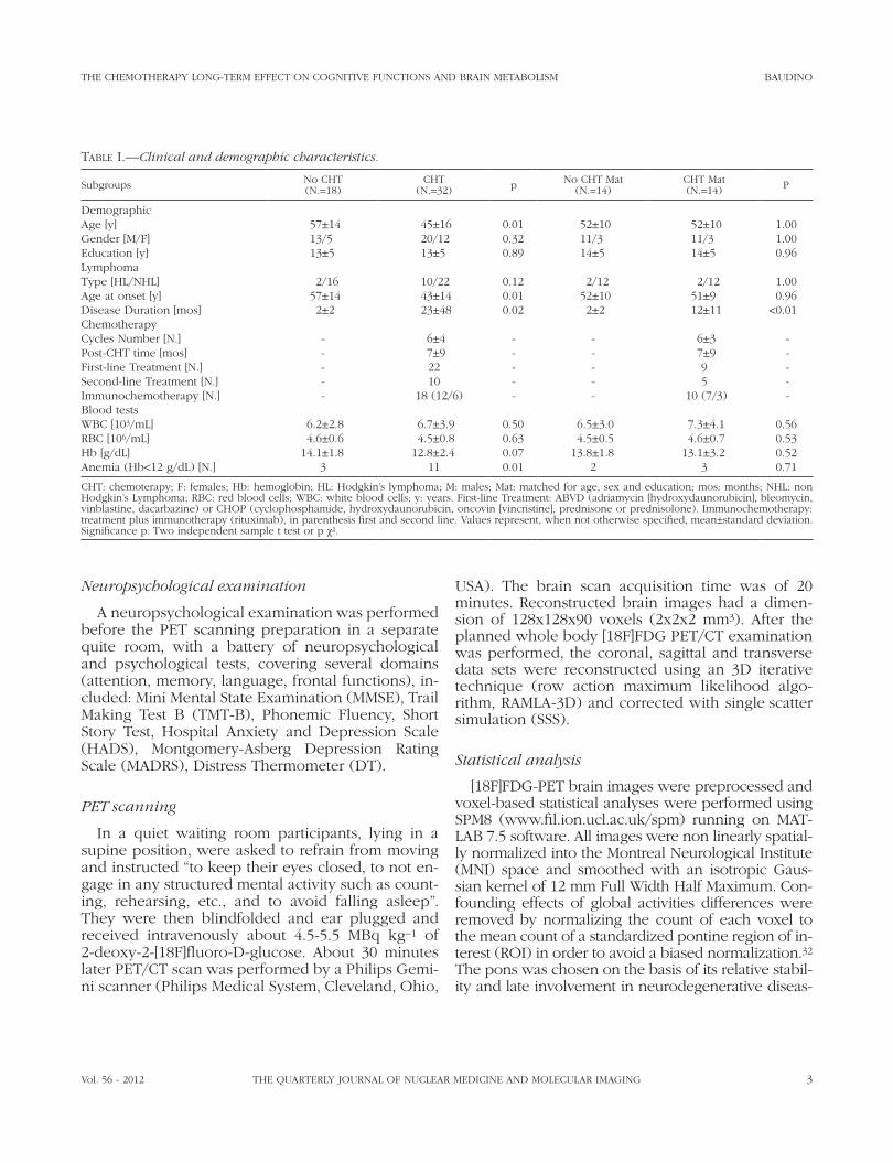
Table I.—�Clinical and demographic characteristics.
Subgroups No CHT(N.=18)
CHT(N.=32) p No CHT Mat
(N.=14)CHT Mat(N.=14) P
DemographicAge [y] 57±14 45±16 0.01 52±10 52±10 1.00Gender [M/F] 13/5 20/12 0.32 11/3 11/3 1.00Education [y] 13±5 13±5 0.89 14±5 14±5 0.96LymphomaType [HL/NHL] 2/16 10/22 0.12 2/12 2/12 1.00Age at onset [y] 57±14 43±14 0.01 52±10 51±9 0.96Disease Duration [mos] 2±2 23±48 0.02 2±2 12±11 <0.01ChemotherapyCycles Number [N.] - 6±4 - - 6±3 -Post-CHT time [mos] - 7±9 - - 7±9 -First-line Treatment [N.] - 22 - - 9 -Second-line Treatment [N.] - 10 - - 5 -Immunochemotherapy [N.] - 18 (12/6) - - 10 (7/3) -Blood testsWBC [103/mL] 6.2±2.8 6.7±3.9 0.50 6.5±3.0 7.3±4.1 0.56RBC [106/mL] 4.6±0.6 4.5±0.8 0.63 4.5±0.5 4.6±0.7 0.53Hb [g/dL] 14.1±1.8 12.8±2.4 0.07 13.8±1.8 13.1±3.2 0.52Anemia (Hb<12 g/dL) [N.] 3 11 0.01 2 3 0.71
CHT: chemoterapy; F: females; Hb: hemoglobin; HL: Hodgkin’s lymphoma; M: males; Mat: matched for age, sex and education; mos: months; NHL: non Hodgkin’s Lymphoma; RBC: red blood cells; WBC: white blood cells; y: years. First-line Treatment: ABVD (adriamycin [hydroxydaunorubicin], bleomycin, vinblastine, dacarbazine) or CHOP (cyclophosphamide, hydroxydaunorubicin, oncovin [vincristine], prednisone or prednisolone). Immunochemotherapy: treatment plus immunotherapy (rituximab), in parenthesis first and second line. Values represent, when not otherwise specified, mean±standard deviation. Significance p. Two independent sample t test or p χ2.

BAUDINO THE CHEMOTHERAPY LONG-TERM EFFECT ON COGNITIVE FUNCTIONS AND BRAIN METABOLISM
4 THE QUARTERLY JOURNAL OF NUCLEAR MEDICINE AND MOLECULAR IMAGING VOL. 56 - 2012
the age impact onto rCMRglc and test scores in the CHT and No CHT Matched groups, then a conjunc-tion analysis between brain areas significantly cor-related which age, C and T was performed (N.=32).
Finally, to link the neurobehavior with metabolic pattern we used a GLM with test scores, age and gender as independent variables and rCMRglc as de-pendent variable (N.=50).
Results
Comparison between No CHT and CHT groups
The total CHT and No CHT groups (Table II) dif-fered in MMSE (P=0.04) and TMT-B (P=0.04) scores and showed a trend in Phonemic Fluency (P=0.08), but did not differ for depression self- (HADS-D) or hetero- (MARDS) evaluated, anxiety (HADS-A) or distress (DT, HADS-Tot).
The CHT and No CHT Matched groups did not dif-fer (Table II) for any psychological (depression, anx-iety, distress) or neuropsychological scores (MMSE, attention, fluency, memory, frontal functions). How-ever a cluster of 3297 voxels involving bilaterally the anterior cingulate cortex (max Z=3.73, P corr. =0.03) and part of frontal cortices showed a lesser rCMRglc (Figure 1, Table III) in the CHT group.
Correlations with C, T and age
In the total CHT group, we observed a negative correlation between C and a bilateral fronto-tempo-ral rCMRglc pattern (Figure 2). A similar pattern was positively correlated with T (Figure 2) and negative-ly with age (Figure 2). The conjoint analysis of the three correlations together gave a very large and sig-nificant cluster involving bilaterally the frontal corti-ces (Ke=13457, max Z=4.42, P corr. <0.01, Figure 2, Table III).
Comparison between age impact onto rCMRglc in No CHT and CHT groups
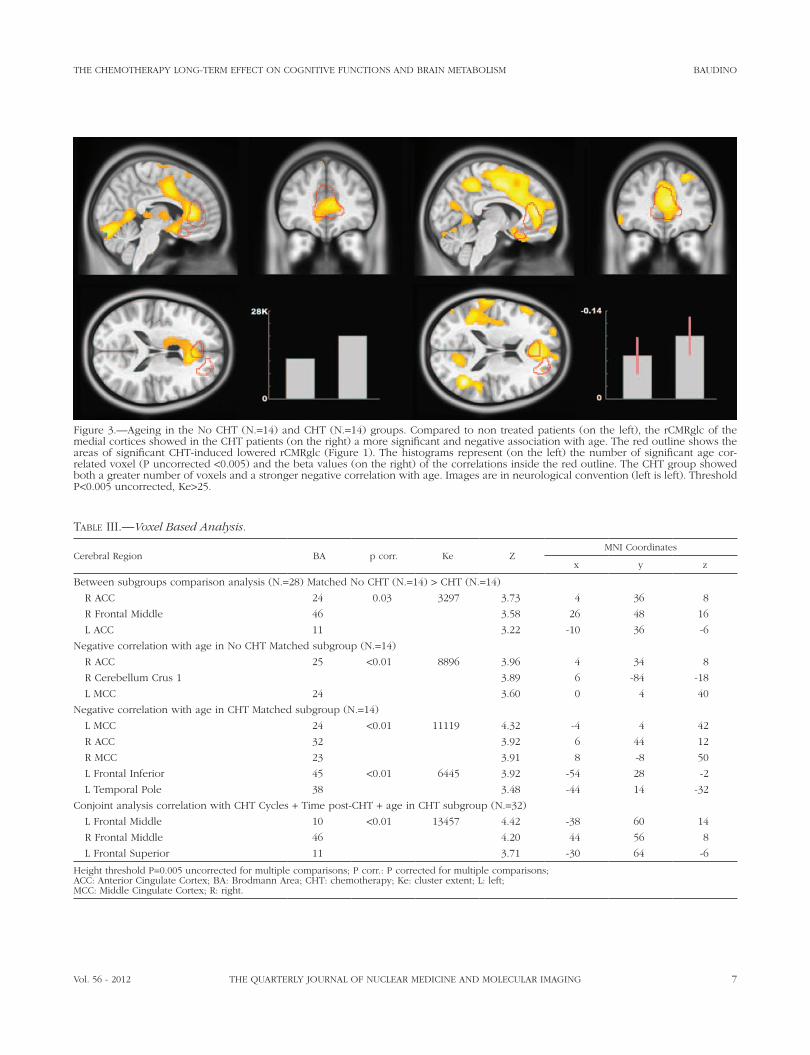
In the No CHT Matched group, age was nega-tively correlated with a large medial cluster involv-ing cingulate cortex and cerebellum (Ke=8896, max Z=3.96, P corr. <0.01, Figure 3, Table III).
The CHT Matched group showed a similar pat-tern (Figure 3, Table III), but more extended, with two large clusters involving medial (Ke=11119, max
es such as Alzheimer disease, a finding leading other investigators to use it as reference region. The ROI was a rectangular multislice region (x/x’=-8/8, y/y’=-32/-24, z/z’=-44/-34; MNI space) sampling 144 voxels on the central pontine region and manually drawn on the PET SPM template using the MRIcro application (http://www.sph.sc.edu/comd/rorden/mricro.html). Both ROI coordinates and dimensions were chosen to avoid low-counts background voxel sampling and to minimize the random noise effect. A previous careful visual inspection of the pons was conducted on each spatially normalized but non smoothed brain scan in order to detect metabolic changes which could alter the ROI measure. The same ROI was then employed on each spatial normalized and smoothed brain image and the pons mean voxel values (Yp) sampled. Using the image calculation tool of SPM, the scaled voxel values (Y ’) of each brain was set at Y ’ = (Y/Yp) were Y was the non scaled (“raw”) voxel value. Only voxel values greater than 80% of the whole brain mean MR-glc were included in the analysis. All SPM results were thresholded at P<0.005 uncorrected for multiple com-parisons, with an extent threshold cluster extent (Ke) of 25 voxels. Statistical inferences were performed by applying the Random Field Theory. Clusters with P≤0.05 corrected for multiple comparisons were con-sidered as significant.
SPSS 13.0 was used for all other statistical analy-ses, P<0.05 was considered as significant.
Between groups rCMRglc comparison analyses were performed using a two independent samples t test after selecting 14 CHT and 14 No CHT pa-tients exactly matched one by one (same sex and age) for a case-control design (Table I). Neuropsy-chological and psychological scores comparison were performed with a two independent samples t test for Matched groups raw scores (14 CHT versus 14 No CHT) and for total groups (18 CHT versus 32 No CHT) corrected scores (sex, age and educa-tion).
Since both animal 33, 34 and human studies sug-gested that CHT dose 9, 15 and number of CHT cycles (C),19 as well as the time (T) elapsed since comple-tion of the treatment 13, 16-18 can modulate the CHT effects on neurobehavior and brain metabolism, we tested this hypothesis on the whole CHT group (N.=32) with a General Linear Model (GLM) using age, gender, C and T as independent variables and rCMRglc as dependent variables.
To better investigate the possible mechanisms un-derlying the CHT induced changes we compared

THE CHEMOTHERAPY LONG-TERM EFFECT ON COGNITIVE FUNCTIONS AND BRAIN METABOLISM BAUDINO
Vol. 56 - 2012 THE QUARTERLY JOURNAL OF NUCLEAR MEDICINE AND MOLECULAR IMAGING 5
(P<0.005) with the rCMRglc except for TMT-B, but the clusters disappeared if we applied a threshold correction for multiple comparison (the pattern was similar to that of the age negative correlation, data not shown).
Discussion
Some neuroimaging studies have shown that CHT could induce structural 21, 22, 25-27, 35 and functional 24, 28, 36 brain changes involving both the cortex and the white matter, with a prevailing involvement of the prefrontal cortex. However, the time course of such changes is less well consistently defined, with a study suggesting a complete recovery of the struc-tural damage few years after the completion of the treatment 21, and others reporting a longer lasting functional impairment.28, 29
Our data strongly support a reversible, but long lasting model (at least after 6 months post-CHT) of CHT induced brain damage (Figures 1, 2, Table III), suggesting that chemotherapy induced cerebral glu-cose metabolic impairment could be transient and reversible over time (Figure 2), paralleling or even preceding the structural recovery of the CHT target-ed brain regions. The neurobehavioral correlate of this metabolic impairment was the significant wors-ening of MMSE (P<0.05), TMT-B (P<0.05) and Flu-
Z=4.32, P corr. <0.01), but also fronto-temporal left areas (Ke=6445, max Z=3.92, P corr. <0.01).
Correlation between test scores and rCMRglc
In the total sample of subjects no psychological or neuropsychological test significantly correlated
Table II.—�Neurobehavioral characteristics.
Subgroups No CHT(N.=18)
CHT(N.=32) p No CHT Mat
(N.=14)CHT Mat(N.=14) P
Neuropsychological testsMMSE 27.6±1.4 26.7±1.5 0.04 28.4±1.3 28.1±1.8 0.63TMT-B 71±37 100±52 0.04 84±24 97±52 0.40Phonemic Fluency 36±9 30±12 0.08 38±8 33±11 0.21Short Story IR - - - 5±2 6±1 0.13Short Story DR - - - 6±2 6±1 0.69Short Story Tot 10±3 10±3 0.96 11±3 12±1 0.43DepressionMADRS 10±5 8±7 0.40 9±6 8±7 0.68HADS-D 4±3 4±4 0.90 4±3 4±3 0.86AnxietyHADS-A 6±3 5±3 0.14 6±3 4±3 0.17DistressDT 3±2 3±2 0.51 3±2 2±2 0.28HADS-Tot 10±5 9±6 0.46 9±5 8±6 0.37
Raw scores for the Mat groups, corrected scores (age, sex and education) for the total groups. CHT: Chemotherapy; DR: delayed recall; DT: distress ther-mometer; HADS: Hospital Anxiety and Depression Scale; IR: Immediate Recall; Mat: matched for age, sex and education; MADRS: Montgomery-Asberg Depression Rating Scale; MMSE: Mini Mental State Examination; TMT-B: Trail Making Test B; Tot: total. Values represent, when not otherwise specified, mean±standard deviation. Significance p. Two independent sample t test.
Figure 1.—Patterns of cortical CHT-induced rCMRglc changes. T-maps obtained by comparisons analysis between No CHT and CHT groups overlaid on canonical brain templates. Images are in neu-rological convention (left is left). Threshold P<0.005 uncorrected, Ke>25 for both rows, only for the fist row’s cluster p corr≤0.05.
BAUDINO THE CHEMOTHERAPY LONG-TERM EFFECT ON COGNITIVE FUNCTIONS AND BRAIN METABOLISM
6 THE QUARTERLY JOURNAL OF NUCLEAR MEDICINE AND MOLECULAR IMAGING VOL. 56 - 2012
brain metabolic and cognitive impairment must be taken with caution. Indeed, we cannot exclude that a subset of brain regions do not recover or recover only partially over time. Candidate regions could be medial areas that showed a long lasting metabolic impairment in the present study (ACC in the No CHT versus CHT comparison, Figures 1, 2, Table III), but not a linear recovery with post-CHT time (the frontal areas recovering were more lateral, Figure 2, Table III). We cannot exclude that these areas recover in a non linear, slower manner or only partially, so all these hypotheses are compatible with our analyses.
To gain more insights on the mechanisms un-derlying these chemotherapy related adverse ef-
ency (P<0.10) scores in the CHT groups (Table II). The results were significant only in the total CHT group as, probably in the Matched groups there was not enough power to reveal the effect (note that the differences between the Matched groups scores would be significant with a larger sample as in the total groups [Table II], we are in fact talking of a subtle subclinical impairment). These results are in agreement with findings emerging from controlled longitudinal neuropsychological studies 13, 16-18 indi-cating that cognitive changes are noticeable close to the end off-therapy, but tend to fully or partially resolve over one year.
However, the assumptions on the reversibility of
Figure 2.—Conjoint analysis of number of cycles, time elapsed after the therapy and aging. The whole group of patients having received chemotherapy showed a significant lowering of rCMRglc with the number of cycles C in the bilateral fronto-temporal cortices (in blue) and a recovering of the same areas rCMRglc with the time T elapsed after the therapy (in red). This pattern was also strongly negatively correlated with the age of the patients (in green). These findings are consistent with a time-dependent heuristic model of CHT-induced rCMRglc changes (on the upper-left). The model take in account moreover the possible cumulative neurotoxic effects of the CHT cycles (red asterisk). On the upper-right the common pattern of C, T and aging (in light blue) evidenced by a conjoin analysis (P<0.005 uncor-rected, Ke>25).
THE CHEMOTHERAPY LONG-TERM EFFECT ON COGNITIVE FUNCTIONS AND BRAIN METABOLISM BAUDINO
Vol. 56 - 2012 THE QUARTERLY JOURNAL OF NUCLEAR MEDICINE AND MOLECULAR IMAGING 7
Figure 3.—Ageing in the No CHT (N.=14) and CHT (N.=14) groups. Compared to non treated patients (on the left), the rCMRglc of the medial cortices showed in the CHT patients (on the right) a more significant and negative association with age. The red outline shows the areas of significant CHT-induced lowered rCMRglc (Figure 1). The histograms represent (on the left) the number of significant age cor-related voxel (P uncorrected <0.005) and the beta values (on the right) of the correlations inside the red outline. The CHT group showed both a greater number of voxels and a stronger negative correlation with age. Images are in neurological convention (left is left). Threshold P<0.005 uncorrected, Ke>25.
Table III.—�Voxel Based Analysis.
Cerebral Region BA p corr. Ke ZMNI Coordinates
x y z
Between subgroups comparison analysis (N.=28) Matched No CHT (N.=14) > CHT (N.=14)
R ACC 24 0.03 3297 3.73 4 36 8
R Frontal Middle 46 3.58 26 48 16
L ACC 11 3.22 -10 36 -6
Negative correlation with age in No CHT Matched subgroup (N.=14)
R ACC 25 <0.01 8896 3.96 4 34 8
R Cerebellum Crus 1 3.89 6 -84 -18
L MCC 24 3.60 0 4 40
Negative correlation with age in CHT Matched subgroup (N.=14)
L MCC 24 <0.01 11119 4.32 -4 4 42
R ACC 32 3.92 6 44 12
R MCC 23 3.91 8 -8 50
L Frontal Inferior 45 <0.01 6445 3.92 -54 28 -2
L Temporal Pole 38 3.48 -44 14 -32
Conjoint analysis correlation with CHT Cycles + Time post-CHT + age in CHT subgroup (N.=32)
L Frontal Middle 10 <0.01 13457 4.42 -38 60 14
R Frontal Middle 46 4.20 44 56 8
L Frontal Superior 11 3.71 -30 64 -6
Height threshold P=0.005 uncorrected for multiple comparisons; P corr.: P corrected for multiple comparisons;ACC: Anterior Cingulate Cortex; BA: Brodmann Area; CHT: chemotherapy; Ke: cluster extent; L: left;MCC: Middle Cingulate Cortex; R: right.
BAUDINO THE CHEMOTHERAPY LONG-TERM EFFECT ON COGNITIVE FUNCTIONS AND BRAIN METABOLISM
8 THE QUARTERLY JOURNAL OF NUCLEAR MEDICINE AND MOLECULAR IMAGING VOL. 56 - 2012
speculate that CHT induces ageing-like and/or ac-celerates ageing-related processes, such as oxidative stress and decreased mitochondrial function, which could lead to the rCMRglc reduction evidenced in patients receiving CHT.
The TMT-B score was the only neuropsychologi-cal measure showing significant correlations with rCMRglc (P<0.005), although not surviving to clus-ter multiple comparison correction. This probably because, among all the tests, it was the one more related to frontal functions and their decline with ageing. Noteworthy, the pattern was similar to the CHT induced pattern reinforcing the link between metabolic and neuropsychological alterations. A confounding effect on our results could be related to the anxiety status connected with the PET exami-nation that may have influenced the neuropsycho-logical setting, but to avoid the risk of drop out of patients, we choose not to separate sessions, how-ever this limitation is overcome by the fact that this bias was equally distributed among groups.
Our data supported that the observed effect of CHT was not an indirect effect of the psychological status (distress, depression, anxiety) or of the chem-otherapy induced bone marrow suppression, as the Matched groups did not differ for clinical mood or blood data (Table II).
Limitations
Obviously a longitudinal study could have an ad-vantage in observing the evolution of metabolism over time, unfortunately, in a clinical setting it is complex to organize with patients highly emotionally and physically stressed. However, it could also have a disadvantage: the same patients after some months will have a longer disease duration and any metabo-lism difference could be related to a aging process. A potential limitation of the study is the heterogeneity of protocols treatment that could limit the possibility to assess CHT effects, for example, steroids could have a specific effect on brain metabolism, however the limited number of patients belonging to the dif-ferent treatments subgroups did not consent to per-form a meaningful statistical analysis.
Conclusions
This study evidenced significant chemotherapy related changes on glucose metabolism in multiple
fects, we looked for a relationship between C and the cerebral glucose metabolism. The voxel-based correlation analysis evidenced a set of brain areas showing a significant negative association between C and the rCMRglc (Figure 2), which partially over-lapped those uncovered by the post-CHT correla-tion analysis described above (Figure 2). These re-sults therefore support a model in which the CHT induced brain damage appears to be related to C, in agreement with previous neuropsychological data 19 reporting a negative relationship between C and the cognitive performance. At this time we cannot dis-criminate between a pure dose dependency, a pure CHT duration dependency or a mixed/interaction form of dependency. The number of CHT cycles, in fact, could be thought as a mere proxy of either the above aspects, since an higher number of cy-cles could correlate with an higher dose, but also with a longer time of exposition. We cannot exclude that the number of cycles represent the number of times that the system came in contact with CHT and the complex cascade of events triggered by this ex-position. So this repeated interaction per se (Figure 2) could be a factor guiding the changes and the adaptation mechanisms activated as a response by the system (e.g. cytokines production, stress system activation, immune system responses).
The pattern of CHT induced brain metabolic im-pairment, markedly characterized by a dispropor-tionate involvement of frontal lobes (Figure 1, Table III), is reminiscent of that seen in aging 37-40 and some age-related neurodegenerative processes.41 This was confirmed by our analysis on age negatively cor-related pattern (Figure 2) and its large overlap with the previous C and T correlation patterns (Figure 2).
Oxidative DNA damage and decreased mito-chondrial function are well established processes underlying brain aging changes,42 findings leading Maccormick 43 to hypothesize that adverse effects associated with CHT might be related to accelera-tion of the ageing process. Inspired by this hypoth-esis we looked for differences in the correlations between age and the rCMRglc in the regions show-ing a lower glucose metabolism in the CHT and No CHT subgroups. This analysis showed that, com-pared to non treated patients, the CMRglc of me-dial cortices in CHT patients had a more negative and more widespread association with age (Figure 3, Table III), meaning that older subjects undergo a higher than expected CHT induced metabolic im-pairment. Taken together, these findings lead us to

THE CHEMOTHERAPY LONG-TERM EFFECT ON COGNITIVE FUNCTIONS AND BRAIN METABOLISM BAUDINO
Vol. 56 - 2012 THE QUARTERLY JOURNAL OF NUCLEAR MEDICINE AND MOLECULAR IMAGING 9
Preliminary results of a longitudinal study of changes in cogni-tive function in breast cancer patients undergoing chemothe-rapy with doxorubicin and cyclophosphamide. Psychooncology 2008;17:1189-95.
15. Schagen SB, Muller MJ, Boogerd W, Mellenbergh GJ, van Dam FS. Change in cognitive function after chemotherapy: a prospec-tive longitudinal study in breast cancer patients. J Natl Cancer Inst 2006;98:1742-5.
16. Wagner LI, Sweet JJ, Butt Z. Trajectory of cognitive impairment during breast cancer treatment: a prospective analysis. ASCO Me-eting Abstracts 8500. Journal of clinical oncology: official journal of the American Society of Clinical Oncology 2006;24:4685.
17. Fan HG, Houede-Tchen N, Yi QL, Chemerynsky I, Downie FP, Sabate K et al. Fatigue, menopausal symptoms, and cognitive function in women after adjuvant chemotherapy for breast can-cer: 1- and 2-year follow-up of a prospective controlled study. J Clin Oncol 2005;23:8025-32.
18. Wefel JS, Lenzi R, Theriault RL, Davis RN, Meyers CA. The co-gnitive sequelae of standard-dose adjuvant chemotherapy in women with breast carcinoma: results of a prospective, rando-mized, longitudinal trial. Cancer 2004;100:2292-9.
19. Ahles TA, Saykin AJ, Furstenberg CT, Cole B, Mott LA, Skalla K et al. Neuropsychologic impact of standard-dose systemic chemo-therapy in long-term survivors of breast cancer and lymphoma. J Clin Oncol 2002;20:485-93.
20. Castellon S, Ganz P, Bower J, Petersen L, Abraham L, Greendale G. Neurocognitive performance in breast cancer survivors ex-posed to adjuvant chemotherapy and tamoxifen. J of Clin Ex-periment Neuropsychol 2004;26:955-69.
21. Inagaki M, Yoshikawa E, Matsuoka Y, Sugawara Y, Nakano T, Akechi T et al. Smaller regional volumes of brain gray and white matter demonstrated in breast cancer survivors exposed to adju-vant chemotherapy. Cancer 2007;109:146-56.
22. Brown MS, Simon JH, Stemmer SM, Stears JC, Scherzinger A, Cagnoni PJ et al. MR and proton spectroscopy of white matter disease induced by high-dose chemotherapy with bone marrow transplant in advanced breast carcinoma. AJNR Am J Neuroradiol 1995;16:2013-20.
23. Stemmer SM, Stears JC, Burton BS, Jones RB, Simon JH. Whi-te matter changes in patients with breast cancer treated with high-dose chemotherapy and autologous bone marrow support. AJNR Am J Neuroradiol 1994;15:1267-73.
24. Ferguson RJ, McDonald BC, Saykin AJ, Ahles TA. Brain structure and function differences in monozygotic twins: possible effects of breast cancer chemotherapy. J Clin Oncol 2007;25:3866-70.
25. Koppelmans V, de Ruiter MB, van der Lijn F, Boogerd W, Sey-naeve C, van der Lugt A et al. Global and focal brain volume in long-term breast cancer survivors exposed to adjuvant chemo-therapy. Breast Cancer Res Treat 2012;132:1099-106.
26. de Ruiter MB, Reneman L, Boogerd W, Veltman DJ, Caan M, Douaud G et al. Late effects of high-dose adjuvant chemothera-py on white and gray matter in breast cancer survivors: Converg-ing results from multimodal magnetic resonance imaging. Hum Brain Mapp 2011 [Epub ahead of print].
27. Brown MS, Stemmer SM, Simon JH, Stears JC, Jones RB, Cagnoni PJ et al. White matter disease induced by high-dose chemother-apy: longitudinal study with MR imaging and proton spectros-copy. AJNR Am J Neuroradiol 1998;19:217-21.
28. Silverman DH, Dy CJ, Castellon SA, Lai J, Pio BS, Abraham L et al. Altered frontocortical, cerebellar, and basal ganglia activity in adjuvant-treated breast cancer survivors 5-10 years after chemo-therapy. Breast Cancer Res Treat 2007;103:303-11.
29. Kesler SR, Bennett FC, Mahaffey ML, Spiegel D. Regional brain activation during verbal declarative memory in metastatic breast cancer. Clin Cancer Res 2009;15:6665-73.
30. de Ruiter MB, Reneman L, Boogerd W, Veltman DJ, van Dam FS, Nederveen AJ et al. Cerebral hyporesponsiveness and cognitive impairment 10 years after chemotherapy for breast cancer. Hum Brain Mapp 2011;32:1206-19.
brain regions, with a prevailing involvement of fron-tal lobes. Such metabolic changes appear to be posi-tively related to the time elapsed from the end of the treatment suggesting that they are transient and rapidly reversible. A subset of these areas undergoes a metabolic impairment proportional to the number of CHT cycles while a subset, including the cerebel-lum and midline cortical regions, presents evidences of partial or delayed metabolic recovery.
References
1. Jenkins V, Shilling V, Deutsch G, Bloomfield D, Morris R, Allan S et al. A 3-year prospective study of the effects of adjuvant treat-ments on cognition in women with early stage breast cancer. Br J Cancer 2006;94:828-34.
2. Mehlsen M, Pedersen AD, Jensen AB, Zachariae R. No indica-tions of cognitive side-effects in a prospective study of breast cancer patients receiving adjuvant chemotherapy. Psychoonco-logy 2009;18:248-57.
3. Donovan KA, Small BJ, Andrykowski MA, Schmitt FA, Munster P, Jacobsen PB. Cognitive functioning after adjuvant chemotherapy and/or radiotherapy for early-stage breast carcinoma. Cancer 2005;104:2499-507.
4. Ahles TA, Silberfarb PM, Herndon J, 2nd, Maurer LH, Kornblith AB, Aisner J et al. Psychologic and neuropsychologic function-ing of patients with limited small-cell lung cancer treated with chemotherapy and radiation therapy with or without warfarin: a study by the Cancer and Leukemia Group B. J Clin Oncol 1998;16:1954-60.
5. Anderson-Hanley C, Sherman ML, Riggs R, Agocha VB, Compas BE. Neuropsychological effects of treatments for adults with can-cer: a meta-analysis and review of the literature. J Int Neuropsy-chol Soc 2003;9:967-82.
6. Freeman JR, Broshek DK. Assessing cognitive dysfunction in breast cancer: what are the tools? Clin Breast Cancer 2002;3(Sup-pl 3):S91-9.
7. Reid-Arndt SA, Yee A, Perry MC, Hsieh C. Cognitive and psy-chological factors associated with early posttreatment functio-nal outcomes in breast cancer survivors. J Psychosoc Oncol 2009;27:415-34.
8. Schagen SB, van Dam FS, Muller MJ, Boogerd W, Lindeboom J, Bruning PF. Cognitive deficits after postoperative adjuvant che-motherapy for breast carcinoma. Cancer 1999;85:640-50.
9. van Dam FS, Schagen SB, Muller MJ, Boogerd W, vd Wall E, Droogleever Fortuyn ME et al. Impairment of cognitive function in women receiving adjuvant treatment for high-risk breast can-cer: high-dose versus standard-dose chemotherapy. J Natl Can-J Natl Can-cer Inst 1998;90:210-8.
10. Wieneke MH, Dienst ER. Neuropsychological Assessment of Co-gnitive Functioning Following Chemotherapy for Breast Cancer. Psycho-Oncology 1995;41:61-6.
11. Jansen CE, Miaskowski C, Dodd M, Dowling G, Kramer J. A metaanalysis of studies of the effects of cancer chemotherapy on various domains of cognitive function. Cancer 2005;104:2222-33.
12. Falleti MG, Sanfilippo A, Maruff P, Weih L, Phillips KA. The natu-re and severity of cognitive impairment associated with adjuvant chemotherapy in women with breast cancer: a meta-analysis of the current literature. Brain Cogn 2005;59:60-70.
13. Collins B, Mackenzie J, Stewart A, Bielajew C, Verma S. Cogni-tive effects of chemotherapy in post-menopausal breast cancer patients 1 year after treatment. Psychooncology 2009;18:134-43.
14. Jansen CE, Dodd MJ, Miaskowski CA, Dowling GA, Kramer J.
BAUDINO THE CHEMOTHERAPY LONG-TERM EFFECT ON COGNITIVE FUNCTIONS AND BRAIN METABOLISM
10 THE QUARTERLY JOURNAL OF NUCLEAR MEDICINE AND MOLECULAR IMAGING VOL. 56 - 2012
Hippocampal hypometabolism predicts cognitive decline from normal aging. Neurobiol Aging 2008;29:676-92.
40. Tumeh PC, Alavi A, Houseni M, Greenfield A, Chryssikos T, Newberg A et al. Structural and functional imaging correlates for age-related changes in the brain. Semin Nucl Med 2007;37:69- 87.
41. Silverman DHS. Brain 18F-FDG PET in the diagnosis of neuro-degenerative dementias: comparison with perfusion SPECT and with clinical evaluations lacking nuclear imaging. Journal of nu-Journal of nu-clear medicine: official publication. Soc Nucl Med 2004;45:594-607.
42. Mattson MP, Magnus T. Ageing and neuronal vulnerability. Nat Rev Neurosci 2006;7:278-94.
43. Maccormick RE. Possible acceleration of aging by adjuvant chemotherapy: a cause of early onset frailty? Med Hypotheses 2006;67:212-5.
Acknowledgements.—Authors would like to thank patients who par-ticipated in the study and their families. We wish to thank Dr. Francesca Giunta for her contribution to this paper and all the NIPGI (Nuclear Imaging, Psychological, Genetic Investigation) Study Group, composed by: authors and Marcella Caglio, Lorys Castelli, Alberto Censon, Tom-maso Costa, Pierpaola Fenoglio, Salvatore Gallone, Lorella Orsucci, An-tonella Parente, Roberto Passera, Debora Tonello, Antonella Varetto, UmbertoVitolo.
Conflicts of interest.—The authors declare no conflict of interest.Received on February 20, 2012.Accepted for publication on September 7, 2012.
31. Ahles TA, Saykin AJ. Candidate mechanisms for chemotherapy-induced cognitive changes. Nat Rev Cancer 2007;7:192-201.
32. Borghammer P, Cumming P, Aanerud J, Gjedde A. Artefac-tual subcortical hyperperfusion in PET studies normalized to global mean: lessons from Parkinson’s disease. Neuroimage 2009;45:249-57.
33. Mignone RG, Weber ET. Potent inhibition of cell proliferation in the hippocampal dentate gyrus of mice by the chemotherapeutic drug thioTEPA. Brain Res 2006;1111:26-9.
34. Rzeski W, Pruskil S, Macke A, Felderhoff-Mueser U, Reiher AK, Hoerster F et al. Anticancer agents are potent neurotoxins in vitro and in vivo. Ann Neurol 2004;56:351-60.
35. Abraham J, Haut MW, Moran MT, Filburn S, Lemiuex S, Kuwa-bara H. Adjuvant chemotherapy for breast cancer: effects on ce-rebral white matter seen in diffusion tensor imaging. Clin Breast Cancer 2008;8:88-91.
36. Eberling JL, Wu C, Tong-Turnbeaugh R, Jagust WJ. Estrogen- and tamoxifen-associated effects on brain structure and function. Neuroimage 2004;21:364-71.
37. de Leon MJ, Convit A, Wolf OT, Tarshish CY, DeSanti S, Rusinek H et al. Prediction of cognitive decline in normal elderly subjects with 2-[(18)F]fluoro-2-deoxy-D-glucose/poitron-emission tom-ography (FDG/PET). Proc Natl Acad Sci U S A 2001;98:10966-71.
38. Kalpouzos G, Chetelat G, Baron JC, Landeau B, Mevel K, Go-deau C et al. Voxel-based mapping of brain gray matter volume and glucose metabolism profiles in normal aging. Neurobiol Aging 2009;30:112-24.
39. Mosconi L, De Santi S, Li J, Tsui WH, Li Y, Boppana M et al.