Formation of Acrolein-derived 2'-Deoxyadenosine Adduct in an Iron-induced Carcinogenesis Model
BBA Clinical 3 (2015) 152–161
Contents lists available at ScienceDirect
BBA Clinical
j ourna l homepage: http : / /www. journa ls .e lsev ie r .com/bba-c l i n i ca l /
Review
Cancer metabolism and oxidative stress: Insights into carcinogenesis andchemotherapy via the non-dihydrofolate reductase effects of methotrexate
Joshua A. Hess a,⁎, Mohamad K. Khasawneh b
a Department of Internal Medicine and Pediatrics, Marshall University School of Medicine, 1600 Medical Center Drive, Huntington, WV 25701, United Statesb Marshall University School of Medicine, Edwards Comprehensive Cancer Center, 1400 Hal Greer Blvd., Huntington, WV 25701, United States
⁎ Corresponding author: Tel.: +1 304 691 1094E-mail address: [email protected] (J.A. Hess).
http://dx.doi.org/10.1016/j.bbacli.2015.01.0062214-6474/© 2015 The Authors. Published by Elsevier B.V
a b s t r a c t
a r t i c l e i n f oArticle history:Received 5 December 2014Received in revised form 8 January 2015Accepted 21 January 2015Available online 7 February 2015
Keywords:Cancer metabolismOxidative stressWarburg effectMethotrexateMitochondria
Methotrexate has been in use as an anti-cancer agent for over 60 years. Though inhibition of dihydrofolate reduc-tase is its best known mechanisms of action, its non-dihydrofolate reductase dependent mechanisms disruptmetabolic pathways resulting in a depletion of NAD(P)H and increasing oxidative stress. These mechanismshighlight a novel dependence of cancer cells on their metabolic abnormalities to buffer oxidative stress and che-motherapeutic agents interfere with these cellular abilities. Mitochondria appear to play a significant role inmaintaining cancer cell viability and alterations inmetabolism seen in cancer cells aid this mitochondrial ability.Further research is needed to understand the effects of other chemotherapeutic agents on these pathways.
© 2015 The Authors. Published by Elsevier B.V. This is an open access article under the CC BY-NC-ND license(http://creativecommons.org/licenses/by-nc-nd/4.0/).
Contents
1. Introduction . . . . . . . . . . . . . . . . . . . . . . . . . . . . . . . . . . . . . . . . . . . . . . . . . . . . . . . . . . . . . . 1522. Methods . . . . . . . . . . . . . . . . . . . . . . . . . . . . . . . . . . . . . . . . . . . . . . . . . . . . . . . . . . . . . . . . 1533. Metabolic dysfunction of the cancer cell and the Warburg effect . . . . . . . . . . . . . . . . . . . . . . . . . . . . . . . . . . . . . . . 1544. Methotrexate . . . . . . . . . . . . . . . . . . . . . . . . . . . . . . . . . . . . . . . . . . . . . . . . . . . . . . . . . . . . . 1545. Mitochondrial roles in oxidative stress and carcinogenesis . . . . . . . . . . . . . . . . . . . . . . . . . . . . . . . . . . . . . . . . . 1566. Mitochondrial metabolism and the cell cycle . . . . . . . . . . . . . . . . . . . . . . . . . . . . . . . . . . . . . . . . . . . . . . . 1577. DNA damage and malignant transformation . . . . . . . . . . . . . . . . . . . . . . . . . . . . . . . . . . . . . . . . . . . . . . . . 1588. Mitochondria, evolution and cancer development . . . . . . . . . . . . . . . . . . . . . . . . . . . . . . . . . . . . . . . . . . . . . 1589. Discussion . . . . . . . . . . . . . . . . . . . . . . . . . . . . . . . . . . . . . . . . . . . . . . . . . . . . . . . . . . . . . . . 158Transparency documents . . . . . . . . . . . . . . . . . . . . . . . . . . . . . . . . . . . . . . . . . . . . . . . . . . . . . . . . . . . 159Acknowledgments . . . . . . . . . . . . . . . . . . . . . . . . . . . . . . . . . . . . . . . . . . . . . . . . . . . . . . . . . . . . . . 159References . . . . . . . . . . . . . . . . . . . . . . . . . . . . . . . . . . . . . . . . . . . . . . . . . . . . . . . . . . . . . . . . . . .
1. Introduction
The origins of modern day chemotherapy can be traced back to theearly days of World War II. On December 2, 1943, the port of Bari,Italy was the target of a German air raid on Allied forces [1–3]. The casu-alties of this air raid far outnumbered those expected for the size of theattack. Though banned by the Geneva Protocol of 1925, Allied forceswere concernedwith the possibility that theGerman armywould resort
. This is an open access article under
to the use of chemical weapons. In August of 1943, President Rooseveltapproved the shipment of a secret cargo of mustard gas via SS Harvey.The explosion of the ship during this raid released its contents ontothe city inflicting an untold number of civilian casualties. With somevariation in details depending on the source, investigations by Dr.Stewart Francis Alexander, a Lieutenant Colonel and expert in chemicalwarfare noted significant suppression of both lymphoid and myeloidtissues in those exposed to the chemical agent [1]. Later research witha related compound, mustine, led to successful reduction of tumormass in a patient with non-Hodgkin's lymphoma and to a realizationthat pharmacotherapy of cancers was feasible.
the CC BY-NC-ND license (http://creativecommons.org/licenses/by-nc-nd/4.0/).
153J.A. Hess, M.K. Khasawneh / BBA Clinical 3 (2015) 152–161
FollowingWorldWar II, a second step forward in chemotherapywasmade by a pathologist at Harvard Medical School. Sidney Farber hadstudied the stimulating effect of folic acid on the proliferation of acutelymphoblastic leukemia cells when given to children with this cancer[4]. At that time, knowledge of the role of folic acid was limited to itseffect as a cofactor in the synthesis of purines. This observation led toone of the first attempts at rational drug design. In collaboration withthe first chemists to successfully synthesize folic acid at Lederle Labora-tories, Farber helped design the folate analog aminopterin [4,3]. Later,this collaboration led to the synthesis of the antibiotic trimethoprimand the chemotherapeutic agent amethopterin, more famously knownas methotrexate [4]. Since that time, the intracellular biochemistries ofboth folic acid and methotrexate have been further explored anddetailed.
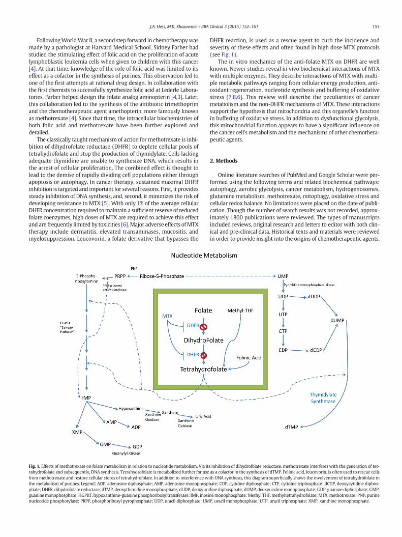
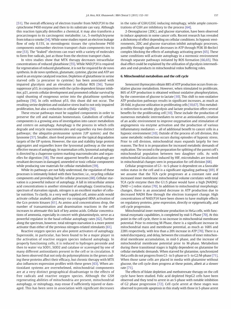
The classically taught mechanism of action for methotrexate is inhi-bition of dihydrofolate reductase (DHFR) to deplete cellular pools oftetrahydrofolate and stop the production of thymidylate. Cells lackingadequate thymidine are unable to synthesize DNA, which results inthe arrest of cellular proliferation. The combined effect is thought tolead to the demise of rapidly dividing cell populations either throughapoptosis or autophagy. In cancer therapy, sustained maximal DHFRinhibition is targeted and important for several reasons. First, it providessteady inhibition of DNA synthesis, and, second, it minimizes the risk ofdeveloping resistance to MTX [5]. With only 1% of the average cellularDHFR concentration required tomaintain a sufficient reserve of reducedfolate coenzymes, high doses of MTX are required to achieve this effectand are frequently limited by toxicities [6].Major adverse effects ofMTXtherapy include dermatitis, elevated transaminases, mucositis, andmyelosuppression. Leucovorin, a folate derivative that bypasses the
Fig. 1. Effects of methotrexate on folate metabolism in relation to nucleotidemetabolism. Via itrahydrofolate and subsequently, DNA synthesis. Tetrahydrofolate ismetabolized further for usefrommethotrexate and restore cellular stores of tetrahydrofolate. In addition to interference wthe metabolism of purines. Legend: ADP, adenosine diphosphate; AMP, adenosine monophospphate;DHFR, dihydrofolate reductase; dTMP, deoxythimidinemonophosphate; dUDP, deoxyuriguaninemonophosphate;HGPRT, hypoxanthine-guaninephosphoribosyltransferase; IMP, inosinucleotide phosphorylase; PRPP, phosphoribosyl pyrophosphate; UDP, uracil diphosphate; UM
DHFR reaction, is used as a rescue agent to curb the incidence andseverity of these effects and often found in high dose MTX protocols(see Fig. 1).
The in vitro mechanics of the anti-folate MTX on DHFR are wellknown. Newer studies reveal in vivo biochemical interactions of MTXwith multiple enzymes. They describe interactions of MTX with multi-ple metabolic pathways ranging from cellular energy production, anti-oxidant regeneration, nucleotide synthesis and buffering of oxidativestress [7,8,6]. This review will describe the peculiarities of cancermetabolism and the non-DHFRmechanisms of MTX. These interactionssupport the hypothesis that mitochondria and this organelle's functionin buffering of oxidative stress. In addition to dysfunctional glycolysis,this mitochondrial function appears to have a significant influence onthe cancer cell's metabolism and themechanisms of other chemothera-peutic agents.
2. Methods
Online literature searches of PubMed and Google Scholar were per-formed using the following terms and related biochemical pathways:autophagy, aerobic glycolysis, cancer metabolism, hydrogenosomes,glutamine metabolism, methotrexate, mitophagy, oxidative stress andcellular redox balance. No limitations were placed on the date of publi-cation. Though the number of search results was not recorded, approx-imately 1800 publications were reviewed. The types of manuscriptsincluded reviews, original research and letters to editor with both clin-ical and pre-clinical data. Historical texts and materials were reviewedin order to provide insight into the origins of chemotherapeutic agents.
s inhibition of dihydrofolate reductase, methotrexate interferes with the generation of tet-as a cofactor in the synthesis of dTMP. Folinic acid, leucovorin, is often used to rescue cellsith DNA synthesis, this diagram superficially shows the involvement of tetrahydrofolate inhate; CDP, cytidine diphosphate; CTP, cytidine triphosphate; dCDP, deoxycytidine diphos-dinediphosphate; dUMP, deoxyuridinemonophosphate; GDP, guanine diphosphate; GMP,nemonophosphate;Methyl THF,methyltetrahydrofolate;MTX,methotrexate; PNP, purineP, uracil monophosphate; UTP, uracil triphosphate; XMP, xanthine monophosphate.
154 J.A. Hess, M.K. Khasawneh / BBA Clinical 3 (2015) 152–161
In cases in which animal studies were reported, attempts were made tosearch for human trials of similar therapeutic scenarios.
3. Metabolic dysfunction of the cancer cell and the Warburg effect
Otto Warburg was awarded the 1931 Nobel Prize for Physiology orMedicine as a result of his research on cellular respiration. He hypothe-sized that cancerous transformation of tissue was a result of an injuredrespiration, ormetabolism. This hypothesis, referred to as “TheWarburgEffect”, describes a dysfunctional metabolic state dominated by glycoly-sis ending in lactate production and present in a variety of cancers [9,10]. Warburg postulated that this dysfunctional metabolism was theprimary cause of cancer. Cancer cell research has since characterized acomplex state of metabolic disarray involving glycolysis, the pentosephosphate pathway, the tricarboxylic acid (TCA) cycle, the electrontransport chain and amino acid metabolism [11,6,12–14]. The derange-ments seen in cancer cells result from a combination of factors includinghypoxia, poor total body and cellular antioxidant reserve,mitochondrialdysfunction and free radical production.
The elementary differences between normal and tumor cell metab-olism are characterized by increased glycolytic flux even in the presenceof adequate oxygen [14]. This change in cellular metabolism is oftconsidered a result, not an instigator, of cancerous transformation.However, the idea of metabolic disarray as an initiator of cancer hasgained additional support in recent years [14]. The metabolic ratios ofglycolysis and mitochondrial oxidative respiration in transformed cellsshow similarities to proliferating non-transformed cells but the abilityof transformed cells to return to non-Warburg, or non-stressed, meta-bolic profile is hindered. Somemay argue against this theory as oxygenconsumption in these transformed cells continues despite the produc-tion of ATP via aerobic glycolysis [15]. The perturbed metabolism oftransformed cells does not fully exclude oxidative phosphorylation asa source of ATP production but cellular oxygen tension, or lack thereof,does influence these cellular processes.
Under normal oxygen tension, hypoxia-inducible factor alpha (HIF-1α) is produced and degraded at an equal rate. The Von Hippel–Lindautumor suppressor gene product, pVHL, has multiple functions, one ofwhich is a recognition subunit of an E3 ubiquitin ligase complex. Prolylhydroxylases (PHD) catalyze the hydroxylation of proline residues onthe oxygen-dependent degradation site of HIF-1α creating a dockingsite for pVHL directed degradation [16]. Degradation of HIF-1α viapVHL is an oxygen-dependent process, converting oxygen and α-ketoglutarate to carbon dioxide, and is a rate-limiting reactiondependent on the functional capacity of pVHL [17]. In the absence ofsufficient reactants the HIF-1α protein stabilizes, binds HIF-b and istransported to the nucleus where it stimulates the transcriptionof genes containing the hypoxia response element. Stabilization ofHIF-1α outside of a true hypoxic environment is referred to aspseudohypoxia. The pseudohypoxic phenotype has been seen in associ-ation with alterations in TCA associated enzymes and may lead tochanges in epigenetic homeostasis with tumor promoting effects [14].Both succinate dehydrogenase and fumarate dehydrogenase possesstumor suppressing functions and loss of these functions has beenobserved in many VHL-associated renal tumors [16]. The defects ofthese mitochondrial enzymes may be from inhibition exogenous tothe enzyme, such as inactivation through reaction with free radicals,or a functional defect secondary to genetic mutation.
The HIF transcription cascade results in increased synthesis ofglucose transporters GLUT1 and GLUT3, 6-phosphofructo-2-kinase,phosphoglycerate kinase 1, pyruvate kinase M2, monocarboxylatetransporter MCT4 (a lactate efflux pump), and pyruvate dehydrogenasekinase (PDK1). When phosphorylated by PDK1, the pyruvate dehydro-genase enzyme is inactivated and this inhibits the conversion of pyru-vate into acetyl-CoA for the TCA cycle [18]. Despite a lack of glycolysisderived acetyl-CoA, TCA cycle activity continues. In oxygen concentra-tions as low as 1%, NADH production maintains a normal rate with
mitochondrial ATP production significantly reduced [19]. As GLUT1and GLUT3 maintain an insulin-independent influx of glucose, activa-tion of insulin receptorswould be associatedwith amuch larger volumeof raw materials for its metabolic processes. If the cell remainsnormoxic, aberrantly expressed PDK1 blocks delivery of glycolysisderived substrate to the TCA cycle. Mitochondria in the pseudohypoxiccell become starved for materials begin catabolizing amino acids, suchas glutamine and asparagine, to supply the TCA cycle with intermedi-ates, a-ketoglutarate and oxaloacetate respectively [20,21,13].
The change in fuel source and pathwaymay offer an explanation forthe inverse relationship between the aggressiveness in tumor growthand proliferation and the drop in serum glutamine concentrations[22]. The conversion of amino acids to TCA intermediates has beenseen as glutamine derived carbon exiting the TCA cycle as malic acidand being catalyzed by malic enzymes to produce NADPH; a reactantin the regeneration of free radical scavengers like glutathione [23]. Thecatabolism of glutamine and asparagine seen in highlymetabolically ac-tive cancers is likely due to the ease with which they are transaminatedto supply TCA intermediates. Additionally, high concentrations of pyru-vate serve as a sink for nitrogen freed from amino acid transaminations[24].
4. Methotrexate
MTX is an inhibitor of dihydrofolate reductase and, more generallyspeaking, many NAD(P)H-dependent oxidoreductases. Unfortunately,its clinical use is associated with various toxicities. High doses of MTXare required to induce inhibition of the entire cellular pool of DHFRand are often associatedwith increased frequency and severity of its ad-verse effects [6]. Folinic acid, or leucovorin, has long been used to rescuecells from MTX toxicity and sidesteps the DHFR blockade in the cell.Beyond very high dose leucovorin rescues, few reports can be found ofmarkedly reducedMTX efficacywith low dose leucovorin or concurrentfolate supplementation. This suggests methotrexate toxicity andpharmacokinetics in chemotherapy may be secondary to its effects oncellular abilities to buffer oxidative flux via non-DHFR mechanisms.Multiple studies report various “antioxidant” therapies using quercetin,L-carnitine, lipoic acid, melatonin, and curcumin are effective in reduc-ing MTX toxicity [25,11,26–28].
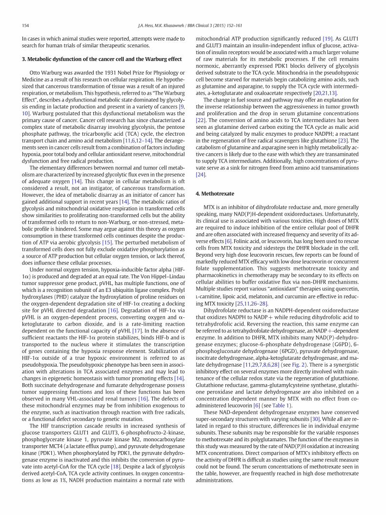
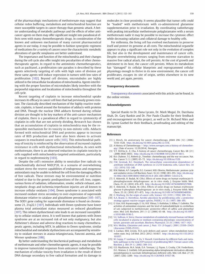
Dihydrofolate reductase is an NADPH-dependent oxidoreductasethat oxidizes NADPH to NADP+ while reducing dihydrofolic acid totetrahydrofolic acid. Reversing the reaction, this same enzyme canbe referred to as tetrahydrofolate dehydrogenase, anNADP+-dependentenzyme. In addition to DHFR, MTX inhibits many NAD(P)-dehydro-genase enzymes; glucose-6-phosphate dehydrogenase (G6PD), 6-phosphogluconate dehydrogenase (6PGD), pyruvate dehydrogenase,isocitrate dehydrogenase, alpha-ketoglutarate dehydrogenase, andma-late dehydrogenase [11,29,7,8,6,28] (see Fig. 2). There is a synergisticinhibitory effect on several enzymes more directly involved withmain-tenance of the cellular redox state via the regeneration of glutathione.Glutathione reductase, gamma-glutamylcysteine synthetase, glutathi-one peroxidase and lactate dehydrogenase are also inhibited on aconcentration dependent manner by MTX with no effect from co-administered leucovorin [6] (see Table 1).
These NAD-dependent dehydrogenase enzymes have conservedsuper-secondary structures with varying subunits [30]. While all are re-lated in regard to this structure, differences lie in individual enzymesubunits. These subunits may be responsible for the variable responsesto methotrexate and its polyglutamates. The function of the enzymes inthis studywasmeasured by the rate of NAD(P)H oxidation at increasingMTX concentrations. Direct comparison of MTX's inhibitory effects onthe activity of DHFR is difficult as studies using the same result measurecould not be found. The serum concentrations of methotrexate seen inthe table, however, are frequently reached in high dose methotrexateadministrations.
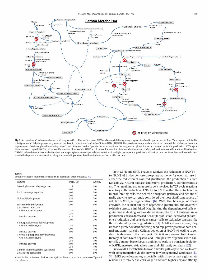
Fig. 2. An overview of carbon metabolism with enzymes affected by methotrexate. MTX can be seen inhibiting many enzymes involved in glucose metabolism. The enzymes inhibited inthis figure are all dehydrogenase enzymes and involved in reduction of NAD+/NADP+ to NADH/NADPH. These reduced compounds are involved in multiple cellular reactions, theregeneration of reduced glutathione being one of them. Also seen in this figure is the incorporation of asparagine and glutamine as carbon sources for the production of TCA cycleintermediates. Legend: NAD+, nicotinamide adenine dinucleotide; NADP+, nicotinamide adenine dinucleotide phosphate; NADH, reduced nicotinamide adenine dinucleotide;NADPH, reduced nicotinamide adenine dinucleotide phosphate; star-shape indicates reaction of multiple reactants and products with various intermediates. Dashed lines indicate ametabolite is present in two locations along the metabolic pathway. Bold lines indicate an irreversible reaction.
Table 1Inhibitory effect of methotrexate on NADPH-dependent oxidoreductases [6].
Enzyme [MTX] μM Activity
2-Oxaloglutarate dehydrogenase 15 50%100 0%
Isocitrate dehydrogenase 100 50%500 0%
Malate dehydrogenase 150 50%400 0%
Succinate dehydrogenase 500 80%Glutathione reductase
CFE-HeLa cell enzyme 80 50%250 10%
Purified enzyme 110 50%200 20%
6-Phosphogluconate dehydrogenaseCFE-Hela cell enzyme 50 50%
200 10%Purified enzyme 100 50%
Glucose-6-phosphate dehydrogenaseCFE-Hela cell enzyme 100 50%
250 10%Purified enzyme 100 50%
250 10%Gamma-glutamylcysteine synthetase 100 46%Glutathione peroxidase 100 50%
Values in this table were obtained directly from the text or via interpretation of figures inthe reference.
155J.A. Hess, M.K. Khasawneh / BBA Clinical 3 (2015) 152–161
Both G6PD and 6PGD enzymes catalyze the reduction of NAD(P)+to NAD(P)H in the pentose phosphate pathway for eventual use ineither the reduction of oxidized glutathione, the production of a freeradicals via NADPH oxidase, cholesterol production, steroidogenesis,etc. The remaining enzymes are largely involved in TCA cycle reactionsresulting in the reduction of NAD+ to NADH within the mitochondria.In proliferating cells, the pentose phosphate pathway and actions ofmalic enzyme are currently considered the most significant source ofcellular NAD(P)+ regeneration [6]. With the blockage of theseenzymes, the cellular ability to regenerate glutathione, and deal withoxidative stress, is inhibited. Highlighting the dependence of cells onglutamine in dealing with oxidative stress, the loss of glutaminase-2production leads to decreasedNAD(P)Hproduction, decreased glutathi-one production and sensitizes cancer cells to oxidative stresses likethose induced by ionizing radiation [31]. As MTX doses increase, theyimpose a greater oxidant buffering handicap, proving fatal for both nor-mal and abnormal cells. Cellular depletion of NAD(P)H leading to celldeath is also seen in the treatment of infectious diseases. Antimicrobialtherapy of both Gram negative and Gram positive organisms with bac-tericidal, but not bacteriostatic, antibiotics leads to a transient depletionof NADH, increased oxidative stress and ultimately cell death [32].
In vivoMTXmetabolism follows a similar pathway to natural folateswith polyglutamation via the enzyme folylpolyglutamyl synthetase [33,34]. MTX polyglutamates, especially with three or more glutamateresidues, are retained in cells longer, and with higher enzyme affinity,
156 J.A. Hess, M.K. Khasawneh / BBA Clinical 3 (2015) 152–161
than unmodified MTX [35]. Cellular concentrations of polyglutamatedmethotrexate rise even in the absence of circulating drug, until cellularefflux begins [33]. Animals fed a diet rich in supplemented glutamineshowed not only a decreased incidence of MTX adverse effects, butalso an enhanced anti-tumor effect in cancer cells [6]. This effect wasalso observed in the treatment of human tumors, and is likely secondaryto MTX polyglutamylation [36].
Intracellular polyglutamated MTX concentrations are an importantfactor in achieving maximum pharmacologic effects in the treatmentof malignancy [35]. Rodent studies have shown that administration oforal glutamine in conjunction with high dose MTX therapy was notonly associated with decreases in enteric mucositis, but also a 300%increase in polyglutamated MTX concentrations within malignant cellswhen compared to normal cells [36]. In a phase I trial based on theserodent studies, nine patients with inflammatory breast cancer wereplaced on a similar regimen. In this human study, one case of grade Imucositis was reported and eight of the nine patients responded tothe chemotherapy regimen with a median survival of 35 months [36].
Asparaginases are frequently used in combination with MTX. Theprocess of MTX polyglutamylation influences the schedule dependentresponse to combination therapy with L-asparaginase. Pre-treatmentof cancer cells with L-asparaginase leads to significant interferencewith MTX polyglutamation [37]. While its primary enzymatic activityis focused on asparagine, L-asparaginases have varying levels of gluta-minase activity that depends on the source of the enzyme. Post-MTXtreatment with L-asparaginase, the deamination of glutamine has asynergistic effect on the activity of L-asparaginase [38,37]. Pretreatmentwith asparaginase decreases tumor cell retention, and lowers thetumoricidal effects, of MTX. The current literature suggests that thiseffect is largely due to a massive deamination of glutamine beingrequired for optimal asparagine deamination [38].
Interference with cellular abilities to maintain the supply ofglutamine, glutamate and glutathione is detrimental, and often fatal,to the cell. Glutaminase 2, a phosphate-activated mitochondrial en-zyme, catalyzes the hydrolysis of glutamine to glutamate [39]. Loss ofthis enzyme results in an effect similar to methotrexate therapy:decreased NADH and glutathione with increased sensitivity to oxidantstress [39]. Combination chemotherapy with metabolically activeagents, i.e., asparaginase, effectively starves the cancer cell of twosources of fuel used to maintain functional output of the TCA cycle. Insettings of glutamine depletion, increased expression of asparagine syn-thetase has been observed in hamster cell lines [40]. Pretreatment ofcancer cells withmethotrexate significantly handicaps the regenerationof NAD(P)H. Glutamine and glutamate shift from supplying intermedi-ates of the TCA cycle into a significant resource sink, de novo synthesisof glutathione. As intracellular levels of glutamine decrease, expressionof asparagine synthetase increases with subsequent glutamine deple-tion and increased ammonia production. With an evolving depletionof glutamine and cellular shift toward increased asparagine synthesis,the addition of L-asparaginase induces a starvation of asparagine andglutamine with increased concentrations of aspartate, glutamate, andammonia. The restriction of both asparagine and glutamine have beenobserved to induce expression of asparagine synthetase, glutaminesynthetase, IGF-I and -II, IGFBP-1, CHOP, ornithine decarboxylase,oncogenes c-JUN and c-FOS and ribosomal proteins L17 and L25 [41].In asparaginase-resistant cells, inhibition of glutamine synthetase hasbeen shown to block proliferation and decrease cellular viability [42].Additionally, as expression of asparagine and glutamine synthetaseincreases there is concomitant stabilization and up-regulation of theMyc and BCL-2 oncoproteins [41].
5. Mitochondrial roles in oxidative stress and carcinogenesis
Endosymbiotic theory is currently the most accepted process ofmitochondrial origin. Two general assumptions accompany thistheory. The first is that mitochondrial acquisition accompanied an
accumulation of atmospheric oxygen. The second marks a separationof aerobic and anaerobic (assumed lack of mitochondria) eukaryotes.The recent inclusion of various anaerobes to eukaryotic lineages is sec-ondary to a much better understanding of variant “mitochondria-like”organelles — i.e., hydrogenosomes, mitosomes. Recent biological andgeochemical theories propose an atmospheric oxygen increased longbefore a noticeable increase in oxygen tension in oceanic waters. Thisatmospheric oxygenation led to oxidation and solubilization of landbound sulfide deposits making ocean waters increasingly anoxic andsulfidic. In the light of this Proterozoic ocean chemistry theory formitochondrial endosymbiosis, one can rationalize that the benefit ofmitochondrial acquisition was not solely an ability to provide ATP viaits utilization of oxygen but its ability to use electrons, as reducedmolecules, to buffer oxidant stresses in the environment. As a moreelectronegative atom than sulfur, increasing concentrations of dissolvedoxygen would make it a much more efficient recipient of those freeelectrons [43].
Contemporarymitochondria are very dynamic organelles. They fuse,divide, power metabolic detoxification, generate stored energy andservemany important regulatory and signaling functions. Recent litera-ture describes a localizing mechanism of mitochondria with definitiveinfluence by the oxygen tension of the cell [44,45]. In hypoxic states,mitochondria in pulmonary artery endothelial cells undergo inwardmigration, via microtubules, and localize around the nucleus [44].Mitochondrial respiration is a significant producer of free radicals withthe electron transport chain being the primary source. This movementis associated with a local increase in production of free radicals andreactive oxygen species in the vicinity of the nucleus [44].
Mechanisms to mitigate damage from oxidative stresses producedwithin the cell have evolved alongside the mitochondria. Superoxidedismutase (SOD), catalase, glutathione (GSH) and thioredoxin are wellknown scavengers of free radicals and their reactive metabolites [46].Intermediates and products of glycolysis may also serve either antioxi-dant roles, such as pyruvate, or serve as a substrate for an antioxidantregenerating pathway as glucose-6-phosphate, glyceraldehyde-3-phosphate via NADH production [47,46]. The absence of adequate oxy-gen, presence of a dysfunctional electron transport chain, or a deficiencyof cofactors places the mitochondria at risk of increased free radicalproduction. A state of normal oxygen tension encouragesmitochondriallocalization in the cell periphery and a subsequent local increase in freeradical concentrations. For a metabolically normal cell, injury inducesthe production of reactive oxygen species produced via NAD(P)Hoxidase and then secretion into the extracellular matrix to initiate theprocess of healing. This peripheral increase in free radicals places cellmembrane components at risk. The hydroxyl radical, regardless of itssource, is one that may freely diffuse through cell membranes. Onemembrane component particularly vulnerable to hydroxyl radicals isarachidonic acid often esterified to membrane phospholipids. A classof eicosanoids called isoprostanes are formed upon reaction of arachi-donic acid with free radicals; i.e., hydroxyl radicals [48]. Isoprostanesare isomers of the cyclo-oxygenase enzyme product prostaglandin F2and will subsequently induce the same inflammatory cascade [49].There is also some evidence to suggest the formation of thromboxaneA2 via isoprostane endoperoxide rearrangement [50]. In a cell withpoor antioxidant reserve, similar hypoxic events increase the likelihoodof subsequent DNA damage from those free radicals.
Mitochondria are arguably the most significant physiologic playersin chemical metabolism. Beyond the ETC., reactions catalyzed via thecytochrome P450 family of enzymes are a significant source of freeradical production. Cytochrome P450 enzymes make up a superfamilyof heme-thiolate proteins involved in steroid synthesis and are respon-sible for 75% of phase I drug metabolism [51–53]. The majority of theseenzymes are associated with NAD(P)H-reductases and found in eitherthe endoplasmic reticulum or the mitochondria [53]. An electron isobtained fromNAD(P)H via the associated reductase enzyme and trans-ferred to the substrate at the enzyme's binding site via its heme group
157J.A. Hess, M.K. Khasawneh / BBA Clinical 3 (2015) 152–161
[51]. The overall efficiency of electron transfer from NAD(P)H to thecytochrome P450 enzyme and then to its substrate can vary. Althoughthis reaction typically detoxifies a chemical, it may also transform aprocarcinogen to its carcinogenic metabolite; i.e., 5-methylchrysenefrom tobacco smoke [54].While some studies report an electron leakageflow of only 0.15%, in steroidogenic tissues the cytochrome P450components outnumber electron transport chain components ten toone [53]. The “leaked” electrons can react with a variety of moleculesto form free radicals, just as those from the electron transport chain.
In vitro studies show that MTX therapy decreases intracellularconcentrations of reduced glutathione [55]. While NAD(P)H is requiredfor regeneration of reduced glutathione, it is not for required for de novosynthesis. In de novo synthesis, glutamate, cysteine, glycine and ATP areused in an enzyme catalyzed reaction. Depletion of glutathione in serinestarved cells (a precursor to cysteine) has been associated withimpaired glycolysis and an elevation in cellular ROS [56]. Tumorsuppressor p53, in conjunctionwith the cyclin-dependent kinase inhib-itor, p21, arrests cellular development and promoted cellular survival byrapid shunting of exogenous serine into the glutathione synthesispathway [56]. In cells without p53, this shunt did not occur. Theresulting serine depletion and oxidative stress lead to not only impairedproliferation, but also a reduction in cellular viability [56].
These rescue pathways must be supplied components in order topreserve the cell and maintain homeostasis. Catabolism of cellularcomponents is a growing area of investigation into cancer metabolismand centers on autophagy. Autophagy is the process in which cellsdegrade and recycle macromolecules and organelles via two distinctpathways; the ubiquitin-proteasome system (UP system) and thelysosome [57]. Smaller, short-lived proteins are the typical degradationtargets of the UP system. However, the size and stability large proteinaggregates and organelles leave the lysosomal pathway as the mosteffectivemeans of autophagy. Inmammalian cells, lysosomal autophagyis directed by a chaperone systemmarkingmacromolecules and organ-elles for digestion [58]. The most apparent benefits of autophagy areresultant decreases in damaged, unneeded or toxic cellular componentswhile producing raw materials for cellular metabolism [59].
While its pathways are not fully understood, the regulation of theseprocesses is intimately linked with their function; i.e., recycling cellularcomponents and providing fuel for cellular processes [60]. Cellular star-vation is a powerful inducer of autophagy. A fall in intracellular aminoacid concentrations is another stimulant of autophagy. Constructing aspectrum of starvation signals, nitrogen is an excellent marker of cellu-lar nutrition. To clarify, in a very well supplied cell, amino acids wouldactivate cellular anabolic pathways via conjugated tRNA activation ofthe Gcn protein kinases [61]. As amino acid concentrations drop, thenumber of transamination and deamination reactions in the cellincreases to attenuate this lack of key amino acids. Cellular concentra-tions of ammonia, especially in concert with glutaminolysis, serve as apowerful regulator in the basal cellular autophagy rates [62]. Furtheralong the spectrum, however, total nitrogen starvation is a more potentactivator than either of the previous nitrogen-related stimulants [61].
Reactive oxygen species are also potent activators of autophagy.Superoxide, in particular, has been found to be a major player inthe activation of reactive oxygen species induced autophagy. Inproperly functioning cells, it is reduced to hydrogen peroxide andthen to water via SOD1, SOD2 and catalase or scavenged by one ofmany different antioxidants present in the cell or in circulation. Ithas been observed that not only do polymorphisms in the genes cod-ing these proteins affect their efficacy, but chronic therapy with MTXalso influences expression profiles with these genes [63]. When an-tioxidant systems are overwhelmed, mitochondrial componentsare at a very distinct geographical disadvantage to the effects offree radicals and reactive oxygen species. Although the GSHregenerating abilities of mitochondria are present, mitochondrialautophagy, or mitophagy, may ensue if sufficiently injured or dam-aged. This has been seen in association with significant decreases
in the ratio of GSH/GSSG inducing mitophagy, while ample concen-trations of GSH are inhibitory to the process [64].
2-Deoxyglucose (2DG), and glucose starvation, have been observedto induce apoptosis in some cancer cells. Recent research has revealeda dichotomy of effect depending on cellular conditions. In hypoxic envi-ronments, 2DG and glucose starvation inhibit autophagic processes,possibly through significant decreases in ATP through PI3K III-Beclin1complex blocking the effects of autophagy activating genes [65]. Thesesame conditions will activate autophagy in a normoxic environmentthrough separate pathways initiated by ROS formation [66,65]. Thisdual effect could be explained by the utilization of glycolysis intermedi-ates and end-products in mitochondrial redox buffering roles.
6. Mitochondrial metabolism and the cell cycle
Senescent thymocytes obtain 88% of ATP production occurs fromox-idative glucose metabolism. However, when stimulated to proliferate,86% of ATP production is obtained without oxidative phosphorylation,by the conversion of glucose to lactate [10]. This shift to non-oxidativeATP production pathways results in significant increases, as much as20-fold, in glucose utilization in proliferating cells [10,67]. Thismetabol-ic redirection to aerobic glycolysis and lactate production offers severalbenefits for the proliferating cell [10]. These include the production ofnumerous metabolic intermediates to serve as antioxidants, creationof an acidic environment to improve oxygenation and stimulation ofangiogenesis via enzyme activation and the production of variousinflammatory mediators — all of additional benefit to cancer cells in ahypoxic environment [10]. Outside of the process of cell division, thissame metabolic redirection occurs during episodes of hypoxic stress.Prior to cell division, mitochondrial mass expands largely for tworeasons. The first is in preparation for increased metabolic demands ofreplication. The second is the preparation for splitting of the parent cell'smitochondrial population between two daughter cells. As withmitochondrial localization induced by HIF, microtubules are involvedin mitochondrial changes seen in preparation for cell division [68].
Cellular progression of G1- to S-phase is dependent on a favorableredox status in the cell and is a significant signaling pathway [69]. Ifwe assume that the TCA cycle progresses at a constant rate andincreased inner membrane mitochondrial volume correlates with totalTCA cycle enzyme then the G1/S phase would have a high [NADH]/[NAD+] redox status [70]. In addition to mitochondrial morphologicchanges, there is an associated decrease in ATP production due todecreased NADH flow through the electron transport change. Highconcentrations of NAD(P)H have been shown to have multiple effectson regulatory proteins, gene expression, directly or epigenetically, andcell cycle progression.
Mitochondrial inner membrane production in HeLa cells, with func-tional enzymatic capabilities, is completed by mid-S-Phase [70]. At thispoint in the cell cycle, there is no increase in mitochondrial membranepotential. Prior to entering M-Phase, there is a significant increase inmitochondrial mass and membrane potential, as much as 168% and228% respectively, with less than a 20% increase in ATP [70]. There is anoted discrepancy, and delay, between the cessation of innermitochon-drial membrane accumulation, in mid-S phase, and the increase ofmitochondrial membrane potential prior to M-phase. Metabolismduring these transitional stages is highly dependent on glutamine forcellular metabolic demands.When starved for glutamine, synchronizedHeLa cells do not progress fromG1- to S-phase or S- toG2/M-phase [71].When those same cells are placed in media with glutamine withoutglucose, the cell cycle does progress at these points, albeit at a slowerrate [71].
The effects of folate depletion and methotrexate therapy on the cellcycle have been studied. Folic acid depleted HepG2 cells have beenobserved and they tend to arrest in an S-phase with notable inhibitionof G2 phase progression [72]. Cell cycle arrest at these stages wasobserved to precede apoptosis in this studywith those in S-phase arrest

158 J.A. Hess, M.K. Khasawneh / BBA Clinical 3 (2015) 152–161
particularly [72]. The literature also supports a methotrexate inducedarrest the cell cycle S-Phase, i.e., an induced folate deficiency. The liter-ature supports the observation of S-phase arrests in both low and highdose methotrexate therapies [73–75]. Interestingly, a thorough evalua-tion of these studies suggests a temporary blockade of G1 to S phase thatoccurs in the first few hours after administration of methotrexate and itappears to be dose related [74,75].
The historical cytotoxic mechanism of MTX is felt to be a disruptionof DNA synthesis via its inhibition of DHFR. However, the antagonisticeffect of MTX on other NADPH-dependent oxidoreductases resultingin decreasingNADHproduction and, subsequently, GSH depletion offersa slightly more accurate model of toxicity [11,29,7,8,6,28]. Depletion ofreduced glutathione, GSH, has been observed to affect the cell cyclesimilarly to both folate depletion and methotrexate therapy. GSHdepletion has not only been observed to restrict cell cycle progressionfrom G1 to S-phase, but also restrict DNA synthesis arresting cells in S-Phase [76]. The authors of this study explain that the slightly lesspronounced inhibition of G1 to S-phase may be due to pathwaysdependent on, in part, a separate pool of thiol proteins [76]. However,the mechanism by which methotrexate interferes with cell cycleprogressions is not fully understood and studies are generally limited.
7. DNA damage and malignant transformation
Cellular hypoxia induces a peri-nuclear mitochondrial migration.Applying the same cellular state of fluctuating oxidative stress andfree radical production in the vicinity of the nucleus would place thenuclear DNA at increased risk of damage. Beyond the risk for geneticdamage and mutation during chromosomal replication, the current ge-nome profile being expressed in a senescent cell would expose manyregulatory and functional sites to an even higher risk of damage. Manycommonly mutated tumor suppressor genes and oncogenes, P53, MYCand BCL as examples, induce metabolic changes in non-transformedcells in periods of high cellular stress [77–83]. Affecting amino acid me-tabolism, mitochondrial function, glycolysis and autophagy, the endfunction of these genes is to temporarily halt cellular self-destructionwhile processes attempt to overcome that stress. A Pyrrhic victory ofsorts, these genes may prolong the life of the cell but at the cost of po-tential mutation if recovery fails, leading to cancerous transformation.
The mitochondrial chromosome is at a much higher risk for damagevia this mechanism. Two important differences are present betweennuclear andmitochondrial DNA, the first being thatmitochondrial chro-mosomes are not protected via histones [84]. In nuclear DNA, geneticmaterial is wrapped around histones and physically protected, tosome degree, from the damaging effects of free radicals. The secondfactor is that the status of cellular oxygen tension would likely haveno significant effect on the proximity of free radicals produced bymitochondria. Low level reactive oxygen species could be severeenough to induce mtDNA damage if not adequately buffered.
Combining the two factors, i.e., a state of cellular hypoxia andincreased free radical production, would put the mitochondrial DNA ata much higher risk for damage than nuclear DNA. Periods of chronicreactive oxygen species exposure have been shown to damage mtDNAand other macromolecules more extensively and with longer lastingeffect, than damage to nuclear DNA [85]. Whether its resultant acutedamage or epigenetic changes through chronic FR exposure of nuclearor mitochondrial DNA, this process could be responsible for “locking”cancer cells into this abnormal metabolism [86–90].
8. Mitochondria, evolution and cancer development
Genetic predispositions aside, the literature supports the postulatethat genetic mutations in most cancers are secondary to severe meta-bolic and mitochondrial dysfunction leading to inappropriate and inad-equate buffering of oxidant stresses. A better understanding of thecomplex interactions of cellular metabolism, mitochondrial function,
and oxidative stress appears to have very widespread implications inthe pathophysiology and treatment of disease [91–97]. Immediateapplication of this review falls on the subject of chemotherapeuticsbut applications may exist for the treatment or prevention of manypathologies.
While exact environmental parameters of Proterozoic oceans areunder debate, an increased level of electrochemical redox stress fromdissolving land bound sulfide deposits prior to oceanic oxygenation iscertainly plausible [43]. Acquisition of an organelle with the sole pur-pose of buffering changes in redox stress would undoubtedly increasecellular fitness, expand the number of tolerable environments andallow for the evolution of more complex cellular processes [86–90]. Co-incidentally or not, the acquisition of these organelles, and subsequentstabilization of environmental redox status, is followed by a massiveincrease in biodiversity known as the Cambrian Explosion that marksthe end of the Proterozoic Era.
Current understandings describe the primary mitochondrial roleas an oxygen dependent power plant with the primary role of ATPproduction via oxidative phosphorylation. Research into mitochondrialorigins and the environmental redox status of early eukaryotic cellsmayfurther expose early roles of the ancestral organelle. Investigations ofmitosomes and hydrogenosomes reveal organelles very similar tomito-chondria, with reduced or non-existent ATP-producing functions [98].Interestingly, all three organelles have in common the ability to createand store reduced molecules for later use by the cell. Some researchsuggests that molecular hydrogen, one product of hydrogenosomes,may serve as an effective antioxidant and has been shown to decreasearea of infarction in rat brains affected by ischemia–reperfusion injuries[99]. The mitochondrial currency of stored reduced molecules,NAD(P)H, and its ratio toNAD+has been shown to have intimate inter-actions in the growth and metastasis of tumors. Excessive concentra-tions of NAD(P)H in relation to NAD+ have been shown to have apositive effect on the growth and metastasis of breast cancers [100].
Many cancers originate in hypoxic microenvironments. Detailedstudies have documented how characteristics of the tumor microenvi-ronment can lead to significant changes in tumor phenotypes. Survivalof these cells appears to be dependent, in part, upon mitochondrialabilities to regenerate free radical scavengers to buffer oxidant stress.Experimental studies have shown a dependence on mitochondrial andcytosolic properties to perpetuate tumors. By placing normal andtumor nuclei in tumor and normal cells respectively, normal daughtercells were produced upon division of the tumor nuclei and normal cellcytoplasm [101]. In the second population of cells, with normal nucleiand tumor cytoplasmic contents, division resulted in the production ofmore tumor cells or cell death [101].
The resultant metabolism of these cells becomes highly dependenton the catabolism of amino acids, particularly glutamine, to preservethe cell via regeneration of free radical scavengers.With this realization,the Warburg effect seemingly covers only part of the metabolicderangements seen in cancer. The increase in glycolysis and lactateproduction may serve as a depository for nitrogen freed from trans-amination reactions needed to fuel enzyme catalyzed reduction ofNAD(P)+. Methotrexate interferes with many enzymes involved inreduction of NAD(P)+ to NAD(P)H and, more significantly, cellularabilities to buffer oxidative stress.
9. Discussion
Acquisition of the earlymitochondrial organelle led to increased cel-lular fitness, in part, due to its ability to buffer redox stresses secondaryto changes in the cells' immediate environment. This buffering functionincreased cellular hardiness allowing survival during environmentalchanges. Although part of a whole, individual cells throughout thebody are subject to changes and stresses in their own microenviron-ments. Cellular metabolites serve as intermediate signals of the extra-cellular environment to effect genetic expression. New understandings
159J.A. Hess, M.K. Khasawneh / BBA Clinical 3 (2015) 152–161
of the pharmacologic mechanisms of methotrexate may suggest thatcellular redox buffering, metabolism and mitochondrial function aremore susceptible targets in cancer therapy than genomic attack. A bet-ter understanding of metabolic pathways and the effects of other anti-cancer agents on themmay offer significant insight into paradoxical ef-fects seen with many chemotherapeutic drugs. In consideration of theknown and unknown mechanism of many current pharmaceuticalagents in use today, it may be possible to fashion synergistic regimensof medications for a variety of cancers once the characteristic metabolicalterations of specific neoplasms are better understood.
The importance and function of the mitochondria and their changesduring the cell cycle also offer insight into peculiarities of other chemo-therapeutic agents. In regard to the antimitotic chemotherapeutics,such as paclitaxel, a proliferation rate paradox is described by theseagents selectively targeting of cells with high rates of division yetthese same agents will induce regression in tumors with low rates ofproliferation [102]. Beyond cell division, microtubules are highlyutilized in the intracellular localization ofmitochondria. Agents interfer-ing with the proper function of microtubules likely interfere with thepurposeful migration and localization of mitochondria throughout thecell cycle.
Recent targeting of cisplatin to increase mitochondrial uptakeincreases its efficacy in cancer cell lines that had previously grown resis-tant. The classically described mechanism of the highly reactive mole-cule, cisplatin, is based around the formation of adducts with proteinsand DNA. Though the nuclear DNA adducts formed during cellulardivision are thought to be key markers of the anti-cancer mechanismof cisplatin, there is a paradoxical effect in regard to cytotoxicity ofcisplatin in cells that are not actively dividing. Research has shownthat an accumulation of cisplatin in mitochondria may be the re-sponsible mechanism for its toxicity in non-mitotic cells. Adductsformed with mitochondrial DNA and proteins appear to increaserates of ROS production and have also been shown to activatecaspases in enucleated cancer cells [103]. This mitochondrial path-way of toxicity is reinforced by the observation of increased cisplatinresistance in cells with dysfunctional mitochondria. As seen withmethotrexate, there is an observed reduction in cisplatin toxicity incells supplied with mitochondria-targeted antioxidants, particularlyin regard to nephrotoxicity [103].
Despite the cell's extensive ability to neutralize free radicals viamitochondrially derived NAD(P)H, in a scenario of overwhelmingoxidant stress the reserve capabilities of endogenous and exogenousantioxidantsmay be unable to defend the cell from the damaging effectsof free radicals. These stresses may be environmental or nutritionrelated or due to the genetic predispositions of the cell. Iron, copper,various forms of radiation, inflammation, smoke, vehicle exhaust, anti-neoplastic drugs and ischemia/reperfusion injuries are all known toincrease cellular oxidants [104]. Down syndrome is associated withincreased oxidant stress secondary to over-expression of superoxidedismutase relative to catalase and glutathione peroxidase [105–107].The SOD1 gene coding for superoxide dismutase is found on chromo-some 21, 21q22.1 [107]. Individuals with Down syndrome have lowerplasma total antioxidant status measured as reduced glutathione[107]. This inborn dysfunctional ratio leads to an increased susceptibil-ity to cellular oxidant stress. It is well known that patients with Downsyndrome are at an increased risk of not only malignancy, but alsoAlzheimer's disease and adverse drug reactions to many chemothera-peutic agents, including MTX. In addition to Down syndrome, similarmitochondrial andmetabolic dysfunctions are accompanied by sensitiv-ity to oxidant stresses in ataxia-telangiectasia, Fanconi anemia, andWerner syndrome [106].
By better understanding the biochemical pathways and metabolismof methotrexate and other chemotherapeutic agents, it may be possibleto improve tumoricidal responses from anticancer radiotherapy. Themechanism of cellular toxicity from irradiation is the result of directDNA damage secondary to free radical formation and its damage to
molecules in close proximity. It seems plausible that tumor cells couldbe “loaded” with methotrexate with co-administered glutaminesupplementation prior to irradiation. By coordinating radiotherapywith peaking intracellular methotrexate polyglutamates with a serummethotrexate nadir it may be possible to increase the cytotoxic effectwhile decreasing radiation and collateral damage to healthy cells.
For millennia, the living cell has evolved mechanisms to preserveitself and protect its genome at all costs. The mitochondrial organelleappears to play a significant role not only in the evolution of complexlife, but also in the development and maintenance of cancer cells.Despite overwhelming stresses ranging from extreme starvation tomassive free radical attack, the cell persists. At the cost of growth anddetriment to its host, the cancer cell persists. When its metabolismhas “damaged” its cellular blueprints and reprogrammed cellularphysiology enough to thrive in its new environment, the cancer cellproliferates, escapes its site of origin, settles elsewhere in its newworld and, yet again, persists.
Transparency documents
Transparency documents associatedwith this article can be found, inthe online version.
Acknowledgments
Special thanks to Dr. Dana Lycans, Dr. Mark Mogul, Dr. DarshanaShah, Dr. Gary Rankin and Dr. Pier Paulo Cluadio for their feedbackand encouragement on this project, as well as Dr. Richard Niles andDr. Lynne Goebel for their feedback, proofreading and editing of themanuscript.
References
[1] J. Hirsch, An anniversary for cancer chemotherapy, JAMA 296 (12) (2006)1518–1520. http://dx.doi.org/10.1001/jama.296.12.1518.
[2] A History of Chemotherapy — http://www.novogen.com/a-history-of-chemother-apy/ (Date of access: 12/29/2014). Novogen.
[3] V.T. DeVita Jr., E. Chu, A history of cancer chemotherapy, Cancer Res. 68 (21)(2008) 8643–8653. http://dx.doi.org/10.1158/0008-5472.CAN-07-6611.
[4] B.A. Chabner, T.G. Roberts Jr., Timeline: chemotherapy and the war on cancer, Nat.Rev. Cancer 5 (1) (2005) 65–72. http://dx.doi.org/10.1038/nrc1529.
[5] F.M. Sirotnak, R.C. Donsbach, The intracellular concentration dependence ofantifolate inhibition of DNA synthesis in L1210 leukemia cells, Cancer Res. 34(12) (1974) 332–340.
[6] R.M. Babiak, A.P. Campello, E.G. Carnieri, M.B. Oliveira, Methotrexate: pentose cycleand oxidative stress, Cell Biochem. Funct. 16 (4) (1998) 283–293. http://dx.doi.org/10.1002/(SICI)1099-0844(1998120)16:4b283::AID-CBF801N3.0.CO;2-E.
[7] E. Akkemik, H. Budak, M. Ciftci, Effects of some drugs on human erythrocyte 6-phosphogluconate dehydrogenase: an in vitro study, J. Enzyme Inhib. Med.Chem. 25 (4) (2010) 476–479. http://dx.doi.org/10.3109/14756360903257900.
[8] E. Akkemik, H. Budak, M. Ciftci, Effects of some drugs on human erythrocyteglucose 6-phosphate dehydrogenase: an in vitro study, J. Enzyme Inhib. Med.Chem. 25 (6) (2010) 871–875. http://dx.doi.org/10.3109/14756360903489581.
[9] O. Warburg, On the origin of cancer cells, Science 123 (3191) (1956) 309–314.[10] K.A. Brand, U. Hermfisse, Aerobic glycolysis by proliferating cells: a protective
strategy against reactive oxygen species, FASEB J. 11 (5) (1997) 388–395.[11] E. Uzar, H.R. Koyuncuoglu, E. Uz, H.R. Yilmaz, S. Kutluhan, S. Kilbas, F. Gultekin, The
activities of antioxidant enzymes and the level of malondialdehyde in cerebellumof rats subjected to methotrexate: protective effect of caffeic acid phenethylester, Mol. Cell. Biochem. 291 (1–2) (2006) 63–68. http://dx.doi.org/10.1007/s11010-006-9196-5.
[12] S.G. Sullivan, A. Stern, Glucosemetabolism of oxidatively stressed human red bloodcells incubated in plasma or medium containing physiologic concentrations oflactate, pyruvate and ascorbate, Biochem. Pharmacol. 33 (9) (1984) 1417–1421.
[13] M.A. Medina, Glutamine and cancer, J. Nutr. 131 (9 Suppl.) (2001) 2539S–2542S(discussion 2550S-2531S).
[14] S. Cardaci, M.R. Ciriolo, TCA cycle defects and cancer: when metabolism tunesredox state, Int.J. Cell Biol. 2012 (2012) 161837. http://dx.doi.org/10.1155/2012/161837.
[15] M. Guppy, P. Leedman, X. Zu, V. Russell, Contribution by different fuels and meta-bolic pathways to the total ATP turnover of proliferating MCF-7 breast cancer cells,Biochem. J. 364 (Pt 1) (2002) 309–315.
[16] E.D. MacKenzie, M.A. Selak, D.A. Tennant, L.J. Payne, S. Crosby, C.M. Frederiksen,D.G. Watson, E. Gottlieb, Cell-permeating alpha-ketoglutarate derivatives alleviatepseudohypoxia in succinate dehydrogenase-deficient cells, Mol. Cell. Biol. 27 (9)(2007) 3282–3289. http://dx.doi.org/10.1128/MCB. 01927-06.
160 J.A. Hess, M.K. Khasawneh / BBA Clinical 3 (2015) 152–161
[17] C.E. Snell, H. Turley, A. McIntyre, D. Li, M. Masiero, C.J. Schofield, K.C. Gatter, A.L.Harris, F. Pezzella, Proline-hydroxylated hypoxia-inducible factor 1alpha (HIF-1alpha) upregulation in human tumours, PLoS One 9 (2) (2014) e88955. http://dx.doi.org/10.1371/journal.pone.0088955.
[18] W. Liu, S.M. Shen, X.Y. Zhao, G.Q. Chen, Targeted genes and interacting proteins ofhypoxia inducible factor-1, Int. J. Biochem. Mol. Biol. 3 (2) (2012) 165–178.
[19] C. Frezza, L. Zheng, D.A. Tennant, D.B. Papkovsky, B.A. Hedley, G. Kalna, D.G.Watson, E. Gottlieb, Metabolic profiling of hypoxic cells revealed a catabolic signa-ture required for cell survival, PLoS One 6 (9) (2011) e24411. http://dx.doi.org/10.1371/journal.pone.0024411.
[20] S. Sookoian, C.J. Pirola, Alanine and aspartate aminotransferase and glutamine-cycling pathway: their roles in pathogenesis of metabolic syndrome, World J.Gastroenterol. 18 (29) (2012) 3775–3781. http://dx.doi.org/10.3748/wjg.v18.i29.3775.
[21] D. Anastasiou, L.C. Cantley, Breathless cancer cells get fat on glutamine, Cell Res. 22(3) (2012) 443–446. http://dx.doi.org/10.1038/cr.2012.5.
[22] A. Mohamed, X. Deng, F.R. Khuri, T.K. Owonikoko, Altered glutamine metabolismand therapeutic opportunities for lung cancer, Clin. Lung cancer 15 (1) (2014)7–15. http://dx.doi.org/10.1016/j.cllc.2013.09.001.
[23] C. Des Rosiers, L. Di Donato, B. Comte, A. Laplante, C. Marcoux, F. David, C.A.Fernandez, H. Brunengraber, Isotopomer analysis of citric acid cycle and gluco-neogenesis in rat liver. Reversibility of isocitrate dehydrogenase and involve-ment of ATP-citrate lyase in gluconeogenesis, J. Biol. Chem. 270 (17) (1995)10027–10036.
[24] A. Le, A.N. Lane, M. Hamaker, S. Bose, A. Gouw, J. Barbi, T. Tsukamoto, C.J. Rojas, B.S.Slusher, H. Zhang, L.J. Zimmerman, D.C. Liebler, R.J. Slebos, P.K. Lorkiewicz, R.M.Higashi, T.W. Fan, C.V. Dang, Glucose-independent glutamine metabolism viaTCA cycling for proliferation and survival in B cells, Cell Metab. 15 (1) (2012)110–121. http://dx.doi.org/10.1016/j.cmet.2011.12.009.
[25] B. Aydin, Quercetin prevents methotrexate-induced hepatotoxicity without inter-fering with methotrexate metabolizing enzymes in liver of mice, J. Appl. Biol. Sci.5 (2) (2011) 75–80.
[26] G. Sener, E. Eksioglu-Demiralp, M. Cetiner, F. Ercan, S. Sirvanci, N. Gedik, B.C. Yegen,L-Carnitine ameliorates methotrexate-induced oxidative organ injury and inhibitsleukocyte death, Cell Biol. Toxicol. 22 (1) (2006) 47–60. http://dx.doi.org/10.1007/s10565-006-0025-0.
[27] P. Abraham, V.K. Kolli, S. Rabi, Melatonin attenuates methotrexate-induced oxida-tive stress and renal damage in rats, Cell Biochem. Funct. 28 (5) (2010) 426–433.http://dx.doi.org/10.1002/cbf.1676.
[28] H. Tabassum, S. Parvez, S.T. Pasha, B.D. Banerjee, S. Raisuddin, Protective effect oflipoic acid against methotrexate-induced oxidative stress in liver mitochondria,Food Chem. Toxicol. 48 (7) (2010) 1973–1979. http://dx.doi.org/10.1016/j.fct.2010.04.047.
[29] N.N. Caetano, A.P. Campello, E.G. Carnieri, M.L. Kluppel, M.B. Oliveira, Effect ofmethotrexate (MTX) on NAD(P)+ dehydrogenases of HeLa cells: malic enzyme,2-oxoglutarate and isocitrate dehydrogenases, Cell Biochem. Funct. 15 (4)(1997) 259–264. http://dx.doi.org/10.1002/(SICI)1099-0844(199712)15:4b259::AID-CBF749N3.0.CO;2-D.
[30] A.S. Kutzenko, V.S. Lamzin, V.O. Popov, Conserved supersecondary structural motifin NAD-dependent dehydrogenases, FEBS Lett. 423 (1) (1998) 105–109.
[31] L. Xiang, G. Xie, C. Liu, J. Zhou, J. Chen, S. Yu, J. Li, X. Pang, H. Shi, H. Liang, Knock-down of glutaminase 2 expression decreases glutathione, NADH, and sensitizescervical cancer to ionizing radiation, Biochim. Biophys. Acta 1833 (12) (2013)2996–3005. http://dx.doi.org/10.1016/j.bbamcr.2013.08.003.
[32] M.A. Kohanski, D.J. Dwyer, B. Hayete, C.A. Lawrence, J.J. Collins, A common mecha-nism of cellular death induced by bactericidal antibiotics, Cell 130 (5) (2007)797–810. http://dx.doi.org/10.1016/j.cell.2007.06.049.
[33] B.A. Chabner, C.J. Allegra, G.A. Curt, N.J. Clendeninn, J. Baram, S. Koizumi, J.C. Drake,J. Jolivet, Polyglutamation of methotrexate. Is methotrexate a prodrug? J. Clin.Invest. 76 (3) (1985) 907–912. http://dx.doi.org/10.1172/JCI112088.
[34] M. Weigand, E. Frei, N. Graf, M. Wiessler, Comparative analysis of methotrexatepolyglutamates in lymphoblast preparations from bone marrow and blood, andthe contribution of residual red blood cells, J. Cancer Res. Clin. Oncol. 126 (7)(2000) 407–411.
[35] T.W. Synold, M.V. Relling, J.M. Boyett, G.K. Rivera, J.T. Sandlund, H. Mahmoud,W.M.Crist, C.H. Pui, W.E. Evans, Blast cell methotrexate-polyglutamate accumulationin vivo differs by lineage, ploidy, and methotrexate dose in acute lymphoblasticleukemia, J. Clin. Invest. 94 (5) (1994) 1996–2001. http://dx.doi.org/10.1172/JCI117552.
[36] I.T. Rubio, Y. Cao, L.F. Hutchins, K.C. Westbrook, V.S. Klimberg, Effect of gluta-mine on methotrexate efficacy and toxicity, Ann. Surg. 227 (5) (1998)772–778 (discussion 778–780).
[37] P. Sur, D.J. Fernandes, T.E. Kute, R.L. Capizzi, L-asparaginase-induced modulation ofmethotrexate polyglutamylation in murine leukemia L5178Y, Cancer Res. 47 (5)(1987) 1313–1318.
[38] E.H. Panosyan, R.S. Grigoryan, I.A. Avramis, N.L. Seibel, P.S. Gaynon, S.E. Siegel, H.J.Fingert, V.I. Avramis, Deamination of glutamine is a prerequisite for optimal aspar-agine deamination by asparaginases in vivo (CCG-1961), Anticancer Res. 24 (2C)(2004) 1121–1125.
[39] W. Hu, C. Zhang, R.Wu, Y. Sun, A. Levine, Z. Feng, Glutaminase 2, a novel p53 targetgene regulating energy metabolism and antioxidant function, Proc. Natl. Acad. Sci.107 (16) (2010) 7455–7460. http://dx.doi.org/10.1073/pnas.1001006107.
[40] S.S. Gong, L. Guerrini, C. Basilico, Regulation of asparagine synthetase gene expres-sion by amino acid starvation, Mol. Cell. Biol. 11 (12) (1991) 6059–6066.
[41] V.I. Avramis, Asparaginases: biochemical pharmacology and modes of drugresistance, Anticancer Res. 32 (7) (2012) 2423–2437.
[42] B.M. Rotoli, J. Uggeri, V. Dall'Asta, R. Visigalli, A. Barilli, R. Gatti, G. Orlandini, G.C.Gazzola, O. Bussolati, Inhibition of glutamine synthetase triggers apoptosis inasparaginase-resistant cells, Cell Physiol. Biochem. 15 (6) (2005) 281–292.http://dx.doi.org/10.1159/000087238.
[43] M. Mentel, W. Martin, Energy metabolism among eukaryotic anaerobes in light ofProterozoic ocean chemistry, Philos. Trans. R. Soc. Lond. Ser. B Biol. Sci. 363 (1504)(2008) 2717–2729. http://dx.doi.org/10.1098/rstb.2008.0031.
[44] M.P. Murphy,Modulatingmitochondrial intracellular location as a redox signal, Sci.Signal. 5 (242) (2012) pe39. http://dx.doi.org/10.1126/scisignal.2003386.
[45] A.B. Al-Mehdi, V.M. Pastukh, B.M. Swiger, D.J. Reed, M.R. Patel, G.C. Bardwell, V.V.Pastukh,M.F. Alexeyev,M.N. Gillespie, Perinuclear mitochondrial clustering createsan oxidant-rich nuclear domain required for hypoxia-induced transcription, Sci.Signal. 5 (231) (2012) ra47. http://dx.doi.org/10.1126/scisignal.2002712.
[46] J. Berridge Michael, Cell signalling biology: module 2 — cell signalling pathways,Biochem. J. (2012) http://dx.doi.org/10.1042/csb0001002.
[47] G.S. Krasnov, A.A. Dmitriev, A.V. Snezhkina, A.V. Kudryavtseva, Deregulation ofglycolysis in cancer: glyceraldehyde-3-phosphate dehydrogenase (GAPDH) as atherapeutic target, Expert Opin. Ther. Targets (2013) http://dx.doi.org/10.1517/14728222.2013.775253.
[48] H. Yin, Y. Zhou, M. Zhu, S. Hou, Z. Li, H. Zhong, J. Lu, T. Meng, J. Wang, L. Xia, Y.Xu, Y. Wu, Role of mitochondria in programmed cell death mediated by arachi-donic acid-derived eicosanoids, Mitochondrion (2012) http://dx.doi.org/10.1016/j.mito.2012.10.003.
[49] A. Strydom, M.J. Dickinson, S. Shende, D. Pratico, Z. Walker, Oxidative stressand cognitive ability in adults with Down syndrome, Prog. Neuro-Psychopharmacol. Biol. Psychiatry 33 (1) (2009) 76–80. http://dx.doi.org/10.1016/j.pnpbp.2008.10.006.
[50] I. Karamouzis, P. Pervanidou, R. Berardelli, S. Iliadis, I. Papassotiriou, M.Karamouzis, G.P. Chrousos, C. Kanaka-Gantenbein, Enhanced oxidative stress andplatelet activation combined with reduced antioxidant capacity in obese prepu-bertal and adolescent girls with full or partial metabolic syndrome, Horm.Metab. Res. 43 (9) (2011) 607–613. http://dx.doi.org/10.1055/s-0031-1284355.
[51] P.B. Danielson, The cytochrome P450 superfamily: biochemistry, evolution anddrug metabolism in humans, Curr. Drug Metab. 3 (6) (2002) 561–597.
[52] M. Ingelman-Sundberg, Human drug metabolising cytochrome P450 enzymes:properties and polymorphisms, Naunyn Schmiedeberg's Arch. Pharmacol. 369(1) (2004) 89–104. http://dx.doi.org/10.1007/s00210-003-0819-z.
[53] I. Hanukoglu, Antioxidant protective mechanisms against reactive oxygen species(ROS) generated by mitochondrial P450 systems in steroidogenic cells, Drug Metab.Rev. 38 (1–2) (2006) 171–196. http://dx.doi.org/10.1080/03602530600570040.
[54] T. Shimada, C.L. Hayes, H. Yamazaki, S. Amin, S.S. Hecht, F.P. Guengerich, T.R. Sutter,Activation of chemically diverse procarcinogens by human cytochrome P-450 1B1,Cancer Res. 56 (13) (1996) 2979–2984.
[55] B.N. Cronstein, Low-dose methotrexate: a mainstay in the treatment of rheuma-toid arthritis, Pharmacol. Rev. 57 (2) (2005) 163–172. http://dx.doi.org/10.1124/pr.57.2.3.
[56] O.D. Maddocks, C.R. Berkers, S.M. Mason, L. Zheng, K. Blyth, E. Gottlieb, K.H.Vousden, Serine starvation induces stress and p53-dependent metabolic remodel-ling in cancer cells, Nature 493 (7433) (2013) 542–546. http://dx.doi.org/10.1038/nature11743.
[57] C.D. Gonzalez, S. Alvarez, A. Ropolo, C. Rosenzvit, M.F. Gonzalez Bagnes, M.I.Vaccaro, Autophagy, Warburg, and Warburg reverse effects in human cancer,Biomed. Res. Int. 2014 (2014) 926729. http://dx.doi.org/10.1155/2014/926729.
[58] J. Cui, Z. Gong, H.M. Shen, The role of autophagy in liver cancer: molecular mech-anisms and potential therapeutic targets, Biochim. Biophys. Acta 1836 (1) (2013)15–26. http://dx.doi.org/10.1016/j.bbcan.2013.02.003.
[59] K.S. Choi, Autophagy and cancer, Exp. Mol. Med. 44 (2) (2012) 109–120. http://dx.doi.org/10.3858/emm.2012.44.2.033.
[60] J.S. Carew, K.R. Kelly, S.T. Nawrocki, Autophagy as a target for cancer therapy: newdevelopments, Cancer Manag. Res. 4 (2012) 357–365. http://dx.doi.org/10.2147/CMAR.S26133.
[61] N. Ecker, A. Mor, D. Journo, H. Abeliovich, Induction of autophagic flux by aminoacid deprivation is distinct from nitrogen starvation-induced macroautophagy,Autophagy 6 (7) (2010) 879–890. http://dx.doi.org/10.4161/auto.6.7.12753.
[62] C.H. Eng, K. Yu, J. Lucas, E. White, R.T. Abraham, Ammonia derived fromglutaminolysis is a diffusible regulator of autophagy, Sci. Signal. 3 (119) (2010)ra31. http://dx.doi.org/10.1126/scisignal.2000911.
[63] F. Barbisan, R. Motta Jde, A. Trott, V. Azzolin, E.B. Dornelles, M. Marcon, T.D. Algarve,M.M. Duarte, C.P. Mostardeiro, T.C. Unfer, K.L. Schott, I.B. da Cruz, Methotrexate-related response on human peripheral bloodmononuclear cells may bemodulatedby the Ala16Val-SOD2 gene polymorphism, PLoS One 9 (10) (2014) e107299.http://dx.doi.org/10.1371/journal.pone.0107299.
[64] M. Deffieu, I. Bhatia-Kissova, B. Salin, A. Galinier, S. Manon, N. Camougrand, Gluta-thione participates in the regulation of mitophagy in yeast, J. Biol. Chem. 284 (22)(2009) 14828–14837. http://dx.doi.org/10.1074/jbc.M109.005181.
[65] H. Xi, J.C. Barredo, J.R. Merchan, T.J. Lampidis, Endoplasmic reticulum stressinduced by 2-deoxyglucose but not glucose starvation activates AMPK throughCaMKKbeta leading to autophagy, Biochem. Pharmacol. (2013) http://dx.doi.org/10.1016/j.bcp.2013.02.037.
[66] Q. Wang, B. Liang, N.A. Shirwany, M.H. Zou, 2-Deoxy-D-glucose treatment ofendothelial cells induces autophagy by reactive oxygen species-mediated activa-tion of the AMP-activated protein kinase, PLoS One 6 (2) (2011) e17234. http://dx.doi.org/10.1371/journal.pone.0017234.
[67] H. Pelicano, D.S. Martin, R.H. Xu, P. Huang, Glycolysis inhibition for anticancertreatment, Oncogene 25 (34) (2006) 4633–4646. http://dx.doi.org/10.1038/sj.onc.1209597.
161J.A. Hess, M.K. Khasawneh / BBA Clinical 3 (2015) 152–161
[68] M. Karbowski, J.H. Spodnik, M. Teranishi, M. Wozniak, Y. Nishizawa, J. Usukura, T.Wakabayashi, Opposite effects of microtubule-stabilizing and microtubule-destabilizing drugs on biogenesis of mitochondria in mammalian cells, J. Cell Sci.114 (Pt 2) (2001) 281–291.
[69] S.G. Menon, E.H. Sarsour, D.R. Spitz, R. Higashikubo, M. Sturm, H. Zhang, P.C.Goswami, Redox regulation of the G1 to S phase transition in the mouse embryofibroblast cell cycle, Cancer Res. 63 (9) (2003) 2109–2117.
[70] S. Sweet, G. Singh, Changes in mitochondrial mass, membrane potential, andcellular adenosine triphosphate content during the cell cycle of human leukemic(HL-60) cells, J. Cell. Physiol. 180 (1) (1999) 91–96. http://dx.doi.org/10.1002/(SICI)1097-4652(199907)180:1b91::AID-JCP10N3.0.CO;2-6.
[71] S.L. Colombo, M. Palacios-Callender, N. Frakich, S. Carcamo, I. Kovacs, S. Tudzarova,S. Moncada, Molecular basis for the differential use of glucose and glutamine in cellproliferation as revealed by synchronized HeLa cells, Proc. Natl. Acad. Sci. U. S. A.108 (52) (2011) 21069–21074. http://dx.doi.org/10.1073/pnas.1117500108.
[72] R.F. Huang, Y.H. Ho, H.L. Lin, J.S. Wei, T.Z. Liu, Folate deficiency induces a cell cycle-specific apoptosis in HepG2 cells, J. Nutr. 129 (1) (1999) 25–31.
[73] S.A. Goncharova, O.S. Frankfurt, Effect of methotrexate on the cell cycle of L1210leukemia, Cell Prolif. 9 (4) (1976) 333–340. http://dx.doi.org/10.1111/j.1365-2184.1976.tb01281.x.
[74] M. Tsurusawa, Niwa, Katano, Fujimoto, Methotrexate cytotoxicity as related toirreversible S phase arrest in mouse L1210 leukemia cells, Jpn. J. Cancer Res. 81(1990) 85–90.
[75] P. Ernst, S.A. Killmann, Perturbation of generation cycle of human leukemicmyelo-blasts in vivo by methotrexate, Blood 38 (6) (1971) 689–705.
[76] J.P. Messina, D.A. Lawrence, Cell cycle progression of glutathione-depleted humanperipheral blood mononuclear cells is inhibited at S phase, J. Immunol. 143 (6)(1989).
[77] C. Zhang, M. Lin, R. Wu, X. Wang, B. Yang, A.J. Levine, W. Hu, Z. Feng, Parkin, a p53target gene, mediates the role of p53 in glucose metabolism and the Warburgeffect, Proc. Natl. Acad. Sci. U. S. A. 108 (39) (2011) 16259–16264. http://dx.doi.org/10.1073/pnas.1113884108.
[78] X.D. Zhang, Z.H. Qin, J. Wang, The role of p53 in cell metabolism, Acta Pharmacol.Sin. 31 (9) (2010) 1208–1212. http://dx.doi.org/10.1038/aps.2010.151.
[79] E. Yamada, R. Singh, Mapping autophagy on to your metabolic radar, Diabetes 61(2) (2012) 272–280. http://dx.doi.org/10.2337/db11-1199.
[80] A. Cimmino, R. Capasso, F. Muller, I. Sambri, L. Masella, M. Raimo, M.L. De Bonis, S.D'Angelo, V. Zappia, P. Galletti, D. Ingrosso, Protein isoaspartate methyltransferaseprevents apoptosis induced by oxidative stress in endothelial cells: role of Bcl-Xldeamidation and methylation, PLoS One 3 (9) (2008) e3258. http://dx.doi.org/10.1371/journal.pone.0003258.
[81] C.V. Dang, Rethinking the Warburg effect with Myc micromanaging glutaminemetabolism, Cancer Res. 70 (3) (2010) 859–862. http://dx.doi.org/10.1158/0008-5472.CAN-09-3556.
[82] S.A. Cook, P.H. Sugden, A. Clerk, Regulation of Bcl-2 family proteins during develop-ment and in response to oxidative stress in cardiac myocytes: association withchanges in mitochondrial membrane potential, Circ. Res. 85 (10) (1999)940–949. http://dx.doi.org/10.1161/01.res.85.10.940.
[83] H. Zirath, A. Frenzel, G. Oliynyk, L. Segerstrom, U.K. Westermark, K. Larsson, M.Munksgaard Persson, K. Hultenby, J. Lehtio, C. Einvik, S. Pahlman, P. Kogner, P.J.Jakobsson, M.A. Henriksson, MYC inhibition induces metabolic changes leadingto accumulation of lipid droplets in tumor cells, Proc. Natl. Acad. Sci. U. S. A. 110(25) (2013) 10258–10263. http://dx.doi.org/10.1073/pnas.1222404110.
[84] S.R. Pieczenik, J. Neustadt, Mitochondrial dysfunction and molecular pathways ofdisease, Exp. Mol. Pathol. 83 (1) (2007) 84–92. http://dx.doi.org/10.1016/j.yexmp.2006.09.008.
[85] Mitochondrial DNA damage is more extensive and persists longer than nuclearDNA damage in human cells following oxidative, stress, 1997.
[86] A. Soltysova, V. Altanerova, C. Altaner, Cancer stem cells, Neoplasma 52 (6) (2005)435–440.
[87] M. Todaro, M.G. Francipane, J.P. Medema, G. Stassi, Colon cancer stem cells: prom-ise of targeted therapy, Gastroenterology 138 (6) (2010) 2151–2162. http://dx.doi.org/10.1053/j.gastro.2009.12.063.
[88] M.S. Wicha, S. Liu, G. Dontu, Cancer stem cells: an old idea—a paradigm shift,Cancer Res. 66 (4) (2006) 1883–1890. http://dx.doi.org/10.1158/0008-5472.CAN-05-3153 (discussion 1895–1886).
[89] R.K. Naviaux, Mitochondrial control of epigenetics, Cancer Biol. Ther. 7 (8) (2008)1191–1193.
[90] A. Vincent, I. Van Seuningen, On the epigenetic origin of cancer stem cells, Biochim.Biophys. Acta 1826 (1) (2012) 83–88. http://dx.doi.org/10.1016/j.bbcan.2012.03.009.
[91] C. Ruggiero, M. Ehrenshaft, E. Cleland, K. Stadler, High-fat diet induces an initial ad-aptation of mitochondrial bioenergetics in the kidney despite evident oxidativestress and mitochondrial ROS production, Am. J. Physiol. Endocrinol. Metab. 300(6) (2011) E1047–E1058. http://dx.doi.org/10.1152/ajpendo.00666.2010.
[92] A.P. Rolo, C.M. Palmeira, Diabetes and mitochondrial function: role of hyperglyce-mia and oxidative stress, Toxicol. Appl. Pharmacol. 212 (2) (2006) 167–178.http://dx.doi.org/10.1016/j.taap.2006.01.003.
[93] R.E. Frye, R. Delatorre, H. Taylor, J. Slattery, S. Melnyk, N. Chowdhury, S.J. James,Redox metabolism abnormalities in autistic children associated with mitochondri-al disease, Transl. Psychiatry 3 (2013) e273. http://dx.doi.org/10.1038/tp.2013.51.
[94] Y.J. Taverne, A.J. Bogers, D.J. Duncker, D. Merkus, Reactive oxygen species and thecardiovascular system, Oxidative Med. Cell. Longev. 2013 (2013) 862423. http://dx.doi.org/10.1155/2013/862423.
[95] A.B. Hwang, D.E. Jeong, S.J. Lee, Mitochondria and organismal longevity, Curr. Ge-nomics 13 (7) (2012) 519–532. http://dx.doi.org/10.2174/138920212803251427.
[96] S.R. Subramaniam, M.F. Chesselet, Mitochondrial dysfunction and oxidativestress in Parkinson's disease, Prog. Neurobiol. (2013) http://dx.doi.org/10.1016/j.pneurobio.2013.04.004.
[97] A. Melo, L. Monteiro, R.M. Lima, D.M. Oliveira, M.D. Cerqueira, R.S. El-Bacha, Oxida-tive stress in neurodegenerative diseases: mechanisms and therapeutic perspec-tives, Oxidative Med. Cell. Longev. 2011 (2011) 467180. http://dx.doi.org/10.1155/2011/467180.
[98] M. Muller, M. Mentel, J.J. van Hellemond, K. Henze, C. Woehle, S.B. Gould, R.Y. Yu,M. van der Giezen, A.G. Tielens, W.F. Martin, Biochemistry and evolution of anaer-obic energy metabolism in eukaryotes, Microbiol. Mol. Biol. Rev. 76 (2) (2012)444–495. http://dx.doi.org/10.1128/MMBR. 05024-11.
[99] S. Ohta, Molecular hydrogen is a novel antioxidant to efficiently reduce oxida-tive stress with potential for the improvement of mitochondrial diseases,Biochim. Biophys. Acta 1820 (5) (2012) 586–594. http://dx.doi.org/10.1016/j.bbagen.2011.05.006.
[100] A.F. Santidrian, A. Matsuno-Yagi, M. Ritland, B.B. Seo, S.E. LeBoeuf, L.J. Gay, T. Yagi,B. Felding-Habermann, Mitochondrial complex I activity and NAD+/NADHbalance regulate breast cancer progression, J. Clin. Invest. 123 (3) (2013)1068–1081. http://dx.doi.org/10.1172/JCI64264.
[101] T.N. Seyfried, R. Flores, A.M. Poff, D.P. D'Agostino, Cancer as a metabolic disease:implications for novel therapeutics, Carcinogenesis (2013) http://dx.doi.org/10.1093/carcin/bgt480.
[102] T.J. Mitchison, The proliferation rate paradox in antimitotic chemotherapy, Mol.Biol. Cell 23 (1) (2012) 1–6. http://dx.doi.org/10.1091/mbc.E10-04-0335.
[103] R. Marullo, E. Werner, N. Degtyareva, B. Moore, G. Altavilla, S.S. Ramalingam, P.W.Doetsch, Cisplatin induces a mitochondrial-ROS response that contributes to cyto-toxicity depending onmitochondrial redox status and bioenergetic functions, PLoSOne 8 (11) (2013) e81162. http://dx.doi.org/10.1371/journal.pone.0081162.
[104] S. Toyokuni, Oxidative stress and cancer: the role of redox regulation, Biotherapy11 (2–3) (1998) 147–154.
[105] C. Campos, R. Guzman, E. Lopez-Fernandez, A. Casado, Evaluation of urinarybiomarkers of oxidative/nitrosative stress in children with Down syndrome, LifeSci. 89 (17–18) (2011) 655–661. http://dx.doi.org/10.1016/j.lfs.2011.08.006.
[106] F.V. Pallardo, A. Lloret, M. Lebel, M. d'Ischia, V.C. Cogger, D.G. Le Couteur, M.N.Gadaleta, G. Castello, G. Pagano, Mitochondrial dysfunction in some oxidativestress-related genetic diseases: ataxia-telangiectasia, Down syndrome, Fanconianaemia and Werner syndrome, Biogerontology 11 (4) (2010) 401–419. http://dx.doi.org/10.1007/s10522-010-9269-4.
[107] S.M. Sulthana, S.N. Kumar,M.G. Sridhar, B.V. Bhat, K.R. Rao, Levels of non enzymaticantioxidants in Down syndrome, Indian J. Pediatr. 79 (11) (2012) 1473–1476.http://dx.doi.org/10.1007/s12098-012-0795-8.
Copyright © 2022 FDOKUMEN