2,4-Diaminopyrimidines as inhibitors of Leishmanial and Trypanosomal dihydrofolate reductase

Free Energy Simulations of Active-Site Mutants of DihydrofolateReductaseDvir Doron,† Vanja Stojkovic,‡,§,# Lokesh Gakhar,§ Alexandra Vardi-Kilshtain,† Amnon Kohen,‡
and Dan Thomas Major*,†
†Department of Chemistry and the Lise Meitner-Minerva Center of Computational Quantum Chemistry, Bar-Ilan University,Ramat-Gan 5290002, Israel‡Department of Chemistry, University of Iowa, Iowa City, Iowa 52242, United States§Protein Crystallography Facility and Department of Biochemistry, University of Iowa, Iowa City, Iowa 52242, United States
*S Supporting Information
ABSTRACT: This study employs hybrid quantum mechanics−molecular mechanics(QM/MM) simulations to investigate the effect of mutations of the active-site residue I14of E. coli dihydrofolate reductase (DHFR) on the hydride transfer. Recent kineticmeasurements of the I14X mutants (X = V, A, and G) indicated slower hydride transferrates and increasingly temperature-dependent kinetic isotope effects (KIEs) with systematicreduction of the I14 side chain. The QM/MM simulations show that when the originalisoleucine residue is substituted in silico by valine, alanine, or glycine (I14V, I14A, andI14G DHFR, respectively), the free energy barrier height of the hydride transfer reactionincreases relative to the wild-type enzyme. These trends are in line with the single-turnoverrate measurements reported for these systems. In addition, extended dynamics simulationsof the reactive Michaelis complex reveal enhanced flexibility in the mutants, and inparticular for the I14G mutant, including considerable fluctuations of the donor−acceptor distance (DAD) and the active-sitehydrogen bonding network compared with those detected in the native enzyme. These observations suggest that theperturbations induced by the mutations partly impair the active-site environment in the reactant state. On the other hand, theaverage DADs at the transition state of all DHFR variants are similar. Crystal structures of I14 mutants (V, A, and G) confirmedthe trend of increased flexibility of the M20 and other loops.
1. INTRODUCTION
Dihydrofolate reductase (DHFR; EC 1.5.1.3) is a ubiquitousenzyme catalyzing the reduction of dihydrofolate (DHF orH2folate) by nicotinamide adenine dinucleotide phosphatehydride (NADPH) to form tetrahydrofolate (THF or H4folate)and NADP+. Its principal function is to maintain intracellularpools of THF, a compound essential for the biosynthesis ofpurines, thymine nucleotides, and several amino acids. Due toits pivotal role in nucleotide biosynthesis in many organisms,DHFR has long been recognized as an important target forvarious therapeutic purposes, in particular anticancer andantibacterial drugs, such as methotrexate (MTX) andtrimethoprim, respectively.1,2 The clinical importance ofDHFR, along with its relatively modest size, has led manyresearchers to study, both experimentally and theoretically, thecatalytic mechanism and kinetics of the DHFR redox reaction.The key chemical step is a stereospecific transfer of the pro-R
hydrogen at the C4 position of the nicotinamide ring inNADPH to the re face of the C6 atom of the pterin ring inDHF, with concomitant protonation at N5 (Scheme 1 andFigure 1).3,4 The X-ray structure of the ternary complex ofEscherichia coli DHFR (ecDHFR) crystallized with folate andthe oxidized cofactor (i.e., E:folate:NADP+)5 is considered toreliably mimic the reactive configuration of the Michaelis
complex of the enzyme. In this conformation, a flexible segmentof ca. 15 amino acid residues within the major domain, termedthe M20 loop, packs against the nicotinamide ring of thecofactor, closing over the active site. This closed conformationmight be associated with the C−H → C transfer (i.e., the M20loop deletion mutant is still active, although it is much slower).6
The interactions exerted by active site residues proximal tothe donor−acceptor moieties are likely to play a role in
Special Issue: William L. Jorgensen Festschrift
Received: June 17, 2014Revised: November 5, 2014Published: November 8, 2014
Scheme 1
Article
pubs.acs.org/JPCB
© 2014 American Chemical Society 906 dx.doi.org/10.1021/jp5059963 | J. Phys. Chem. B 2015, 119, 906−916
modulating the relative orientation of the reacting fragments asthey approach one another along the hydride transfer reactionpathway. In that sense, the donor−acceptor distance (DAD) isa useful, chemically intuitive variable to follow. Thus, it isdesirable to examine the contribution of the enzyme environ-ment by scrutinizing the influence of direct structuralperturbations of the reacting fragments and the DAD, andespecially the relation between these and the free energy profileof the reaction. In this regard, recent studies of transition pathsampling (TPS) simulations by Schwartz and co-workers haveshown that unlike other enzymes (such as human lactatedehydrogenase7) in which nonequilibrated, fast (subpico-second) dynamics was suggested to be coupled to the reactioncoordinate, the protein motions in DHFR are presumably notdirectly coupled to the compressive promoting vibration alongthe DAD axis as the enzymatic reaction crosses the transitionstate.8 Other computational studies have addressed thequestion of correlation between ns-ps motions and the reactioncoordinate, as well as effects of mutations on the DADfluctuations on both ground state (GS) and transition state(TS).9−19
Recently, we measured the single turnover rates at 25 °C andextracted the intrinsic kinetic isotope effect (KIE) over atemperature range of 5−45 °C for a series of active-site mutantsof ecDHFR, in which the size of the side chain of residue 14 wassystematically decreased from Ile in the wild-type (WT)enzyme to Val, Ala and Gly.20,21 In this approach, theexperiments yielded a controlled change with minimalalteration of the active-site electrostatics, while focusing onthe changes to the carbon DAD. It was found that a smaller sidechain behind the nicotinamide ring resulted in slower hydridetransfer rates and increased temperature dependency of theintrinsic KIEs. In addition, molecular mechanics (MM)molecular dynamics (MD) simulations at room temperaturefor the enzyme ground state (GS, i.e. the Michaelis complex)were performed. These simulations suggested that the WTenzyme had an exclusive population characterized by narrowdistributions of short GS DADs and angle between the planesof the nicotinamide and pterin rings, around the average values(3.58 Å and 29°, respectively). As expected, the mutantsdeviated significantly from these ideal values, with largeraverage DADs and a concomitant reduction in the populationof the presumed reactive conformer with similar DADs to WT
DHFR. Although not necessarily related, both lower rates andsteeper temperature dependence of the intrinsic KIEs werecorrelated with the longer GS DADs and broader distributionsof the GS DADs. Within the framework of a Marcus-like model,the temperature dependence of the KIEs only reflects thedistribution of DADs at the TS (tunneling ready state, or QMdelocalized TS) but is not affected by the GS DADs. Takentogether, the hydrophobic side chain of I14 assists the hydridetransfer by both, restricting the distribution and dynamics ofthe GS DAD and that of the DAD distribution at the TS.In this work, we seek to complement the experiments
performed in ref 21 from a theoretical vantage point byconducting extensive hybrid quantum mechanics (QM) andclassical mechanics (CM) simulations of the hydride transferreaction in ecDHFR. The free energy profiles were obtained forthe WT ecDHFR and the aforementioned mutations of residue14. Our approach takes advantage of the hybrid QM(AM1-SRP)/MM methodology employed in our earlier studies ofDHFR22−26 and formate dehydrogenase27 systems, which hasbeen shown to yield an accurate potential energy surface of themodeled reaction, as well as the integrated path integral (PI)and umbrella sampling (US) method.28−32 This paperdemonstrates the ability of state-of-the-art computationaltechniques to discriminate between mutations that alter theactive site in a serial fashion (e.g., WT vs I14 active-sitemutants). As a final introductory note, we stress that thecurrent work integrates many of the important contributions ofJorgensen and co-workers to the field of molecular simulations:all-atom force-fields, the TIPnP water models and free energymethods, and the interfacing between force-fields and QMmethods.33−35
2. METHODSConstruction of Mutant Enzymes. The initial coordinates
of the mutant enzymes were constructed in silico by changingthe Ala residue, I14, in the original WT ecDHFR structure(used in our introductory study22) to Val, Ala, or Gly. Thesetup and the MD simulations of all systems were carried outon the basis of procedures similar to those employed for theWT DHFR.22−24 Briefly, the protonation states of all polaramino acid residue side chains were adjusted to pH 7, and theprotonation states of the His residues (either the neutraltautomeric forms or the positively charged form) weredetermined so as to match the hydrogen bonding patterns inthe nearest environment. The N5 position on the pterin ring ofthe substrate (dihydrofolate) was protonated. The HBUILDfacility in the program CHARMM was applied to add hydrogenatoms. Fourteen sodium ions were added to the four iso-charged WT and mutant systems to neutralize the overallnegative charge.
Potential Energy Surface. The potential energy surface inthe current study is described by a hybrid QM/MMHamiltonian,36 where the QM region is treated by a modifiedAM137 semiempirical Hamiltonian, denoted AM1-SRP (specificreaction parameters).38 This Hamiltonian has been designed toreproduce high-level calculations for an assortment ofelectronic and thermodynamic properties for reactionsinvolving various nicotinamide and pterin derivatives.22 TheQM region includes fragments of DHF and NADPH, which areproximal to the reaction center, whereas the MM regioncontains the remaining ligand atoms, the entire protein, watermolecules, and sodium ions. The water molecules wererepresented by the three-point charge TIP3P model.33
Figure 1. Portion of the protein structure and the bound ligands in theMichaelis complex of E. coli DHFR, based on X-ray data for theternary complex E:folate:NADP+ (PDB ID 1RX2). The active site isshown with the main species addressed in this work: the substrate(folate, magenta), the cofactor NADP+ (blue); and three amino acidresidues, including I14, A7 (both hydrogen bonded to the proximalcarboxamide group of the nicotinamide moiety), and D27 and Y100,the only ionizable active site residues, hydrogen bonded to the pterinring in folate. The donor and the acceptor carbons are denoted by ared arrow from donor to acceptor.
The Journal of Physical Chemistry B Article
dx.doi.org/10.1021/jp5059963 | J. Phys. Chem. B 2015, 119, 906−916907

Hydrogen link atoms were placed across the bonds intersectedby the QM/MM boundary, and the QM/MM interactions weretreated by electrostatic embedding. A detailed QM/MMpartitioning scheme and a thorough description of thedevelopment of the AM1-SRP Hamiltonian is providedelsewhere.22 In modeling the MM region, we used the all-atom CHARMM3639−42 force field, which has been reopti-mized on the basis of studies of protein folding, assembly, andfunctionally relevant conformational changes. Such force-fieldfine-tuning was recently shown to be essential in order tocorrectly treat proteins in micro- and millisecond simulations.43
Molecular Dynamics Simulations. Periodic boundaryconditions were employed to solvate the Michaelis complexusing a pre-equilibrated cubic water box (∼65 Å × ∼65 Å ×∼65 Å), while treating long-range electrostatic interactions withthe Ewald summation technique (64 × 64 × 64 FFT grid, κ =0.340 Å−1).44 All systems were fully minimized, heated upgradually to 298 K for 25 ps, and equilibrated at thattemperature for 1 ns at the MM level of theory. Each systemwas thereafter re-equilibrated using the QM(AM1-SRP)/MMpotential over the course of 200 ps. All equilibrations wereconducted with the isothermal−isobaric (NPT) ensemble at 1atm and the target temperature was controlled by the extendedconstant pressure/temperature (CPT) method45 and theHoover thermostat.46 The leapfrog integration scheme47 wasused to propagate the equations of motions, and the SHAKEalgorithm48 was applied to constrain all MM bonds involvinghydrogen atoms, allowing a time step of 1 fs. During theequilibration, several nuclear Overhauser effect (NOE)restraints were imposed on key hydrogen bond interactionsbetween the ligands and the surrounding residues, and removed100 ps before moving on to the production phase. All enzymesimulations used a development version of the CHARMMprogram.49,50 Complementary details of the MD simulationsare available in our earlier work.Potential of Mean Force. The US technique51 was used to
determine the classical-mechanical potential of mean force(CM-PMF) for the hydride transfer reaction at five targettemperatures (5, 15, 25, 35, 45 °C). The reaction coordinatewas defined as the antisymmetric reactive stretch coordinate,ζasym, namely, the difference between the lengths of thebreaking C4−H and forming H−C6 bonds. A total of 17individual US MD simulations (“windows”) were performedalong discrete, evenly spaced values of ζasym from −2.0 to 2.0 Å.Each window was subject to an appropriate harmonic restraint,which keeps ζasym in the desired region, and an umbrellapotential (roughly the negative of the PMF) as a function ofζasym. In order to efficiently update the biasing potential asnecessary, each window was sampled in multiple successiveseries with a predetermined number of MD steps. A typicalsimulation starts with a short equilibration (2 ps), followed bycollection of the probability densities of configurations (ρ)along the reaction coordinate, ζasym. Whenever the biasingpotential is updated, the subsequent simulation commenceswith a short equilibration, and the accompanying equilibrationdata is discarded. The cumulative simulation time per windowwas 275−375 ps. The statistics for the reaction coordinate wassorted into bins of width 0.01 Å. The positions and velocities ofthe last recorded configuration in a specific window were usedto start its successor, to maintain continuity of propagation. Inparticular, the phase space variables reached at the end of themost current simulations at 25 °C were also the starting pointsfor the simulations at both 15 and 35 °C. Similarly, simulations
at 5 and 45 °C were each initiated from the respective endpoints determined in the simulations at the “neighboring”temperatures, 15 and 35 °C. CM-PMF curves and surfaces werecomputed using a multidimensional version of the weightedhistogram analysis method (WHAM).52
QM PMFs were obtained by PI-US simulations as describedin our earlier work.22
Materials and Methods. The mutants were expressed,purified, and stored as reported for other DHFR mutants.53
Prior to preparation of ternary complexes for crystallization,purified proteins were loaded onto a Superdex-75 (GE) gelfiltration column pre-equilibrated with the buffer containing 20mM Tris-HCl, pH 7.5. DHFR eluted in a single peak, whichwas tested for monodispersity using dynamic light scattering ona NanoStar instrument (Wyatt Technology). Concentration ofthe pooled fractions was measured using UV absorbance at 280nm and Bradford assay. Due to its low solubility, folate wasadded to a dilute protein solution at 10-fold molar excessrelative to the concentration of the DHFR variant. The samplewas incubated on ice for at least 1 h, after which it was filteredusing Milipore 0.22 μm filter. The binary complex was thenconcentrated to a final concentration of ∼15 mg/mL (asdetermined by a Bradford assay). NADP+ was added as a solidto the concentrated binary DHFR-Folate complex at 5-foldmolar excess of DHFR.All enzyme-Folate-NADP+ ternary complexes were crystal-
lized via the hanging drop vapor diffusion method by mixing400 nl crystallization solution and 400 nl of prepared ternarycomplex using TTP LabTech Mosquito, except I14G DHFRwhere crystals were obtained through macroseeding into 4 μLdrops (2 μL crystallization solution and 2 μL of proteincomplex).To describe in greater detail, I14G-NADP+-Folate crystal-
lized in the P21212 space group after macroseeding under thefollowing conditions: 16 mg/mL I14G DHFR, 0.1 M Tris, pH8.5, 0.9 M LiCl, and 36% w/v PEG 6,000; at 18 °C. Crystalsappeared within 7 days after macroseeding.I14V-NADP+-Folate ternary complex crystallized in the P21
space group under the following conditions: 16 mg/mL I14VDHFR, 0.1 M Tris, pH 8.5, 0.1 M LiCl, and 34.2% w/v PEG6,000; at 18 °C, within 2 weeks.I14A-NADP+-Folate ternary complex was crystallized in P21
space group under the following conditions: 15 mg/mL I14ADHFR, 0.2 M Lithium acetate, and 20% w/v PEG 3,350; at 18°C, within 7 days.
Data Collection and Structure Determination. Crystalswere flashed-cooled in liquid nitrogen, and data were collectedat 100 K at the 4.2.2 synchotron beamline at the Advance LightSource of the Lawrence Berkley National Laboratory. The datawere processed using either XDS54 (for the I14A and I14Gmutants), or d*TREK (for the I14V mutant).55 The structureswere solved by molecular replacement in PHASER,56 part ofthe CCP4 suite,57 using the structure of WT ecDHFR (PDB1RX2) with the ligands removed to generate an initial model.Refinement and model building for all structures were doneusing REFMAC58 and COOT.59 All structures were refinedwith good geometry (RMS deviations in bond lengths were0.013−0.03 Å, RMS deviation in bond angles were 1.72−2.54°), with the possible exception of the I14A structure, whichshowed higher overall temperature factor for molecule B. Theseproblems are probably a result of static disorder. Dataprocessing and final refinement statistics are given in Table S4.
The Journal of Physical Chemistry B Article
dx.doi.org/10.1021/jp5059963 | J. Phys. Chem. B 2015, 119, 906−916908

3. RESULTSFree Energy Profiles. We obtained the CM-PMFs for the
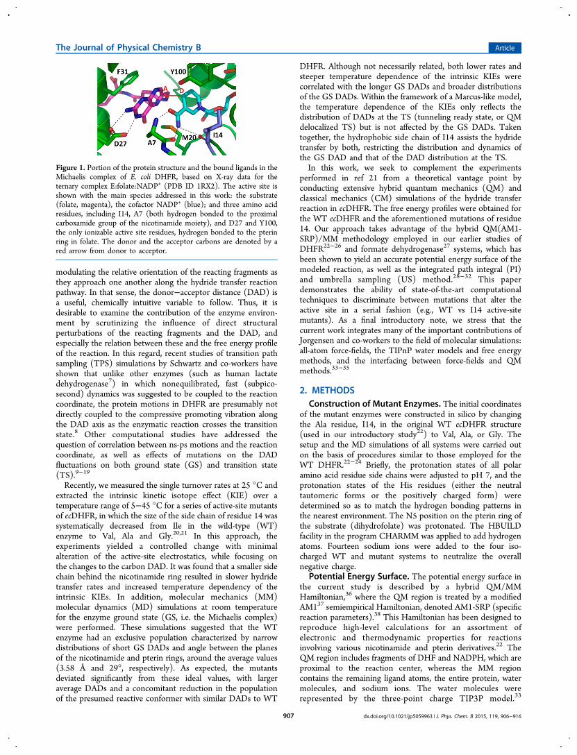
catalyzed transfer of hydride from NADPH to DHF in the wild-type DHFR (WT DHFR) and three I14 mutants, namely I14V,I14A, and I14G DHFR (Figure 1), at 5, 15, 25, 35, and 45 °C.Figure 2 shows the CM-PMFs for WT DHFR and the active-
site mutants at 25 °C. The CM-PMFs at 5, 15, 25, 35, and 45°C are provided as Supporting Information (Figure S1 andTable S1). Examination of the CM-PMFs at 25 °C providessome important findings. The free energy of activation for theWT enzyme, ΔG‡, is 16.8 kcal/mol, while the reaction freeenergy, ΔGr, is −9.7 kcal/mol. The presence of a slightly lessbulky side chain in the I14V DHFR system causes a slightincrease in the free energy barrier (18.5 kcal/mol) relative toWT DHFR. Further truncation of the side chain brings abouteven higher free energy barriers, as evident by the ΔG‡ valuefor the I14A and I14G systems (19.8 and 21.1 kcal/mol,respectively), indicative of a progressive reduction in rate as themutation becomes more substantial. This general trend is inaccordance with our experimental work in refs 20−21, whichfound a correlation between reduced side chain volume anddiminished single-turnover rates of the hydride transfer. At thesame time, the reaction becomes less exothermic as the freeenergy barrier increases in the series in the WT−I14V-I14G,indicating a diminished driving force for the hydride transfercaused by the mutation. These trends are reminiscent of recentQM/MM studies of the remote mutations G121 V, M42W, andG121 V-M42W in ecDHFR, conducted by us25 and others.17
An exception to the above said trend is I14A DHFR, whoseestimated free energy of reaction is comparable to that of thenative enzyme. Interestingly, the temperature seemingly haslittle effect on the computed CM-PMF (Figure S1 and TableS1).The quantum corrected ΔG‡’s obtained from combined
classical free energy and quantum PI simulations are comparedwith the values obtained from the single-turnover rates of ref 21and the Eyring equation. To assess the quality of the results, wecompare them to kinetic measurements for the WTDHFR53,60−62 and the I14 mutants,21 all of which wereconducted using a similar experimental protocol under thesame conditions (pH 7). Notably, the presteady-state single-turnover rates at pH 7 are not representative of the C−H → C
transfer step per se, as it is still somewhat masked bynonchemical steps such as ligand binding and conformationalrearrangements (i.e., kinetic complexity), as evident from asmaller observed KIE relative to its intrinsic value.63 Moreover,available data from different kinetic studies of WT ecDHFRshow a linear increase of the activation barrier with pH for thesingle turnover measurements (13.4,64 14.2−14.3,53,60,61,6516.766-16.9,62 and 17.565 kcal/mol at pH 6.5, 7, 9, and 9.5,respectively)further indicating that the single-turnover ratemeasurements must include the N5-DHF protonation step,which precedes the hydride transfer step calculated here. In ourmodels, the titratable/ionizable residues in both the native andmutated proteins were constructed considering a neutral pHenvironment. As can be inferred from Table S1, inclusion ofNQEs lowered the free energy barrier from classical MDsimulations by 2.0−2.4 kcal/mol, which is similar to our earlierwork,22 that estimated by Hammes-Schiffer and co-workers(2.4 kcal/mol),9 where the transferring hydrogen nucleus wasrepresented as a three-dimensional wave function, and lower by∼1 kcal/mol than that predicted by Gao and co-workers usingeither ensemble-averaged variational transition state theorywith multidimensional tunneling (EA-VTST/MT)67,68 or thePI-UM17 approaches. Overall, the calculated free energybarriers are in good agreement with the experimental ones,and basically conform to the general trend due to the I14mutations.
Impact of Mutation on Structure. From a structuralstandpoint, the varying magnitudes of the experimentallyderived free energy barriers in the WT enzyme and the I14mutants may reflect differences in the relative probabilities ofsampling conformations that are conducive to the hydridetransfer process. These conformations are directed towardgenerating reactant and transition state configurations withfavorable geometry of the reactants and optimized stabilizinginteractions within the active-site environment.13 In this work,the starting structures of all three mutants were created bypoint mutation based on the coordinates of the ternary complexof WT DHFR with folate and NADP+ (PDB ID: 1RX2),5
crystallized in the P212121 space group, in which the M20 loopadopts the closed conformation. The basic premise in oursimulation approach was that the mutant enzymes adopt astructure similar to that of the WT enzyme, and that MDpropagation allows the mutants to relax into their correctBoltzmann distribution. The analogous ternary complexes forall three mutations of I14 in ecDHFR were crystallized and theirstructures determined. In particular, the I14A and I14Vstructures were only obtained in the P21 space group, wherethe M20 loop is always observed in the open conformation (i.e.,also in WT) as the open conformation is seemingly stabilizedby crystal contacts. It is plausible that a transient population ofthe open conformational state may play a role in the catalyticcycle since: (i) the open conformation facilitates both substratebinding and product release; and (ii) replacement of the part ofthe M20 loop (residues 16−19) with glycine still yields anenzyme that exhibits some catalytic activity.6 However, in thiswork these structures are not employed for modeling purposes,as we focus on the closed conformation; it is not clear whetherthe closed conformation is a prerequisite for enzyme activity.6
On the other hand, the crystal structure of I14G DHFR wascrystallized in the P21212 space group, where both the M20loop and the F-G loop (in direct contact with M20 loop) arepartially disordered. This suggests that greater flexibility in theM20 loop may be observed in simulations of the I14G mutant.
Figure 2. Classical mechanical potentials of mean force for the hydridetransfer in WT DHFR and Ile14 mutants, computed at 25 °C. Theantisymmetric stretch coordinate, ζasym, is defined as the difference inthe distance of the transferring hydrogen from the donor carbon of thecofactor and the acceptor carbon of the substrate.
The Journal of Physical Chemistry B Article
dx.doi.org/10.1021/jp5059963 | J. Phys. Chem. B 2015, 119, 906−916909

Comparison of the X-ray structures of the WT and I14GDHFR (Figure 3) shows noticeable conformational differences
with regard to the M20 loop arrangement. In the WT enzyme,the side chain of residue M20 packs against the pterin ring ofthe substrate and is also close to the nicotinamide moiety of thecofactor. Consequently, the sulfur atom in the M20 side chainis within van der Waals contact distance with both the N5 atomof the pterin ring (3.68 Å) and the N7 position of thenicotinamide’s carboxamide group (3.33 Å). This proximityallows the lone pair of the sulfur to maintain interactions withthe bound ligands either via hydrogen bonding or throughdative bonding, in addition to the hydrogen bonding betweenthe N7 atom and the carbonyl oxygen of I14, which supportsthe nicotinamide ring in its binding site. Overall, thisilluminates the possible role of the M20 residue as a mediatorbetween the cofactor and the substrate, directing their relativeposition, thus facilitating the hydride transfer.69 We also notethat the M20 residue (and the M20 loop as a whole) adopts avery similar orientation in the complex of ecDHFR with theanticancer agent methotrexate (MTX) and NADPH (PDB ID1RX3).5 Conversely, in the crystal structure of the I14Gmutant, the M20 loop is not well ordered, and the M20 sidechain no longer points toward the ligands but is rather directedaway from the active site. It seems that the M20 loop adopts aconformation unattainable in the WT DHFR, suggesting thatI14G mutation might lead to enhanced flexibility of the loop.Assuming that the M20 loop is nonetheless partially closedduring catalysis, we may ascribe the observed reduced rate ofthe hydride transfer in I14G DHFR relative to the WTenzyme21 to a greater reorganization cost during the catalyticstep. This slowing down corresponds to ΔΔG‡ of ca. 4 kcal/mol.It is instructive to see whether and how the proximal
mutations affect the intrinsic structural characteristics of thespecies that are directly involved in the hydride transferreaction. To this end, we followed several geometric variables
during the course of the PMF simulations, including theantisymmetic stretch reaction coordinate, ζasym; the DAD,which was treated as a spectator coordinate (i.e., the spectatorcoordinate is not directly subject to a biasing force), denoted asζDAD; and the donor−hydride−acceptor angle (∠D−H−A), allcritical parameters of the reaction progress. Comparison ofthese properties at the TS is provided in Tables 1 and 2 at 25
°C (and at other temperatures in the Supporting Information,Tables S2 and S3). The slightly negative value of theantisymmetric stretch coordinate at the saddle point of thePMF, (ζasym
‡ ∼ −0.16 to −0.11 Å), suggests that the transitionstate is somewhat early, as can be seen from the PMF plots(Figure 2), and as expected from exothermic reactions (i.e., theHammond Postulate).22 This feature does not appear to bemonotonically intensified or weakened with variation of thetemperature in any of the DHFR systems, contrary to what hasbeen observed previously.68
The DAD in the GS (Table 2) is slightly elongated as afunction of mutant perturbation (i.e., DAD(WT) ∼ DAD-(I14V) < DAD(I14A) ∼ DAD(I14G)). The average DADcoordinate value obtained at the saddle point, ζDAD
‡ , is 2.63−2.65 Å, irrespective of the DHFR variants and temperature. Thisdistance is similar to that calculated previously for the transitionstate by other researchers. Using the EVB approach, Hammes-Schiffer and co-workers obtained transition state DAD values of2.739−2.7410 Å, Miller and co-workers reported a value of 2.72Å,16 while Warshel and co-workers reported values in the range2.6 to >3 Å.14,70,71 Gao and co-workers obtained values rangingfrom 2.6867 to 2.7117 Å, using the QM(AM1)/MMHamiltonian with a simple valence bond (SVB) correction.Interestingly, our results resemble the DAD in the optimizedgeometries of gas phase models, calculated at the AM1-SRPlevel (2.63 Å), and M06/6-31+G(d,p) and B3LYP-D/6-31+G(d,p) levels (2.66 Å).22 This indicates that in the currentsimulations, the enzyme exerts a minor effect on the DAD.Furthermore, at these short DADs we expect only moderate
Figure 3. Superimposition of the X-ray crystal structures of the ternarycomplexes of WT (blue) and I14G (orange) DHFR. In the WTstructure, both folate and NADP+ ligands are bound (shown as sticks),whereas for I14G the nicotinamide-ribosyl moiety of NADP+ ismissing. The denoted amino acid residues (I14, G14, M20) aredisplayed as balls and sticks, with carbon, nitrogen, oxygen, and sulfuratoms colored gray, purple, red, and yellow, respectively. Theinteractions of the sulfur atom of the side chain of residue M20 inWT DHFR with the N5 position of the pterin ring (folate) and the N7position of the nicotinamide ring (NADP+) are marked with greenlines, in addition to the hydrogen bond between N7 with the carbonyloxygen of I14. In I14G DHFR, the M20 loop is located far from theligands, incapable of making such contacts.
Table 1. Comparison of the CM and QM Transition StatePositions for the Reaction in Wild-Type DHFR and I14Mutants at 25 °C, in Terms of the Antisymmetric StretchCoordinate, ζasym, and the Ensemble-Averaged Donor−Acceptor Distance (DAD) (Both in Å Units)
ζasym‡ a ⟨ζDAD⟩ζ asym
‡ d
CMb QMc CMb QMc
WT −0.16 −0.18 2.65 2.67±0.01 ±0.01 (0.06) (0.05)
I14V −0.13 −0.13 2.63 2.66±0.01 ±0.01 (0.06) (0.05)
I14A −0.13 −0.13 2.64 2.66±0.01 ±0.01 (0.06) (0.05)
I14G −0.11 −0.13 2.65 2.65±0.01 ±0.01 (0.06) (0.05)
aDetermined from the saddle point of the classical mechanical PMFsurface, obtained by combining the probability density statisticscollected in the US simulations. bA bin size of 0.02 Å was used.cNuclear quantum effects corrections for hydrogen were obtainedfrom PI simulations, fitted to a cubic polynomial expression as afunction of ζasym, and added to the CM-PMF. dObtained byBoltzmann averaging of the DAD at ζasym
‡ . Standard deviations areshown in parentheses.
The Journal of Physical Chemistry B Article
dx.doi.org/10.1021/jp5059963 | J. Phys. Chem. B 2015, 119, 906−916910

tunneling effects, based on the fairly small NQE on the effectivefree energy barrier.In addition, it should be noted that our findings agree with
other recent computational studies, where the respectivechanges in the DAD at the reactant state (the Michaeliscomplex) and transition state of mutants of ecDHFR werefound to be minor, if not negligible. For example, WT andG121V DHFR were predicted by Watney et al. to have anaverage DAD of 3.31 and 3.33 Å in the reactant state,respectively, while at the transition state, both systems werefound to have an average DAD of 2.74 Å;10 Likewise, Fan et al.computed an average DAD of 3.50 Å for the Michaelis complexin WT DHFR and 3.46 Å for the M42W/G121V doublemutant, whereas these two had nearly identical average DAD atthe transition state (2.71 and 2.70 Å, respectively).17
The invariability of the DAD at the TS in the mutantenzymes, relative to the WT, does not reflect the KIEs, theirtemperature dependence, or other components of a Marcus-likemodel. In the Marcus-like model the DAD is not simply the C−C distance of the reactive symmetric stretch coordinate at thesaddle point of the PMF. Rather, in such phenomenologicalmodels, the relevant DADs constitute an ensemble ofconfigurations, which are conducive to hydride transfer via adiffuse quantum mechanical state (i.e., tunneling ready state),which has nonzero contribution to the rate even at 3.0Å.21,66,72,73 Recently, we have shown that standard biasedsimulation methods might not be capable of capturing the finedetails of transition state structures in enzymes.26 Such slightdifferences do not have significant effects on the PMF, butcould explain the substantial effects of the I14 mutants on theKIEs and their temperature dependence.The donor−hydride−acceptor (D−H−A) angle is another
useful geometric measure for tracking of the hydride ionposition. The bending of the D−H−A angle at the transitionstate can quantify the extent to which the hydride transition
path is bent relative to a hypothetical linear displacement alongthe axis connecting the DAD carbons. Generally speaking, inWT and I14V DHFR, this angle adopts an average of ca. 150°in the reactant state (Table 2). Fluctuations are observedtoward the transition state during which the ∠D−H−A iswidened by ∼5°, until it reaches a peak value. We note, though,that the maximum average ∠D−H−A is obtained slightlybefore or after, but not precisely at ζasym
‡ , suggesting that thechange in the D−H−A angle as the hydride transfers is not fullysynchronized with the translation of the hydride. Pu et al. haveobserved this asynchronous nature of hydride transfer in DHFRpreviously.74 As shown in Table 2, the average ∠D−H−A atζasym‡ is in the range 154−161° for both WT and mutantDHFRs. This result agrees well with that reported for the WTenzyme by Garcia-Viloca et al. (157°),67 who employed aQM(AM1-SVB)/MM Hamiltonian. However, the angle issomewhat smaller compared to earlier computational studies ofecDHFR (162−166°)9,75 and in vacuo calculations of modeltransition state structures (162°).22 Much like our findings forthe average DAD, the temperature hardly affects the variationof the average ∠D−H−A (Table S3).The interactions between the pterin−nicotinamide complex
with the enzyme matrix provide additional important structuralinformation pertaining to the effect of the active-site mutations.In our simulations, the M20 loop is packed similarly against thereactive species in the GS and TS (Table 2 and S3). Thiscontrasts the findings of Fan et al, where a slightly looser TSthan GS was observed.17 Furthermore, the hydrogen bondsbetween the nicotinamide moiety and backbone amide of A7and I14 are kept largely intact in the simulations of WT and allmutants (Tables 2 and S3). However, in the GS, some of thesehydrogen bonds are loosened, while at the TS, these hydrogenbonds are tightened.We have also monitored various structural properties in the
course of the extended QM/MM MD simulations of the
Table 2. Ensemble-Averaged Structural Properties of WT DHFR and I14 Mutant at 25 °C at the Ground and Transition Statesa
WT I14V I14A I14G
GS TS GS TS GS TS GS TS
C4N−H4N (Å) 1.11 1.29 1.11 1.31 1.11 1.31 1.11 1.31(0.03) (0.03) (0.03) (0.03) (0.03) (0.03) (0.03) (0.03)
C6−H4N (Å) 2.98 1.40 2.95 1.38 3.05 1.41 3.04 1.40(0.04) (0.03) (0.03) (0.03) (0.03) (0.03) (0.03) (0.03)
C4N−C6 (DAD) (Å) 3.96 2.65 3.94 2.63 4.07 2.64 4.06 2.65(0.09) (0.06) (0.10) (0.06) (0.08) (0.06) (0.09) (0.06)
rehybridization (Å) −1.2 −0.1 −1.1 0.0 −1.2 0.0 −1.2 −0.02(0.2) (0.2) (0.2) (0.2) (0.1) (0.2) (0.1) (0.2)
C4N−H4N−C6 (∠D−H−A) (deg) 149 161 151 156 155 154 155 157(9) (8) (12) (7) (9) (7) (11) (7)
N5−S(Met20) (Å) 3.8 4.1 4.3 3.9 4.0 3.7 4.3 4.2(0.3) (0.5) (0.4) (0.3) (0.6) (0.5) (0.4) (0.6)
N7N−S(Met20) (Å) 3.8 3.7 4.1 3.8 4.2 4.2 4.1 4.2(0.3) (0.4) (0.4) (0.3) (0.3) (0.4) (0.5) (0.4)
N7N−O(Ala7) (Å) 3.0 2.9 2.9 3.0 3.0 3.0 3.0 2.9(0.2) (0.1) (0.1) (0.2) (0.2) (0.2) (0.3) (0.1)
N7N−O(Ile14) (Å) 3.1 3.3 3.0 3.1 3.1 3.1 3.1 3.0(0.2) (0.3) (0.2) (0.2) (0.2) (0.2) (0.3) (0.2)
O7N−N(Ala7) (Å) 3.6 3.0 3.2 3.0 3.3 3.0 3.3 3.0(0.3) (0.1) (0.2) (0.1) (0.2) (0.1) (0.2) (0.1)
aThe properties were calculated from the structural data recorded during MD simulations, sorted into bins of ζasym with a width of 0.04 Å, andaveraged over each bin. The values shown thus correspond to the ensemble of configurations falling within the bin associated with ζGS and ζasym
‡ .Standard deviations per that bin are shown in parentheses.
The Journal of Physical Chemistry B Article
dx.doi.org/10.1021/jp5059963 | J. Phys. Chem. B 2015, 119, 906−916911

Michaelis complex in the DHFR variants, covering dynamicswithin the active site and beyond. These include geometricchanges within the reactants (such as the DAD), as well ashydrogen bonds between the bound ligands and surroundingprotein residues. Figures 4 and 5 present selected time series
plots describing the evolution of WT and I14V, I14A, and I14GDHFR during MD at 25 °C. A validation set of time-seriesusing an independent system setup is presented in Figure S8and S9.A rather striking difference between the GS dynamics of the
WT and I14G ligand−protein complexes is demonstrated bythe evolution of the DAD (Figure 4) and the hydrogen bondsinvolving the nicotinamide ring (Figure 5). In the WT enzyme,as well as in I14V and I14A, the DAD is rather stable,
fluctuating around ca. 4.0 Å. However, in the I14G mutant, theDAD is subject to abrupt elongation, manifested by themovement of the nicotinamide−ribose moiety away from thepterin ring, in accordance with earlier MM MD simulations.21
Analysis of the trajectories of the I14G complex reveals that themain changes in the NADPH geometry contributing to theincreasing DAD often involve considerable rotation of theentire nicotinamide unit around the bond between theanomeric carbon of the ribose (the C1′ position) and the N1position, as well as rotation of the amide group with respect tothe dihydropyridine ring. As expected, these changes areaccompanied by considerable weakening of the hydrogen bondsbetween the cofactor amide and the active-site residues 7 and14. The M20 loop is slightly opened during this process,although a complete opening of the loop is not observed. In thecase of I14V DHFR, we observe a rapid elongation andshortening of the DAD and the Ala7-NH---O-amide(NADPH)H-bond between ca. 0.5−1.0 ns of the MD simulations. In thesecond set of simulations of the Michaelis complex of the WTand I14 mutants (Figure S8 and S9), the same key active siteinteractions were monitored. In the case of the WT enzyme,these interactions remain intact throughout the 10 ns of thesimulations. However, in all the I14 mutants, these interactionsare lost as the nicotinamide moiety partially unbinds. Thus, ourMD study of the Michaelis complex of WT DHFR shows thatthe closed conformation is stable for the duration of thesimulations, while for the I14 mutants there is some unbindingof the nicotinamide−ribosyl moiety. In particular, the I14Gmutant simulations show that the bound conformation is notstable. Nonetheless, the closed state is likely to bethermodynamically accessible during the hydride transfer step.
Figure 4. Time evolution series of the donor−acceptor distance duringa 10 ns long MD trajectory of the Michaelis complex in WT and I14V,I14A, and I14G mutants of DHFR at 25 °C.
Figure 5. Time evolution series of selected hydrogen bonds during a 10 ns long MD trajectory of the Michaelis complex in WT and I14V, I14A, andI14G mutants of DHFR at 25 °C.
The Journal of Physical Chemistry B Article
dx.doi.org/10.1021/jp5059963 | J. Phys. Chem. B 2015, 119, 906−916912

4. DISCUSSION
The current multiscale MD simulation study investigated thehydride transfer in WT and I14 mutants of DHFR at severaldifferent temperatures relevant to our earlier experiments.21
The simulations reproduce the effect of the active-sitemutations on the free energy barrier height of the hydridetransfer reaction in ecDHFR, in reasonable agreement withsingle-turnover rate measurements. The free energy simulationspredict greater ΔG‡’s for I14 mutants relative to the WTDHFR system. Furthermore, the increase in the free energybarriers are related to the extent of the side-chain perturbation,that is, ΔG‡(WT) < ΔG‡(I14V) < ΔG‡(I14A) < ΔG‡(I14G).The computed free energy barriers were also found to be ratherinsensitive to the temperatures investigated herein. ExtendedQM/MM MD simulations of the Michaelis complexes atambient temperature show that the I14 mutants exhibit greaterstructural fluctuations than the WT enzyme, in accordance withour earlier MD simulations. This is particularly true for the GSDAD in I14G where the fluctuations are significant relative tothose detected in the native enzyme. However, the TSconfigurations obtained from the current QM/MM MDsimulations of the DHFR mutants exhibit similar active-sitearchitecture to that seen in the TS ensemble of the WTenzyme, with very little variation in the average DADs. Changesin temperature were not found to modify these average DADs,and their influence is limited to minimal variations in thesampling distribution of the DADs at the TS. Based on thecurrent simulations, the differences in activation free energymay be ascribed to a slight increase in the reorganization costsas the reaction proceeds from GS to TS in the mutant enzymes.We note that we initiated PMF simulations starting fromsystems equilibrated for 1, 5, and 10 ns. These simulations allgave similar DADs, PMFs and nuclear quantum effects.From a computational perspective, one may ask whether the
TS configurations generated by classical or quantum-correctedUS simulations are representative of the hypothesized“tunneling ready state” (TRS) in the ecDHFR-catalyzedreaction. As noted above, in our multidimensional simulationapproach, each distinct region in the phase space (“window”) issampled by restricting the antisymmetric stretch coordinate,ζasym, to certain positions, whereas no biasing forces areformally applied to the DAD coordinate. It is assumed that theDAD coordinate, ζDAD, independently adapts itself to therestrained configuration according to Boltzmann distribution,as other degrees of freedom do. In practice, the motion alongthe DAD cannot be completely separated from that associatedwith the C−H---C stretch, and therefore, the bias on ζasyminevitably affects the ζDAD as well, though indirectly. This partialcoupling may be estimated by computing the Jacobian;however, this correction is expected to be small, as the D−H−A angle is nearly linear. Moreover, it is similar for the WTand different mutants at different temperatures. Anotherinherent assumption in the US technique is the completerelaxation of the enzyme-solvent environment along thereaction coordinate. This assumes a slow, progressive climbingof the reaction barrier. Furthermore, the motion along thereaction coordinate is not treated explicitly in such biasedsimulations, as the bias must be applied to allow barrier passingin a reasonable time. Thus, the barrier crossing motion itself isperturbed. This might explain the lack of temperaturedependence we observe in the DAD, and the variation in theWT and mutant. These aspects warrant further studies.
Another computational aspect concerns sampling time andconformational effects, in particular the different conforma-tional states associated with the M20 loop (Figure S7). Asnoted above, the initial configurations of both native andmutant Michaelis complexes of DHFR were modeled in thisstudy (as in many other in silico investigations of ecDHFR)assuming that the M20 loop adopts a closed conformationduring catalysis. In all four DHFR variants studied, the M20loop remained in the closed, or partially closed, conformationthroughout the US MD simulations. On the other hand, thecrystal structure of I14G DHFR reported herein, where largeparts of the M20 and F-G loops are poorly ordered, theposition of M20 residue suggests that M20 loop faces awayfrom the ligands, resulting in fewer stabilizing interactions withthe reacting fragments (Figure 3). Although the prolonged MDequilibration of the Michaelis complex indicated intensifiedstructural fluctuations in the I14G mutant relative to the WTenzyme (Figures 4 and 5), we did not observe a completeopening of the M20 loop that can be attributed toconformational exchange. The limited conformational spacesampled by the DHFR variants is also manifested in the factthat the RMSD values of the M20 loop residues with respect tothe starting coordinates did not exceed 1.5 Å.The conformational stability of the Michaelis complex
suggested by our simulations does not contradict availableNMR data, given that NMR dispersion experiments for the WTenzyme observed conformational transitions (e.g., betweenclosed to occluded states) only on the millisecond time-scale;76,77 however, the current QM/MM MD simulations arerestricted to the multiple-nanosecond time-scale and thus maynot be able capture such large-scale processes. Indeed, Brooksand co-workers employed enhanced sampling simulations inorder to elucidate the conformational transition pathway andthe corresponding free energy profile in ecDHFR.78 Theircalculations showed that when the nicotinamide cofactor is inits binding pocket, the closed state of the enzyme is the mostthermodynamically favorable conformation. However, becausethe free energy barriers separating the different states of theM20 loop are small (≤5 kcal/mol), the loop can practicallypopulate multiple alternate conformations by thermal fluctua-tions at all times. In their studies, the flexibility of the M20 loopwas quantified by measuring the Cα RMSD of the M20 looprelative to the reference crystallographic coordinates of theclosed and occluded complexes. In particular, they found thatthe closed conformational ensemble contains two substates,including the “tightly-closed” conformation (RMSD 0.25−1.0Å), in which the interactions of the cofactor and the substratewith the M20 side chain are enhanced, and the nicotinamideand pterin rings are oriented in an approximately coplanarfashion, as well as the more abundant “partially closed/open”conformation (RMSD 1.5−3.5 Å), where these two rings orientthemselves approximately orthogonally to each other (seeFigure 8 in ref 69). However, it was argued that it is only theformer, minor substate that increases the pKa of the N5 atom indihydrofolate to a value as high as 6.5, thereby establishingappropriate conditions for a reaction to occur. Thus, the“tightly closed” conformation is considered as the catalyticallyactive form.69
Moreover, using enhanced sampling simulations, the freeenergy profile along the entire conformational change pathwaywas delineated (see Scheme 3 in ref 78), which suggested thatthe transition between the closed and occluded states occursthrough an intermediate “open” conformation.78 The structural
The Journal of Physical Chemistry B Article
dx.doi.org/10.1021/jp5059963 | J. Phys. Chem. B 2015, 119, 906−916913

characteristics of the “tightly closed” complex described in thework of Brooks and co-workers are comparable to thoseobserved in our own simulations of the Michaelis complex,although the mutant GS displayed some “partially closed/open”characteristics. Assuming that these two substates coexist inequilibrium, it will be essential to properly sample both states,and this will have implications on the evaluation of equilibriumproperties obtained by ensemble averaging, such as partitionfunctions. It might thus be important to consider multipleconformations of the Michaelis complex, each having its ownreactivity, to account for experimentally observed enzymekinetics. We also note that earlier studies of Brooks and co-workers have shown that distal mutations of ecDHFR alter therelative energy among the different M20 loop conformations,with respect to the WT enzyme.11 In closing, we note thatrecent high-resolution crystal structures of ecDHFR atcryogenic and room temperatures have highlighted that cryo-cooling of crystals can perturb the energy landscape inDHFR.79
5. CONCLUSIONSIn summary, we have applied a hybrid quantum-classicalmethodology that can shed light on the interplay betweenenzyme kinetics, active-site architecture, structural dynamics,and nuclear quantum effects. Our strategy combines QM(AM1-SRP)/MM MD simulations with a PI-US approach to accountfor these aspects in catalysis of the hydride transfer reaction byWT ecDHFR and three I14 mutants. The free energy profilescomputed in this work are in good agreement with availableexperimental single-turnover rate data obtained at a singletemperature (25 °C). Structural analysis indicates that theMichaelis complex configuration is sensitive to I14 mutations,whereas the TSs obtained with US are rather insensitive both tothe I14 mutations and temperature. Further studies are neededto elucidate other experimental findings such as KIEs and theirtemperature dependence.
■ ASSOCIATED CONTENT*S Supporting InformationClassical and quantum free-energy surfaces (near the TS); CMand QM DAD distributions (at the TS); data tables with thefree energies of activation and reaction and the KIEs for WTand I14V, I14A, and I14G DHFR at various temperatures;detailed comparison of geometric properties in the GS and TSof the four DHFR variants at 5, 25, and 45 °C; furtherdiscussion of X-ray diffraction findings is also provided. Thismaterial is available free of charge via the Internet at http://pubs.acs.org.
■ AUTHOR INFORMATIONCorresponding Author*E-mail: [email protected] Address#Department of Cellular and Molecular Pharmacology,University of California, San Francisco, San Francisco, CA94158, United StatesNotesThe authors declare no competing financial interest.
■ ACKNOWLEDGMENTSThis work has been supported by the Israel Science Foundationand the United States-Israel Binational Science Foundation
(Grant # 2012340). The work was also supported by the Cy-Tera Project (NEA YΠOΔOMH/ΣTPATH/0308/31), whichis cofunded by the European Regional Development Fund andthe Republic of Cyprus through the Research PromotionFoundation. We thank Dr. Jay Nix at beamline 4.2.2 at theAdvanced Light Source, Lawrence Berkeley National Labo-ratory for assistance with remote diffraction data collection. Wethank the organizers and scientists at the workshop “CCP4school: From data collection to structure refinement andbeyond - June 2013” for assistance with data processing andrefinement of the I14G DHFR mutant.
■ REFERENCES(1) Hitchings, G. H.; Burchall, J. J. Inhibition of Folate Biosynthesisand Function as a Basis for Chemotherapy. Adv. Enzymol. Relat. AreasMol. Biol. 1965, 417−468.(2) Huennekens, F. In Search of Dihydrofolate Reductase. Protein Sci.1996, 5, 1201.(3) Charlton, P.; Young, D.; Birdsall, B.; Feeney, J.; Roberts, G.Stereochemistry of Reduction of Folic Acid Using DihydrofolateReductase. J. Chem. Soc., Chem. Commun. 1979, 922−924.(4) Frey, P. A.; Hegeman, A. D. Enzymatic Reaction Mechanisms;Oxford University Press: New York, 2007.(5) Sawaya, M. R.; Kraut, J. Loop and Subdomain Movements in theMechanism of Escherichia coli Dihydrofolate Reductase: Crystallo-graphic Evidence. Biochemistry 1997, 36, 586−603.(6) Li, L.; C.J, F.; Wright, P. E.; Benkovic, S. J. Functional Role of aMobile Loop of Escherichia coli Dihydrofolate Reductase in Transition-State Stabilization. Biochemistry 1992, 31, 7826−7833.(7) Quaytman, S. L.; Schwartz, S. D. Reaction Coordinate of anEnzymatic Reaction Revealed by Transition Path Sampling. Proc. Natl.Acad. Sci. U.S.A. 2007, 104, 12253−12258.(8) Dametto, M.; Antoniou, D.; Schwartz, S. D. Barrier Crossing inDihydrofolate Reductase Does Not Involve a Rate-PromotingVibration. Mol. Phys. 2012, 110, 531−536.(9) Agarwal, P. K.; Billeter, S. R.; Hammes-Schiffer, S. NuclearQuantum Effects and Enzyme Dynamics in Dihydrofolate ReductaseCatalysis. J. Phys. Chem. B 2002, 106, 3283−3293.(10) Watney, J. B.; Agarwal, P. K.; Hammes-Schiffer, S. Effect ofMutation on Enzyme Motion in Dihydrofolate Reductase. J. Am.Chem. Soc. 2003, 125, 3745−3750.(11) Rod, T. H.; Radkiewicz, J. L.; Brooks, C. L., III CorrelatedMotion and the Effect of Distal Mutations in Dihydrofolate Reductase.Proc. Natl. Acad. Sci. U.S.A. 2003, 100, 6980−6985.(12) Thorpe, I. F.; Brooks, C. L., III Barriers to Hydride Transfer inWild Type and Mutant Dihydrofolate Reductase from E. coli. J. Phys.Chem. B 2003, 107, 14042−14051.(13) Sergi, A.; Watney, J. B.; Wong, K. F.; Hammes-Schiffer, S.Freezing a Single Distal Motion in Dihydrofolate Reductase. J. Phys.Chem. B 2006, 110, 2435−2441.(14) Liu, H.; Warshel, A. Origin of the Temperature Dependence ofIsotope Effects in Enzymatic Reactions: The Case of DihydrofolateReductase. J. Phys. Chem. B 2007, 111, 7852−7861.(15) Adamczyk, A. J.; Cao, J.; Kamerlin, S. C. L.; Warshel, A.Catalysis by Dihydrofolate Reductase and Other Enzymes Arises fromElectrostatic Preorganization, Not Conformational Motions. Proc.Natl. Acad. Sci. U.S.A. 2011, 108, 14115−14120.(16) Boekelheide, N.; Salomon-Ferrer, R.; Miller, T. F. Dynamics andDissipation in Enzyme Catalysis. Proc. Natl. Acad. Sci. U.S.A. 2011,108, 16159−16163.(17) Fan, Y.; Cembran, A.; Ma, S.; Gao, J. Connecting ProteinConformational Dynamics with Catalytic Function as Illustrated inDihydrofolate Reductase. Biochemistry 2013, 52, 2036−2049.(18) Ruiz-Pernia, J. J.; Luk, L. Y. P.; García-Meseguer, R.; Martí, S.;Loveridge, E. J.; Tunon, I.; Moliner, V.; Allemann, R. K. IncreasedDynamic Effects in a Catalytically Compromised Variant of Escherichiacoli Dihydrofolate Reductase. J. Am. Chem. Soc. 2013, 135, 18689−18696.
The Journal of Physical Chemistry B Article
dx.doi.org/10.1021/jp5059963 | J. Phys. Chem. B 2015, 119, 906−916914

(19) Luk, L. Y. P.; Ruiz-Pernía, J. J.; Dawson, W. M.; Roca, M.;Loveridge, E. J.; Glowacki, D. R.; Harvey, J. N.; Mulholland, A. J.;Tunon, I.; Moliner, V.; et al. Unraveling the Role of Protein Dynamicsin Dihydrofolate Reductase Catalysis. Proc. Natl. Acad. Sci. U.S.A. 2013,110, 16344−16349.(20) Stojkovic, V.; Perissinotti, L. L.; Lee, J.; Benkovic, S. J.; Kohen,A. The Effect of Active-Site Isoleucine to Alanine Mutation on theDHFR Catalyzed Hydride-Transfer. Chem. Commun. 2010, 46, 8974−8976.(21) Stojkovic, V.; Perissinotti, L. L.; Willmer, D.; Benkovic, S. J.;Kohen, A. Effects of the Donor−Acceptor Distance and Dynamics onHydride Tunneling in the Dihydrofolate Reductase CatalyzedReaction. J. Am. Chem. Soc. 2012, 134, 1738−1745.(22) Doron, D.; Major, D. T.; Kohen, A.; Thiel, W.; Wu, X. HybridQuantum and Classical Simulations of the Dihydrofolate ReductaseCatalyzed Hydride Transfer Reaction on an Accurate Semi-EmpiricalPotential Energy Surface. J. Chem. Theory Comput. 2011, 7, 3420−3437.(23) Doron, D.; Kohen, A.; Major, D. T. Collective ReactionCoordinate for Hybrid Quantum and Molecular Mechanics Simu-lations: A Case Study of the Hydride Transfer in DihydrofolateReductase. J. Chem. Theory Comput. 2012, 8, 2484−2496.(24) Engel, H.; Doron, D.; Kohen, A.; Major, D. T. MomentumDistribution as a Fingerprint of Quantum Delocalization in EnzymaticReactions: Open-Chain Path-Integral Simulations of Model Systemsand the Hydride Transfer in Dihydrofolate Reductase. J. Chem. TheoryComput. 2012, 8, 1223−1234.(25) Roston, D.; Kohen, A.; Doron, D.; Major, D. T. Simulations ofRemote Mutants of Dihydrofolate Reductase Reveal the Nature of aNetwork of Residues Coupled to Hydride Transfer. J. Comput. Chem.2014, 35, 1411−1417.(26) Doron, D.; Kohen, A.; Nam, K.; Major, D. T. How Accurate AreTransition States from Simulations of Enzymatic Reactions? J. Chem.Theory Comput. 2014, 10, 1863−1871.(27) Vardi-Kilshtain, A.; Major, D. T.; Kohen, A.; Engel, H.; Doron,D. Hybrid Quantum and Classical Simulations of the FormateDehydrogenase Catalyzed Hydride Transfer Reaction on an AccurateSemiempirical Potential Energy Surface. J. Chem. Theory Comput.2012, 8, 4786−4796.(28) Hwang, J. K.; Chu, Z. T.; Yadav, A.; Warshel, A. Simulations ofQuantum Mechanical Corrections for Rate Constants of Hydride-Transfer Reactions in Enzymes and Solutions. J. Phys. Chem. 1991, 95,8445−8448.(29) Hwang, J. K.; Warshel, A. A Quantized Classical Path Approachfor Calculations of Quantum Mechanical Rate Constants. J. Phys.Chem. 1993, 97, 10053−10058.(30) Major, D. T.; Gao, J. L. Implementation of the BisectionSampling Method in Path Integral Simulations. J. Mol. Graph. Model.2005, 24, 121−127.(31) Major, D. T.; Garcia-Viloca, M.; Gao, J. L. Path IntegralSimulations of Proton Transfer Reactions in Aqueous Solution UsingCombined QM/MM Potentials. J. Chem. Theory Comput. 2006, 2,236−245.(32) Major, D. T.; Gao, J. L. An Integrated Path Integral and Free-Energy Perturbation-Umbrella Sampling Method for ComputingKinetic Isotope Effects of Chemical Reactions in Solution and inEnzymes. J. Chem. Theory Comput. 2007, 3, 949−960.(33) Jorgensen, W. L.; Chandrasekhar, J.; Madura, J. D.; Impey, R.W.; Klein, M. L. Comparison of Simple Potential Functions forSimulating Liquid Water. J. Chem. Phys. 1983, 79, 926−935.(34) Jorgensen, W. L.; Maxwell, D. S.; Tirado-Rives, J. Developmentand Testing of the Opls All-Atom Force Field on ConformationalEnergetics and Properties of Organic Liquids. J. Am. Chem. Soc. 1996,118, 11225−11236.(35) Jorgensen, W. L.; Tirado-Rives, J. Molecular Modeling ofOrganic and Biomolecular Systems Using BOSS and MCPRO. J.Comput. Chem. 2005, 26, 1689−700.(36) Warshel, A.; Levitt, M. Theoretical Studies of EnzymicReactions: Dielectric, Electrostatic and Steric Stabilization of the
Carbonium Ion in the Reaction of Lysozyme. J. Mol. Biol. 1976, 103,227−249.(37) Dewar, M. J. S.; Zoebisch, E. G.; Healy, E. F.; Stewart, J. J. P.AM1: A New General Purpose Quantum Mechanical MolecularModel. J. Am. Chem. Soc. 1985, 107, 3902−3909.(38) Rossi, I.; Truhlar, D. G. Parameterization of NDDOWavefunctions Using Genetic Algorithms. An Evolutionary Approachto Parameterizing Potential Energy Surfaces and Direct DynamicsCalculations for Organic Reactions. Chem. Phys. Lett. 1995, 233, 231−236.(39) MacKerell, A. D., Jr.; Bashford, D.; Bellott; Dunbrack, R. L.;Evanseck, J. D.; Field, M. J.; Fischer, S.; Gao, J.; Guo, H.; Ha, S.; et al.All-Atom Empirical Potential for Molecular Modeling and DynamicsStudies of Proteins. J. Phys. Chem. B 1998, 102, 3586−3616.(40) MacKerell, A. D., Jr.; Feig, M.; Brooks, C. L., III Extending theTreatment of Backbone Energetics in Protein Force Fields:Limitations of Gas-Phase Quantum Mechanics in Reproducing ProteinConformational Distributions in Molecular Dynamics Simulations. J.Comput. Chem. 2004, 25, 1400−1415.(41) Best, R. B.; Zhu, X.; Shim, J.; Lopes, P. E. M.; Mittal, J.; Feig,M.; MacKerell, A. D., Jr. Optimization of the Additive CHARMM All-Atom Protein Force Field Targeting Improved Sampling of theBackbone ϕ, ψ and Side-Chain χ1 and χ2 Dihedral Angles. J. Chem.Theory Comput. 2012, 8, 3257−3273.(42) Best, R. B.; Mittal, J.; Feig, M.; MacKerell, A. D., Jr. Inclusion ofMany-Body Effects in the Additive CHARMM Protein CMAPPotential Results in Enhanced Cooperativity of α-Helix and β-HairpinFormation. Biophys. J. 2012, 103, 1045−1051.(43) Dror, R. O.; Dirks, R. M.; Grossman, J.; Xu, H.; Shaw, D. E.Biomolecular Simulation: A Computational Microscope for MolecularBiology. Annu. Rev. Biophys. 2012, 41, 429−452.(44) Nam, K.; Gao, J.; York, D. M. An Efficient Linear-Scaling EwaldMethod for Long-Range Electrostatic Interactions in Combined QM/MM Calculations. J. Chem. Theory Comput. 2004, 1, 2−13.(45) Andersen, H. C. Molecular Dynamics Simulations at ConstantPressure and/or Temperature. J. Chem. Phys. 1980, 72, 2384−2393.(46) Hoover, W. G. Canonical Dynamics: Equilibrium Phase-SpaceDistributions. Phys. Rev. A 1985, 31, 1695−1697.(47) Hockney, R. W. The Potential Calculation and SomeApplications. Meth. Comput. Phys. 1970, 9, 135−211.(48) Ryckaert, J.-P.; Ciccotti, G.; Berendsen, H. J. C. NumericalIntegration of the Cartesian Equations of Motion of a System withConstraints: Molecular Dynamics of N-Alkanes. J. Comput. Phys. 1977,23, 327−341.(49) Brooks, B. R.; Bruccoleri, R. E.; Olafson, B. D.; States, D. J.;Swaminathan, S.; Karplus, M. CHARMM: A Program for Macro-molecular Energy, Minimization, and Dynamics Calculations. J.Comput. Chem. 1983, 4, 187−217.(50) Brooks, B. R.; Brooks, C. L., III; MacKerell, A. D., Jr.; Nilsson,L.; Petrella, R. J.; Roux, B.; Won, Y.; Archontis, G.; Bartels, C.;Boresch, S.; et al. CHARMM: The Biomolecular Simulation Program.J. Comput. Chem. 2009, 30, 1545−1614.(51) Torrie, G. M.; Valleau, J. P. Nonphysical Sampling Distributionsin Monte Carlo Free-Energy Estimation: Umbrella Sampling. J.Comput. Phys. 1977, 23, 187−199.(52) Kumar, S.; Rosenberg, J. M.; Bouzida, D.; Swendsen, R. H.;Kollman, P. A. The Weighted Histogram Analysis Method for Free-Energy Calculations on Biomolecules. I. The Method. J. Comput.Chem. 1992, 13, 1011−1021.(53) Cameron, C. E.; Benkovic, S. J. Evidence for a Functional Roleof the Dynamics of Glycine-121 of Escherichia coli DihydrofolateReductase Obtained from Kinetic Analysis of a Site-Directed Mutant.Biochemistry 1997, 36, 15792−15800.(54) Kabsch, W. Xds. Acta. Cryst. D 2010, 66, 125−132.(55) Pflugrath, J. W. The Finer Things in X-Ray Diffrcation DataCollection. Acta. Cryst. D 1999, 55, 1718−1725.(56) McCoy, A. J.; Grosse-Kunstleve, R. W.; Adama, P. D.; Winn, M.D.; Storoni, L. C.; Read, R. J. Phaser Crystallographic Software. J. Appl.Crystallogr. 2007, 40, 658−674.
The Journal of Physical Chemistry B Article
dx.doi.org/10.1021/jp5059963 | J. Phys. Chem. B 2015, 119, 906−916915

(57) Collaborative Computational Project, N., The CCP4 Suite:Programs for Protein Crystallography. Acta. Cryst. D 1994, 50, 760−763.(58) Murshundov, G. N.; Vagin, A. A.; Dodson, E. J. Refinement ofMacromolecular Structures by the Maximum-Likelihood Method.Acta. Cryst. D 1997, 53, 240−255.(59) Emsley, P.; Cowtan, K. Coot: Model-Building Tools forMolecular Graphics. Acta. Cryst. D 2004, 60, 2126−2132.(60) Rajagopalan, P. T. R.; Lutz, S.; Benkovic, S. J. CouplingInteractions of Distal Residues Enhance Dihydrofolate ReductaseCatalysis: Mutational Effects on Hydride Transfer Rates. Biochemistry2002, 41, 12618−12628.(61) Swanwick, R. S.; Maglia, G.; Tey, L.-h.; Allemann, R. K.Coupling of Protein Motions and Hydrogen Transfer During Catalysisby Escherichia coli Dihydrofolate Reductase. Biochem. J. 2006, 394,259−265.(62) Loveridge, E. J.; Allemann, R. K. Effect of pH on HydrideTransfer by Escherichia coli Dihydrofolate Reductase. ChemBioChem2011, 12, 1258−1262.(63) Sikorski, R. S.; Wang, L.; Markham, K. A.; Rajagopalan, P. T. R.;Benkovic, S. J.; Kohen, A. Tunneling and Coupled Motion in theEscherichia coli Dihydrofolate Reductase Catalysis. J. Am. Chem. Soc.2004, 126, 4778−4779.(64) Fierke, C. A.; Johnson, K. A.; Benkovic, S. J. Construction andEvaluation of the Kinetic Scheme Associated with DihydrofolateReductase from Escherichia coli. Biochemistry 1987, 26, 4085−4092.(65) Loveridge, E. J.; Tey, L.-H.; Behiry, E. M.; Dawson, W. M.;Evans, R. M.; Whittaker, S. B. M.; Gunther, U. L.; Williams, C.;Crump, M. P.; Allemann, R. K. The Role of Large-Scale Motions inCatalysis by Dihydrofolate Reductase. J. Am. Chem. Soc. 2011, 133,20561−20570.(66) Wang, L.; Goodey, N. M.; Benkovic, S. J.; Kohen, A.Coordinated Effects of Distal Mutations on Environmentally CoupledTunneling in Dihydrofolate Reductase. Proc. Natl. Acad. Sci. U.S.A.2006, 103, 15753−15758.(67) Garcia-Viloca, M.; Truhlar, D. G.; Gao, J. Reaction-PathEnergetics and Kinetics of the Hydride Transfer Reaction Catalyzed byDihydrofolate Reductase. Biochemistry 2003, 42, 13558−13575.(68) Pu, J.; Ma, S.; Gao, J.; Truhlar, D. G. Small TemperatureDependence of the Kinetic Isotope Effect for the Hydride TransferReaction Catalyzed by Escherichia coli Dihydrofolate Reductase. J. Phys.Chem. B 2005, 109, 8551−8556.(69) Khavrutskii, I.; Price, D.; Lee, J.; Brooks, C. L., IIIConformational Change of the Methionine 20 Loop of Escherichiacoli Dihydrofolate Reductase Modulates pKa of the BoundDihydrofolate. Protein Sci. 2007, 16, 1087.(70) Liu, H.; Warshel, A. The Catalytic Effect of DihydrofolateReductase and Its Mutants Is Determined by Reorganization Energies.Biochemistry 2007, 46, 6011−6025.(71) Roca, M.; Liu, H.; Messer, B.; Warshel, A. On the Relationshipbetween Thermal Stability and Catalytic Power of Enzymes.Biochemistry 2007, 46, 15076−15088.(72) Roston, D.; Cheatum, C. M.; Kohen, A. Hydrogen Donor-Acceptor Fluctuations from Kinetic Isotope Effects: A Phenomeno-logical Model. Biochemistry 2012, 51, 6860−6870.(73) Klinman, J. P.; Kohen, A. Hydrogen Tunneling Links ProteinDynamics to Enzyme Catalysis. Annu. Rev. Biochem. 2013, 82, 471−496.(74) Pu, J.; Ma, S.; Garcia-Viloca, M.; Gao, J.; Truhlar, D. G.; Kohen,A. Nonperfect Synchronization of Reaction Center Rehybridization inthe Transition State of the Hydride Transfer Catalyzed byDihydrofolate Reductase. J. Am. Chem. Soc. 2005, 127, 14879−14886.(75) Castillo, R.; Andres, J.; Moliner, V. Catalytic Mechanism ofDihydrofolate Reductase Enzyme. A Combined Quantum-Mechan-ical/Molecular-Mechanical Characterization of Transition StateStructure for the Hydride Transfer Step. J. Am. Chem. Soc. 1999,121, 12140−12147.(76) McElheny, D.; Schnell, J. R.; Lansing, J. C.; Dyson, H. J.;Wright, P. E. Defining the Role of Active-Site Loop Fluctuations in
Dihydrofolate Reductase Catalysis. Proc. Natl. Acad. Sci. U.S.A. 2005,102, 5032−5037.(77) Boehr, D. D.; McElheny, D.; Dyson, H. J.; Wright, P. E. TheDynamic Energy Landscape of Dihydrofolate Reductase Catalysis.Science 2006, 313, 1638−1642.(78) Arora, K.; Brooks, C. L., III Functionally ImportantConformations of the Met20 Loop in Dihydrofolate Reductase ArePopulated by Rapid Thermal Fluctuations. J. Am. Chem. Soc. 2009,131, 5642−5647.(79) Keedy, D. A.; van den Bedem, H.; Sivak, D. A.; Petsko, G. A.;Ringe, D.; Wilson, M. A.; Fraser, J. S. Crystal Cryocooling DistortsConformational Heterogeneity in a Model Michaelis Complex ofDHFR. Structure 2014, 22, 899−910.
The Journal of Physical Chemistry B Article
dx.doi.org/10.1021/jp5059963 | J. Phys. Chem. B 2015, 119, 906−916916
Copyright © 2022 FDOKUMEN