Protective role of glutathione reductase in paraquat induced neurotoxicity
Upload
anglais-upvdCategory
view
4download
0
ATR3 encodes a diflavin reductase essential forArabidopsis embryo development
Janani Varadarajan1, Jocelyne Guilleminot2, Claude Saint-Jore-Dupas3, Benoıt Piegu2, Marie-Edith Chaboute4,
Veronique Gomord3, Ronald C. Coolbaugh1, Martine Devic2 and Valerie Delorme2
1Department of Botany and Plant Pathology, Purdue University, West Lafayette, IN 47907-1155, USA; 2Laboratoire Genome et Developpement des
Plantes, UMR-CNRS-IRD 5096, Universite de Perpignan Via Domitia 58 Avenue Paul Alduy, 66860 Perpignan-Cedex, France; 3Centre National de la
Recherche Scientifique, Unite Mixte de Recherche 6037, IFRMP 23, UFR des Sciences, Universite de Rouen, 76821 Mont-Saint-Aignan Cedex, France;
4Institut de Biologie Moleculaire des Plantes du CNRS, 12 rue du General Zimmer, 67084 Strasbourg-Cedex, France
Author for correspondence:Valerie Delorme
Tel: +33 4 6866 2242
Email: [email protected]
Received: 16 December 2009Accepted: 19 February 2010
New Phytologist (2010) 187: 67–82doi: 10.1111/j.1469-8137.2010.03254.x
Key words: CIAPIN1, cytochrome P450reductase (CPR), Dre2, embryogenesis,FRE-1, NR1, TAH18.
Summary
• The Arabidopsis genome possesses two confirmed Cytochrome P450 Reductase
(CPR) genes, ATR1 and ATR2, together with a third putative homologue, ATR3,
which annotation is questionable.
• Phylogenetic analysis classified ATR3 as a CPR-like protein sharing homologies
with the animal cytosolic dual flavin reductases, NR1 and Fre-1, distinct from the
microsomal CPRs, ATR1 and ATR2. Like NR1 and Fre-1, ATR3 lacks the N-
terminal endoplasmic reticulum (ER) anchor domain of CPRs and is localized in the
cytoplasm. Recombinant ATR3 in plant soluble extracts was able to reduce
cytochrome c but failed to reduce the human P450 CYP1A2.
• Loss of ATR3 function resulted in early embryo lethality indicating that this
reductase activity is essential. A yeast 2-hybrid screen identified a unique
interaction of ATR3 with the homologue of the human anti-apoptotic CIAPIN1
and the yeast Dre2 protein.
• This interaction suggests two possible roles for ATR3 in the control of cell death
and in chromosome segregation at mitosis. Consistent with these results, the
promoter of ATR3 is activated during cell cycle progression. Together these results
demonstrated that ATR3 belongs to the NR1 subfamily of diflavin reductases
whose characterized members are involved in essential cellular functions.
Introduction
Diflavin reductases are enzymes which emerged as a genefusion of ferredoxin reductase and flavodoxin. The enzymesof this family tightly bind two flavin cofactors, FAD (Flavinadenine dinucleotide) and FMN (Flavin mononucleotide),and catalyse transfer of the reducing equivalents from thetwo-electron donor NADPH to a variety of one-electronacceptors (Murataliev et al., 2004). Cytochrome P450reductase (CPR) was the first enzyme of this family to beisolated (Dignam & Strobel, 1975; Yasukochi & Masters,1976), followed by several other dual flavin enzymes, sulfitereductase in bacteria (Ostrowski et al., 1989) as well asthree proteins identified in humans: nitric oxide synthase(Bredt et al., 1991; Schmidt et al., 1992), methionine
synthase reductase (Leclerc et al., 1998; Olteanu &Banerjee, 2001) and a cytoplasmic protein NR1 with yetunknown functions that is expressed in cancer cells (Paineet al., 2000). Cytochrome P450 reductase is the most exten-sively characterized member of this family and the overallstructural resemblance is the highest between CPR andNR1 or its homologue, Fre-1 (flavin reductase-1), reportedin Caenorhabditis elegans (Kwasnicka et al., 2003).
In contrast to animals, where a single CPR is responsiblefor the electron transport to diverse P450s (57 genes encod-ing P450s annotated in the human genome; Huang et al.,2005), higher plants possess multiple CPRs to meet thehigh reductive demand for P450-mediated reactions (272genes encoding P450s annotated in the Arabidopsis gen-ome, (Werck-Reichhart et al., 2002)). In plants, polyploidy
NewPhytologist Research
� The Authors (2010)
Journal compilation � New Phytologist Trust (2010)
New Phytologist (2010) 187: 67–82 67www.newphytologist.com
contributes to gene duplication and may result in functionalredundancy or divergence. The Arabidopsis genome con-tains two authentic and one putative CPR genes namedATR (Arabidopsis Thaliana P450 Reductase). ATR1 andATR2 are functionally active P450 reductases, as demon-strated by reconstitution of the CYP73A5 activity (Urbanet al., 1997; Mizutani & Ohta, 1998). By contrast, noP450 reductase activity has been reported for the putativeCPR encoded by ATR3. ATR1 and ATR2 are more closelyrelated to each other than they are to the third potentialCPR. ATR3 deviates in the strictly conserved FMN, FAD,NADPH-binding domains from all other CPRs, making itsannotation as a third CPR questionable.
The endoplasmic reticulum (ER) membrane is generallyaccepted as the primary subcellular site for eukaryoticP450s and CPRs. Little amino acid sequence identity isfound between the first 100 residues of ATR1 and ATR2except for a hydrophobic stretch that is critical for the bind-ing of CPRs to microsomal membrane. ATR1 encodes asingle protein while ATR2 encodes either ATR2-1 orATR2-2 depending on the choice of the initiation codon.In ATR2-1, the ER membrane anchoring signal is precededby a predicted chloroplast targeting signal (Urban et al.,1997). ATR2-2 possesses only the ER signal peptide. Todate, there is no published data confirming the subcellularlocalization of ATR1 and ATR2. This leaves open thehypothesis that ATR1 and ATR2-2 may be ER anchoredand ATR2-1 targeted to the chloroplast also housing P450-mediated reactions. A similar organization of the N-terminiof CPRs exists in poplar. However, green fluorescent pro-tein (GFP) fusion experiments demonstrated that thehomologues of ATR1 and ATR2 in hybrid poplarare localized to the ER and not in the chloroplast in trans-genic Arabidopsis (Ro et al., 2002). As ATR3 lacks this N-terminal extension, ATR3 is probably not resident in the ER.
Here we report an integrative approach showing thatATR3 is the homologue of NR1 in Arabidopsis. ATR3 pos-sesses cytochrome c reductase activity but no detectableP450 reductase activity. Genetic analysis of ATR3 loss offunction revealed that ATR3 is an essential gene for earlyembryo development. An ATR3-interacting partner homol-ogous to the human CIAPIN1 was identified by a yeasttwo-hybrid screen. Together, these results provide new per-spectives on the elucidation of the functions associated withthe NR1 subfamily of diflavin reductases.
Materials and Methods
Plant materials
The Arabidopsis thaliana atr3-1 mutant (EYL4, INRAcollection of T-DNA lines) was of the Wassilewskijaecotype (Bechtold et al., 1993). The atr3-2 mutant (SALK_123688) was of Columbia 0 ecotype (Alonso et al., 2003).
Arabidopsis plants were grown as previously described(Ronceret et al., 2005). Tobacco plants were grown under18 h : 6 h light : dark period at 22–25�C. Tobacco BY-2cell line was grown as previously described (Nagata et al.,1992).
Phylogenetic analyses
blastp (Altschul et al., 1997) searches were performed toscreen the protein database of NCBI and find proteinshomologous to our targets (ATR3 ⁄ CPR or CIAPIN).ATR3 ⁄ CPR sequences were aligned using clustalw andthe alignment was modified manually using seaview
(Galtier et al., 1996). Consensus sequences were obtainedby using the program cons from the emboss suite (Riceet al., 2000). The accession numbers are provided in thelegend of Supporting Information Fig. S1. CIAPIN1 pro-tein sequences were aligned using m-coffee (Wallace et al.,2006), and their accession numbers are listed in Table S3.Phylogenetic tree construction was performed withPhylogenies by Maximum Likelihood (PhyML; Guindon& Gascuel, 2003)). The invoked options in the maximumlikelihood analysis with PhyML program were 100 boot-strap replications, Whelan & Goldman (WAG) substitu-tion model (Whelan & Goldman, 2001), shape parameterof gamma distribution estimate from data and BIONJ treeas a starting point for the iterative ML searches.
Genotyping and reverse-transcription polymerasechain reaction (RT-PCR) expression studies
For atr3-1, the T-DNA insertion site was identified byPCR walking (Devic et al., 1997). For reverse genetics withthe SALK line, two primers, 5¢-gctcagtctagcccggttgc-3¢ and5¢-accaagcttcaacattgtacctc-3¢, were designed from the geno-mic sequences flanking the T-DNA insertion and used incombination with T-DNA primers to verify the insertionand the hemizygous or homozygous status of the plants.RNA extraction and RT-PCR were performed as previouslydescribed (Ronceret et al., 2005) using the two primers, 5¢-acctcatcatgaggaggctg-3¢ and 5¢-cctgatgggtgttgatcatctc-3¢ forexpression of the ATR3 gene.
Complementation of the atr3-1 mutation
A 5.7-kb genomic fragment of ATR3 including a 1.6-kbpromoter was amplified using the Phusion polymerase(Finnzymes, Espoo, Finland) with the gateway primers5¢-attB1 ⁄ cgcgttgagcgcgtgtcgtcgatttctg-3¢ and 5¢-acatcatcttc-acttttaattaatttg ⁄ attB2-3¢. The PCR product was recom-bined into pDONR221 entry vector (Invitrogen),sequenced and shuttled into pHGWFS7 destination vector(Karimi et al., 2002). Heterozygous atr3-1 plants weretransformed by the floral dip protocol (Clough & Bent,
68 Research
NewPhytologist
� The Authors (2010)
Journal compilation � New Phytologist Trust (2010)
New Phytologist (2010) 187: 67–82
www.newphytologist.com

1998). Siliques of the T2 progeny of nine independentprimary transformants were opened and seeds counted.
Analysis of promoter expression in synchronized BY-2cells
A 1.6-kb promoter of ATR3 was amplified using thePhusion polymerase and the primers 5¢-cgggtacccgttgagc-gcgtgtcgtc-3¢ and 5¢-cgccatggcgccgccactgccaccac-3¢ designedto introduce, respectively, KpnI and NcoI restriction sites.The digested PCR product was introduced into pLuk07 toreplace the original 35S CaMV promoter (Mankin et al.,1997). The promoter–luciferase fusion was subcloned inthe KpnI–XbaI sites of the pCGN1549 binary vector(Calgene, Davis, CA, USA). Sequenced construct was intro-duced into Agrobacterium tumefaciens LBA 4404 and usedto transform tobacco BY-2 cells (Chaboute et al., 2000).Luciferase activity, DNA synthesis and mitotic index weredetermined as previously described (Chaboute et al., 2002).
Glucuronidase (GUS) histochemistry and embryophenotype
A 1.6 kb promoter of ATR3 was amplified from Col0 geno-mic DNA using the primers 5¢-attB1 ⁄ cgcgttgagcgcgtgtc-gtcgatttctg-3¢ and 5¢-tcgccgccactgccaccacccgtccg ⁄ attB2-3¢.The PCR product was recombined into pDONR221 entryvector, sequenced and shuttled into pKGWFS7 destinationvector (Karimi et al., 2002). Columbia 0 plants were trans-formed and five independent homozygous ATR3promoter::GUS transgenic lines were obtained. Drug treat-ment of ATR3 promoter::GUS transgenic plants using5 mg ml)1 colchicine (Sigma-Aldrich) or 5 lg ml)1 aphi-dicolin (Sigma-Aldrich) were carried out as previouslydescribed (Wang & Liu, 2006) except that seed imbibitionwas done in the presence of the drug for 24 h or 48 h andthat the GUS staining protocol differed. In one set of exper-iments, the GUS staining solution differed from the recipedescribed in Blazquez et al. (1997) by the absence of metha-nol (no fixative) while in the second set, the GUS stainingsolution contained methanol (with fixative), as described inBlazquez et al. (1997) cited by Wang & Liu (2006).Dimethyl sulfoxide (DMSO) (10 ll ml)1 final) was used asa control as both drugs were soluble in this solvent. Studyof the embryo phenotype and GUS histochemistry of theATR3::promoter expression in the tapetum at floral stage10 was performed as described previously (Ronceret et al.,2005).
Expression in tobacco BY-2 and epidermal cells
Two sequential runs of PCR (25 cycles) were necessary toamplify the ATR3 CDS minus stop from cDNAs from 10-wk-old seedlings using the primers 5¢-attB1 ⁄ cgatgggagaaaa-
acaaaggaagctgcttg-3¢ and 5¢-aagaccaagcttcaacattgtacctcc ⁄attB2-3¢. The ATR3 CDS was fused to EGFP intopK7FWG2 destination vector (Karimi et al., 2002). Stableand transient expression of the ATR3::GFP and mRFP-HDEL respectively in tobacco BY-2 and epidermal cellswere performed as described earlier (Chaboute et al., 2000;Saint-Jore et al., 2002).
Enzyme activity assays
The 35S CaMV promoter::ATR3::EGFP was engineeredindependently at Purdue University and ligated to the Not1site of the binary vector pMCB302 (a gift from Dr RobertPruitt, Purdue University). Agroinfiltration of Nicotianabenthamiana leaves was performed (Wroblewski et al.,2005). Infiltrated leaves were detached 5 d later for proteinextraction and immunochemical analysis. Cytoplasmic andmicrosomal fractions were isolated (Ro et al., 2002) andanalysed by sodium dodecyl sulfate–polyacrylamide gel elec-trophoresis (SDS–PAGE) and immunoblot analyses. Thecytochrome c reductase enzymatic reaction, consistingof 50 mM TES buffer (N-Tris(hydroxymethyl)methyl-2-aminoethanesulfonic acid), 1 mM KCN, 50 lm cyto-chrome c, and 0.1 mM NADPH in a final volume of 1 ml,was initiated by the addition of 20–30 lg of microsomal orsoluble protein fractions. The rate of reduction of cyto-chrome c over 1 min at 550 nm was measured with aShimadzu UV2100 spectrophotometer. The reference sam-ple contained all the reaction components except theprotein. The specific activity was calculated as lmol ofcytochrome c reduced min)1 mg)1 microsomal or solubleprotein, using an extinction coefficient of 21 mM)1 cm)1 at550 nm for cytochrome c. In vitro reconstitution of theO-deethylase activity of CYP1A2 was performed asdescribed by Yamazaki and Shimada (2006), using humanCPR or soluble and microsomal ATR3::GFP expressed inN. benthamiana.
SDS-PAGE and immunoblot analysis
Five micrograms of microsomal and cytoplasmic proteinsamples from N. benthamiana were resolved by 10% SDS-PAGE and transferred to polyvinyl difluoride membranes(Millipore). The blot was probed with GFP antibodies (BDBiosciences, San Jose, CA, USA) at 1 : 1000 v : v dilution.After treatment with appropriate secondary antibodies, thereactive bands were detected with the alkaline phosphatasesubstrate BCIP ⁄ NBT (Bio-Rad).
Yeast two-hybrid (Y2H) assays
For the Y2H assays, the open reading frame (ORF) ofATR3 was amplified using Pfu polymerase and primers 5¢-cagtggatccgcatgggagaaaaacaaag-3¢ and pER3-Rev designed
NewPhytologist Research 69
� The Authors (2010)
Journal compilation � New Phytologist Trust (2010)
New Phytologist (2010) 187: 67–82
www.newphytologist.com

ScCPR
RnCPRMmCPR
HsCPR
1000
DmCPR
1000
CeCPR
985
OsttaCPRChlamyCPR
GmCPRVrCPR
VsCPR
763
PtdCPR1
1000
AtATR1
611
CeryCPR
509
PsoCPR
892
PmCPRTcCPR
1000
1000
OsCPR1OsCPR3
TaCPR1000
OsCPR2
379
HtCPRAaCPR
PcCPR
1000
CrCPRPtdCPR2PtdCPR3
PsaCPRMtCPR
1000
1000
AtATR2
797
493
523
417
951
964
827
999
1000
730
1000
1000
839
PtdATR3
MtATR3AtATR3
881
OsATR3
971
OsttaATR3
1000
ChlamyATR3
782
RnNR1MmNR1
HsNR1
1000
631
1000
FRE-1
DmATR3
526
777
ScATR3/TAH18
687
1000
0.05
substitutions/site(a)
(b)
70 Research
NewPhytologist
� The Authors (2010)
Journal compilation � New Phytologist Trust (2010)
New Phytologist (2010) 187: 67–82
www.newphytologist.com

to introduce BamHI and Xho1 restriction sites. The digestedPCR product was ligated to the corresponding sites of pSosplasmid (Stratagene Agilent Technologies, La Jolla, CA,USA) in-frame with the N-terminus hSOS gene to yield thepSos-ATR3 translational fusion construct. The ArabidopsiscDNA library (SRS.Y2H library), prepared with theCytotrap XR system from whole Arabidopsis seedlings(Wassilewskija), was a gift from Dr Zhixiang Chen(Department of Botany and Plant Pathology, PurdueUniversity, West Lafayette, IN, USA). To identify ATR3-interacting Arabidopsis proteins, cdc25H cells were trans-formed with pSos-ATR3 and the SRS.Y2H library and thepositive yeast colonies harboring the candidate interactorswere subsequently screened. DNA sequencing of the puta-tive interacting candidate was performed with the primers5¢-cgtcaaggagaaaaaaccccg-3¢ and 5¢-cgtacacgcgtctgtacagaa-3¢. To further verify the interaction, yeast cotransformantsharboring appropriate bait plasmids and SRS.Y2H librarycDNAs isolated from the putative positives were tested forgalactose-dependent growth at 37�C in retransformationassays.
Coimmunoprecipitation assay
For translating the AtCIAPIN1 homologue in vitro, theORF of At5g18400 was amplified from pMyr-UP with Taqpolymerase and primers 5¢-atggattcgatgatgaatcag-3¢ and 5¢-tatgtcagcttcaaggaagttttgag-3¢. The PCR product was ligatedto the pGEMT-Easy vector (Promega). 35S-labelledAtCIAPIN1 homologous protein was synthesized in vitrowith the TNT-T7 Coupled Reticulocyte System(Promega). The 35S-labeled AtCIAPIN1 homologous pro-tein was incubated with recombinant ATR3 from yeast in afinal volume of 400 ll made up with 1 · phosphate-buf-fered saline (PBS) supplemented with protease-inhibitorcocktail (Complete mini, EDTA-free; Roche) shaking for2 h at room temperature (RT). The reactions were thenincubated with 1 : 100 (v : v) dilution of ATR3 antibodiesat RT for 2 h and the immune complexes were capturedwith Protein A agarose beads (1 : 8 v :v) shaking at RT for2 h. The bead-bound immune complexes were pelleted bycentrifugation at 12 000 g for 30–45 s, the pellet waswashed five times with 1· PBS buffer, and the bound
protein complexes were eluted from the beads with 1·Laemmli sample buffer. The eluted proteins were resolvedby 10% SDS-PAGE and the 35S-AtCIAPIN1 homologousprotein was detected by autoradiography.
Results
ATR3 belongs to the same clade as NR1 and Fre-1
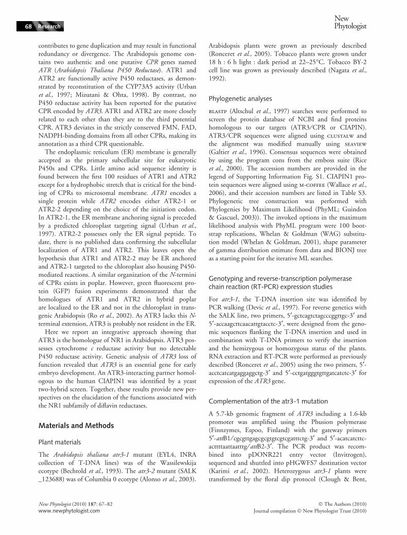
Three CPR genes, ATR1-3, are annotated in the Arabidopsisgenome (Paquette et al., 2009) and registered in TAIR(http://www.arabidopsis.org/browse/genefamily/cytochromeb5.jsp). ATR3 (At3g02280) encodes a protein of623 amino acids showing 25.7% and 24% identity withATR1 and ATR2, respectively, whereas these last two share60.5% identity. ATR3 shares 42.1% and 35.4% identitywith NR1 and Fre-1, respectively, while in humans, NR1shares 32.7% identity with CPR. These data raise the ques-tion as to whether ATR3 is an authentic CPR or a homo-logue of NR1 and Fre-1. To address this question, aClustalw alignment was used to compare plant and animalCPRs together with plant ATR3 and animal NR1 homo-logues available at the time of the analysis (Altschul et al.,1997). A phylogenetic tree was derived and showed thatATR3, NR1 and their homologues form a group distinctfrom the CPRs (Fig. 1a).
The CPR and ATR3 ⁄ NR1 groups belong to a family ofenzymes that shuttles two electrons from NADPH to itsphysiological redox partner, via two cofactors, FAD andFMN. We compared the FMN, FAD and NADPH-bind-ing domains between CPR and ATR3 ⁄ NR1 sequences(Figs 1b, S1). A similar domain arrangement was found inCPR and ATR3 sequences. However, a greater degree ofconservation exists among plant ⁄ animal ATR3 ⁄ NR1sequences than plant ⁄ animal CPRs, in particular for theFMN-binding regions.
The rate of hybrid transfer from NADPH to theFAD ⁄ NADPH domain of NR1 is c. 200-fold slower thanwith CPR as measured in rat (Finn et al., 2003). Theauthors noted the substitution of a cysteine residue (Cys630in CPRs) by an alanine in NR1. In CPRs, Cys630 formspart of the catalytic triad with Ser457 and Asp675.Substitution of Cys630 by an alanine residue in CPR
Fig. 1 ATR3 belongs to the clade of NR1 ⁄ Fre1 diflavin reductases. (a) Phylogenetic tree with per cent bootstrap value (1000 bootstrap) of 30cytochrome P450 reductase (CPR) sequences and 12 ATR3-like sequences from 25 plant (in bold) and animal species. Aa, Artemisia annua;At, Arabidopsis thaliana; Ce, Caenorhabditis elegans; Cery, Centaurium erythraea; Chlamy, Chlamydomonas reinhardtii; Cr, Catharanthusroseus; Dm, Drosophila melanogaster; Gm, Glycine max; Hs, Homo sapiens; Ht, Helianthus tuberosus; Mm, Mus musculus; Mt, Medicago
truncatula; Os, Oryza sativa; Ostta, Ostreococcus tauri; Pc, Petroselinum crispum; Pm, Pseudotsuga menziesii; Ps, Pisum sativum; Pso,Papaver somniferum; Ptd, Populus balsamifera subsp. trichocarpa · Populus deltoids; Rn, Rattus norvegicus; Sc, Saccharomyces cerevisiae;Ta, Triticum aestivum; Tc, Taxus chinensis; Vr, Vigna radiata; Vs, Vicia sativa. (b) Sequence alignment for FMN, FAD and NADPH cofactor-binding regions of consensus of plant ⁄ animal CPRs and plant ⁄ animal ATR3s. The protein consensus was obtained retaining only sequencesfrom genome-sequencing projects ensuring that both CPR and ATR3 were available. Comprehensive alignment for all sequences is presentedas in the Supporting Information, Fig. S1. Identical and similar residues are respectively black and grey box-shaded. The percentage of identitywas calculated for each binding region and · indicates in the table which consensus sequences were compared. Asterisks indicate Ser457,Asp675 and Cys630, residues of the catalytic triad in CPRs.
NewPhytologist Research 71
� The Authors (2010)
Journal compilation � New Phytologist Trust (2010)
New Phytologist (2010) 187: 67–82
www.newphytologist.com

decreases the enzymatic activity 49-fold (Shen et al.,1999). Interestingly, this alanine variant is found in allATR3 ⁄ NR1 sequences with the exception of poplar ATR3where a serine replaces the cysteine and yeast where thecysteine remains (Figs 1b, S1).
The major difference in domain organization betweenCPR and ATR3 ⁄ NR1 sequences is associated with the N-terminal region. CPRs contain a hydrophobic N-terminalanchor domain which tethers the protein to the endoplasmicreticulum. This domain is absent in ATR3 ⁄ NR1 proteins.
(a) (b) (c) (d)
(e) (f) (g) (h)
(i) (j) (k) (l)
(m) (n)
(o)
72 Research
NewPhytologist
� The Authors (2010)
Journal compilation � New Phytologist Trust (2010)
New Phytologist (2010) 187: 67–82
www.newphytologist.com

ATR3 is localized in the cytoplasm and the nucleus
To determine ATR3 localization in vivo, GFP was used as atag and ATR3::GFP was transiently expressed together withthe mRFP-HDEL ER marker in tobacco leaf epidermalcells (Fig. 2a–d) and stably expressed in tobacco BY-2 cells(Fig. 2e–h). In both expression systems, mRFP-HDELhighlighted the cortical ER (Figs 2b, S2) while ATR3::GFPwas found in the cytoplasm and in the nucleus but excludedfrom the nucleolus (Fig. 2a,d,e). As the ER is localized inthe cytosol, a coincident yellow signal results from the over-lap of red ER and green cytoplasm emissions (Fig. 2c,g,k).However, there is no evidence of a fluorescent yellowlabelled network typical and diagnostic of the cortical ER(Fig. S2). As a control, cytosolic GFP (cGFP) was co-expressed with the mRFP-HDEL and the micrographs(Fig. 2i–l) were comparable to that obtained forATR3::GFP and mRFPHDEL (Fig. 2e–h). These resultsclearly show that ATR3::GFP resides in the cytoplasm andthe nucleus.
ATR3 reduces cytochrome c but not the human P450CYP1A2
To establish the catalytic properties of ATR3, recombinantATR3::GFP was transiently expressed in N. benthamianaby agroinfiltration. The ATR3::GFP fluorescence patternobserved in the epidermal cells of N. benthamiana showed astrong accumulation in the cytosol similar to that observedin Fig. 2 (data not shown). Immunoblot analysis of thesoluble and microsomal fractions isolated from agroinfiltratedleaves was performed using GFP antibodies (Fig. 2m). Inleaf samples expressing GFP alone, a band of c. 27 kDa wasobserved in the soluble fraction. No free GFP was detectedin leaves over-expressing ATR3::GFP. The recombinantATR3::GFP protein was detected predominantly in thesoluble fraction and to a lesser extent in the microsomalfraction where contamination with soluble ATR3::GFPcannot be excluded. Cytochrome c, an artificial acceptor ofelectrons from CPRs, was used as a substrate for assaying
the reductase activity of CPRs (Murataliev et al., 2004).Cytochrome c reduction by ATR3 or ATR3::GFP was firstassayed in yeast in order to confirm that the ATR3::GFPfusion was functional. The activities measured were equiva-lent with and without the GFP tag (ATR3::GFP4.78 ± 0.66 lmol min)1 mg)1 while vector was only1.32 ± 0.4 lmol min)1 mg)1; ATR3 6.3 ± 0.14 lmolmin)1 mg)1 while vector was only 1.86 ± 0.09 l lmolmin)1 mg)1; each value is the mean ± SE from three inde-pendent protein preparations). Subsequently, the rate ofreduction of cytochrome c by recombinant ATR3::GFP inthe soluble and microsomal fractions of agroinfiltratedleaves was determined in the presence of NADPH. Bothfractions were functional, although a higher activity wasmeasured in the soluble fraction (Fig. 2n). As the recombi-nant ATR3::GFP expressed in N. benthamiana could beappropriately modified and suited for enzymatic activity,the plant cytosolic and microsomal ATR3::GFP were testedfor cytochrome P450 reductase activity by in vitro reconsti-tution experiments with CYP1A2. In contrast to the humanCPR, neither soluble nor microsomal ATR3::GFP was ableto reconstitute the O-deethylase activity of CYP1A2 in vitro(Fig. 2o). These results indicate that ATR3, like NR1, has acytochrome c reductase activity but fails to reconstitute aP450-mediated reaction.
Genetic analysis of ATR3 loss of function
We have characterized an embryo-defective (emb) mutant(EYL4) by screening the INRA collection of T-DNA inser-tion lines and determined that the T-DNA has inserted intothe fourth exon of ATR3 (Fig. 3a). The line was renamedatr3-1. Heterozygous atr3-1 ⁄ + plants produced a 2 : 1 ratioof KanR : KanS plants, which is consistent with a singlelocus of T-DNA integration linked to an homozygous lethalemb mutation (1157 KanR : 561 KanS seedlings, v2 = 0.36).To confirm that the emb phenotype resulted from the dis-ruption of ATR3, heterozygous atr3-1 ⁄ + plants were trans-formed with a T-DNA carrying a wild-type ATR3 gene andthe hygromycin resistance marker. The segregation analysis
Fig. 2 ATR3 is localized in the cytoplasm and the nucleus, has a cytochrome c reductase activity but fails to reconstitute a P450-mediated reac-tion. (a–d) Localization of ATR3::GFP and mRFP-HDEL in tobacco leaf epidermal cells using confocal microscopy. (a–c) cortical view showingthat the majority of ATR3::GFP is in the cytoplasm (a) and does not colocalize with the endoplasmic reticulum (ER) network labeled withmRFP-HDEL (b,c). (d) Merged nuclear view showing that ATR3::GFP is cytoplasmic and nuclear but excluded from the nucleolus. (e–l)Nucleus views of tobacco BY-2 cells stably coexpressing ATR3::GFP and mRFP-HDEL (e–h) or cGFP and mRFP-HDEL (i–l). ATR3::GFP andcGFP highlight the cytoplasm (e–i) whereas mRFP-HDEL resides in the ER (f–j). Merged images show that the proteins are located to distinctcompartments (g–k). Panels h and l are transmitted light images corresponding to panels e–g and i–k, respectively. (a,e,i) ATR3::GFP (488 nmargon ion laser line; k emission: 493–538 nm); (b,f,j) mRFP-HDEL (543 nm HeNe laser line; k emission: 580–620 nm); (c,d,g,k) mergedimages. Bars, 8 lm; N, nucleus; n, nucleolus. (m) Soluble (S) and microsomal (M) fractions from Nicotiana benthamiana expressing GFP orATR3::GFP were resolved by a 10% sodium dodecyl sulfate–polyacrylamide gel electrophoresis (SDS–PAGE). After transfer, the blot wasprobed with GFP antibody. Protein band(s) corresponding to ATR3::GFP fusion protein and to GFP are indicated by arrow and asterisk, respec-tively. (n) Cytochrome c reduction by GFP and ATR3::GFP from microsomal and soluble fractions expressed in tobacco. The control indicatedas GFP corresponds to the vector, which includes the GFP lacking a fusion and represents the background activity. (o) O-deethylation of7-ethoxycoumarin by CYP1A2 to form 7-hydroxycoumarin in the presence of human cytochrome P450 reductase (CPR) or ATR3.
NewPhytologist Research 73
� The Authors (2010)
Journal compilation � New Phytologist Trust (2010)
New Phytologist (2010) 187: 67–82
www.newphytologist.com

of the progeny of nine independent T1 lines is provided inthe Supporting Information, Table S1. Four T1 plants werehomozygous for the atr3 mutation (lines 1, 2, 5 and 7) and
would not exist unless there is complementation. Thisexperiment demonstrated that ATR3 plays an essential rolein embryogenesis for which ATR1 and ATR2 are unable tocompensate. An independent atr3 allele (SALK_123688)was identified in the SALK Institute collection (Alonsoet al., 2003). The T-DNA has inserted into the lastexon of ATR3 (Fig. 3a). Siliques of SALK_123688 ⁄ + plants contained 50% aborted seeds and furtheranalyses highlighted that the SALK_123688 line has twodistinct emb loci located on the same chromosome ingenetic opposition (emb1 + : + emb2). Back-crosses allowedus to isolate the atr3-2 allele. Crosses between atr3-1 andatr3-2 heterozygous plants demonstrated that the twomutations were allelic (655 wild type seeds, 200 emb seeds,v2 = 1.11).
The phenotype of atr3 ⁄ atr3 seeds was studied in detail.The percentage of emb seeds in the siliques is, on average,27% for the atr3-1 allele (total seed number, 1481; embseeds, 400; v2 = 3.19) and 24.2% for the atr3-2 allele(total seed number, 1032; emb seeds, 250; v2 = 0.33),values not significantly different from the 25% expectedfor a recessive lethal sporophytic mutation. However, thetransmission of the T-DNA was determined in reciprocalcrosses in order to test the possibility of additional gameto-phytic lethality (Table S2). No bias of transmission wasobserved and this confirmed the sporophytic nature of theatr3 mutation. The major stage of arrest of embryo devel-opment was two- to four-cell stages in atr3-1 and eightcells in atr3-2 (Fig. 3b). This difference in the severity ofthe phenotype may be caused by the position of theT-DNA insertion in the ATR3 gene (Fig. 3a). In atr3-2,the production of a nearly complete ATR3 protein,although missing a functional catalytic triad, may allow theembryo to develop further compared with atr3-1 embryos.Fig. 3(c–k) presents typical mutant embryos observed foratr3-1 (c,d,e) and atr3-2 (f,g,h) alleles when the wild-typeseeds of the same silique were at globular (i), triangular (j)and heart (k) stages. The endosperm nuclei were able todivide and the number of nucleoli was consistent with thenumber usually present in a wild-type seed containing atwo- to eight-cell stage embryo. This suggests that thedevelopment of the embryo and the endosperm are arrestedat the same time. Moreover, in cases when the embryoeventually produces more than eight cells (Fig. 3g,h), itfails to perform periclinal divisions, which is considered asthe first step of the radial patterning of the embryo (Mayer& Jurgens, 1998).
ATR3 is regulated during cell cycle progression
Genes that play essential functions in early embryogenesismay be involved in cell division. For example, TITAN andPILZ genes act at late phases of the cell cycle and areinvolved in the mechanisms of chromosome separation
LB LB
atr3-1, EYL4
2 3 4 5 6 7 8 9 10 11 12 13
At3g02280AtATR3
400 bp
1ATG
atr3-2, SALK_123688
+1
atr3-1, EYL4atr3-1, EYL4(a)
(b)
2 3 4 5 6 7 8 9 10 11 12 13
At3g02280AtATR3
400 bp
1ATG
atr3-2, SALK_123688
+1
(c-k) Phenotypes of atr3-1 and atr3-2 mutant embryos
Distribution of embryo phenotypes for atr3-1 and atr3-2 allelesWT globular to heart 1 cell 2 cells 4 cells 8 cells >8 cells ndatr3-1 (n = 336) 9.20% 58.35% 31.85% 0.00% 0.00% 0.60% 100.00%atr3-2 (n = 142) 9.85% 21.85% 17.60% 26.00% 12.00% 12.70% 100.00%
atr3-1 atr3-2 wt(f)*
** **
**
*
E(i)(c)
E E
S
E
S
E
S
E
S
E
S
E
S
E
S
E
S
E
S
E
S
(j)
E
S
(g)
E
S
(d)
E
S
E
S
E
S E
S
(k)E
S
(e) (h)
E
S
SS
Fig. 3 Genetic analysis of ATR3 loss of function. (a) Schematicrepresentation of intron–exon structure and T-DNA integration inthe ATR3 gene. Black boxes, exon; black line, intron. LB, T-DNA leftborder. (b) Distribution of embryo phenotypes for atr3-1 and atr3-2
alleles. (c–k) Phenotypes of atr3-1 and atr3-2 mutant embryos. (c)2-cell atr3-1 embryo and (f), 8-cell atr3-2 embryo when wild type isat globular stage (i). (d) 2–4 cell atr3-1 embryo and (g), atr3-2
embryo when wild type is at triangular stage (j). (e) Four-cell atr3-1
embryo and (h), atr3-2 embryo when wild type is at heart stage (k).(g,h) atr3-2 embryos with more than eight cells. Bar, 20 lm.Asterisks point to individual endosperm nucleoli. Black arrows markthe limit between embryo and suspensor. E, embryo proper; S,suspensor.
74 Research
NewPhytologist
� The Authors (2010)
Journal compilation � New Phytologist Trust (2010)
New Phytologist (2010) 187: 67–82
www.newphytologist.com

(Steinborn et al., 2002; Tzafrir et al., 2002). DNA poly-merase epsilon (Jenik et al., 2005; Ronceret et al., 2005)and thymidylate kinase genes (Ronceret et al., 2008) act atearly phases of the cell cycle and are transcribed at theG1 ⁄ S-phase transition of the cell cycle. A detailed examina-tion of the promoter region of ATR3 revealed the presenceof conserved cis-elements, CHR (cell cycle homologyregion) and CDE (cell cycle-dependent expression), knownto be involved in transcriptional cell cycle regulation oftobacco RNR2 gene (Chaboute et al., 2000). A reverseCHR ⁄ CDE at )1100 bp and a forward CDE in the 5¢leader sequence are found upstream from the initiationcodon of ATR3 (Fig. S3). A 1.6 kb promoter region
containing the potential CHR and CDE cis-elements wasfused to luciferase (Fig. 4a) and used to stably transformtobacco BY-2 cells. Cells were synchronized by aphidicolinand luciferase activity was measured at different time-pointsof the cell cycle. A peak of luciferase activity was visible 4–5 h after drug washout occurring after DNA synthesis (Sphase) and preceding the maximal mitotic index reached at8–9 h (M phase; Fig. 4b). This induction peak was repro-ducible in three independent transformation experiments(Fig. 4c). These results demonstrated that the ATR3 pro-moter is most probably activated at the G2 and G2 ⁄ M-phase transition of the cell cycle.
Developmental expression of ATR3
The level of expression of the ATR3 gene is very low dur-ing the Arabidopsis plant life cycle, as recorded in micro-array databases and confirmed by RT-PCR experimentswhere 45 cycles are required to detect its expression com-pared with 23 cycles for the constitutive control EF1a(Fig. 5a). Despite its low level of expression, the ATR3band of 267 bp was detected in every sample tested. Thespatial and temporal expression of ATR3 duringArabidopsis development was analysed in five independenttransgenic lines containing the GUS gene under the con-trol of the 1.6 kb ATR3 promoter. Overall, GUS expres-sion was not detected throughout plant and embryodevelopment with the exception of dry seeds 24–48 h afterimbibition and the tapetum of the anthers (Fig. 5b) atthe floral stage 10 according to (Bowman, 1994). mRNA
1
ATG
LuciferaseCDE
+1
1CHR CDE
CDE
500.00
600.00
Time after aphidicolin removal (h)
(a)
(b)
(c)
25
30
500.00
600.00
0.00
100.00
200.00
300.00
400.00
0
5
10
15
20
90.00MG2S
0.00
100.00
200.00
300.00
400.00
CP
M µ
g–1
of
pro
tein
sR
LU
µg
–1 o
f p
rote
ins
RL
U µ
g–1
of
pro
tein
s
Mit
oti
c in
dex
(%
)
0.00
10.00
20.00
30.00
40.00
50.00
60.00
70.00
0.00
10.00
20.00
30.00
40.00
50.00
60.00
70.00
80.00
Time after aphidicolin removal (h)
60
80
100
120
20
25
30
35
40
0
20
40
0
5
10
15
0
0 1 2 3 4 5 6 7 8 9 10
0 1 2 3 4 5 6 7 8 9 10
MS G2 G1
0 2 4 6 8 10 12 14 16Time after aphidicolin removal (h)
Mit
oti
c in
dex
(%
)
T1 T2 T3
MI 1 MI 2 MI 3
Fig. 4 Cell cycle activation of the ATR3 promoter. (a) Schematicrepresentation of ATR3 promoter::luciferase construct. The 1.6 kbATR3 promoter corresponds to the sequence upstream of the ATG.Gray line, promoter region upstream of the +1 transcription; opentriangle, cell cycle dependent expression (CDE) sequence; tinted tri-angle, cell cycle homology region (CHR) sequence. Orientation ofthe triangle mimics the orientation of the cis-element. (b)Measurement of the ATR3 promoter activity in synchronized stablytransformed BY-2 cells. Cells were synchronized by aphidicolin.After removal of the drug, DNA synthesis (upper panel, triangles)was determined by 3H thymidine pulse experiments on the basis oftotal proteins (counts per minute, CPM. Mitotic index (upperpanel, squares) was determined by microscopic observation of 4,6-diamidino-2-phenylindole (DAPI) stained cells. Luciferase activity(lower panel, diamonds) was measured on cells harvested at differ-ent time points and expressed in RLU (relative light unit) per totalprotein content. The position of cell cycle phases is indicatedbetween graphs. (c) Luciferase activity (RLU) measured for threeindependent transformation experiments (T1, diamonds; T2,squares; T3, triangles) and their corresponding mitotic index (MI 1,diamonds; MI 2, squares; MI 3, triangles, dotted lines). The positionof cell cycle phases is indicated above the graph. The G2 inductionpeak was reproducible in the three independent transformationexperiments. The T2 experiment is the one presented in more detailin (b).
NewPhytologist Research 75
� The Authors (2010)
Journal compilation � New Phytologist Trust (2010)
New Phytologist (2010) 187: 67–82
www.newphytologist.com

EF1 α(23 cycles)
ATR3
Se R L St Cl FB Fl IS MS C G
250
896
bp
GUS–Fix GUS +Fix4, 5
Obs 2, 3
Obs 4, 5
ATR3(45 cycles)
896267
G2 M 1 cm
842404420
Time (HAI)
Dry seed
Rootprotuding
G1
SAphidicolin
Colchicine
Synchronisation
1
2
3
72
2, 3
G2 MS
WashG2 M
S
G2 MS
4 hSAphidicolin
Colchicine
4
5Wash
249 172 287
Treatment24 h block and release
DMSO Aphidicolin Colchicine DMSO Aphidicolin Colchicine
225 189
Treatment48 h block
(f)
(d)
(c)
(a) (b)
(e)
(g) (h)
GUS nega 249 172 287 GUS negative 225 189Number of seeds 284 217 316GUS ti
Number of seeds 248 208 363255tive
GUS positive 35 45 29Percentage 12.3% 20.7% 9.2%
255GUS positive 23 19 108Percentage 9.3% 9.1% 29.8%
Fig. 5 Cell cycle regulation of the ATR3 promoter::GUS fusion after seed imbibition. (a) Expression of the ATR3 gene in Arabidopsis. Reverse-transcription polymerase chain reaction (RT-PCR) was performed with RNA from 10-d-old seedlings (Se), roots (R), rosette leaves (L), stem(St), cauline leaves (Cl), flower buds (FB), flowers (Fl), siliques after fertilization up to torpedo stage (immature stage, IM) and siliques at maturecotyledon stage (MS). Lane C shows the control with no DNA and lane G, the control with genomic DNA. Expression of EF1a was used as apositive and constitutive control. (b) illustrates ATR3 promoter::GUS expression in the tapetum at floral stage 10 according to (Bowman,1994). Glucuronidase (GUS) staining was carried out for 48 h. Bar = 20 lm (c) Graphic representation of cell cycle progression during seedimbibition and germination based on (Barroco et al., 2005) (lane 1) and seed imbibition experiments carried out using aphidicolin andcolchicine (lanes 2–5). Time is given in hours after seed imbibition (HAI). Lanes 2–3, 24 h block and release experiment. Drugs were removedat 24 HAI; GUS staining was carried out for 24 h in the absence of fixative allowing cells to proceed through the cell cycle after drug washout;cytological observations were performed at 48 HAI. Lanes 4–5, 48 h block experiment. Drugs were removed at 48 HAI; GUS staining wascarried out for 24 h in the presence of fixative preventing cells to proceed through the cell cycle after drug washout; cytological observationswere done at 72 HAI. Black arrows (downward) indicate when drug was added and red arrows (upward) when drug was washed out. Bluearrows indicate when GUS staining was performed for 24 h and whether fixative was added (+Fix) or not ()Fix). Green arrows indicate whenseeds were cleared in Hoyer’s and observed. Phases of the cell cycle are indicated. Arrowhead indicates radicle protrusion. (d) ATR3promoter::GUS expression in Arabidopsis roots after 24 HAI in the presence of dimethyl sulfoxide (DMSO) as a control or 5 lg ml)1 aphidicolinor 5 lg ml)1 colchicine. (e) ATR3 promoter::GUS expression in Arabidopsis roots after 48 HAI in the presence of DMSO as a control or5 lg ml)1 aphidicolin or 5 lg ml)1 colchicine. (f–h) Illustrate ATR3 promoter::GUS expression in Arabidopsis roots with DMSO (f), 5 lg ml)1
aphidicolin for the 24 h block and release experiment (g) and 5 lg ml)1 colchicine for the 48 h block experiment (h), Bar = 20 lm.
76 Research
NewPhytologist
� The Authors (2010)
Journal compilation � New Phytologist Trust (2010)
New Phytologist (2010) 187: 67–82
www.newphytologist.com

in situ hybridizations were performed but no signal wasdetected throughout embryo development, suggesting thatthe level of expression of the ATR3 gene is below thethreshold of detection.
As the ATR3 promoter is activated during cell cycle pro-gression and GUS expression is occurring in imbibedseeds, experiments similar to those described in Wang &Liu (2006) were performed. Barroco et al. (2005) havedemonstrated that most cells of dry seeds are arrested inthe G1 phase. During imbibition, DNA synthesis is initi-ated in the radicle. A major transition through S phasetoward G2 was detected when the radicle starts to pro-trude 42 h after seed imbibition (HAI), while entry intoM phase occurred after radicle protrusion (Fig. 5c, lane1). Dry seeds of two homozygous ATR3 promoter::GUSlines showing the highest GUS activity were imbibed in5 lg ml)1 aphidicolin or 5 mg ml)1 colchicine, whicharrests cells at S phase or M phase, respectively (Fig. 5c,lines 2–5). A DMSO treatment was used as a control asboth drugs were solubilized in this solvent. The two linesexhibited similar responses to drug treatments. In the firstexperiment (Fig. 5c), seed imbibition was carried out insterile water for 24 h in the presence of DMSO as a con-trol or in the presence of aphidicolin (lane 2) or in thepresence of colchicine (lane 3). The seeds were thenwashed with sterile water to remove the drug and incu-bated in GUS staining buffer without methanol () Fix)for 24 h. Cytological observations were performed at 48HAI (including the period of GUS staining). Under suchconditions, the absence of fixative allows cells synchronizedby aphidicolin to proceed through the G2 ⁄ M phase duringGUS staining. The number of roots expressing GUS was c.twofold higher (Fig. 5d) and at a higher level than theDMSO control (Fig. 5f,g). By contrast, colchicine treat-ment had no effect on GUS expression, consistent withthe removal of the drug before cells advanced throughmitosis.
The second experiment (Fig. 5c) was carried out in asimilar manner, with the exception of the time for seedimbibition (48 h) and the addition of methanol (+ Fix) inthe GUS staining solution. Cytological observations wereperformed at 72 HAI including 24 h of GUS staining. Inthis experiment the presence of fixative does not allow cellsto proceed through the cell cycle after drug washout. Thenumber of GUS-positive radicles observed for the aphidico-lin treatment was not significantly different from theDMSO control indicating that cells arrested at S phase didnot express the reporter gene (Fig. 5e). By contrast, thenumber of GUS-positive radicles observed for the colchi-cine treatment was threefold higher (Fig. 5e) and at higherlevel than the DMSO control (Fig. 5f,h), indicating thatcells went through the G2 ⁄ M-phase transition and werearrested at M phase before fixation. Together, these experi-ments confirmed that the ATR3 promoter is activated
during cell cycle progression, most probably at G2 andG2 ⁄ M phases.
ATR3 interacts with a protein similar to humanCIAPIN1
The SRS.Y2H system (Cytotrap; Stratagene) which detectsinteractions in the cytoplasm (Aronheim et al., 1997) wasused to identify proteins that interact with ATR3 in orderto provide insight to its biological function. The system isbased on the ability of protein interactions to rescue thegalactose-dependent growth of the cdc25H mutant yeaststrain at the restrictive temperature of 37�C. Screening ofan Arabidopsis whole-seedling cDNA library with ATR3(pSos-ATR3) as bait yielded a single cDNA clone(At5g18400) encoding the homologue of the human cyto-kine-induced inhibitor of apoptosis protein 1 (CIAPIN1).Sequence alignment of animal CIAPIN1 and plantCIAPIN1 homologues available for genomes entirelysequenced showed that the average 35% amino acid iden-tities and 50% similarities are concentrated in the twofunctional domains predicted for the human CIAPIN1(Hao et al., 2008): a generic methyltransferase motif inthe N-terminal region and Zn-ribbon-like motifs includingseveral conserved cysteines in the C-terminal region(Fig. 6a). A phylogenetic tree was derived from an m-cof-
fee alignment of 67 CIAPIN1 sequences representative ofdifferent phylum (Fig. 6b). This tree indicated that thereis clearly a common ancestor for animal and plant lin-eages. The closest sequence of At5g18400 is the Drosophilasimulans sequence (49% identity, 64% similarities) in theMetazoa phylum and the Picea sitchensis sequence (50%identity, 68% similarities) in the Streptophyta phylum.We did not find any other paralogous gene in any of thegenomes entirely sequenced to date. Therefore, we canconsider that At5g18400 (hereafter designed AtCIAPIN1)is a unique gene and corresponds to the homologue ofhuman CIAPIN1. In retransformation experiments, thegrowth of cdc25H cells cotransformed with pSos-ATR3and pMyrAtCIAPIN1 was rescued on SD ⁄ galactose butnot on SD ⁄ glucose at 37�C, thereby confirming the speci-ficity of the protein interaction (Fig. 6c). The negativecontrols did not grow on galactose at 37�C, whereas thepositive control (pSos-MAFB and pMyr-MAFB) grew wellon SD ⁄ galactose. To confirm the interaction betweenATR3 and AtCIAPIN1 in vitro, coimmunoprecipitationassays were performed with ATR3 antibodies. 35S-AtCIAPIN1 coimmunoprecipitated with the solublerecombinant ATR3, but not with the control yeastfraction (Fig. 6d). ATR3 antibodies did not immuno-precipitate the 35S-AtCIAPIN1 in the absence of recombi-nant ATR3. These results suggest that AtCIAPIN1 may bean acceptor ⁄ donor protein requiring ATR3 reductaseactivity for its function.
NewPhytologist Research 77
� The Authors (2010)
Journal compilation � New Phytologist Trust (2010)
New Phytologist (2010) 187: 67–82
www.newphytologist.com

(a)
SD/GalactoseSD/Glucose
25°C 37°C
MAFB MAFB
ATR3 AtCIAPIN1
Bait
pSOS vector
Prey
pMyr vector
Empty Empty
ATR3 Empty
Empty AtCIAPIN1
25°C 37°C
Pull-down with ATR3 -Ab
30 kDa
-- Vec-S
AtC
IAPI
N1
-- ATR3-S
AtCIAPIN1 AtCIAPIN1AtCIAPIN1
(c)
(d)
68
(b)
78 Research
NewPhytologist
� The Authors (2010)
Journal compilation � New Phytologist Trust (2010)
New Phytologist (2010) 187: 67–82
www.newphytologist.com

Discussion
The Arabidopsis ATR3 gene encodes a diflavinreductase, the homologue of NR1
Several lines of evidence have led us to question the identityof ATR3 as an authentic CPR. The phylogenetic tree clus-ters ATR3 with the animal oxidoreductases NR1 and Fre-1distinct from the clade containing the plant and animalCPRs. The FMN, FAD, NADPH-binding domains ofplant ATR3 proteins display a higher percentage of identitywith their animal counterparts (67%, 45% and 55%,respectively) than with plant CPRs (32%, 40%, and 45%respectively) and the cysteine residue essential for the cata-lytic activity of CPRs is often replaced by an alanine. Allplant and animal ATR3 ⁄ NR1 proteins lack an ER mem-brane anchor domain at their N-terminal end, a characteris-tic of authentic CPRs (Wang et al., 1997). Plant andanimal CPRs are membrane bound enzymes, whereasATR3 was found in the cytosol and the nucleus, a localiza-tion similar to that of NR1 (Paine et al., 2000).
Although CYP1A2 is a human P450 enzyme and unli-kely to constitute an optimal partner for ATR3, ATR1 andATR2 have been shown to support the catalytic activity ofthe human P450 enzyme with differing efficiencies(Louerat-Oriou et al., 1998). Like ATR3, NR1 also failedto reconstitute the O-deethylase activity of the CYP1A2in vitro (Paine et al., 2000). In conclusion, we showed thatATR3 possesses a cytochrome c reductase activity in thecytosol but we were unable to demonstrate a P450 reductaseactivity in agreement with its subcellular localization. Theseresults suggest that reduction of the P450 enzymes attachedto the endoplasmic reticulum is an unlikely physiologicalrole for both ATR3 and NR1. The observation that loss ofATR3 resulted in an emb phenotype consolidated ourhypothesis that ATR3 plays an essential role, distinct fromthat of ATR1 and ATR2. Knocking out the unique CPRgene results in defects in establishing the polarity in early C.
elegans embryo (Rappleye et al., 2003) and embryoniclethality in mouse (Shen et al., 2002). Therefore, neitherFre-1 nor NR1 were able to compensate the loss of CPRfunction. Taken together, the sequence homologies, subcel-lular localization, enzymatic activities and loss of functionphenotype, demonstrated that ATR3 is the homologue ofhuman NR1 and C. elegans Fre-1 and belongs to the NR1subfamily of diflavin reductases.
Is ATR3 a novel regulator of cell death in plants?
As ATR3 has physiological partners other than P450s, thenext step towards the understanding of ATR3 functionwas to identify ATR3-interacting proteins. Recent workon NR1 suggested a role in cell death and indicated afunctional cooperation between NR1 and DcpS, a scaven-ger decapping enzyme that plays an important role in the3¢–5¢ mRNA decay pathway after the degradation by theexosome. Overexpression of NR1 in human embryonickidney cells enhances the cytotoxic action of the artificialelectron acceptor menadione and DcpS physically interactswith NR1 and modulates its activity promoting cell sur-vival (Kwasnicka et al., 2003). This information promptedus to search for a homologue of DcpS in Arabidopsis.Structural and biochemical studies have revealed that acentral histidine in a histidine triad (HIT) domain inDcpS is responsible for hydrolysis of the cap structure (Liuet al., 2002). The best Arabidopsis DcpS candidatesdisplayed identities only in the histidine triad domain andthis limited similarity may question their functionalhomologies. These candidates when tested in yeast two-hybrid assays, failed to interact with ATR3 (data notshown).
The yeast two-hybrid assay strategy was pursued byscreening an Arabidopsis seedling cDNA library. We identi-fied an Arabidopsis protein homologous to CIAPIN1 as anATR3 interacting partner. Human CIAPIN1 was shown tobe a mediator of the RAS signalling pathway and to play a
Fig. 6 ATR3 interacts with the Arabidopsis homologue of human CIAPIN1. (a) Sequence alignment of animal and plant CIAPIN1 proteinsdisplaying DUF689 domain (Pfam PF05093) which contains several uncharacterized proteins displaying an N-terminal generic methyltransferasemotif (dotted line) and a C-terminal zinc ribbon-like motif (plain line) including several conserved cysteines (highlighted by asterisks). TheCIAPIN1 proteins retained for the alignment here are from entirely sequenced genomes and their accession numbers are listed in Fig. S3.Arth, Arabidopsis thaliana (At5g18400); Orsa, Oryza sativa japonica; Mumu, Mus musculus; Hosa, Homo sapiens; Drme, Drosophilamelanogaster; Dare, Danio rerio; Sace, Saccharomyces cerevisiae. (b) Phylogenetic tree of 67 CIAPIN1 sequences of the following phyla:Apicomplexa (API: 6), Bacillariophyta (BAC: 2), Chlorophyta (CHL: 1), Choanoflagellida (CHO: 1), Ciliophora (CIL: 2), Fungi (FUN: 16),Metazoa (MET: 30), Mycetozoa (MYC: 1), Streptophyta (STR: 8). Accession numbers are listed in Table S3. The number of replicate trees inwhich the associated taxa clustered together in the bootstrap test (1000 replicates) is shown next to the branches. Arabidopsis and humansequences are indicated in bold type. (c) SRS-Y2H retransformation assays. Yeast cotransformed with pSos-ATR3 and pMyr-AtCIAPIN1plasmids were selected for galactose-dependent growth at 37�C. Positive control, pSos-MAFB + pMyr-MAFB; negative controls:pSos + pMyr-AtCIAPIN1, pSos-ATR3 + pMyr, pSos + pMyr. MAFB (v-Maf musculo-aponeurotic fibrosarcoma oncogene homolog B)belongs to the leucine zipper family of transcription factors and forms homodimers (Cytotrap two-hybrid system, Stratagene). (d) Co-immunoprecipitation of AtCIAPIN1 with ATR3. 35S-AtCIAPIN1 translated in vitro was tested for interaction with the yeast cytosolic solubleATR3 (ATR3-S) in co-immunoprecipitation assays with ATR3 antibodies. Controls included soluble fraction (Vector-S) from yeasttransformed with empty vector. Arrow indicates the 35S-AtCIAPIN1.
NewPhytologist Research 79
� The Authors (2010)
Journal compilation � New Phytologist Trust (2010)
New Phytologist (2010) 187: 67–82
www.newphytologist.com

vital role in fetal liver hematopoiesis (Shibayama et al.,2004). CIAPIN1 is described as a novel anti-apoptotic mol-ecule that does not show any homology to known apoptosisregulatory molecules such as Bcl-2 family members or casp-ases (Shibayama et al., 2004). Microarray analysis revealedreduced expression of Bcl-xL and Jak2 in fetal liver ofciapin1 null mice (Shibayama et al., 2004) and depletion ofCIAPIN1 was shown to trigger apoptosis in HCC cellsdownregulating Bcl-2 and Bcl-xL and upregulating Bax (Liet al., 2008). This suggests that CIAPIN1 might exert itsfunction by modulating the expression of apoptosis regula-tory genes. Although no interaction has been reportedbetween the human NR1 and CIAPIN1, such an interactionwas observed in yeast. The genome of yeast encodes both aCPR (ScCPR) and an ATR3 ⁄ NR1 (TAH18) diflavin reduc-tase (Figs 1a, S1). The interaction between TAH18 andDre2, the yeast homologue of CIAPIN1 was found to con-trol oxidative stress-induced cell death (Vernis et al., 2009).Dre2-Tah18 interaction takes place in the cytoplasm in theabsence of oxidative stress. The tah18 and dre2 mutatedalleles are synthetic lethal in yeast. Moreover, humanCIAPIN1 is able to replace the yeast Dre2 in vivo andphysically interacts with TAH18. The involvement of theDre2–TAH18 complex in apoptosis, together with therole of human CIAPIN1, indicates a possible role forATR3 in programmed cell death via an interaction withAtCIAPIN1.
In plants, the cell death is critical for the establishment ofpolarity at early stages of plant embryogenesis, when the dif-ferentiation of the temporary basal organ, the suspensor, isfollowed by its programmed elimination (Bozhkov et al.,2005). However, the observation of mutant embryos foratr3-1 and atr3-2 alleles did not reveal any abnormalities inthe suspensor development as the embryo developmentarrests much earlier than the torpedo stage coincident withsuspensor degeneration. Interestingly, we observed a tran-sient expression of ATR3 in the tapetum during antherdevelopment at floral stage 10 according to (Bowman,1994). All the programmed cell death (PCD) hallmarkshave been reported for tapetum degeneration and the PCDsignal commences at the tetrad stage (Kawanabe et al.,2006) corresponding to floral stage 9 according to(Bowman, 1994). Therefore, PCD signalling precedesATR3 expression (as monitored by GUS activity) at stage10, which occurs before cells degenerate at stage 11–12.This chronology is consistent with an involvement ofATR3 ⁄ AtCIAPIN1 complex in PCD. However, the embry-onic lethality of ATR3 prevented us from further addressingits role during the later stages of the plant life cycle. Byexample, a putative role of ATR3 in the PCD of tapetumcells cannot be functionally tested in the heterozygousatr3 ⁄ + plants. Similarly, a role of ATR3 in germination andin plant growth could not be investigated in the knockoutlines.
Alternative function of ATR3 in DNA replication andcell division
Is the function of ATR3 uniquely restricted to the controlof cell death? Although the Dre2–TAH18 complex controlscell death in yeast, this process is conditioned by stress. Thisraises the question of the nature of the essential function ofDre2–TAH18 interaction in absence of stress.
TAH18 function was identified as a gene involved inimpaired DNA replication and possibly DNA repair ⁄ DNAintegrity check (Vernis et al., 2009). In the same way, thermo-sensitive dre2 mutant exhibits a cin phenotype (cin forincreased chromosome instability) and this phenotype wasscored as severe in chromosome transmission fidelity (CTF)assays (Ben-Aroya et al., 2008). In the same way, expressionof the human CIAPIN1 influences the rate of cell division.Ectopic expression of CIAPIN1 by cDNA transfectionresulted in suppression of cell proliferation and inhibitionof cell cycle progression while knockdown of CIAPIN1with siRNA accelerated cell proliferation and promoted cellcycle progression in these cells (Hao et al., 2009). In thiscontext, the confirmation of ATR3 as a gene essential forearly embryogenesis brings a novel perspective to the role ofATR3-AtCIAPIN1 interaction in cell division. We(Ronceret et al., 2005, 2008), and others, have demon-strated the importance of cell cycle related genes at thegametophytic and sporophytic transition and at earlyembryo development. Furthermore, the transcription ofATR3 itself is regulated during the cell cycle at the G2 andG2 ⁄ M-phase transition. Similarly loss of cdc5, expressed atthe G2 ⁄ M transition, also arrested Arabidopsis embryodevelopment at an early stage (Lin et al., 2007). The controlof ATR3 transcription could constitute a mechanism to reg-ulate AtCIAPIN1 activity during the cell cycle.
Based on our results and the comparison with the func-tions of their human and yeast homologues, we proposethat ATR3-AtCIAPIN1 interaction in Arabidopsis mayconstitute an important yet previously unrecognized com-ponent in the general mechanisms of cell division and celldeath.
Acknowledgements
We thank Nicole Bechtold and Roger Voisin for their workon the INRA T-DNA insertion collection. We thankDaniele Werck-Reichhardt, Loıc Faye, Thomas Roscoe,Jean-Philippe Reichheld and Christophe Riondet forcritical reading of the manuscript. The work of VD, JG andMD was supported by the GENOPLANTE AF015 grant.CSJD and VG are supported by the Centre National de laRecherche Scientifique (CNRS), France. Plant Gatewayvectors were provided by Functional Genomics Division ofthe Department of Plant Systems Biology (VIB-GhentUniversity, Belgium).
80 Research
NewPhytologist
� The Authors (2010)
Journal compilation � New Phytologist Trust (2010)
New Phytologist (2010) 187: 67–82
www.newphytologist.com

References
Alonso JM, Stepanova AN, Leisse TJ, Kim CJ, Chen H, Shinn P,
Stevenson DK, Zimmerman J, Barajas P, Cheuk R et al. 2003.
Genome-wide insertional mutagenesis of Arabidopsis thaliana. Science301: 653–657.
Altschul SF, Madden TL, Schaffer AA, Zhang J, Zhang Z, Miller W,
Lipman DJ. 1997. Gapped BLAST and PSI-BLAST: a new generation
of protein database search programs. Nucleic Acids Research 25: 3389–
3402.
Aronheim A, Zandi E, Hennemann H, Elledge SJ, Karin M. 1997.
Isolation of an AP-1 repressor by a novel method for detecting protein–
protein interactions. Molecular and Cellular Biology 17: 3094–3102.
Barroco RM, Van Poucke K, Bergervoet JH, De Veylder L, Groot SP,
Inze D, Engler G. 2005. The role of the cell cycle machinery in
resumption of postembryonic development. Plant Physiology 137: 127–
140.
Bechtold N, Ellis J, Pelletier G. 1993. In planta Agrobacterium mediated
gene transfer by infiltration of adult Arabidopsis thaliana plants. ComptesRendus. Academie des Sciences 316: 1194–1199.
Ben-Aroya S, Coombes C, Kwok T, O’Donnell KA, Boeke JD, Hieter P.
2008. Toward a comprehensive temperature-sensitive mutant repository
of the essential genes of Saccharomyces cerevisiae. Molecular Cell 30: 248–
258.
Blazquez MA, Soowal LN, Lee I, Weigel D. 1997. LEAFY expression and
flower initiation in Arabidopsis. Development 124: 3835–3844.
Bowman J. 1994. Arabidopsis, an atlas of morphology and development. New
York, NY, USA: Springer-Verlag.
Bozhkov PV, Filonova LH, Suarez MF. 2005. Programmed cell death in
plant embryogenesis. Current Topics in Developmental Biology 67: 135–
179.
Bredt DS, Hwang PM, Glatt CE, Lowenstein C, Reed RR, Snyder SH.
1991. Cloned and expressed nitric oxide synthase structurally resembles
cytochrome P-450 reductase. Nature 351: 714–718.
Chaboute ME, Clement B, Philipps G. 2002. S phase and meristem-
specific expression of the tobacco RNR1b gene is mediated by an E2F
element located in the 5¢ leader sequence. Journal of Biological Chemistry277: 17845–17851.
Chaboute ME, Clement B, Sekine M, Philipps G, Chaubet-Gigot N.
2000. Cell cycle regulation of the tobacco ribonucleotide reductase small
subunit gene is mediated by E2F-like elements. Plant Cell 12: 1987–
2000.
Clough SJ, Bent AF. 1998. Floral dip: a simplified method for
Agrobacterium-mediated transformation of Arabidopsis thaliana. PlantJournal 16: 735–743.
Devic M, Albert S, Delseny M, Roscoe T. 1997. Efficient PCR walking
on plant genomic DNA. Plant Physiology and Biochemistry 35: 331–339.
Dignam JD, Strobel HW. 1975. Preparation of homogeneous NADPH-
cytochrome P-450 reductase from rat liver. Biochemical and BiophysicalResearch Communications 63: 845–852.
Finn RD, Basran J, Roitel O, Wolf CR, Munro AW, Paine MJ, Scrutton
NS. 2003. Determination of the redox potentials and electron transfer
properties of the FAD- and FMN-binding domains of the human
oxidoreductase NR1. European Journal of Biochemistry 270: 1164–1175.
Galtier N, Gouy M, Gautier C. 1996. SEAVIEW and PHYLO_WIN:
two graphic tools for sequence alignment and molecular phylogeny.
Computer Applications in the Biosciences 12: 543–548.
Guindon S, Gascuel O. 2003. A simple, fast, and accurate algorithm to
estimate large phylogenies by maximum likelihood. Systematic Biology52: 696–704.
Hao Z, Li X, Qiao T, Fan D. 2008. Successful expression and purification
of human CIAPIN1 in baculovirus–insect cell system and application of
this system to investigation of its potential methyltransferase activity.
International Journal of Biological Macromolecules 42: 27–32.
Hao Z, Li X, Qiao T, Li S, Lv Y, Fan D. 2009. Downregulated expression
of CIAPIN1 may contribute to gastric carcinogenesis by accelerating cell
proliferation and promoting cell cycle progression. Cancer Biology &Therapy 8: 1064–1070.
Huang N, Pandey AV, Agrawal V, Reardon W, Lapunzina PD, Mowat
D, Jabs EW, Van Vliet G, Sack J, Fluck CE et al. 2005. Diversity and
function of mutations in p450 oxidoreductase in patients with Antley–
Bixler syndrome and disordered steroidogenesis. American Journal ofHuman Genetics 76: 729–749.
Jenik PD, Jurkuta RE, Barton MK. 2005. Interactions between the cell
cycle and embryonic patterning in Arabidopsis uncovered by a mutation
in DNA polymerase epsilon. Plant Cell 17: 3362–3377.
Karimi M, Inze D, Depicker A. 2002. GATEWAY vectors for
Agrobacterium-mediated plant transformation. Trends in Plant Science 7:
193–195.
Kawanabe T, Ariizumi T, Kawai-Yamada M, Uchimiya H, Toriyama K.
2006. Abolition of the tapetum suicide program ruins
microsporogenesis. Plant and Cell Physiology 47: 784–787.
Kwasnicka DA, Krakowiak A, Thacker C, Brenner C, Vincent SR. 2003.
Coordinate expression of NADPH-dependent flavin reductase, Fre-1,
and Hint-related 7meGMP-directed hydrolase, DCS-1. Journal ofBiological Chemistry 278: 39051–39058.
Leclerc D, Wilson A, Dumas R, Gafuik C, Song D, Watkins D, Heng
HH, Rommens JM, Scherer SW, Rosenblatt DS et al. 1998. Cloning
and mapping of a cDNA for methionine synthase reductase, a
flavoprotein defective in patients with homocystinuria. Proceedings of theNational Academy of Sciences, USA 95: 3059–3064.
Li X, Pan Y, Fan R, Jin H, Han S, Liu J, Wu K, Fan D. 2008.
Adenovirus-delivered CIAPIN1 small interfering RNA inhibits HCC
growth in vitro and in vivo. Carcinogenesis 29: 1587–1593.
Lin Z, Yin K, Zhu D, Chen Z, Gu H, Qu LJ. 2007. AtCDC5
regulates the G2 to M transition of the cell cycle and is critical for
the function of Arabidopsis shoot apical meristem. Cell Research 17:
815–828.
Liu H, Rodgers ND, Jiao X, Kiledjian M. 2002. The scavenger mRNA
decapping enzyme DcpS is a member of the HIT family of
pyrophosphatases. EMBO Journal 21: 4699–4708.
Louerat-Oriou B, Perret A, Pompon D. 1998. Differential redox and
electron-transfer properties of purified yeast, plant and human
NADPH-cytochrome P-450 reductases highly modulate cytochrome P-
450 activities. European Journal of Biochemistry 258: 1040–1049.
Mankin SL, Allen CG, Thompson WF. 1997. Introduction of a plant
intron into the luciferase gene of Photinus pyralis. Molecular BiologyReports 15: 186–196.
Mayer U, Jurgens G. 1998. Pattern formation in plant embryogenesis: a
reassessment. Seminars in Cell & Developmental Biology 9: 187–193.
Mizutani M, Ohta D. 1998. Two isoforms of NADPH:cytochrome P450
reductase in Arabidopsis thaliana. Gene structure, heterologous
expression in insect cells, and differential regulation. Plant Physiology116: 357–367.
Murataliev MB, Feyereisen R, Walker FA. 2004. Electron transfer by
diflavin reductases. Biochimica et Biophysica Acta 1698: 1–26.
Nagata T, Nemoto Y, Hasezawa S. 1992. Tobacco BY-2 cell line as the
‘‘Hela’’ cell in the cell biology of higher plants. International Review ofCytology 132: 1–30.
Olteanu H, Banerjee R. 2001. Human methionine synthase reductase, a
soluble P-450 reductase-like dual flavoprotein, is sufficient for NADPH-
dependent methionine synthase activation. Journal of BiologicalChemistry 276: 35558–35563.
Ostrowski J, Barber MJ, Rueger DC, Miller BE, Siegel LM, Kredich
NM. 1989. Characterization of the flavoprotein moieties of NADPH-
sulfite reductase from Salmonella typhimurium and Escherichia coli.Physicochemical and catalytic properties, amino acid sequence deduced
from DNA sequence of cysJ, and comparison with NADPH-
NewPhytologist Research 81
� The Authors (2010)
Journal compilation � New Phytologist Trust (2010)
New Phytologist (2010) 187: 67–82
www.newphytologist.com

cytochrome P-450 reductase. Journal of Biological Chemistry 264:
15796–15808.
Paine MJ, Garner AP, Powell D, Sibbald J, Sales M, Pratt N, Smith T,
Tew DG, Wolf CR. 2000. Cloning and characterization of a novel
human dual flavin reductase. Journal of Biological Chemistry 275: 1471–
1478.
Paquette SM, Jensen K, Bak S. 2009. A web-based resource for the
Arabidopsis P450, cytochromes b(5), NADPH-cytochrome P450
reductases, and family 1 glycosyltransferases (http://www.P450.kvl.dk).
Phytochemistry 70(17-18): 1940–1947.
Rappleye CA, Tagawa A, Le Bot N, Ahringer J, Aroian RV. 2003.
Involvement of fatty acid pathways and cortical interaction of the
pronuclear complex in Caenorhabditis elegans embryonic polarity. BMCDevelopmental Biology 3: 8.
Rice P, Longden I, Bleasby A. 2000. EMBOSS: the European molecular
biology open software suite. Trends in Genetics 16: 276–277.
Ro DK, Ehlting J, Douglas CJ. 2002. Cloning, functional expression,
and subcellular localization of multiple NADPH-cytochrome
P450 reductases from hybrid poplar. Plant Physiology 130: 1837–
1851.
Ronceret A, Gadea-Vacas J, Guilleminot J, Lincker F, Delorme V, Lahmy
S, Pelletier G, Chaboute ME, Devic M. 2008. The first zygotic division
in Arabidopsis requires de novo transcription of thymidylate kinase.
Plant Journal 53: 776–789.
Ronceret A, Guilleminot J, Lincker F, Gadea-Vacas J, Delorme V,
Bechtold N, Pelletier G, Delseny M, Chaboute ME, Devic M. 2005.
Genetic analysis of two Arabidopsis DNA polymerase epsilon subunits
during early embryogenesis. Plant Journal 44: 223–236.
Saint-Jore CM, Evins J, Batoko H, Brandizzi F, Moore I, Hawes C.
2002. Redistribution of membrane proteins between the Golgi
apparatus and endoplasmic reticulum in plants is reversible and not
dependent on cytoskeletal networks. Plant Journal 29: 661–678.
Schmidt HH, Smith RM, Nakane M, Murad F. 1992. Ca2+ ⁄ calmodulin-
dependent NO synthase type I: a biopteroflavoprotein with
Ca2+ ⁄ calmodulin-independent diaphorase and reductase activities.
Biochemistry 31: 3243–3249.
Shen AL, O’Leary KA, Kasper CB. 2002. Association of multiple
developmental defects and embryonic lethality with loss of microsomal
NADPH-cytochrome P450 oxidoreductase. Journal of BiologicalChemistry 277: 6536–6541.
Shen AL, Sem DS, Kasper CB. 1999. Mechanistic studies on the reductive
half-reaction of NADPH-cytochrome P450 oxidoreductase. Journal ofBiological Chemistry 274: 5391–5398.
Shibayama H, Takai E, Matsumura I, Kouno M, Morii E, Kitamura Y,
Takeda J, Kanakura Y. 2004. Identification of a cytokine-induced
antiapoptotic molecule anamorsin essential for definitive hematopoiesis.
Journal of Experimental Medicine 199: 581–592.
Steinborn K, Maulbetsch C, Priester B, Trautmann S, Pacher T, Geiges
B, Kuttner F, Lepiniec L, Stierhof YD, Schwarz H et al. 2002. The
Arabidopsis PILZ group genes encode tubulin-folding cofactor
orthologs required for cell division but not cell growth. Genes andDevelopment 16: 959–971.
Tzafrir I, McElver JA, Liu Cm CM, Yang LJ, Wu JQ, Martinez A, Patton
DA, Meinke DW. 2002. Diversity of TITAN functions in Arabidopsis
seed development. Plant Physiology 128: 38–51.
Urban P, Mignotte C, Kazmaier M, Delorme F, Pompon D. 1997.
Cloning, yeast expression, and characterization of the coupling of two
distantly related Arabidopsis thaliana NADPH-cytochrome P450
reductases with P450 CYP73A5. Journal of Biological Chemistry 272:
19176–19186.
Vernis L, Facca C, Delagoutte E, Soler N, Chanet R, Guiard B, Faye G,
Baldacci G. 2009. A newly identified essential complex, Dre2-Tah18,
controls mitochondria integrity and cell death after oxidative stress in
yeast. PLoS ONE 4: e4376.
Wallace IM, O’Sullivan O, Higgins DG, Notredame C. 2006. M-Coffee:
combining multiple sequence alignment methods with T-Coffee.
Nucleic Acids Research 34: 1692–1699.
Wang C, Liu Z. 2006. Arabidopsis ribonucleotide reductases are critical
for cell cycle progression, DNA damage repair, and plant development.
Plant Cell 18: 350–365.
Wang M, Roberts DL, Paschke R, Shea TM, Masters BS, Kim JJ. 1997.
Three-dimensional structure of NADPH-cytochrome P450 reductase:
prototype for FMN- and FAD-containing enzymes. Proceedings of theNational Academy of Sciences, USA 94: 8411–8416.
Werck-Reichhart D, Bak S, Paquette S. 2002. Cytochromes P450.
Rockville, MD, USA: American Society of Plant Biologists.
Whelan S, Goldman N. 2001. A general empirical model of protein
evolution derived from multiple protein families using a
maximum-likelihood approach. Molecular Biology and Evolution 18:
691–699.
Wroblewski T, Tomczak A, Michelmore R. 2005. Optimization of
Agrobacterium-mediated transient assays of gene expression in lettuce,
tomato and Arabidopsis. Plant Biotechnology Journal 3: 259–273.
Yamazaki H, Shimada T. 2006. Cytochrome P450 reconstitution systems.
Methods Mol Biol. 320: 61–71.
Yasukochi Y, Masters BS. 1976. Some properties of a detergent-
solubilized NADPH-cytochrome c (cytochrome P-450) reductase
purified by biospecific affinity chromatography. Journal of BiologicalChemistry 251: 5337–5344.
Supporting Information
Additional supporting information may be found in theonline version of this article.
Fig. S1 Sequence alignment for FMN, FAD and NADPHcofactors-binding regions of 15 CPRs and 10 ATR3sretrieved from genome sequencing projects.
Fig. S2 Cortical views of BY-2 suspension cultured cells co-expressing ATR3::GFP and mRFP-HDEL (a–d) or cGFPand mRFP-HDEL (e–h).
Fig. S3 Sequence of the 1.6 kb ATR3 promoter regionwith the putative cell cycle regulation boxes CHR and CDEhighlighted.
Table S1 Complementation experiment of the atr3 muta-tion by the At3g02280 gene.
Table S2 Results of T-DNA transmission in reciprocalcrosses for atr3-1 ⁄ +.
Table S3 Accession numbers of CIAPIN1 sequencesincluded in phylogenetic analyses.
Please note: Wiley-Blackwell are not responsible for thecontent or functionality of any supporting information sup-plied by the authors. Any queries (other than missing mate-rial) should be directed to the New Phytologist CentralOffice.
82 Research
NewPhytologist
� The Authors (2010)
Journal compilation � New Phytologist Trust (2010)
New Phytologist (2010) 187: 67–82
www.newphytologist.com
Copyright © 2022 FDOKUMEN