FORMULATION AND EVALUATION OF NANOPARTICLES OF HMG -CoA REDUCTASE INHIBITOR
17
Online Published (2011) ISSN: 0976-7908 Ramani et al www.pharmasm.com 1135 PHARMA SCIENCE MONITOR AN INTERNATIONAL JOURNAL OF PHARMACEUTICAL SCIENCES FORMULATION AND EVALUATION OF NANOPARTICLES OF HMG -CoA REDUCTASE INHIBITOR Vinod Ramani* 1 , Sachin Chuhan 1 , Jibin Joshi 2 , Tejas Ghelani 1 , Gajanan Deshmukh 1 , Seth AK 1 , Jignesh Patel 1 , Merin Philips 3 , Rajdeep Gupta 4 1 Sumandeep Vidyapeeth University, Department of Pharmacy, Baroda, India 2 Malik Deenar College of pharmacy, Kassaragod, India 3 Sri Ramakrishna Institute of Pharmacy, Coimbatore, India 4 Sidmack Labs. Pvt. Ltd. ABSTRACT Poor water solubility has been attributed to almost half of the 150,000 new molecular entities (NMEs) synthesized annually by pharmaceutical companies, and is also claimed to reduce the performance of more than 10% of successfully marketed drugs. Nanocrystal technology has become the fastest selling drug in the transplant market. Nanotechnologies offer new ways to address these drug delivery challenges and are being applied in a wide range of healthcare settings. Simvastatin is a poorly soluble lipid- lowering agent. Its Water solubility is very low, approximately 30 μg/mL and poorly absorbed from the gastrointestinal (GI) tract. This work is an attempt to overcome the poor solubility and dissolution rate of simvastatin by using Nanosuspension technology. PVP and Tween 80 with Soybean Lecithin were used at different ratios as the surfactants. The formulations were done by Emulsion-solvent evaporation method followed by freeze- drying. The formulated nanoparticles were subjected to characterization studies like Particle size analysis, X-ray diffraction studies, Differential Scanning Colorimetry, Scanning electron microscopy and UV analysis. The dissolution test of tablets containing the nanometric drug flakes revealed that, within 30 minutes, 89.76% (w/w) of the simvastatin in the tablet was dissolved. In comparison, the dissolution test of the conventional tablets revealed that under these testing conditions only 45.97% (w/w) simvastatin was dissolved. This result demonstrates the significant advantage of simvastatin nanoparticles over the conventional particulate drug and the feasibility of the proposed method. Keywords: Simvastatin, Solubility, Nanocrystal, Dissolution rate, Nanosuspension. INTRODUCTION Simvastatin is a poorly soluble lipid-lowering agent which is used for the treatment of primary hypercholesterolemia. When given orally, Simvastatin (a lactone) undergoes hydrolysis and is converted to the β, δ-dihydroxy acid form, a potent competitive inhibitor of 3-hydroxyglutaryl-CoA reductase the enzyme that catalyzes the
Transcript of FORMULATION AND EVALUATION OF NANOPARTICLES OF HMG -CoA REDUCTASE INHIBITOR

Online Published (2011) ISSN: 0976-7908 Ramani et al
www.pharmasm.com 1135
PHARMA SCIENCE MONITOR AN INTERNATIONAL JOURNAL OF PHARMACEUTICAL SCIENCES
FORMULATION AND EVALUATION OF NANOPARTICLES OF
HMG -CoA REDUCTASE INHIBITOR
Vinod Ramani*1, Sachin Chuhan1, Jibin Joshi2, Tejas Ghelani1, Gajanan Deshmukh1,
Seth AK1, Jignesh Patel1, Merin Philips3, Rajdeep Gupta4 1 Sumandeep Vidyapeeth University, Department of Pharmacy, Baroda, India 2 Malik Deenar College of pharmacy, Kassaragod, India 3 Sri Ramakrishna Institute of Pharmacy, Coimbatore, India 4 Sidmack Labs. Pvt. Ltd.
ABSTRACT Poor water solubility has been attributed to almost half of the 150,000 new molecular entities (NMEs) synthesized annually by pharmaceutical companies, and is also claimed to reduce the performance of more than 10% of successfully marketed drugs. Nanocrystal technology has become the fastest selling drug in the transplant market. Nanotechnologies offer new ways to address these drug delivery challenges and are being applied in a wide range of healthcare settings. Simvastatin is a poorly soluble lipid-lowering agent. Its Water solubility is very low, approximately 30 μg/mL and poorly absorbed from the gastrointestinal (GI) tract. This work is an attempt to overcome the poor solubility and dissolution rate of simvastatin by using Nanosuspension technology. PVP and Tween 80 with Soybean Lecithin were used at different ratios as the surfactants. The formulations were done by Emulsion-solvent evaporation method followed by freeze-drying. The formulated nanoparticles were subjected to characterization studies like Particle size analysis, X-ray diffraction studies, Differential Scanning Colorimetry, Scanning electron microscopy and UV analysis. The dissolution test of tablets containing the nanometric drug flakes revealed that, within 30 minutes, 89.76% (w/w) of the simvastatin in the tablet was dissolved. In comparison, the dissolution test of the conventional tablets revealed that under these testing conditions only 45.97% (w/w) simvastatin was dissolved. This result demonstrates the significant advantage of simvastatin nanoparticles over the conventional particulate drug and the feasibility of the proposed method. Keywords: Simvastatin, Solubility, Nanocrystal, Dissolution rate, Nanosuspension. INTRODUCTION
Simvastatin is a poorly soluble lipid-lowering agent which is used for the
treatment of primary hypercholesterolemia. When given orally, Simvastatin (a lactone)
undergoes hydrolysis and is converted to the β, δ-dihydroxy acid form, a potent
competitive inhibitor of 3-hydroxyglutaryl-CoA reductase the enzyme that catalyzes the

Online Published (2011) ISSN: 0976-7908 Ramani et al
www.pharmasm.com 1136
rate-limiting step of cholesterol biosynthesis.[1] Water solubility of Simvastatin is very
low, approximately 30 μg/mL.[2] It is practically insoluble in water and poorly absorbed
from the gastrointestinal (GI) tract. Therefore, it is very important to introduce effective
methods to enhance the solubility and dissolution rate of drug, substantially leading to its
bioavailability. Improvement of the aqueous solubility in such a case is a valuable goal
that leads to enhancing therapeutic efficacy. It is reported that the absolute bioavailability
of simvastatin is 5% after a 40 mg oral dose.[3]
This work is an attempt to overcome the poor solubility and dissolution rate of
simvastatin by using Nanosuspension technology. PVP and Tween 80 with Soybean
Lecithin were used at different ratios as the surfactants. The formulations were done by
Emulsion-solvent evaporation method followed by freeze-drying. The formulated
nanoparticles were subjected to characterization studies like Particle size analysis, X-ray
diffraction studies, Differential Scanning Colorimetry, Scanning electron microscopy and
UV analysis.
MATERIALS AND METHODS
Simvastatin obtained as gift sample (Sangrose laboratories Pvt. Ltd, Kerala,
India), PVP (Loba Chemie, Mumbai,India), Soybean Lecithin (Himedia Laboratories,
Mumbai, India), Ethanol (Changshu Yangyuan Chemicals, China), Lactose
Monohydrate (Chemdays chemicals, Ahmedabad), Powdered Cellulose- Direct
compressible grad (Degussa corporation), Crospovidone (Loba Chemie, Mumbai,India),
Tween 80 (Suvidhinath lab,Vdodara,India) All other reagents and chemicals used were of
analytical reagent grade.
FORMULATION OF SIMVASTATIN NANOPARTICLES [4,5,6]
Simvastatin nanoparticles were prepared by Microemulsion-solvent evaporation
method at temperature (23±3°C) by adding the surfactants (PVP, Tween-80 and soybean
lecithin) in the different ratio (as per table 1) to the Ethanol solution to create an oil
(organic) phase. Simvastatin was also added to the oil phase in drug-loaded
microemulsions. Afterward the oil phase was mixed with the distilled water and
magnetically stirred at until optically transparent systems were formed. The resulted
nanoparticles were centrifuged and freeze dried for the complete removal of solvents.

Online Published (2011) ISSN: 0976-7908 Ramani et al
www.pharmasm.com 1137
DETERMINATION OF λ max [7]
A stock solution of 1 mg/ml of Simvastatin was prepared by dissolving 100 mg of
drug in small quantity of ethanol and sonicated for few minutes and diluted with 100 ml
of phosphate buffer (pH 6.8). The stock solution was serially diluted to get solution in the
range of 20µg/ml and λ max of the solution was found out by scanning from 200 - 400 nm.
DETERMINATION OF CALIBRATION CURVE [7]
A stock solution of 1 mg/ml of Simvastatin was prepared by dissolving 100 mg of
drug in small quantity of methanol and sonicated for few minutes and diluted with 100 ml
of phosphate buffer (pH 6.8). The stock solution was serially diluted to get solutions in
the range of 10-50 µg/ml and λ max of the solution was found out. The absorbance of the
different diluted solutions was measured in a UV-Visible spectrophotometer at 238 nm.
A calibration curve was plotted by taking concentration of solution in X axis and
absorbance in Y axis and correlation coefficient ‘r’ was calculated.
DETERMINATION OF DRUG CONTENT
A 50mg of the prepared formulations were weighed and dissolved in minimum
quantity of Ethanol and made up to 100ml with phosphate buffer (6.8 pH). From the
aliquots 1 ml was taken and diluted to 10 ml with the buffer and absorbance was taken in
UV- Visible spectrophotometer at 238 nm. From the absorbance total drug content in the
batches were calculated.
SATURATION SOLUBILITY STUDIES [8,9,10]
Saturation solubility studies were conducted as per the method. [1] Weighed
amount of Simvastatin (pure drug) and the nanoparticles equivalent to 10 mg of the drug
were separately introduced into 25 ml stoppered conical flasks containing 10 ml
phosphate buffer (6.8 pH). The sealed flasks were agitated on a rotary shaker for 24 hrs at
37° C and equilibrated for 2 days. An aliquot was passed through 0.45 μm membrane
filter and the filtrate was suitably diluted and analyzed for drug content on a UV
Spectrophotometer.
CHARACTERIZATION OF NANOPARTICLES
PARTICLE SIZE ANALYSIS

Online Published (2011) ISSN: 0976-7908 Ramani et al
www.pharmasm.com 1138
Size of the formed drug particles was measured by dynamic laser light scattering
diffractometer (Nanotrac, Ultra) at SASTRA University, Thanjavur. Before analysis, the
drug suspension was diluted by deionized water to 0.2 mg/ml.
X-RAY DIFFRACTION STUDY [11]
X-ray diffraction analysis was employed to detect the crystallinity of the pure
drug and the formulations, which was conducted using a XRD-6000 diffractometer
(Shimadzu, Japan) at CIF, Pondicherry University. The powder was placed in a glass
sample holder. CuK radiation was generated at 30mA and 40 kV. Samples were scanned
from 5° to 50° with a step size of 0.02° and the scan speed was 3° min-1
DIFFERENTIAL SCANNING COLORIMETRY (DSC)[12]
Differential scanning colorimetry (DSC) was conducted on Diamond DSC
Calorimeter at CIF, Pondicherry University. The samples were equilibrated at 20° C for
half hour and then heated to 220° C at 10° C/ml in a N2 atmosphere.
SCANNING ELECTRON MICROSCOPY (SEM)
Particle morphology was observed using scanning electron microscopy (SEM),
JSM-6390 (JEOL, Japan) at SASTRA University, Thanjavur. The samples and an
appropriate amount of pure drug were fixed on an SEM stub using double-sided adhesive
tape and coated with Pt at 50 mA for 6 min through a sputter-coater (KYKY SBC-12,
Beijing, China). A scanning electron microscope with a secondary electron detector was
used to obtain digital images of the samples at an accelerating voltage of 20 kV.
PREPARATION OF SIMVASTATIN TABLETS [4]
The lyophilized product equivalent to 10 mg of simvastatin was weighed and
mixed with 75 mg each of Lactose monohydrate and powdered cellulose.5 mg of
crospovidone was added and triturated carefully in mortar and pestle. The tablet was
prepared by using direct compression method. Formula for the 1 tablet of simvastatin has
been given in table 2.
IN-VITRO DISSOLUTION STUDIES [13]

Online Published (2011) ISSN: 0976-7908 Ramani et al
www.pharmasm.com 1139
Parameters
Instrument : USP Dissolution apparatus
Type : Paddle method
Medium : 900 ml Phosphate buffer pH 6.8
Temperature : 37± 0.5ºC
RPM : 50
Testing time : 30 min
Amount withdrawn : 1 ml
λ max : 238 nm
Sample : Tablets of Pure drug and Nanoparticles
Pure drug tablets and tablets prepared with flakes (Nanoparticles) were placed in the
dissolution flask. Dissolution studies were conducted as per the parameters.
TABLE 1: FORMULATION SCHEME
Formulation Drug Simvastatin (mg)
Surfactant used Ethanol (ml)
Distilled water(ml)
F1 50 Tween 80: PVP(1:1) 20 80
F2 50 Tween 80: PVP(1:2) 20 80
F3 50 Tween 80: PVP(1:3) 20 80
F4 50 Tween 80:soybean lecithin(1:1) 20 80
F5 50 Tween 80:soybean lecithin(1:2) 20 80
F6 50 Tween 80:soybean lecithin(1:3) 20 80
F7 50 PVP: soybean lecithin(1:1) 20 80
F8 50 PVP: soybean lecithin(1:2) 20 80
F9 50 PVP: soybean lecithin(1:3) 20 80
Online Published (2011) ISSN: 0976-7908 Ramani et al
www.pharmasm.com 1140
TABLE 2: COMPOSITION OF 10 MG SIMVASTATIN (1 TABLET)
Tablet composition
Lyophilized product
Lactose monohydrate
Powdered cellulose
Crospovidone
By weight (mg) 50 75 75 5 By percentage 24% 37% 37% 2%
TABLE 3: CALIBRATION CURVE DATA OF SIMVASTATIN IN 6.8 PH
PHOSPHATE BUFFER AT 238 NM.
Concentration (μg/ml) Absorbance at 238 nm
0 0.000
10 0.210
20 0.415
30 0.612
40 0.809
50 1.038
Figure 1
Calibration curve of simvastatin
Online Published (2011) ISSN: 0976-7908 Ramani et al
www.pharmasm.com 1141
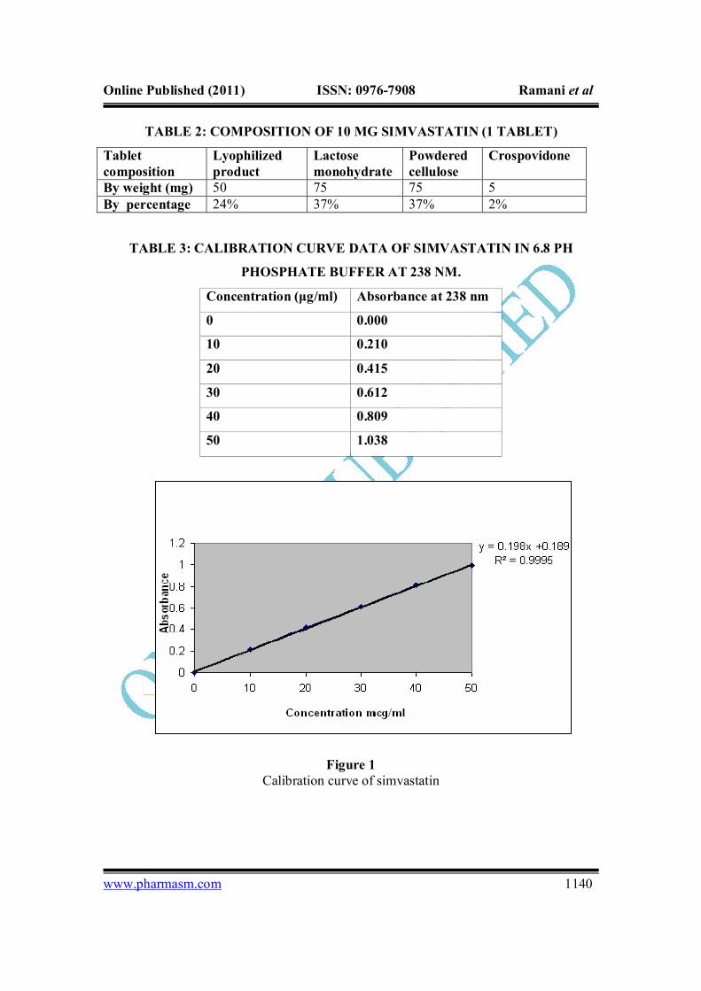
TABLE 4: SIMVASTATIN NANOPARTICLES ASSAY DATA
Theoretical drug content
Assayed drug content Formulations Surfactant
Amount (mg)
Expressed in %
Amount (mg)
Expressed in %
F1 Tween 80: PVP(1:1)
50 100 43.6 87.2
F4
Tween 80:soybean lecithin(1:1)
50 100 45.2 90.4
F7 PVP: soybean lecithin(1:1)
50 100 46.9 93.8
TABLE 5: SATURATION SOLUBILITY DATA OF BATCHES COMPARED
WITH PURE DRUG
Sr. No Formulation Amount per ml (µg) 1 Simvastatin 74.2 2 F1 163.8 3 F4 198.5 4 F7 275.3
TABLE 6: PARTICLE SIZE DATA OF THE FORMULATIONS
Formulations Average (nm)
F1 343 F4 220 F7 113 Simvastatin (pure drug) 28.3 µm
Online Published (2011) ISSN: 0976-7908 Ramani et al
www.pharmasm.com 1142
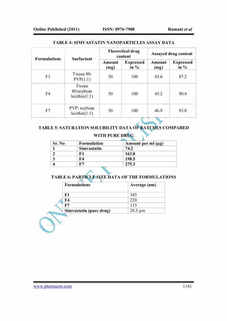
Figure 2 Particle Size Distribution for Pure Drug Simvastatin
Figure 3 Particle Size Distribution for F1
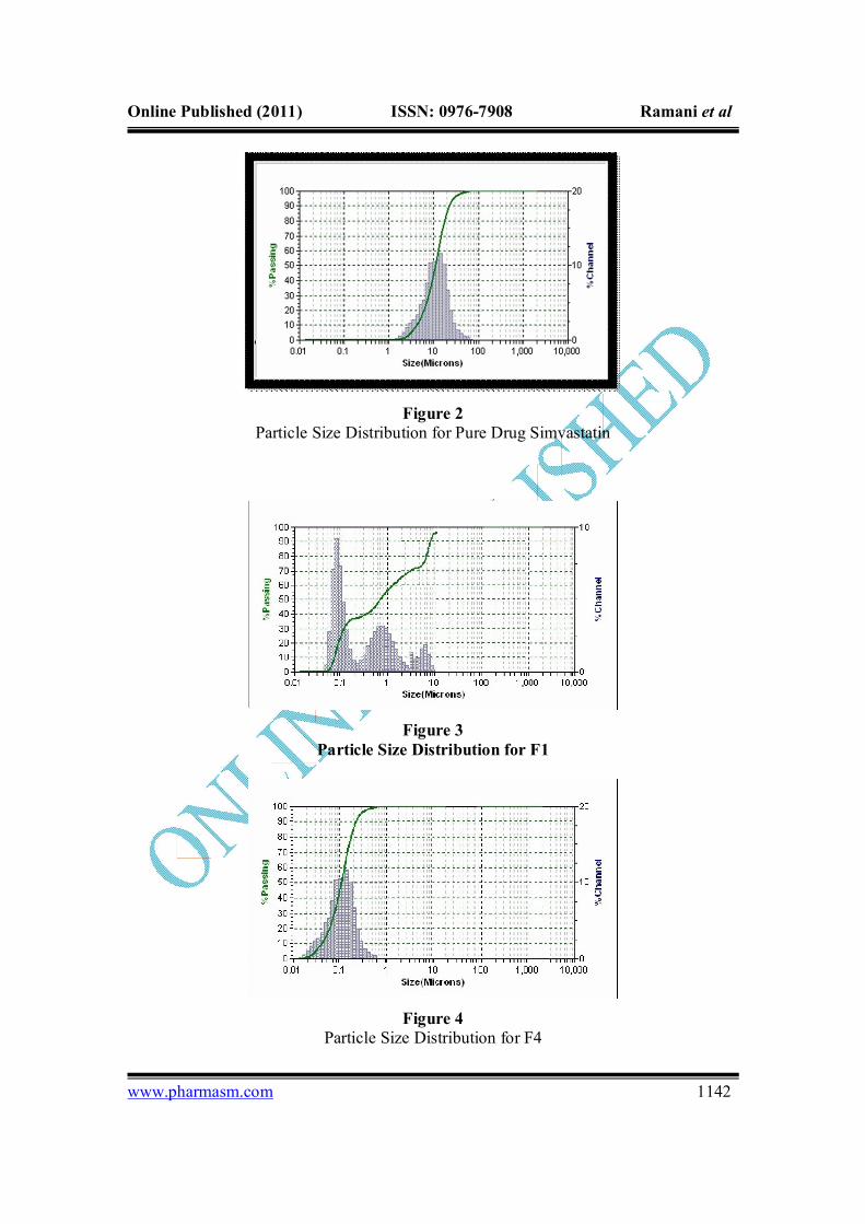
Figure 4 Particle Size Distribution for F4

Online Published (2011) ISSN: 0976-7908 Ramani et al
www.pharmasm.com 1143
Figure 5 Particle Size Distribution for F7
Figure 6 XRD Pattern of Pure Drug (Simvastatin)
Online Published (2011) ISSN: 0976-7908 Ramani et al
www.pharmasm.com 1144
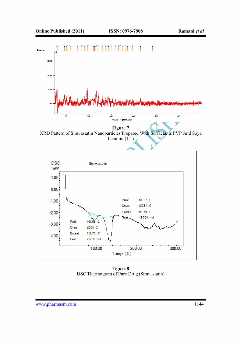
Figure 7 XRD Pattern of Simvastatin Nanoparticles Prepared With Surfactants PVP And Soya
Lecithin (1:1)
Figure 8
DSC Thermogram of Pure Drug (Simvastatin)

Online Published (2011) ISSN: 0976-7908 Ramani et al
www.pharmasm.com 1145
Figure 9 DSC Thermogram Simvastatin Nanoparticles
Figure 10 SEM Image of Raw Simvastatin Particles

Online Published (2011) ISSN: 0976-7908 Ramani et al
www.pharmasm.com 1146
Figure 11 SEM Image of Simvastatin Nanosuspension
Figure 12 SEM Image of Simvastatin Nanoparticle After Freeze Drying
Online Published (2011) ISSN: 0976-7908 Ramani et al
www.pharmasm.com 1147
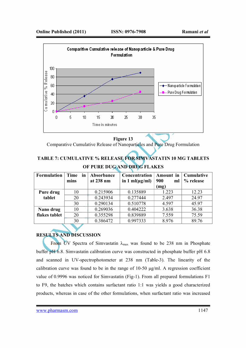
Figure 13 Comparative Cumulative Release of Nanoparticles and Pure Drug Formulation
TABLE 7: CUMULATIVE % RELEASE FOR SIMVASTATIN 10 MG TABLETS
OF PURE DUG AND DRUG FLAKES
Formulation Time in mins
Absorbance at 238 nm
Concentration in 1 ml(µg/ml)
Amount in 900 ml (mg)
Cumulative % release
10 0.215906 0.135889 1.223 12.23 20 0.243934 0.277444 2.497 24.97
Pure drug tablet
30 0.290134 0.510778 4.597 45.97 10 0.269036 0.404222 3.638 36.38 20 0.355298 0.839889 7.559 75.59
Nano drug flakes tablet
30 0.386472 0.997333 8.976 89.76
RESULTS AND DISCUSSION
From UV Spectra of Simvastatin λmax was found to be 238 nm in Phosphate
buffer pH 6.8. Simvastatin calibration curve was constructed in phosphate buffer pH 6.8
and scanned in UV-spectrophotometer at 238 nm (Table-3). The linearity of the
calibration curve was found to be in the range of 10-50 μg/ml. A regression coefficient
value of 0.9996 was noticed for Simvastatin (Fig-1). From all prepared formulations F1
to F9, the batches which contains surfactant ratio 1:1 was yields a good characterized
products, whereas in case of the other formulations, when surfactant ratio was increased

Online Published (2011) ISSN: 0976-7908 Ramani et al
www.pharmasm.com 1148
from 1:1 to 1:2 and onwards, not producing physically acceptable product. Even during
manufacturing process, the turbidity was also not disappeared, so those batches such as
F2, F3, F5, F6, F8 and F9 was not considered for the further study. The evaluation test
was carried out only for F1, F4 and F7 batches.
Nanoparticles of simvastatin were formulated by emulsification method followed
by solvent evaporation. The surfactants viz soya lecithin, PVP and Tween 80 in the
different ratio in Ethanol were tried in combination for optimization. From the Table 4
data it was clearly evident that the assayed drug content in the formulations was found to
be within the range of ± 10% of the theoretical amount. Assay data also indicates that the
method used for the formulation produced good yield and was suitable and reproducible
in nature.
The solubility data of pure and flakes of simvastatin particles are shown in Table
5. In the case of commercial simvastatin particles, the equilibrium solubility
(approximately 74.2µg/ml) was reached rapidly. In contrast, the maximum supersaturated
concentrations from nanoparticles were about 275.3µg/ml for particles prepared with
surfactants PVP and soya lecithin (1:1) at 0.2% concentration.
Pure simvastatin used for the study was characterized by relatively large particles
with average value of about 28.3 µm as reported .The nanoparticles prepared after
emulsification and solvent evaporation showed a drastic decrease in the particle size
when compared to the pure drug particles. The results shown in Table 6 that the
technique of Microemulsion method can be utilized as an effective tool in the reduction
of particle size to the nano scale range. As per Noyes-Whitney equation, the decrease in
the particle size will have a positive effect on the drug dissolution rate. Hence this
decrease in the particle size achieved will have a significant effect in the drug solubility
and dissolution characteristics.
XRD pattern of the pure drug and selected formulation are shown in fig 6 and fig
7. Characteristic diffraction peaks were observed for commercial simvastatin. On the
other hand, the nano formulations particles prepared with surfactants PVP and soya
lecithin (1:1) at 0.2% concentration i.e. (F7) was characterized by less intensity of the
diffraction peak when compared to that of simvastatin. This clearly indicates the
reduction in the crystallanity of the precipitated simvastatin nanoparticles.

Online Published (2011) ISSN: 0976-7908 Ramani et al
www.pharmasm.com 1149
The DSC curves of commercial simvastatin (fig 8) shows a broad endotherm
ranging from 30 to 120°C indicating the loss of water and the sharp endotherm at 138.97°
C might be due to the melting point of simvastatin. However no sharp endotherm was
seen at 139°C for the DSC curves of the nanoparticles (fig 9) prepared of PVP: soybean
lecithin (1:1) 0.2%. This shows the crystallinity of the drug has been reduced
significantly in the nanoparticles. The DSC results were in support of the XRD analysis
which also showed decrease in drug crystallinity.
The SEM images of pure drug particles and the nanoparticles are presented in fig
10, fig 11, and fig 12. While unprocessed simvastatin particles have appeared as
irregular- shaped crystals, a drastic change in the morphology and shape of drug was
observed for the nanoparticles prepared of PVP: soybean lecithin (1:1).
The dissolution test of tablets containing the nanometric drug flakes revealed that,
within 30 minutes, 89.76% (w/w) of the simvastatin in the tablet was dissolved. In
comparison, the dissolution test of the conventional tablets revealed that under these
testing conditions only 45.97% (w/w) simvastatin was dissolved (Table 7). This result
demonstrates the significant advantage of simvastatin nanoparticles over the conventional
particulate drug and the feasibility of the proposed method (Fig 13).
CONCLUSION
The prime objective of present work was accomplished by nanosuspension
technology using Soya lecithin with PVP and Tween 80 as surfactants. The method used
for the formulation was Emulsion-solvent evaporation followed by freeze drying. This
method can be used as an effective tool for preparation of nanosized formulations.
Simvastatin nanoparticles prepared by this method showed significant improvement in
aqueous solubility as well as dissolution characteristics which may significantly improve
its oral bioavailability. Further studies in animal models can be done to show the
effectiveness of prepared nanoparticles in-vivo.
REFERENCES
1. Carlucci G, Mazzeo P, Biordi L, Bologna M: Simultaneous determination of
simvastatin and its hydroxy acid form in human plasma by highperformance
liquid chromatography with UV detection. Journal of Pharmaceutical and
Biomedical Analysis 1992; 10: 693-697.

Online Published (2011) ISSN: 0976-7908 Ramani et al
www.pharmasm.com 1150
2. O'Neil MJ: The Merck index—an encyclopedia of chemicals, drugs and
biologicals 2006; 14: 1471-1472.
3. Patel RP, Patel MM: Inclusion complexion—physico-chemical characterization
and in vitro dissolution behavior of simvastatin-cyclodextrin inclusion
compounds. Drug Delivery Technology 2007; 7: 50-56.
4. Magdassi S and Margulis-Goshen K: Formation of simvastatin nanoparticles from
microemulsion. Nanotechnology Biology and Medicine in Nanomedicine. 2009; 5:
274–281.
5. Holister P, Weener J-W, Harper T: Nanopartical technology white papers.
Nanoparticles. 2003; 3: 1-11.
6. Kim M-S, Jin S-J, Kim J-S, Park HJ, Song H-S, Neubertb RHH, Hwang S-J:
Preparation, characterization and in vivo evaluation of amorphous atorvastatin
calcium nanoparticles using supercritical antisolvent (SAS) process. Europian
Journal of Pharmaceutics and Biopharmaceutics 2008; 69: 454-465.
7. Lei W and Asgharnejad M: Second Derivative UV Spectrometric determination
of simvastatin in its tablet dosage form. Journal of Pharmaceutical and
Biomedical Analysis 2000; 21: 1243-1248
8. Arunkumar N, Deecaraman M, Rani C: Nanosuspension technology and its
applications in drug delivery. Asian Journal of Pharmaceutics 2009; 3: 168-173.
9. Hecq J, Deleers M, Fanara D, Vrandex H, Amighi K: Preparation and
characterization of nanocrystals for solubility and dissolution rate enhancement of
nifedipine. International Journal of Pharmaceutics 2005; 299: 167-177.
10. Arunkumar N, Deecaraman M, Rani C, Mohanraj K P, Venkateskumar K:
Preparation and solid state characterization of atorvastatin nanosuspensions for
enhanced solubility and dissolution. International Journal of PharmTech
Research 2009; 1: 1725-1730.
11. Yuichi T, Atsutoshi I, Hiiroko S, Toshio O, Keiji Y: Characterization and
quantitation of Clarithromycin polymorphs by powder X-Ray diffractometry and
solid state NMR spectroscopy. Chemical & Pharmaceutical Bulletin 2002; 50:
1128-1130.
12. Kocbek P, Baumgartner S, Kristi J: Preparation and evaluation of
nanosuspensions for enhancing dissolution of poorly soluble drugs. International
Journal of Pharmaceutics 2006; 312(1-2):179-186.
Online Published (2011) ISSN: 0976-7908 Ramani et al
www.pharmasm.com 1151
13. Kristl J, Baumgartner S, Planinsˇek O, Dolenc A: Advantages of celecoxib
nanosuspension formulation and transformation into tablets. International Journal
of Pharmaceutics 2009; 376(1-2): 204-212.
14. Brahmankar DM, Jaiswal SB: Have described the various important properties of
drug that affect drug dissolution. Biophamaceutics and pharmacokinetics 2001:
25-31.
15. Muller RH and Keck CM: Drug nanocrystals of poorly soluble drugs produced by
high pressure homogenization. Europian Journal of Pharmaceutics and
Biopharmaceutics 2006; 62: 3–16.
16. Solans C, Izquierdo P, Nolla J, Azemar N, Garcia-Celma MJ: Nano-emulsions.
Current Opinion in Colloid & Interface Science 2005; 10: 102 – 110.
17. Carlucci G, Mazzeo P, Biordi L, Bologna M: Simultaneous determination of
simvastatin and its hydroxy acid form in human plasma by highperformance
liquid chromatography with UV detection. Journal of Pharmaceutical and
Biomedical Analysis 1992; 10: 693-697.
18. Hwang S-J, Jun SW, Kim M-S, Kim J-S, Park HJ, Lee S, Woo J-S: Preparation
and characterization of simvastatin/hydroxypropyl-b-cyclodextrin inclusion
complex using supercritical antisolvent (SAS) process. Europian Journal of
Pharmaceutics and Biopharmaceutics 2007; 66: 413–421.
19. Verma S, Lan Y, Gokhale R, Diane J: Quality by Design Approach to Understand
the Process of Nanosuspension Preparation, International Journal of
Pharmaceutics 2009; 377(1-2): 185-198.
20. Lei W and Asgharnejad M: Second Derivative UV Spectrometric determination
of simvastatin in its tablet dosage form. Journal of Pharmaceutical and
Biomedical Analysis 2000; 21: 1243-1248
21. Kocbek P, Baumgartner S, Kristi J: Preparation and evaluation of
nanosuspensions for enhancing dissolution of poorly soluble drugs. International
Journal of Pharmaceutics 2006; 312(1-2): 179-186.
For Correspondence: Vinod Ramani Department of Pharmacy, Sumandeep Vidyapeeth University, Baroda, India Email: [email protected]