The Catabolite Control Protein E (CcpE) Affects Virulence Determinant Production and Pathogenesis of...
26
Virginie Molle and Markus Bischoff Bals, Greg Somerville, Mathias Herrmann, Sunderkötter, Christoph Beisswenger, Robert Schmidt-Hohagen, Rosmarie Gaupp, Cord Christiane Wolz, Janina Eisenbeis, Kerstin Nippe, Meike Voss, Bettina Schulthess, Torsten Hartmann, Grègory Baronian, Nadine and pathogenesis of Staphylococcus aureus affects virulence determinant production The catabolite control protein E (CcpE) Microbiology: published online September 5, 2014 J. Biol. Chem. 10.1074/jbc.M114.584979 Access the most updated version of this article at doi: . JBC Affinity Sites Find articles, minireviews, Reflections and Classics on similar topics on the Alerts: When a correction for this article is posted • When this article is cited • to choose from all of JBC's e-mail alerts Click here http://www.jbc.org/content/early/2014/09/05/jbc.M114.584979.full.html#ref-list-1 This article cites 0 references, 0 of which can be accessed free at at UNIV OF NEBRASKA - Lincoln on September 15, 2014 http://www.jbc.org/ Downloaded from at UNIV OF NEBRASKA - Lincoln on September 15, 2014 http://www.jbc.org/ Downloaded from
-
Upload
uni-tuebingen1 -
Category
Documents
-
view
0 -
download
0
Transcript of The Catabolite Control Protein E (CcpE) Affects Virulence Determinant Production and Pathogenesis of...

Virginie Molle and Markus BischoffBals, Greg Somerville, Mathias Herrmann, Sunderkötter, Christoph Beisswenger, RobertSchmidt-Hohagen, Rosmarie Gaupp, Cord Christiane Wolz, Janina Eisenbeis, KerstinNippe, Meike Voss, Bettina Schulthess, Torsten Hartmann, Grègory Baronian, Nadine and pathogenesis of Staphylococcus aureusaffects virulence determinant production The catabolite control protein E (CcpE)Microbiology:
published online September 5, 2014J. Biol. Chem.
10.1074/jbc.M114.584979Access the most updated version of this article at doi:
.JBC Affinity SitesFind articles, minireviews, Reflections and Classics on similar topics on the
Alerts:
When a correction for this article is posted•
When this article is cited•
to choose from all of JBC's e-mail alertsClick here
http://www.jbc.org/content/early/2014/09/05/jbc.M114.584979.full.html#ref-list-1
This article cites 0 references, 0 of which can be accessed free at
at UN
IV O
F NE
BR
ASK
A - L
incoln on September 15, 2014
http://ww
w.jbc.org/
Dow
nloaded from
at UN
IV O
F NE
BR
ASK
A - L
incoln on September 15, 2014
http://ww
w.jbc.org/
Dow
nloaded from

1
The catabolite control protein E (CcpE) affects virulence determinant production and pathogenesis of Staphylococcus aureus
Torsten Hartmann1, Grégory Baronian2, Nadine Nippe3, Meike Voss4, Bettina Schulthess5, Christiane Wolz6, Janina Eisenbeis1, Kerstin Schmidt-Hohagen7, Rosmarie Gaupp1, Cord
Sunderkötter8, Christoph Beisswenger4, Robert Bals4, Greg Somerville9, Mathias Herrmann1, Virginie Molle2, and Markus Bischoff1,*
1Institute of Medical Microbiology and Hygiene, University of Saarland, Homburg/Saar, Germany 2Laboratoire de Dynamique des Interactions Membranaires Normales et Pathologiques, Université Montpellier 2, CNRS, UMR 5235, Montpellier, France 3Institute of Immunology, University of Münster, Münster, Germany 4Department of Internal Medicine V–Pulmonology, Allergology and Critical Care Medicine, Saarland University Medical Centre, Homburg/Saar, Germany 5Institute of Medical Microbiology, University of Zürich, Zürich, Switzerland 6Institute of Medical Microbiology and Hygiene, University Hospital of Tübingen, Tübingen, Germany 7Institute for Biochemistry, Biotechnology and Bioinformatics, Technische Universität Braunschweig, Braunschweig, Germany 8Department of Dermatology, University of Münster, Münster, Germany 9School of Veterinary Medicine and Biomedical Sciences, University of Nebraska, Lincoln, Nebraska, USA
*Corresponding authors: MB: Tel: (+49) 6841 162 39 63, Fax: (+49) 6841 162 39 85, E-mail: [email protected]
Running title: Attenuation of pathogenesis by CcpE of S. aureus.
Keywords: Staphylococcus aureus, virulence factor production, regulation, pathogenesis, hla
Background: Carbon metabolism and virulence are often linked in pathogenic bacteria.
Results: Deletion of the catabolite control protein E (CcpE) affects the expression of virulence factors and pathogenicity of Staphylococcus aureus.
Conclusion: Our data suggest that CcpE acts as an attenuator of virulence in S. aureus.
Significance: CcpE may serve to link S. aureus nutritional status to virulence determinant biosynthesis.
Carbon metabolism and virulence determinant production are often linked in pathogenic bacteria, and several regulatory elements have been reported to mediate this linkage in Staphylococcus aureus. Previously, we described a novel protein, catabolite control protein E (CcpE) that functions as regulator of the tricarboxylic acid cycle. Here we demonstrate that CcpE also regulates virulence
determinant biosynthesis and pathogenesis. Specifically, deletion of ccpE in S. aureus strain Newman revealed that CcpE affects transcription of virulence factors such as capA, the first gene in the capsule biosynthetic operon; hla, encoding α-toxin; and psmα , encoding the phenol soluble modulin cluster α . Electrophoretic mobility shift assays demonstrated that CcpE binds to the hla promoter. Mice challenged with S. aureus strain Newman or its isogenic ΔccpE derivative revealed increased disease severity in the ΔccpE mutant using two animal models; an acute lung infection model and a skin infection model. Complementation of the mutant with the ccpE wild-type allele restored all phenotypes, demonstrating that CcpE is negative regulator of virulence in S. aureus.
Carbon catabolite repression is a common mechanism utilized by bacteria to optimize their transcriptomes in response to the availability of carbon sources [reviewed in (1)]. Similarly, many
http://www.jbc.org/cgi/doi/10.1074/jbc.M114.584979The latest version is at JBC Papers in Press. Published on September 5, 2014 as Manuscript M114.584979
Copyright 2014 by The American Society for Biochemistry and Molecular Biology, Inc.
at UN
IV O
F NE
BR
ASK
A - L
incoln on September 15, 2014
http://ww
w.jbc.org/
Dow
nloaded from

2
pathogenic bacteria use this same mechanism to link the nutritional status with the transcription of virulence factors [reviewed in (2)]. In Staphylococcus aureus, carbon catabolite repression is mediated by several regulators such as the catabolite control protein A (CcpA), a glucose-responsive member of the LacI/GalR family of transcriptional regulators (3), CodY, a pleiotropic repressor that responds to GTP and branched-chain amino acids (BCAA) (4), and RpiRc, a putative ribose-responsive regulator that belongs to the RpiR family of transcriptional regulators (5). Recently, we identified CcpE as another potential carbon catabolite responsive element of S. aureus that controls transcription of tricaboxylic acid (TCA) cycle genes (6). In addition to regulating metabolism, CcpA, CodY, and RpiRc also regulate virulence factor expression (3-5).
CcpA regulates the expression of exotoxins, such as α-toxin (encoded by hla) and toxic shock syndrome toxin-1 (encoded by tst), and capsule formation in a glucose-responsive manner (3,7,8). In addition, CcpA promotes biofilm formation under in vitro conditions (9) and alters antibiotic susceptibility in methicillin-resistant S. aureus (MRSA) and glycopeptide intermediary resistant S. aureus (GISA) (3). More recently, CcpA was reported to mediate proline and arginine auxotrophies during in vitro growth (10,11), and to contribute to infectivity of S. aureus in a murine model of staphylococcal abscess formation (10).
CodY in S. aureus regulates the expression of virulence factors such as the cap operon (encoding proteins required for capsule biosynthesis), coa (encoding coagulase), fnbA (encoding fibronectin binding protein A), hla, icaADBC (encoding factors required for synthesis of polysaccharide intercellular adhesin), and spa (encoding protein A) (4,12,13). Although inactivation of codY did not markedly affect infectivity of S. aureus strain Newman in a murine abscess model, it restored the virulence of a mutant lacking the major (p)ppGpp synthase/hydrolase enzyme RSH to wild-type levels, suggesting that RSH-dependent derepression of CodY-regulated genes is important for virulence of S. aureus (14). More recently, it was found that inactivation of codY decreased the infectivity of the community-associated MRSA (CA-MRSA) USA300 isolate 923 in two murine infection models (15).
The pentose phosphate pathway (PPP)- regulator RpiRc alters the synthesis of several virulence factors such as protein A, capsular polysaccharide, and hemolysins, to decrease the
transcription of RNAIII, the regulatory RNA of the agr locus and a major regulator of virulence factor production in S. aureus, and to promote biofilm formation under in vitro conditions (5). These observations on RpiRc suggest that this regulator might also affect virulence in vivo; however, this has not been tested.
CcpE directly affects transcription of the aconitase-encoding gene citB, increases tricarboxylic acid (TCA) cycle activity during in vitro growth (6), and decreases pigment production in S. aureus (16). Since both TCA cycle activity (17-22) and pigment production (16,23,24) affect virulence determinant synthesis and/or infectivity of S. aureus, it is likely that CcpE modulates the expression of virulence factors and pathogenicity in this medically important pathogen. To test this hypothesis, we assessed the effect of ccpE deletion in S. aureus strain Newman (25) on the transcription of select virulence factors and on its role in infectivity using two unrelated murine infection models. Our data demonstrate that CcpE affects the transcription of virulence determinants, and infectivity of S. aureus in both in vivo infection models. EXPERIMENTAL PROCEDURES Bacterial strains and culture conditions. The bacterial strains and plasmids used in this study are listed in Table 1. Strains were grown in Luria-Bertani Lennox (LB-L) medium (BD, Heidelberg, Germany) at 37°C and aerated at 230 rpm with a flask-to-medium volume ratio of 10:1. The ΔccpE mutants HOM 354 and HOM 355 were obtained by phage-transducing the lox66-aphAIII-lox71–tagged ccpE deletion of THa (6) into strains 923 (26) and SA564 (27), respectively. Transcriptional analyses. For Northern blot experiments, overnight cultures of S. aureus were diluted to an A600 of 0.05 into fresh pre-warmed LB-L and grown at 37°C with 230 rpm of aeration. Samples were removed from the cultures at the indicated times and centrifuged at 9,000 × g and 4°C for 2 min, the culture supernatants were discarded, and the cell pellets were snap frozen in liquid nitrogen. Total RNAs were isolated according to (28), and blotting, hybridization, and labeling were performed as described (29). Primer pairs hla-F/hla-R and RNAIII-F/RNAIII-R (Table 2) were used to generate digoxigenin-labeled hla- and RNAIII-specific probes by PCR labeling, respectively.
For the quantification of transcripts by real-time reverse transcription PCR (qRT-PCR), RNA
at UN
IV O
F NE
BR
ASK
A - L
incoln on September 15, 2014
http://ww
w.jbc.org/
Dow
nloaded from

3
isolations and qRT-PCRs were carried out essentially as described (30). The cDNA (20 ng per reaction) was used for real-time amplification using primers listed in Table 2. mRNA levels were normalized against the mRNA level of gyrB, which is constitutively expressed under the conditions analyzed (31). The amounts of transcripts were expressed as the n-fold difference relative to the control gene (2-ΔCT, where ΔCT represents the difference in threshold cycle between the target and control genes). Electrophoretic mobility shift assays. DNA probes for electrophoretic mobility shift assays (EMSAs) were generated by PCR using S. aureus strain Newman chromosomal DNA as a template, and primer pairs (Table 2) that amplified the DNA regions preceding the capA, hla, hld, purA, and psmα ORFs. The 5’-ends of the double-stranded PCR products were labeled using [γ-32P] ATP and T4 polynucleotide kinase. A typical assay mixture contained (in 20 µl) 10 mM Tris-HCl, pH 7.5; 50 mM KCl; 1 mM dithiothreitol; 5 mM MgCl2; 0.1 µg of nonspecific competitor (poly(dI-dC)); 2,5% (v/v) glycerol, 0,05% (v/v) Igepal; radioactive DNA probe (2000 cpm ml-1) and various amounts (0, 15, 65, 130, and 200 nM) of purified CcpE. After 20 min of incubation at room temperature, 20 µl of this mixture was loaded into a native 5% (w/v) polyacrylamide Tris borate-EDTA Ready Gel (Bio-Rad) and electrophoresed in 1% Tris borate-EDTA (v/v) buffer for 1 h at 100 V cm-1. Radioactive species were detected by autoradiography using direct exposure to films. Radioactivity labeled promoter probes shifting with CcpE were additionally coincubated with increasing amounts of a nonspecific promoter probe and cold competitor, respectively, to demonstrate specificity of the shifting reaction. Luciferase assay. For assaying luciferase activities of S. aureus cells harboring plasmid pSB20235 (32), bacteria were cultivated in LB-L supplemented with 10 µg ml-1 chloramphenicol. Luciferase measurements were carried out essentially as described (33). 200 µl samples of the cell suspensions were removed at the time points indicated, and transferred to the wells of a 96-well clear-bottomed black plate (Greiner). The plate was placed in a Wallac Victor2 1420 Multilabel Counter (Perkin Elmer), and luminescence readings were taken for 5 s at 37°C. Capsular polysaccharide 5 (CP-5) production. CP-5 production was determined by indirect immunofluorescence as described (3), using mouse immunoglobulin M monoclonal antibodies
to CP-5 (34). Quantification of CP-5-positive cells was done by determining the numbers of 4’,6-diamidino-2-phenylindole (DAPI) and CY-3-positive cells using the software program CellC (35) (Institute of Signal Processing, Tampere University of Technology, Finland). Immune fluorescence intensities were analyzed using the MetaVue™ Research Imaging System (Molecular Devices). Briefly, from each image 80 bacteria detected by DAPI were randomly selected and the regions transferred to the corresponding CY-3 stained image. Intensities of the single bacteria were measured and the distribution of intensities analyzed by the software program GraphPad Prsim (GraphPad Software, Inc.). Pigment measurements. Bacteria were harvested after 24 h of growth on tryptic soy agar (TSA) and carotenoids were extracted as described (36). The optical densities at 465 nm of the methanol extracts were measured and normalized in reference to the values obtained with the wild-type extracts, which were set at 100. Animal models. Eight weeks old female C57BL/6N mice were purchased from Charles River Laboratories (Sulzfeld, Germany) and kept under specific pathogen-free conditions according to the regulations of German veterinary law. All animal studies were performed with the approval of the local State Review Boards.
The murine lung infection model was done essentially as described (37). Eight weeks old C57BL/6N mice were slightly anesthetized by intraperitoneal injection of 2.6 mg of ketaminhydrochloride (Pfizer, Berlin, Germany) and 0.18 mg of xylazinhydrochloride (Bayer, Leverkusen, Germany) per mouse and infected intranasally with 5 × 10⁷ colony forming units (CFU) of S. aureus. Twenty-four hours post infection, the animals were euthanized, the tracheae were cannulated and a bronchoalveolar lavage was performed (three times with 1 ml of phosphate buffered saline [PBS]). The bronchoalveolar lavage fluid (BALF) was centrifuged at 300 × g for 10 min at 4°C to obtain alveolar cells, which were suspended in 1 ml of PBS. Total cell numbers in BALF were determined using a Neubauer hemocytometer. To identify the bacterial load of the lungs 24 h post infection, whole lungs were homogenized in 1 ml of PBS, and serial dilutions plated onto sheep blood agar (SBA). CFU were counted after incubation overnight at 37°C.
The footpad swelling model was carried out as described (38). Age-matched mice were inoculated subcutaneously with 1 × 10⁷ CFU of S.
at UN
IV O
F NE
BR
ASK
A - L
incoln on September 15, 2014
http://ww
w.jbc.org/
Dow
nloaded from

4
aureus into the left hind footpad, and footpad swelling was measured daily with a micrometric caliper in reference to the uninfected footpad. Cytokine determinations. Levels of murine interleukin-1β (IL-1β), keratinocyte-derived chemokine (KC), and granulocyte-colony stimulating factor (G-CSF) in cell-free BALFs and lung homogenates were determined by commercially available sandwich-type ELISAs, according to the manufacturer’s instructions (R&D Systems, Wiesbaden-Nordenstadt, Germany). Statistical analyses. Statistical significance was assessed using the Mann-Whitney U test. p values <0.05 were considered significant. RESULTS Influence of a ccpE deletion on RNAIII transcription. Given the importance of the agr locus for virulence determinant production in S. aureus [reviewed in (39,40)], we tested whether CcpE affects transcription of this regulatory system. Northern blot analysis revealed that all three strains, Newman, TH01, and TH01c, produced RNAIII transcripts in a growth phase-dependent manner, with a peak transcription rate at the transition from the exponential growth phase to post-exponential growth phase (i.e., 6 h) (Fig. 1). However, deletion of ccpE increased the post-exponential growth phase accumulation of RNAIII transcripts in TH01, suggesting that CcpE negatively affects RNAIII transcription. To test this suggestion, we transformed an RNAIII transcriptional reporter plasmid, pSB2035, (32) into strains Newman, TH01, and TH01c. This plasmid harbors a gfp-luxABCDE dual reporter system under the control of the RNAIII transcription-driving agr P3 promoter. Similar to the Northern blot data (Fig. 1B), luciferase activity assays revealed a growth phase-dependent transcription of RNAIII (Fig. 1C). In addition, we observed that deletion of ccpE increased transcription of RNAIII, confirming that the increased amount of RNAIII in the TH01 mutant is due to increased RNAIII transcription and not due to decreased RNAIII degradation. To test whether CcpE might exert this effect via direct binding to the agr P3 promoter, electrophoretic mobility shift assays (EMSA) were performed with purified CcpE and a radioactively labeled PCR-probe covering the agr P2/3 promoter (Fig. 1D). No mobility shifts were observed over a range of
protein concentrations, suggesting that CcpE does not directly interact with the agr P3 promoter to modulate RNAIII transcription.
CcpE directly influences hla transcription. α-toxin is a major virulence factor of S. aureus, and its synthesis is regulated at multiple levels, including transcriptional and post-transcriptional mechanisms (3,41-44). Regulation of hla transcription is also influenced by the carbon catabolite responsive elements CcpA and CodY (3,4,15,45); hence, we hypothesized that hla transcription might be regulated by CcpE as well. Support for this hypothesis can be seen in the Northern blot analysis of hla transcription (Fig. 2A), where hla mRNA levels are much greater in the ΔccpE mutant strain TH01 in all growth phases relative to the wild-type and the cis-complemented derivative strain TH01c. To quantify the effect of ccpE deletion on hla transcription, we performed qRT-PCRs on strains Newman, TH01, and TH01c throughout a complete growth cycle (Fig. 2B). Consistent with our Northern blot data, we observed a growth phase-dependent transcription of hla, with a peak in the post-exponential growth phase (9 h). Deletion of ccpE resulted in a massive up-regulation (30 to 60-fold) of hla transcription in strain TH01. Complementation of TH01 with a ccpE wild-type allele restored hla mRNA levels to those seen in the wild-type strain. To exclude that this effect of CcpE was specific for S. aureus strain Newman, we deleted ccpE in two genetically unrelated S. aureus strains, the CA-MRSA USA300 isolate 923 (26) and the low passage human isolate SA564 (27), and assessed hla transcription of these strain pairs (Fig. 2C). Deletion of ccpE again strongly increased the transcription of hla in both strains, suggesting that the repressive effect of CcpE on hla transcription is independent of the genetic background. To assess whether CcpE directly regulates transcription of hla, we performed EMSAs with the hla promoter as probe (Fig. 2D). A clear and dose-dependent shift of CcpE with the radioactively labeled hla promoter probe was observed, which was not affected by the addition of a nonspecific promoter probe (data not shown) but was invertable by adding excessive amounts of cold competitor, suggesting that CcpE directly controls transcription of hla. CcpE promotes capsule formation. Capsular polysaccharide is another important virulence factor of S. aureus, whose synthesis is intimately linked to the nutritional status of the bacterium (3,5,12). Our results (Fig. 3) demonstrate that in
at UN
IV O
F NE
BR
ASK
A - L
incoln on September 15, 2014
http://ww
w.jbc.org/
Dow
nloaded from

5
addition to CcpA, CodY, and RpiRc, CcpE also modulates transcription of the cap operon and the elaboration of a capsule. As expected, when S. aureus was cultivated in LB-L, the first gene of the cap operon (capA) was predominantly transcribed during the later stages of growth (Fig. 3A). Deletion of ccpE in TH01 strongly decreased accumulation of capA mRNA throughout the growth cycle. Cis-complementation of TH01 with the wild-type ccpE allele restored capA mRNA levels to those found in the isogenic wild-type strain Newman. Consistent with the transcriptional data, a reduced number of capsular polysaccharide positive cells was observed with the ΔccpE mutant (Fig. 3B). While about 69±6% of the Newman cells and 75±8% of the TH01c cells incubated with the CP-5 antibodies produced clear fluorescence signals after 24 h of growth in LB-L, in the TH01 cell pool only 35±7% of the cells emitted detectable amounts of fluorescence. Similarly, an approximately 10-fold decrease in the mean fluorescence intensity per cell were observed with the ΔccpE mutant (Fig. 3B) when compared to cells of the wild type and the complemented derivative TH01c. To determine if the CcpE-dependent regulation of capA was due to an interaction with the capA promoter, EMSAs were performed with CcpE and a radioactively labeled probe of the capA promoter. In contrast to the hla promoter, CcpE did not shift the capA promoter probe at any of the CcpE concentrations tested (Fig. 3C), indicating that CcpE indirectly influences cap operon transcription and capsule formation.
CcpE alters transcription of the phenol-soluble modulin α (psmα) cluster. Phenol-soluble modulins (PSM) are a small group of cytolytic and immunomodulating peptides that are important virulence determinants of S. aureus, especially in CA-MRSA USA300 isolates [reviewed in (46)]. The S. aureus Newman genome harbors two psm operons, psmα and psmβ, which are transcriptionally affected by regulators such as SarA and AgrA (40,47). To determine if CcpE influences psm transcription, we assessed psmα and psmβ transcription using qRT-PCR. Deletion of ccpE had a negligible effect on psmβ transcription (data not shown); however, we observed a significant reduction of psmα transcripts in strain TH01 compared to the wild-type strain (Fig. 4A). Complementation of TH01 with a ccpE wild-type allele restored psmα mRNA levels to that seen in the wild-type strain. EMSAs performed using CcpE and the psmα promoter as a probe failed to shift the radiolabeled
probe with any of the protein concentrations tested (Fig. 4B), suggesting an indirect effect of CcpE on psmα transcription. CcpE decreases pigment production. Most S. aureus strains produce the carotenoid pigment staphyloxanthin, which is responsible for the yellowish-orange appearance of this bacterium (48). In line with a previous publication (16), we noticed an increase in pigment production after 24 h of growth on tryptic soy agar, and this phenotype was reverted by introducing a functional ccpE into this mutant (Fig. 5). The synthesis of staphyloxanthin is encoded within the crtOPQMN operon (48); hence, to determine if CcpE affects transcription of crtOPQMN, we assessed crtM mRNA levels in strains Newman, TH01, and TH01c using qRT-PCR. Contrary to the findings reported by Lan and colleagues (16), our results suggest that transcription of crtOPQMN appears to be independent of CcpE (Fig. 5C). Similarly, inactivation of ccpE in strains 923 and SA564 significantly increased the pigment contents of mutant cells compared to wild-type, without affecting crtM transcription, suggesting that this phenomenon is not strain-dependent (Fig. 5). CcpE attenuates virulence in two murine infection models. Deletion of ccpE in S. aureus strain Newman augmented transcription of the global virulence regulator RNAIII (Fig. 1) and increased α-toxin (hla) mRNA (Fig. 2). Given the effect of CcpE on virulence factor transcription in vitro, we hypothesized that CcpE might alter infectivity of S. aureus in vivo. To address this hypothesis, we assessed the ability of the strains Newman, TH01, and TH01c to cause disease in two different murine infection models. In a murine pneumonia model, C57BL/6N mice were infected intranasally with either strain Newman, or TH01, or TH01c, and the bacterial load in the lungs and the total amount of eukaryotic cells in bronchoalveolar lavage fluids (BALFs) at 24 h post infection were determined (Fig. 6). Strain TH01 significantly increased the bacterial load in the lungs of mice relative to the wild-type and complemented strains (Fig. 6A). Similarly, we observed a significant increase in total cells in BALFs of the TH01 challenged mice (Fig. 6B), indicating a more severe infection. This increase in total cell numbers correlated with an increased number of neutrophils in BALFs of TH01 challenged mice (Fig. 6C), and this also correlated with increased concentrations of the neutrophil mobilization stimulating factor granulocyte colony-stimulating factor (G-CSF) (49) (Fig. 6D).
at UN
IV O
F NE
BR
ASK
A - L
incoln on September 15, 2014
http://ww
w.jbc.org/
Dow
nloaded from

6
Complementation of the ΔccpE mutant restored all virulence traits back to wild-type levels, confirming that all observed alterations were caused by CcpE. To exclude that this CcpE effect is specific for strain Newman, we additionally infected mice intranasally with strain SA564 and its ΔccpE derivative HOM 355, respectively. In line with our observations made with the strain triplet Newman/TH01/TH01c, we observed significantly increased CFU numbers in the lung tissues of mice that have been infected with the SA564 ccpE mutant (Fig. 7), demonstrating that this virulence diminishing effect of CcpE is not specific for strain Newman. To substantiate these findings in another in vivo model, we utilized a murine footpad infection model (38). In this model, bacteria are inoculated into the left hind footpad of mice and footpad swelling ratios are determined on a daily basis for up to 12 days (Fig. 8). Consistent with our observations using a lung infection model, we observed enhanced footpad swelling in mice challenged with the Newman ΔccpE mutant relative to the isogenic wild-type and complemented strains (Fig. 8). Swelling was most significantly increased early in the infection process (days 1 to 4) and in the later stages of the infection (days 8 to 12) when compared to the values obtained with the wild-type and TH01c challenged mice groups.
DISCUSSION
The nosocomial pathogen S. aureus is known to link its virulence factor production with central metabolic pathways (3-5,8,9,13,22). This linkage is mediated via at least three metabolite responsive regulators; namely, CcpA, (3,8), CodY (13), and RpiRc (5). Data presented here demonstrate that CcpE represents a fourth regulatory protein that connects virulence factor synthesis with central metabolism, specifically the TCA cycle (Fig. 9) (6).
While most effects of CcpE on virulence factor synthesis were indirect, possibly via regulation of TCA cycle activity (22,27,50), a direct link between CcpE and hla transcription was established. In a murine pneumonia model, α-toxin is a key virulence determinant involved in the pathogenesis of S. aureus (51,52); specifically, the level of α-toxin correlated with disease severity in this animal model (53). Mechanistically, α-toxin increases cytokine synthesis, enhances neutrophil recruitment, and stimulates the NLRP3 (NOD-like receptor family, pyrin domain containing 3) inflammasome in the
lungs, leading to a massive inflammatory response and tissue destruction (54,55). Consistent with these observations, deletion of ccpE increased hla transcription (Fig. 2) and increased the bacterial loads and neutrophil contents in the lungs of mice (Figs. 6 and 7), suggesting that CcpE might influence the virulence of S. aureus during lung infections via transcriptional regulation of hla.
In addition to directly interacting with hla, ccpE deletion increased RNAIII levels, which likely contributed to the altered pathogenesis of the ΔccpE mutant in both animal models. RNAIII is the RNA regulator of the agr locus encoded quorum sensing system [reviewed in (39,40)] and it codes for a small lytic peptide called δ-toxin, which is a chemoattractant for neutrophils (50). RNAIII is predominantly transcribed when a threshold level of bacteria is achieved (56,57). In its regulatory function, RNA promotes the expression of many exoproteins including α-toxin, either directly or via the control of a repressor protein known as Rot (43,58). Mutations in agr have been shown to attenuate virulence in several animal models (59-63) including murine models of pneumonia (52,64) and skin infections (65-67). When the peptide δ-toxin is translated from RNAIII, it is produced in two forms; one without an N-terminal formyl group on the methionine, and one containing a formylated methionine (50). Formylated δ-toxin is a potent neutrophil chemoattractant, suggesting that increased neutrophils in the lungs on TH01 infected mice may be due to an increase in δ-toxin synthesis.
Alterations in the synthesis of virulence factors and RNAIII will likely alter the immune response to the infection. The BALF cytokine profiles of mice infected with strains Newman, TH01, and TH01c were similar with respect to keratinocyte-derived chemokine and IL-1β, however, G-CSF was higher in BALFs and lung homogenates from TH01 challenged mice relative to mice infected with the wild-type strain. G-CSF was originally characterized in hematopoietic cells to stimulate the proliferation and differentiation of neutrophil granulocyte precursors. In addition, G-CSF functions to recruit polymorphonuclear leukocytes (PMNs) to the lung (68), and its expression in lung tissue is stimulated by microbial infections (69-72). Recently, Hua et al. (73) observed in a mouse pneumonia model that pre-immunization with an anti-α-toxin antibody significantly decreased the G-CSF contents in BALFs of mice infected with S. aureus. Based on this observation, it is reasonable to speculate that an increase in α-toxin synthesis (Fig. 2) would increase G-CSF
at UN
IV O
F NE
BR
ASK
A - L
incoln on September 15, 2014
http://ww
w.jbc.org/
Dow
nloaded from

7
production (Fig. 6D), resulting in an increase in neutrophil recruitment (Fig. 6C). Model of CcpE-mediated virulence regulation. Transcription of RNAIII is primarily promoted by AgrA, the response regulator of the two-component system encoded by the agr locus (74). In addition, AgrA also promotes transcription of the psm operons (40). Because we observed divergent effects of CcpE on RNAIII and psmα transcription (Figs. 1, 4), we can largely exclude that CcpE modulates RNAIII production via activation of AgrA. Similarly, the agr system promotes capsule synthesis (75-77); however, capA mRNA levels were decreased in the ΔccpE mutant despite an increase in RNAIII transcript levels (Figs. 1 and 3). Interestingly, Somerville and colleagues observed increased RNAIII levels and an impaired capsule biosynthesis in TCA cycle mutants in which the aconitase-encoding gene citB (syn. acnA) was inactivated (20,27), demonstrating a link between TCA cycle activity, capsule formation and RNAIII production. It is possible that CcpE modulates RNAIII transcription and capsule biosynthesis via regulation of TCA cycle activity. However, the effect of TCA cycle inactivation on capsule synthesis is tied to a lack of oxaloacetate for gluconeogenesis (20), and it is still unclear how TCA cycle activity affects transcription of RNAIII. A potential factor might be aconitase
itself. This key enzyme of TCA cycle is reported in B. subtilis to act as a bifunctional protein that possesses enzymatic activity and functions as an RNA-binding regulatory protein (78-80). Similar to B. subtilis, apo-aconitase binds to iron-responsive elements in mRNA (Somerville, unpublished data), raising the possibility of a direct interaction between aconitase and the highly structured RNAIII. Additionally, CcpE might affect RNAIII synthesis and capsule formation via pH alterations. We have recently shown that in vitro cultivation of the ccpE deletion mutant in LB-L led to a significantly reduced alkalinization of the culture medium during the later stages of growth (6-12 h) compared to the wild-type culture (6). Alkaline growth conditions were previously reported to repress RNAIII production (81), and to augment capsule formation (82,83), consistent with our findings of increased RNAIII transcription and decreased capA transcription in TH01 during the later growth stages in LB-L (Figs. 1 and 3).
In conclusion, CcpE modulates the
expression of several major virulence factors of S. aureus, which affects its pathogenesis. Given its mostly repressive effect on virulence determinant production, it can be assumed that CcpE serves as an attenuator of virulence in this clinically important pathogen.
REFERENCES
1. Görke, B., and Stülke, J. (2008) Carbon catabolite repression in bacteria: many ways to make the
most out of nutrients. Nature reviews. Microbiology 6, 613-624 2. Poncet, S., Milohanic, E., Maze, A., Nait Abdallah, J., Ake, F., Larribe, M., Deghmane, A. E., Taha,
M. K., Dozot, M., De Bolle, X., Letesson, J. J., and Deutscher, J. (2009) Correlations between carbon metabolism and virulence in bacteria. Contributions to microbiology 16, 88-102
3. Seidl, K., Stucki, M., Ruegg, M., Goerke, C., Wolz, C., Harris, L., Berger-Bachi, B., and Bischoff, M. (2006) Staphylococcus aureus CcpA affects virulence determinant production and antibiotic resistance. Antimicrobial agents and chemotherapy 50, 1183-1194
4. Majerczyk, C. D., Sadykov, M. R., Luong, T. T., Lee, C., Somerville, G. A., and Sonenshein, A. L. (2008) Staphylococcus aureus CodY negatively regulates virulence gene expression. Journal of bacteriology 190, 2257-2265
5. Zhu, Y., Nandakumar, R., Sadykov, M. R., Madayiputhiya, N., Luong, T. T., Gaupp, R., Lee, C. Y., and Somerville, G. A. (2011) RpiR homologues may link Staphylococcus aureus RNAIII synthesis and pentose phosphate pathway regulation. Journal of bacteriology 193, 6187-6196
6. Hartmann, T., Zhang, B., Baronian, G., Schulthess, B., Homerova, D., Grubmuller, S., Kutzner, E., Gaupp, R., Bertram, R., Powers, R., Eisenreich, W., Kormanec, J., Herrmann, M., Molle, V., Somerville, G. A., and Bischoff, M. (2013) Catabolite Control Protein E (CcpE) Is a LysR-type Transcriptional Regulator of Tricarboxylic Acid Cycle Activity in Staphylococcus aureus. The Journal of biological chemistry 288, 36116-36128
7. Seidl, K., Bischoff, M., and Berger-Bachi, B. (2008) CcpA mediates the catabolite repression of tst in Staphylococcus aureus. Infection and immunity 76, 5093-5099
at UN
IV O
F NE
BR
ASK
A - L
incoln on September 15, 2014
http://ww
w.jbc.org/
Dow
nloaded from

8
8. Seidl, K., Muller, S., Francois, P., Kriebitzsch, C., Schrenzel, J., Engelmann, S., Bischoff, M., and Berger-Bachi, B. (2009) Effect of a glucose impulse on the CcpA regulon in Staphylococcus aureus. BMC microbiology 9, 95
9. Seidl, K., Goerke, C., Wolz, C., Mack, D., Berger-Bachi, B., and Bischoff, M. (2008) Staphylococcus aureus CcpA affects biofilm formation. Infection and immunity 76, 2044-2050
10. Li, C., Sun, F., Cho, H., Yelavarthi, V., Sohn, C., He, C., Schneewind, O., and Bae, T. (2010) CcpA mediates proline auxotrophy and is required for Staphylococcus aureus pathogenesis. Journal of bacteriology 192, 3883-3892
11. Nuxoll, A. S., Halouska, S. M., Sadykov, M. R., Hanke, M. L., Bayles, K. W., Kielian, T., Powers, R., and Fey, P. D. (2012) CcpA regulates arginine biosynthesis in Staphylococcus aureus through repression of proline catabolism. PLoS pathogens 8, e1003033
12. Majerczyk, C. D., Dunman, P. M., Luong, T. T., Lee, C. Y., Sadykov, M. R., Somerville, G. A., Bodi, K., and Sonenshein, A. L. (2010) Direct targets of CodY in Staphylococcus aureus. Journal of bacteriology 192, 2861-2877
13. Pohl, K., Francois, P., Stenz, L., Schlink, F., Geiger, T., Herbert, S., Goerke, C., Schrenzel, J., and Wolz, C. (2009) CodY in Staphylococcus aureus: a regulatory link between metabolism and virulence gene expression. Journal of bacteriology 191, 2953-2963
14. Geiger, T., Goerke, C., Fritz, M., Schafer, T., Ohlsen, K., Liebeke, M., Lalk, M., and Wolz, C. (2010) Role of the (p)ppGpp synthase RSH, a RelA/SpoT homolog, in stringent response and virulence of Staphylococcus aureus. Infection and immunity 78, 1873-1883
15. Montgomery, C. P., Boyle-Vavra, S., Roux, A., Ebine, K., Sonenshein, A. L., and Daum, R. S. (2012) CodY deletion enhances in vivo virulence of community-associated methicillin-resistant Staphylococcus aureus clone USA300. Infection and immunity 80, 2382-2389
16. Lan, L., Cheng, A., Dunman, P. M., Missiakas, D., and He, C. (2010) Golden pigment production and virulence gene expression are affected by metabolisms in Staphylococcus aureus. Journal of bacteriology 192, 3068-3077
17. Chatterjee, I., Herrmann, M., Proctor, R. A., Peters, G., and Kahl, B. C. (2007) Enhanced post-stationary-phase survival of a clinical thymidine-dependent small-colony variant of Staphylococcus aureus results from lack of a functional tricarboxylic acid cycle. Journal of bacteriology 189, 2936-2940
18. Gaupp, R., Schlag, S., Liebeke, M., Lalk, M., and Gotz, F. (2010) Advantage of upregulation of succinate dehydrogenase in Staphylococcus aureus biofilms. Journal of bacteriology 192, 2385-2394
19. Massilamany, C., Gangaplara, A., Gardner, D. J., Musser, J. M., Steffen, D., Somerville, G. A., and Reddy, J. (2011) TCA cycle inactivation in Staphylococcus aureus alters nitric oxide production in RAW 264.7 cells. Molecular and cellular biochemistry 355, 75-82
20. Sadykov, M. R., Mattes, T. A., Luong, T. T., Zhu, Y., Day, S. R., Sifri, C. D., Lee, C. Y., and Somerville, G. A. (2010) Tricarboxylic acid cycle-dependent synthesis of Staphylococcus aureus Type 5 and 8 capsular polysaccharides. Journal of bacteriology 192, 1459-1462
21. Sheldon, J. R., Marolda, C. L., and Heinrichs, D. E. (2014) TCA cycle activity in Staphylococcus aureus is essential for iron-regulated synthesis of staphyloferrin A, but not staphyloferrin B: the benefit of a second citrate synthase. Molecular microbiology
22. Zhu, Y., Xiong, Y. Q., Sadykov, M. R., Fey, P. D., Lei, M. G., Lee, C. Y., Bayer, A. S., and Somerville, G. A. (2009) Tricarboxylic acid cycle-dependent attenuation of Staphylococcus aureus in vivo virulence by selective inhibition of amino acid transport. Infection and immunity 77, 4256-4264
23. Liu, C. I., Liu, G. Y., Song, Y., Yin, F., Hensler, M. E., Jeng, W. Y., Nizet, V., Wang, A. H., and Oldfield, E. (2008) A cholesterol biosynthesis inhibitor blocks Staphylococcus aureus virulence. Science (New York, N.Y.) 319, 1391-1394
24. Liu, G. Y., Essex, A., Buchanan, J. T., Datta, V., Hoffman, H. M., Bastian, J. F., Fierer, J., and Nizet, V. (2005) Staphylococcus aureus golden pigment impairs neutrophil killing and promotes virulence through its antioxidant activity. The Journal of experimental medicine 202, 209-215
25. Duthie, E. S. (1952) Variation in the antigenic composition of staphylococcal coagulase. Journal of general microbiology 7, 320-326
26. Boyle-Vavra, S., Yin, S., and Daum, R. S. (2006) The VraS/VraR two-component regulatory system required for oxacillin resistance in community-acquired methicillin-resistant Staphylococcus aureus. FEMS microbiology letters 262, 163-171
at UN
IV O
F NE
BR
ASK
A - L
incoln on September 15, 2014
http://ww
w.jbc.org/
Dow
nloaded from

9
27. Somerville, G. A., Chaussee, M. S., Morgan, C. I., Fitzgerald, J. R., Dorward, D. W., Reitzer, L. J., and Musser, J. M. (2002) Staphylococcus aureus aconitase inactivation unexpectedly inhibits post-exponential-phase growth and enhances stationary-phase survival. Infection and immunity 70, 6373-6382
28. Cheung, A. L., Eberhardt, K. J., and Fischetti, V. A. (1994) A method to isolate RNA from gram-positive bacteria and mycobacteria. Analytical biochemistry 222, 511-514
29. McCallum, N., Karauzum, H., Getzmann, R., Bischoff, M., Majcherczyk, P., Berger-Bachi, B., and Landmann, R. (2006) In vivo survival of teicoplanin-resistant Staphylococcus aureus and fitness cost of teicoplanin resistance. Antimicrobial agents and chemotherapy 50, 2352-2360
30. Chatterjee, I., Becker, P., Grundmeier, M., Bischoff, M., Somerville, G. A., Peters, G., Sinha, B., Harraghy, N., Proctor, R. A., and Herrmann, M. (2005) Staphylococcus aureus ClpC is required for stress resistance, aconitase activity, growth recovery, and death. Journal of bacteriology 187, 4488-4496
31. Valihrach, L., and Demnerova, K. (2012) Impact of normalization method on experimental outcome using RT-qPCR in Staphylococcus aureus. Journal of microbiological methods 90, 214-216
32. Qazi, S. N., Counil, E., Morrissey, J., Rees, C. E., Cockayne, A., Winzer, K., Chan, W. C., Williams, P., and Hill, P. J. (2001) agr expression precedes escape of internalized Staphylococcus aureus from the host endosome. Infection and immunity 69, 7074-7082
33. Schmitt, J., Joost, I., Skaar, E. P., Herrmann, M., and Bischoff, M. (2012) Haemin represses the haemolytic activity of Staphylococcus aureus in an Sae-dependent manner. Microbiology (Reading, England) 158, 2619-2631
34. Hoeger, P. H., Lenz, W., Boutonnier, A., and Fournier, J. M. (1992) Staphylococcal skin colonization in children with atopic dermatitis: prevalence, persistence, and transmission of toxigenic and nontoxigenic strains. The Journal of infectious diseases 165, 1064-1068
35. Selinummi, J., Seppala, J., Yli-Harja, O., and Puhakka, J. A. (2005) Software for quantification of labeled bacteria from digital microscope images by automated image analysis. BioTechniques 39, 859-863
36. Morikawa, K., Maruyama, A., Inose, Y., Higashide, M., Hayashi, H., and Ohta, T. (2001) Overexpression of sigma factor, sigma(B), urges Staphylococcus aureus to thicken the cell wall and to resist beta-lactams. Biochemical and biophysical research communications 288, 385-389
37. Seiler, F., Hellberg, J., Lepper, P. M., Kamyschnikow, A., Herr, C., Bischoff, M., Langer, F., Schafers, H. J., Lammert, F., Menger, M. D., Bals, R., and Beisswenger, C. (2013) FOXO transcription factors regulate innate immune mechanisms in respiratory epithelial cells. Journal of immunology (Baltimore, Md. : 1950) 190, 1603-1613
38. Nippe, N., Varga, G., Holzinger, D., Loffler, B., Medina, E., Becker, K., Roth, J., Ehrchen, J. M., and Sunderkotter, C. (2011) Subcutaneous infection with S. aureus in mice reveals association of resistance with influx of neutrophils and Th2 response. The Journal of investigative dermatology 131, 125-132
39. Pragman, A. A., and Schlievert, P. M. (2004) Virulence regulation in Staphylococcus aureus: the need for in vivo analysis of virulence factor regulation. FEMS immunology and medical microbiology 42, 147-154
40. Queck, S. Y., Jameson-Lee, M., Villaruz, A. E., Bach, T. H., Khan, B. A., Sturdevant, D. E., Ricklefs, S. M., Li, M., and Otto, M. (2008) RNAIII-independent target gene control by the agr quorum-sensing system: insight into the evolution of virulence regulation in Staphylococcus aureus. Molecular cell 32, 150-158
41. Cheung, A. L., and Ying, P. (1994) Regulation of alpha- and beta-hemolysins by the sar locus of Staphylococcus aureus. Journal of bacteriology 176, 580-585
42. Ingavale, S., van Wamel, W., Luong, T. T., Lee, C. Y., and Cheung, A. L. (2005) Rat/MgrA, a regulator of autolysis, is a regulator of virulence genes in Staphylococcus aureus. Infection and immunity 73, 1423-1431
43. Morfeldt, E., Taylor, D., von Gabain, A., and Arvidson, S. (1995) Activation of alpha-toxin translation in Staphylococcus aureus by the trans-encoded antisense RNA, RNAIII. The EMBO journal 14, 4569-4577
44. Oscarsson, J., Kanth, A., Tegmark-Wisell, K., and Arvidson, S. (2006) SarA is a repressor of hla (alpha-hemolysin) transcription in Staphylococcus aureus: its apparent role as an activator of hla in the prototype strain NCTC 8325 depends on reduced expression of sarS. Journal of bacteriology 188, 8526-8533
at UN
IV O
F NE
BR
ASK
A - L
incoln on September 15, 2014
http://ww
w.jbc.org/
Dow
nloaded from

10
45. Leiba, J., Hartmann, T., Cluzel, M. E., Cohen-Gonsaud, M., Delolme, F., Bischoff, M., and Molle, V. (2012) A novel mode of regulation of the Staphylococcus aureus catabolite control protein A (CcpA) mediated by Stk1 protein phosphorylation. The Journal of biological chemistry 287, 43607-43619
46. Peschel, A., and Otto, M. (2013) Phenol-soluble modulins and staphylococcal infection. Nature reviews. Microbiology 11, 667-673
47. Zielinska, A. K., Beenken, K. E., Joo, H. S., Mrak, L. N., Griffin, L. M., Luong, T. T., Lee, C. Y., Otto, M., Shaw, L. N., and Smeltzer, M. S. (2011) Defining the strain-dependent impact of the Staphylococcal accessory regulator (sarA) on the alpha-toxin phenotype of Staphylococcus aureus. Journal of bacteriology 193, 2948-2958
48. Pelz, A., Wieland, K. P., Putzbach, K., Hentschel, P., Albert, K., and Gotz, F. (2005) Structure and biosynthesis of staphyloxanthin from Staphylococcus aureus. The Journal of biological chemistry 280, 32493-32498
49. Suzuki, S., Kobayashi, M., Chiba, K., Horiuchi, I., Wang, J., Kondoh, T., Hashino, S., Tanaka, J., Hosokawa, M., and Asaka, M. (2002) Autocrine production of epithelial cell-derived neutrophil attractant-78 induced by granulocyte colony-stimulating factor in neutrophils. Blood 99, 1863-1865
50. Somerville, G. A., Cockayne, A., Durr, M., Peschel, A., Otto, M., and Musser, J. M. (2003) Synthesis and deformylation of Staphylococcus aureus delta-toxin are linked to tricarboxylic acid cycle activity. Journal of bacteriology 185, 6686-6694
51. Bubeck Wardenburg, J., Bae, T., Otto, M., Deleo, F. R., and Schneewind, O. (2007) Poring over pores: alpha-hemolysin and Panton-Valentine leukocidin in Staphylococcus aureus pneumonia. Nature medicine 13, 1405-1406
52. Bubeck Wardenburg, J., Patel, R. J., and Schneewind, O. (2007) Surface proteins and exotoxins are required for the pathogenesis of Staphylococcus aureus pneumonia. Infection and immunity 75, 1040-1044
53. Bubeck Wardenburg, J., and Schneewind, O. (2008) Vaccine protection against Staphylococcus aureus pneumonia. The Journal of experimental medicine 205, 287-294
54. Bartlett, A. H., Foster, T. J., Hayashida, A., and Park, P. W. (2008) Alpha-toxin facilitates the generation of CXC chemokine gradients and stimulates neutrophil homing in Staphylococcus aureus pneumonia. The Journal of infectious diseases 198, 1529-1535
55. Kebaier, C., Chamberland, R. R., Allen, I. C., Gao, X., Broglie, P. M., Hall, J. D., Jania, C., Doerschuk, C. M., Tilley, S. L., and Duncan, J. A. (2012) Staphylococcus aureus alpha-hemolysin mediates virulence in a murine model of severe pneumonia through activation of the NLRP3 inflammasome. The Journal of infectious diseases 205, 807-817
56. Geisinger, E., Chen, J., and Novick, R. P. (2012) Allele-dependent differences in quorum-sensing dynamics result in variant expression of virulence genes in Staphylococcus aureus. Journal of bacteriology 194, 2854-2864
57. Novick, R. P. (2003) Autoinduction and signal transduction in the regulation of staphylococcal virulence. Mol Microbiol 48, 1429-1449
58. Boisset, S., Geissmann, T., Huntzinger, E., Fechter, P., Bendridi, N., Possedko, M., Chevalier, C., Helfer, A. C., Benito, Y., Jacquier, A., Gaspin, C., Vandenesch, F., and Romby, P. (2007) Staphylococcus aureus RNAIII coordinately represses the synthesis of virulence factors and the transcription regulator Rot by an antisense mechanism. Genes & development 21, 1353-1366
59. Kobayashi, S. D., Malachowa, N., Whitney, A. R., Braughton, K. R., Gardner, D. J., Long, D., Bubeck Wardenburg, J., Schneewind, O., Otto, M., and Deleo, F. R. (2011) Comparative analysis of USA300 virulence determinants in a rabbit model of skin and soft tissue infection. The Journal of infectious diseases 204, 937-941
60. Abdelnour, A., Arvidson, S., Bremell, T., Ryden, C., and Tarkowski, A. (1993) The accessory gene regulator (agr) controls Staphylococcus aureus virulence in a murine arthritis model. Infection and immunity 61, 3879-3885
61. Gillaspy, A. F., Hickmon, S. G., Skinner, R. A., Thomas, J. R., Nelson, C. L., and Smeltzer, M. S. (1995) Role of the accessory gene regulator (agr) in pathogenesis of staphylococcal osteomyelitis. Infection and immunity 63, 3373-3380
62. Cheung, G. Y., Wang, R., Khan, B. A., Sturdevant, D. E., and Otto, M. (2011) Role of the accessory gene regulator agr in community-associated methicillin-resistant Staphylococcus aureus pathogenesis. Infection and immunity 79, 1927-1935
at UN
IV O
F NE
BR
ASK
A - L
incoln on September 15, 2014
http://ww
w.jbc.org/
Dow
nloaded from

11
63. Cheung, A. L., Eberhardt, K. J., Chung, E., Yeaman, M. R., Sullam, P. M., Ramos, M., and Bayer, A. S. (1994) Diminished virulence of a sar-/agr- mutant of Staphylococcus aureus in the rabbit model of endocarditis. The Journal of clinical investigation 94, 1815-1822
64. Montgomery, C. P., Boyle-Vavra, S., and Daum, R. S. (2010) Importance of the global regulators Agr and SaeRS in the pathogenesis of CA-MRSA USA300 infection. PloS one 5, e15177
65. Chua, K. Y., Monk, I. R., Lin, Y. H., Seemann, T., Tuck, K. L., Porter, J. L., Stepnell, J., Coombs, G. W., Davies, J. K., Stinear, T. P., and Howden, B. P. (2014) Hyperexpression of alpha-hemolysin explains enhanced virulence of sequence type 93 community-associated methicillin-resistant Staphylococcus aureus. BMC microbiology 14, 31
66. Mayville, P., Ji, G., Beavis, R., Yang, H., Goger, M., Novick, R. P., and Muir, T. W. (1999) Structure-activity analysis of synthetic autoinducing thiolactone peptides from Staphylococcus aureus responsible for virulence. Proceedings of the National Academy of Sciences of the United States of America 96, 1218-1223
67. Wright, J. S., 3rd, Jin, R., and Novick, R. P. (2005) Transient interference with staphylococcal quorum sensing blocks abscess formation. Proceedings of the National Academy of Sciences of the United States of America 102, 1691-1696
68. Zhang, P., Bagby, G. J., Kolls, J. K., Welsh, D. A., Summer, W. R., Andresen, J., and Nelson, S. (2001) The effects of granulocyte colony-stimulating factor and neutrophil recruitment on the pulmonary chemokine response to intratracheal endotoxin. Journal of immunology (Baltimore, Md. : 1950) 166, 458-465
69. Koyama, S., Sato, E., Masubuchi, T., Takamizawa, A., Kubo, K., Nagai, S., and Izumi, T. (1998) Alveolar type II-like cells release G-CSF as neutrophil chemotactic activity. The American journal of physiology 275, L687-693
70. Koyama, S., Sato, E., Nomura, H., Kubo, K., Miura, M., Yamashita, T., Nagai, S., and Izumi, T. (2000) The potential of various lipopolysaccharides to release IL-8 and G-CSF. American journal of physiology. Lung cellular and molecular physiology 278, L658-666
71. Saba, S., Soong, G., Greenberg, S., and Prince, A. (2002) Bacterial stimulation of epithelial G-CSF and GM-CSF expression promotes PMN survival in CF airways. American journal of respiratory cell and molecular biology 27, 561-567
72. Balamayooran, G., Batra, S., Theivanthiran, B., Cai, S., Pacher, P., and Jeyaseelan, S. (2012) Intrapulmonary G-CSF rescues neutrophil recruitment to the lung and neutrophil release to blood in Gram-negative bacterial infection in MCP-1-/- mice. Journal of immunology (Baltimore, Md. : 1950) 189, 5849-5859
73. Hua, L., Hilliard, J. J., Shi, Y., Tkaczyk, C., Cheng, L. I., Yu, X., Datta, V., Ren, S., Feng, H., Zinsou, R., Keller, A., O'Day, T., Du, Q., Cheng, L., Damschroder, M., Robbie, G., Suzich, J., Stover, C. K., and Sellman, B. R. (2014) Assessment of an anti-alpha-toxin monoclonal antibody for prevention and treatment of Staphylococcus aureus-induced pneumonia. Antimicrobial agents and chemotherapy 58, 1108-1117
74. Reyes, D., Andrey, D. O., Monod, A., Kelley, W. L., Zhang, G., and Cheung, A. L. (2011) Coordinated regulation by AgrA, SarA, and SarR to control agr expression in Staphylococcus aureus. Journal of bacteriology 193, 6020-6031
75. Gupta, R. K., Alba, J., Xiong, Y. Q., Bayer, A. S., and Lee, C. Y. (2013) MgrA activates expression of capsule genes, but not the alpha-toxin gene in experimental Staphylococcus aureus endocarditis. The Journal of infectious diseases 208, 1841-1848
76. van Wamel, W., Xiong, Y. Q., Bayer, A. S., Yeaman, M. R., Nast, C. C., and Cheung, A. L. (2002) Regulation of Staphylococcus aureus type 5 capsular polysaccharides by agr and sarA in vitro and in an experimental endocarditis model. Microbial pathogenesis 33, 73-79
77. Luong, T., Sau, S., Gomez, M., Lee, J. C., and Lee, C. Y. (2002) Regulation of Staphylococcus aureus capsular polysaccharide expression by agr and sarA. Infection and immunity 70, 444-450
78. Alen, C., and Sonenshein, A. L. (1999) Bacillus subtilis aconitase is an RNA-binding protein. Proceedings of the National Academy of Sciences of the United States of America 96, 10412-10417
79. Serio, A. W., Pechter, K. B., and Sonenshein, A. L. (2006) Bacillus subtilis aconitase is required for efficient late-sporulation gene expression. Journal of bacteriology 188, 6396-6405
80. Pechter, K. B., Meyer, F. M., Serio, A. W., Stulke, J., and Sonenshein, A. L. (2013) Two roles for aconitase in the regulation of tricarboxylic acid branch gene expression in Bacillus subtilis. Journal of bacteriology 195, 1525-1537
at UN
IV O
F NE
BR
ASK
A - L
incoln on September 15, 2014
http://ww
w.jbc.org/
Dow
nloaded from

12
81. Regassa, L. B., and Betley, M. J. (1992) Alkaline pH decreases expression of the accessory gene regulator (agr) in Staphylococcus aureus. Journal of bacteriology 174, 5095-5100
82. Anderson, K. L., Roux, C. M., Olson, M. W., Luong, T. T., Lee, C. Y., Olson, R., and Dunman, P. M. (2010) Characterizing the effects of inorganic acid and alkaline shock on the Staphylococcus aureus transcriptome and messenger RNA turnover. FEMS immunology and medical microbiology 60, 208-250
83. Pane-Farre, J., Jonas, B., Forstner, K., Engelmann, S., and Hecker, M. (2006) The sigmaB regulon in Staphylococcus aureus and its regulation. International journal of medical microbiology : IJMM 296, 237-258
FOOTNOTES This study was supported by the grants BI 1350/1-1 and BI 1350/1-2 provided through the Deutsche Forschungsgemeinschaft (DFG). BS was supported by SNF grant Nr. 31-117707. GB and VM were supported by the region Languedoc-Roussillon-Chercheur d'Avenir grant, and CW by SFB766 project A7. GAS was supported by funds provided through the National Institutes of Health grant AI087668. We thank A. Honecker for excellent technical assistance. FIGURE LEGENDS Figure 1. Effect of the ccpE deletion on RNAIII transcription in S. aureus Newman. (A), Growth characteristics of S. aureus strains Newman (black symbols), TH01 (white symbols), and TH01c (grey symbols) cultured in LB-L at 37°C and 230 rpm. Time points of sampling for downstream applications (reporter assays, qRT-PCRs) are indicated by arrows. (B) Northern blot of RNAIII transcription in strains Newman, TH01 (ΔccpE), and the complemented TH01c during growth in LB-L. Approximate transcript sizes are indicated on the left. Ethidium bromide-stained 16S rRNA are presented to indicate equivalent RNA loading. (C) agr P3 promoter driven luciferase activities of plasmid pSB2035 harboring derivatives of strains Newman (black bars), TH01 (white bars), and TH01c (grey bars) during growth in LB-L. Luciferase activities were determined at the time points indicated. Data shown are the means ± SD of six independent experiments. Mann–Whitney U test; *, P<0.05; **, P<0.01. (D) Binding activity of CcpE to the agr P2/3 promoter region. The PCR-amplified DNA fragments were radioactively labeled and incubated with the amount of purified CcpE indicated. The results are representative of at least two independent experiments. Figure 2. Effect of the ccpE deletion on hla transcription in S. aureus. (A) Northern blot of hla transcription in strains Newman, TH01 (ΔccpE), and the complemented TH01c during growth in LB-L. Approximate transcript sizes are indicated on the left. Ethidium bromide-stained 16S rRNA are presented to indicate equivalent RNA loading. (B) Quantitative transcript analysis of hla by qRT-PCR of strains Newman (black bars), TH01 (white bars), and TH01c (grey bars) during growth in LB-L. (C) Quantitative transcript analysis of hla by qRT-PCR of strains 923, HOM 354 (923 ΔccpE), SA564, and HOM 355 (SA564 ΔccpE) after 9 h of growth in LB-L. mRNA levels are expressed relative to gyrase B (in numbers of copies per copy of gyrB). The data presented in B and C are the means ± S.D. of three independent experiments each determined in duplicate. Mann–Whitney U test; *, P<0.05; **, P<0.01. (D) Binding activity of CcpE to the hla promoter of strain Newman. The PCR-amplified DNA fragments (100 ng/lane) were radioactively labeled and incubated with the amount of purified CcpE in absence and presence of cold competitor as indicated. The results are representative of at least two independent experiments. Figure 3. Effect of the ccpE deletion on capsule formation in S. aureus Newman. (A) Quantitative transcript analysis of capA by qRT-PCR of strains Newman (black bars), TH01 (ΔccpE, white bars), and TH01c (complemented derivative, grey bars) during growth in LB-L. mRNA levels are expressed relative to gyrase B (in numbers of copies per copy of gyrB). The data presented are the means ± S.D. of three independent experiments each determined in duplicate. Mann–Whitney U test; *, P<0.05; **, P<0.01. (B) CP-5 expression of strains Newman, TH01, and TH01c during growth in LB-L. Bacteria were grown to an A600 of 0.5, stained with 4’,6-diamidino-2-phenylindole (DAPI), marked with CP-5-specific monoclonal antibodies, and stained with Cy-3-conjugated anti-mouse antibodies (CY-3). Numbers in parentheses indicate the mean fluorescence intensities ± S.D. per cell (n=80). (C) Binding activity of CcpE to the cap
at UN
IV O
F NE
BR
ASK
A - L
incoln on September 15, 2014
http://ww
w.jbc.org/
Dow
nloaded from

13
promoter. The PCR-amplified DNA fragments were radioactively labeled and incubated with the amount of purified CcpE indicated. The results in B and C are representative of at least two independent experiments. Figure 4. Effect of the ccpE deletion on psmα transcription in S. aureus Newman. (A) Quantitative transcript analysis of psmα by qRT-PCR of strains Newman (black bars), TH01 (white bars), and TH01c (grey bars) during growth in LB-L. mRNA levels are expressed relative to gyrase B (in numbers of copies per copy of gyrB). The data presented are means ± S.D. of three independent experiments each determined in duplicate. Mann–Whitney U test; *, P<0.05; **, P<0.01. (B) Binding activity of CcpE to the psmα promoter. The PCR-amplified DNA fragments were radioactively labeled and incubated with the amount of purified CcpE indicated. The results are representative of at least two independent experiments. Figure 5. Effect of the ccpE deletion on pigment production in S. aureus. (A) Pigmentation displays of S. aureus strains grown for 24 h at 37°C on tryptic soy agar plates (TSA). (B) Measurement of carotenoid pigment contents in S. aureus cells grown for 24 h at 37°C on TSA. The relative optical density units at 465 nm were normalized to those of wild types, which were set at 100. The data presented are means ± S.D. of five independent experiments. Mann–Whitney U test; *, P<0.05; **, P<0.01. (C) Quantitative transcript analysis of crtM by qRT-PCR of S. aureus cells grown for 24 h at 37°C on TSA. mRNA levels are expressed relative to gyrase B (in numbers of copies per copy of gyrB). The data presented are means ± S.D. of three independent experiments each determined in duplicate. Mann–Whitney U test. Figure 6. Effect of the ccpE deletion on infectivity of S. aureus Newman in an acute murine lung infection model. C57BL/6 mice were infected intranasally with 5 × 10⁷ cells of S. aureus strain Newman (black symbols), TH01 (ΔccpE, white symbols), and TH01c (complemented mutant, grey symbols), respectively (n=8-10 per group). Mice were euthanized 24 h post infection, BALFs collected, and lungs homogenized in PBS to determine the bacterial loads and cytokine concentrations in this tissue. (A) Bacterial loads in the lungs of infected mice. (B) Total eukaryotic cell contents in BALFs. Each symbol represents an individual mouse, Horizontal bars indicate the median of all observations. (C) Ratios of monocytes and neutrophils in BALFs of infected mice. (D) Granulocyte colony-stimulating factor (G-CSF) concentrations in lungs and cell-free BALFs of infected mice. Data are presented as means ± SEM (n=8-10). Mann–Whitney U test; *, P<0.05; **, P<0.01. Figure 7. Effect of the ccpE deletion on infectivity of S. aureus SA564 in an acute murine lung infection model. C57BL/6 mice were infected intranasally with 5 × 10⁷ cells of S. aureus strain SA564 (black symbols) and HOM355 (ΔccpE, white symbols), respectively (n=7-8 per group). Mice were euthanized 24 h post infection, and lungs homogenized in PBS to determine the bacterial loads in this tissue. Each symbol represents an individual mouse, Horizontal bars indicate the median of all observations. Mann–Whitney U test; *, P<0.05. Figure 8. Effect of the ccpE deletion on infectivity of S. aureus Newman in a systemic murine footpad infection model. 1 x 10⁷ cells of S. aureus strain Newman (black symbols) and its derivatives TH01 (ΔccpE, white symbols), and TH01c (complemented mutant, grey symbols) were injected subcutaneously into the footpads of C57BL/6 mice, and swelling of the footpads were measured in reference to the uninfected footpads at the time points indicated. Data shown represent the means ± SD of 8 mice per group. Mann–Whitney U test; *, P<0.05. Figure 9. Proposed regulatory role of CcpE on virulence factor production and pathogenicity of S. aureus. Under glucose-rich conditions, tricarboxylic acid (TCA) cycle activity is repressed in S. aureus via CcpA. When glucose concentrations become growth limiting, the transcription of TCA cycle genes is de-repressed in a CcpE-dependent manner, which directly promotes TCA cycle activity via direct transcriptional control of the aconitase encoding gene citB. An active TCA cycle augments capsule formation via an increased transcription of the cap operon. Additionally, it decreases pigment production and transcription of RNAIII, the master virulence regulator of the agr locus. CcpE also directly interferes with hla transcription, leading to reduced α-toxin synthesis, thereby decreasing the synthesis of cytokines, reducing the attraction of neutrophils, and impairing the pathogen-driven stimulation of the NLRP3 inflammasome in lungs. CcpE also enhances transcription of the psmα cluster by a yet unidentified mechanism, thereby increasing lysis of white blood cells and stimulating an inflammatory response. Experimentally proven positive effects of CcpE are depicted by green diagonal arrows and negative effects by red diagonal arrows. Direct regulation of CcpE is
at UN
IV O
F NE
BR
ASK
A - L
incoln on September 15, 2014
http://ww
w.jbc.org/
Dow
nloaded from

14
displayed by black connecting lines, and indirect regulation by gray lines. Arrows indicate a stimulatory effect, and perpendicular lines a repressive effect.
at UN
IV O
F NE
BR
ASK
A - L
incoln on September 15, 2014
http://ww
w.jbc.org/
Dow
nloaded from

15
TABLES Table 1. Strains and plasmids used in this study Strain or plasmid Relevant genotype or characteristic(s)a Reference or
source S. aureus
923 CA-MRSA, clinical isolate of pulsotype USA300, Oxar (26) HOM 354 923 ∆ccpE::lox66-aphAIII-lox71, Oxar, Kanr This study HOM 355 SA564 ∆ccpE::lox66-aphAIII-lox71, Kanr Newman Laboratory strain (ATCC 25904); CP-5 producer (25) SA564 Low passage human isolate (27) THa RN4220 ∆ccpE::lox66-aphAIII-lox71, Kanr (6) TH01 Newman ∆ccpE::lox72 (6) TH01c TH01 harboring plasmid pTH2c cis-integrated at the
NWMN_0640 locus, leading to a duplication of the NWMN_0640 gene, ccpE+, Tcr
(6)
Plasmids pSB2035 E. coli-S. aureus shuttle plasmid, harboring the cat
gene conferring chloramphenicol resistance, and a gfp-lux dual reporter system under the control of the agr P3 promoter; Cmr
(32)
aAbbreviations: CA-MRSA, community associated MRSA; Cmr, chloramphenicol resistant; Kanr, kanamycin resistant; MLST, multi locus sequence type; Oxar, methicillin/oxacillin resistant; Tcr, tetracycline resistant; ORF, open reading frame
at UN
IV O
F NE
BR
ASK
A - L
incoln on September 15, 2014
http://ww
w.jbc.org/
Dow
nloaded from
16
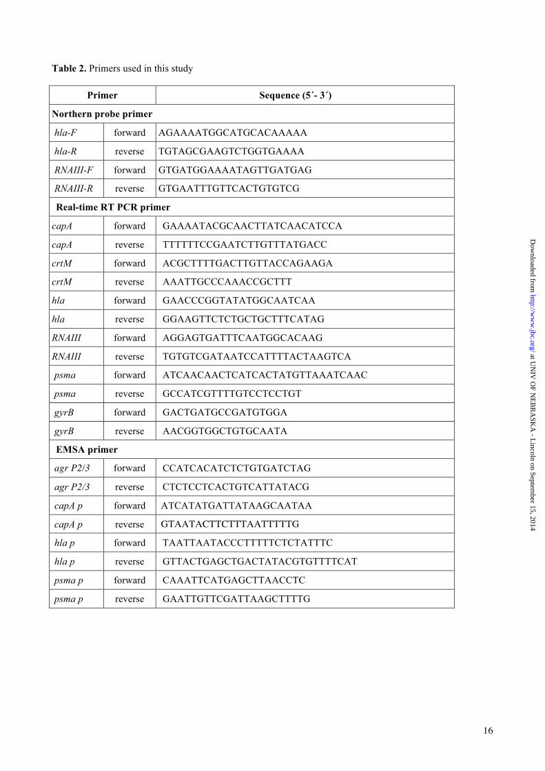
Table 2. Primers used in this study
Primer Sequence (5´- 3´)
Northern probe primer
hla-F forward AGAAAATGGCATGCACAAAAA
hla-R reverse TGTAGCGAAGTCTGGTGAAAA
RNAIII-F forward GTGATGGAAAATAGTTGATGAG
RNAIII-R reverse GTGAATTTGTTCACTGTGTCG
Real-time RT PCR primer
capA forward GAAAATACGCAACTTATCAACATCCA
capA reverse TTTTTTCCGAATCTTGTTTATGACC
crtM forward ACGCTTTTGACTTGTTACCAGAAGA
crtM reverse AAATTGCCCAAACCGCTTT
hla forward GAACCCGGTATATGGCAATCAA
hla reverse GGAAGTTCTCTGCTGCTTTCATAG
RNAIII forward AGGAGTGATTTCAATGGCACAAG
RNAIII reverse TGTGTCGATAATCCATTTTACTAAGTCA
psma forward ATCAACAACTCATCACTATGTTAAATCAAC
psma reverse GCCATCGTTTTGTCCTCCTGT
gyrB forward GACTGATGCCGATGTGGA
gyrB reverse AACGGTGGCTGTGCAATA
EMSA primer
agr P2/3 forward CCATCACATCTCTGTGATCTAG
agr P2/3 reverse CTCTCCTCACTGTCATTATACG
capA p forward ATCATATGATTATAAGCAATAA
capA p reverse GTAATACTTCTTTAATTTTTG
hla p forward TAATTAATACCCTTTTTCTCTATTTC
hla p reverse GTTACTGAGCTGACTATACGTGTTTTCAT
psma p forward CAAATTCATGAGCTTAACCTC
psma p reverse GAATTGTTCGATTAAGCTTTTG
at UN
IV O
F NE
BR
ASK
A - L
incoln on September 15, 2014
http://ww
w.jbc.org/
Dow
nloaded from

Fig. 1
CcpE (µg)
A
B
RNAIII 0.5 knt
16S
3 6 12 9
Newman TH01 TH01c
3 6 12 9 3 6 12 9 Time (h)
100
200
300
400
Luci
fera
se a
ctiv
ity
(RLU
/A60
0 1)
C
0 0.3 0.6 1.5 2
agr P2/3
0
** ** ** ** ** ** ** * *
Time [h]
A60
0
10
1
0.1
0.01
D
3 6 9 12
Time [h]
at UN
IV O
F NE
BR
ASK
A - L
incoln on September 15, 2014
http://ww
w.jbc.org/
Dow
nloaded from
Virginie
Texte tapé à la machine
17

Fig. 2
A
B
hla 1 knt
16S
Time (h)
40
80
120
Cop
ies
hla/
copy
gyr
B
C
0
** ** ** ** ** **
D
3 6 12 9
3 6 12 9
Newman TH01 TH01c
3 6 12 9 3 6 12 9
Time (h)
Cop
ies
hla/
copy
gyr
B
0
3
6
20
40
60 ** **
0 0.3 0.6 1.5 2 CcpE (µg)
hla P
Cold competitor (x-fold) 5 10 25 50 100 0
at UN
IV O
F NE
BR
ASK
A - L
incoln on September 15, 2014
http://ww
w.jbc.org/
Dow
nloaded from
Virginie
Texte tapé à la machine
18

Fig. 3
C
B
0 0.3 0.6 1.5 2
CcpE (µg)
TH01
(3.1±9.1)
TH01c
(41.2±20.5)
capA p
A
1
2
3
4
Cop
ies
capA
/cop
y gy
rB
0
** ** ** ** ** **
3 6 12 9
Time (h)
Newman
(30.0±18.5)
DAPI CY-3 Overlay
at UN
IV O
F NE
BR
ASK
A - L
incoln on September 15, 2014
http://ww
w.jbc.org/
Dow
nloaded from
Virginie
Texte tapé à la machine
19

Fig. 4
B
0 0.3 0.6 1.5 2
CcpE (µg)
psmα p
A
25
50
75
100
Cop
ies
psmα
/cop
y gy
rB
0
** ** ** ** ** ** ** **
3 6 12 9
Time (h)
at UN
IV O
F NE
BR
ASK
A - L
incoln on September 15, 2014
http://ww
w.jbc.org/
Dow
nloaded from
Virginie
Texte tapé à la machine
20

Fig. 5
0
100
200
300
400
Rel
ativ
e op
tical
den
sitie
s a
t 465
nm
[%]
A
B
C
Cop
ies
crtM
/cop
y gy
rB
* * * **
0
0.5
1.0
1.5
at UN
IV O
F NE
BR
ASK
A - L
incoln on September 15, 2014
http://ww
w.jbc.org/
Dow
nloaded from
Virginie
Texte tapé à la machine
21

Fig. 6
A B
1
2
3
4
5
6
7
Newman TH01 TH01c
log 1
0 CFU
/g
4
5
6
Newman TH01 TH01c
log 1
0 cel
ls/m
l
** * * **
C D
0
20
40
60
80
100 * * * *
Monocytes Neutrophils
Diff
eren
tial c
ell c
ount
in B
ALF
(%
of c
ells
)
** **
Tissue BALF G
-CS
F (n
g/m
l)
0
2
4
6
at UN
IV O
F NE
BR
ASK
A - L
incoln on September 15, 2014
http://ww
w.jbc.org/
Dow
nloaded from
Virginie
Texte tapé à la machine
22

Fig. 7
1
2
3
4
5
6
7 *
log 1
0 CFU
/g
SA564 HOM 355
at UN
IV O
F NE
BR
ASK
A - L
incoln on September 15, 2014
http://ww
w.jbc.org/
Dow
nloaded from
Virginie
Texte tapé à la machine
23

0 2 4 6 8 10 12 0
1
2
3
4
Foot
pad-
swel
ling
(mm
)
Days post infection
* *
* * * * *
*
*
Fig. 8
at UN
IV O
F NE
BR
ASK
A - L
incoln on September 15, 2014
http://ww
w.jbc.org/
Dow
nloaded from
Virginie
Texte tapé à la machine
24

Fig. 9
CcpE
hla
psmα
cap
RNAIII
Capsule formation
α-toxin
Host
Phenol soluble
modulins
Protection from opsonization
Cell lysis
Cytokine synthesis
Neutrophil recruitment
NLRP3 inflammasome
stimulation
Massive inflammatory response, tissue destruction
Low glucose S. aureus
at UN
IV O
F NE
BR
ASK
A - L
incoln on September 15, 2014
http://ww
w.jbc.org/
Dow
nloaded from
Virginie
Texte tapé à la machine
25
Virginie
Texte tapé à la machine