The C-terminus of the metabotropic glutamate receptor 1b regulates dimerization of the receptor
12
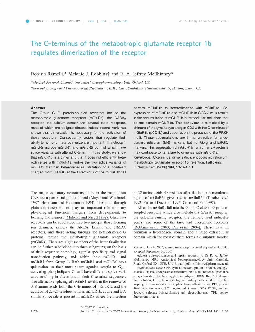
The C-terminus of the metabotropic glutamate receptor 1b regulates dimerization of the receptor Rosaria Remelli,* Melanie J. Robbins and R. A. Jeffrey McIlhinney* *Medical Research Council Anatomical Neuropharmacology Unit, Oxford, UK Neurophysiology and Pharmacology, Psychiatry CEDD, GlaxoSmithKline Pharmaceuticals, Harlow, Essex, UK The major excitatory neurotransmitters in the mammalian CNS are aspartic and glutamic acid (Mayer and Westbrook 1987; Hollmann and Heinemann 1994). These act through glutamate receptors and play an important role in many physiological functions, ranging from development, to learning and memory (Malenka and Nicoll 1993). Glutamate receptors can be subdivided into two groups, those forming ion channels, namely the AMPA, kainate and NMDA receptors, and those acting through the heterotrimeric G proteins, termed the metabotropic glutamate receptors (mGluRs). There are eight members of the latter family that can be further subdivided into three subgroups, on the basis of their sequence homology, agonist specificity and signal transduction pathway, and within these mGluR1 and mGluR5 form Group 1. Both mGluR1 and mGluR5 have quisqualate as their most potent agonist, couple to G q/11 activating phospholipase C, and have different splice vari- ants, resulting in alterations in their C-terminal sequences. The alternative splicing of mGluR1 results in the removal of 318 amino acids from the C-terminus of mGluR1a and the addition of 22–26 residues to form mGluR1b, c, d, e and f. A similar splice site is present in mGluR5 where the insertion of 32 amino acids 49 residues after the last transmembrane region of mGluR5a gives rise to mGluR5b (Tanabe et al. 1992; Pin and Duvoisin 1995; Conn and Pin 1997). All of the mGluRs fall into the Group C class of G protein- coupled receptors which also include the GABA B receptor, the calcium sensing receptor, the retinoic acid inducible genes, and some of the taste and pheromone receptors (Robbins et al. 2000; Pin et al. 2004). These have in common a heptahelical domain and a large extracellular domain which for most of them forms a disulphide bonded Received July 4, 2007; revised manuscript received September 4, 2007; accepted September 26, 2007. Address correspondence and reprint requests to Dr R. A. Jeffrey McIlhinney, MRC Anatomical Neuropharmacology Unit, Mansfield Road, Oxford OX1 3TH, UK. E-mail: [email protected] Abbreviations used: CFP, cyan fluorescent protein; EndoH, endogly- cosidase H; ER, endoplasmic reticulum; FRET, fluorescence resonance energy transfer; HA, haemagglutinin antigen; HBSS, Hank’s Balanced Salt Solution; HEK, human embryonic kidney cells; mGluR, metabo- tropic glutamate receptor; PBS, phosphate-buffered saline; PDI, protein disulphide isomerase; ROI, region of interest; SDS–PAGE, sodium dodecyl sulphate–polyacrylamide gel electrophoresis; YFP, yellow fluorescent protein. Abstract The Group C G protein-coupled receptors include the metabotropic glutamate receptors (mGluRs), the GABA B receptor, the calcium sensor and several taste receptors, most of which are obligate dimers, indeed recent work has shown that dimerization is necessary for the activation of these receptors. Consequently factors that regulate their ability to homo- or heterodimerize are important. The Group 1 mGluRs include mGluR1 and mGluR5 both of which have splice variants with altered C-termini. In this study, we show that mGluR1b is a dimer and that it does not efficiently hete- rodimerize with mGluR1a, unlike the two splice variants of mGluR5 that can heterodimerize. Mutation of a positively charged motif (RRKK) at the C-terminus of the mGluR1b tail permits mGluR1b to heterodimerize with mGluR1a. Co- expression of mGluR1a and mGluR1b in COS-7 cells results in the accumulation of mGluR1b in intracellular inclusions that do not contain mGluR1a. This behaviour is mimicked by a chimera of the lymphocyte antigen CD2 with the C-terminus of mGluR1b (pCD1b) and depends on the presence of the RRKK motif. These accumulations are immunoreactive for endo- plasmic reticulum (ER) markers, but not Golgi and ERGIC markers. This segregation of mGluR1b from other ER proteins may contribute to its failure to dimerize with mGluR1a. Keywords: C-terminus, dimerization, endoplasmic reticulum, metabotropic glutamate receptor 1b, retention, trafficking. J. Neurochem. (2008) 104, 1020–1031. d JOURNAL OF NEUROCHEMISTRY | 2008 | 104 | 1020–1031 doi: 10.1111/j.1471-4159.2007.05034.x 1020 Journal Compilation ȑ 2007 International Society for Neurochemistry, J. Neurochem. (2008) 104, 1020–1031 ȑ 2007 The Authors
-
Upload
independent -
Category
Documents
-
view
5 -
download
0
Transcript of The C-terminus of the metabotropic glutamate receptor 1b regulates dimerization of the receptor
The C-terminus of the metabotropic glutamate receptor 1bregulates dimerization of the receptor
Rosaria Remelli,* Melanie J. Robbins� and R. A. Jeffrey McIlhinney*
*Medical Research Council Anatomical Neuropharmacology Unit, Oxford, UK
�Neurophysiology and Pharmacology, Psychiatry CEDD, GlaxoSmithKline Pharmaceuticals, Harlow, Essex, UK
The major excitatory neurotransmitters in the mammalianCNS are aspartic and glutamic acid (Mayer and Westbrook1987; Hollmann and Heinemann 1994). These act throughglutamate receptors and play an important role in manyphysiological functions, ranging from development, tolearning and memory (Malenka and Nicoll 1993). Glutamatereceptors can be subdivided into two groups, those formingion channels, namely the AMPA, kainate and NMDAreceptors, and those acting through the heterotrimeric Gproteins, termed the metabotropic glutamate receptors(mGluRs). There are eight members of the latter family thatcan be further subdivided into three subgroups, on the basisof their sequence homology, agonist specificity and signaltransduction pathway, and within these mGluR1 andmGluR5 form Group 1. Both mGluR1 and mGluR5 havequisqualate as their most potent agonist, couple to Gq/11
activating phospholipase C, and have different splice vari-ants, resulting in alterations in their C-terminal sequences.The alternative splicing of mGluR1 results in the removal of318 amino acids from the C-terminus of mGluR1a and theaddition of 22–26 residues to form mGluR1b, c, d, e and f. Asimilar splice site is present in mGluR5 where the insertion
of 32 amino acids 49 residues after the last transmembraneregion of mGluR5a gives rise to mGluR5b (Tanabe et al.1992; Pin and Duvoisin 1995; Conn and Pin 1997).
All of the mGluRs fall into the Group C class of G protein-coupled receptors which also include the GABAB receptor,the calcium sensing receptor, the retinoic acid induciblegenes, and some of the taste and pheromone receptors(Robbins et al. 2000; Pin et al. 2004). These have incommon a heptahelical domain and a large extracellulardomain which for most of them forms a disulphide bonded
Received July 4, 2007; revised manuscript received September 4, 2007;accepted September 26, 2007.Address correspondence and reprint requests to Dr R. A. Jeffrey
McIlhinney, MRC Anatomical Neuropharmacology Unit, MansfieldRoad, Oxford OX1 3TH, UK. E-mail: [email protected] used: CFP, cyan fluorescent protein; EndoH, endogly-
cosidase H; ER, endoplasmic reticulum; FRET, fluorescence resonanceenergy transfer; HA, haemagglutinin antigen; HBSS, Hank’s BalancedSalt Solution; HEK, human embryonic kidney cells; mGluR, metabo-tropic glutamate receptor; PBS, phosphate-buffered saline; PDI, proteindisulphide isomerase; ROI, region of interest; SDS–PAGE, sodiumdodecyl sulphate–polyacrylamide gel electrophoresis; YFP, yellowfluorescent protein.
Abstract
The Group C G protein-coupled receptors include the
metabotropic glutamate receptors (mGluRs), the GABAB
receptor, the calcium sensor and several taste receptors,
most of which are obligate dimers, indeed recent work has
shown that dimerization is necessary for the activation of
these receptors. Consequently factors that regulate their
ability to homo- or heterodimerize are important. The Group 1
mGluRs include mGluR1 and mGluR5 both of which have
splice variants with altered C-termini. In this study, we show
that mGluR1b is a dimer and that it does not efficiently hete-
rodimerize with mGluR1a, unlike the two splice variants of
mGluR5 that can heterodimerize. Mutation of a positively
charged motif (RRKK) at the C-terminus of the mGluR1b tail
permits mGluR1b to heterodimerize with mGluR1a. Co-
expression of mGluR1a and mGluR1b in COS-7 cells results
in the accumulation of mGluR1b in intracellular inclusions that
do not contain mGluR1a. This behaviour is mimicked by a
chimera of the lymphocyte antigen CD2 with the C-terminus of
mGluR1b (pCD1b) and depends on the presence of the RRKK
motif. These accumulations are immunoreactive for endo-
plasmic reticulum (ER) markers, but not Golgi and ERGIC
markers. This segregation of mGluR1b from other ER proteins
may contribute to its failure to dimerize with mGluR1a.
Keywords: C-terminus, dimerization, endoplasmic reticulum,
metabotropic glutamate receptor 1b, retention, trafficking.
J. Neurochem. (2008) 104, 1020–1031.
d JOURNAL OF NEUROCHEMISTRY | 2008 | 104 | 1020–1031 doi: 10.1111/j.1471-4159.2007.05034.x
1020 Journal Compilation � 2007 International Society for Neurochemistry, J. Neurochem. (2008) 104, 1020–1031� 2007 The Authors

dimer (Romano et al. 1996; Tsuji et al. 2000; Pin and Acher2002). Biochemical and structural studies have shown thateach of these extracellular regions forms a Venus Flytrapdomain between which there are direct interactions (Kuni-shima et al. 2000; Tsuji et al. 2000; Liu et al. 2004a). Fromstructural and functional studies, it has become apparent thatagonist binding causes a change in the relative orientation ofthe Venus Flytrap domains which results in the activation ofthese receptors (Kunishima et al. 2000; Bessis et al. 2002;Tsuchiya et al. 2002; Kniazeff et al. 2004a,b). Consequently,the dimerization of these receptors is important for theirfunction and for most of these receptors this seems to bedriven by their extracellular domains and takes place in theendoplasmic reticulum (ER) (Ray et al. 1999; Robbins et al.1999; Ray and Hauschild 2000; Romano et al. 2001; Liuet al. 2004a; Villemure et al. 2005). Indeed, soluble domainsof the extracellular domains of mGluR1 and mGluR4 aresecreted as dimers (Han and Hampson 1999; Robbins et al.1999; Kunishima et al. 2000; Selkirk et al. 2002) althoughthe cysteine that forms the disulphide bridge between theextracellular domains is not essential for dimer formation,nor for functioning of the receptor (Tsuji et al. 2000;Romano et al. 2001). With the exception of the reportedheterodimerization of the mGluR1 and calcium sensingreceptor, there has been no evidence for the heterodimeriza-tion of other mGluRs, in fact mGluR1 and mGluR5 do notform heterodimers when co-expressed in cells (Romanoet al. 1996; Robbins et al. 1999; Gama et al. 2001).However, given that mGluR1a and mGluR1b are identicalat their N-terminus and that this is the region that drives thedimerization of the receptors, it was a surprise to discoverthat these two subtypes of mGluR1 did not heterodimerizewhen co-expressed in human embryonic kidney (HEK) 293cells (Robbins et al. 1999).
We have previously shown that an RRKK motif at the C-terminus of mGluR1b can cause its retention in the ER butthat, on mutation of this to AAAA, the receptor can trafficnormally to the cell surface (Chan et al. 2001). This result,together with our observation of the lack of heterodimeriza-tion between mGluR1a and mGluR1b, led us to speculatethat the RRKK motif in mGluR1b might regulate itsdimerization with other mGluR1 isoforms. Therefore wehave studied the dimerization and trafficking of mGluR1b,and mutants thereof, in transfected cells. In this study, weprovide the first direct evidence that mGluR1b is a dimer andshow that the RRKK motif prevents it from efficientlydimerizing with other forms of mGluR1. Consistent withthis, in rat cerebellum, where both mGluR1a and mGluR1bare co-expressed in Purkinje cells, co-immunoprecipitationexperiments did not reveal heterodimerization of the twoisoforms of the receptor. Immunocytochemical studies withcell compartment specific antibodies showed that mGluR1b,and a chimaeric molecule containing its C-terminus, isapparently segregated from other membrane proteins in the
ER and this may contribute to its failure to dimerize withother mGluR1 splice variants.
Materials and methods
MaterialsProtein N-glycosidase F (pNGaseF) and Endoglycosidase H
(EndoH) were purchased from Roche Biochemicals (Lewes, UK),
and Lipofectamine 2000 from Invitrogen (Carlsbad, CA, USA).
JetPEI was supplied by Autogen Bioclear (Colne, UK). Horseradish
peroxidase-conjugated anti-mouse and rabbit antibodies were
purchased from Promega (Southampton, UK) and the Talon resin
was from BD Biosciences (San Diego, CA, USA). Antibodies used
in this study were rabbit anti-GluR1a C- and N-terminal and a rabbit
anti-mGluR1b C-terminal antibody that have been described
previously (Ciruela et al. 1999a; Chan et al. 2001), an anti-myc
mouse monoclonal antibody clone 9E10 produced in house from a
hybridoma, a rabbit anti-haemagglutinin antigen (HA) (BAbCO,
Berkeley, CA, USA), rabbit anti-sec24 a gift from Dr David
Stephens (University of Bristol, Bristol, UK), mouse anti-GM130
(BD Biosciences, Oxford, UK), mouse anti-protein disulphide
isomerase (PDI; Stressgen, San Diego, CA, USA), mouse anti-
ERGIC 53 (Alexis Biochemicals, San Diego, CA, USA), a mouse
monoclonal anti-CD2 a gift from Dr Neil Barclay (Oxford, UK),
Alexa 568-coupled goat anti-rabbit, Alexa 488-coupled goat anti-
rabbit, Alexa 488-coupled goat anti-mouse and Alexa 568-coupled
goat anti-mouse (Molecular Probes, Eugene, OR, USA).
Methods
Recombinant constructsN-terminally FLAG-tagged mGluR1a, mGluR1b and their mutants
have been described previously (Ciruela et al. 1999b; Chan et al.2001). A BglII/XhoI digest was performed on pcDNA3-myc-
HisA (Invitrogen) and the vector gel purified. The C-terminal tail
of mGluR1b was amplified by PCR using the primers 5¢-GGC-AGCAAGAAGAAGATCTGCACCCGG-3¢ and 5¢-GCAGTGTGG-GGGTTTCTCGAGCTGCGCATGTGC-3¢ and the product digested
with BglII/XhoI and ligated into the pcDNA3 myc-HisA vector to
give pcDNA3myc-His-1. The mGluR1b, in a pcDNA3 vector
(Ciruela et al. 1999a), was digested with BglII and the N-terminal
region isolated as the smaller fragment. This was then ligated into
the BglII digested, calf intestinal alkaline phosphatase treated
pcDNA3myc-His-1 to yield the final mGluR1b-mycHis tagged at
the C-terminus. Exactly the same steps, except using mGluRMM18
as the substrate in the PCR reaction, yielded mGluRMM18-mycHis.
To produce the yellow fluorescent (YFP) and cyan fluorescent (CFP)
protein forms of mGluR1b and mGluRMM18, the plasmids
encoding the myc-His versions of the proteins were digested with
BamHI and XhoI to excise the entire cDNA fragment of each, and
these ligated into BglII XhoI digested pEYFP-N1 and pECFP-N1
vectors (Clontech, Mountain View, CA, USA).
The mGluR5a and mGluR5b cDNAs and their C-terminally HA-
tagged versions in pcDNA3 were gifts from Dr Francesco Ferraguti
and Dr Corrado Corti and have been described previously (Mion
et al. 2001). Both mGluR5a and 5b-mycHis versions were prepared
by amplifying the C-terminal of mGluR5-HA in pcDNA3 from
� 2007 The AuthorsJournal Compilation � 2007 International Society for Neurochemistry, J. Neurochem. (2008) 104, 1020–1031
mGluR1b C-terminus regulates receptor dimerization | 1021

beyond an EcoR47III site to the stop codon with the primers
5¢-CGCAGGATGCACAGCAACAGGT-3¢ and 5¢-CAGTTCTAG-ACAACGATGAAGA-3¢. The PCR product and the mGluR5 in
pcDNA3 were digested with EcoR47III and XbaI and ligated to
produce an mGluR5 without a stop codon and with an XbaI site at
the C-terminus. The whole sequence was then removed from the
pcDNA3 using EcoRI and XbaI and ligated into pcDNA3-mycHis
to give mGluR5a or 5b-mycHis.
The production of the constructs pCD1b and pCDMM18,
containing the C-termini of mGluR1b and mGluRMM18, respec-
tively, fused with the N-terminus and transmembrane domain of the
lymphocyte antigen CD2 has been described previously (Chan et al.2001). To produce CD2-YFP the plasmid containing CD2 fused to
the C-terminal tail of mGluR1a previously described (Chan et al.2001) was digested with BamHI and NotI to remove the mGluR1a
sequence. YFP (pEYFP-N1; Clontech) was amplified by PCR with
the primers 5¢-GACTCAGATCTCGAGCTAAGCTTCGAATTC-3¢and 5¢-GATCTAGAGTCGCGGCCGCTTTACTTGTAC-3¢ contain-ing 5¢ BglII and 3¢ NotI sites. The PCR product was gel purified,
digested with the appropriate enzymes, and ligated into the digested
CD2 plasmid to generate the construct CD2-YFP. For all constructs
the sequence changes were confirmed by DNA sequencing.
Tissue cultureHuman embryonic kidney 293 cells and COS-7 cells were grown in
Dulbecco’s Modified Eagle’s Medium (Sigma Chemical Co., Poole,
UK) supplemented with 10% (v/v) foetal calf serum (Invitrogen),
2 mmol/L L-glutamine, 50 U/mL penicillin, 50 lg/mL streptomycin
(all from Invitrogen) at 37�C, in a humidified atmosphere of 5%
CO2. For microscopy the cells were plated onto glass coverslips.
Cells were either transfected using a calcium phosphate protocol or
jetPEI, a polyethylenimine-derived transfection reagent (Qbiogene,
Cambridge, UK). The calcium phosphate method was adapted from
(Jordan et al. 1996). The transfection mixture consists of: 540 lLsterile water, 10 lg DNA, 186 lL of 1 mol/L CaCl2 and 750 lL of
2· HEPES-buffered saline pH 7.2. The mixture was gently mixed
and left for 45 s to allow the formation of the precipitate. An
appropriate volume was added directly to the cells (750 lL for a 25-
cm2 flask, 200 lL for a well of a 6-well plate). JetPEI was used
according to the manufacturer’s instructions. For transient transfec-
tions with cDNAs encoding mGluR5, the medium was replaced
with glutamate-free medium [Glutamax Dulbecco’s Modified
Eagle’s Medium, 10% (v/v) dialysed foetal calf serum] at 6 or
24 h after transfection, to avoid agonist-induced degradation of the
receptor.
Endoglycosidase treatment and SDS–PAGE analysisMembranes prepared from cells transfected with the proteins of
interest were digested with pNGaseF or EndoH as described earlier.
Samples were prepared for polyacrylamide gel electrophoresis
(PAGE) by heating at 60�C for 15 min in 62.5 mmol/L Tris–HCl pH
6.6 containing 1% sodium dodecyl sulphate (SDS), 0.01% brom-
ophenol blue, 20 mmol/L dithiothreitol and 10% glycerol (SDS
sample buffer). The proteins separated by SDS–PAGE were
transferred to a polyvinylidene difluoride membrane using a BioRad
Transblot semi-dry transfer system (BioRad, Hemel Hempstead,
UK) and immunoblotted as described previously (Ciruela et al.1999b, 2000).
Membrane preparation and immunoprecipitationMembranes were prepared from transfected cells and rat
cerebellum as described by (Chan et al. 2001). For the immu-
noprecipitation from rat cerebellum, synaptic plasma membranes
were solubilized at 20�C for 10 min, in 50 mmol/L Tris–HCl (pH
7.2) containing 0.1 mol/L NaCl (Tris–saline), 1% SDS and
protease inhibitors, with a protein : detergent ratio of 1 : 5. The
supernatant was diluted 1 : 10 with 1% Triton X-100 in Tris–
saline and centrifuged at 100 000 gav for 1 h at 4�C. The
supernatant was removed and the mGluR1a receptor was
incubated with rabbit-anti-mGluR1a antibody (5 lg) overnight at
4�C, and the immunocomplexes isolated by adding protein G
Sepharose, 50 lL of a 1 : 1 aqueous suspension. After rotation
for 2 h at 4�C, the Sepharose beads were exhaustively washed
and the bound proteins eluted by incubation with SDS sample
buffer.
To investigate the dimerization of mGluRs, a modification of
the co-immunoprecipitation was performed as follows: cell
membranes were prepared 48 h after transfection of HEK293
cells with the two proteins, one of which was myc-His tagged. The
membranes were solubilized in Tris–saline containing 1% SDS, in
order to disrupt completely any weak non-covalent interactions.
The sample was then diluted 1 : 10 in Tris–saline containing 1%
Triton X-100, imidazole added to 2.5 mmol/L, and centrifuged at
15 000 gav for 15 min, at 4�C. The supernatant was rotated for 1 h
with 50 lL Talon resin (Clontech), which strongly binds to the His
tag. After extensive washing with 1% Triton X-100 in Tris–saline
containing 2.5 mmol/L imidazole, the proteins were eluted with a
small volume of Tris–saline containing 1% Triton X-100 and
0.25 mol/L imidazole. As a negative control, the precipitation of
the FLAG-tagged receptor was performed in parallel omitting the
myc-His-tagged receptor. The eluate was heated at 60�C for
30 min after the addition of SDS sample buffer. The eluted
proteins were then analysed by SDS–PAGE and immunoblotting,
together with a fraction of the supernatant, as a control for the
input material.
ImmunocytochemistryCells to be processed were cultured on 22-mm diameter
borosilicate glass coverslips in a six-well plate and transfected
as appropriate. At 48 h post-transfection, the medium was
aspirated and the cells carefully washed in phosphate-buffered
saline (PBS) and fixed with 4% (w/v) p-formaldehyde in PBS for
5 min at 20�C. The fixed cells were washed in Tris–saline, pH
7.4 for 5 min, and where necessary, permeabilized with 0.25%
Triton X-100 for 5 min at 20�C. After incubation in blocking
buffer [1% (w/v) bovine serum albumin and 1% (v/v) normal
goat serum in PBS] for 30 min, each coverslip was overlaid with
150 lL of primary antibody appropriately diluted in blocking
buffer and incubated for at least 1 h at 20�C in a moist chamber.
After three washes in blocking buffer, the coverslips were
incubated with a 1/1000 dilution of the appropriate fluorophore-
conjugated secondary antibody for 1 h at 20�C. The coverslips
were then washed three times in PBS and mounted on
microscope slides in Vectashield mounting medium (Vector
Laboratories, Peterborough, UK) and sealed with clear nail
varnish. The images were taken using Zeiss LSM510 confocal
scanning microscope mounted on an Axiovert 100M inverted
Journal Compilation � 2007 International Society for Neurochemistry, J. Neurochem. (2008) 104, 1020–1031� 2007 The Authors
1022 | R. Remelli et al.

microscope (Carl Zeiss Ltd, Welwyn Garden City, UK). Anti-
bodies were used at the following dilutions rabbit anti-HA
1/1000, mouse anti-myc 1/10, rabbit anti-sec24 1/100, mouse
anti-ERGIC 53 1/100, rabbit anti-PDI 1/400, rabbit anti-GM130
1/400, rabbit anti-mGluR1a C-terminus 1/300, rabbit anti-
mGluR1a N-terminus 1/100, rabbit anti-mGluR1b C-terminus
1/300 and mouse anti-CD2 1/20.
Calcium imagingHuman embryonic kidney cells 293, 24 h after transfection with
the different mGluR constructs or a control plasmid (pcDNA3),
were subcultured onto poly-D-lysine coated coverslips and
incubated with glutamate-free medium for a further 24 h. The
cells were washed twice with HEPES-buffered Hank’s Balanced
Salt Solution containing 2 mmol/L CaCl2 (HBSS), and then
incubated at 20�C in 1 mL HBSS containing 1 lmol/L Fluo-4
(Molecular Probes) and 0.03% pluronic F-127 (Invitrogen) for
1 h. After three 5 min washes with HBSS the cells were imaged
on a Zeiss LSM510 confocal scanning microscope, using the 20·lens of the Axiovert 100M inverted microscope. Images were
scanned every 2 s using a 488 nm excitation and 530–550 nm
emission filters. After monitoring the cells for 2 min, glutamate at
20 lmol/L was used to stimulate the cells. The cell responses
were quantified using the Zeiss LSM software and the results
plotted as a ratio of the Fluorescence emission (F) divided by the
initial unstimulated fluorescence (F0). For the control samples
transfected with pcDNA3 0.5% Triton X-100 was added at the
indicated time to show that these cells had loaded with Fluo-4.
Data were collected from a minimum of 14 cells and the results
analysed in GraphPad Prism (GraphPad Software, San Diego,
CA, USA) using the Kruskal–Wallis method for analysis of
variance.
FRET analysisThe fluorescence resonance energy transfer (FRET) occurring
between the mGluRMM18 or mGluR1b-YFP and mGluRMM18
or mGluR1b-CFP subunits was determined using acceptor
photobleaching (Nashmi et al. 2003; Liu et al. 2004b). Cells
were transfected with equal amounts of the YFP and CFP
mGluR1 constructs, and after 48 h, imaged on a Zeiss LSM510
scanning confocal microscope, using a 40· oil immersion lens.
The YFP and CFP were excited using the 514 and 458 nm lines
of the He–Ne laser and the bleaching of the YFP carried out by
repetitive scanning (30 iterations) of the region of interest (ROI)
on the cells using 90% of full laser power at 514 nm. The YFP
emission was collected through a long pass LP530 nm filter and
the CFP fluorescence through a band pass filter BP480–520 nm.
ROI were defined in the ER of the cells, taking care to avoid
large intracellular inclusions, using a pinhole of 206 lm. After
taking five 1 s scans across the cell, the ROI was bleached and
five additional scans were collected. The FRET signal was
calculated as the fractional increase in the CFP fluorescence
signal in the ROI after the bleach thus: [(CFPpost-bleach )CFPpre-bleach)/CFPpost-bleach] · 100, after correction for the back-
ground signal, and the weak increase in CFP fluorescence seen
after the photobleaching protocol. Data was collected from a
minimum of 30 cells and the results analysed in GraphPad using
the Kruskal–Wallis method for ANOVA.
Results
Characterization of the mGluR1b-mycHis constructsIn our previous studies we used antibodies to the C-terminalregion of mGluR1a to perform immunoprecipitations formcells labelled with 35S-methionine (Robbins et al. 1999). Inorder to allow for more stringent solubilization conditionsprior to affinity-isolation of the receptors, we preparedC-terminally myc-His-tagged version of mGluR1b and itsmutant form in which the RRKK motif was changed toAAAA (mGluRMM18). These were examined for theirexpression in transfected COS-7 cells and cell surfaceexpression by immunofluorescence. This showed that themGluRMM18-mycHis was present at the cell surface, givinga granular pattern of staining, whilst the mGluR1b-mycHisconstruct gave a much weaker granular pattern of immuno-reactivity (Fig. 1a). Immunostaining of permeabilized cellsexpressing these constructs showed that both were present inthe cells at comparable levels and that the mGluR1b-mycHisformed intracellular inclusions that were not present in themGluRMM18-mycHis cells (see also Fig. 5). Endoglycosi-dase treatment of the constructs expressed in HEK293 cellsshowed that the mGluR1b-mycHis was fully EndoH sensi-tive indicating that it was predominantly retained in the ER.In contrast, the mGluRMM18-mycHis-tagged receptor wasEndoH resistant, consistent with our previously reportedfindings on mGluR1b (Chan et al. 2001) (Fig. 1b). It shouldbe noted that that the dimer and monomer bands for theglycosylated forms of mGluRMM18-mycHis appear tomigrate more slowly than those formed by mGluR1b-mycHisindicating that the former is more fully glycosylated. Thetotally de-glycosylated pNGaseF digested protein core ofthe two proteins is, as expected, the same size. It shouldbe noted that the apparently SDS-resistant dimers of theshort splice variants of mGluR1, present in these SDS-denaturing gels have been reported elsewhere and areformed during the processing of the samples (Mary et al.1998; Mateos et al. 1998; Chan et al. 2001; Francesconi andDuvoisin 2002).
All of the mGluR1b constructs gave a similar peakcalcium response following challenge with 20 lmol/L glu-tamate, indicating that they were functionally coupled(Fig 1c). The mGluRMM18-mycHis receptor calcium re-sponse appears to be more prolonged than that of eithermGluR1b or mGluR1b-mycHis, and this may reflect itsgreater cell surface expression. The peak response evoked bymGluR1b was not significantly different from that ofmGluRMM18, despite the fact that the wild type has amuch lower surface expression compared with the mutant.This suggests that a small number of functional receptors onthe cell surface are sufficient to induce a maximal response,which might be limited by other factors such as G proteinavailability. Therefore, the elevated surface expression of
� 2007 The AuthorsJournal Compilation � 2007 International Society for Neurochemistry, J. Neurochem. (2008) 104, 1020–1031
mGluR1b C-terminus regulates receptor dimerization | 1023

mGluRMM18 would only increase the receptor reserve,without altering the maximal response.
Dimerization of mGluR1b with itself and mGluR1aAs the mGluRs form disulphide bonded dimers, SDS-solubilization will not disrupt them, but will guarantee thedisassociation of non-covalent or inadvertent associationscaused by the solubilization. In order to maximize recoveryof the target proteins during their subsequent affinityisolation, the SDS-solubilized samples were diluted into1% Triton X-100 to reduce the SDS concentration to 0.1%,prior to the isolation step. Thus using the myc-His-taggedconstructs, affinity-isolations were performed, using Talon
resin, from SDS-solubilized cell membranes to test if thehomodimerization of mGluR1b with itself and heterodimer-ization with mGluRMM18 could be detected. As can beseen in Fig. 2a, co-expression of mGluR1b-mycHis andFLAG-tagged mGluR1b and immunoprecipitation of themGluR1b-mycHis, resulted in the co-immunoprecipitation ofFLAG-tagged mGluR1b, indicating that mGluR1b can formhomodimers of these constructs in the cells. However, ifFLAG-tagged mGluR1b and mGluRMM18-mycHis wereco-expressed together then we consistently failed toco-immunoprecipitate the two proteins, suggesting that theydo not heterodimerize to a significant extent under theseconditions (Fig. 2a).
Having established that the Talon resin could be used todemonstrate the dimerization of mGluR1b, the dimerizationof mGluR1a and mGluR1b was examined using co-expres-sion of both mGluR1b-mycHis and mGluRMM18-mycHiswith FLAG-tagged-mGluR1a. As can be seen in Fig. 2b,mGluR1a is only weakly co-immunoprecipitated withmGluR1b-mycHis, but significantly more of it is broughtdown when it is co-expressed with mGluRMM18-mycHis. In
Mr X 10–3
**
*
0
1
2
3
4
5mGluRMM18-mycHismGluR1bmGluR1b-mycHisCon
Time (s)
F/F0
(i) (ii)
(iv)(iii)
(a)
(b)
(c)
20 40 60 80 100 120 140 160 180 200
Fig. 1 (a) Immunofluorescence of mGluR1b-mycHis and mGluRMM18-
mycHis in COS-7 cells. COS-7 cells were transfected with either
mGluR1b-mycHis [a(ii and iv)] or mGluRMM18-mycHis [a(i and iii)].
After 48 h the cells were fixed and immunostained for the receptor
using a rabbit N-terminal anti-mGluR1b antibody. In panels [a(iii and
iv)], the cells were treated with 0.25% Triton X-100 prior to the primary
antibody incubation to permeabilize the cells. The scale bar is 10 lm
and the arrows show the inclusions formed by the mGluR1b-mycHis.
(b) Endoglycosidase treatment of membranes from HEK293 cells
expressing either mGluR1b-mycHis or mGluRMM18-mycHis. Mem-
branes were prepared from the cells 48 h after transfection and trea-
ted with the indicated endoglycosidases. After SDS–PAGE and
western blotting the receptors were detected using an anti-myc anti-
body. The mGluR1b shows almost complete EndoH sensitivity, as
indicated by its reduction in size, whereas the mGluRMM18-mycHis is
almost completely EndoH resistant. The position of the molecular
weight markers are indicated by the arrows and the position of the
monomer and dimer forms of mGluR1b are indicated by the single and
double asterisks respectively. Similar results were obtained in three
separate experiments. (c) Calcium responses evoked by stimulating
mGluR1b, mGluR1b-mycHis and mGluRMM18-mycHis. HEK293 cells
were transfected with the indicated constructs and after 48 h their
calcium responses to 20 lmol/L glutamate determined using the cal-
cium sensitive dye, Fluo-4. The time of addition of the glutamate, or
0.5% Triton X-100 to the control cells, is indicated by the arrow. All of
the mGluR1b constructs gave a response to glutamate with no sig-
nificant difference in the mean peak responses (mGluR1b 3.55 ± 0.31,
mGluR1b-mycHis 3.6 ± 0.35 and mGluRMM18-mycHis 4.1 ± 0.38).
The results are the mean ± SEM responses from 14, 16 and 21 cells
respectively. However, the area under the curve for the mGluRMM18-
mycHis responses was significantly different from that of the
mGluR1b and mGluR1b-mycHis (p < 0.05), and these were not dif-
ferent from each other. Similar results were obtained in two separate
experiments.
Journal Compilation � 2007 International Society for Neurochemistry, J. Neurochem. (2008) 104, 1020–1031� 2007 The Authors
1024 | R. Remelli et al.

three experiments results similar to those shown here wereobtained, in two more we failed to co-immunoprecipitatemGluR1a and mGluR1b suggesting that the two mGluR1subtypes are only weakly associated. If mGluR1b andmGluR1a were to heterodimerize then we reasoned thateither the mGluR1a would act as a chaperone for mGluR1bcausing it to move to the cell surface and become EndoHresistant or the mGluR1a associated with mGluR1b would beER retained and become EndoH sensitive. Therefore, we
examined the endoglycosidase sensitivity of the two receptorisoforms after their co-expression in HEK293 cells. In thisexperiment, the glycosylation status of both mGluR1a and 1bwas unchanged when they are co-expressed, suggesting thatthey do not associate to any significant extent, consistentwith the co-immunoprecipitation experiments (Fig. 2c).
To further examine the possibility that mGluR1b ormGluRMM18 were dimerizing, a FRET analysis wasperformed to see whether the C-termini of the different
Transfected with:mGluR1a-FlagmGluR1b-mycHisMM18-mycHis
Probed for:
Anti-FLAG
Anti-Myc
Probed for:
Anti-Myc
Anti-FLAG
Transfected with:
mGluR1b-Flag
mGluR1b-mycHis
MM18-mycHis
Lys
250
100
150
Transfected with:
Mr X 10–3
**
*
250
100150
250
100150
Mr X 10–3
**
*
**
*
250
100
150
250
150
Mr X10–3
*
**
mGluR1a+mGluR1b mGluR1b mGluR1amGluR1a+mGluR1b
Probed with: anti-mGluR1b anti-mGluR1a
Ppt Lys Ppt Lys PptLys Ppt Lys Ppt Lys Ppt
Lys Ppt
C F H C CC F FF H HH
(a)
(c)
(b)
Fig. 2 (a) Talon isolation of FLAG-tagged mGluR1b, by mGluR1b-
mycHis or mGluRMM18-mycHis, from SDS-solubilized cell mem-
branes from cells transfected with the indicated cDNAs. HEK293 cells
were transfected as shown and after 48 h cell membranes were pre-
pared from the cells. The solubilized material was incubated with Talon
resin to isolate the (His)6 form of the receptor and the isolate analysed
on SDS–polyacrylamide gels followed by western blotting for the
indicated epitope tag. The starting material is indicated by Lys and the
Talon isolate by Ppt. Note that whilst mGluR1b and mGluRMM18 form
homodimers they do not heterodimerize as shown by the absence of a
Talon isolated FLAG-tagged band in the lane where both FLAG-
tagged mGluR1b and mGluRMM18-mycHis are co-expressed. Similar
results were obtained in three separate experiments. (b) Talon
isolation of FLAG-tagged mGluR1a, by mGluR1b-mycHis and
mGluRMM18-mycHis, from SDS-solubilized cell membranes from
cells transfected with the indicated cDNAs. Note the relatively weak
co-isolation of FLAG-tagged mGluR1a with mGluR1b-mycHis
compared with when FLAG-tagged mGluR1a is co-expressed with
mGluRMM18-mycHis. Similar results were obtained in three separate
experiments and in two further experiments no co-immunoprecipitation
of mGluR1a with mGluR1b was seen. (c) Endoglycosidase sensitivity
of mGluR1b and mGluR1a when expressed alone or together.
HEK293 cells were transfected with the indicated cDNAs and after
48 h membranes were prepared from the cells. These were subjected
to treatment with no enzyme (C) or with EndoH (H) or pNGaseF (F).
The digests were analysed on 5% SDS–polyacrylamide gels followed
by immunoblotting for the different receptors. The endoglycosidase
sensitivity of both mGluR1a and mGluR1b is not altered when they are
co-expressed, suggesting that they do not associate and are not
trafficking together to the cell surface. In all the panels, the position of
the molecular weight markers are indicated by the arrows and the
position of the monomer and dimer forms of mGluR1b are indicated by
the single and double asterisks respectively. Similar results were
obtained in two separate experiments.
� 2007 The AuthorsJournal Compilation � 2007 International Society for Neurochemistry, J. Neurochem. (2008) 104, 1020–1031
mGluR1b C-terminus regulates receptor dimerization | 1025

receptors when expressed as YFP/CFP C-terminally taggedpairs, were in proximity. The FRET signal was determined byphotobleaching the YFP versions of the proteins andmonitoring the increase in the CFP fluorescence in theirCFP forms, because of the acceptor YFP being bleached. Theresults showed a mean fluorescence increase for mGluR1b-YFP/CFP pair of 9.9% ± 3.9 (SD; 32 cells), with themGluRMM18-YFP/CFP pair giving 9.7% ± 3.5 (SD; 35)FRET. The mGluRMM18-YFP/mGluR1b-CFP pair howeveronly gave a 3.2% ± 3.4 (SD; 35) FRET signal. The FRETvalues obtained from mGluRMM18-YFP/CFP pair, and themGluR1b-YFP/CFP pair, were not significantly different, butboth were significantly different from the mGluRMM18-YFPand mGluR1b-CFP pair (p > 0.01). Although the FRETsignals are low they are comparable with those reported forsimilar C-terminal truncated constructs of mGluR1a (Tatey-ama et al. 2004). These data are also consistent with themGluR1b and mGluRMM18 forming dimers, whereas themGluRMM18/mGluR1b pair do not.
This prompted us to ask if the alternative splice variants ofthe other Group 1 mGluRs, namely mGluR5a and 5b, wereable to heterodimerize, or whether their alternatively splicedC-termini also prevented heterodimerization. Therefore,mGluR5a and 5b carrying different epitope tags were co-expressed in HEK293 cells and immunoprecipitated sepa-rately. The results show clearly that the two mGluR5 splicevariants do indeed heterodimerize (Fig. 3).
mGluR1a and mGluR1b do not dimerize in the ratcerebellumTogether these data show that the affinity isolation methoddescribed here can demonstrate the dimerization of thedifferent mGluRs and provides convincing evidence that theRRKK motif in the C-terminus of mGluR1b is involved inthe prevention of heterodimerization of mGluR1b withmGluR1a and can also prevent the association of mGluR1bwith its mutated form mGluRMM18-mycHis.
It remains possible that, in the brain, where the mGluR1breceptor is fully glycosylated (Chan et al. 2001), mGluR1acould act as a chaperone for mGluR1b, especially in thecerebellum where the two splice variants are co-expressed inPurkinje cells (Mateos et al. 2000). Therefore, we solubilizedcerebellar membranes from rat brains using SDS andimmunoprecipitated mGluR1a using a C-terminal antibody.The resulting immunoprecipitate was split into two aliquotsand each immunoblotted for either mGluR1a or mGluR1b.The results show that whilst we obtained good solubilizationof the mGluRs and recovered the mGluR1a in good yield, wecould not detect mGluR1b in these precipitates (Fig. 4). Thesame results were obtained in two separate experiments.Thus in cerebellar synaptic membranes, mGluR1a andmGluR1b are not associated in heterodimers.
The ER retention motif causes segregation of mGluR1aand mGluR1b and causes the latter to form intracellularER-derived accumulationsAs mGluR1b does not heterodimerize with mGluRMM18 ormGluR1a we examined the intracellular distribution ofmGluR1a, when co-expressed with either mGluR1b ormGluRMM18 in COS-7 cells. The results shown in Fig. 5
Transfected with: mGluR5a-FlagmGluR5a-mycHismGluR5b-mycHis
Probed for:
Flag
Myc
Lys
250
150
250
150
Mr X 10–3
Ppt Lys Ppt Lys Ppt
Fig. 3 Talon isolation of FLAG-tagged mGluR5a, by mGluR5a-
mycHis or mGluR5b-mycHis, from SDS-solubilized cell membranes
from cells transfected with the indicated cDNAs. HEK293 cells were
transfected as shown and after 48 h cell membranes were prepared
from the cells. The solubilized material was incubated with Talon resin
to isolate the (His)6 form of the receptor and the isolate analysed on
SDS–polyacrylamide gels followed by western blotting for the indi-
cated epitope tag. The starting material is indicated by Lys and the
Talon isolate by Ppt. The position of the molecular weight markers are
indicated by the arrows. Similar results were obtained in three sepa-
rate experiments.
Probed for: anti-mGluR1a anti-mGluR1b
250
100
150
Mr X 10–3
*
**
Tot Pel Ppt Tot Pel Ppt
Fig. 4 Immunoprecipitation of mGluR1a from rat cerebellar mem-
branes. The soluble material after a 100 000gav centrifugation was
immunoprecipitated for mGluR1a using an anti-mGluR1a C-terminal
antibody, and the immunoprecipitate split into two aliquots before
analysing the total lysate (Tot), the 100 000 gav pellet (Pel) and the
precipitate (Ppt) on a 5% SDS–polyacrylamide gel followed by
immunoblotting for either mGluR1a or mGluR1b as indicated. Similar
results were obtained in two separate experiments. The position of the
molecular weight markers are indicated by the arrows and the position
of the monomer and dimer forms of mGluR1b are indicated by the
single and double asterisks respectively. Similar results were obtained
in two separate experiments.
Journal Compilation � 2007 International Society for Neurochemistry, J. Neurochem. (2008) 104, 1020–1031� 2007 The Authors
1026 | R. Remelli et al.

demonstrate that mGluRMM18 and mGluR1a show analmost complete overlap in their intracellular distribution(Fig. 5 lower row), whereas mGluR1b, although clearlypresent in the ER with mGluR1a, gives multiple brightpuncta of immunofluorescence that do not coincide with theimmunostaining of mGluR1a (Fig. 5 upper row). Thissuggests that mGluR1b may be segregating from mGluR1awithin the ER. To examine whether this behaviour ofmGluR1b is conferred solely by the C-terminal tail, we madeuse of a reporter molecule, the lymphocyte surface antigen,CD2, where we replaced its C-terminus with that ofmGluR1b to give pCD1b. This construct has been shownto convert CD2 from a fully glycosylated surface expressedprotein into an ER retained EndoH sensitive form (Chanet al. 2001). We then co-expressed CD2-YFP with pCD1b inCOS-7 cells, and following fixation and permeabilization,immunostained the cells for pCD1b with a C-terminal anti-mGluR1b antibody. CD2-YFP was clearly apparent atthe cell surface (Fig. 6 upper row) whilst pCD1b wasretained in the cells in large intracellular inclusions (Fig. 6lower row). However where these inclusions were present,the fluorescence of CD2-YFP was significantly diminishedsuggesting that it was being excluded, at least in part, fromthese areas of the ER (Fig. 6 lower panel). Furthermore, theobservation that CD2 was still being trafficked to the cellsurface suggests that despite the formation of the pCD1b
inclusions, the normal cell processing of membrane proteinswas maintained. In order to see if the C-terminal tail ofmGluR1b was allowing the CD2 to progress further than theER we examined the localization of the pCD1b in differentcell compartments. The results show that as before the largeintracellular inclusions formed by pCD1b are immunoreac-tive with the ER marker PDI, but not with the Golgi markerGM130, or the exit site marker Sec 24 nor with theintermediate compartment marker ERGIC 53 (Fig. 7). Inter-estingly, neither the intracellular distribution of the latter twoproteins appears to be altered despite the formation of theinclusions containing pCD1b (Fig. 7; left hand column,lower two rows). Thus, as suggested by the surfaceexpression of CD2-YFP, the intracellular machinery fortrafficking proteins to the cell membrane seems to continueto function despite the sequestration of pCD1b into theER-derived inclusions.
Discussion
The use of the myc-His-tagged versions of mGluR1b andmGluR1MM18 in this study confirms and extends ourprevious finding that the RRKK motif in the C-terminus ofmGluR1b retains the majority of the receptor in the ER(Chan et al. 2001). Despite this some receptor does arrive atthe plasma membrane where it is functional, but this protein,
Fig. 5 Co-expression of mGluR1a with
FLAG-tagged mGluR1b and co-expres-
sion of mGluR1a with FLAG-tagged
mGluRMM18. COS-7 cells were transfect-
ed with the indicated constructs using a
1 : 1 ratio of DNA and 48 h later they were
fixed, permeabilized and immunoreacted
with an anti-FLAG antibody (mGluR1b and
mGluRMM18) and with an anti-mGluR1a
antibody (mGluR1a). The secondary anti-
bodies were an Alexa 488-coupled goat
anti-mouse antibody, and an Alexa 568-
coupled goat anti-rabbit antibody. The scale
bar represents (10 lm) and the right hand
column shows the merged images of the
red and green channels. The arrows indi-
cate the intracellular accumulations formed
by mGluR1b but not mGluRMM18.
� 2007 The AuthorsJournal Compilation � 2007 International Society for Neurochemistry, J. Neurochem. (2008) 104, 1020–1031
mGluR1b C-terminus regulates receptor dimerization | 1027

previously quantified at 10% of the total, is EndoH sensitive,unlike mGluR1b found in the brain (Chan et al. 2001).Similar arginine-rich motifs have been described in othermembrane proteins such as the NMDA receptor NR1 subunit(Standley et al. 2000), potassium channels (Zerangue et al.1999), GABAB R1 (Margeta-Mitrovic 2002) and kainatereceptors (Ren et al. 2003) and have been suggested toregulate the surface expression of these proteins. Thesestudies have led to the suggestion that either RXR or F/Y/RRXR, where F/Y are any hydrophobic or aromatic aminoacids and X is any amino acid, provide ER retention motifs atthe appropriate place in the C-terminus of membrane proteins(Zerangue et al. 2001; Michelsen et al. 2005). The RRKKmotif in mGluR1b does not conform to this consensus, andthis may suggest that the domain in mGluR1b acts differentlyfrom other members of this type of ER retention motif.
Generally the ER retention of these proteins by thecharged residues is overcome by the association of theretained protein with another receptor subunit as is the casefor the GABAB receptor, potassium channels and NMDAreceptor. In the latter case additional forward traffickingsignals may involve the phosphorylation of residues flankingthe retention motif and/or interaction with a PDZ-bindingprotein (Scott et al. 2001, 2003; Xia et al. 2001). Alterna-tively, the oligomerization of receptor subunits recruitsadditional proteins to facilitate the trafficking of the receptor,or channel complex, from the ER, as happens with thepotassium channels and their recruitment of the 14-3-3proteins after oligomerization (O’Kelly et al. 2002). Onecandidate for a partner protein to facilitate the trafficking ofmGluR1b is mGluR1a, but as shown here it does notefficiently associate with mGluR1b in transiently transfectedcells, or in the cerebellum. Consequently, in the brain wheremGluR1b is fully glycosylated (Chan et al. 2001) there mustexist other proteins that facilitate the trafficking of this
receptor subtype, in this context it is interesting to note that asubunit of the GABAA receptor has been reported to promotethe cell surface expression of GABAB R1 subunits (Bala-subramanian et al. 2004).
The mechanism by which the arginine-based ER retentionmotifs operate is currently unclear, although clearly it causesthe accumulation of mGluR1b in an ER-derived cellcompartment. Indeed, the apparent sequestration ofmGluR1b or a chimera of it into the ER-derived compart-ment may be one mechanism by which its dimerization withmGluR1a may be minimized. The ER accumulation ofmGluR1b could result from its retrieval from the cis-Golgiapparatus or by its failure to utilize the normal traffickingmechanisms from the ER. This could reflect a failure of thereceptor to interact with the coat protein 1or 2 complexes,respectively (Bonifacino and Lippincott-Schwartz 2003), andthere is some evidence that at least some of the arginine-based motifs can bind coat protein 1 proteins (Yuan et al.2003; Brock et al. 2005), although other studies suggestdifferent mechanisms (Zerangue et al. 2001). As we couldnot detect the presence of mGluR1b or pCD1b in the Golgiapparatus, or in the ERGIC compartment it would seem thatit is either actively retained in the ER or fails to leave it. Ineither case, its exit from the ER should involve either theformation, or the disruption, of a protein interaction. As theC-terminal tail of mGluR1b can act to restrict CD2 to the ER,it seems probable that this sequence of amino acids issufficient to cause the ER retention. However, despite severalattempts using the yeast two hybrid system and proteomicapproaches, with both the normal mGluR1b C-terminus orthat derived from mGluRMM18, we have so far failed toidentify proteins interacting with this region of the receptor.
Our studies indicate that one function of the RRKK motifis to prevent the heterodimerization of mGluR1b withmGluR1a. Although using the more stringent solubilization
Fig. 6 COS-7 cells were transfected with
the CD2-YFP and pCD1b constructs using
a 1 : 1 ratio of DNA, and 48 h later they
were fixed (upper panel) or fixed and per-
meabilized (lower panel). The cells in the
upper panel were incubated with a mouse
antibody to CD2 before being reacted with
an Alexa 568-coupled anti mouse antibody
to reveal the surface CD2. In the lower pa-
nel, the permeabilized cells were incubated
with a rabbit anti-mGluR1b antibody before
being reacted with an Alexa 568-coupled
anti-rabbit antibody, to detect the intracel-
lular pCD1b. The left hand column shows
the YFP signal, the middle column shows
the Alexa 568 signal and the right column
panel shows the merged signals. The scale
bar represents (10 lm).
Journal Compilation � 2007 International Society for Neurochemistry, J. Neurochem. (2008) 104, 1020–1031� 2007 The Authors
1028 | R. Remelli et al.

conditions, permitted by the use of the Talon resin, to isolatepotential heterodimers of mGluR1a and mGluR1b, did revealoccasional low levels of heterodimerization, these were muchless than those found when the RRKK motif was removed bymutation. Likewise, no heterodimerization of these receptorsubtypes was detected in rat cerebellum isolates, where bothare expressed in the Purkinje cells. Interestingly, we wereable to show clear homodimerization of both mGluR1b andmGluRMM18, but not heterodimerization of mGluR1b andmGluRMM18. This represents the first clear evidence thatmGluR1b does exist as a homodimer and strongly suggeststhat the RRKK motif can regulate this dimerization.
As mGluR5a and mGluR5b, the other members of themGluR Group 1 family, appear to heterodimerize efficientlyand are well expressed at the cell surface, the question arisesas to why the mGluR1 family has this RRKK motif thatprevents heterodimerization of the receptor. In many cases,
the heterodimerization between receptor subtypes, or evenbetween different receptors, can result in a change ofpharmacological properties [as observed for example forthe opioid receptors (Jordan and Devi 1999) and tastereceptors (Zhao et al. 2003)] or desensitization profile (as forthe somatostatin receptor SSTR2/SSTR3 heterodimers;(Pfeiffer et al. 2001). As the functional differences betweenthe two mGluR1 splice variants are minor (Pin et al. 1992;Joly et al. 1995; Mary et al. 1997) heterodimerization wouldnot be expected to have a huge impact on the receptorproperties. Nevertheless, heterodimerization would affect thearray of proteins interacting with the dimeric mGluR. ThusmGluR1a, but not mGluR1b, can interact with the proteinsTamalin and Homer (Brakeman et al. 1997; Tu et al. 1998;Kitano et al. 2002). The interaction with Homer in particularcan lead to association of mGluR1a with other proteins suchas the IP3 receptor and Shank (Tu et al. 1998, 1999). Indeed,
Fig. 7 Intracellular accumulations of pCD1b
contain endoplasmic reticulum markers.
COS-7 cells were transfected with pCD1b
and 48 h later fixed using cold methanol
and immunostained for pCD1b and the
indicated marker proteins. pCD1b was
detected using a rabbit anti-mGluR1b anti-
body in all the panels apart from the bottom
row, where the mouse anti-CD2 was used.
The secondary antibodies were an Alexa
568-coupled goat anti-mouse antibody, and
an Alexa 488-coupled goat anti-rabbit anti-
body. The right hand column shows the
merged images and the scale bar repre-
sents (10 lm).
� 2007 The AuthorsJournal Compilation � 2007 International Society for Neurochemistry, J. Neurochem. (2008) 104, 1020–1031
mGluR1b C-terminus regulates receptor dimerization | 1029

interaction of Homer and mGluR1a appears to modulate theconstitutive activation of the receptor (Ango et al. 2001), andthis and other Homer-mediated interactions can be modulatedby the activity-induced expression of the dominant negativeshort variant Homer 1a (Minami et al. 2003). Therefore, thelack of heterodimerization between mGluR1a and mGluR1bkeeps mGluR1b insensitive to the actions of Homer1a,creating a receptor pool with a functional profile similar tomGluR1a but differently regulated upon neuronal activation.
Acknowledgements
The authors would like to thank Dr David Stephens, Department of
Biochemistry, Bristol University for the generous donation of an
anti-Sec24 antibody and Professor Neil Barclay, William Dunn
School of Pathology, Oxford for the gift of the plasmid for CD2 and
the antibody OX34. RR was the recipient of a CASE studentship
from the BBSRC and GlaxoSmithKline.
References
Ango F., Prezeau L., Muller T., Tu J. C., Xiao B., Worley P. F., Pin J. P.,Bockaert J. and Fagni L. (2001) Agonist-independent activation ofmetabotropic glutamate receptors by the intracellular proteinHomer. Nature 411, 962–965.
Balasubramanian S., Teissere J. A., Raju D. V. and Hall R. A. (2004)Hetero-oligomerization between GABAA and GABAB receptorsregulates GABAB receptor trafficking. J. Biol. Chem. 279, 18840–18850.
Bessis A. S., Rondard P., Gaven F., Brabet I., Triballeau N., Prezeau L.,Acher F. and Pin J. P. (2002) Closure of the Venus flytrap moduleof mGlu8 receptor and the activation process: insights frommutations converting antagonists into agonists. Proc. Natl Acad.Sci. U. S. A. 99, 11097–11102.
Bonifacino J. S. and Lippincott-Schwartz J. (2003) Coat proteins:shaping membrane transport. Nat. Rev. Mol. Cell Biol. 4, 409–414.
Brakeman P. R., Lanahan A. A., O’Brien R., Roche K., Barnes C. A.,Huganir R. L. and Worley P. F. (1997) Homer: a protein thatselectively binds metabotropic glutamate receptors. Nature 386,284–288.
Brock C., Boudier L., Maurel D., Blahos J. and Pin J. P. (2005)Assembly-dependent surface targeting of the heterodimeric GA-BAB receptor is controlled by COPI but not 14-3-3.Mol. Biol. Cell16, 5572–5578.
Chan W. Y., Soloviev M. M., Ciruela F. and McIlhinney R. A. J. (2001)Molecular determinants of metabotropic glutamate receptor 1Btrafficking. Mol. Cell. Neurosci. 17, 577–588.
Ciruela F., Soloviev M. M. and McIlhinney R. A. J. (1999a) Co-expression of metabotropic glutamate receptor type 1 alpha withHomer-1a/Vesl-1S increases the cell surface expression of thereceptor. Biochem. J. 341, 795–803.
Ciruela F., Soloviev M. M. and McIlhinney R. A. J. (1999b) Cell surfaceexpression of the metabotropic glutamate receptor type 1a is reg-ulated by the C-terminal tail. FEBS Lett. 448, 91–94.
Ciruela F., Soloviev M. M., Chan W. Y. and McIlhinney R. A. J. (2000)Homer-1c/Vesl-1L modulates the cell surface targeting of metab-otropic glutamate receptor type 1 alpha: evidence for an anchoringfunction. Mol. Cell. Neurosci. 15, 36–50.
Conn P. J. and Pin J. P. (1997) Pharmacology and functions of metab-otropic glutamate receptors. Ann. Rev. Pharmacol. Toxicol. 37,205–237.
Francesconi A. and Duvoisin R. M. (2002) Alternative splicing unmasksdendritic and axonal targeting signals in metabotropic glutamatereceptor 1. J. Neurosci. 22, 2196–2205.
Gama L., Wilt S. G. and Breitwieser G. E. (2001) Heterodimerization ofcalcium sensing receptors with metabotropic glutamate receptors inneurons. J. Biol. Chem. 276, 39053–39059.
Han G. and Hampson D. R. (1999) Ligand binding to the amino-terminaldomain of the mGluR4 subtype of metabotropic glutamate recep-tor. J. Biol. Chem. 274, 10008–10013.
Hollmann M. and Heinemann S. (1994) Cloned glutamate receptors.Annu. Rev. Neurosci. 17, 31–108.
Joly C., Gomeza J., Brabet I., Curry K., Bockaert J. and Pin J. P. (1995)Molecular, functional, and pharmacological characterization of themetabotropic glutamate receptor type 5 splice variants: comparisonwith mGluR1. J. Neurosci. 15, 3970–3981.
Jordan B. A. and Devi L. A. (1999) G-protein heterodimerizationmodulates receptor function. Nature 399, 697–700.
Jordan M., Schallhorn A. and Wurm F. M. (1996) Transfectingmammalian cells: optimization of critical parameters affectingcalcium phosphate precipitate formation. Nucleic Acids Res. 24,596–601.
Kitano J., Kimura K., Yamazaki Y., Soda T., Shigemoto R., Nakajima Y.and Nakanishi S. (2002) Tamalin, a PDZ domain-containing pro-tein, links a protein complex formation of group 1 metabotropicglutamate receptors and the guanine nucleotide exchange factorcytohesins. J. Neurosci. 22, 1280–1289.
Kniazeff J., Bessis A. S., Maurel D., Ansanay H., Prezeau L. and Pin J.P. (2004a) Closed state of both binding domains of homodimericmGlu receptors is required for full activity. Nat. Struct. Mol. Biol.11, 706–713.
Kniazeff J., Saintot P. P., Goudet C., Liu J., Charnet A., Guillon G. andPin J. P. (2004b) Locking the dimeric GABA(B) G-protein-coupledreceptor in its active state. J. Neurosci. 24, 370–377.
Kunishima N., Shimada Y., Tsuji Y., Sato T., Yamamoto M., KumasakaT., Nakanishi S., Jingami H. and Morikawa K. (2000) Structuralbasis of glutamate recognition by a dimeric metabotropic glutamatereceptor. Nature 407, 971–977.
Liu J., Maurel D., Etzol S., Brabet I., Ansanay H., Pin J. P. and RondardP. (2004a) Molecular determinants involved in the allosteric controlof agonist affinity in the GABAB receptor by the GABAB2 sub-unit. J. Biol. Chem. 279, 15824–15830.
Liu J., Ernst S. A., Gladycheva S. E., Lee Y. Y., Lentz S. I., Ho C. S., LiQ. and Stuenkel E. L. (2004b) Fluorescence resonance energytransfer reports properties of syntaxin1a interaction with Munc18-1in vivo. J. Biol. Chem. 279, 55924–55936.
Malenka R. C. and Nicoll R. A. (1993) NMDA-receptor-dependentsynaptic plasticity: multiple forms and mechanisms. Trends Neu-rosci. 16, 521–527.
Margeta-Mitrovic M. (2002) Assembly-dependent trafficking assays inthe detection of receptor-receptor interactions. Methods 27, 311–317.
Mary S., Stephan D., Gomeza J., Bockaert J., Pruss R. M. and Pin J. P.(1997) The rat mGlu1d receptor splice variant shares functionalproperties with the other short isoforms of mGlu1 receptor. Eur. J.Pharmacol. 335, 65–72.
Mary S., Gomeza J., Prezeau L., Bockaert J. and Pin J. P. (1998) Acluster of basic residues in the carboxyl-terminal tail of the shortmetabotropic glutamate receptor 1 variants impairs their couplingto phospholipase C. J. Biol. Chem. 273, 425–432.
Mateos J. M., Azkue J., Benitez R., Sarria R., Losada J., Conquet F.,Ferraguti F., Kuhn R., Knopfel T. and Grandes P. (1998)Immunocytochemical localization of the mGluR1b metabotropicglutamate receptor in the rat hypothalamus. J. Comp. Neurol. 390,225–233.
Journal Compilation � 2007 International Society for Neurochemistry, J. Neurochem. (2008) 104, 1020–1031� 2007 The Authors
1030 | R. Remelli et al.
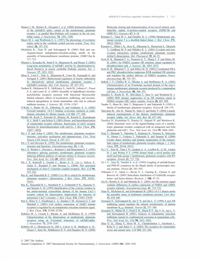
Mateos J. M., Benitez R., Elezgarai I. et al. (2000) Immunolocalizationof the mGluR1b splice variant of the metabotropic glutamatereceptor 1 at parallel fiber-Purkinje cell synapses in the rat cere-bellar cortex. J. Neurochem. 74, 1301–1309.
Mayer M. L. and Westbrook G. L. (1987) The physiology of excitatoryamino acids in the vertebrate central nervous system. Prog. Neu-robiol. 28, 197–276.
Michelsen K., Yuan H. and Schwappach B. (2005) Hide and run.Arginine-based endoplasmic-reticulum-sorting motifs in theassembly of heteromultimeric membrane proteins. EMBO Rep 6,717–722.
Minami I., Kengaku M., Smitt P. S., Shigemoto R. and Hirano T. (2003)Long-term potentiation of mGluR1 activity by depolarization-in-duced Homer1a in mouse cerebellar Purkinje neurons. Eur. J.Neurosci. 17, 1023–1032.
Mion S., Corti C., Neki A., Shigemoto R., Corsi M., Fumagalli G. andFerraguti F. (2001) Bidirectional regulation of neurite elaborationby alternatively spliced metabotropic glutamate receptor 5(mGluR5) isoforms. Mol. Cell. Neurosci. 17, 957–972.
Nashmi R., Dickinson M. E., McKinney S., Jareb M., Labarca C., FraserS. E. and Lester H. A. (2003) Assembly of alpha4beta2 nicotinicacetylcholine receptors assessed with functional fluorescentlylabeled subunits: effects of localization, trafficking, and nicotine-induced upregulation in clonal mammalian cells and in culturedmidbrain neurons. J. Neurosci. 23, 11554–11567.
O’Kelly I., Butler M. H., Zilberberg N. and Goldstein S. A. (2002)Forward transport. 14-3-3 binding overcomes retention in endo-plasmic reticulum by dibasic signals. Cell 111, 577–588.
Pfeiffer M., Koch T., Schroder H., Klutzny M., Kirscht S., KreienkampH. J., Hollt V. and Schulz S. (2001) Homo- and heterodimerizationof somatostatin receptor subtypes. Inactivation of sst(3) receptorfunction by heterodimerization with sst(2A). J. Biol. Chem. 276,14027–14036.
Pin J. P. and Acher F. (2002) The metabotropic glutamate receptors:structure, activation mechanism and pharmacology. Curr. DrugTargets CNS Neurol. Disord. 1, 297–317.
Pin J. P. and Duvoisin R. (1995) The metabotropic glutamate receptors:structure and functions. Neuropharmacology 34, 1–26.
Pin J. P., Waeber C., Prezeau L., Bockaert J. and Heinemann S. F. (1992)Alternative splicing generates metabotropic glutamate receptorsinducing different patterns of calcium release in Xenopus oocytes.Proc. Natl Acad. Sci. USA 89, 10331–10335.
Pin J. P., Kniazeff J., Goudet C., Bessis A. S., Liu J., Galvez T.,Acher F., Rondard P. and Prezeau L. (2004) The activationmechanism of class-C G-protein coupled receptors. Biol. Cell 96,335–342.
Ray K. and Hauschild B. C. (2000) Cys-140 is critical for metabotropicglutamate receptor-1 dimerization. J. Biol. Chem. 275, 34245–34251.
Ray K., Hauschild B. C., Steinbach P. J., Goldsmith P. K., Hauache O.and Spiegel A. M. (1999) Identification of the cysteine residues inthe amino-terminal extracellular domain of the human Ca(2+)receptor critical for dimerization. Implications for function ofmonomeric Ca(2+) receptor. J. Biol. Chem. 274, 27642–27650.
Ren Z., Riley N. J., Needleman L. A., Sanders J. M., Swanson G. T. andMarshall J. (2003) Cell surface expression of GluR5 kainatereceptors is regulated by an endoplasmic reticulum retention signal.J. Biol. Chem. 278, 52700–52709.
Robbins M. J., Ciruela F., Rhodes A. and McIlhinney R. A. (1999)Characterization of the dimerization of metabotropic glutamatereceptors using an N-terminal truncation of mGluR1alpha.J. Neurochem. 72, 2539–2547.
Robbins M. J., Michalovich D., Hill J., Calver A. R., Medhurst A. D.,Gloger I., Sims M., Middlemiss D. N. and Pangalos M. N. (2000)
Molecular cloning and characterization of two novel retinoic acid-inducible orphan G-protein-coupled receptors (GPRC5B andGPRC5C). Genomics 67, 8–18.
Romano C., Yang W. L. and Omalley K. L. (1996) Metabotropic glu-tamate receptor 5 is a disulfide-linked dimer. J. Biol. Chem. 271,28612–28616.
Romano C., Miller J. K., Hyrc K., Dikranian S., Mennerick S., TakeuchiY., Goldberg M. P. and OMalley K. L. (2001) Covalent and non-covalent interactions mediate metabotropic glutamate receptormGlu(5) dimerization. Mol. Pharmacol. 59, 46–53.
Scott D. B., Blanpied T. A., Swanson G. T., Zhang C. F. and Ehlers M.D. (2001) An NMDA receptor ER retention signal regulated byphosphorylation and. J. Neurosci. 21, 3063–3072.
Scott D. B., Blanpied T. A. and Ehlers M. D. (2003) Coordinated PKAand PKC phosphorylation suppresses RXR-mediated ER retentionand regulates the surface delivery of NMDA receptors. Neuro-pharmacology 45, 755–767.
Selkirk J. V., Challiss R. A., Rhodes A. and McIlhinney R. A. (2002)Characterization of an N-terminal secreted domain of the type-1human metabotropic glutamate receptor produced by a mammaliancell line. J. Neurochem. 80, 346–353.
Standley S., Roche K. W., McCallum J., Sans N. and Wenthold R. J.(2000) PDZ domain suppression of an ER retention signal inNMDA receptor NR1 splice variants. Neuron 28, 887–898.
Tanabe Y., Masu M., Ishii T., Shigemoto S. and Nakanishi S. (1992) Afamily of metabotropic glutamate receptors. Neuron 8, 169–179.
Tateyama M., Abe H., Nakata H., Saito O. and Kubo Y. (2004) Ligand-induced rearrangement of the dimeric metabotropic glutamatereceptor 1alpha. Nat. Struct. Mol. Biol. 11, 637–642.
Tsuchiya D., Kunishima N., Kamiya N., Jingami H. and Morikawa K.(2002) Structural views of the ligand-binding cores of a metabo-tropic glutamate receptor complexed with an antagonist and bothglutamate and Gd3+. Proc. Natl Acad. Sci. USA 99, 2660–2665.
Tsuji Y., Shimada Y., Takeshita T., Kajimura N., Nomura S., SekiyamaN., Otomo J., Usukura J., Nakanishi S. and Jingami H. (2000)Cryptic dimer interface and domain organization of the extracel-lular region of metabotropic glutamate receptor subtype 1. J. Biol.Chem. 275, 28144–28151.
Tu J. C., Xiao B., Yuan J. P., Lanahan A. A., Leoffert K., Li M., LindenD. J. and Worley P. F. (1998) Homer binds a novel proline richmotif and links group 1 metabotropic glutamate receptors with IP3receptors. Neuron 21, 717–726.
Tu J. C., Xiao B., Naisbitt S. et al. (1999) Coupling of mGluR/Homerand PSD-95 complexes by the Shank family of postsynaptic den-sity proteins. Neuron 23, 583–592.
Villemure J. F., Adam L., Bevan N. J., Gearing K., Chenier S. andBouvier M. (2005) Subcellular distribution of GABA(B) receptorhomo- and hetero-dimers. Biochem. J. 388, 47–55.
Xia H., Hornby Z. D. and Malenka R. C. (2001) An ER retention signalexplains differences in surface expression of NMDA and AMPAreceptor subunits. Neuropharmacology 41, 714–723.
Yuan H., Michelsen K. and Schwappach B. (2003) 14-3-3 dimers probethe assembly status of multimeric membrane proteins. Curr. Biol.13, 638–646.
Zerangue N., Schwappach B., Jan Y. N. and Jan L. Y. (1999) A new ERtrafficking signal regulates the subunit stoichiometry of plasmamembrane KATP channels. Neuron 22, 537–548.
Zerangue N., Malan M. J., Fried S. R., Dazin P. F., Jan Y. N., Jan L. Y.and Schwappach B. (2001) Analysis of endoplasmic reticulumtrafficking signals by combinatorial screening in mammalian cells.Proc. Natl Acad. Sci. USA 98, 2431–2436.
Zhao G. Q., Zhang Y., Hoon M. A., Chandrashekar J., Erlenbach I.,Ryba N. J. and Zuker C. S. (2003) The receptors for mammaliansweet and umami taste. Cell 115, 255–266.
� 2007 The AuthorsJournal Compilation � 2007 International Society for Neurochemistry, J. Neurochem. (2008) 104, 1020–1031
mGluR1b C-terminus regulates receptor dimerization | 1031