Specific Delivery of Therapeutic RNAs to Cancer Cells via the Dimerization Mechanism of phi29 Motor...
22
Specific Delivery of Therapeutic RNAs to Cancer Cells via the Dimerization Mechanism of phi29 Motor pRNA SONGCHUAN GUO 1 , NUSKA TSCHAMMER 2 , SULMA MOHAMMED 1 , and PEIXUAN GUO 1,* 1 Department of Pathobiology and Purdue Cancer Research Center, Purdue University, West Lafayette, IN 47907 2 Biomolecular Science Center, University of Central Florida, Orlando, FL 32816 Abstract The application of small RNA in therapy has been hindered by the lack of an efficient and safe delivery system to target specific cells. Packaging RNA (pRNA), part of the DNA-packaging motor of bacteriophage phi29(φ29), was manipulated by RNA nanotechnology to make chimeric RNAs that form dimers via interlocking right- and left-hand loops. Fusing pRNA with receptor-binding RNA aptamer, folate, small interfering RNA (siRNA), ribozyme, or another chemical group did not disturb dimer formation or interfere with the function of the inserted moieties. Incubation of cancer cells with the pRNA dimer, one subunit of which harbored the receptor-binding moiety and the other harboring the gene-silencing molecule, resulted in their binding and entry into the cells, and subsequent silencing of anti/proapoptotic genes. The chimeric pRNA complex was found to be processed into functional double-stranded siRNA by Dicer (RNA-specific endonuclease). Animal trials confirmed the suppression of tumorigenicity of cancer cells by ex vivo delivery. It has been reported [Shu, D., Moll, W.-D., Deng, Z., Mao, C., and Guo, P. (2004). Nano Lett. 4:1717–1724] that RNA can be used as a building block for bottom-up assembly in nanotechnology. The assembly of protein-free 25-nm RNA nanoparticles reported here will allow for repeated long-term administration and avoid the problems of short retention time of small molecules and the difficulties in the delivery of particles larger than 100 nm. OVERVIEW SUMMARY The successful use of small RNA for therapeutic purposes requires a safe and efficient delivery system capable of targeting specific cells. φ29 motor RNA (pRNA) was manipulated to produce chimeric RNAs that formed dimers through connection by right- and left-hand loops. Fusion of the pRNA with a variety of therapeutic and chemical compounds did not impede the formation of dimers or interfere with moiety function. Incubation of cells with dimer, one subunit of which carried a gene-silencing molecule and the other a receptor-binding moiety, resulted in successful binding, entry, and silencing of apoptotic genes. Animal trials further confirmed suppression of the tumorigenicity of cancer cells by ex vivo delivery. The dimeric RNA chimera was processed into double-stranded small interfering RNA (siRNA) by Dicer. These protein-free 25-nm nanoparticles will allow for repeated and long-term administration escaping immunoresponse and avoid the short retention time of smaller molecules and the undeliverability of larger molecules. *Corresponding author. Address reprint requests to: Dr. Peixuan Guo, Purdue Cancer Research Center, B-36 Hansen Life Science Research Building, Purdue University, West Lafayette, IN 47907, [email protected]. NIH Public Access Author Manuscript Hum Gene Ther. Author manuscript; available in PMC 2010 March 12. Published in final edited form as: Hum Gene Ther. 2005 September ; 16(9): 1097–1109. doi:10.1089/hum.2005.16.1097. NIH-PA Author Manuscript NIH-PA Author Manuscript NIH-PA Author Manuscript
-
Upload
independent -
Category
Documents
-
view
3 -
download
0
Transcript of Specific Delivery of Therapeutic RNAs to Cancer Cells via the Dimerization Mechanism of phi29 Motor...

Specific Delivery of Therapeutic RNAs to Cancer Cells via theDimerization Mechanism of phi29 Motor pRNA
SONGCHUAN GUO1, NUSKA TSCHAMMER2, SULMA MOHAMMED1, and PEIXUAN GUO1,*1 Department of Pathobiology and Purdue Cancer Research Center, Purdue University, WestLafayette, IN 479072 Biomolecular Science Center, University of Central Florida, Orlando, FL 32816
AbstractThe application of small RNA in therapy has been hindered by the lack of an efficient and safedelivery system to target specific cells. Packaging RNA (pRNA), part of the DNA-packaging motorof bacteriophage phi29(φ29), was manipulated by RNA nanotechnology to make chimeric RNAsthat form dimers via interlocking right- and left-hand loops. Fusing pRNA with receptor-bindingRNA aptamer, folate, small interfering RNA (siRNA), ribozyme, or another chemical group did notdisturb dimer formation or interfere with the function of the inserted moieties. Incubation of cancercells with the pRNA dimer, one subunit of which harbored the receptor-binding moiety and the otherharboring the gene-silencing molecule, resulted in their binding and entry into the cells, andsubsequent silencing of anti/proapoptotic genes. The chimeric pRNA complex was found to beprocessed into functional double-stranded siRNA by Dicer (RNA-specific endonuclease). Animaltrials confirmed the suppression of tumorigenicity of cancer cells by ex vivo delivery. It has beenreported [Shu, D., Moll, W.-D., Deng, Z., Mao, C., and Guo, P. (2004). Nano Lett. 4:1717–1724]that RNA can be used as a building block for bottom-up assembly in nanotechnology. The assemblyof protein-free 25-nm RNA nanoparticles reported here will allow for repeated long-termadministration and avoid the problems of short retention time of small molecules and the difficultiesin the delivery of particles larger than 100 nm.
OVERVIEW SUMMARYThe successful use of small RNA for therapeutic purposes requires a safe and efficient deliverysystem capable of targeting specific cells. φ29 motor RNA (pRNA) was manipulated to producechimeric RNAs that formed dimers through connection by right- and left-hand loops. Fusionof the pRNA with a variety of therapeutic and chemical compounds did not impede theformation of dimers or interfere with moiety function. Incubation of cells with dimer, onesubunit of which carried a gene-silencing molecule and the other a receptor-binding moiety,resulted in successful binding, entry, and silencing of apoptotic genes. Animal trials furtherconfirmed suppression of the tumorigenicity of cancer cells by ex vivo delivery. The dimericRNA chimera was processed into double-stranded small interfering RNA (siRNA) by Dicer.These protein-free 25-nm nanoparticles will allow for repeated and long-term administrationescaping immunoresponse and avoid the short retention time of smaller molecules and theundeliverability of larger molecules.
*Corresponding author. Address reprint requests to: Dr. Peixuan Guo, Purdue Cancer Research Center, B-36 Hansen Life ScienceResearch Building, Purdue University, West Lafayette, IN 47907, [email protected].
NIH Public AccessAuthor ManuscriptHum Gene Ther. Author manuscript; available in PMC 2010 March 12.
Published in final edited form as:Hum Gene Ther. 2005 September ; 16(9): 1097–1109. doi:10.1089/hum.2005.16.1097.
NIH
-PA Author Manuscript
NIH
-PA Author Manuscript
NIH
-PA Author Manuscript

INTRODUCTIONThe development of molecular gene therapy is one of the most promising applications ofmodern biological science. Small interfering RNA (siRNA) (Elbashir et al., 2001;Brummelkamp et al., 2002; Carmichael, 2002; Li et al., 2002; McCaffrey et al., 2002),ribozymes (Blount and Uhlenbeck, 2002; Hoeprich et al., 2003), and antisense RNA (Kumarand Carmichael, 1998) have shown significant potential as novel molecular approaches todownregulate specific gene expression. siRNA was also successfully used for knocking downhuman immunodeficiency virus (HIV)-related gene expression (Novina et al., 2002; Akkinaet al., 2003). To be successfully applied in the treatment of cancer and infectious diseases,siRNAs must be able to (1) enter cells despite size limitations on membrane penetration, (2)survive degradation by nucleases within the cells, (3) traffic into the appropriate cellcompartment, (4) fold correctly in the cell, if fused to a carrier, and (5) home to specific cells.Hence, the development of a safe, efficient, specific, and nonpathogenic system for the deliveryof therapeutic RNA is highly desirable.
We discovered a 117-nucleotide bacteriophage φ29-encoded packaging RNA (pRNA) thatplays a novel and essential role in packaging DNA into procapsids (Fig. 1) (Guo et al.,1987). Six copies of pRNA form a hexameric ring to drive the DNA-packaging motor (Trottierand Guo, 1997;Guo et al., 1998;Zhang et al., 1998). pRNA dimers are the building blocks ofhexamers (Chen et al., 2000). Hand-in-hand interaction of the right and left interlocking loopscan be manipulated to produce desired stable dimers, trimers, or hexamers (Guo et al.,1998;Zhang et al., 1998;Chen et al., 1999;Shu et al., 2003). The size of pRNA dimer is about25 nm (Hoeprich and Guo, 2002;Shu et al., 2004).
Our work indicates that RNA, and especially pRNA, can serve as a building block to buildnanomaterials via bottom-up assembly (Shu et al., 2004). The structural and molecular featuresof φ29 pRNA allow its easy manipulation, making it possible to redesign its parts as gene-targeting and delivery vehicles. The pRNA molecule contains intermolecular interactiondomains and a 5′/3′ helical domain (Fig. 1) (Zhang et al., 1995; Garver and Guo, 1997; Chenet al., 1999, 2000). Replacement or insertion of the 5′/3′ helical domain does not interfere withdimer formation (Chen et al., 1999).
The feasibility of these ideas was tested by the construction of chimeric pRNA dimers. Onesubunit of the dimer contained a receptor-binding RNA aptamer or folate for cell recognition,and the other harbored a moiety of siRNA, ribozyme, or chemical group. The dimers weredelivered to specific cells to silence the genes for green fluorescent protein (GFP), luciferase,survivin, and other pro/anti-apoptotic members of the BCL-2 family in a variety of cancer cells.
MATERIALS AND METHODSIn vitro synthesis and physical characterization of RNA
RNAs were prepared as described (Zhang et al., 1995). DNA oligonucleotides (oligos) weresynthesized with the desired sequences and used to produce double-stranded DNA bypolymerase chain reaction (PCR). The DNA products containing the T7 promoter were clonedinto plasmids or used as a substrate for direct in vitro transcription. All pRNA chimeras weretreated with calf intestinal alkaline phosphatase (CIP) to remove the 5′-phosphate and eliminatePKR (protein kinase RNA-activated) and interferon effects (Kim et al., 2004) or synthesizedin the presence of SH-AMP, biotin–AMP, or CoA (Li et al., 2005; Khaled et al., 2005).Methods of electrophoresis and cryo-atomic force microscopy (cryo-AFM) for the detectionof pRNA dimer have been described (Chen et al., 2000; Shu et al., 2003). Magnesium (10mM) was included in all buffers to maintain the folding of pRNA and the formation of dimers(Chen et al., 2000; Mat-Arip et al., 2001).
GUO et al. Page 2
Hum Gene Ther. Author manuscript; available in PMC 2010 March 12.
NIH
-PA Author Manuscript
NIH
-PA Author Manuscript
NIH
-PA Author Manuscript

Transfection assay for monomeric chimeric pRNA subunitsFor Drosophila S2 cells, various siRNAs and GFP-coding plasmid pMT-GFP werecotransfected in a 24-well plate, using Cellfectin (Invitrogen, Carlsbad, CA). The expressionof GFP was induced by overnight incubation with CuSO4 at 0.5 mM (Li et al., 2002).
For luciferase assay of monomer pRNA/siRNA, various chimeric siRNAs were cotransfectedinto mouse fibroblast PA317-PAR cells with both plasmid DNA pGL3 encoding fireflyluciferase and pRL-TK (Promega, Madison, WI) encoding Renilla luciferase. Luciferaseactivities were measured in a dual reporter assay system (Promega) 1 day after transfection.
For survivin knockdown assay, MDA-231, PC-3, A-549, T47D, and MCF-7 cells weretransfected with various RNAs at 20 pmol/well in 24-well plates, using Lipofectamine2000(Invitrogen). The next day, cells were observed under a phase-contrast microscope and scoredon the basis of viability.
For pro-B FL5.12A cells, 107 cells were resuspended in 500 μl of RPMI 1640 with 10% fetalbovine serum (FBS). For D1 cells, the cells were resuspended in hypo-osmolar buffer.Electroporation was performed with an ElectroSquarePorator (ECM 830; BTX MolecularDelivery Systems/Harvard Apparatus, Holliston, MA). FL5.12A cells were electroporated at200 V (three pulses) and D1 cells were electroporated at 180 V (one pulse). After a shortincubation on ice, cells were resuspended in 10 ml of complete medium with cytokine andincubated for 2 hr. Cell viability was measured by trypan blue assay before transfection withMirus reagent (Mirus Bio, Madison, WI). Cells (106) were incubated with the RNA constructs(100 nM) overnight, washed twice with Hanks’ balanced salt solution, and incubated for anadditional 24 to 48 hr in the medium with or without cytokine.
Chimeric pRNA/siRNA processing by DicerChimeric siRNA was incubated with purified recombinant RNA-specific endonuclease (Dicer;Genlantis, San Diego, CA) for 2 hr at 37°C. RNA was labeled with 32P at the 5′ end, using T4polynucleotide kinase (New England BioLabs).
Functional delivery assays for chimeric pRNA dimer harboring CD4 aptamer and survivinsiRNA
CD4hi, CD4lo, and CD4neg cells were seeded in a 96-well plate. Cells (5 × 10⁴ per sample)were washed once. Cells were incubated with 100 nM RNA dimer for 30 min. After rinsing,cells were further incubated for 24 or 48 hr, with or without cytokines. Cell viability wasmeasured by trypan blue assay.
CD4 receptor-binding assay by confocal microscopyD1 cells were grown in RPMI 1640 with 10% FBS, penicillin–streptomycin (50 IU/ml), 0.1%2-mercaptoethanol, and interleukin-7 (IL-7, 50 ng/ml). FL5.12A cells were grown in completemedium supplemented with IL-3 (2 ng/ml). For the expression of CD4 in D1 cells, the L3T4(mouse CD4) insert was subcloned into pcDNA 6/V5-HisB (Invitrogen). Stable lines wereselected by antibiotic resistance. D1-CD4 cells, expressing high levels of CD4, were furtherisolated by fluorescence-activated cell sorting (FACS). The D1-CD4 cell line was maintainedin complete medium supplemented with IL-7 (50 ng/ml) and blasticidin HCl (2.5 mg/ml).Coverslips coated with poly-L-lysine (200 μg/ml) were incubated overnight with cells. Beforefixing, coverslips with cells were rinsed and treated for 30 min in a 65 nM solution of dimericRNA complex. Coverslips with cells were fixed with 4% paraformaldehyde and mounted inGel/Mount (Biomeda). Images were captured with a confocal microscope (LSM 510 NLO;Carl Zeiss).
GUO et al. Page 3
Hum Gene Ther. Author manuscript; available in PMC 2010 March 12.
NIH
-PA Author Manuscript
NIH
-PA Author Manuscript
NIH
-PA Author Manuscript

Specific gene knockdown mediated by folate receptorFolate dimer was prepared by mixing folate-pRNA(7–106) B-a′ and pRNA/siRNA(fireflyluciferase) A-b′ with 10 mM Mg2+. KB cells were seeded in a six-well plate in folate-freemedium. After being washed with phosphate-buffered saline (PBS) containing MgCl2, thepremixed dimer RNA (1.75 μM) was then added to the cells and incubated for 3 hr at 37°C.RNase inhibitor (SUPERase · IN, 1 unit/μl; Ambion, Austin, TX) was added to the bindingbuffer. After incubation, free RNA was washed off and pGL3 and pRL-TK plasmids wereintroduced into the cells, using Lipofectamine2000 (Promega). Luciferase activities weremeasured the next day.
Double-labeling and flow cytometryMCF-7 cells were transfected with RNA samples at a 100 nM concentration. Cells were stainedwith annexin V and propidium iodide (PI) followed by flow cytometry assay.
Animal studiesPathogen-free male 5-week-old athymic nude mice (Harlan, Indianapolis, IN) were housed ina specific pathogen-free environment. Food supplies and instruments were autoclaved, and allmanipulations were performed in a laminar flow hood. Animals were randomly assigned toexperimental groups. Cells were inoculated subcutaneously at the right axilla of the forelimb.
RESULTSNomenclature of RNA subunits
To simplify description of the construction of RNA complexes, upper-case and lower-caseletters are used to represent the right- and left-hand loops of the pRNA, respectively (Fig. 1A).Matched letters indicate complementarity, whereas different letters indicatenoncomplementary loops. For example, pRNA(A-b′) contains right-hand loop A(5′G45G46A47C48) and left-hand loop b′ (3′U85G84C83G82), which can pair with the left-handloop a′ (3′C85C84U83G82) and right-hand loop B (5′A45C46G47C48), respectively, of pRNA(B-a′) (Fig. 1). pRNA/aptamer(CD4) denotes a pRNA chimera that harbors an aptamer that bindsCD4, and pRNA/siRNA(GFP) represents a pRNA chimera that harbors an siRNA targetinggreen fluorescent protein (GFP). pRNA/ribozyme(survivin) represents a chimeric pRNAharboring a hammerhead ribozyme against survivin.
Construction of chimeric pRNA subunits harboring foreign moietiesConstruction of chimeric pRNA harboring siRNA—pRNA contains a double-strandedhelical domain at 5′/3′ end and an intermolecular binding domain, which fold independentlyof each other. Complementary modification studies have revealed that altering the primarysequences of any nucleotide of the helical region does not affect pRNA structure and foldingas long as the two strands are paired (Fig. 1) (Zhang et al., 1994). Extensive studies revealedthat siRNA is a double-stranded RNA helix (Elbashir et al., 2001;Brummelkamp et al.,2002;Carmichael, 2002;Li et al., 2002). To test whether it is possible to replace the helicalregion in pRNA with double-stranded siRNA, a variety of chimeric pRNAs with differenttargets were constructed to carry siRNA connected to nucleotides 29 and 91 of the pRNA (Fig.1), resulting in pRNA/siRNA(GFP), pRNA/siRNA(luciferase), pRNA/siRNA(BAD), andpRNA/siRNA(survivin) that were competent in gene sliencing (see below).
Construction of chimeric pRNA harboring receptor-binding aptamer orribozyme—To achieve specific delivery of therapeutic complexes, it is often necessary toincorporate a moiety that recognizes signature molecules on cell surfaces. In comparison withantibodies and phage-displayed peptides, RNA aptamer is an attractive alternative because it
GUO et al. Page 4
Hum Gene Ther. Author manuscript; available in PMC 2010 March 12.
NIH
-PA Author Manuscript
NIH
-PA Author Manuscript
NIH
-PA Author Manuscript

avoids the induction of immune responses (Goldsby et al., 2002). Using the SELEX (systematicevolution of ligands by exponential enrichment) approach (Gold, 1995; Ellington and Szostak,1990), a number of RNA aptamers were obtained that specifically recognize a particular cellsurface receptor such as CD4 (Kraus et al., 1998). One CD4-binding RNA aptamer was chosento construct chimeric pRNA/aptamer(CD4) via a mutual 5′/3′ end connection (Fig. 1D). ThepRNA vector was reorganized into a circularly permuted form, with the nascent 5′ and 3′ endsrelocated to residues 71 and 75, respectively, of the original pRNA sequence. The 71/75 endis located in a tightly folded area to bury and protect the ends from exonuclease degradationin vivo (Hoeprich et al., 2003). Similar rules were followed to construct the chimeric pRNA/ribozyme(survivin). All aptamers or ribozymes fused to the pRNA were functional (see below).
Construction of chimeric pRNA harboring folate—Folate receptors are overexpressedin various types of tumors such as human nasopharyngeal epidermal carcinoma, but aregenerally absent in normal adult tissues. Many therapeutic reagents such as low molecularweight drugs, antisense oligonucleotides, and protein toxins have been conjugated to folateand then delivered to tumor cells (Sudimack and Lee, 2000; Lu and Low, 2003). The samestrategy was employed in this study to deliver siRNA to folate receptor-overexpressing tumorcells. The folate molecule was incorporated into the 5′ end of RNA and formed a dimer witha pRNA/siRNA chimera to achieve specific delivery (Fig. 1D). To increase the accessibilityof the folate molecule to folate receptor on the cell surface, folate-labeled RNA was designedto be a 5′ overhang, in which nucleotides 107 to 117 of pRNA were truncated.
Processing of chimeric pRNA/siRNA complex into double-stranded siRNA by DicerTo determine whether the chimeric complexes could be processed into functional siRNA,chimeric pRNA/siRNA was subjected to treatment with purified recombinant Dicer, which iswell known for its function of processing long double-stranded RNA into 22-bp siRNAs invitro and in vivo (Hannon, 2002). The chimeric pRNA/siRNA complex used as the substratein this study harbored a 29-bp double-stranded siRNA connected to the pRNA intermolecularinteraction domain from nucleotides 29 to 91 (Fig. 2A). Two additional uridines were used tolink the siRNA to the pRNA domain to help enhance processing efficiency by increasing theΔG for the folding of the loop. Purified chimeric pRNA/siRNA complex was labeled at the 5′end with 32P and incubated with Dicer, and the digestion product was then analyzed ondenaturing PAGE/urea gels. As shown in Fig. 2, digestion of pRNA/siRNA by Dicer for 30min to 2 hr resulted in the production of 22-base siRNA with high efficiency. This resultconfirms that the chimeric pRNA/siRNA was cleaved and released the functional double-stranded siRNA located at the 5′/3′ ends.
Functional assay of chimeric pRNA subunits by transfection pRNA/siRNA(GFP)—To test the function of pRNA/siRNA(GFP), GFP-expressing plasmid was cotransfected withvarious RNA chimeras into cells. Fluorescence microscopy revealed that pRNA/siRNA(GFP)effectively inhibited GFP gene expression in a dose-dependent manner (Fig. 3A). In contrast,such inhibitory effects were not observed with a control construct containing site-directedmutations within siRNA sequences. Nonspecific inhibition by pRNA vector was ruled outthrough a control (Fig. 3B) with the vector alone (nucleotides 23–97). The effect of genesilencing on mRNA level was further demonstrated by Northern blot (Fig. 3D). In both assays,chimeric pRNA/siRNA(GFP) exhibited equivalent or superior inhibitory effects on GFP geneexpression compared with chemically synthesized double-stranded siRNA(GFP) (Fig. 3C andD).
pRNA/siRNA(luciferase)—In addition to the GFP-specific chimeric siRNA, pRNA/siRNAconstructs against luciferase were also constructed and tested. Two chimeric pRNA/siRNAconstructs targeting either firefly luciferase or Renilla luciferase were introduced into cells by
GUO et al. Page 5
Hum Gene Ther. Author manuscript; available in PMC 2010 March 12.
NIH
-PA Author Manuscript
NIH
-PA Author Manuscript
NIH
-PA Author Manuscript

transient transfection in separate experiments, and the expression levels of both luciferaseswere then measured simultaneously in a dual reporter assay. When the targeted luciferase wasexamined, the nontargeted luciferase served as the internal control. As shown in Fig. 4A, eachconstruct was found to suppress its target gene efficiently and specifically. No silencing of theluciferase genes occurred when mutations were introduced into the siRNA of the pRNAcomplexes. In addition, the silencing effect of pRNA/siRNA(firefly) was found to be moreefficient than that of hairpin siRNA(firefly) alone (Fig. 4B).
pRNA/siRNA(survivin) and pRNA/ribozyme(survivin) knock down antiapoptosisfactor survivin and initiate cell death—To evaluate the effectiveness of therapeutic RNAmolecules in treating cancer, it is necessary to suppress genes involved in tumor developmentand progression. Survivin was chosen as a target because it inhibits apoptosis and is detectedonly in cancer cells and not in normal adult cells. It has been shown that the suppression ofsurvivin induces the apoptosis of cancer cells (Grossman et al., 2001; Pennati et al., 2003).
The function and specificity of pRNA/siRNA(survivin) were first examined in breast andprostate cancer cells. The apoptosis of breast cancer cells transfected with pRNA/siRNA(survivin) was assessed with annexin V–propidium iodide (PI) double-staining followed byflow cytometry analysis. As shown in Fig. 5.I.B, cells transfected with pRNA/siRNA(survivin)were shown at a much higher percentage in the bottom right area representing apoptotic cells,compared with those treated with mutant chimeric siRNA or 5S RNA as negative controls (Fig.5.I.A, C, and D). The effects of chimeric siRNA on cell survival were also evaluated by cellmorphology studies in breast cancer cell line MDA-231 and prostate cancer cell line PC-3.After transfection, the majority of cells shrank and were detached from the cell culture plate,whereas the control pRNA/siRNA(mutant) did not induce cell death (Fig. 5.II).
The knockdown effect by pRNA/ribozyme(survivin) was also tested. When several humancancer cell lines were transfected with pRNA/ribozyme(survivin), more than 80% of the cellswere dead 24 hr after transfection.
pRNA/siRNA(BAD) silences proapoptosis factor and prevents cell death—Inducing cell death by knocking down anti-apoptosis factors would be ideal in cancer therapy,but it might not be sufficient to demonstrate the specificity of the RNA delivery strategy,because cell death can potentially be caused by nonspecific effects, if any occur. To rule outthe possibility of nonspecific cytotoxicity, proapoptosis factor BAD in the Bcl-2 family(Khaled et al., 2001) was selected as the target, because silencing proapoptotic factors wouldprevent apoptosis instead of causing cell death (Kumar and Vaux, 2002). Thus, the knockdowneffect from siRNA could be distinguished from the nonspecific toxicity of the RNAcomponents. To this end, IL-3-dependent pro-B cell line FL5.12A was employed. Withdrawalof IL-3 could induce the expression of BAD, leading to apoptosis of FL5.12A cells (Khaledand Durum, 2002). pRNA/siRNA(BAD) was constructed and introduced into the pro-B cellline, and Western blot indicated that there was a significant decrease in BAD protein (Fig. 6B),whereas the mutant controls and pRNA vector alone resulted in only minor decreases in BADprotein compared with untreated cells. The viability assay revealed that the transfection ofpRNA/siRNA(BAD) protected FL5.12A cells from death on IL-3 removal, and did not causecell death in the presence of IL-3 (Fig. 6A). These results demonstrate that chimeric pRNA/siRNA can specifically silence the expression of targeted proapoptotic genes and preventgrowth factor withdrawal-induced cell death. In contrast, cell death was induced by pRNA/siRNA(Survivin) in the presence of IL-3, and exacerbated on IL-3 withdrawal, compared withthe mutant chimeric siRNA control (Fig. 6C).
GUO et al. Page 6
Hum Gene Ther. Author manuscript; available in PMC 2010 March 12.
NIH
-PA Author Manuscript
NIH
-PA Author Manuscript
NIH
-PA Author Manuscript

Assembly of pRNA dimers composed of chimeric subunits harboring individual functionalsubunits
Effective gene therapy requires at least two features: specific cell recognition and the silencingof specific gene(s) in cells. Construction of a RNA molecule with both functionalities wouldsatisfy these requirements, but direct fusion or conjugation of one RNA with two or moremoieties could lead to misfolding and loss of function. Construction of RNA dimer is analternative approach for achieving these two goals.
Extensive studies of φ29 pRNA revealed that two pRNA subunits form a dimer through theinteraction of the complementary left- and right-hand loops of pRNA monomers (Guo et al.,1998; Zhang et al., 1998; Chen et al., 1999). The extendable 5′/3′ ends of dimer provide twoseparated sites to carry extra sequences without altering the secondary structure of the pRNAvectors or the inserted sequences (Shu et al., 2004). The dimer formation mechanism wastherefore used to construct the RNA complex to deliver functional moieties for (1) specificrecognition mediated by receptor-binding RNA aptamer or folate, and (2) regulation of cellfunctions (growth, death, physiology, etc.) mediated by siRNA or ribozyme.
The sequence responsible for intermolecular pRNA/pRNA interaction is located betweenresidues 23 and 97 (Chen et al., 1999). Chimeric monomer subunits were intentionally designedto possess either A-b′ or B-a′ to match with each other. When pRNA A-b′ and B-a′ were mixedin a 1:1 molar ratio in the presence of 10 mM Mg2+, pRNA dimers were produced with highefficiency, as confirmed by gel electrophoresis and cryo-AFM imaging (Fig. 1C). In AFMimages, the monomer exhibits a checkmark shape and the dimer is twice as large as themonomer. Dimers were generated from monomer subunits despite the replacement of the 5′/3′ helix with double-stranded siRNA or connection of the 5′/3′ end to an aptamer or ribozyme.The correct folding of pRNA chimera was also confirmed by φ29 in vitro assembly inhibitionassay as previously reported (Guo et al., 1987, 1998).
Functional assay of chimeric pRNA dimer by incubationBinding assay of chimeric pRNA containing CD4 aptamer—CD4 is a receptordisplayed on the surface of certain T lymphocytes. A CD4-overexpressing T cell line (referredto as CD4hi) was developed from a murine IL-7-dependent pro-T cell line D1 (referred to asCD4low) (Kim et al., 2003) that normally expresses undetectable levels of endogenous CD4.Incubation of CD4hi cells with RNA dimer composed of pRNA(A-b′)/aptamer(CD4) andpRNA(B-a′)/FITC (fluorescein isothiocyanate) revealed strong and specific binding (Fig. 7.I),because binding was not detected in FL5.12A cells with no CD4 expression (Fig. 7.I.c) orFITC–dimer without aptamer(CD4) (data not shown).
Incubation with dimer harboring both aptamer(CD4) and siRNA(survivin)—Thestrategy of RNA dimer-mediated gene delivery is that the receptor-binding moiety mediatescell recognition and subsequent internalization, and the siRNA is then released to downregulatespecific genes. Dimers containing both pRNA/siRNA(survivin) and pRNA/aptamer(CD4)were incubated with cells with different levels of CD4 expression. The CD4hi cells respondedmost strongly to the pRNA dimeric complex, which showed more than 30% reduction in cellviability in the presence of IL-7. Upon removal of IL-7, both CD4low and CDhi D1 cellsexhibited severe cell death compared with CD4-negative FL5.12A cells. The level of cell deathwas correlated with the expression level of CD4. These results suggest that CD4-mediatedentry of survivin siRNA led to the suppression of cell viability and that such effects weredependent on the level of CD4 molecules on the cell surface. When incubated simply withcancer cells, pRNA/siRNA(survivin) or pRNA/ribozyme(survivin) did not cause cell death forup to 72 hr in the absence of transfection reagent (Fig. 8). This indicates that the chimericpRNA did not show nonspecific cytotoxicity on incubation.
GUO et al. Page 7
Hum Gene Ther. Author manuscript; available in PMC 2010 March 12.
NIH
-PA Author Manuscript
NIH
-PA Author Manuscript
NIH
-PA Author Manuscript

Binding assay of chimeric pRNA conjugated with folate—Specific cell binding ofpRNA/folate monomer was demonstrated by flow cytometry (Fig. 9A). When humannasopharyngeal epidermal carcinoma KB cells with endogenously overexpressed folatereceptors were incubated with FITC-labeled pRNA/folate, 97.3% of the cells were FITCpositive. Adding free folate as a blocking reagent to the incubation buffer decreased the FITC-positive cells to only 0.7% (Fig. 9A), suggesting that the binding of pRNA to cells is specificand dependent on folate receptor. FITC-labeled pRNA without folate conjugation did notexhibit binding.
The possibility of using folate to carry dimeric siRNA chimera was also demonstrated throughthe use of radiolabeled RNA. First, the RNA dimer was generated by mixing equal amountsof pRNA(A-b′) and 3H-labeled pRNA(B-a′) in the presence of Mg2+. The folate-dimer showedmuch stronger binding compared with the control dimer without folate labeling (Fig. 9B).When free folate was included as a blocking reagent, the binding of folate-labeled heterodimerRNA to cells diminished.
Functional assay for pRNA/siRNA(firefly or Renilla) after delivery to cells byincubation—After the specific knockdown of chimeric siRNA against luciferase (Fig. 4A)and the specific binding of folate-labeled dimer (Fig. 9A) were demonstrated, the gene-silencing effects by incubation were further investigated. To determine whether folate-mediated targeting of chimeric siRNA to folate receptor-overexpressing cells can lead to theentry of the RNA complex and knockdown of a specific gene, dimers composed of pRNA/siRNA(firefly) and pRNA/folate were incubated with folate receptor-positive KB cells, toallow the binding and entry of RNA mediated by folate. Dual reporter assay revealed a dramaticdecrease in firefly luciferase expression after incubation (Fig. 9C). A control RNA withoutfolate labeling, on the other hand, did not significantly interfere with target protein expression.The specificity of pRNA/siRNA(firefly) in dimers was demonstrated when Renilla luciferasewas used as an internal control.
Animal trials to demonstrate specific suppression of tumorigenicity of cancer cells by exvivo delivery of chimeric siRNA against survivin
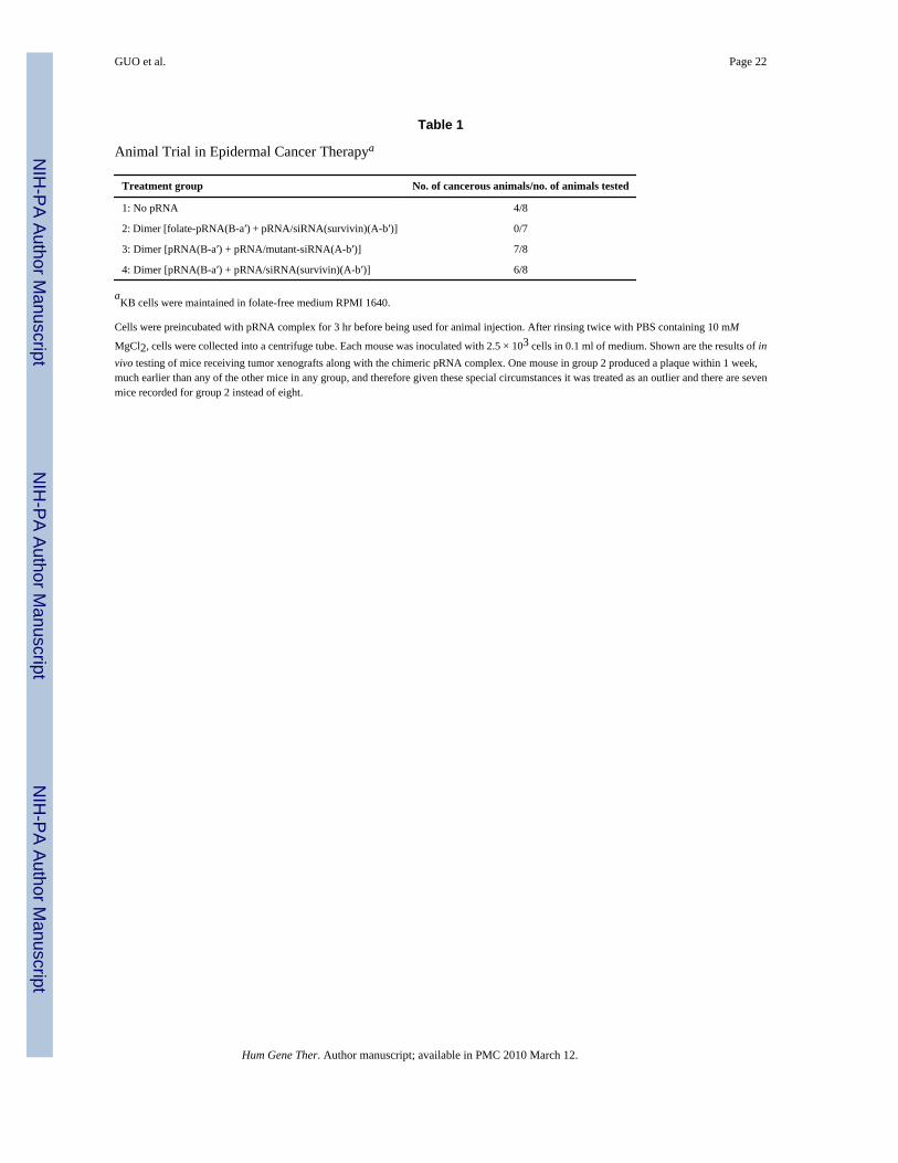
Animal trials were conducted to test the specificity by ex vivo delivery, using a dimer containingboth pRNA(A-b′)/folate and pRNA(B-a′)/siRNA(survivin). The potential of this RNA dimerto suppress tumor formation was tested in athymic nude mice. Nasopharyngeal epidermalcarcinoma (KB) cells were incubated with dimeric RNA with or without the blocking reagent,free folate, before being introduced into the nude mice by axillary injection. The mice receivingcells alone developed tumors within 3 weeks, whereas none of the mice receiving cellspretreated with the dimers with pRNA(A-b′)/folate and pRNA(B-a′)/siRNA(survivin)developed tumors (Table 1). The inhibition of tumor formation is specific because the controldimer RNA without folate conjugation used in control mice groups did not affect tumordevelopment.
DISCUSSIONφ29 pRNA has a tendency to form dimers, which are the building blocks of hexamers (Fig.10), as a result of the interaction of interlocking loops of each pRNA. This paper hasdemonstrated the production of dimers to deliver therapeutic RNA to specific cells. In thefuture, chimeric hexamers could also be assembled via hand-in-hand interaction. Thus, becausethere are six chimeric pRNAs in the hexamer, there would be six positions available to carrymolecules for cell recognition, therapy, and detection. Besides the receptor-binding aptamers,siRNA, ribozyme, and folate reported here, other materials such as fluorescent dyes, heavymetal, quantum dots, fluorescent beads, or radioisotopes can also be conjugated for the
GUO et al. Page 8
Hum Gene Ther. Author manuscript; available in PMC 2010 March 12.
NIH
-PA Author Manuscript
NIH
-PA Author Manuscript
NIH
-PA Author Manuscript

detection of cancer signatures at different stages of development. The reported methods forconjugating folate and FITC could be used for the conjugation of chemical drugs, andendosome-disrupting chemicals could be added to promote the release of siRNA from theendosome after delivery to improve therapeutic efficacy (Fig. 10). Nucleotide derivatives suchas 2′-fluoro-2′-deoxy-CTP, 2′-fluoro-2′-deoxy-UTP, or Spiegelmer will be incorporated intothe RNA to produce stable in vitro RNA transcripts that are resistant to RNase digestion(Soutschek et al., 2004).
This polyvalent RNA complex can also potentially be used to treat chronic viral infections,such as those caused by HIV and hepatitis B virus, by targeting the specific virus glycoproteinsincorporated on the infected cell surface. It is well established in the scientific community thatRNAs do not induce a detectable immune response except when complexed with proteins(Madaio et al., 1984; Goldsby et al., 2002). The use of RNA as a delivery vehicle could avoidthe problems of immune response and the rejection of protein vectors after repeated long-termdrug administration. The use of such a 25 nm RNA complex would provide a longer turnovertime in the body than other small molecules would offer. The toxicity or other adverse effectsof the pRNA chimera were further mitigated by the conjugation of a small DNA oligo(Manuscript in preparation).
AcknowledgmentsThis work was supported by NIH grants, ROI GM59944, and DOD prostate cancer grant W81XWH-050158 and DODbreast cancer grant DAMD17-03-1-0589 to P.G. We thank Guangxin Shen and Paul Robison for the flow cytometryassays; Suresh Mittal, David Riese, Phillip Low, and Shou-wei Ding for providing cell lines; we thank Taejin Lee forluciferase assays and Anoop Tiwari and Jeremy Hall for their assistance in the preparation of this manuscript; wethank Annette Khaled, Carol Koons, Richard Roll, Alex Cole, and Faqing Huang for their contributions to thismanuscript.
ReferencesAKKINA R, BANERJEA A, BAI J, ANDERSON J, LI MJ, ROSSI J. siRNAs, ribozymes and RNA
decoys in modeling stem cell-based gene therapy for HIV/AIDS. Anticancer Res 2003;23:1997–2005.[PubMed: 12894572]
BLOUNT KF, UHLENBECK OC. The hammerhead ribozyme. Biochem Soc Trans 2002;30:1119–1122.[PubMed: 12440986]
BRUMMELKAMP TR, BERNARDS R, AGAMI R. A system for stable expression of short interferingRNAs in mammalian cells. Science 2002;296:550–553. [PubMed: 11910072]
CARMICHAEL GG. Medicine: Silencing viruses with RNA. Nature 2002;418:379–380. [PubMed:12140542]
CHEN C, ZHANG C, GUO P. Sequence requirement for hand-in-hand interaction in formation of pRNAdimers and hexamers to gear φ29 DNA translocation motor. RNA 1999;5:805–818. [PubMed:10376879]
CHEN C, SHENG S, SHAO Z, GUO P. A dimer as a building block in assembling RNA: A hexamerthat gears bacterial virus φ29 DNA-translocating machinery. J Biol Chem 2000;275:17510–17516.[PubMed: 10748150]
ELBASHIR SM, HARBORTH J, LENDECKEL W, YALCIN A, WEBER K, TUSCHL T. Duplexes of21-nucleotide RNAs mediate RNA interference in cultured mammalian cells. Nature 2001;411:494–498. [PubMed: 11373684]
ELLINGTON AD, SZOSTAK JW. In vitro selection of RNA molecules that bind specific ligands. Nature1990;346:818–822. [PubMed: 1697402]
GARVER K, GUO P. Boundary of pRNA functional domains and minimum pRNA sequence requirementfor specific connector binding and DNA packaging of phage φ29. RNA 1997;3:1068–1079. [PubMed:9292504]
GOLD L. The SELEX process: a surprising source of therapeutic and diagnositc compounds. HarveyLect 1995;91:47–57. [PubMed: 9127985]
GUO et al. Page 9
Hum Gene Ther. Author manuscript; available in PMC 2010 March 12.
NIH
-PA Author Manuscript
NIH
-PA Author Manuscript
NIH
-PA Author Manuscript

GOLDSBY, RA.; KINDT, TJ.; OSBORNE, BA.; KUBY, J. Immunology. W.H. Freeman and Company;New York: 2002. Antigens; p. 57-61.
GROSSMAN D, KIM PJ, SCHECHNER JS, ALTIERI DC. Inhibition of melanoma tumor growth invivo by survivin targeting. Proc Natl Acad Sci USA 2001;98:635–640. [PubMed: 11149963]
GUO P, ERICKSON S, ANDERSON D. A small viral RNA is required for in vitro packaging ofbacteriophage f29 DNA. Science 1987;236:690–694. [PubMed: 3107124]
GUO P, ZHANG C, CHEN C, TROTTIER M, GARVER K. Inter-RNA interaction of phage φ29 pRNAto form a hexameric complex for viral DNA transportation. Mol Cell 1998;2:149–155. [PubMed:9702202]
HANNON GJ. RNA interference. Nature 2002;418:244–251. [PubMed: 12110901]HOEPRICH S, GUO P. Computer modeling of three-dimensional structure of DNA-packaging RNA
(pRNA) monomer, dimer, and hexamer of φ29 DNA. J Biol Chem 2002;277:20794–20803.[PubMed: 11886855]
HOEPRICH S, ZHOU Q, GUO S, QI G, WANG Y, GUO P. Bacterial virus φ29 pRNA as a hammerheadribozyme escort to destroy hepatitis B virus. Gene Ther 2003;10:1258–1267. [PubMed: 12858191]
KHALED AR, DURUM SK. Lymphocide: Cytokines and the control of lymphoid homeostasis. Nat RevImmunol 2002;2:817–830. [PubMed: 12415306]
KHALED AR, REYNOLDS DA, YOUNG HA, THOMPSON CB, MUEGGE K, DURUM SK.Interleukin-3 withdrawal induces an early increase in mitochondrial membrane potential unrelatedto the Bcl-2 family: Roles of intracellular pH, ADP transport, and F0F1-ATPase. J Biol Chem2001;276:6453–6462. [PubMed: 11102440]
KHALED A, GUO S, LI F, GUO P. Controllable self-assembly of nanoparticles for specific delivery ofmultiple therapeutic molecules to cancer cells using RNA nanotechnology. Nano letters. 2005 (inpress).
KIM DH, LONGO M, HAN Y, LUNDBERG P, CANTIN E, ROSSI JJ. Interferon induction by siRNAsand ssRNAs synthesized by phage polymerase. Nat Biotechnol 2004;22:321–325. [PubMed:14990954]
KIM K, KHALED AR, REYNOLDS D, YOUNG HA, LEE CK, DURUM SK. Characterization of aninterleukin-7-dependent thymic cell line derived from a p53−/− mouse. J Immunol Methods2003;274:177–184. [PubMed: 12609543]
KRAUS E, JAMES W, BARCLAY AN. Cutting edge: Novel RNA ligands able to bind CD4 antigenand inhibit CD4+ T lymphocyte function. J Immunol 1998;160:5209–5212. [PubMed: 9605115]
KUMAR M, CARMICHAEL GG. Antisense RNA: Function and fate of duplex RNA in cells of highereukaryotes. Microbiol Mol Biol Rev 1998;62:1415–1434. [PubMed: 9841677]
KUMAR S, VAUX DL. Apoptosis: A Cinderella caspase takes center stage. Science 2002;297:1290–1291. [PubMed: 12193776]
LI H, LI WX, DING SW. Induction and suppression of RNA silencing by an animal virus. Science2002;296:1319–1321. [PubMed: 12016316]
LI N, YU C, HUANG F. Novel cyanine-AMP conjugates for efficient 5′ RNA fluorescent labeling byone-step transcription and replacement of [gamma-32P]ATP in RNA structural investigation.Nucleic Acids Res 2005;33:e37. [PubMed: 15731330]
LU Y, LOW PS. Immunotherapy of folate receptor-expressing tumors: Review of recent advances andfuture prospects. J Control Release 2003;91:17–29. [PubMed: 12932634]
MADAIO MP, HODDER S, SCHWARTZ RS, STOLLAR BD. Responsiveness of autoimmune andnormal mice to nucleic acid antigens. J Immunol 1984;132:872–876. [PubMed: 6690621]
MAT-ARIP Y, GARVER K, CHEN C, SHENG S, SHAO Z, GUO P. Three-dimensional interaction ofφ29 pRNA dimer probed by chemical modification interference, cryo-AFM, and cross-linking. J BiolChem 2001;276:32575–32584. [PubMed: 11371551]
McCAFFREY AP, MEUSE L, PHAM TT, CONKLIN DS, HANNON GJ, KAY MA. RNA interferencein adult mice. Nature 2002;418:38–39. [PubMed: 12097900]
NOVINA CD, MURRAY MF, DYKXHOORN DM, BERESFORD PJ, RIESS J, LEE SK, COLLMANRG, LIEBERMAN J, SHANKAR P, SHARP PA. siRNA-directed inhibition of HIV-1 infection. NatMed 2002;8:681–686. [PubMed: 12042777]
GUO et al. Page 10
Hum Gene Ther. Author manuscript; available in PMC 2010 March 12.
NIH
-PA Author Manuscript
NIH
-PA Author Manuscript
NIH
-PA Author Manuscript

SHU D, HUANG L, HOEPRICH S, GUO P. Construction of φ29 DNA-packaging RNA (pRNA)monomers, dimers and trimers with variable sizes and shapes as potential parts for nanodevices. JNanosci Nanotech 2003;3:295–302.
PENNATI M, BINDA M, COLELLA G, FOLINI M, CITTI L, VILLA R, DAIDONE MG, ZAFFARONIN. Radiosensitization of human melanoma cells by ribozyme-mediated inhibition of survivinexpression. J Invest Dermatol 2003;120:648–654. [PubMed: 12648230]
SHU D, MOLL D, DENG Z, MAO C, GUO P. Bottom-up assembly of RNA arrays and superstructuresas potential parts in nanotechnology. Nano Lett 2004;4:1717–1724.
SOUTSCHEK J, AKINC A, BRAMLAGE B, CHARISSE K, CONSTIEN R, DONOGHUE M,ELBASHIR S, GEICK A, HADWIGER P, HARBORTH J, JOHN M, KESAVAN V, LAVINE G,PANDEY RK, RACIE T, RAJEEV KG, ROHL I, TOUDJARSKA I, WANG G, WUSCHKO S,BUMCROT D, KOTELIANSKY V, LIMMER S, MANOHARAN M, VORNLOCHER HP.Therapeutic silencing of an endogenous gene by systemic administration of modified siRNAs. Nature2004;432:173–178. [PubMed: 15538359]
SUDIMACK J, LEE RJ. Targeted drug delivery via the folate receptor. Adv Drug Deliv Rev2000;41:147–162. [PubMed: 10699311]
TROTTIER M, GUO P. Approaches to determine stoichiometry of viral assembly components. J Virol1997;71:487–494. [PubMed: 8985375]
ZHANG CL, LEE CS, GUO P. The proximate 5′ and 3′ ends of the 120-base viral RNA (pRNA) arecrucial for the packaging of bacteriophage f29 DNA. Virology 1994;201:77–85. [PubMed: 8178491]
ZHANG CL, TELLINGHUISEN T, GUO P. Confirmation of the helical structure of the 5′/3′ termini ofthe essential DNA packaging pRNA of phage f29. RNA 1995;1:1041–1050. [PubMed: 8595559]
ZHANG F, LEMIEUX S, WU X, ST-ARNAUD S, McMURRAY CT, MAJOR F, ANDERSON D.Function of hexameric RNA in packaging of bacteriophage φ29 DNA in vitro. Mol Cell 1998;2:141–147. [PubMed: 9702201]
GUO et al. Page 11
Hum Gene Ther. Author manuscript; available in PMC 2010 March 12.
NIH
-PA Author Manuscript
NIH
-PA Author Manuscript
NIH
-PA Author Manuscript

FIG. 1.Sketch of sequence and structure of pRNA chimeras. (A) φ29 pRNA sequence and secondarystructure. The right- and left-hand loops are circled in orange and green, respectively. Thedouble-stranded helical domain on the 5′/3′ ends is framed, and the domain for dimer formationis shaded. The curved line points to the two interacting loops. (B) Three-dimensional structureof pRNA dimer. (C) Native polyacrylamide gel showing monomer and dimer of the pRNAchimeras exhibiting different migration rates. Below the gel are cryo-AFM images of ′29 pRNAmonomer and dimer. The colors reflect the thickness and height of the molecule; the brighterthe color, the thicker or taller the molecule. (D) Design of chimeric pRNA dimers harboringforeign moieties (see Nomenclature of RNA Subunits, under Results).
GUO et al. Page 12
Hum Gene Ther. Author manuscript; available in PMC 2010 March 12.
NIH
-PA Author Manuscript
NIH
-PA Author Manuscript
NIH
-PA Author Manuscript

FIG. 2.Processing of chimeric pRNA/siRNA complex by Dicer. The structures of pRNA/siRNA andpRNA vector are shown in (A) and (B). Processing of pRNA/siRNA into 22-bp siRNAs byrecombinant Dicer. The chimeric pRNA/siRNA with 5′-end 32P labeling was incubated withpurified recombinant Dicer for 30 min and 2 hr, respectively, and then separated on a denaturingPAGE/urea gel. A radiolabeled 22-nucleotide RNA was used as a molecular weight marker.
GUO et al. Page 13
Hum Gene Ther. Author manuscript; available in PMC 2010 March 12.
NIH
-PA Author Manuscript
NIH
-PA Author Manuscript
NIH
-PA Author Manuscript

FIG. 3.Functional assay of chimeric pRNA/siRNA(GFP) by transfection. (A–C) Fluorescencemicroscopy images showing the silencing of GFP gene by transfection. (A) Dose-dependentsilencing of GFP gene by chimeric pRNA/siRNA(GFP) (left column). A mutant pRNA/siRNA(right column) served as negative control. (B) GFP expression of cells transfected with variousRNAs: (a) no RNA; (b) synthesized double-stranded siRNA(GFP); (c) double-stranded siRNA(LacZ) control; (d) pRNA/siRNA(GFP); (e) pRNA/siRNA(mutant); (f) pRNA vector alone.(C) Comparison of the performance of (a) chimeric pRNA/siRNA(GFP) and (b) conventionaldouble-stranded siRNA(GFP) at the same molar concentration; (c) control with no siRNAtreatment. (D) Northern blot to examine the effect of chimeric pRNA/siRNA(GFP) on GFPmRNA level after transfection. Lanes 1 and 2 show the effects of two different constructs ofpRNA/siRNA(GFP); lane 3, double-stranded siRNA; lane 4, cells without RNA treatment.rRNA was used as loading control.
GUO et al. Page 14
Hum Gene Ther. Author manuscript; available in PMC 2010 March 12.
NIH
-PA Author Manuscript
NIH
-PA Author Manuscript
NIH
-PA Author Manuscript

FIG. 4.Functional assay of chimeric pRNA/siRNA targeting luciferase by transfection. (A) Dualreporter luciferase assay showing the specific knockdown of firefly luciferase or Renillaluciferase expression by pRNA/siRNA(firefly) or pRNA/siRNA(Renilla), respectively, in adose-dependent manner. (B) Comparison of the activities of conventional hairpin siRNA(luciferase) and pRNA/siRNA(luciferase). pRNA/siRNA(mutant) with mutations in siRNAsequences was included as a nonspecific control.
GUO et al. Page 15
Hum Gene Ther. Author manuscript; available in PMC 2010 March 12.
NIH
-PA Author Manuscript
NIH
-PA Author Manuscript
NIH
-PA Author Manuscript

FIG. 5.Apoptosis and cell death induced by transfection of chimeric pRNA harboring siRNA targetingsurvivin. (I) Breast cancer MCF-7 cells were transfected with pRNA/siRNA(survivin) andapoptosis was monitored by PI–annexin V double-labeling followed by flow cytometry. Cellsin the bottom right quadrant represent apoptotic cells. (II) Breast cancer cells (MDA-231) andprostate cancer cells (PC-3) were transfected with 20 pmol of pRNA/siRNA(survivin) in 24-well plates and images were taken 24 hr after transfection. The mutant pRNA/siRNA wastransfected in parallel as a negative control.
GUO et al. Page 16
Hum Gene Ther. Author manuscript; available in PMC 2010 March 12.
NIH
-PA Author Manuscript
NIH
-PA Author Manuscript
NIH
-PA Author Manuscript

FIG. 6.Functional assay of pRNA/siRNA chimera targeting proapoptotic factor BAD. (A) pRNA/siRNA(BAD) and control siRNAs (10 nM) were introduced into pro-B cells by electroporation,combined with a transfection reagent on day 1. Cells were washed to remove IL-3 on day 2and assayed for viability on day 3. (B) BAD protein levels were compared in cells transfectedwith chimeric siRNA(BAD) or two mutant controls containing different mutations withinsiRNA sequences. Control cells were treated with pRNA alone. Cell lysates were prepared(Khaled et al., 2001) on day 3 and proteins were separated by 12% SDS–PAGE followed byWestern blot with BAD antibody (Cell Signaling Technology, Beverly, MA). Numbers belowthe panel indicate the remaining BAD level, expressed as a percentage compared with controlcells. (C) Pro-B cells transfected with pRNA/siRNA(survivin) or mutant were grown incomplete medium containing IL-3 or deprived of IL-3. The impact of RNA on cell morphologywas observed by microscopy.
GUO et al. Page 17
Hum Gene Ther. Author manuscript; available in PMC 2010 March 12.
NIH
-PA Author Manuscript
NIH
-PA Author Manuscript
NIH
-PA Author Manuscript

FIG. 7.Specific delivery of chimeric pRNA/siRNA by CD4 receptor. (I) Green circles correspondingto the binding of FITC-labeled pRNA dimer containing CD4-binding aptamer to lymphocyteswere shown by confocal microscopy (a) and the entry of RNA was shown as a green spot insidethe cell (d). Texas Red-labeled transferrin was used as a positive control of internalization (b).No binding was observed in the control cell line without CD4 expression (c). (II) Incubationof RNA dimer led to the specific suppression of cell viability, as measured by trypan blueexclusion assay. Three different cell lines with no, low, or high CD4 expression level wereincubated with the pRNA/siRNA(survivin)-pRNA/aptamer(CD4) complex in the presence orabsence of IL-7.
GUO et al. Page 18
Hum Gene Ther. Author manuscript; available in PMC 2010 March 12.
NIH
-PA Author Manuscript
NIH
-PA Author Manuscript
NIH
-PA Author Manuscript

FIG. 8.Toxicity assay of chimeric pRNA targeting survivin. HeLaT4 cells were seeded in 24-wellplates so that they will be 30–50% confluent at the time of incubation. Twenty-four hours afterseeding, the indicated amount of RNA was added into each well. Cells were further incubatedin incubator for 24, 48, and 72 hr and the number of viable cells was counted by hemacytometer.The relative survival rates displayed were obtained by dividing the number of surviving cellsin each treatment by the number of surviving cells without treatment with RNA.
GUO et al. Page 19
Hum Gene Ther. Author manuscript; available in PMC 2010 March 12.
NIH
-PA Author Manuscript
NIH
-PA Author Manuscript
NIH
-PA Author Manuscript

FIG. 9.Specific delivery of chimeric pRNA/siRNA by folate-pRNA. (A) Flow cytometry analyses ofthe binding of FITC-labeled folate-pRNA to KB cells. Left: Cells were incubated with folate-pRNA labeled with FITC. Middle: Cells were preincubated with free folate, which served asa blocking agent to compete with folate-pRNA for binding to the receptor. Right: Binding wasalso tested using folate-free pRNA labeled with FITC as a negative control. The percentagesof FITC-positive cells are shown in the top right quadrants. (B) Specific binding of folate-pRNA dimer to KB cells. After incubation of cells with the [3H]folate-pRNA dimer in thepresence (middle column) or absence (left column) of free folate, cells were isolated andsubjected to scintillation counting. The right-hand column represent 3H-labeled dimer withoutfolate labeling as a negative control. (C) In a knockdown assay by incubation, folate-chimericdimer complex containing pRNA(B-a′)/folate and pRNA(A-b′)/siRNA(firefly) was incubatedwith KB cells for 3 hr to allow the binding and entry of RNA. The luciferase level was measuredthe next day in the dual reporter system. The control dimer was identical to the folate dimerexcept for its lack of folate labeling.
GUO et al. Page 20
Hum Gene Ther. Author manuscript; available in PMC 2010 March 12.
NIH
-PA Author Manuscript
NIH
-PA Author Manuscript
NIH
-PA Author Manuscript

FIG. 10.The potential use of pRNA hexamers as polyvalent gene delivery vectors. Six copies of pRNAhave been found to form a hexameric ring to drive the DNA-packaging motor of bacterial virusφ29. There would therefore be six positions available to carry foreign moieties for targeting,therapy, and detection.
GUO et al. Page 21
Hum Gene Ther. Author manuscript; available in PMC 2010 March 12.
NIH
-PA Author Manuscript
NIH
-PA Author Manuscript
NIH
-PA Author Manuscript
NIH
-PA Author Manuscript
NIH
-PA Author Manuscript
NIH
-PA Author Manuscript
GUO et al. Page 22
Table 1
Animal Trial in Epidermal Cancer Therapya
Treatment group No. of cancerous animals/no. of animals tested
1: No pRNA 4/8
2: Dimer [folate-pRNA(B-a′) + pRNA/siRNA(survivin)(A-b′)] 0/7
3: Dimer [pRNA(B-a′) + pRNA/mutant-siRNA(A-b′)] 7/8
4: Dimer [pRNA(B-a′) + pRNA/siRNA(survivin)(A-b′)] 6/8
aKB cells were maintained in folate-free medium RPMI 1640.
Cells were preincubated with pRNA complex for 3 hr before being used for animal injection. After rinsing twice with PBS containing 10 mM
MgCl2, cells were collected into a centrifuge tube. Each mouse was inoculated with 2.5 × 10³ cells in 0.1 ml of medium. Shown are the results of invivo testing of mice receiving tumor xenografts along with the chimeric pRNA complex. One mouse in group 2 produced a plaque within 1 week,much earlier than any of the other mice in any group, and therefore given these special circumstances it was treated as an outlier and there are sevenmice recorded for group 2 instead of eight.
Hum Gene Ther. Author manuscript; available in PMC 2010 March 12.