Therapeutic Intradiscal Procedures for Lumbosacral ...
22
Therapeutic Intradiscal Procedures for Lumbosacral Radiculopathy Guiseppe Bonaldi and Alessandro Cianfoni HISTORICAL OVERVIEW In the early 1900s, degenerative and traumatic interverte- bral disc pathology were first established as a key cause of low back pain and sciatica, and in 1934, Mixter and Barr [1] described the herniated nucleus pulposus. Radicular pain, which is usually caused by herniation of an interver- tebral disc, is a common problem with an annual incidence of 5 per 1000 [2,3]. In 60% to 80% of patients experiencing their first episode of radicular pain, the symptoms recede to a nondisabling level within a period of 6 weeks [3]. The remaining group of patients qualifies for spinal imaging, usually magnetic resonance imaging (MRI). Patients suf- fering from radicular pain, in which spinal imaging shows an herniated disc compressing the nerve root involved in the radiculopathy, have historically been considered pos- sible candidates for open surgical discectomy, with the intent of providing decompression of the nerve root by removing the herniated disc. Numerous surgical treat- ments for discogenic pain have been developed, ranging from disc excision with laminectomy, microdiscectomy, spinal fusion, to artificial disc replacement. Because of the considerable morbidity and convalescence period inher- ent to conventional lumbar disc surgery, there has been an ongoing search for less invasive methods of treatment [4]. Multiple percutaneous minimally invasive interven- tional techniques to achieve disc decompression have been described. Percutaneous access to the disc was first used in the 1950s to biopsy the disc using needles [5,6]. In 1963, Lyman Smith, an orthopedic surgeon in Chicago, first described a minimally invasive aĴempt to treat sciat- ica through a percutaneous injection of chymopapain into the disc, with intent of achieving enzymatic chemolysis of the nucleus pulposus and of its protruding fragments compressing the nerve root [7–9]. Although this tech- nique is still widely used in the world, reports of severe allergic reactions, and catastrophic complications follow- ing inadvertent injection of chymopapain into the sub- arachnoid space, have dampened the early enthusiasm for this procedure. Smith opened the path, and from the seventies onwards, many other percutaneous techniques have been proposed, involving mechanical removal of the nucleus pulposus, with different types of instruments, with or without fiberscopic vision, or different types of energy (radiofrequency [RF], laser, coblation, etc.) for the reduction-decompression of the nucleus pulposus, and of its protruding components [10–16]. RATIONALE Spinal pain is the result of a complex interplay of mechanical, biochemical, and biomechanical processes. Radiculopathy arises from direct neural compression by disc herniations and associated inflammatory and ischemic phenomena. Symptoms can also arise from a disc protrusion because of the effect on heavily innervated surrounding structures such as the outer annulus and posterior longitudinal lig- ament. The severity of symptoms does not always corre- late with the extent of the herniation [17]. Sensitization of the central nervous system has also been suggested to be a possible causative factor of chronicity in some spinal pain conditions. With surgical approaches there is direct visualization of the herniated disc, and removal of the portion of the disc compressing the adjacent nerve root. Percutaneous discectomy techniques aĴempt nerve root decompression indirectly by decreasing the central disc pressure [18]. The treatment principle of percutaneous disc decompression is based on the concept of the interverte- bral disc being a closed hydraulic system. This system con- sists of the nucleus pulposus, containing a large amount of water, surrounded by the inelastic annulus fibrosus. An increase in water content of the nucleus pulposus leads to a disproportional increase of intradiscal pressure. On the other hand a decrease of intradiscal volume causes a disproportionally large decrease in intradiscal pressure [19,20]. Central decompression is achieved by the removal of material from the nucleus pulposus. The most oĞen stated goal of central nuclear decompression is to lower the pressure in the nucleus and to allow room for the herni- ated fragment to recede inward. The theory postulates that intact outer annular fibers will be able to contract enough to reduce the tension on both the nerve root and annu- lus. Additional suggested effects of central decompression include denaturation and fibrotic changes in the nucleus pulposus, which should in turn limit the ability of the nuclear matrix to aĴract water, thereby causing a long-last- ing pressure reduction [21], and reinforce the inner annular fibers, reducing the tendency of the central components of the disc to herniate toward the spinal canal [22]. Although 24 Depalma_864716_PTR_CH24_14-02-11_217-238.indd 217 Depalma_864716_PTR_CH24_14-02-11_217-238.indd 217 2/12/2011 2:10:19 PM 2/12/2011 2:10:19 PM
-
Upload
khangminh22 -
Category
Documents
-
view
1 -
download
0
Transcript of Therapeutic Intradiscal Procedures for Lumbosacral ...
Therapeutic Intradiscal Procedures for Lumbosacral Radiculopathy
Guiseppe Bonaldi and Alessandro Cianfoni
HISTORICAL OVERVIEW
In the early 1900s, degenerative and traumatic interverte-bral disc pathology were fi rst established as a key cause of low back pain and sciatica, and in 1934, Mixter and Barr [1] described the herniated nucleus pulposus. Radicular pain, which is usually caused by herniation of an interver-tebral disc, is a common problem with an annual incidence of 5 per 1000 [2,3]. In 60% to 80% of patients experiencing their fi rst episode of radicular pain, the symptoms recede to a nondisabling level within a period of 6 weeks [3]. The remaining group of patients qualifi es for spinal imaging, usually magnetic resonance imaging (MRI). Patients suf-fering from radicular pain, in which spinal imaging shows an herniated disc compressing the nerve root involved in the radiculopathy, have historically been considered pos-sible candidates for open surgical discectomy, with the intent of providing decompression of the nerve root by removing the herniated disc. Numerous surgical treat-ments for discogenic pain have been developed, ranging from disc excision with laminectomy, microdiscectomy, spinal fusion, to artifi cial disc replacement. Because of the considerable morbidity and convalescence period inher-ent to conventional lumbar disc surgery, there has been an ongoing search for less invasive methods of treatment [4]. Multiple percutaneous minimally invasive interven-tional techniques to achieve disc decompression have been described. Percutaneous access to the disc was fi rst used in the 1950s to biopsy the disc using needles [5,6]. In 1963, Lyman Smith, an orthopedic surgeon in Chicago, fi rst described a minimally invasive a empt to treat sciat-ica through a percutaneous injection of chymopapain into the disc, with intent of achieving enzymatic chemolysis of the nucleus pulposus and of its protruding fragments compressing the nerve root [7–9]. Although this tech-nique is still widely used in the world, reports of severe allergic reactions, and catastrophic complications follow-ing inadvertent injection of chymopapain into the sub-arachnoid space, have dampened the early enthusiasm for this procedure. Smith opened the path, and from the seventies onwards, many other percutaneous techniques have been proposed, involving mechanical removal of the nucleus pulposus, with diff erent types of instruments, with or without fi berscopic vision, or diff erent types of energy (radiofrequency [RF], laser, coblation, etc.) for the
reduction-decompression of the nucleus pulposus, and of its protruding components [10–16].
RATIONALE
Spinal pain is the result of a complex interplay of mechanical, biochemical, and biomechanical processes. Radiculopathy arises from direct neural compression by disc herniations and associated infl ammatory and ischemic phenomena. Symptoms can also arise from a disc protrusion because of the eff ect on heavily innervated surrounding structures such as the outer annulus and posterior longitudinal lig-ament. The severity of symptoms does not always corre-late with the extent of the herniation [17]. Sensitization of the central nervous system has also been suggested to be a possible causative factor of chronicity in some spinal pain conditions. With surgical approaches there is direct visualization of the herniated disc, and removal of the portion of the disc compressing the adjacent nerve root. Percutaneous discectomy techniques a empt nerve root decompression indirectly by decreasing the central disc pressure [18]. The treatment principle of percutaneous disc decompression is based on the concept of the interverte-bral disc being a closed hydraulic system. This system con-sists of the nucleus pulposus, containing a large amount of water, surrounded by the inelastic annulus fi brosus. An increase in water content of the nucleus pulposus leads to a disproportional increase of intradiscal pressure. On the other hand a decrease of intradiscal volume causes a disproportionally large decrease in intradiscal pressure [19,20]. Central decompression is achieved by the removal of material from the nucleus pulposus. The most o en stated goal of central nuclear decompression is to lower the pressure in the nucleus and to allow room for the herni-ated fragment to recede inward. The theory postulates that intact outer annular fi bers will be able to contract enough to reduce the tension on both the nerve root and annu-lus. Additional suggested eff ects of central decompression include denaturation and fi brotic changes in the nucleus pulposus, which should in turn limit the ability of the nuclear matrix to a ract water, thereby causing a long-last-ing pressure reduction [21], and reinforce the inner annular fi bers, reducing the tendency of the central components of the disc to herniate toward the spinal canal [22]. Although
24
Depalma_864716_PTR_CH24_14-02-11_217-238.indd 217Depalma_864716_PTR_CH24_14-02-11_217-238.indd 217 2/12/2011 2:10:19 PM2/12/2011 2:10:19 PM
218 Part I • Lumbosacral Spine • Radiculopathy/Radicular Pain
of performing physician and of their patients, but have not yet proven their defi nite effi cacy in reliable long-term randomized controlled trials (RCTs). Gibson and Waddell [35] in the 2009 Cochrane Collaboration review presented the results from 40 RCTs and 2 quasi-randomized con-trolled trials of surgical interventions for lumbar disc pro-lapse. This review indicated that the place for other forms of discectomy other than traditional open discectomy is unresolved. Trials of percutaneous discectomy and laser discectomy suggest that clinical outcomes following treat-ment are at best fair, although the importance of patient selection is acknowledged. The authors concluded that discectomy provides faster relief from the acute a ack of sciatica, although any positive or negative eff ects on the long-term natural history of the underlying disc disease are unclear. However, the potential medical and economic benefi ts are too high to justify discarding percutaneous techniques as experimental or ineff ective on the sole basis of insuffi cient scientifi c proof [4,36].
Recent evidence-based guidelines from the American Society of Interventional Pain Physicians-Interventional Pain Management conclude that level II and III evidence exists supporting a variety of intradiscal therapeutic pro-cedures for lumbosacral radiculopathy [37]. The lack of good scientifi c evidence rests in part on the diffi culty in designing a powerful scientifi c trial, in consideration of the heterogeneous, and somehow subjective patients’ selection criteria, the diff erence in techniques, and operators’ skills, the control choice, and the overly subjective outcome mea-surements [38]. One controlled trial comparing laser disc decompression and open surgery is on its way [39]. It might be argued that open surgical techniques suff er from the same lack of high-quality scientifi c evidence with regard to the same pathology [40], especially in the long term, while being more invasive, carrying a much higher rate of potentially severe complications, and being more expen-sive. Finally, it should be noted that comparing results of percutaneous minimally invasive techniques with surgi-cal results is probably methodologically incorrect, because the disc decompression has diff erent indications, and tar-gets a diff erent type of pathology, or a diff erent stage of the same pathology (i.e., contained disc herniations; please see later), when compared with open surgery.
INDICATIONS
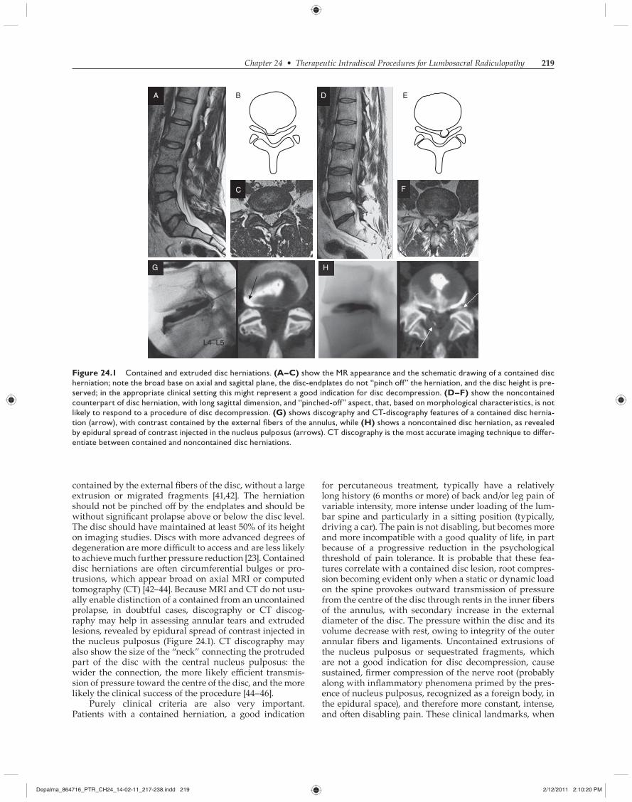
Percutaneous decompression has been shown eff ective in relieving radicular pain and to a lesser extent axial pain from contained disc protrusions. Patient selection criteria includes the presence of a contained disc herniation docu-mented by spinal imaging, causing radicular pain greater than axial pain, for 6 months or longer, and the patient having failed conservative measures, including anti-infl ammatory and analgesic medications and physical therapy. Imaging and clinical correlation is of utmost importance. In doubtful cases, diagnostic selective nerve root blocks, facet blocks, and provocative discography might help target the cor-rect pain generator. The success of the procedure depends greatly on selecting the lesions to treat (Figure 24.1): the protruding nucleus pulposus must be at least partially
sounding logical, there is li le proof that this phenomena actually occurs in humans in vivo. Immediate pressure drop in the nucleus pulposus with diff erent percutaneous disc decompression techniques has been experimentally proven in animals and in human cadavers, with larger eff ects on well-hydrated disc without signs of advanced degeneration and volume loss [23–25]. Although an exper-imental increase in pressure in the nucleus pulposus of a disc with annular tear transmits increased tension forces to the outer annular fi bers [26,27], there are no studies showing the opposite phenomenon of a decreased tension in the outer fi bers of the annulus following disc decom-pression. Fibrous changes in the nucleus have been shown in rabbit discs weeks a er laser disc decompression [24]. An MRI longitudinal study failed to demonstrate visible evidence of disc remodeling 6 weeks a er percutaneous disc decompression [28]. Nevertheless, relief of radiocul-opathy has been documented even in absence of a radio-graphically evident reduction in total disc volume [29]. There are, however, no studies that correlate the amount of nucleus removed with a decrease in tensional pres-sure on the outer annulus, and no one has studied how outcome correlates with the amount of nucleus removed. On the other hand, an interesting study has showed the potential of disc decompression, performed with coblation in porcine models, to activate biochemical as opposed to morphologic changes, by promoting a favorable humoral pain mediator shi , and initiating a repair response in the disc [30]; analogous result were obtained on rabbits’ discs by another group of researchers [31]. Disc decompression can be accomplished with chemical, mechanical, and ther-mal devices. The goal is to allow suffi cient tissue removal while minimizing collateral tissue damage and avoiding destabilization of the discovertebral unit [18]. Although open surgery is eff ective, it has well-known disadvantages, including epidural scarring, damage to bone, denervation of paraspinal muscles with consequent lumbar instabil-ity, long postoperative inactivity, and the frequent “failed back-surgery syndrome.” Patients with the la er are in fact o en untreatable and severely disabled. The benefi ts of percutaneous discectomy are greater than just avoidance of open surgery. Small contained disc protrusions have been shown to be less likely than larger disc extrusions to undergo spontaneous resorption [32] and are associated with worse surgical outcomes following discectomy [33]. Fortunately, this is the subtype of herniation most respon-sive to percutaneous techniques. Finally, percutaneous disc procedures have the advantage of a high patient psy-chological acceptance, tolerance, and satisfaction.
It should be stated here that there are new published data [34] suggesting the potential for intradiscal pro-cedures to accelerate the degenerative disc processes. Although the discs targeted by decompression proce-dures are symptomatic and already aff ected by some form of degeneration, and we do not know at present the clinical signifi cance of these phenomena, this aspect should be disclosed to patients in the informed consent process. From a purely evidence-based standpoint, the percutaneous minimally invasive disc decompression procedures have stood the test of time, having been per-formed for more than 40 years, with overall satisfaction
Depalma_864716_PTR_CH24_14-02-11_217-238.indd 218Depalma_864716_PTR_CH24_14-02-11_217-238.indd 218 2/12/2011 2:10:20 PM2/12/2011 2:10:20 PM
Chapter 24 • Therapeutic Intradiscal Procedures for Lumbosacral Radiculopathy 219
for percutaneous treatment, typically have a relatively long history (6 months or more) of back and/or leg pain of variable intensity, more intense under loading of the lum-bar spine and particularly in a si ing position (typically, driving a car). The pain is not disabling, but becomes more and more incompatible with a good quality of life, in part because of a progressive reduction in the psychological threshold of pain tolerance. It is probable that these fea-tures correlate with a contained disc lesion, root compres-sion becoming evident only when a static or dynamic load on the spine provokes outward transmission of pressure from the centre of the disc through rents in the inner fi bers of the annulus, with secondary increase in the external diameter of the disc. The pressure within the disc and its volume decrease with rest, owing to integrity of the outer annular fi bers and ligaments. Uncontained extrusions of the nucleus pulposus or sequestrated fragments, which are not a good indication for disc decompression, cause sustained, fi rmer compression of the nerve root (probably along with infl ammatory phenomena primed by the pres-ence of nucleus pulposus, recognized as a foreign body, in the epidural space), and therefore more constant, intense, and o en disabling pain. These clinical landmarks, when
contained by the external fi bers of the disc, without a large extrusion or migrated fragments [41,42]. The herniation should not be pinched off by the endplates and should be without signifi cant prolapse above or below the disc level. The disc should have maintained at least 50% of its height on imaging studies. Discs with more advanced degrees of degeneration are more diffi cult to access and are less likely to achieve much further pressure reduction [23]. Contained disc herniations are o en circumferential bulges or pro-trusions, which appear broad on axial MRI or computed tomography (CT) [42–44]. Because MRI and CT do not usu-ally enable distinction of a contained from an uncontained prolapse, in doubtful cases, discography or CT discog-raphy may help in assessing annular tears and extruded lesions, revealed by epidural spread of contrast injected in the nucleus pulposus (Figure 24.1). CT discography may also show the size of the “neck” connecting the protruded part of the disc with the central nucleus pulposus: the wider the connection, the more likely effi cient transmis-sion of pressure toward the centre of the disc, and the more likely the clinical success of the procedure [44–46].
Purely clinical criteria are also very important. Patients with a contained herniation, a good indication
A AB
C
D E
F
G
L4–L5
H
Figure 24.1 Contained and extruded disc herniations. (A–C) show the MR appearance and the schematic drawing of a contained disc
herniation; note the broad base on axial and sagittal plane, the disc-endplates do not “pinch off” the herniation, and the disc height is pre-
served; in the appropriate clinical setting this might represent a good indication for disc decompression. (D–F) show the noncontained
counterpart of disc herniation, with long sagittal dimension, and “pinched-off” aspect, that, based on morphological characteristics, is not
likely to respond to a procedure of disc decompression. (G) shows discography and CT-discography features of a contained disc hernia-
tion (arrow), with contrast contained by the external fi bers of the annulus, while (H) shows a noncontained disc herniation, as revealed
by epidural spread of contrast injected in the nucleus pulposus (arrows). CT discography is the most accurate imaging technique to differ-
entiate between contained and noncontained disc herniations.
Depalma_864716_PTR_CH24_14-02-11_217-238.indd 219Depalma_864716_PTR_CH24_14-02-11_217-238.indd 219 2/12/2011 2:10:20 PM2/12/2011 2:10:20 PM
220 Part I • Lumbosacral Spine • Radiculopathy/Radicular Pain
TECHNIQUES OF PERCUTANEOUS LUMBAR DISC ACCESS
Fluoroscopic Guidance
To safely perform intradiscal lumbar procedures, adequate anteroposterior (AP), laterolateral (LL), and oblique fl uoro-scopic views must be obtained. These characteristics can be off ered by C-arms, single-plane or biplane angiography units. By no means, one should undertake these proce-dures with the aid of only a fi xed fl uoroscopy unit. The fl uoroscopy table should be completely radiotransparent.
AP and LL Views of the Disc Space
For the interventionalists and radiology technologists, approaching the fl uoroscopic-guided spine procedures, the concept of AP and LL views should be reshaped. The spine has natural lordotic and kyphotic curvatures along the sagi al plane, as well as possible additional curvatures and rotations on the coronal and axial plane, which can be due to conformational deformity, such as in scoliosis and rotoscoliosis, or due to the patient positioning on the fl uo-roscopy table. Therefore, the classical 0° AP position of the tube defi ning AP view, and 90° LL position defi ning LL view, should be completely abandoned, and substituted by level-specifi c AP and LL views, that is, the AP views of L2-L3 and L4-L5 disc spaces will certainly have diff erent tube obliquity (Figure 24.2). These views are irrespective of any predetermined tube angulation, and of the patient’s body positioning, and should only be defi ned by the actual fl uoroscopic appearance of the vertebral body at the level
they last for at least 6 to 8 weeks, justify open surgery as the treatment of choice.
Contraindications include sequestered herniation, herniation greater than one third of the sagi al diameter of the spinal canal, progressive neurologic defi cit, infec-tion, bony deformity not allowing a safe percutaneous image-guided disc access, or other bone lesions which could compress a root and cause radicular symptoms [47]. Applying strict selection criteria, Onik estimated that only 5% to 10% of the patients with disc herniation who even-tually undergo surgery would be eligible for percutaneous disc decompression [48]. Given its low morbidity, however, disclosing the lesser likelihood of clinical success of the procedure, the minimally invasive therapeutic option can be ethically off ered to a wider range of patients, such as the ones with partially uncontained prolapses, as an a empt to avoid surgery, or when the risks of open surgery are higher because of age, general medical conditions, or other contraindications [48]. This typically applies to patients who have already undergone open surgery at the same level, because of the possibility of symptomatic epidural scar, and to elderly people. In fact, an observational study on a large cohort of patients reports these subgroups of patients as good responders to automated disc decompres-sion (APLD) [22]. Although the satisfactory outcome can be a ributed to several factors, the one that supersedes all is that in these patients, with a nerve root confi ned and compressed in a small space, either due to epidural fi brosis or arthropathic degenerative bone changes, even a small reduction in the volume of the disc by disc decompres-sion might result in radicular decompression and clinical improvement.
A
L3-L4L3-L4
L4-L5
L4-L5
L5-S1
L5-S1
B C D
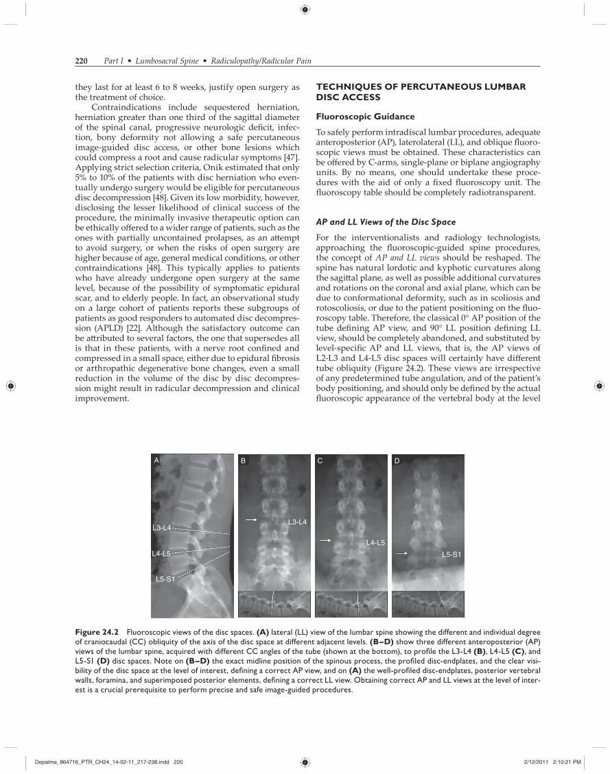
Figure 24.2 Fluoroscopic views of the disc spaces. (A) lateral (LL) view of the lumbar spine showing the different and individual degree
of craniocaudal (CC) obliquity of the axis of the disc space at different adjacent levels. (B–D) show three different anteroposterior (AP)
views of the lumbar spine, acquired with different CC angles of the tube (shown at the bottom), to profi le the L3-L4 (B), L4-L5 (C), and
L5-S1 (D) disc spaces. Note on (B–D) the exact midline position of the spinous process, the profi led disc-endplates, and the clear visi-
bility of the disc space at the level of interest, defi ning a correct AP view, and on (A) the well-profi led disc-endplates, posterior vertebral
walls, foramina, and superimposed posterior elements, defi ning a correct LL view. Obtaining correct AP and LL views at the level of inter-
est is a crucial prerequisite to perform precise and safe image-guided procedures.
Depalma_864716_PTR_CH24_14-02-11_217-238.indd 220Depalma_864716_PTR_CH24_14-02-11_217-238.indd 220 2/12/2011 2:10:21 PM2/12/2011 2:10:21 PM
Chapter 24 • Therapeutic Intradiscal Procedures for Lumbosacral Radiculopathy 221
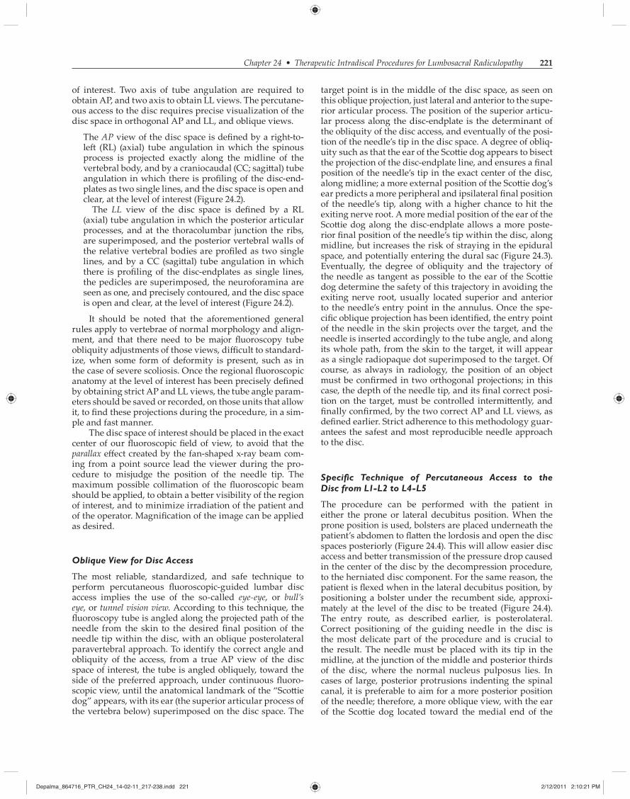
target point is in the middle of the disc space, as seen on this oblique projection, just lateral and anterior to the supe-rior articular process. The position of the superior articu-lar process along the disc-endplate is the determinant of the obliquity of the disc access, and eventually of the posi-tion of the needle’s tip in the disc space. A degree of obliq-uity such as that the ear of the Sco ie dog appears to bisect the projection of the disc-endplate line, and ensures a fi nal position of the needle’s tip in the exact center of the disc, along midline; a more external position of the Sco ie dog’s ear predicts a more peripheral and ipsilateral fi nal position of the needle’s tip, along with a higher chance to hit the exiting nerve root. A more medial position of the ear of the Sco ie dog along the disc-endplate allows a more poste-rior fi nal position of the needle’s tip within the disc, along midline, but increases the risk of straying in the epidural space, and potentially entering the dural sac (Figure 24.3). Eventually, the degree of obliquity and the trajectory of the needle as tangent as possible to the ear of the Sco ie dog determine the safety of this trajectory in avoiding the exiting nerve root, usually located superior and anterior to the needle’s entry point in the annulus. Once the spe-cifi c oblique projection has been identifi ed, the entry point of the needle in the skin projects over the target, and the needle is inserted accordingly to the tube angle, and along its whole path, from the skin to the target, it will appear as a single radiopaque dot superimposed to the target. Of course, as always in radiology, the position of an object must be confi rmed in two orthogonal projections; in this case, the depth of the needle tip, and its fi nal correct posi-tion on the target, must be controlled intermi ently, and fi nally confi rmed, by the two correct AP and LL views, as defi ned earlier. Strict adherence to this methodology guar-antees the safest and most reproducible needle approach to the disc.
Specifi c Technique of Percutaneous Access to the Disc from L1-L2 to L4-L5
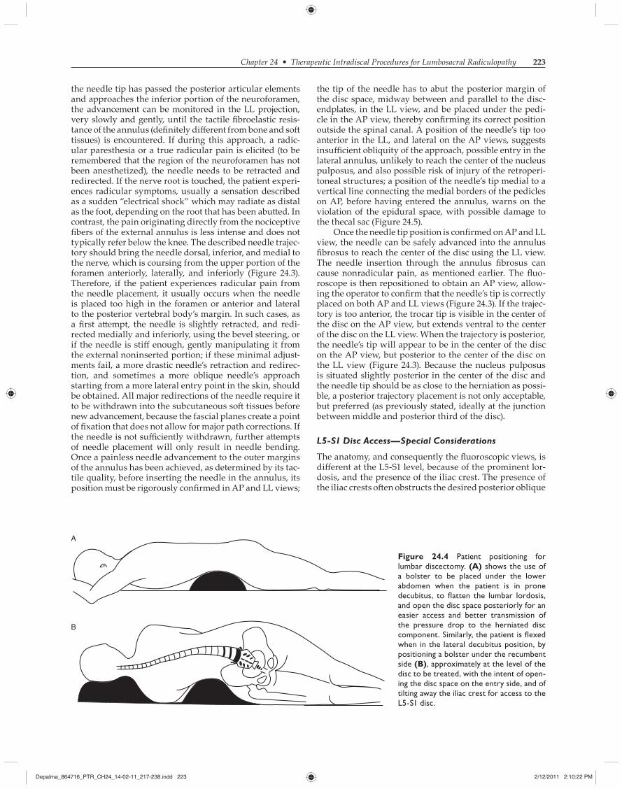
The procedure can be performed with the patient in either the prone or lateral decubitus position. When the prone position is used, bolsters are placed underneath the patient’s abdomen to fl a en the lordosis and open the disc spaces posteriorly (Figure 24.4). This will allow easier disc access and be er transmission of the pressure drop caused in the center of the disc by the decompression procedure, to the herniated disc component. For the same reason, the patient is fl exed when in the lateral decubitus position, by positioning a bolster under the recumbent side, approxi-mately at the level of the disc to be treated (Figure 24.4). The entry route, as described earlier, is posterolateral. Correct positioning of the guiding needle in the disc is the most delicate part of the procedure and is crucial to the result. The needle must be placed with its tip in the midline, at the junction of the middle and posterior thirds of the disc, where the normal nucleus pulposus lies. In cases of large, posterior protrusions indenting the spinal canal, it is preferable to aim for a more posterior position of the needle; therefore, a more oblique view, with the ear of the Sco ie dog located toward the medial end of the
of interest. Two axis of tube angulation are required to obtain AP, and two axis to obtain LL views. The percutane-ous access to the disc requires precise visualization of the disc space in orthogonal AP and LL, and oblique views.
The AP view of the disc space is defi ned by a right-to-le (RL) (axial) tube angulation in which the spinous process is projected exactly along the midline of the vertebral body, and by a craniocaudal (CC; sagi al) tube angulation in which there is profi ling of the disc-end-plates as two single lines, and the disc space is open and clear, at the level of interest (Figure 24.2). The LL view of the disc space is defi ned by a RL (axial) tube angulation in which the posterior articular processes, and at the thoracolumbar junction the ribs, are superimposed, and the posterior vertebral walls of the relative vertebral bodies are profi led as two single lines, and by a CC (sagi al) tube angulation in which there is profi ling of the disc-endplates as single lines, the pedicles are superimposed, the neuroforamina are seen as one, and precisely contoured, and the disc space is open and clear, at the level of interest (Figure 24.2).
It should be noted that the aforementioned general rules apply to vertebrae of normal morphology and align-ment, and that there need to be major fl uoroscopy tube obliquity adjustments of those views, diffi cult to standard-ize, when some form of deformity is present, such as in the case of severe scoliosis. Once the regional fl uoroscopic anatomy at the level of interest has been precisely defi ned by obtaining strict AP and LL views, the tube angle param-eters should be saved or recorded, on those units that allow it, to fi nd these projections during the procedure, in a sim-ple and fast manner.
The disc space of interest should be placed in the exact center of our fl uoroscopic fi eld of view, to avoid that the parallax eff ect created by the fan-shaped x-ray beam com-ing from a point source lead the viewer during the pro-cedure to misjudge the position of the needle tip. The maximum possible collimation of the fl uoroscopic beam should be applied, to obtain a be er visibility of the region of interest, and to minimize irradiation of the patient and of the operator. Magnifi cation of the image can be applied as desired.
Oblique View for Disc Access
The most reliable, standardized, and safe technique to perform percutaneous fl uoroscopic-guided lumbar disc access implies the use of the so-called eye-eye, or bull’s eye, or tunnel vision view. According to this technique, the fl uoroscopy tube is angled along the projected path of the needle from the skin to the desired fi nal position of the needle tip within the disc, with an oblique posterolateral paravertebral approach. To identify the correct angle and obliquity of the access, from a true AP view of the disc space of interest, the tube is angled obliquely, toward the side of the preferred approach, under continuous fl uoro-scopic view, until the anatomical landmark of the “Sco ie dog” appears, with its ear (the superior articular process of the vertebra below) superimposed on the disc space. The
Depalma_864716_PTR_CH24_14-02-11_217-238.indd 221Depalma_864716_PTR_CH24_14-02-11_217-238.indd 221 2/12/2011 2:10:21 PM2/12/2011 2:10:21 PM
222 Part I • Lumbosacral Spine • Radiculopathy/Radicular Pain
a broad base and encompasses both sides, the side of the patient’s symptoms should be chosen. There are two com-pelling reasons to do so: fi rst, to place the needle tip as close to the herniation as possible, particularly when the herniation is lateralized; second, to avoid the possibility of bilateral symptoms in the event of complications. In rare circumstances, a contralateral approach is preferred, such as when a correct needle positioning inside the disc is impossible from one side. That situation can arise when the needle repeatedly abuts the exiting nerve root, pre-cluding safe advancement into the annulus, such in cases of conjoined nerve root, a fl a ened root compressed by the disc herniation, or in case of other anatomical variants. When a conjoined nerve root is present, both roots can exit from a single foramen, and one root can take a more cau-dal and inferior course, which places it in the line of a cor-rectly placed needle. In case of a far lateral herniation, it is possible that the nerve root is in an abnormal position and pushed posteriorly resulting in obliteration of the space between the nerve and the anterior surface of the facet. An ossifi ed bridging posterolateral osteophyte might also make the disc access impossible from one side.
The procedure is monitored fl uoroscopically, using the oblique view, and advancing the needle along the eye-eye trajectory. Intermi ent fl uoroscopic control in the LL projec-tion confi rms that the needle is approaching the disc paral-lel to the disc-endplates, and serves as depth control. Once
disc-endplate line, is chosen. The most eff ective and safest way to accurately place the needle, as mentioned earlier, is to choose a path that passes laterally tangent and then anteriorly to the superior articular process of the zygapo-physeal complex (Figure 24.3). Once the skin entry point has been marked under fl uoroscopy, overlying the target point on the oblique fl uoroscopic view, a skin wheal is raised. Immediately therea er, a 22- to 25-gauge × 12- to 15-cm spinal needle is inserted and aimed just slightly more posteriorly than the anticipated route to the disc, aiming to touch the bone of the superior articular process of the facet. The bone contact is the depth control in this maneuver, assuring that the anesthetic will be delivered superfi cial to the foraminal space, in order not to numb the nerve root. It is critical that local anesthetic is not depos-ited anterior to the articular processes, because this would neutralize the warning role that radicular paresthesia has for the performing physician, alerting an undesired, too close proximity of the needle tip to the nerve root. Local anesthetic is then injected through the needle and the nee-dle is gradually withdrawn toward the skin, allowing for anesthetization of the underlying spinal musculature.
The spinal needle of small caliber used to deliver local anesthesia can also be used to test if the trajectory from the skin to the annulus is accurate. Choosing which side to enter the disc depends upon the side of the symptom-atic contained herniation. When the disc herniation has
A
E F G
B C D
Figure 24.3 Fluoroscopy and CT correlates of disc access anatomy. (A) shows the fl uoroscopic tube angle to obtain a correct eye-eye
(EE) view for a right posterolateral percutaneous access to the disc L3-L4 (B), as also shown on the correspondent 3D volume rendering
CT model (C). The CC angle of the tube is such that the disc space is well profi led at the level of interest, and the right-to-left (RL) obliq-
uity is such that the superior articular process (ear of the Scottie dog) of L4 is superimposed on the midpoint of the disc-endplate line.
This ensures an access-window for the needle (white dot on B and C) posterior, inferior, and medial to the exiting nerve root (D). (E,F) show the fi nal location of the needle (arrow) in the center of the nucleus pulposus on the AP and LL views. (G) shows the axial CT section
through the disc space, and the ideal needle path (dashed arrow); note that steep obliquity, tangent to the superior articular process of
the facet, is necessary to have a correct access to the disc, and to avoid the exiting nerve root (arrowhead).
Depalma_864716_PTR_CH24_14-02-11_217-238.indd 222Depalma_864716_PTR_CH24_14-02-11_217-238.indd 222 2/12/2011 2:10:21 PM2/12/2011 2:10:21 PM
Chapter 24 • Therapeutic Intradiscal Procedures for Lumbosacral Radiculopathy 223
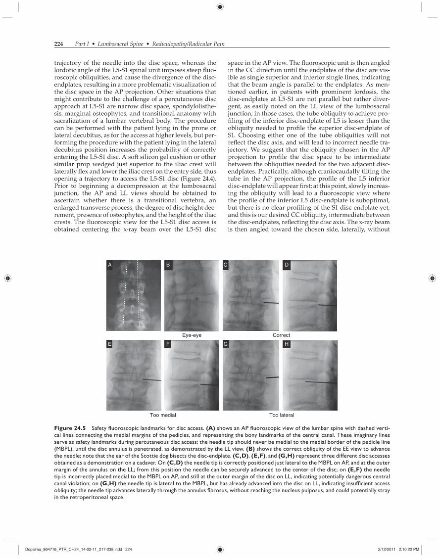
the tip of the needle has to abut the posterior margin of the disc space, midway between and parallel to the disc-endplates, in the LL view, and be placed under the pedi-cle in the AP view, thereby confi rming its correct position outside the spinal canal. A position of the needle’s tip too anterior in the LL, and lateral on the AP views, suggests insuffi cient obliquity of the approach, possible entry in the lateral annulus, unlikely to reach the center of the nucleus pulposus, and also possible risk of injury of the retroperi-toneal structures; a position of the needle’s tip medial to a vertical line connecting the medial borders of the pedicles on AP, before having entered the annulus, warns on the violation of the epidural space, with possible damage to the thecal sac (Figure 24.5).
Once the needle tip position is confi rmed on AP and LL view, the needle can be safely advanced into the annulus fi brosus to reach the center of the disc using the LL view. The needle insertion through the annulus fi brosus can cause nonradicular pain, as mentioned earlier. The fl uo-roscope is then repositioned to obtain an AP view, allow-ing the operator to confi rm that the needle’s tip is correctly placed on both AP and LL views (Figure 24.3). If the trajec-tory is too anterior, the trocar tip is visible in the center of the disc on the AP view, but extends ventral to the center of the disc on the LL view. When the trajectory is posterior, the needle’s tip will appear to be in the center of the disc on the AP view, but posterior to the center of the disc on the LL view (Figure 24.3). Because the nucleus pulposus is situated slightly posterior in the center of the disc and the needle tip should be as close to the herniation as possi-ble, a posterior trajectory placement is not only acceptable, but preferred (as previously stated, ideally at the junction between middle and posterior third of the disc).
L5-S1 Disc Access—Special Considerations
The anatomy, and consequently the fl uoroscopic views, is diff erent at the L5-S1 level, because of the prominent lor-dosis, and the presence of the iliac crest. The presence of the iliac crests o en obstructs the desired posterior oblique
the needle tip has passed the posterior articular elements and approaches the inferior portion of the neuroforamen, the advancement can be monitored in the LL projection, very slowly and gently, until the tactile fi broelastic resis-tance of the annulus (defi nitely diff erent from bone and so tissues) is encountered. If during this approach, a radic-ular paresthesia or a true radicular pain is elicited (to be remembered that the region of the neuroforamen has not been anesthetized), the needle needs to be retracted and redirected. If the nerve root is touched, the patient experi-ences radicular symptoms, usually a sensation described as a sudden “electrical shock” which may radiate as distal as the foot, depending on the root that has been abu ed. In contrast, the pain originating directly from the nociceptive fi bers of the external annulus is less intense and does not typically refer below the knee. The described needle trajec-tory should bring the needle dorsal, inferior, and medial to the nerve, which is coursing from the upper portion of the foramen anteriorly, laterally, and inferiorly (Figure 24.3). Therefore, if the patient experiences radicular pain from the needle placement, it usually occurs when the needle is placed too high in the foramen or anterior and lateral to the posterior vertebral body’s margin. In such cases, as a fi rst a empt, the needle is slightly retracted, and redi-rected medially and inferiorly, using the bevel steering, or if the needle is stiff enough, gently manipulating it from the external noninserted portion; if these minimal adjust-ments fail, a more drastic needle’s retraction and redirec-tion, and sometimes a more oblique needle’s approach starting from a more lateral entry point in the skin, should be obtained. All major redirections of the needle require it to be withdrawn into the subcutaneous so tissues before new advancement, because the fascial planes create a point of fi xation that does not allow for major path corrections. If the needle is not suffi ciently withdrawn, further a empts of needle placement will only result in needle bending. Once a painless needle advancement to the outer margins of the annulus has been achieved, as determined by its tac-tile quality, before inserting the needle in the annulus, its position must be rigorously confi rmed in AP and LL views;
A
B
Figure 24.4 Patient positioning for
lumbar discectomy. (A) shows the use of
a bolster to be placed under the lower
abdomen when the patient is in prone
decubitus, to fl atten the lumbar lordosis,
and open the disc space posteriorly for an
easier access and better transmission of
the pressure drop to the herniated disc
component. Similarly, the patient is fl exed
when in the lateral decubitus position, by
positioning a bolster under the recumbent
side (B), approximately at the level of the
disc to be treated, with the intent of open-
ing the disc space on the entry side, and of
tilting away the iliac crest for access to the
L5-S1 disc.
Depalma_864716_PTR_CH24_14-02-11_217-238.indd 223Depalma_864716_PTR_CH24_14-02-11_217-238.indd 223 2/12/2011 2:10:22 PM2/12/2011 2:10:22 PM
224 Part I • Lumbosacral Spine • Radiculopathy/Radicular Pain
space in the AP view. The fl uoroscopic unit is then angled in the CC direction until the endplates of the disc are vis-ible as single superior and inferior single lines, indicating that the beam angle is parallel to the endplates. As men-tioned earlier, in patients with prominent lordosis, the disc-endplates at L5-S1 are not parallel but rather diver-gent, as easily noted on the LL view of the lumbosacral junction; in those cases, the tube obliquity to achieve pro-fi ling of the inferior disc-endplate of L5 is lesser than the obliquity needed to profi le the superior disc-endplate of S1. Choosing either one of the tube obliquities will not refl ect the disc axis, and will lead to incorrect needle tra-jectory. We suggest that the obliquity chosen in the AP projection to profi le the disc space to be intermediate between the obliquities needed for the two adjacent disc-endplates. Practically, although craniocaudally tilting the tube in the AP projection, the profi le of the L5 inferior disc-endplate will appear fi rst; at this point, slowly increas-ing the obliquity will lead to a fl uoroscopic view where the profi le of the inferior L5 disc-endplate is suboptimal, but there is no clear profi ling of the S1 disc-endplate yet, and this is our desired CC obliquity, intermediate between the disc-endplates, refl ecting the disc axis. The x-ray beam is then angled toward the chosen side, laterally, without
trajectory of the needle into the disc space, whereas the lordotic angle of the L5-S1 spinal unit imposes steep fl uo-roscopic obliquities, and cause the divergence of the disc-endplates, resulting in a more problematic visualization of the disc space in the AP projection. Other situations that might contribute to the challenge of a percutaneous disc approach at L5-S1 are narrow disc space, spondylolisthe-sis, marginal osteophytes, and transitional anatomy with sacralization of a lumbar vertebral body. The procedure can be performed with the patient lying in the prone or lateral decubitus, as for the access at higher levels, but per-forming the procedure with the patient lying in the lateral decubitus position increases the probability of correctly entering the L5-S1 disc. A so silicon gel cushion or other similar prop wedged just superior to the iliac crest will laterally fl ex and lower the iliac crest on the entry side, thus opening a trajectory to access the L5-S1 disc (Figure 24.4). Prior to beginning a decompression at the lumbosacral junction, the AP and LL views should be obtained to ascertain whether there is a transitional vertebra, an enlarged transverse process, the degree of disc height dec-rement, presence of osteophytes, and the height of the iliac crests. The fl uoroscopic view for the L5-S1 disc access is obtained centering the x-ray beam over the L5-S1 disc
A B C D
E F G H
Too medial
Eye-eye Correct
Too lateral
Figure 24.5 Safety fl uoroscopic landmarks for disc access. (A) shows an AP fl uoroscopic view of the lumbar spine with dashed verti-
cal lines connecting the medial margins of the pedicles, and representing the bony landmarks of the central canal. These imaginary lines
serve as safety landmarks during percutaneous disc access; the needle tip should never be medial to the medial border of the pedicle line
(MBPL), until the disc annulus is penetrated, as demonstrated by the LL view. (B) shows the correct obliquity of the EE view to advance
the needle; note that the ear of the Scottie dog bisects the disc-endplate. (C,D), (E,F), and (G,H) represent three different disc accesses
obtained as a demonstration on a cadaver. On (C,D) the needle tip is correctly positioned just lateral to the MBPL on AP, and at the outer
margin of the annulus on the LL; from this position the needle can be securely advanced to the center of the disc; on (E,F) the needle
tip is incorrectly placed medial to the MBPL on AP, and still at the outer margin of the disc on LL, indicating potentially dangerous central
canal violation; on (G,H) the needle tip is lateral to the MBPL, but has already advanced into the disc on LL, indicating insuffi cient access
obliquity; the needle tip advances laterally through the annulus fi brosus, without reaching the nucleus pulposus, and could potentially stray
in the retroperitoneal space.
Depalma_864716_PTR_CH24_14-02-11_217-238.indd 224Depalma_864716_PTR_CH24_14-02-11_217-238.indd 224 2/12/2011 2:10:22 PM2/12/2011 2:10:22 PM
Chapter 24 • Therapeutic Intradiscal Procedures for Lumbosacral Radiculopathy 225
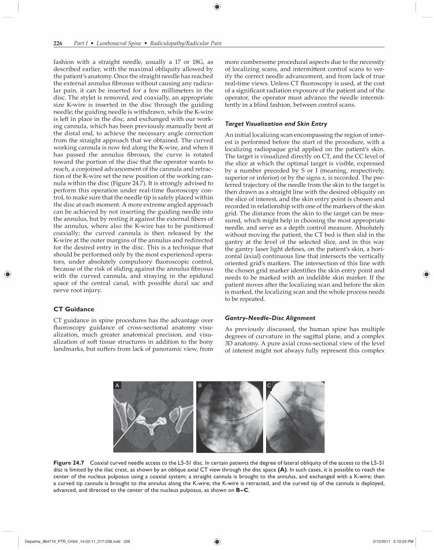
projected on the lateral third of the S1 endplate; any fur-ther obliquity of the x-ray beam brings the iliac crest to obstruct the path from the desired skin entry point and the disc space. Consequently, the entry route has to be less oblique (which means that the entry point in the skin is closer to the midline of the spine, for the needle to pass medially to the iliac crest), or must originate from a more cephalad starting point. With both approaches (more medial and more cephalad), there are instances in which straight instrumentation will not enter the disc correctly. If the trajectory of the needle is not obliquely angled enough, as discussed in the previous paragraph, it might be impossible to position the needle’s tip in the desired position in the center of the nucleus pulposus, as it will tend to be too lateral and anterior in the disc. If the trajec-tory comes from a more cephalad entry point, the needle might still enter the disc, but will not be parallel to the disc-endplates, and therefore it will not advance in the disc space to the center of the nucleus pulposus. If the cor-rect intradiscal position cannot be achieved with a straight cannula, a curved needle can be used. Although some operators might use a curved cannula as the introducing needle alone, this technique is very dependent on per-sonal skills and expertise, and might require several a empts to achieve the proper trajectory. The most reli-able and safe technique for a curved needle approach is most likely the coaxial technique. With the coaxial tech-nique, the access is performed in a standard eye-eye
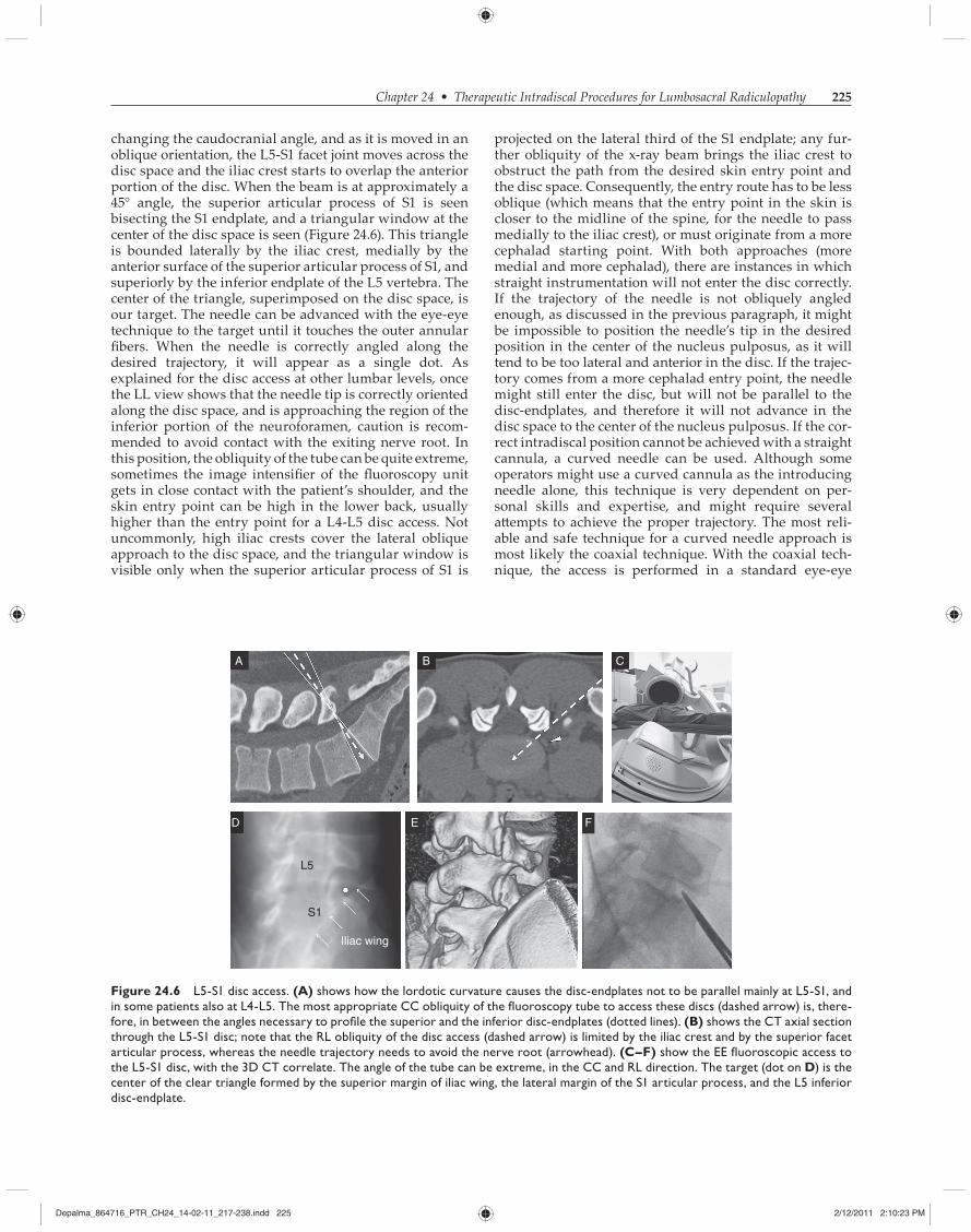
changing the caudocranial angle, and as it is moved in an oblique orientation, the L5-S1 facet joint moves across the disc space and the iliac crest starts to overlap the anterior portion of the disc. When the beam is at approximately a 45° angle, the superior articular process of S1 is seen bisecting the S1 endplate, and a triangular window at the center of the disc space is seen (Figure 24.6). This triangle is bounded laterally by the iliac crest, medially by the anterior surface of the superior articular process of S1, and superiorly by the inferior endplate of the L5 vertebra. The center of the triangle, superimposed on the disc space, is our target. The needle can be advanced with the eye-eye technique to the target until it touches the outer annular fi bers. When the needle is correctly angled along the desired trajectory, it will appear as a single dot. As explained for the disc access at other lumbar levels, once the LL view shows that the needle tip is correctly oriented along the disc space, and is approaching the region of the inferior portion of the neuroforamen, caution is recom-mended to avoid contact with the exiting nerve root. In this position, the obliquity of the tube can be quite extreme, sometimes the image intensifi er of the fl uoroscopy unit gets in close contact with the patient’s shoulder, and the skin entry point can be high in the lower back, usually higher than the entry point for a L4-L5 disc access. Not uncommonly, high iliac crests cover the lateral oblique approach to the disc space, and the triangular window is visible only when the superior articular process of S1 is
A B C
D E F
L5
S1
Iliac wing
Figure 24.6 L5-S1 disc access. (A) shows how the lordotic curvature causes the disc-endplates not to be parallel mainly at L5-S1, and
in some patients also at L4-L5. The most appropriate CC obliquity of the fl uoroscopy tube to access these discs (dashed arrow) is, there-
fore, in between the angles necessary to profi le the superior and the inferior disc-endplates (dotted lines). (B) shows the CT axial section
through the L5-S1 disc; note that the RL obliquity of the disc access (dashed arrow) is limited by the iliac crest and by the superior facet
articular process, whereas the needle trajectory needs to avoid the nerve root (arrowhead). (C–F) show the EE fl uoroscopic access to
the L5-S1 disc, with the 3D CT correlate. The angle of the tube can be extreme, in the CC and RL direction. The target (dot on D) is the
center of the clear triangle formed by the superior margin of iliac wing, the lateral margin of the S1 articular process, and the L5 inferior
disc-endplate.
Depalma_864716_PTR_CH24_14-02-11_217-238.indd 225Depalma_864716_PTR_CH24_14-02-11_217-238.indd 225 2/12/2011 2:10:23 PM2/12/2011 2:10:23 PM
226 Part I • Lumbosacral Spine • Radiculopathy/Radicular Pain
more cumbersome procedural aspects due to the necessity of localizing scans, and intermi ent control scans to ver-ify the correct needle advancement, and from lack of true real-time views. Unless CT fl uoroscopy is used, at the cost of a signifi cant radiation exposure of the patient and of the operator, the operator must advance the needle intermit-tently in a blind fashion, between control scans.
Target Visualization and Skin Entry
An initial localizing scan encompassing the region of inter-est is performed before the start of the procedure, with a localizing radiopaque grid applied on the patient’s skin. The target is visualized directly on CT, and the CC level of the slice at which the optimal target is visible, expressed by a number preceded by S or I (meaning, respectively, superior or inferior) or by the signs ±, is recorded. The pre-ferred trajectory of the needle from the skin to the target is then drawn as a straight line with the desired obliquity on the slice of interest, and the skin entry point is chosen and recorded in relationship with one of the markers of the skin grid. The distance from the skin to the target can be mea-sured, which might help in choosing the most appropriate needle, and serve as a depth control measure. Absolutely without moving the patient, the CT bed is then slid in the gantry at the level of the selected slice, and in this way the gantry laser light defi nes, on the patient’s skin, a hori-zontal (axial) continuous line that intersects the vertically oriented grid’s markers. The intersection of this line with the chosen grid marker identifi es the skin entry point and needs to be marked with an indelible skin marker. If the patient moves a er the localizing scan and before the skin is marked, the localizing scan and the whole process needs to be repeated.
Gantry-Needle-Disc Alignment
As previously discussed, the human spine has multiple degrees of curvature in the sagi al plane, and a complex 3D anatomy. A pure axial cross-sectional view of the level of interest might not always fully represent this complex
fashion with a straight needle, usually a 17 or 18G, as described earlier, with the maximal obliquity allowed by the patient’s anatomy. Once the straight needle has reached the external annulus fi brosus without causing any radicu-lar pain, it can be inserted for a few millimeters in the disc. The stylet is removed, and coaxially, an appropriate size K-wire is inserted in the disc through the guiding needle; the guiding needle is withdrawn, while the K-wire is le in place in the disc, and exchanged with our work-ing cannula, which has been previously manually bent at the distal end, to achieve the necessary angle correction from the straight approach that we obtained. The curved working cannula is now fed along the K-wire, and when it has passed the annulus fi brosus, the curve is rotated toward the portion of the disc that the operator wants to reach, a conjoined advancement of the cannula and retrac-tion of the K-wire set the new position of the working can-nula within the disc (Figure 24.7). It is strongly advised to perform this operation under real-time fl uoroscopy con-trol, to make sure that the needle tip is safely placed within the disc at each moment. A more extreme angled approach can be achieved by not inserting the guiding needle into the annulus, but by resting it against the external fi bers of the annulus, where also the K-wire has to be positioned coaxially; the curved cannula is then released by the K-wire at the outer margins of the annulus and redirected for the desired entry in the disc. This is a technique that should be performed only by the most experienced opera-tors, under absolutely compulsory fl uoroscopic control, because of the risk of sliding against the annulus fi brosus with the curved cannula, and straying in the epidural space of the central canal, with possible dural sac and nerve root injury.
CT Guidance
CT guidance in spine procedures has the advantage over fl uoroscopy guidance of cross-sectional anatomy visu-alization, much greater anatomical precision, and visu-alization of so tissue structures in addition to the bony landmarks, but suff ers from lack of panoramic view, from
A B C
Figure 24.7 Coaxial curved needle access to the L5-S1 disc. In certain patients the degree of lateral obliquity of the access to the L5-S1
disc is limited by the iliac crest, as shown by an oblique axial CT view through the disc space (A). In such cases, it is possible to reach the
center of the nucleus pulposus using a coaxial system; a straight cannula is brought to the annulus, and exchanged with a K-wire; then
a curved tip cannula is brought to the annulus along the K-wire; the K-wire is retracted, and the curved tip of the cannula is deployed,
advanced, and directed to the center of the nucleus pulposus, as shown on B–C.
Depalma_864716_PTR_CH24_14-02-11_217-238.indd 226Depalma_864716_PTR_CH24_14-02-11_217-238.indd 226 2/12/2011 2:10:24 PM2/12/2011 2:10:24 PM
Chapter 24 • Therapeutic Intradiscal Procedures for Lumbosacral Radiculopathy 227
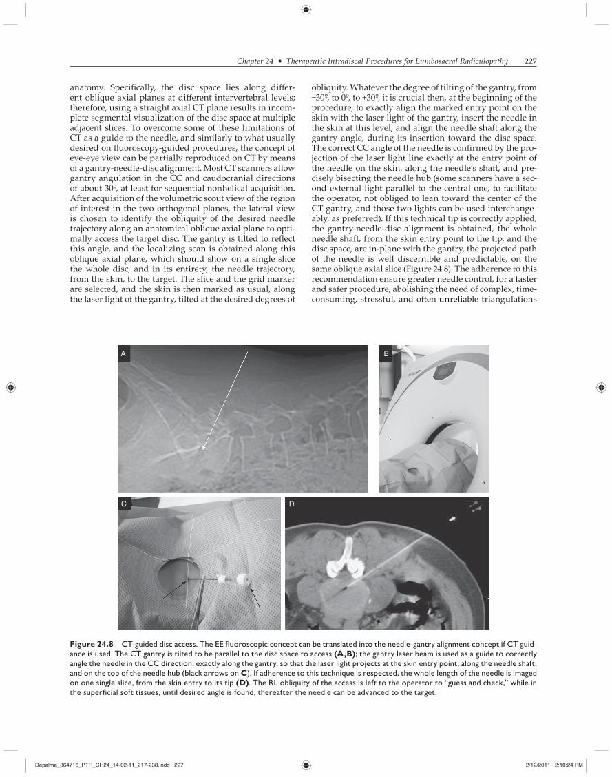
obliquity. Whatever the degree of tilting of the gantry, from −30º, to 0º, to +30º, it is crucial then, at the beginning of the procedure, to exactly align the marked entry point on the skin with the laser light of the gantry, insert the needle in the skin at this level, and align the needle sha along the gantry angle, during its insertion toward the disc space. The correct CC angle of the needle is confi rmed by the pro-jection of the laser light line exactly at the entry point of the needle on the skin, along the needle’s sha , and pre-cisely bisecting the needle hub (some scanners have a sec-ond external light parallel to the central one, to facilitate the operator, not obliged to lean toward the center of the CT gantry, and those two lights can be used interchange-ably, as preferred). If this technical tip is correctly applied, the gantry-needle-disc alignment is obtained, the whole needle sha , from the skin entry point to the tip, and the disc space, are in-plane with the gantry, the projected path of the needle is well discernible and predictable, on the same oblique axial slice (Figure 24.8). The adherence to this recommendation ensure greater needle control, for a faster and safer procedure, abolishing the need of complex, time-consuming, stressful, and o en unreliable triangulations
anatomy. Specifi cally, the disc space lies along diff er-ent oblique axial planes at diff erent intervertebral levels; therefore, using a straight axial CT plane results in incom-plete segmental visualization of the disc space at multiple adjacent slices. To overcome some of these limitations of CT as a guide to the needle, and similarly to what usually desired on fl uoroscopy-guided procedures, the concept of eye-eye view can be partially reproduced on CT by means of a gantry-needle-disc alignment. Most CT scanners allow gantry angulation in the CC and caudocranial directions of about 30º, at least for sequential nonhelical acquisition. A er acquisition of the volumetric scout view of the region of interest in the two orthogonal planes, the lateral view is chosen to identify the obliquity of the desired needle trajectory along an anatomical oblique axial plane to opti-mally access the target disc. The gantry is tilted to refl ect this angle, and the localizing scan is obtained along this oblique axial plane, which should show on a single slice the whole disc, and in its entirety, the needle trajectory, from the skin, to the target. The slice and the grid marker are selected, and the skin is then marked as usual, along the laser light of the gantry, tilted at the desired degrees of
A B
C D
Figure 24.8 CT-guided disc access. The EE fl uoroscopic concept can be translated into the needle-gantry alignment concept if CT guid-
ance is used. The CT gantry is tilted to be parallel to the disc space to access (A,B); the gantry laser beam is used as a guide to correctly
angle the needle in the CC direction, exactly along the gantry, so that the laser light projects at the skin entry point, along the needle shaft,
and on the top of the needle hub (black arrows on C). If adherence to this technique is respected, the whole length of the needle is imaged
on one single slice, from the skin entry to its tip (D). The RL obliquity of the access is left to the operator to “guess and check,” while in
the superfi cial soft tissues, until desired angle is found, thereafter the needle can be advanced to the target.
Depalma_864716_PTR_CH24_14-02-11_217-238.indd 227Depalma_864716_PTR_CH24_14-02-11_217-238.indd 227 2/12/2011 2:10:24 PM2/12/2011 2:10:24 PM
228 Part I • Lumbosacral Spine • Radiculopathy/Radicular Pain
of the inserted needle, with one slice above and one below in which no needle be visualized; if this safety condition is not respected, there is risk improper needle placement, with resultant risk of injury in a complex and delicate ana-tomical region such as the spine.
If the CC obliquity of the needle can be precisely con-trolled with the gantry-needle alignment technique, the desired RL obliquity of the needle in the CT-guided pro-cedures is le to the experience and ability of the operator in reproducing on the patient the trajectory chosen on the
that the operator is obliged to imagine, to predict where the needle tip will be located a er a certain depth of advance-ment, as usually happens when the needle is off -plane with the gantry, and imaged partially on multiple adjacent sections. The gantry-needle alignment greatly reduces the number of CT sections required at each CT control of the procedure (usually three to six slices are suffi cient), and consequently reduce the procedure time and the radiation dose. It is imperative that the control CT views obtained intermi ently during the procedure show the whole length
Moveable depth marker
Probe tip
Removeable collection chamber
Activation switch
1.5 mm cannula
H2O
H2O
Aspirated nucleus
Vacuum
Nucleuspulposus
VENTURISUCTIONEFFECT
EVACUATIONTUBE
SALINESTREAM
IN FLOWTUBE
Waste
SterileFluid Supply
SpineJetPercResector
Sealed WasteContainment
Spi
neJe
t
SpineJetPower
Console
Variable PowerFoot Pedal
Saline
A B
C
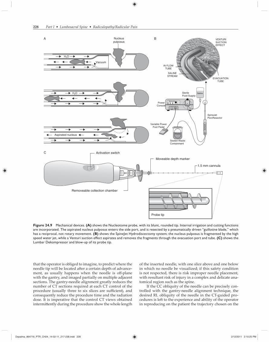
Figure 24.9 Mechanical devices. (A) shows the Nucleotome probe, with its blunt, rounded tip. Internal irrigation and cutting functions
are incorporated. The aspirated nucleus pulposus enters the side port, and is resected by a pneumatically driven “guillotine blade,” which
has a reciprocal, not rotary movement. (B) shows the SpineJet Hydrodiscectomy system; the nucleus pulposus is fragmented by the high
speed water jet, while a Venturi suction effect aspirates and removes the fragments through the evacuation port and tube. (C) shows the
Lumbar Dekompressor and blow-up of its probe tip.
Depalma_864716_PTR_CH24_14-02-11_217-238.indd 228Depalma_864716_PTR_CH24_14-02-11_217-238.indd 228 2/12/2011 2:10:25 PM2/12/2011 2:10:25 PM

Chapter 24 • Therapeutic Intradiscal Procedures for Lumbosacral Radiculopathy 229
or not, or entail use of diff erent types of energy, such as RF, laser, coblation, etc. The main goal of these procedures is mainly nonselective removal of the nucleus pulposus, with the aim of a global decompression and decrease of disc volume and intradiscal pressure, as previously dis-cussed. The indications for each of these modalities are substantially identical.
Chemical
In 1959, Hirsch [49] suggested the use of proteolytic enzymes for the treatment of discal herniations. Lyman Smith, an orthopedic surgeon in Illinois, undertook a series of experimental studies in 1963 to demonstrate that intradiscal injection of a proteolytic enzyme was a possi-ble nonsurgical treatment for disc removal. In 1964, in the Journal of the American Medical Association, he published his fi rst report suggesting that the enzyme chymopapain, derived from the papaya, might prove eff ective in the treat-ment of herniated lumbar discs that have not ruptured through the encircling posterior longitudinal ligament [50]. The initially enthusiastic response to this approach has given way to a much greater degree of caution because of the reported complications of anaphylaxis and neural injuries [51–53], mostly owing to technical errors of some practitioners.
Chymopapain remains the most widely evaluated and clinically tested substance for the purpose of chemi-cal, percutaneous treatment of disc herniations, although other enzymes such as collagenase [54] and chondroitinase ABC [55] have been proposed. Chymopapain is no longer available in the US market, although it still is in other countries.
Chymopapain is a proteolytic enzyme of vegetal origin, extracted from the latex of Carica Papaya, and has a molec-ular weight of 27,000 [56]. The enzymatic action of chymo-papain is not highly specifi c and is exerted on numerous substrates such as hemoglobin, casein in milk, and on noncollagenous proteins of fi brocartilage. Like any proteo-lytic enzyme, chymopapain is immunogenic. Intradiscal injection of chymopapain may cause allergic reactions in humans. A small quantity of immunoreactive chymopa-pain appears in the plasma immediately following injec-tion. As much as 3% of the North American population has been sensitized to papaya enzymes, due to the inges-tion of chymopapain in the form of fruits or food additives. Certain subjects have developed an infraclinical sensitiv-ity. This risk of anaphylactic reaction justifi es precautions in the selection of patients, ruling out potential allergic subjects (generically hyperallergic or with previous his-tory of anaphylactic reactions, history of allergy to papaya, or previous exposure to parenteral chymopapain, with IgE antibodies to chymopapain). In the usually adminis-tered dosage, chymopapain acts only on the proteoglycans [57–59]. Its activity takes place on the glucosaminoglycans-carrying protein. The cleavage leads to a depolymeriza-tion of the large proteoglycan molecules, and liberation of the polysaccharide groupings which lose their capacity to retain water. This hydrolytic action of chymopapain on the proteoglycans of discal tissue, well demonstrated by sev-eral experimental studies realized in vitro on normal and
cross-sectional image of interest on the CT-consolle. Most commonly, in skilled hands, a satisfactorily RL trajectory is obtained with a maximum of three a empts of nee-dle insertion at a point no deeper than the paravertebral muscles.
Posterolateral Oblique Approach
Not diff erently from the posterolateral oblique disc approach used under fl uoroscopic guidance, on CT, once the correct axial obliquity of the scan is obtained, and the gantry is aligned with the disc space (i.e., on the single axial image slicing through the disc space there is no disc-endplate bone visualized), local anesthesia is administered to the skin, fascia, and paravertebral muscles, paying a en-tion not to inject deep to the posterior articular elements, to keep a normal sensitivity of the exiting nerve root at that level. The working needle is also aligned with the gan-try, obliqued as desired in the RL direction, and is inserted and advanced from a posterolateral approach to be lateral and tangent as possible to the lateral aspect of the supe-rior articular process of the inferior vertebral level, and to course posterior to the visualized exiting nerve root. Once contact with the external annulus is reached, with no radicular pain, the needle is advanced through the annu-lus fi brosus and to the center of the nucleus pulposus.
Transdural Posterior Approach
If desired, using an appropriately small caliber needle (21–22 G), the disc space can be accessed from a posterior transdural approach at the lower lumbar levels, below the position of the conus medullaris. This access is feasible with straight needles when local anatomy permits an axial oblique plane parallel to the disc space passing through the interlaminar space. At certain levels, and in certain patients, the oblique plane passing through the disc space corresponds posteriorly to the bony laminae, which would clearly obstruct the needle path. The path of the needle is just lateral to midline where the spinous process is, enter-ing the central canal through the ligamentum fl avum, piercing the dura along the posterior and anterior aspect of the dural sac, and thereby entering the disc from the pos-terior longitudinal ligament. The nerve roots of the cauda equina easily allow this needle access, when performed gently. Although this is a potentially easy and eff ective access to the center of the nucleus pulposus, in selected instances, it carries additional risks of cerebrospinal fl uid leak and headache, cerebrospinal fl uid infection, epidural hematoma.
MODALITIES
Since the 1960s, many diff erent techniques for percuta-neous removal of the nucleus pulposus or its protruding components have been proposed. In the following section, the most diff used among them, and still present in current clinical practice, are described. They may achieve the goal in many diff erent ways. The proposed instruments for disc decompression are chemical, mechanical, automated
Depalma_864716_PTR_CH24_14-02-11_217-238.indd 229Depalma_864716_PTR_CH24_14-02-11_217-238.indd 229 2/12/2011 2:10:25 PM2/12/2011 2:10:25 PM

230 Part I • Lumbosacral Spine • Radiculopathy/Radicular Pain
approximately 80% of his patients experienced improve-ment a er this procedure. Variations on this method have been subsequently popularized by Kambin [67,68] in Philadelphia and Suezawa [69] in Switzerland. Using Craig-type biopsy instruments under fl uoroscopic control, Kambin inserted a large trocar through the lateral annu-lus fi brosus, grasped the herniated disc, and removed it. He reported excellent results with no signifi cant compli-cations in 85% of 50 patients. Suezawa used the instru-ments designed by Hij ikata and in addition he inserted a discoscope through a contralateral approach. This was essentially a fi beroptic system used to visualize, from the contralateral side, the disc material being removed. Excellent results were reported in 67% of 47 patients, although the majority of these patients showed complicat-ing factors, such as spinal stenosis.
In another development, Jacobson [70], a neurosurgeon in Miami, designed his own instruments and used a direct lateral approach to remove herniated discs percutaneously in more than 300 patients. With the patient under general anesthesia, a 10- to 11-mm cannula was introduced through the lateral annulus. Using his own patented instruments, Jacobson grasped and removed disc material with overall good results in terms of pain relief. Unfortunately, unac-ceptable injury of bowel and peripheral nerves occurred. Friedman [11] studied Jacobson’s technique in cadavers and demonstrated that the anatomical variations were such, that an unacceptably high rate of morbidity and potential mortality could be expected with this technique. Friedman therefore recommended against its use.
A er surveying the previous techniques and assess-ing their potential problems, Gary Onik working with engineers from Surgical Dynamics, Inc, designed his own instruments for lumbar discectomy in 1984 and intro-duced it in clinical practice in 1985 [71–74]. The technique was called “automated” percutaneous lumbar discectomy, because it involves a mechanical probe, Nucleotome, which removes the nucleus pulposus by a “suction and cu ing” action (Figure 24.9A). The device is now manu-factured by Clarus Medical, LLC. The probe tip, exclud-ing the handle, is 20.2 cm long and has an outer diameter of 2.2 mm. The negative pressure for aspiration is gen-erated by the vacuum-generating console. A vacuum is created that draws nuclear material into the side port, which is located a few millimeters proximal to the distal tip of the probe. The cu ing blade for fragmentation of nucleus pulposus aspirated through the port works with a reciprocal, not rotatory motion. This type of movement is a safety feature because the “guillotine” blade is con-tained within the probe. Consequently, only the nuclear material that is drawn into the port can be cut. The mate-rial aspirated from the inner disc and exiting through the metallic probe is ultimately deposited into a fi lter in a dis-posable collection bo le. The extracted nucleus pulposus is thus available for quantitative and macroscopic qual-itative evaluation, or even for histology examination. A sequence of devices is used for introduction of the probe inside the disc, the last one being a cannula, straight with an outer diameter of 2.8 mm, or a curved one, with an outer diameter of 3.8 mm, be er suited for access to the L5-S1 disc space, when the direct path from the skin is
pathologic human discs, and in vivo, in numerous animal species, leads to diminution of the volume of the disc and of its herniated fragment, thus reducing the compression of the nuclear material on the nerve root. Diminution of the water content can indeed relieve the hydraulic intra-discal pressure. The nonspecifi c proteolytic action of the chymopapain can result in severe neurologic complica-tions [60] if misplaced outside of the disc. Adherence to a precise and proven technique is mandatory. The proce-dure must be performed under local anesthesia, because general anesthesia can mask early sign of an allergic reac-tion, and might result in possible nerve root puncture, which could in turn also lead to intrathecal injection. An 18- to 22-gauge, two-needle technique is preferable [61,62], with the 22-gauge needle advanced coaxially in the center of the disc. Injection of a small amount of contrast medium at this point can rule out epidural or intrathecal injection. The enzyme is injected slowly, in 10 to 20 minutes, to let it be accepted by the nuclear material (the positively charged enzyme will bind with the negatively charged nuclear matrix), thus reducing the risk of epidural migration. The needle is then withdrawn and the patient kept under observation for 20 to 30 minutes for possible systemic aller-gic reactions. The patient can be discharged from the hos-pital the same day.
Collagenase, an enzyme synthesized by the Clostridium histolyticum, splits collagen fi bers, particularly type-2 fi bers, mainly found in the nucleus pulposus. Wi enberg, in a randomized, prospective study, observed at 5 years 72% of good results in the chymopapain group and 52% in the collagenase group [63].
Alcohol was proposed in France for chemical lysis of the nucleus pulposus and its herniating components by the group of Tournade in Colmar [64] in 1999. Alcohol is used as a lytic and necrotizing agent in many interven-tional procedures (ablation of tumors, vascular malforma-tions, nerve and ganglion blocks, including the Gasserian ganglion). Alcohol produces a molecular split of proteogly-cans and glucosaminoglycans. The main advantage over chymopapain is the absence of allergic reactions.
Recently, Théron [65] proposed, for the same pur-pose, alcohol linked to a more viscous agent, a gel of eth-ylcellulose, commercially called Discogel, opacifi ed with tungsten powder. The aim by combining the alcohol with a viscouse material is be er control over the diff usion of the lytic agent, thus limiting the risk of damaging adjacent structures. The agent remains injectable through a small-bore needle, and injection can be followed under fl uoros-copy, much like contrast medium for discography.
Mechanical
In 1975, Hij ikata [66], in Japan, published his results with a series of patients who underwent lumbar discectomy performed percutaneously. Rather than relying on enzy-matic dissolution of the herniated disc, he used specially designed instruments placed through a 5-mm cannula inserted through the lateral annulus. A circular incision was made in the annulus, and the herniated disc mate-rial was grasped with modifi ed pituitary-type rongeurs. In his initial published fi ndings, Hij ikata reported that
Depalma_864716_PTR_CH24_14-02-11_217-238.indd 230Depalma_864716_PTR_CH24_14-02-11_217-238.indd 230 2/12/2011 2:10:25 PM2/12/2011 2:10:25 PM
Chapter 24 • Therapeutic Intradiscal Procedures for Lumbosacral Radiculopathy 231
The Dekompressor, proposed by the Stryker Company in 2003, is a single-use probe, introduced through a 15-mm cannula, intended for percutaneous discectomy in the lumbar, thoracic, and cervical spine. Under fl uoroscopic imaging, the Dekompressor utilizes an Archimedes pump principle to remove nucleus material from the disc. The rotating screw blade is spun by a disposable rotational motor (Figure 24.9C). The single-use probe is smaller than Onik’s Nucleotome, with no need for a console or other external control to make it operate properly. Unlike the Nucleotome and the SpineJet, the Dekompressor does not entail a hydration of the inner disc to favor tissue removal (purely mechanical extraction). Case series are available [81–85], although no controlled studies have to date been performed. The level of evidence for clinical eff ectiveness, based on USPSTF criteria [79], is level III for short- and long-term relief [86].
RF Ablation
The use of thermal energy to modulate and ablate tissue is not new. Electrical current, in one form or another, has been applied to human tissues as a surgical modality for more than 100 years. RF energy occupies a range upon the elec-tromagnetic radiation spectrum. The frequency at which the device operates determines the absorption characteris-tics and tissue eff ects. Electrosurgical units based on stan-dard monopolar or bipolar devices generally operate from 200 to 500 kHz and they are limitedly applied or avoided to prevent unwanted tissue destruction. Devices operating in this frequency range cause the electrode that comes in con-tact with the tissue to become hot, therefore acting like true heat cautery. RF in the radiowave range (between 1.7 MHz and 4.0 MHz) of the radiation spectrum emits energy that is nonthermal with optimal controlled absorption charac-teristics of water-rich tissues, with minimal tissue alter-ation. High-frequency radiosurgery, above 1500 kHz (1.5 MHz), transmits pure radiowaves to the tissue without heating the electrode. The heat for this ablation is gener-ated by a natural resistance of the tissue, which comes in the path of the waves released through the electrode tip of the device. The cellular water in the so tissues gets heated and when the temperature reaches 100°C, it starts boiling, and produces steam, which results in cellular molecular dissolution of individual tissue cells. The cells exposed to these waves are destroyed whereas the surrounding tis-sues remain unaff ected. This property of radiofrequencies eliminates the possibility of undesired damage to the nor-mal tissues, while improving the surgical precision.
The Disc-FX discectomy system is proposed by Elliquence, LLC, formerly Ellman Innovations, LLC, New York. It works in bipolar mode at 1.7 MHz. In particular, the bipolar Trigger-Flex probe is used to obtain a radio-wave energy application both for removal of nucleus mate-rial and a modulation of weak collagen fi brils and sealing of annular tears (shrinking or eliminating defects in the annulus) as well as contributing to depopulating nerve fi bers sensitizing the outer annulus due to its smooth thermal eff ect.
The two diff erent eff ects in the nucleus (ablation for decompression) and in the annulus (annulus modulation)
covered by the iliac crest. The reason for a larger diam-eter in the la er is that it is internally coated by a Tefl on layer, which reduces friction and favors the sliding of the fl exible but straight probe. APLD as proposed by Onik has lost the favor of most operators, and discussion of the reasons of that falls beyond the limits of this chapter. One of the authors (GB) of this chapter has developed a large experience with very good results, depending on a strict and wise selection of patients, and still uses it in few, very selected cases [75]. In any case, Onik’s remains the per-cutaneous procedure that removes the largest amounts of nuclear material from within the intervertebral disc. Another advantage, when comparing APLD with physi-cal techniques that blindly destroy the disc (such as laser, RF or coblation), is that the surgeon can verify directly and visually the quantity of disc material removed, as well as its “quality.” The extracted nucleus pulposus can be observed as it passes through the transparent tubing that connects to the fi lter. How much nucleus is taken out and how degenerated it is, are important procedural and prognostic pieces of information. For example, viewing the quantity of removed nuclear tissue and comparing it with the amount that was anticipated to be extracted from interpreting the preoperative imaging provides critical information to determine whether the probe worked in the correct intranuclear location. Observing blood coming from the disc could suggest the presence of unexpected degeneration, or of painful granulation tissue inside the disc, or prompt arrest of the procedure so as not to dam-age the endplate cartilage. Another important safety fea-ture is that, once the Nucleotome is safely within the disc, it is unable, unlike other devices, to cut its way out of the disc space to cause injury to vital structures.
The notion that Onik’s proposal was meritorious is suggested by the recent resurgence of new devices designed to mechanically remove the nucleus pulposus in an “automated” mode by aspiration. Observational stud-ies are available, together with four RCTs [75–78]. The level of evidence for clinical eff ectiveness of APLD, as deter-mined based on the US Preventive Services Task Force (USPSTF) criteria [79], using fi ve levels, ranging from I to III with three subcategories in level II, is level II-2 for short and long-term relief [80]. However, APLD does not appear to compare favorably against chymopapain injection and open discectomy [76,78].
A similar hydraulic aspiration principle is utilized by the SpineJet probe, produced by HydroCision. The disposable SpineJet probe simultaneously cuts and aspi-rates nucleus; a round atraumatic tip design reduces risks of annular puncture and endplate damage. The SpineJet Hydrosurgery system utilizes a reusable power con-sole with foot pedal activation (Figure 24.9B). Both the Nucleotome and the SpineJet are fl uid-based systems, that is, the inner disc material is hydrated while the probe’s aspiration action is active for tissue removal: consequently they can, unlike every other system (purely mechanical, thermal, laser, etc.) effi ciently ablate tissue regardless of patient’s age and disc hydration. Moreover, internal irri-gation with sterile saline is a vehicle for easy aspiration, to prevent accumulation of nuclear material and consequent clogging inside the probe.
Depalma_864716_PTR_CH24_14-02-11_217-238.indd 231Depalma_864716_PTR_CH24_14-02-11_217-238.indd 231 2/12/2011 2:10:25 PM2/12/2011 2:10:25 PM

232 Part I • Lumbosacral Spine • Radiculopathy/Radicular Pain
process, bipolar voltage pulses at 100 kHz are applied to the active electrode at the distal end of the device, which produces a strong electric fi eld region around the elec-trode. The electrolytes in the surrounding conductive medium (e.g., sodium ions resident within the nucleus pulposus) respond to the electric fi elds, and if the voltage is suffi ciently large, a localized fi nely focused plasma fi eld (ionized vapor) is produced between the electrode and adjacent tissue [23,90]. The plasma fi eld comprises a com-plex mixture of gas-phase radical chemically reactive and nonreactive molecules and a very small fraction of ionized particles (predominately positive ions and electrons), some of which can break molecular bonds in the adjacent tissue by energetic particle bombardment and chemical reactions. The organic molecules in the disc material (particularly long-chain molecules such as collagen) are thought to be susceptible to fragmentation by the plasma particles, resulting in their conversion into liquid and gaseous prod-ucts that are subsequently desorbed from the targeted site. Water molecules (which compose a signifi cant fraction of most types of tissue) can be fragmented into excited and groundstate hydroxyl radicals and hydrogen atoms. Both of these species are chemically active and can cleave long-chain molecules (e.g., collagen) into smaller fragments that are either more easily liquefi ed or gasifi ed. Moreover, electrons emi ed from the electrodes at the distal end of
are obtained by means of two diff erent waveforms gen-erated by an external source; the ablation in the nucleus is achieved using a more aggressive waveform, called “Bipolar Turbo,” the modulation of the annulus using a smoother waveform “called Bipolar Hemo.”
The bipolar Trigger-Flex probe is fl exible and steerable (Figure 24.10A), and consequently can be oriented to oper-ate either in the nucleus or along the posterior annulus. At the same time, the fl exible probe allows a more targeted removal of the protruding nucleus, thus relieving tension on the innervated and irritated annulus.
Neither observational studies nor RCTs have been pub-lished in the literature. In our personal but limited experi-ence of the fi rst 30 cases, the procedure showed short-term results comparable with those of Nucleoplasty. The action of the probe on the posterior annulus and its nerve fi bers should allow treatments also of patients suff ering from purely discogenic back pain.
Coblation
Percutaneous plasma RF-based discectomy, with com-mercial name Nucleoplasty (ArthroCare, Sunnyvale, CA), is conducted by using a bipolar radio-frequency-based device, which functions via a plasma-mediated process [87–89], to perform precise removal of disc tissue. In this
A
B
C
B
Figure 24.10 Thermal-radiofrequency-
laser devices. (A) represents a schematic
drawing of the proposed action of the
Disc-Fx; the steerable probe allows topo-
graphic nuclear material removal at the site
of internal annular disruption, and ther-
mal modulation of the nerve fi bers in the
posterior annulus, using a less aggressive
waveform. (B) shows the active tip of the
Arthrocare spine wand, introduced in the
nucleus pulposus through a beveled 17 G nee-
dle; the slightly curved tip allows the creation
of multiple coblation channels upon rotating
the proble at multiple passes. (C) shows the
CT-guided coaxial insertion of the laser fi ber
through a 21G cannula into the extruded disc
fragment, through a translaminar and trans-
dural paraspinous approach.
Depalma_864716_PTR_CH24_14-02-11_217-238.indd 232Depalma_864716_PTR_CH24_14-02-11_217-238.indd 232 2/12/2011 2:10:25 PM2/12/2011 2:10:25 PM
Chapter 24 • Therapeutic Intradiscal Procedures for Lumbosacral Radiculopathy 233
amount of energy put into a given tissue. The majority of surgical lasers fall in the invisible portion of the elec-tromagnetic radiation spectrum. The absorption charac-teristic of the medium largely determines the extent of penetration in particular tissue types. Application of any laser requires the surgeon to completely understand the characteristics of the specifi c laser for safe and eff ective use. The way in which light interacts with a substance largely depends upon its wavelength. Penetration depth at a certain wavelength is mostly aff ected by absorption by specifi c molecules, such as water (the principal compo-nent of the nucleus pulposus), hematoproteins, pigments, nucleic acids, and so on. As a laser is absorbed by the tis-sue, several surgical eff ects take place: at 60°C protein denaturation and coagulation of blood vessels, near 100°C evaporation of intracellular water causing shrinkage and tissue loss, beyond this point vaporization will occur. In general, the therapeutic eff ect of a laser signifi cantly depends on penetration depth, in eff ect determining whether tissue removal or hemostasis will be predomi-nant. Intradiscal decompression is obtained by shrink-age of the water-rich nucleus pulposus by vaporization. The evaporation of water and the increase in temperature causes protein denaturation and subsequent renaturation, causing a structural change of nucleus pulposus, limit-ing its capability to a ract water [21,99]. An increase of intradiscal volume of only 1.0 mL causes the intradiscal pressure to rise by as much 312 kPa or 2340 mm Hg. On the other hand, a decrease of intradiscal volume causes a disproportionately large decrease of intradiscal pres-sure. Other benefi cial eff ects of the laser action are postu-lated, such as shrinkage of collagen fi brils with reduction of disc volume [100,101] and destruction of nociceptors in the annulus or in the granulation, vascularized tissue growing in degenerated discs. Many types of lasers have been reported in the literature for spine applications [102], including the following: Nd:YAG (whose active medium is a crystal of y rium, aluminum, and garnet doped with neodymium ions, and whose beam is in the near infra-red) at 1054 nm and at 1320 nm, KTP (a beam generated by a neodymium:YAG laser is directed through a potassium titanyl phosphate crystal to produce a beam in the green visible spectrum) at 531 nm, CO 2 at 10,600 nm, Ho:YAG (YAG doped holmium) laser at 2100 nm, diode (semicon-ductor) laser at 810–890–940–980 nm. The Ho:YAG laser is be er suited for open or endoscopic surgery, under direct visual control, because of the more mechanical eff ects and risk of endplate damage from energy sca ering [103–105]. Yeung, Casper, Chiu, and Knight further report utility of Holmium laser to remodel extradiscal bony architecture during endoscopic spine surgery [105–107]. Nd:YAG at 1054 nm and KTP 532 are the most popular for intradiscal treatments [21,108–112]. KTP lasers are similar to Nd:YAG lasers in both action and eff ects [113,114], as are diode lasers [115]. The la er have the advantage of a much less expensive and less cumbersome power unit. Advantages of the 980 wavelength (peak absorption of water) of the diode laser are maximum absorption for well-hydrated, so tissue like the nucleus pulposus, with great thermal eff ect and consequent effi cient shrinking eff ect [115,116], with minimal thermal damage of surrounding tissues
the device when the voltage is applied can develop suffi -ciently high energies not only to cause the water molecules to fragment but also to directly dissociate the chemical bonds in the nearby targeted tissue structures (in this case disc tissue) into smaller fragments [89]. The net result is a reduction of so tissue volume and eff ective excision of the so tissues within the nucleus. Small channels are created in the nucleus, and they are thermally treated, producing a zone of thermal coagulation. Thus, Nucleoplasty com-bines coagulation and controlled tissue ablation (patented Coblation technology). The plasma radio-frequency-based process has been reported to have minimal histopatho-logic eff ect on tissues immediately adjacent to the treated site [23,90], particularly annulus, endplates, neural ele-ments, and nerve roots [90]. The temperature is kept below 70°C to minimize tissue damage and avoid tissue charring. Because of the mechanism of action on hydrated nuclear components, tissue ablation and consequently intradiscal decompression are supposedly higher in younger patients and in hydrated, nonadvancedly degenerated discs [23]. Actually, an exclusion criterion for lumbar Nucleoplasty must be considered a disc height less than 50% [91]. The coblation probe is introduced through a very thin, 17 G cannula (Figure 24.10B), probably the narrower among nonchemical procedures, laser excepted. This also allows for bending of the cannula and probe, for access to diffi -cult L5-S1 levels. A large experience is available and since 2000 many tenths thousands of patients have been treated using coblation technique, both at lumbar and cervical lev-els [47,92–96]. Many observational studies are available but not RCTs. The level of evidence for clinical eff ectiveness, based on USPSTF criteria [79], is level II-3 in managing pre-dominantly lower limb radicular pain, with no evidence for axial back pain [91,97].
Laser
Percutaneous laser disc decompression (PLDD) was intro-duced by Choy et al. [21,98] in 1986. By 2002, more than 35,000 PLDDs had been performed [99]. The term laser is an acronym standing for Light Amplifi cation by the Stimulated Emission of Radiation. Laser is a form of light, and light is made up of electromagnetic energy. Laser energy is formed by energizing an active lasing medium. With the introduction of energy from an outside source, the atoms absorb the energy causing the electrons to rise to a higher excited state. When the electron returns to the normal, nonexcited state, the energy initially absorbed is given off as a photon, and the photon bundle has unique properties characteristic to that particular medium. Lasers are generally classifi ed according to the medium they use to produce the laser light. Solid-state, gas, liquid, and semiconductor are all common types of lasers. The radiant energy of the laser beam can be transformed into heat energy that produces medical and surgical eff ects in tissue, such as coagulation, vaporization, or cu ing. The total power output of a laser is measured in wa s, the power density, measured in wa s per square centi-meter, and it determines the thermal eff ect in the target tissue; the energy density (measured in power density X time) of joules per square centimeter indicates the total
Depalma_864716_PTR_CH24_14-02-11_217-238.indd 233Depalma_864716_PTR_CH24_14-02-11_217-238.indd 233 2/12/2011 2:10:26 PM2/12/2011 2:10:26 PM
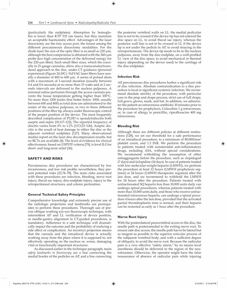
234 Part I • Lumbosacral Spine • Radiculopathy/Radicular Pain
(particularly the endplates). Absorption by hemoglo-bin is lower than KTP 532 nm lasers, but they maintain an acceptable haemostatic eff ect. Advantages of the laser discectomy are the tiny access port, the tiniest among the diff erent percutaneous discectomy modalities. For the diode laser the size of the optic fi ber is as small as 220 μm, although the best compromise is obtained with the 360-μm probe (too high concentration of the delivered energy for the 220-μm fi ber). Such small fi ber sizes, which fi ts coaxi-ally in 21-gauge cannulas, allow also a transcanalar/trans-dural approach to the disc, under CT guidance (personal experience) (Figure 24.10C). Nd:YAG laser fi bers have usu-ally a diameter of 400 to 600 μm. A series of pulsed shots with a maximum of 1-second duration (usually between 0.4 and 0.6 seconds) at no more than 15 wa s and at 2 sec-onds intervals are delivered to the nucleus pulposus. A minimal saline perfusion through the access cannula pre-vents the tissue temperature ge ing higher than 100°C. No more than 1200 joules (but be er below 1000, usually between 600 and 800) as total dose are administered to the center of the nucleus pulposus, in two to three diff erent positions of the fi ber tip, always under fl uoroscopic control of the proper position of the device. The most frequently described complication of PLDD is spondylodiscitis both aseptic and septic [10,117–122]. The reported frequency of discitis varies from 0% to 1.2% [117,123–126]. Aseptic dis-citis is the result of heat damage to either the disc or the adjacent vertebral endplates [127]. Many observational studies report on the laser disc decompression results, but no RCTs are available [4]. The level of evidence for clinical eff ectiveness, based on USPSTF criteria [79], is level II-2 for short- and long-term relief [36].
SAFETY AND RISKS
Percutaneous disc procedures are characterized by low invasiveness, and low risk profi le; nevertheless, they pre-sent potential risks [22,74–78]. The main risks associated with these procedures are infection, bleeding, nerve root injury, thecal sac injury, disc-endplate injury, injury to the retroperitoneal structures, and colonic perforation.
General Technical Safety Principles
Comprehensive knowledge and extremely precise use of the radiologic projections and landmarks are prerequi-sites to perform these procedures. Thorough use of pre-cise oblique working eye-eye fl uoroscopic technique, with intermi ent AP and LL verifi cation of device position, or needle-gantry alignment in CT-guided procedures, is mandatory. Adherence to a safe technique will dramati-cally impact the outcome and the probability of realizing a side eff ect or complication. An incorrect projection means that the cannula and the intradiscal device is actually working away from the place where it is supposed to, not eff ectively operating on the nucleus or, worse, damaging vital or functionally important structures.
As discussed earlier in the technique paragraph, main safety landmarks in fl uoroscopy are a line connecting the medial border of the pedicles on AP, and a line connecting
the posterior vertebral walls on LL: the medial pedicular line is not to be crossed if the device tip has not entered the disc space on LL, to avoid thecal sac injury, whereas the posterior wall line is not to be crossed in LL if the device tip is not under the pedicle in AP, to avoid straying in the retroperitoneum. The device tip needs to be in the nucleus pulposus, away from the disc-endplate, on a well-profi led LL view of the disc space, to avoid mechanical or thermal injury (depending on the device used) to the cartilage of the disc-endplates.
Infection Risk
All percutaneous disc procedures harbor a signifi cant risk of disc infection. Absolute contraindication to a disc pro-cedure is local or signifi cant systemic infection. We recom-mend absolute sterility of the procedure, with particular care in the prep and drape process, strict use of full drape, full gown, gloves, mask, and hat. In addition, we adminis-ter the patient an intravenous antibiotic 10 minutes prior to the procedure for prophylaxis, Cefazoline 1 g intravenous, or, in case of allergy to penicillin, ciprofl oxacine 400 mg intravenous.
Bleeding Risk
Although there are diff erent policies at diff erent institu-tions [128], we set our threshold for a safe performance of an intradiscal procedure, to a minimum of 80,000/mm3 platelet count, and 1.3 INR. We perform the procedure in patients treated with nonsteroidal anti-infl ammatory drugs, including ASA, without special considerations. We recommend witholding the assumption of other antiaggregants before the procedure, such as clopidogrel (7 days) and ticlopidine (14 days). In case of patients treated with low-molecular-weight heparin (LMWH), we perform the procedure at least 12 hours (LMWH prophylaxis reg-imen) or 24 hours (LMWH therapeutic regimen) a er the last dose, and we recommend to withhold the LMWH for 24 hours a er the procedure. Patients treated with unfractionated SQ heparin less than 10,000 units daily can undergo spinal procedures, whereas patients treated with more than 10,000 units daily, and those who receive unfrac-tionated intravenous heparin, can undergo a spinal proce-dure 4 hours a er the last dose, provided that the activated partial thromboplastin time is normal, and their heparin can be restarted as early as 1 hour a er the procedure.
Nerve Root Injury
With the posterolateral paravertebral access to the disc, the needle path is posteromedial to the exiting nerve root. To ensure safe disc access, the needle path has to be lateral but as tangent as possible to the superior articular process of the subjacent vertebral body, and with a suffi cient degree of obliquity, to avoid the nerve root. Because the radicular pain is a very eff ective “safety alarm,” by no means local anesthesia should be delivered to the region of the neu-roforamen. Otherwise, the operator might have the false reassurance of absence of radicular pain while injuring
Depalma_864716_PTR_CH24_14-02-11_217-238.indd 234Depalma_864716_PTR_CH24_14-02-11_217-238.indd 234 2/12/2011 2:10:26 PM2/12/2011 2:10:26 PM
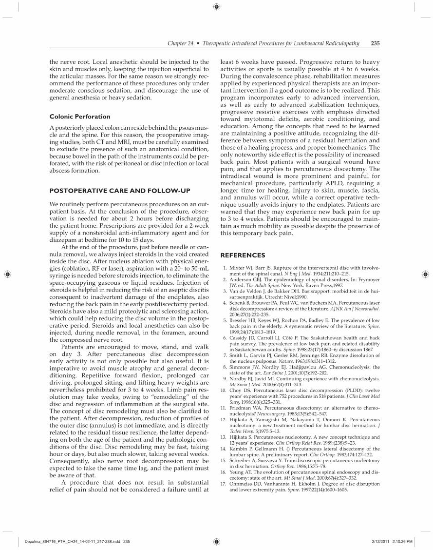
Chapter 24 • Therapeutic Intradiscal Procedures for Lumbosacral Radiculopathy 235
the nerve root. Local anesthetic should be injected to the skin and muscles only, keeping the injection superfi cial to the articular masses. For the same reason we strongly rec-ommend the performance of these procedures only under moderate conscious sedation, and discourage the use of general anesthesia or heavy sedation.
Colonic Perforation
A posteriorly placed colon can reside behind the psoas mus-cle and the spine. For this reason, the preoperative imag-ing studies, both CT and MRI, must be carefully examined to exclude the presence of such an anatomical condition, because bowel in the path of the instruments could be per-forated, with the risk of peritoneal or disc infection or local abscess formation.
POSTOPERATIVE CARE AND FOLLOW-UP
We routinely perform percutaneous procedures on an out-patient basis. At the conclusion of the procedure, obser-vation is needed for about 2 hours before discharging the patient home. Prescriptions are provided for a 2-week supply of a nonsteroidal anti-infl ammatory agent and for diazepam at bedtime for 10 to 15 days.
At the end of the procedure, just before needle or can-nula removal, we always inject steroids in the void created inside the disc. A er nucleus ablation with physical ener-gies (coblation, RF or laser), aspiration with a 20- to 50-mL syringe is needed before steroids injection, to eliminate the space-occupying gaseous or liquid residues. Injection of steroids is helpful in reducing the risk of an aseptic discitis consequent to inadvertent damage of the endplates, also reducing the back pain in the early postdiscectomy period. Steroids have also a mild proteolytic and sclerosing action, which could help reducing the disc volume in the postop-erative period. Steroids and local anesthetics can also be injected, during needle removal, in the foramen, around the compressed nerve root.
Patients are encouraged to move, stand, and walk on day 3. A er percutaneous disc decompression early activity is not only possible but also useful. It is imperative to avoid muscle atrophy and general decon-ditioning. Repetitive forward fl exion, prolonged car driving, prolonged si ing, and li ing heavy weights are nevertheless prohibited for 3 to 4 weeks. Limb pain res-olution may take weeks, owing to “remodeling” of the disc and regression of infl ammation at the surgical site. The concept of disc remodeling must also be clarifi ed to the patient. A er decompression, reduction of profi les of the outer disc (annulus) is not immediate, and is directly related to the residual tissue resilience, the la er depend-ing on both the age of the patient and the pathologic con-ditions of the disc. Disc remodeling may be fast, taking hour or days, but also much slower, taking several weeks. Consequently, also nerve root decompression may be expected to take the same time lag, and the patient must be aware of that.
A procedure that does not result in substantial relief of pain should not be considered a failure until at
least 6 weeks have passed. Progressive return to heavy activities or sports is usually possible at 4 to 6 weeks. During the convalescence phase, rehabilitation measures applied by experienced physical therapists are an impor-tant intervention if a good outcome is to be realized. This program incorporates early to advanced intervention, as well as early to advanced stabilization techniques, progressive resistive exercises with emphasis directed toward mytotomal defi cits, aerobic conditioning, and education. Among the concepts that need to be learned are maintaining a positive a itude, recognizing the dif-ference between symptoms of a residual herniation and those of a healing process, and proper biomechanics. The only noteworthy side eff ect is the possibility of increased back pain. Most patients with a surgical wound have pain, and that applies to percutaneous discectomy. The intradiscal wound is more prominent and painful for mechanical procedure, particularly APLD, requiring a longer time for healing. Injury to skin, muscle, fascia, and annulus will occur, while a correct operative tech-nique usually avoids injury to the endplates. Patients are warned that they may experience new back pain for up to 3 to 4 weeks. Patients should be encouraged to main-tain as much mobility as possible despite the presence of this temporary back pain.
REFERENCES
1. Mixter WJ, Barr JS. Rupture of the intervertebral disc with involve-ment of the spinal canal. N Eng J Med. 1934;211:210–215.
2. Anderson GBJ. The epidemiology of spinal disorders. In: Frymoyer JW, ed. The Adult Spine. New York: Raven Press;1997.
3. Van de Velden J, de Bakker DH. Basisrapport: morbiditeit in de hui-sartsenpraktij k. Utrecht: Nivel;1990.
4. Schenk B, Brouwer PA, Peul WC, van Buchem MA. Percutaneous laser disk decompression: a review of the literature. AJNR Am J Neuroradiol. 2006;27(1):232–235.
5. Bressler HB, Keyes WJ, Rochon PA, Badley E. The prevalence of low back pain in the elderly. A systematic review of the literature. Spine. 1999;24(17):1813–1819.
6. Cassidy JD, Carroll LJ, Côté P. The Saskatchewan health and back pain survey. The prevalence of low back pain and related disability in Saskatchewan adults. Spine. 1998;23(17):1860–6; discussion 1867.
7. Smith L, Garvin PJ, Gesler RM, Jennings RB. Enzyme dissolution of the nucleus pulposus. Nature. 1963;198:1311–1312.
8. Simmons JW, Nordby EJ, Hadjipavlou AG. Chemonucleolysis: the state of the art. Eur Spine J. 2001;10(3):192–202.
9. Nordby EJ, Javid MJ. Continuing experience with chemonucleolysis. Mt Sinai J Med. 2000;67(4):311–313.
10. Choy DS. Percutaneous laser disc decompression (PLDD): twelve years’ experience with 752 procedures in 518 patients. J Clin Laser Med Surg. 1998;16(6):325–331.
11. Friedman WA. Percutaneous discectomy: an alternative to chemo-nucleolysis? Neurosurgery. 1983;13(5):542–547.
12. Hij ikata S, Yamagishi M, Nakayama T, Oomori K. Percutaneous nucleotomy: a new treatment method for lumbar disc herniation. J Toden Hosp. 5;1975:5–13.
13. Hij ikata S. Percutaneous nucleotomy. A new concept technique and 12 years’ experience. Clin Orthop Relat Res. 1989;(238):9–23.
14. Kambin P, Gellmann H. () Percutaneous lateral discectomy of the lumbar spine. A preliminary report. Clin Orthop. 1983;174:127–132.
15. Schreiber A, Suezawa Y. Transdiscoscopic percutaneous nucleotomy in disc herniation. Orthop Rev. 1986;15:75–78.
16. Yeung AT. The evolution of percutaneous spinal endoscopy and dis-cectomy: state of the art. Mt Sinai J Med. 2000;67(4):327–332.
17. Ohnmeiss DD, Vanharanta H, Ekholm J. Degree of disc disruption and lower extremity pain. Spine. 1997;22(14):1600–1605.
Depalma_864716_PTR_CH24_14-02-11_217-238.indd 235Depalma_864716_PTR_CH24_14-02-11_217-238.indd 235 2/12/2011 2:10:26 PM2/12/2011 2:10:26 PM
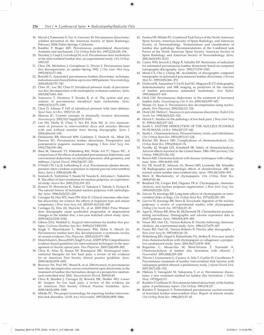
236 Part I • Lumbosacral Spine • Radiculopathy/Radicular Pain
42. Fardon DF, Mile e PC; Combined Task Forces of the North American Spine Society, American Society of Spine Radiology, and American Society of Neuroradiology. Nomenclature and classifi cation of lumbar disc pathology. Recommendations of the Combined task Forces of the North American Spine Society, American Society of Spine Radiology, and American Society of Neuroradiology. Spine. 2001;26(5):E93–E113.
43. Castro WH, Jerosch J, Hepp R, Schulitz KP. Restriction of indication for automated percutaneous lumbar discectomy based on computed tomographic discography. Spine. 1992;17:1239–1243.
44. Moon CT, Cho J, Chang SK. Availability of discographic computed tomography in automated percutaneous lumbar discectomy. J Korean Med Sci. 1995;10(5):368–372.
45. Dullerud R, Amundsen T, Lie H, Juel NG, Magnaes B. CT-diskography, diskomanometry and MR imaging as predictors of the outcome of lumbar percutaneous automated nucleotomy. Acta Radiol. 1995;36(6):613–619.
46. Onik GM. Percutaneous diskectomy in the treatment of herniated lumbar disks. Neuroimaging Clin N Am. 2000;10(3):597–607.
47. Sharps LS, Isaac Z. Percutaneous disc decompression using nucleo-plasty. Pain Physician. 2002;5(2):121–126.
48. Onik GM, Helms C. Nuances in percutaneous discectomy. Radiol Clin North Am. 1998;36(3):523–532.
49. Hirsch C. Studies on the pathology of low back pain. J Bone Joint Surg Br. 1959;41-B(2):237–243.
50. Smith L. ENZYME DISSOLUTION OF THE NUCLEUS PULPOSUS IN HUMANS. JAMA. 1964;187:137–140.
51. Smith L. Chemonucleolysis. Personal history, trials, and tribulations. Clin Orthop Relat Res. 1993;(287):117–124.
52. Kitchel SH, Brown MD. Complications of chemonucleolysis. Clin Orthop Relat Res. 1992;(284):63–74.
53. Nordby EJ, Wright GH, Scholfi eld SR. Safety of chemonucleolysis. Adverse eff ects reported in the United States. 1982–1991 (review). Clin Orthop. 1990;251:144–152.
54. Brown MD. Chemonucleolysis with disease: techniques with collage-nase. Spine. 1991;16:816–819.
55. Fry TR, Eurell JC, Johnson AL, Brown MD, Losonsky JM, Schaeff er DJ. Radiographic and histologic eff ects of chondroitinase ABC on normal canine lumbar intervertebral disc. Spine. 1991;16(7):816–819.
56. Stern IJ . Biochemistry of chymopapain. Clin Orthop Relat Res. 1969;67:42–46.
57. Bradford DS, Cooper KM, Oegema TR Jr. Chymopapain, chemonu-cleolysis, and nucleus pulposus regeneration. J Bone Joint Surg Am. 1983;65(9):1220–1231.
58. Garvin PJ, Jennings RB. Long-term eff ects of chymopapain on inter-vertebral disks of dogs. Clin Orthop Relat Res. 1973;(92):281–295.
59. Garvin PJ, Jennings RB, Stern IJ . Enzymatic digestion of the nucleus pulposus: a review of experimental studies with chymopapain. Orthop Clin North Am. 1977;8(1):27–35.
60. Agre K, Wilson RR, Brim M, McDermo DJ. Chymodiactin postmar-keting surveillance. Demographic and adverse experience data in 29,075 patients. Spine. 1984;9(5):479–485.
61. Fraser RD, Osti OL, Vernon-Roberts B. Discitis following chemonu-cleolysis. An experimental study. Spine. 1986;11(7):679–687.
62. Fraser RD, Osti OL, Vernon-Roberts B. Discitis a er discography. J Bone Joint Surg Br. 1987;69(1):26–35.
63. Wi enberg RH, Oppel S, Rubenthaler FA, Steff en R. Five-year results from chemonucleolysis with chymopapain or collagenase: a prospec-tive randomized study. Spine. 2001;26(17):1835–1841.
64. Riquelme C, Musacchio M, Mont’Alverne F, Tournade A. Chemonucleolysis of lumbar disc herniation with ethanol. J Neuroradiol. 2001;28(4):219–229.
65. Theron J, Guimaraens L, Casasco A, Sola T, Cuellar H, Courtheoux P. Percutaneous treatment of lumbar intervertebral disk hernias with radiopaque gelifi ed ethanol: a preliminary study. J Spinal Disord Tech. 2007;20(7):526–532.
66. Hij ikata S, Yamagishi M, Nakayama T, et al. Percutaneous discec-tomy: a new treatment method for lumbar disc herniation. J Toden Hosp. 1975(Jpn);5:5.
67. Kambin P, Gellman H. Percutaneous lateral discectomy of the lumbar spine. A preliminary report. Clin Orthop. 1983;174:127.
68. Kambin P, Sampson S. Posterolateral percutaneous suction-excision of herniated lumbar intervertebral discs. Report of interim results. Clin Orthop Relat Res. 1986;(207):37–43.
18. Hirsch J, Pomerantz S, Yoo A, Growney M. Percutaneous discectomy: syllabus presented at: the American Society of Spine Radiology; February 2008; Palms Springs, California.
19. Kambin P, Brager MD. Percutaneous posterolateral discectomy. Anatomy and mechanism. Clin Orthop Relat Res. 1987;(223):145–154.
20. Nerubay J, Caspi I, Levinkopf M, et al. Percutaneous laser nucleolysis of the intervertebral lumbar disc: an experimental study. Clin Orthop. 1997;337.
21. Choy DS, Michelsen J, Getrajdman G, Diwan S. Percutaneous laser disc decompression: an update–Spring 1992. J Clin Laser Med Surg. 1992;10(3):177–184.
22. Bonaldi G. Automated percutaneous lumbar discectomy: technique, indications and clinical follow-up in over 1000 patients. Neuroradiology. 2003;45(10):735–743.
23. Chen YC, Lee SH, Chen D. Intradiscal pressure study of percutane-ous disc decompression with nucleoplasty in human cadavers. Spine. 2003;28(7):661–665.
24. Yonezawa T, Onomura T, Kosaka R, et al. The system and pro-cedures of percutaneous intradiscal laser nucleotomy. Spine. 1990;15(11):1175–1185.
25. Choy D, Altman P. Fall of intradiscal pressure with laser ablation. Spine State Art Rev. 1993;7:23–29.
26. Maroon JC. Current concepts in minimally invasive discectomy. Neurosurgery. 2002;51(5 Suppl):S137–S145.
27. Lee SH, Derby R, Chen Y, Seo KS, Kim MJ. In vitro measure-ment of pressure in intervertebral discs and annulus fi brosus with and without annular tears during discography. Spine J. 2004;4(6):614–618.
28. Delamarter RB, Howard MW, Goldstein T, Deutsch AL, Mink JH, Dawson EG. Percutaneous lumbar discectomy. Preoperative and postoperative magnetic resonance imaging. J Bone Joint Surg Am. 1995;77(4):578–584.
29. Shea M, Takeuchi TY, Wi enberg RH, White AA 3rd, Hayes WC. A comparison of the eff ects of automated percutaneous diskectomy and conventional diskectomy on intradiscal pressure, disk geometry, and stiff ness. J Spinal Disord. 1994;7(4):317–325.
30. O’Neill CW, Liu JJ, Leibenberg E, et al. Percutaneous plasma decom-pression alters cytokine expression in injured porcine intervertebral discs. Spine J. 2004;4(1):88–98.
31. Iwatsuki K, Yoshimine T, Sasaki M, Yasuda K, Akiyama C, Nakahira R. The eff ect of laser irradiation for nucleus pulposus: an experimen-tal study. Neurol Res. 2005;27(3):319–323.
32. Komori H, Shinomiya K, Nakai O, Yamaura I, Takeda S, Furuya K. The natural history of herniated nucleus pulposus with radiculopa-thy. Spine. 1996;21(2):225–229.
33. Carragee EJ, Han MY, Suen PW, Kim D. Clinical outcomes a er lum-bar discectomy for sciatica: the eff ects of fragment type and anular competence. J Bone Joint Surg Am. 2003;85-A(1):102–108.
34. Carragee EJ, Don AS, Hurwitz EL, et al. 2009 ISSLS Prize Winner: does discography cause accelerated progression of degeneration changes in the lumbar disc: a ten-year matched cohort study. Spine. 2009;34(21):2338–2345.
35. Gibson JNA, Waddell G. Surgical interventions for lumbar disc pro-lapse. Cochrane Database Syst Rev. 2009;(1):CD001350.
36. Singh V, Manchikanti L, Benyamin RM, Helm S, Hirsch JA. Percutaneous lumbar laser disc decompression: a systematic review of current evidence. Pain Physician. 2009;12(3):573–588.
37. Manchikanti L, Boswell MV, Singh V, et al.; ASIPP-IPM. Comprehensive evidence-based guidelines for interventional techniques in the man-agement of chronic spinal pain. Pain Physician. 2009;12(4):699–802.
38. Chou R, Atlas SJ, Stanos SP, Rosenquist RW. Nonsurgical inter-ventional therapies for low back pain: a review of the evidence for an American Pain Society clinical practice guideline. Spine. 2009;34(10):1078–1093.
39. Brouwer PA, Peul WC, Brand R, et al. Eff ectiveness of percutaneous laser disc decompression versus conventional open discectomy in the treatment of lumbar disc herniation; design of a prospective random-ized controlled trial. BMC Musculoskelet Disord. 2009;10:49.
40. Chou R, Baisden J, Carragee EJ, Resnick DK, Shaff er WO, Loeser JD. Surgery for low back pain: a review of the evidence for an American Pain Society Clinical Practice Guideline. Spine. 2009;34(10):1094–1109.
41. Mile e PC. The proper terminology for reporting lumbar interverte-bral disk disorders. AJNR Am J Neuroradiol. 1997;18(10):1859–1866.
Depalma_864716_PTR_CH24_14-02-11_217-238.indd 236Depalma_864716_PTR_CH24_14-02-11_217-238.indd 236 2/12/2011 2:10:26 PM2/12/2011 2:10:26 PM
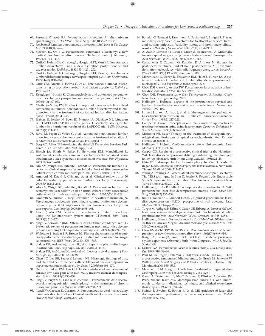
Chapter 24 • Therapeutic Intradiscal Procedures for Lumbosacral Radiculopathy 237
94. Bonaldi G, Baruzzi F, Facchine i A, Fachine i P, Lunghi S. Plasma radio-frequency-based diskectomy for treatment of cervical herni-ated nucleus pulposus: feasibility, safety, and preliminary clinical results. AJNR Am J Neuroradiol. 2006;27(10):2104–2111.
95. Al-Zain F, Lemcke J, Killeen T, Meier U, Eisenschenk A. Minimally invasive spinal surgery using nucleoplasty: a 1-year follow-up study. Acta Neurochir (Wien). 2008;150(12):1257–1262.
96. Calisaneller T, Ozdemir O, Karadeli E, Altinors N. Six months post-operative clinical and 24 hour post-operative MRI examina-tions a er nucleoplasty with radiofrequency energy. Acta Neurochir (Wien). 2007;149(5):495–500; discussion 500.
97. Manchikanti L, Derby R, Benyamin RM, Helm S, Hirsch JA. A sys-tematic review of mechanical lumbar disc decompression with nucleoplasty. Pain Physician. 2009;12(3):561–572.
98. Choy DSJ, Case RB, Ascher PW. Percutaneous laser ablation of lum-bar disc. Ann Meet Orthop Res Soc. 1987;1:19.
99. Choy DSJ. Percutaneous Laser Disc Decompression: A Practical Guide. New York: Springer-Verlag; 2003.
100. Hellinger J. Technical aspects of the percutaneous cervical and lumbar laser-disc-decompression and -nucleotomy. Neurol Res. 1999;21(1):99–102.
101. Hilbert J, Brawn A, Papp J, et al. Erfahrungen mit der perkutanen Laserdiscudekom–pression bei lumbalem banscheibenschaden. Orthop Prax. 1995;31:217–221.
102. Camper D. Current concepts in minimally invasive approaches to treating the lumbar spine using laser energy. Operative Techniques in Sports Medicine. 1998;6(3):174–181.
103. Miriutora NF. Laser Therapy in the treatment of discogenic neu-rological manifestations of spinal osteochondrosis. Vopr Kurortol Fizioter. 2000;3:30–33.
104. Hellinger J. Holmiun-YAG-assistierte off ene Nukleotomie. Laser Med Surg. 1995;11:86–87.
105. Casper GD. Results of a prospective clinical trial of the Holmium-YAG laser disc decompression utilizing a side-fi ring fi ber: four year follow-up (abstract). Fi h Intern Cong. IMLAS. 1998;4:22–25.
106. Chiu JC. Endoscopic lumbar foraminoplasty. In: Kim D, Fessler R, Regan J, eds. Endoscopic Spine Surgery and Instrumentation. New York: Thieme Medical; 2004: 212–229.
107. Yeung AT, Yeung CA. Posterolateral selective endoscopic discectomy, The YESS technique. In: Kim D, Fessler R, Regan J, eds. Endoscopic Spine Surgery and Instrumentation: Percutaneous Procedures. New York: Thieme; 2005:201–211.
108. Hellinger J, Linke R, Heller H. A biophysical explanation for Nd:YAG percutaneous laser disc decompression success. J Clin Laser Med Surg. 2001;19(5):235–238.
109. Brat H, Bouziane F, Lambert J, et al. CT guided percutaneous laser-disc-decompression (PLDD): prospective clinical outcome. Laser Med Sci. 2003;18(suppl 2):16.
110. Turgut M, Açikgöz B, Kilinç K, Ozcan OE, Erbengi A. Eff ect of Nd:YAG laser on experimental disc degeneration. Part I. Biochemical and radio-graphical analysis. Acta Neurochir (Wien). 1996;138(11):1348–1354.
111. Hellinger J, Stern S. Nonendoskopiche PLDN-Nd-YAG 1064nm-Eine 10-Jhres-bilanz als Megastudie und Metaanalyse. Dornier Med Tech. 2000;Newsle er:S2.
112. Choy DS, Ascher PW, Ranu HS, et al. Percutaneous laser disc decom-pression. A new therapeutic modality. Spine. 1992;17(8):949–956.
113. Knight M, Patko JA, Wan S. KTP 523 laser disc decompression—6 years experience (Abstract). Fi h Intern Congress. IMLAS. Sevilla, Spain,1998.
114. Liebler WA. Percutaneous laser disc nucleotomy. Clin Orthop Relat Res. 1995;(310):58–66.
115. Paul M, Hellinger J. ND-YAG (1064) versus diode (940 nm) PLDN: a prospective randomised blinded study. In: Brock M, Schwarz W, Wille C, eds. Spinal Surgery and Related Disciplines. Bologna, Italy: Monduzzi; 2000:555–558.
116. Menche i PPM, Longo L. Diode laser treatment of migrated disc-case report. Laser Med Sci. 2003;18(suppl 2):S1–S29.
117. Gangi A, Dietemann JL, Ide C, Brunner P, Klinkert A, Warter JM. Percutaneous laser disk decompression under CT and fl uoro-scopic guidance: indications, technique, and clinical experience. Radiographics. 1996;16(1):89–96.
118. Steiner P, Zweifel K, Botnar R, et al. MR guidance of laser disc decompression: preliminary in vivo experience. Eur Radiol. 1998;8(4):592–597.
69. Suezawa Y, Jacob HA. Percutaneous nucleotomy. An alternative to spinal surgery. Arch Orthop Trauma Surg. 1986;105(5):287–295.
70. Jacobson S. Lumbar percutaneous diskectomy. Bull Hosp Jt Dis Orthop Inst. 1988;48(1):67–74.
71. Maroon JC, Onik G. Percutaneous automated discectomy: a new method for lumbar disc removal. Technical note. J Neurosurg. 1987;66(1):143–146.
72. Onik G, Helms CA, Ginsberg L, Hoaglund FT, Morris J. Percutaneous lumbar diskectomy using a new aspiration probe: porcine and cadaver model. Radiology. 1985;155(1):251–252.
73. Onik G, Helms CA, Ginsburg L, Hoaglund FT, Morris J. Percutaneous lumbar diskectomy using a new aspiration probe. AJR Am J Roentgenol. 1985;144(6):1137–1140.
74. Onik GM, Morris J, Helms C, et al. Percutaneous lumbar discec-tomy using an aspiration probe: initial patient experience. Radiology. 1987;162:129.
75. Krugluger J, Knahr K. Chemonucleolysis and automated percutane-ous discectomy–a prospective randomized comparison. Int Orthop. 2000;24(3):167–169.
76. Cha erjee S, Foy PM, Findlay GF. Report of a controlled clinical trial comparing automated percutaneous lumbar discectomy and micro-discectomy in the treatment of contained lumbar disc herniation. Spine. 1995;20(6):734–738.
77. Haines SJ, Jordan N, Boen JR, Nyman JA, Oldridge NB, Lindgren BR; LAPDOG/LEAPDOG Investigators. Discectomy strategies for lumbar disc herniation: results of the LAPDOG trial. J Clin Neurosci. 2002;9(4):411–417.
78. Revel M, Payan C, Vallee C, et al. Automated percutaneous lumbar discectomy versus chemonucleolysis in the treatment of sciatica. A randomized multicenter trial. Spine. 1993;18(1):1–7.
79. Berg AO, Allan JD. Introducing the third US Preventive Services Task Force. Am J Prev Med. 2001;20(3 Suppl):3–4.
80. Hirsch JA, Singh V, Falco FJ, Benyamin RM, Manchikanti L. Automated percutaneous lumbar discectomy for the contained herni-ated lumbar disc: a systematic assessment of evidence. Pain Physician. 2009;12(3):601–620.
81. Alò KM, Wright RE, Sutcliff e J, Brandt SA. Percutaneous lumbar dis-cectomy: clinical response in an initial cohort of fi y consecutive patients with chronic radicular pain. Pain Pract. 2004;4(1):19–29.
82. Amore i N, David P, Grimaud A, et al. Clinical follow-up of 50 patients treated by percutaneous lumbar discectomy. Clin Imaging. 2006;30(4):242–244.
83. Alò KM, Wright RE, Sutcliff e J, Brandt SA. Percutaneous lumbar dis-cectomy: one-year follow-up in an initial cohort of fi y consecutive patients with chronic radicular pain. Pain Pract. 2005;5(2):116–124.
84. Amore i N, Huchot F, Flory P, Brunner P, Chevallier P, Bruneton JN. Percutaneous nucleotomy: preliminary communication on a decom-pression probe (Dekompressor) in percutaneous discectomy. Ten case reports. Clin Imaging. 2005;29(2):98–101.
85. Lierz P, Alo KM, Felleiter P. Percutaneous lumbar discectomy using the Dekompressor system under CT-control. Pain Pract. 2009;9(3):216–220.
86. Singh V, Benyamin RM, Da a S, Falco FJ, Helm S 2nd, Manchikanti L. Systematic review of percutaneous lumbar mechanical disc decom-pression utilizing Dekompressor. Pain Physician. 2009;12(3):589–599.
87. Woloszko J, Stalder KR, Brown IG. Plasma characteristics of repeti-tively-pulsed electrical discharges in saline solutions used for surgi-cal procedures. IEEE Trans. 2002;30:1376–1383.
88. Stalder KR, Woloszko J, Brown IG, et al. Repetitive plasma discharges in saline solutions. App Phys Le . 2001;79:4503–4505.
89. Stalder KR, McMillen DF, Woloszko J. Electrosurgical plasmas. J Phys D: Appl Phys. 2005;38:1728–1738.
90. Chen YC, Lee SH, Saenz Y, Lehman NL. Histologic fi ndings of disc, end plate and neural elements a er coblation of nucleus pulposus: an experimental nucleoplasty study. Spine J. 2003;3(6):466–470.
91. Derby R, Baker RM, Lee CH. Evidence-informed management of chronic low back pain with minimally invasive nuclear decompres-sion. Spine J. 2008;8(1):150–159.
92. Singh V, Piryani C, Liao K, Nieschulz S. Percutaneous disc decom-pression using coblation (nucleoplasty) in the treatment of chronic discogenic pain. Pain Physician. 2002;5(3):250–259.
93. Nardi PV, Cabezas D, Cesaroni A. Percutaneous cervical nucleoplasty using coblation technology. Clinical results in fi y consecutive cases. Acta Neurochir Suppl. 2005;92:73–78.
Depalma_864716_PTR_CH24_14-02-11_217-238.indd 237Depalma_864716_PTR_CH24_14-02-11_217-238.indd 237 2/12/2011 2:10:26 PM2/12/2011 2:10:26 PM
238 Part I • Lumbosacral Spine • Radiculopathy/Radicular Pain
124. Bosacco SJ, Bosacco DN, Berman AT, Cordover A, Levenberg RJ, Stellabo e J. Functional results of percutaneous laser discectomy. Am J Orthop. 1996;25(12):825–828.
125. Nerubay J, Caspi I, Levinkopf M. Percutaneous carbon dioxide laser nucleolysis with 2- to 5-year followup. Clin Orthop Relat Res. 1997;(337):45–48.
126. Agarwal S, Bhagwat AS. Ho: Yag laser-assisted lumbar disc decom-pression: a minimally invasive procedure under local anesthesia. Neurol India. 2003;51(1):35–38.
127. Smuck M, Benny B, Han A, Levin J. Epidural fi brosis following percutaneous disc decompression with coblation technology. Pain Physician. 2007;10(5):691–696.
128. Layton KF, Kallmes DF, Horlocker TT. Recommendations for anti-coagulated patients undergoing image-guided spinal procedures. AJNR Am J Neuroradiol. 2006;27(3):468–470.
119. Schatz SW, Talalla A. Preliminary experience with percutaneous laser disc decompression in the treatment of sciatica. Can J Surg. 1995;38(5):432–436.
120. Ascher PW. Laser trends in minimally invasive treatment: athero-sclerosis, disk herniations. J Clin Laser Med Surg. 1991;9(1):49–57.
121. Siebert WE, Berendsen BT, Tollgaard J. [Percutaneous laser disk decompression. Experience since 1989]. Orthopade. 1996;25(1): 42–48.
122. Knight M, Goswami A. Lumbar percutaneous KTP532 wave-length laser disc decompression and disc ablation in the man-agement of discogenic pain. J Clin Laser Med Surg. 2002;20(1):9–13; discussion 15.
123. Ohnmeiss DD, Guyer RD, Hochschuler SH. Laser disc decom-pression. The importance of proper patient selection. Spine. 1994;19(18):2054–8; discussion 2059.
Depalma_864716_PTR_CH24_14-02-11_217-238.indd 238Depalma_864716_PTR_CH24_14-02-11_217-238.indd 238 2/12/2011 2:10:26 PM2/12/2011 2:10:26 PM