STK11/LKB1 Deficiency Promotes Neutrophil Recruitment and ...
Published Ahead of Print 11 August 2014. 10.1128/MCB.00758-14.
2014, 34(20):3843. DOI:Mol. Cell. Biol. Stephanie Weng and Dong-Er ZhangKei-ichiro Arimoto, Christoph Burkart, Ming Yan, Dan Ran, Dimerization and ActivationEpidermal Growth Factor Receptor Ligand-Dependent and -IndependentDevelopment by Enhancing Plakophilin-2 Promotes Tumor
http://mcb.asm.org/content/34/20/3843Updated information and services can be found at:
These include:
REFERENCEShttp://mcb.asm.org/content/34/20/3843#ref-list-1This article cites 41 articles, 9 of which can be accessed free at:
CONTENT ALERTS more»articles cite this article),
Receive: RSS Feeds, eTOCs, free email alerts (when new
http://journals.asm.org/site/misc/reprints.xhtmlInformation about commercial reprint orders: http://journals.asm.org/site/subscriptions/To subscribe to to another ASM Journal go to:
on Novem
ber 4, 2014 by guesthttp://m
cb.asm.org/
Dow
nloaded from
on Novem
ber 4, 2014 by guesthttp://m
cb.asm.org/
Dow
nloaded from
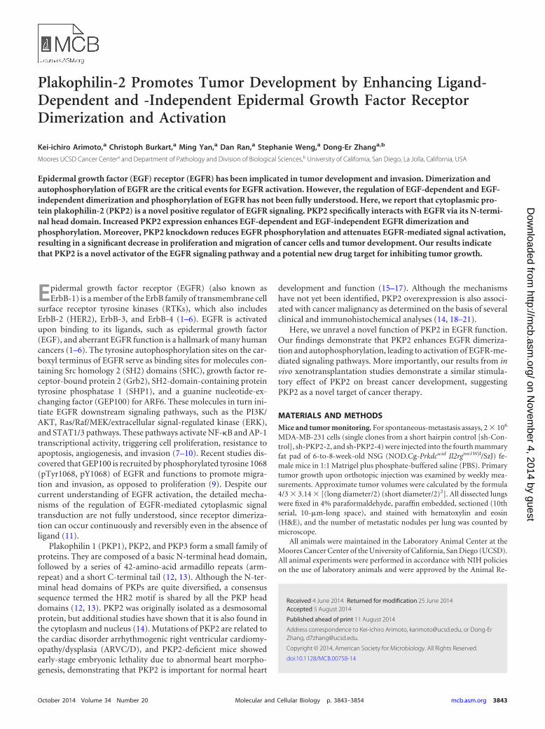
Plakophilin-2 Promotes Tumor Development by Enhancing Ligand-Dependent and -Independent Epidermal Growth Factor ReceptorDimerization and Activation
Kei-ichiro Arimoto,a Christoph Burkart,a Ming Yan,a Dan Ran,a Stephanie Weng,a Dong-Er Zhanga,b
Moores UCSD Cancer Centera and Department of Pathology and Division of Biological Sciences,b University of California, San Diego, La Jolla, California, USA
Epidermal growth factor (EGF) receptor (EGFR) has been implicated in tumor development and invasion. Dimerization andautophosphorylation of EGFR are the critical events for EGFR activation. However, the regulation of EGF-dependent and EGF-independent dimerization and phosphorylation of EGFR has not been fully understood. Here, we report that cytoplasmic pro-tein plakophilin-2 (PKP2) is a novel positive regulator of EGFR signaling. PKP2 specifically interacts with EGFR via its N-termi-nal head domain. Increased PKP2 expression enhances EGF-dependent and EGF-independent EGFR dimerization andphosphorylation. Moreover, PKP2 knockdown reduces EGFR phosphorylation and attenuates EGFR-mediated signal activation,resulting in a significant decrease in proliferation and migration of cancer cells and tumor development. Our results indicatethat PKP2 is a novel activator of the EGFR signaling pathway and a potential new drug target for inhibiting tumor growth.
Epidermal growth factor receptor (EGFR) (also known asErbB-1) is a member of the ErbB family of transmembrane cell
surface receptor tyrosine kinases (RTKs), which also includesErbB-2 (HER2), ErbB-3, and ErbB-4 (1–6). EGFR is activatedupon binding to its ligands, such as epidermal growth factor(EGF), and aberrant EGFR function is a hallmark of many humancancers (1–6). The tyrosine autophosphorylation sites on the car-boxyl terminus of EGFR serve as binding sites for molecules con-taining Src homology 2 (SH2) domains (SHC), growth factor re-ceptor-bound protein 2 (Grb2), SH2-domain-containing proteintyrosine phosphatase 1 (SHP1), and a guanine nucleotide-ex-changing factor (GEP100) for ARF6. These molecules in turn ini-tiate EGFR downstream signaling pathways, such as the PI3K/AKT, Ras/Raf/MEK/extracellular signal-regulated kinase (ERK),and STAT1/3 pathways. These pathways activate NF-�B and AP-1transcriptional activity, triggering cell proliferation, resistance toapoptosis, angiogenesis, and invasion (7–10). Recent studies dis-covered that GEP100 is recruited by phosphorylated tyrosine 1068(pTyr1068, pY1068) of EGFR and functions to promote migra-tion and invasion, as opposed to proliferation (9). Despite ourcurrent understanding of EGFR activation, the detailed mecha-nisms of the regulation of EGFR-mediated cytoplasmic signaltransduction are not fully understood, since receptor dimeriza-tion can occur continuously and reversibly even in the absence ofligand (11).
Plakophilin 1 (PKP1), PKP2, and PKP3 form a small family ofproteins. They are composed of a basic N-terminal head domain,followed by a series of 42-amino-acid armadillo repeats (arm-repeat) and a short C-terminal tail (12, 13). Although the N-ter-minal head domains of PKPs are quite diversified, a consensussequence termed the HR2 motif is shared by all the PKP headdomains (12, 13). PKP2 was originally isolated as a desmosomalprotein, but additional studies have shown that it is also found inthe cytoplasm and nucleus (14). Mutations of PKP2 are related tothe cardiac disorder arrhythmogenic right ventricular cardiomy-opathy/dysplasia (ARVC/D), and PKP2-deficient mice showedearly-stage embryonic lethality due to abnormal heart morpho-genesis, demonstrating that PKP2 is important for normal heart
development and function (15–17). Although the mechanismshave not yet been identified, PKP2 overexpression is also associ-ated with cancer malignancy as determined on the basis of severalclinical and immunohistochemical analyses (14, 18–21).
Here, we unravel a novel function of PKP2 in EGFR function.Our findings demonstrate that PKP2 enhances EGFR dimeriza-tion and autophosphorylation, leading to activation of EGFR-me-diated signaling pathways. More importantly, our results from invivo xenotransplantation studies demonstrate a similar stimula-tory effect of PKP2 on breast cancer development, suggestingPKP2 as a novel target of cancer therapy.
MATERIALS AND METHODSMice and tumor monitoring. For spontaneous-metastasis assays, 2 � 106
MDA-MB-231 cells (single clones from a short hairpin control [sh-Con-trol], sh-PKP2-2, and sh-PKP2-4) were injected into the fourth mammaryfat pad of 6-to-8-week-old NSG (NOD.Cg-Prkdcscid Il2rgtm1Wjl/SzJ) fe-male mice in 1:1 Matrigel plus phosphate-buffered saline (PBS). Primarytumor growth upon orthotopic injection was examined by weekly mea-surements. Approximate tumor volumes were calculated by the formula4/3 � 3.14 � [(long diameter/2) (short diameter/2)2]. All dissected lungswere fixed in 4% paraformaldehyde, paraffin embedded, sectioned (10thserial, 10-�m-long space), and stained with hematoxylin and eosin(H&E), and the number of metastatic nodules per lung was counted bymicroscope.
All animals were maintained in the Laboratory Animal Center at theMoores Cancer Center of the University of California, San Diego (UCSD).All animal experiments were performed in accordance with NIH policieson the use of laboratory animals and were approved by the Animal Re-
Received 4 June 2014 Returned for modification 25 June 2014Accepted 5 August 2014
Published ahead of print 11 August 2014
Address correspondence to Kei-ichiro Arimoto, [email protected], or Dong-ErZhang, [email protected].
Copyright © 2014, American Society for Microbiology. All Rights Reserved.
doi:10.1128/MCB.00758-14
October 2014 Volume 34 Number 20 Molecular and Cellular Biology p. 3843–3854 mcb.asm.org 3843
on Novem
ber 4, 2014 by guesthttp://m
cb.asm.org/
Dow
nloaded from
search Committee of the Office for the Protection of Research Subjects atthe University of California, San Diego.
Mass spectrometry database. As a tool for searching for candidatemolecules that interact with EGFR, we used the EMBL-EBI IntAct data-base (EBI-702413).
Cell culture, transfection, retroviral infection, and luciferase re-porter assays. 293T, A431, and MDA-MB-468 cells were grown in Dul-becco’s modified Eagle’s medium (DMEM) supplemented with glu-tamine, penicillin-streptomycin, and 10% fetal bovine serum (FBS).MDA-MB-231, HT29, and HCT116 cells were further supplemented with5% MEM (where MEM is minimum essential medium) nonessentialamino acid solution (Gibco). 32D cells were grown in RPMI 1640 me-dium supplemented with 15% FBS and 5% conditional media fromWEHI-3B cells. Plasmids were transfected using polyethyleneimine (PEI).For the transient expression of mouse PKP2 to 32D cells, Nucleofector kitsfor 32D cells (Lonza) were used. Small interfering RNA (siRNA) wastransfected using Lipofectamine 2000 (Invitrogen) at an interval of 24 h.At 72 h after the first siRNA transfection, cell lysates were prepared forexamination. For the retrovirus infection, spin infection (3,000 rpm, 3 h)was performed. Analysis of AP-1 and NF-�B dual-luciferase (Luc) assayswere carried out using Promega’s luciferase assay kit. As an internal con-trol, pRL-SV was transfected.
Antibodies and reagents. Antibodies were purchased from commer-cial sources as follows: anti-EGFR (Abcam), immunoprecipitation (IP)-specific anti-EGFR (Cell Signaling), anti-phospho-EGFR (Tyr845, -992,-1045, -1068, and -1173 [Cell Signaling] and Tyr 1086 [Invitrogen]), anti-PKP2 (ARP; American Research Products, Inc.), anti-ERK (BD Biosci-ence), anti-phospho-ERK (Cell Signaling), anti-phospho-MEK1 (CellSignaling), anti-Grb2 (Cell Signaling), anti-SHP1 (Cell Signaling), anti-GEP100 (Sigma), anti-green fluorescent protein (anti-GFP), and anti-�-tubulin (Sigma). Antibodies against FLAG (anti-FLAG; M2), Myc (9E10),and hemagglutinin (HA) (12CA5 or 3F10) were purchased from Sigma,Santa Cruz, and Roche, respectively. Recombinant human EGF was pur-chased from PeproTech. Lapatinib (S1028) was purchased from Selleck.
cDNA construction. Human PKP2 cDNA was cloned into C-termi-nally tagged pCAG vector, pcDNA3.1 vector, and glutathione S-trans-ferase (GST) (6p-1) vector. Murine PKP2 cDNA was cloned into C-ter-minal FLAG pCAG vector and murine stem cell virus (MSCV)-internalribosome entry site (IRES)-Puro (MIP) retroviral vector. HumanpLNCX2-EGFR-GFP was kindly provided by Frank Furnari. HumanEGFR and HER2 were cloned into pCMV5-FLAG and pcDNA3-Myc-Hisvector. Human PKP1 and PKP3 cDNAs were cloned into C-terminallytagged pCAG vector. Human Grb2 and Shc cDNAs were cloned intopcDNA3.1 vector. Deletion mutants of PKP2 and EGFR and Sh-PKP2-4-resistant PKP2 (T249A/T252A/G255A) were made by standard PCR pro-cedures.
Knockdown, RNA isolation, and quantitative reverse transcription-PCR (qRT-PCR) analysis. For knockdown of PKP2, the following siRNAswere used: si-PKP2-1 (5=-GAGACUACCCAAAAGCAAAUU-3= [Ambion]);si-PKP2-2 (5=-GCUCCUAAAAGUUCAGAAUUU-3= [Ambion]); andsi-PKP2-3 (5=-CAAAAGUAUUGGAUGUUUUUU-3= [Ambion]).
Control siRNA was purchased from Ambion.Short hairpin RNA (shRNA) plasmids for PKP2 and control experi-
ments were purchased from the SuperArray Bioscience Corporation. In-sertion sequences for shRNAs were as follows: for sh-PKP2-1, GCACACACGGAACTGCATCAT; for sh-PKP2-2, TGACTCACTGGTCCATTATGT; for sh-PKP2-3, CACGAGTGCTTCCAGAAATCT; for sh-PKP2-4,CAGCAGTGTTCCTGAGTATGT; and for the short hairpin control (sh-Control), GGAATCTCATTCGATGCATAC.
RNA was extracted with an RNeasy microkit (Qiagen). For qRT-PCRanalyses, equal amounts of RNA were reverse transcribed by the use ofqScript (Quanta Biosciences) and the resulting cDNA templates were sub-jected to qRT-PCR using a SYBR green detection system on a CFX96thermal cycler (Bio-Rad).
Primer sequences for PKP2 were as follows: for RT-PKP2-Fw, GAT
GTT TTG GCA GTC GAA GCA G; and for RT-PKP2-Rev, AAT GGAATG CCA CAG CCA CTC.
Western blotting and immunoprecipitation. All samples were dena-tured in 1� sample buffer (50 mM Tris-HCl [pH 6.8], 2% SDS, 2-mer-captethanol, 10% glycerol, and 1% bromophenol blue) for 5 min at100°C. Cells were lysed in a radioimmunoprecipitation assay (RIPA) buf-fer composed of 25 mM Tris-HCl (pH 8.0), 150 mM NaCl, 1 mM EDTA,1 mM dithiothreitol (DTT), 0.1% SDS, 1% Nonidet P-40, and 0.5% so-dium deoxycholate. To analyze immune complexes, cells were lysed inbinding buffer containing 25 mM Tris-HCl (pH 8.0), 150 mM NaCl, 1mM EDTA, and 0.5% Nonidet P-40 for coimmunoprecipitation assays.The cell lysates were centrifuged (10,000 � g) at 4°C for 5 min. All lysisbuffers in this study contained proteinase and phosphatase inhibitors(Roche). Soluble fractions were precleared by the use of protein G-Sep-harose at 4°C for 15 min. Precleared cell lysates were immunoprecipitatedfor �1 to �4 h with the indicated antibodies. Immunocomplexes wereadsorbed to the protein G-Sepharose and, after three washes, were elutedby boiling for 5 min. FLAG-tagged proteins were immunoprecipitatedwith anti-FLAG M2-agarose (Sigma). For quantification, Fujifilm Multi-Gauge V3.0 software was used.
GST pulldown assay. To purify full-length EGFR or the cytoplasmicregion of EGFR (EGFR amino acids [aa] 644 to 1210), 293T cells express-ing EGFR-FLAG or EGFR aa 644 to 1210 (cytoplasmic region)-FLAGwere lysed in RIPA buffer with 1� proteinase inhibitor mixture, and celllysates were immunoprecipitated with anti-FLAG M2 agarose in RIPAbuffer overnight at 4°C. After stringent washing with RIPA buffer, thebound protein was eluted using 50 �g/ml FLAG peptide (Sigma). For GSTpulldown, bacterial cell lysates expressing GST or GST-PKP2 were incu-bated with GST-Sepharose 4B beads (Roche) in binding buffer (50 mMTris [pH 7.4], 150 mM NaCl, 1% Nonidet P-40, and 1� proteinase inhib-itor mixture) for 3 h at 4°C and washed 5 times with binding buffer.Purified EGFR-FLAG or EGFR (aa 644 to 1210)-FLAG was then incu-bated in GST or GST-PKP2 columns. After washing with binding buffer,bound proteins were eluted and analyzed by Western blotting with theindicated antibodies.
Chemical cross-linking. Twenty-four hours after transfection understarvation conditions, monolayer cells were washed twice with ice-cold phos-phate-buffered saline containing 0.33 mM MgCl2 and 0.9 mM CaCl2[PBS(�)] and chemically cross-linked for 20 min at room temperature withfreshly prepared 1.5 mM bis(sulfosuccinimidyl) suberate (BS3; Pierce, Rock-ford, IL). To terminate the reaction, a final concentration of 20 mM glycinewas added for an additional 5 min. For immunoblot analysis, the cells werewashed twice with ice-cold PBS(�) and lysed with 10 mM iodoacetamidecontaining RIPA buffer. Samples were separated using 6% SDS-PAGE gelsand transferred at 20 V overnight with standard transfer buffer (48 mM Tris,39 mM glycine, 0.0375% SDS, and 20% methanol).
Proliferation assay. Cell growth was monitored by trypan blue count-ing or an MTS [3-(4,5-dimethylthiazol-2-yl)-5-(3-carboxymethoxyphe-nyl)-2-(4-sulfophenyl)-2H-tetrazolium] assay. For the trypan bluecounting, cells were seeded in 12-well plates at a density of 5 � 104 cells/well in triplicate. The MTS assay was conducted using CellTiter 96AQueous One Solution reagent (MTS assay reagent) according to theinstructions of the manufacturer (Promega). A total of 5 � 103 cells wereseeded in each well of a 96-well plate on day 0.
Invasion assay. A Matrigel invasion assay was performed with BD Bio-coat Matrigel chambers (BD Bioscience). A layer of dried Matrigel insertionswas rehydrated with DMEM for 2 h at 37°C. Aliquots (0.5 ml) of DMEMcontaining 10% FBS were added to each of the lower chambers of the 24-wellMatrigel invasion chambers, and 1 � 105 cells (MDA-MB-468 and MDA-MB-231 cells, each cell line processed in duplicate) diluted in 0.5-ml aliquotsof DMEM containing 0.5% FBS were then added to each of the upper cham-bers. After incubation for 22 h, cells were fixed by the use of ice-cold metha-nol, stained using Giemsa, and counted using light microscopy.
Statistical methods. Statistical significance was determined by Stu-dent’s t test using statcel2 software.
Arimoto et al.
3844 mcb.asm.org Molecular and Cellular Biology
on Novem
ber 4, 2014 by guesthttp://m
cb.asm.org/
Dow
nloaded from
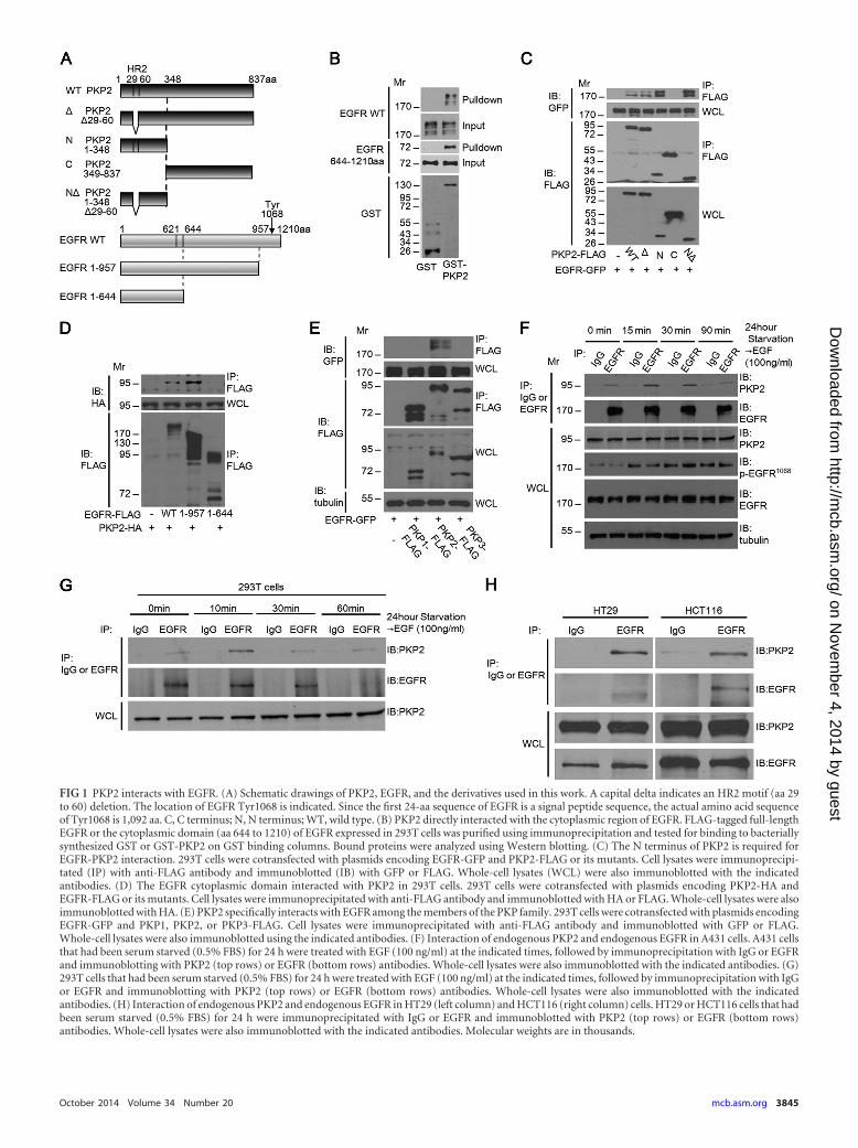
FIG 1 PKP2 interacts with EGFR. (A) Schematic drawings of PKP2, EGFR, and the derivatives used in this work. A capital delta indicates an HR2 motif (aa 29to 60) deletion. The location of EGFR Tyr1068 is indicated. Since the first 24-aa sequence of EGFR is a signal peptide sequence, the actual amino acid sequenceof Tyr1068 is 1,092 aa. C, C terminus; N, N terminus; WT, wild type. (B) PKP2 directly interacted with the cytoplasmic region of EGFR. FLAG-tagged full-lengthEGFR or the cytoplasmic domain (aa 644 to 1210) of EGFR expressed in 293T cells was purified using immunoprecipitation and tested for binding to bacteriallysynthesized GST or GST-PKP2 on GST binding columns. Bound proteins were analyzed using Western blotting. (C) The N terminus of PKP2 is required forEGFR-PKP2 interaction. 293T cells were cotransfected with plasmids encoding EGFR-GFP and PKP2-FLAG or its mutants. Cell lysates were immunoprecipi-tated (IP) with anti-FLAG antibody and immunoblotted (IB) with GFP or FLAG. Whole-cell lysates (WCL) were also immunoblotted with the indicatedantibodies. (D) The EGFR cytoplasmic domain interacted with PKP2 in 293T cells. 293T cells were cotransfected with plasmids encoding PKP2-HA andEGFR-FLAG or its mutants. Cell lysates were immunoprecipitated with anti-FLAG antibody and immunoblotted with HA or FLAG. Whole-cell lysates were alsoimmunoblotted with HA. (E) PKP2 specifically interacts with EGFR among the members of the PKP family. 293T cells were cotransfected with plasmids encodingEGFR-GFP and PKP1, PKP2, or PKP3-FLAG. Cell lysates were immunoprecipitated with anti-FLAG antibody and immunoblotted with GFP or FLAG.Whole-cell lysates were also immunoblotted using the indicated antibodies. (F) Interaction of endogenous PKP2 and endogenous EGFR in A431 cells. A431 cellsthat had been serum starved (0.5% FBS) for 24 h were treated with EGF (100 ng/ml) at the indicated times, followed by immunoprecipitation with IgG or EGFRand immunoblotting with PKP2 (top rows) or EGFR (bottom rows) antibodies. Whole-cell lysates were also immunoblotted with the indicated antibodies. (G)293T cells that had been serum starved (0.5% FBS) for 24 h were treated with EGF (100 ng/ml) at the indicated times, followed by immunoprecipitation with IgGor EGFR and immunoblotting with PKP2 (top rows) or EGFR (bottom rows) antibodies. Whole-cell lysates were also immunoblotted with the indicatedantibodies. (H) Interaction of endogenous PKP2 and endogenous EGFR in HT29 (left column) and HCT116 (right column) cells. HT29 or HCT116 cells that hadbeen serum starved (0.5% FBS) for 24 h were immunoprecipitated with IgG or EGFR and immunoblotted with PKP2 (top rows) or EGFR (bottom rows)antibodies. Whole-cell lysates were also immunoblotted with the indicated antibodies. Molecular weights are in thousands.
October 2014 Volume 34 Number 20 mcb.asm.org 3845
on Novem
ber 4, 2014 by guesthttp://m
cb.asm.org/
Dow
nloaded from
RESULTSPKP2 interacts with EGFR. In efforts to gain mechanistic insightinto EGFR signaling, we utilized a mass spectrometry database(EMBL EBI-702413) to identify proteins that coimmunoprecipi-tated with EGFR. We found that a cytoplasmic protein, PKP2,interacted with EGFR. Several clinical studies have indicated thatPKP2 expression is abnormally high in various cancers and thatPKP2 is a potential biomarker for cancer diagnosis (14, 18–21).However, the molecular basis for PKP2 expression in cancer isunclear. We therefore examined the potential role of PKP2 inmediating EGFR signaling and tumor growth.
We first verified the interaction between PKP2 and EGFR. Aschematic diagram of PKP2, EGFR, and their mutants used in this
report is shown in Fig. 1A. Pulldown analysis revealed that GST-PKP2 purified from Escherichia coli interacted with both full-length EGFR-FLAG and the EGFR cytoplasmic domain (aa 644 to1210)-FLAG purified from 293T cells (Fig. 1B). In order to deter-mine the regions involved in this interaction, truncation and de-letion mutants of PKP2 and EGFR were used for coimmunopre-cipitation studies. Deletion of the (N-terminal) head domain ofPKP2 abolished binding, indicating that it is essential for this in-teraction, and the homology region of PKP family members (HR2motif; aa 29 to 60) was not required for this interaction (Fig. 1Aand C). In addition, the cytoplasmic (C-terminal; aa 644 to 957)domain of EGFR is sufficient for its interaction with PKP2 (Fig. 1Aand D). Interestingly, among the plakophilin family members,
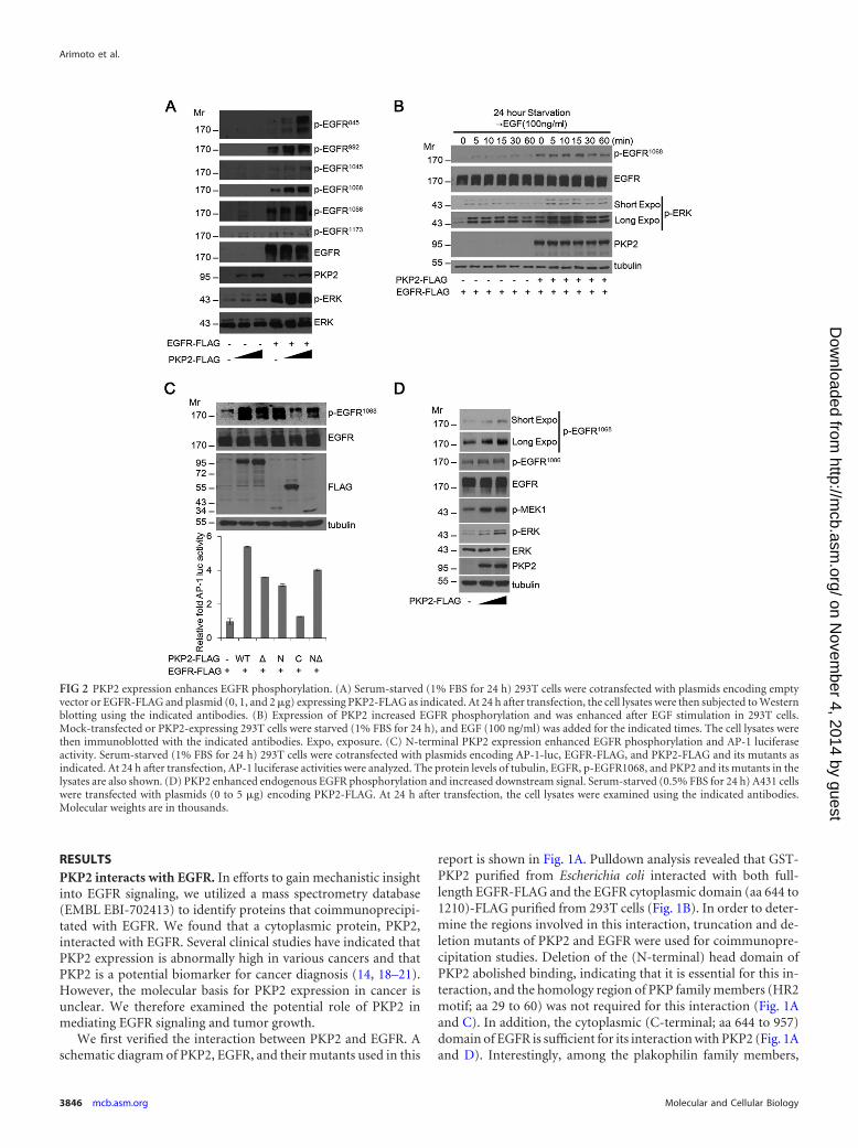
FIG 2 PKP2 expression enhances EGFR phosphorylation. (A) Serum-starved (1% FBS for 24 h) 293T cells were cotransfected with plasmids encoding emptyvector or EGFR-FLAG and plasmid (0, 1, and 2 �g) expressing PKP2-FLAG as indicated. At 24 h after transfection, the cell lysates were then subjected to Westernblotting using the indicated antibodies. (B) Expression of PKP2 increased EGFR phosphorylation and was enhanced after EGF stimulation in 293T cells.Mock-transfected or PKP2-expressing 293T cells were starved (1% FBS for 24 h), and EGF (100 ng/ml) was added for the indicated times. The cell lysates werethen immunoblotted with the indicated antibodies. Expo, exposure. (C) N-terminal PKP2 expression enhanced EGFR phosphorylation and AP-1 luciferaseactivity. Serum-starved (1% FBS for 24 h) 293T cells were cotransfected with plasmids encoding AP-1-luc, EGFR-FLAG, and PKP2-FLAG and its mutants asindicated. At 24 h after transfection, AP-1 luciferase activities were analyzed. The protein levels of tubulin, EGFR, p-EGFR1068, and PKP2 and its mutants in thelysates are also shown. (D) PKP2 enhanced endogenous EGFR phosphorylation and increased downstream signal. Serum-starved (0.5% FBS for 24 h) A431 cellswere transfected with plasmids (0 to 5 �g) encoding PKP2-FLAG. At 24 h after transfection, the cell lysates were examined using the indicated antibodies.Molecular weights are in thousands.
Arimoto et al.
3846 mcb.asm.org Molecular and Cellular Biology
on Novem
ber 4, 2014 by guesthttp://m
cb.asm.org/
Dow
nloaded from
only PKP2 specifically associated with EGFR (Fig. 1E). To test theassociation between PKP2 and EGFR endogenously, coimmuno-precipitation studies were done in A431, 293T, HT29, andHCT116 cells (Fig. 1F, G, and H). A ligand-independent interac-tion between endogenous PKP2 and EGFR was also detected inthese cells.
Our data demonstrate that PKP2 associates with EGFR endog-enously and is the only member of the plakophilin family to do so.
PKP2 expression enhances EGFR phosphorylation. To testwhether PKP2 can regulate EGFR signaling, PKP2 was coex-pressed with EGFR. Interestingly, levels of phosphorylated EGFRwere substantially enhanced by the increasing amounts of PKP2under serum starvation conditions (Fig. 2A). Furthermore, thelevel of phosphorylated EGFR (Tyr1068) after EGF treatment inPKP2-expressing 293T cells was enhanced compared to the levelin mock-transfected cells (Fig. 2B). Through the use of PKP2 de-letion mutants, it was observed that the HR2 motif deleted the Nterminus (N�) and not the C terminus of PKP2, which corre-
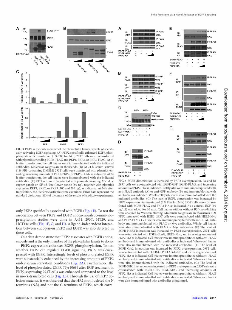
FIG 3 PKP2 is the only member of the plakophilin family capable of specifi-cally activating EGFR signaling. (A) PKP2 specifically enhanced EGFR phos-phorylation. Serum-starved (1% FBS for 24 h) 293T cells were cotransfectedwith plasmids encoding EGFR-FLAG and PKP1, PKP2, or PKP3-FLAG. At 24h after transfection, the cell lysates were immunoblotted with the indicatedantibodies. Molecular weights are in thousands. (B) At 24 h, serum-starved(1% FBS-containing DMEM) 293T cells were transfected with plasmids en-coding increasing amounts of PKP1, PKP2, or PKP3-FLAG as indicated. At 24h after transfection, the cell lysates were immunoblotted with the indicatedantibodies. (C) 293T cells were transfected with plasmids encoding AP-1-Luc(upper panel) or NF-�B-Luc (lower panel) (50 ng), together with plasmidsexpressing PKP1, PKP2, or PKP3 (100 and 200 ng), as indicated. At 24 h aftertransfection, the luciferase activities were examined. Error bars represent thestandard deviations (SD) of the means of the results of triplicate experiments.
FIG 4 EGFR dimerization is increased by PKP2 overexpression. (A and B)293T cells were cotransfected with EGFR-GFP, EGFR-FLAG, and increasingamounts of PKP2-HA as indicated. Cell lysates were immunoprecipitated withanti-FLAG antibody (A) or anti-GFP antibody (B) and immunoblotted withantibodies as indicated. Whole-cell lysates were also immunoblotted with theindicated antibodies. (C) The level of EGFR dimerization was increased byPKP2 expression. Serum-starved (1% FBS for 24 h) 293T cells were cotrans-fected with EGFR-FLAG and PKP2-HA as indicated. As a control, EGF (10ng/ml) was added for 10 min. Cell lysates with or without BS3 cross-linkingwere analyzed by Western blotting. Molecular weights are in thousands. (D)PKP2 interacted with HER2. 293T cells were cotransfected with HER2-Mycand PKP2-FLAG. Cell lysates were immunoprecipitated with anti-FLAG anti-body and immunoblotted with FLAG or Myc antibodies. Whole-cell lysateswere also immunoblotted with FLAG or Myc antibodies. (E) The level ofEGFR-HER2 interaction was increased by PKP2 overexpression. 293T cellswere cotransfected with EGFR-FLAG, HER2-Myc, and increasing amounts ofPKP2-HA as indicated. Cell lysates were immunoprecipitated with anti-FLAGantibody and immunoblotted with antibodies as indicated. Whole-cell lysateswere also immunoblotted with the indicated antibodies. (F) The level ofEGFR-Grb2 interaction was increased by PKP2 overexpression. 293T cellswere cotransfected with EGFR-GFP, FLAG-Grb2, and increasing amounts ofPKP2-HA as indicated. Cell lysates were immunoprecipitated with anti-FLAGantibody and immunoblotted with antibodies as indicated. Whole-cell lysateswere also immunoblotted with the indicated antibodies. (G) The level ofEGFR-SHC interaction was increased by PKP2 overexpression. 293T cells werecotransfected with EGFR-GFP, FLAG-SHC, and increasing amounts ofPKP2-HA as indicated. Cell lysates were immunoprecipitated with anti-FLAGantibody and immunoblotted with antibodies as indicated. Whole-cell lysateswere also immunoblotted with antibodies as indicated.
PKP2 Functions as a Novel Activator of EGFR Signaling
October 2014 Volume 34 Number 20 mcb.asm.org 3847
on Novem
ber 4, 2014 by guesthttp://m
cb.asm.org/
Dow
nloaded from
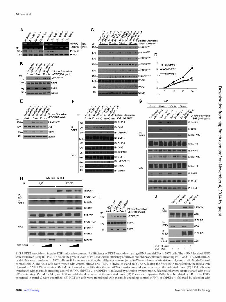
FIG 5 PKP2 knockdown impairs EGF-induced responses. (A) Efficiency of PKP2 knockdown using siRNA and shRNA in 293T cells. The mRNA levels of PKP2were visualized using RT-PCR. To assess the protein levels of PKP2 to test the efficiency of siRNAs and shRNAs, plasmids encoding PKP1 and PKP2 with siRNAsor shRNAs were transfected to 293T cells. At 48 h after transfection, the cell lysates were subjected to Western blot analysis. si-Control, control siRNA; sh-Control,control shRNA. (B) A431 cells were treated with control siRNA or si-PKP2-2 (twice, at 0 and 48 h). At 72 h after the first siRNA transfection, the media werechanged to 0.5% FBS-containing DMEM. EGF was added at 98 h after the first siRNA transfection and was harvested at the indicated times. (C) A431 cells weretransfected with plasmids encoding control shRNA, shPKP2-2, or shPKP2-4, followed by selection by puromycin. Selected cells were serum starved with 0.5%FBS-containing DMEM for 24 h, and EGF was added and harvested at the indicated times. (D) The ratios of tyrosine 1068-phosphorylated EGFR to total EGFRpresented in panel C were quantified. (E) HCT116 cells were transfected with plasmids encoding control shRNA or shPKP2-4, followed by selection with
Arimoto et al.
3848 mcb.asm.org Molecular and Cellular Biology
on Novem
ber 4, 2014 by guesthttp://m
cb.asm.org/
Dow
nloaded from
sponds to the fact that the EGFR-interacting region (Fig. 1A andC) could enhance both the phosphorylation of EGFR and the ac-tivation of the downstream target AP-1 promoter-luciferase activ-ity (Fig. 2C). Furthermore, ectopic expression of PKP2 increasedphosphorylation of endogenous EGFR, MEK1, and ERK dose de-pendently (Fig. 2D).
PKP2 is the only member of the plakophilin family capable ofspecifically activating EGFR signaling. To examine if other pla-kophilin family proteins can regulate EGFR function, we coex-pressed EGFR and PKP family proteins. PKP2 expression readilyenhanced EGFR phosphorylation; however, the other two PKPfamily proteins, PKP1 and PKP3, had no effect on EGFR phos-phorylation (Fig. 3A), which agrees with their lack of interactionwith EGFR (Fig. 1E). Furthermore, PKP2 expression increased thelevel of ERK phosphorylation dose dependently, while neitherPKP1 nor PKP3 had an effect on the phosphorylation of ERK (Fig.3B). Consistent with the PKP2 levels, AP-1 and NF-�B reporter-luciferase activity was increased upon PKP2 expression (Fig. 3C,top and bottom, respectively). However, PKP3 increased NF-�Bactivity (Fig. 3C, bottom), suggesting that PKP3 has a novel effecton NF-�B that is independent of EGFR phosphorylation and ac-tivation.
Thus, these results indicate that PKP2 specifically interactswith EGFR and activates EGFR signaling.
PKP2 activates the EGFR signaling pathway via enhance-ment of EGFR dimerization followed by autophosphorylation.Because EGFR autophosphorylation requires EGFR dimerization,we analyzed whether PKP2 expression affects the dimerization ofEGFR. First, we found that expression of PKP2 clearly enhancedthe EGFR-EGFR interaction (Fig. 4A and B) and markedly in-creased the dimer/monomer ratio of EGFR under starvation con-ditions (Fig. 4C). Interestingly, PKP2 also interacted with HER2(ErbB2) and strengthened the level of interaction between EGFRand HER2, suggesting that PKP2 is able to facilitate both EGFR-EGFR and EGFR-HER2 dimerization (Fig. 4D and E).
Phosphotyrosines on EGFR recruit cytosolic signaling mole-cules such as SHC and Grb2 and trigger a series of intracellularpathways, culminating in cellular proliferation, migration, inva-sion, and tumorigenicity (7–10). To examine whether PKP2 ex-pression is involved in the recruitment of these molecules, weanalyzed the interaction between EGFR and SHC or Grb2 in thepresence of increasing amounts of PKP2 expression. Their levelsof interaction increased dose dependently with PKP2 expression(Fig. 4F and G).
From this data, we found that PKP2 enhances EGFR dimeriza-tion and its subsequent autophosphorylation, as well as down-stream target activation.
PKP2 knockdown results in a substantial reduction in EGF-induced EGFR activation. To examine the physiological roles ofPKP2 in EGFR-mediated signaling, we assessed phosphorylationof EGFR and its downstream signaling molecules in PKP2 knock-down cells. Upon siRNA and shRNA transfection, PKP2 knock-down efficiency was confirmed at both the mRNA and proteinlevels (Fig. 5A). PKP2 reduction by siRNA and two different tar-geting shRNAs substantially impaired EGF-induced phosphory-lation of EGFR in A431 cells (Fig. 5B and C, top rows). Quantifi-cation of the ratio of phosphorylated EGFR (Y1068) to total EGFRclearly correlated with the expression level of PKP2 (Fig. 5D).Similar results were also observed in HCT116 cells (Fig. 5E).Moreover, in accordance with the levels of phosphorylated EGFRin control and sh-PKP2-4-expressing cells, the levels of interac-tions between EGFR and Grb2, GEP100, and SHP-1 were substan-tially reduced in PKP2 knockdown MDA-MB-468 cells (Fig. 5F).Similar results were also observed in A431 cells (Fig. 5G). In addi-tion, increased expression of PKP2 in sh-PKP2-4-expressing A431cells restored the interactions between EGFR and Grb2, GEP100,and SHP-1 (Fig. 5H). Furthermore, the levels of phosphorylatedEGFR in sh-PKP2-4-expressing A431 cells were restored by ex-pression of sh-PKP2-4-resistant PKP2 (PKP2shR) (Fig. 5I). Im-portantly, EGFR-EGFR interaction was substantially reduced insh-PKP2-4-expressing A431 cells compared with short hairpincontrol-expressing A431 cells, further suggesting that PKP2 hasthe ability to facilitate EGFR dimerization (Fig. 5J).
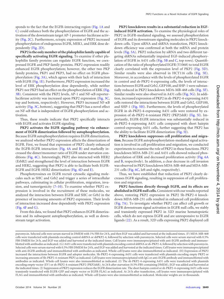
PKP2 knockdown suppresses cell proliferation and migra-tion. Because EGFR autophosphorylation-mediated signal activa-tion is involved in cell proliferation and migration, we conductedexperiments to examine the role of PKP2 in these functions. PKP2knockdown MDA-MB-231 and A431 cells showed reduced phos-phorylation of ERK and decreased proliferation activity (Fig. 6Aand B, respectively). In addition, a clear decrease in cell invasionwas observed in PKP2 knockdown MDA-MB-468 and MDA-MB-231 cells (Fig. 6C, left and right, respectively).
Thus, we have established that reduction of PKP2 clearly de-creases EGFR signaling, resulting in suppression of cell prolifera-tion and migration.
PKP2 functions directly through EGFR, and its effects areabolished in EGFR null cells. Consistent with our results reportedabove, restoring PKP2 expression in PKP2 Sh-PKP2-4 knock-down MDA-MB-231 cells resulted in enhanced cell proliferation(Fig. 7A). To investigate whether PKP2 can affect cell growth orEGFR downstream signal activation in EGFR null cells, we stablyand transiently expressed PKP2 in 32D murine hematopoieticcells, which do not express EGFR and are unresponsive to EGFRligands (22). As a result, 32D cells expressing PKP2 displayed cell
puromycin. Selected cells were serum starved in DMEM with 1% FBS for 24 h, and then EGF was added and harvested at the indicated times. (F) MDA-MB-468cells were transfected with plasmids encoding control shRNA or shPKP2-4, followed by selection with puromycin. Selected cells were serum starved with 0.5%FBS DMEM for 24 h, and EGF was added and harvested at the indicated times. Cell lysates were immunoprecipitated with anti-EGFR antibody and immuno-blotted with antibodies as indicated. (G) A431 cells were transfected with plasmids encoding control shRNA or sh-PKP2-4, followed by selection with puromycin.Selected cells were serum starved with 0.5% FBS DMEM for 24 h, and EGF was added and harvested at the indicated times. Cell lysates were immunoprecipitatedwith anti-EGFR antibody and immunoblotted with antibodies as indicated. Whole-cell lysates were also immunoblotted as indicated. (H) Expression of PKP2increased the interactions between EGFR and adopter molecules. Sh-PKP2-4-expressing A431 cells were transfected with plasmids encoding empty PKP2 orincreasing amounts of Sh-PKP2-4-resistant PKP2 as indicated. Cell lysates were immunoprecipitated with IgG or anti-EGFR antibody and immunoblotted withantibodies as indicated. Whole-cell lysates were also immunoblotted as indicated. (I) The sh-PKP2-4-expressing A431 cells were transfected with plasmidsencoding empty vector (EV) or sh-PKP2-4-resistant PKP2 (PKP2shR). At 24 h after starvation (0.5% FBS-containing DMEM), EGF was added and harvested atthe indicated times. (J) Suppression of PKP2 expression reduces the EGFR dimerization rate. The short hairpin control or sh-PKP2-4-expressing A431 cells weretransiently transfected with EGFR-GFP and empty vector or EGFR-FLAG as indicated. At 24 h after transfection, cell lysates were immunoprecipitated withFLAG and immunoblotted with antibodies as indicated. Whole-cell lysates were also immunoblotted as indicated. Molecular weights are in thousands.
PKP2 Functions as a Novel Activator of EGFR Signaling
October 2014 Volume 34 Number 20 mcb.asm.org 3849
on Novem
ber 4, 2014 by guesthttp://m
cb.asm.org/
Dow
nloaded from
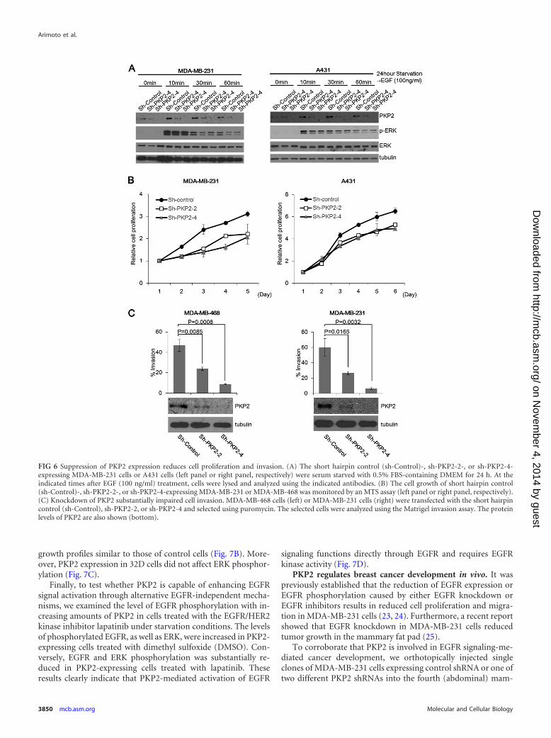
growth profiles similar to those of control cells (Fig. 7B). More-over, PKP2 expression in 32D cells did not affect ERK phosphor-ylation (Fig. 7C).
Finally, to test whether PKP2 is capable of enhancing EGFRsignal activation through alternative EGFR-independent mecha-nisms, we examined the level of EGFR phosphorylation with in-creasing amounts of PKP2 in cells treated with the EGFR/HER2kinase inhibitor lapatinib under starvation conditions. The levelsof phosphorylated EGFR, as well as ERK, were increased in PKP2-expressing cells treated with dimethyl sulfoxide (DMSO). Con-versely, EGFR and ERK phosphorylation was substantially re-duced in PKP2-expressing cells treated with lapatinib. Theseresults clearly indicate that PKP2-mediated activation of EGFR
signaling functions directly through EGFR and requires EGFRkinase activity (Fig. 7D).
PKP2 regulates breast cancer development in vivo. It waspreviously established that the reduction of EGFR expression orEGFR phosphorylation caused by either EGFR knockdown orEGFR inhibitors results in reduced cell proliferation and migra-tion in MDA-MB-231 cells (23, 24). Furthermore, a recent reportshowed that EGFR knockdown in MDA-MB-231 cells reducedtumor growth in the mammary fat pad (25).
To corroborate that PKP2 is involved in EGFR signaling-me-diated cancer development, we orthotopically injected singleclones of MDA-MB-231 cells expressing control shRNA or one oftwo different PKP2 shRNAs into the fourth (abdominal) mam-
FIG 6 Suppression of PKP2 expression reduces cell proliferation and invasion. (A) The short hairpin control (sh-Control)-, sh-PKP2-2-, or sh-PKP2-4-expressing MDA-MB-231 cells or A431 cells (left panel or right panel, respectively) were serum starved with 0.5% FBS-containing DMEM for 24 h. At theindicated times after EGF (100 ng/ml) treatment, cells were lysed and analyzed using the indicated antibodies. (B) The cell growth of short hairpin control(sh-Control)-, sh-PKP2-2-, or sh-PKP2-4-expressing MDA-MB-231 or MDA-MB-468 was monitored by an MTS assay (left panel or right panel, respectively).(C) Knockdown of PKP2 substantially impaired cell invasion. MDA-MB-468 cells (left) or MDA-MB-231 cells (right) were transfected with the short hairpincontrol (sh-Control), sh-PKP2-2, or sh-PKP2-4 and selected using puromycin. The selected cells were analyzed using the Matrigel invasion assay. The proteinlevels of PKP2 are also shown (bottom).
Arimoto et al.
3850 mcb.asm.org Molecular and Cellular Biology
on Novem
ber 4, 2014 by guesthttp://m
cb.asm.org/
Dow
nloaded from
mary fat pads of immune-deficient NSG (NOD.Cg-Prkdcscid
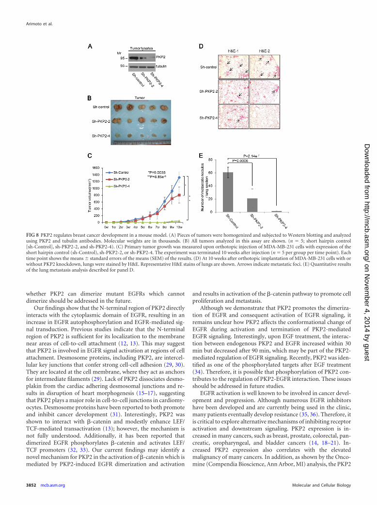
Il2rgtm1Wjl/SzJ) mice. Mice were monitored for tumor growthweekly and were sacrificed at 10 weeks after injection. MDA-MB-231 tumor tissues were subjected to Western blotting to confirmprotein levels of PKP2. As expected, short hairpin control (sh-Control) tumors still expressed PKP2. Tumor cells expressing sh-PKP2-2 expressed half the amount of PKP2 expressed by the shorthairpin control, and shPKP2-4-expressing tumors had nearly noexpression of PKP2 (Fig. 8A). The results indicate that sh-PKP2-2-expressing cells reduced the growth rate of tumors by around40% compared to the control and that Sh-PKP2-4-expressing cellswere severely impaired in their tumor growth capacity in NSGmice (Fig. 8B and C). In direct correlation with their effect onprimary tumor growth, sh-PKP2-2- and sh-PKP2-4-expressingcells formed 65% and 98% fewer metastatic nodules than shorthairpin control cells, respectively (Fig. 8D and E).
Taking the findings together, we conclude that PKP2 plays anovel and important role in facilitating breast cancer cell growthand metastasis.
DISCUSSION
In this report, we are the first to identify PKP2 as a novel regulatorof both EGF-independent and -dependent activation of EGFR.Notably, we found that PKP2 is the only member of the plakophi-lin family that can stimulate EGFR phosphorylation. This stimu-lation by PKP2 is also seen in the absence of EGF (0.5% to 1%serum), indicating that misregulation of PKP2 expression, as is
often observed in cancer, can have functional consequences evenin the absence of ligand. We showed that PKP2-induced enhance-ment of EGFR and ERK phosphorylation was completely abol-ished in the presence of lapatinib, suggesting that PKP2 can di-rectly activate EGFR signaling in EGFR-expressing cancer cells.We also confirmed that expression of PKP2 did not activate down-stream EGFR signaling in EGFR null cells. We demonstrated thatknockdown of PKP2 significantly impaired EGF-induced EGFRautophosphorylation and recruitment of adaptor molecules toEGFR. Furthermore, diminished expression of PKP2 in MDA-MB-231 breast cancer cells reduced their proliferation and meta-static potential and ultimately decreased tumor development invivo.
Previous work by others has identified other cytoplasmic acti-vators of EGF receptor tyrosine kinases, such as YES (Yamaguchisarcoma virus oncogene) and cytohesins (26–28). However, theirroles in EGFR activation are limited. YES-mediated EGFR phos-phorylation occurs only on the endosome, mildly increases phos-phorylation on limited sites, and requires EGF stimulation andpIgA-pIgR transcytosis (26, 27). Furthermore, cytohesins do notinfluence receptor dimerization but function as conformationalactivators of dimerized receptors after EGF stimulation (26, 27).In contrast, PKP2 has a broader effect on EGFR activation and canenhance receptor dimerization and autophosphorylation even inthe absence of ligand stimulation. The function of PKP2 in EGFRsignaling does not overlap that of YES and cytohesins, suggestingthat PKP2 functions as a novel “cytoplasmic EGF.” The issue of
FIG 7 PKP2 functions directly through EGFR, and its effects are abolished in EGFR null cells. (A) The sh-PKP2-4-expressing MDA-MB-231 cells weretransfected with plasmids encoding empty vector (EV) or sh-PKP2-4-resistant PKP2 (PKP2shR). After selection, these cells were monitored by trypan bluecounting. (B) 32D cells stably expressing MIP or MIP-mouse PKP2 were monitored by trypan blue counting. (C) Mouse PKP2 was transiently expressed in 32Dcells as indicated. Twenty-four hours after transfection, cell lysates were immunoblotted with the indicated antibodies. (D) Lapatinib treatment completelyimpaired the enhancement of PKP2-mediated EGFR phosphorylation. Serum-starved (1% FBS for 24 h) 293T cells were cotransfected with plasmids encodingEGFR-FLAG and PKP2-FLAG as indicated. The cells were treated with DMSO or lapatinib (final 1 �M) for 24 h. At 24 h after transfection, the cell lysates weresubjected to Western blotting using the indicated antibodies. Molecular weights are in thousands.
PKP2 Functions as a Novel Activator of EGFR Signaling
October 2014 Volume 34 Number 20 mcb.asm.org 3851
on Novem
ber 4, 2014 by guesthttp://m
cb.asm.org/
Dow
nloaded from
whether PKP2 can dimerize mutant EGFRs which cannotdimerize should be addressed in the future.
Our findings show that the N-terminal region of PKP2 directlyinteracts with the cytoplasmic domain of EGFR, resulting in anincrease in EGFR autophosphorylation and EGFR-mediated sig-nal transduction. Previous studies indicate that the N-terminalregion of PKP2 is sufficient for its localization to the membranenear areas of cell-to-cell attachment (12, 13). This may suggestthat PKP2 is involved in EGFR signal activation at regions of cellattachment. Desmosome proteins, including PKP2, are intercel-lular key junctions that confer strong cell-cell adhesion (29, 30).They are located at the cell membrane, where they act as anchorsfor intermediate filaments (29). Lack of PKP2 dissociates desmo-plakin from the cardiac adhering desmosomal junctions and re-sults in disruption of heart morphogenesis (15–17), suggestingthat PKP2 plays a major role in cell-to-cell junctions in cardiomy-ocytes. Desmosome proteins have been reported to both promoteand inhibit cancer development (31). Interestingly, PKP2 wasshown to interact with -catenin and modestly enhance LEF/TCF-mediated transactivation (13); however, the mechanism isnot fully understood. Additionally, it has been reported thatdimerized EGFR phosphorylates -catenin and activates LEF/TCF promoters (32, 33). Our current findings may identify anovel mechanism for PKP2 in the activation of -catenin which ismediated by PKP2-induced EGFR dimerization and activation
and results in activation of the -catenin pathway to promote cellproliferation and metastasis.
Although we demonstrate that PKP2 promotes the dimeriza-tion of EGFR and consequent activation of EGFR signaling, itremains unclear how PKP2 affects the conformational change ofEGFR during activation and termination of PKP2-mediatedEGFR signaling. Interestingly, upon EGF treatment, the interac-tion between endogenous PKP2 and EGFR increased within 30min but decreased after 90 min, which may be part of the PKP2-mediated regulation of EGFR signaling. Recently, PKP2 was iden-tified as one of the phosphorylated targets after EGF treatment(34). Therefore, it is possible that phosphorylation of PKP2 con-tributes to the regulation of PKP2-EGFR interaction. These issuesshould be addressed in future studies.
EGFR activation is well known to be involved in cancer devel-opment and progression. Although numerous EGFR inhibitorshave been developed and are currently being used in the clinic,many patients eventually develop resistance (35, 36). Therefore, itis critical to explore alternative mechanisms of inhibiting receptoractivation and downstream signaling. PKP2 expression is in-creased in many cancers, such as breast, prostate, colorectal, pan-creatic, oropharyngeal, and bladder cancers (14, 18–21). In-creased PKP2 expression also correlates with the elevatedmalignancy of many cancers. In addition, as shown by the Onco-mine (Compendia Bioscience, Ann Arbor, MI) analysis, the PKP2
FIG 8 PKP2 regulates breast cancer development in a mouse model. (A) Pieces of tumors were homogenized and subjected to Western blotting and analyzedusing PKP2 and tubulin antibodies. Molecular weights are in thousands. (B) All tumors analyzed in this assay are shown. (n 5; short hairpin control[sh-Control], sh-PKP2-2, and sh-PKP2-4). (C) Primary tumor growth was measured upon orthotopic injection of MDA-MB-231 cells with expression of theshort hairpin control (sh-Control), sh-PKP2-2, or sh-PKP2-4. The experiment was terminated 10 weeks after injection (n 5 per group per time point). Eachtime point shows the means � standard errors of the means (SEM) of the results. (D) At 10 weeks after orthotopic implantation of MDA-MB-231 cells with orwithout PKP2 knockdown, lungs were stained by H&E. Representative H&E stains of lungs are shown. Arrows indicate metastatic foci. (E) Quantitative resultsof the lung metastasis analysis described for panel D.
Arimoto et al.
3852 mcb.asm.org Molecular and Cellular Biology
on Novem
ber 4, 2014 by guesthttp://m
cb.asm.org/
Dow
nloaded from
expression levels in cancers of breast (37), lung (38), bladder (39),and thyroid gland (40) were significantly higher than the PKP2expression levels in the normal tissues of these organs. Our studiesdemonstrated that PKP2 enhances EGFR-mediated signaling byfacilitating the dimerization of EGFR and activating downstreamsignaling pathways, as well as promoting cancer cell proliferationand tumor metastasis. The newly identified role of PKP2 in EGFRactivation described in this report identifies a clear and novel mo-lecular mechanism to explain the clinical correlations with PKP2.Development of a small molecule that disrupts protein-proteininteractions by binding with high affinity to “hot spots,” whichcontribute most to the protein-protein interaction, could be ap-plied to preventing the association between the N terminus ofPKP2 and the cytoplasmic domain of EGFR (41). Altogether, ourfindings demonstrate a novel role of PKP2 in EGFR signaling andsuggest PKP2 as a potential target for therapeutic intervention.
ACKNOWLEDGMENTS
We thank Paul S. Mischel and Frank Furnari (Ludwig Institute for CancerResearch, UCSD) and Gen-Sheng Feng (Department of Pathology,UCSD) for critical reading of the manuscript. We thank Kunitada Shimo-tohno (National Center for Global Health and Medicine, Japan) for plas-mid vectors (C-terminally tagged pCAG vector, GST [6p-1], and C-ter-minally Myc-tagged pcDNA3.1 vector), Jo Minji (Steven Goniaslaboratory, UCSD) for MDA-MB-468 cells, and David Cheresh (MooresCancer Center, UCSD) for MDA-MB-231 cells. We thank all members ofthe D.-E.Z. laboratory and Keun Il Kim (Sookmyung Women’s Univer-sity, South Korea) for helpful discussion. We also thank Chuyi Cheng andYue Zhang (Division of Biological Sciences, UCSD) for their technicalassistance in this study.
This work was supported by funding from the U.S. National Institutesof Health (R01CA177305 and R01HL091549). Kei-ichiro Arimoto is aJapan Society for the Promotion of Science (JSPS) Postdoctoral Fellow forResearch Abroad.
REFERENCES1. Blume-Jensen P, Hunter T. 2001. Oncogenic kinase signalling. Nature
411:355–365. http://dx.doi.org/10.1038/35077225.2. Bogdan S, Klambt C. 2001. Epidermal growth factor receptor signal-
ing. Curr. Biol. 11:R292–R295. http://dx.doi.org/10.1016/S0960-9822(01)00167-1.
3. Hynes NE, Lane HA. 2005. ERBB receptors and cancer: the complexity oftargeted inhibitors. Nat. Rev. Cancer 5:341–354. http://dx.doi.org/10.1038/nrc1609.
4. Hynes NE, MacDonald G. 2009. ErbB receptors and signaling pathwaysin cancer. Curr. Opin. Cell Biol. 21:177–184. http://dx.doi.org/10.1016/j.ceb.2008.12.010.
5. Yarden Y, Pines G. 2012. The ERBB network: at last, cancer therapy meetssystems biology. Nat. Rev. Cancer 12:553–563. http://dx.doi.org/10.1038/nrc3309.
6. Lemmon MA, Schlessinger J. 2010. Cell signaling by receptor tyrosinekinases. Cell 141:1117–1134. http://dx.doi.org/10.1016/j.cell.2010.06.011.
7. Batzer AG, Rotin D, Urena JM, Skolnik EY, Schlessinger J. 1994.Hierarchy of binding sites for Grb2 and Shc on the epidermal growthfactor receptor. Mol. Cell. Biol. 14:5192–5201.
8. Hsu JM, Chen CT, Chou CK, Kuo HP, Li LY, Lin CY, Lee HJ, WangYN, Liu M, Liao HW, Shi B, Lai CC, Bedford MT, Tsai CH, Hung MC.2011. Crosstalk between Arg 1175 methylation and Tyr 1173 phosphory-lation negatively modulates EGFR-mediated ERK activation. Nat. CellBiol. 13:174 –181. http://dx.doi.org/10.1038/ncb2158.
9. Morishige M, Hashimoto S, Ogawa E, Toda Y, Kotani H, Hirose M,Wei S, Hashimoto A, Yamada A, Yano H, Mazaki Y, Kodama H, Nio Y,Manabe T, Wada H, Kobayashi H, Sabe H. 2008. GEP100 links epider-mal growth factor receptor signalling to Arf6 activation to induce breastcancer invasion. Nat. Cell Biol. 10:85–92. http://dx.doi.org/10.1038/ncb1672.
10. Rozakis-Adcock M, McGlade J, Mbamalu G, Pelicci G, Daly R, Li W,
Batzer A, Thomas S, Brugge J, Pelicci PG, Schlessinger J, Pawson T.1992. Association of the Shc and Grb2/Sem5 SH2-containing proteins isimplicated in activation of the Ras pathway by tyrosine kinases. Nature360:689 – 692. http://dx.doi.org/10.1038/360689a0.
11. Chung I, Akita R, Vandlen R, Toomre D, Schlessinger J, Mellman I.2010. Spatial control of EGF receptor activation by reversible dimerizationon living cells. Nature 464:783–787. http://dx.doi.org/10.1038/nature08827.
12. Bass-Zubek AE, Godsel LM, Delmar M, Green KJ. 2009. Plakophilins:multifunctional scaffolds for adhesion and signaling. Curr. Opin. CellBiol. 21:708 –716. http://dx.doi.org/10.1016/j.ceb.2009.07.002.
13. Chen X, Bonne S, Hatzfeld M, van Roy F, Green KJ. 2002. Proteinbinding and functional characterization of plakophilin 2. Evidence for itsdiverse roles in desmosomes and beta-catenin signaling. J. Biol. Chem.277:10512–10522. http://dx.doi.org/10.1074/jbc.M108765200.
14. Mertens C, Kuhn C, Moll R, Schwetlick I, Franke WW. 1999.Desmosomal plakophilin 2 as a differentiation marker in normal andmalignant tissues. Differentiation 64:277–290. http://dx.doi.org/10.1046/j.1432-0436.1999.6450277.x.
15. Gerull B, Heuser A, Wichter T, Paul M, Basson CT, McDermott DA,Lerman BB, Markowitz SM, Ellinor PT, MacRae CA, Peters S, Gross-mann KS, Drenckhahn J, Michely B, Sasse-Klaassen S, Birchmeier W,Dietz R, Breithardt G, Schulze-Bahr E, Thierfelder L. 2004. Mutations inthe desmosomal protein plakophilin-2 are common in arrhythmogenicright ventricular cardiomyopathy. Nat. Genet. 36:1162–1164. http://dx.doi.org/10.1038/ng1461.
16. Grossmann KS, Grund C, Huelsken J, Behrend M, Erdmann B, FrankeWW, Birchmeier W. 2004. Requirement of plakophilin 2 for heart mor-phogenesis and cardiac junction formation. J. Cell Biol. 167:149 –160.http://dx.doi.org/10.1083/jcb.200402096.
17. MacRae CA, Birchmeier W, Thierfelder L. 2006. Arrhythmogenic rightventricular cardiomyopathy: moving toward mechanism. J. Clin. Invest.116:1825–1828. http://dx.doi.org/10.1172/JCI29174.
18. Cheng AS, Culhane AC, Chan MW, Venkataramu CR, Ehrich M, NasirA, Rodriguez BA, Liu J, Yan PS, Quackenbush J, Nephew KP, YeatmanTJ, Huang TH. 2008. Epithelial progeny of estrogen-exposed breast pro-genitor cells display a cancer-like methylome. Cancer Res. 68:1786 –1796.http://dx.doi.org/10.1158/0008-5472.CAN-07-5547.
19. Papagerakis S, Shabana AH, Depondt J, Gehanno P, Forest N. 2003.Immunohistochemical localization of plakophilins (PKP1, PKP2, PKP3,and p0071) in primary oropharyngeal tumors: correlation with clinicalparameters. Hum. Pathol. 34:565–572. http://dx.doi.org/10.1016/S0046-8177(03)00174-6.
20. Schwarz J, Ayim A, Schmidt A, Jager S, Koch S, Baumann R, DunneAA, Moll R. 2006. Differential expression of desmosomal plakophilins invarious types of carcinomas: correlation with cell type and differentiation.Hum. Pathol. 37:613– 622. http://dx.doi.org/10.1016/j.humpath.2006.01.013.
21. Takahashi H, Nakatsuji H, Takahashi M, Avirmed S, Fukawa T, Take-mura M, Fukumori T, Kanayama H. 2012. Up-regulation of plakophi-lin-2 and down-regulation of plakophilin-3 are correlated with invasive-ness in bladder cancer. Urology 79:240.e1–240.e8. http://dx.doi.org/10.1016/j.urology.2011.08.049.
22. Wang LM, Kuo A, Alimandi M, Veri MC, Lee CC, Kapoor V, EllmoreN, Chen XH, Pierce JH. 1998. ErbB2 expression increases the spectrumand potency of ligand-mediated signal transduction through ErbB4. Proc.Natl. Acad. Sci. U. S. A. 95:6809 – 6814. http://dx.doi.org/10.1073/pnas.95.12.6809.
23. Anderson NG, Ahmad T, Chan K, Dobson R, Bundred NJ. 2001.ZD1839 (Iressa), a novel epidermal growth factor receptor (EGFR) ty-rosine kinase inhibitor, potently inhibits the growth of EGFR-positivecancer cell lines with or without erbB2 overexpression. Int. J. Cancer 94:774 –782. http://dx.doi.org/10.1002/ijc.1557.
24. Hirsch DS, Shen Y, Wu WJ. 2006. Growth and motility inhibition ofbreast cancer cells by epidermal growth factor receptor degradation iscorrelated with inactivation of Cdc42. Cancer Res. 66:3523–3530. http://dx.doi.org/10.1158/0008-5472.CAN-05-1547.
25. Nickerson NK, Mohammad KS, Gilmore JL, Crismore E, Bruzzaniti A,Guise TA, Foley J. 2012. Decreased autocrine EGFR signaling in meta-static breast cancer cells inhibits tumor growth in bone and mammaryfat pad. PLoS One 7:e30255. http://dx.doi.org/10.1371/journal.pone.0030255.
PKP2 Functions as a Novel Activator of EGFR Signaling
October 2014 Volume 34 Number 20 mcb.asm.org 3853
on Novem
ber 4, 2014 by guesthttp://m
cb.asm.org/
Dow
nloaded from
26. Arteaga CL. 2011. ERBB receptors in cancer: signaling from the inside.Breast Cancer Res. 13:304. http://dx.doi.org/10.1186/bcr2829.
27. Bill A, Schmitz A, Albertoni B, Song JN, Heukamp LC, Walrafen D,Thorwirth F, Verveer PJ, Zimmer S, Meffert L, Schreiber A, ChatterjeeS, Thomas RK, Ullrich RT, Lang T, Famulok M. 2010. Cytohesins arecytoplasmic ErbB receptor activators. Cell 143:201–211. http://dx.doi.org/10.1016/j.cell.2010.09.011.
28. Su T, Bryant DM, Luton F, Verges M, Ulrich SM, Hansen KC, Datta A,Eastburn DJ, Burlingame AL, Shokat KM, Mostov KE. 2010. A kinasecascade leading to Rab11-FIP5 controls transcytosis of the polymeric im-munoglobulin receptor. Nat. Cell Biol. 12:1143–1153. http://dx.doi.org/10.1038/ncb2118.
29. Chidgey M, Dawson C. 2007. Desmosomes: a role in cancer? Br. J. Cancer96:1783–1787. http://dx.doi.org/10.1038/sj.bjc.6603808.
30. Desai BV, Harmon RM, Green KJ. 2009. Desmosomes at a glance. J. CellSci. 122:4401– 4407. http://dx.doi.org/10.1242/jcs.037457.
31. Dusek RL, Attardi LD. 2011. Desmosomes: new perpetrators in tumoursuppression. Nat. Rev. Cancer 11:317–323. http://dx.doi.org/10.1038/nrc3051.
32. Krejci P, Aklian A, Kaucka M, Sevcikova E, Prochazkova J, Masek JK,Mikolka P, Pospisilova T, Spoustova T, Weis M, Paznekas WA, WolfJH, Gutkind JS, Wilcox WR, Kozubik A, Jabs EW, Bryja V, Salazar L,Vesela I, Balek L. 2012. Receptor tyrosine kinases activate canonicalWNT/beta-catenin signaling via MAP kinase/LRP6 pathway and directbeta-catenin phosphorylation. PLoS One 7:e35826. http://dx.doi.org/10.1371/journal.pone.0035826.
33. Lee CH, Hung HW, Hung PH, Shieh YS. 2010. Epidermal growth factorreceptor regulates beta-catenin location, stability, and transcriptional ac-tivity in oral cancer. Mol. Cancer 9:64. http://dx.doi.org/10.1186/1476-4598-9-64.
34. Heibeck TH, Ding SJ, Opresko LK, Zhao R, Schepmoes AA, Yang F,Tolmachev AV, Monroe ME, Camp DG, II, Smith RD, Wiley HS, Qian
WJ. 2009. An extensive survey of tyrosine phosphorylation revealing newsites in human mammary epithelial cells. J. Proteome Res. 8:3852–3861.http://dx.doi.org/10.1021/pr900044c.
35. Seshacharyulu P, Ponnusamy MP, Haridas D, Jain M, Ganti AK, BatraSK. 2012. Targeting the EGFR signaling pathway in cancer therapy. ExpertOpin. Ther. Targets 16:15–31. http://dx.doi.org/10.1517/14728222.2011.648617.
36. Yamaguchi H, Chang SS, Hsu JL, Hung MC. 1 April 2013. Signalingcross-talk in the resistance to HER family receptor targeted therapy. On-cogene http://dx.doi.org/10.1038/onc.2013.74.
37. Finak G, Bertos N, Pepin F, Sadekova S, Souleimanova M, Zhao H,Chen H, Omeroglu G, Meterissian S, Omeroglu A, Hallett M, Park M.2008. Stromal gene expression predicts clinical outcome in breast cancer.Nat. Med. 14:518 –527. http://dx.doi.org/10.1038/nm1764.
38. Hou J, Aerts J, den Hamer B, van Ijcken W, den Bakker M, Riegman P,van der Leest C, van der Spek P, Foekens JA, Hoogsteden HC, GrosveldF, Philipsen S. 2010. Gene expression-based classification of non-smallcell lung carcinomas and survival prediction. PLoS One 5:e10312. http://dx.doi.org/10.1371/journal.pone.0010312.
39. Sanchez-Carbayo M, Socci ND, Lozano J, Saint F, Cordon-Cardo C.2006. Defining molecular profiles of poor outcome in patients with inva-sive bladder cancer using oligonucleotide microarrays. J. Clin. Oncol. 24:778 –789. http://dx.doi.org/10.1200/JCO.2005.03.2375.
40. He H, Jazdzewski K, Li W, Liyanarachchi S, Nagy R, Volinia S, CalinGA, Liu CG, Franssila K, Suster S, Kloos RT, Croce CM, de la ChapelleA. 2005. The role of microRNA genes in papillary thyroid carcinoma.Proc. Natl. Acad. Sci. U. S. A. 102:19075–19080. http://dx.doi.org/10.1073/pnas.0509603102.
41. Wells JA, McClendon CL. 2007. Reaching for high-hanging fruit in drugdiscovery at protein-protein interfaces. Nature 450:1001–1009. http://dx.doi.org/10.1038/nature06526.
Arimoto et al.
3854 mcb.asm.org Molecular and Cellular Biology
on Novem
ber 4, 2014 by guesthttp://m
cb.asm.org/
Dow
nloaded from
Copyright © 2022 FDOKUMEN