A kinetically stable plant subtilase with unique peptide mass fingerprints and dimerization...
12
This article appeared in a journal published by Elsevier. The attached copy is furnished to the author for internal non-commercial research and education use, including for instruction at the authors institution and sharing with colleagues. Other uses, including reproduction and distribution, or selling or licensing copies, or posting to personal, institutional or third party websites are prohibited. In most cases authors are permitted to post their version of the article (e.g. in Word or Tex form) to their personal website or institutional repository. Authors requiring further information regarding Elsevier’s archiving and manuscript policies are encouraged to visit: http://www.elsevier.com/copyright
-
Upload
jntukakinada -
Category
Documents
-
view
3 -
download
0
Transcript of A kinetically stable plant subtilase with unique peptide mass fingerprints and dimerization...

This article appeared in a journal published by Elsevier. The attachedcopy is furnished to the author for internal non-commercial researchand education use, including for instruction at the authors institution
and sharing with colleagues.
Other uses, including reproduction and distribution, or selling orlicensing copies, or posting to personal, institutional or third party
websites are prohibited.
In most cases authors are permitted to post their version of thearticle (e.g. in Word or Tex form) to their personal website orinstitutional repository. Authors requiring further information
regarding Elsevier’s archiving and manuscript policies areencouraged to visit:
http://www.elsevier.com/copyright

Author's personal copy
A kinetically stable plant subtilase with unique peptide mass fingerprints anddimerization properties
Subhash Chandra Yadav a,1, M.V. Jagannadham b, Suman Kundu c,⁎, Medicherla V. Jagannadham a,⁎a Molecular Biology Unit, Institute of Medical Sciences, Banaras Hindu University, Varanasi 221005, Indiab Proteomics Lab, Center for Cellular and Molecular Biology, Uppal Road, Hyderabad 500 007, Indiac Department of Biochemistry, University of Delhi South Campus, Benito Juarez Road, New Delhi 110021, India
a b s t r a c ta r t i c l e i n f o
Article history:Received 23 August 2008Received in revised form 27 September 2008Accepted 27 September 2008Available online 8 October 2008
Keywords:MilinPlant subtilase homodimerizationKinetically stable protein (KSP)De novo sequencingPeptide mass fingerprintingDifferential subunit glycosylationMALDI TOF
Milin, a potent molluscicide from the latex of Euphorbia milii, holds promise in medicinal biochemistry.Electrophoresis, size exclusion chromatography, mass spectrometry and other biochemical characteristicsidentify milin as a homodimeric, plant subtilisin-like serine protease, the first of its kind. The subunits ofmilin are differentially glycosylated affecting dimer association, solubility and proteolytic activity. Thedimeric dissociation is SDS-insensitive and strongly temperature dependent but does not appear to be linkedby disulfide bridges. N-terminal sequence of acid hydrolyzed peptide fragments shows no homology toknown serine protease. Peptide mass fingerprinting and de novo sequencing of the tryptic fragments alsodid not identify putative domains in the protein. Milin seems to be a novel plant enzyme with subunitassociation partly similar to human herpes virus serine proteases and partly to penicillin binding proteins.Its behaviour on SDS-PAGE gels and other properties is like “kinetically stable” proteins. Suchsubunit association and properties might play a critical role in its physiological function and in controllingSchistosomiasis.
© 2008 Elsevier B.V. All rights reserved.
1. Introduction
Schistosomiasis, a communicable disease of concern especially indeveloping countries, is still a major cause of morbidity and mortality.The risk of transmission of this disease can be controlled by the use ofnatural molluscicides, a relatively inexpensive treatment. However,the search for a suitable natural molluscicidewith desirable propertieshas not yet yielded fruitful and confirmatory results. Latices of plantsbelonging to the Euphorbiaceae family are potential molluscicides,with latex of Euphorbia milii reported to be the most effective.Recently, we have shown that a putative serine protease from the latexof this plant, called milin, can be a potent molluscicide [1].
For milin to be considered as an attractive natural drug for use asan anti-Schistosomiasis [1] agent, it must be thoroughly characterizedin several aspects. The finding that though there are reports of12,000–35,000 species of latex producing plants [2] only those fromthe Euphorbiaceae family and especially Euphorbia milii are the mosteffective, points to the fact that milin could be a novel protein ofmedical importance. We have purified the protein to homogeneity
from latex and the preliminary biochemical characteristics have beenreported [3]. The protein has catalytic properties of a serine protease;however, its N-terminal sequence (12 amino acids) shows nohomology with known serine proteases of either the trypsin orsubtilisin or serine-carboxypeptidase family [3]. Typical of secretedproteins, it is glycosylated. It also exhibits relatively high stabilityagainst various denaturants.
Contrary to the perception that plant serine proteases are rare, thelast decade has seen several reports of such proteases in a wide rangeof tissues and organs [4]. However, most reports are only preliminaryand the enzymes are poorly characterized except cucumisin, the firstplant serine protease to be isolated. Their discovery in plants in largenumber means that they have important biological functions toperform. Yet, their primary structure, three-dimensional structure andbiophysical characteristics are largely unknown. The presence ofstructural domains other than the proteolytic ones, their typical foldsand denaturation behavior are yet unexplored. Milin, with itstherapeutic (and yet unknown physiological) importance and uniquecharacteristics can very well serve as a model plant serine protease.
The unique N-terminal amino acid sequence of milin necessitatesthat further peptide sequences of the protein be generated. Such anexercise would establish the novelty of the enzyme in its primarystructure compared to known serine proteases. Knowing furthersequences would also help identify the presence of conserved domains,if any. The sequence information along with its already studiedbiochemical characteristics would probably help in placing the new
Biophysical Chemistry 139 (2009) 13–23
⁎ Corresponding authors. Kundu is to be contacted at Tel.: +91 9899007460; fax: +9111 24115270. Jagannadham, Tel.: +91 542 2367936; fax: +91 542 2367568.
E-mail addresses: [email protected] (S. Kundu), [email protected](M.V. Jagannadham).
1 Present Address: Biotech Division, Institute of Himalayan Bioresources Technology,Palampur (H.P.) India.
0301-4622/$ – see front matter © 2008 Elsevier B.V. All rights reserved.doi:10.1016/j.bpc.2008.09.019
Contents lists available at ScienceDirect
Biophysical Chemistry
j ourna l homepage: ht tp : / /www.e lsev ie r.com/ locate /b iophyschem

Author's personal copy
protease in a proper family of serineproteases. Finally, itwould be usefulin ongoing attempts at solving the three-dimensional structure of theprotease. Hence, de novo sequencing of the enzyme at various positionsalong its primary structure was accomplished by matrix-assisted laserdesorption/ionization time of flight (MALDI TOF TOF).
Various serine proteases from animal and virus sources are wellstudied [5–7]. Many of them are multimeric and subunit associationplays a key role in their functional attributes and stability. Amongothers,the trypsin-like serine protease Granzyme A, localized in the cytoplas-mic granules of activated lymphocytes and natural killer cells, wasreported in the homodimeric form [8]. Similarly, the disulphide linkeddimeric serine protease Factor IX was reported where the dimerizationis essential for its activity [7]. The dimeric forms of proteins areadvantageous in secretion machinery as well since large area of proteinsurfaces are protected in the dimer [9] and increase unusual proteaseactivity [10]. The activation of HIV 1 protease requires dimerizationwhere the dimer interfaces of protease and extra protease domaininfluence the activation [11]. Even chymotrypsin dimerizes undercertain conditions [12]. Yet all plant serine proteases reported so farhave been found to be mainly monomeric, in spite of most of themhaving highmolecularweight [4]. Herewe suggest the failure of normalsodium dodecyl sulfate polyacrylamide gel electrophoresis (SDS-PAGE)to detect oligomerization of highly stable plant serine protease likemilin. In the present report, the oligomerizationproperties ofmilin havebeen investigated in light of the fact that subunit association is provingto be vital for biological function for a wide range of protein classes,including but not limited to tumor necrosis factor receptor (TNFRs),epidermal growth factor receptors (EGFRs), serpins, GTPase etc. [13–17].
2. Materials and methods
2.1. Material
Latex from Euphorbia milii was used to purify milin as describedpreviously[3]. Protein concentration was determined using an extinc-tion coefficient of ε2801% =29 [3]. Guanidine hydrochloride (GuHCl) wasprocured from Sigma Chemical Company, USA. All other chemicalswere of highest purity available commercially. The samples wereprepared in Millipore water and filtered through 0.45 μM filters.
2.2. SDS-PAGE
Electrophoresis was carried out under standard denaturing SDS-PAGE conditions where samples were heated at 95 °C for 5 min in 4%β-mercaptoethanol and 2% SDS. In addition, other harsh denaturingconditions were also used. For example, the protein (100 μg) wasincubated at increasing concentration of GuHCl (final volume 10 μl) for24 h and the corresponding samples were loaded on the gel withheating (for 5 min at 95 °C) and without heating. Heating the proteinin presence of SDS at 140 °C for different time periods (2, 5, 8,10,12,15,20, 25 min) both in absence and presence of reducing agent was usedto monitor the effect of temperature on dissociation of dimer andstability of milin. Mineral oil was layered on the protein samples toprevent evaporation.
2.3. Native PAGE and gel filtration
For native PAGE, the protein sampleswere loaded on the gel withoutheating under non-denaturing and non-reducing conditions. Bovineserum albumin (BSA) and ovalbumin in the native form were usedas references. Gel filtration experiments were carried out on SephacrylS-200 under gravity. The column was equilibrated and protein elutedwith 50 mM potassium phosphate buffer, pH 8.0, containing 100 mMNaCl at a flow rate of 0.5 ml/min at 25 °C. The fractions size was 0.5 ml.The elutionwasmonitored byabsorbancemeasurements at 280nm. Thecolumnwas pre-calibrated with standard molecular weight markers.
2.4. Mass spectrometry
Mass spectrometry (MS) was used to determine the molecularmass of milin. The purity and oligomerization state of the proteinwerealso revealed in the process. The protein solution was extensivelydialyzed against Millipore water and 0.8 μl of the sample was spottedon a MALDI plate. Then 1 μl of sinapinic acid was used as matrix afterproteins samples were dried. BSAwas used as standard. The datawereobtained in MALDI-TOF TOF 4800 using linear spectrum.
2.5. Deglycosylation of milin by TFMS
Deglycosylation of milin was achieved using Tri fluoro methanesulphonic acid (TFMS), a prominent chemical deglycosylation agent[18]. Lyophilized milin (1 mg) was incubated at −20 °C with 150 μl ofpre-chilled working TFMS (−20 °C) solution (having anisole, 1:1 v/v)for 30 min. The reaction was neutralized by adding 6% pre-chilledpyridine. The pyridine solution was diluted using 1:1 v/v methanol:water solution. The reaction solution was desalted by dialysis.Precipitated deglycosylated milin was solubilized in 1.5 M GuHCl atneutral pH and used for further studies. For SDS-PAGE, precipitateddeglycosylated milin was heated with sample buffer for 15 min at95 °C and loaded on 15% polyacrylamide gels.
2.6. Proteolytic activity of milin
The ability of the enzyme to hydrolyze peptide bonds was routinelytested as described before using casein as substrate [3]. Active enzymewas indication of functional native protein while specific activity asreported before was indication of the purity of the protein [3].Deglycosylated milin solubilized in 1.5 M GuHCl, as prepared above,was also assayed against casein. Native, glycosylated enzyme in 1.5 MGuHCl was used as reference for such measurements.
2.7. N-terminal sequence and homology search
N-terminal sequence of the subunits of milin was determined asmentioned previously. The protein was subjected to SDS-PAGE asdescribed above. Protein bands on the gel were transferred to PVDFmembrane by Western blot method. The PVDF membrane was thenbriefly stained with Coomassie and the corresponding bands werecarefully and neatly excised. Homology of the N-terminal sequenceof the enzyme to known proteases was searched using NCBI blastsearch (www.ncbi.nlm.nih.gov), MEROPS database (www.merops.sanger.ac.uk) and manual inspection of serine protease sequences inpublished literature.
2.8. Circular dichroism
The circulardichroism (CD) spectra ofmilin under various conditionswere recorded on a JASCO 715A spectropolarimeter, pre-calibratedwith0.1% d-10-camphorsulfonic acid solution. Secondary structures in theprotein were monitored using far-ultraviolet CD (far-UV CD) spectra inthe wavelength range 180–260 nm, with a protein concentration of0.1 mg/ml in a 1 mm path length cuvette. Whereas the changes in thetertiary structure were measured with a 10 mm path length cuvette inthe wavelength region of 260–340 nm with a protein concentration of1 mg/ml.
The results were expressed asmean residue ellipticity [θ] MRW, usingthe equation:
θ½ �MRW ¼ θobsX MRW=10:c:l ð1Þ
where θobs, c, and l represent respectively the observed ellipticity indegrees, protein concentration in mg/ml and the path length of the lightin cm. Meanweight of amino acid residues (MRW) was taken as 110. The
14 S.C. Yadav et al. / Biophysical Chemistry 139 (2009) 13–23
Author's personal copy
secondary structural content (α-helix, β-sheets) of the proteins wascalculated using software provided by JASCO.
2.9. Thermal and chemical denaturant induced unfolding of milin
The protein samples were incubated in the cuvette at the desiredtemperature for 20 min prior to spectral measurements. Thetemperature was controlled using a Julabo F 25 water bath attacheddirectly to the cell holder in CD and fluorescence. The temperature ofthe sample inside the cuvette was measured using a thermocoupleconnected to a digital multimeter. The fluorescence measurementswere carried out on a Perkin-Elmer LS-50B spectrofluorimeter. Theprotein concentration was 0.01 mg/ml for all fluorescence measure-ments. Tryptophan was selectively excited at 292 nm. The emissionwas recorded from 300 to 400 nm with 10 and 5 nm slit widths forexcitation and emission, respectively.
Calorimetric measurements were performedwith a Microcal MC-2differential scanning calorimeter. Protein solutions (1.25 mg/ml) wereextensively dialyzed against 0.01 M buffer at the desired pH. Theprotein concentration and pH of the samples were rechecked andadjusted to desired concentration. All solutions were degassed undervacuum before being loaded in to the calorimeter cells. Thecalorimetric experiments were conducted at a scan rate of 60 °C/h.Buffer baselines were obtained under the same conditions andsubtracted from the sample curves.
Urea-induced denaturation was performed by incubating theprotein sample at a desired denaturant concentration for approxi-mately 24 h at 25 °C to attain equilibrium. The final concentrations ofthe protein and denaturant in each sample were determined byspectrophotometry and refractive index respectively. Measurementswere done using both CD and fluorescence.
2.10. Preparation of acid hydrolysis fragments
Milin (200 μg) was heated in the presence of 0.1 MHCl for10min at80 °C. Mild acid hydrolyzed samplewas loaded on SDS-PAGE. Differentbands obtained by acid hydrolysis were transferred to the PVDFmembrane and sequenced by N-terminal sequencing as describedabove.
2.11. Tryptic digestion of milin for MS analysis
SDS-PAGE gel loaded with milin was extensively washed withwater after Coomassie Blue staining/destaining. The bands of interestwere excised and cut into ~1×1 mm squares with glass capillary. 2–3excised squares were taken and destained further using destainingsolution (100 μl of 50 mM ammonium bicarbonate in 50% acetonitrileand 0.1% TFA) for 20 min. The washing was repeated three times withfresh destaining solution each time. The supernatants were discarded.The gel pieces were soaked in 100 μl of 100% acetonitrile for 5 min tillthey turned white and opaque. Acetonitrile was subsequentlyremoved and gel was dried in speed-vac for 30 min. A workingsolution of trypsin was prepared using sequencing grade trypsin fromPromega (20 μg vial). Contents of this vial were dissolved in 1.0 ml of20 mM ammonium bicarbonate and 10 aliquots (100 μl) wereprepared. These aliquots were frozen at −70 °C for future use. Thedried gel pieces were rehydrated in trypsin solution (10 μl per gelpiece) and incubated at 37 °C for 24 h. The digested peptides wereextracted in 50 µl of 50% acetonitrile: 0.1% TFA solution in 0.5 mlEppendorf tubes and agitated for 20 min. The supernatant wasremoved and the extraction was repeated with fresh extractionsolution. The extract was dried in the Speed-Vac and reconstituted in10 µl 0.1% TFA. 1 µl of rehydrated tryptic digested sample was spottedon polished stainless steel MALDI target and left for drying. An equalvolume of a saturated solution of α-cyano-4-hydroxycinnamic acid(HCCA) in 0.1% TFA/50% acetonitrile was spotted on the dried sample.
The mixture was allowed to dry at room temperature and used forMALDI MS analysis.
2.12. de novo sequencing by MALDI TOF TOF
Peptide masses were determined using Matrix Assisted LaserDesorption Ionization -Time of Flight (MALDI-TOF) (4800 MALDI TOFTOF, Applied Biosystems) at Center for Cellular and Molecular Biology(CCMB), Hyderabad, India. Positive-ion mass spectra were recorded inboth the linear and reflectivemodes [19]. MALDI relies on the utilizationof a matrix compound capable of absorbing ultraviolet (UV) light. Thematrices were of approximately 10,000-foldmolar excess of the peptideon final deposition. The solvent was allowed to evaporate and co-crystallized analyte molecules embedded in matrix crystals wereacquired. Calibration for protein mass fingerprint (PMF) samples(digests) were performed both externally, using a mixture of ninepeptides ranging from m/z 757.40 to 3147.47 and internally by usingautolytic tryptic fragments. All sampleswere analyzed in reflectormodebefore and after derivatization to obtain PMF spectra. The instrumentwas switched to PSD mode and ion selector was set to them/z values ofprecursor ions. The sample probe is then placed into the MS at highvacuum [19,20] because MALDI is a competitive process in which theionization of an analytemay be inhibited dramatically by thepresence ofothers [20]. Thus, in tryptic peptide mixtures, arginine-containingpeptides ionize preferentially due to the strong gas phase basicity of thisamino acid [21,22].
3. Results
The preliminary biochemical characteristics of milin have beenreported elsewhere[3]. On classical SDS-PAGE, milin showed a singleband at ~52 kDa [3]. Since the plant serine proteases reported so far areall monomeric with molecular weight in the range of 20–125 kDa [4],we took milin to be monomeric as well. However, discrepancies werenoticed during extensive work with the protein and we sought toinvestigate the oligomerization properties of the enzyme as well as itsother characteristics.
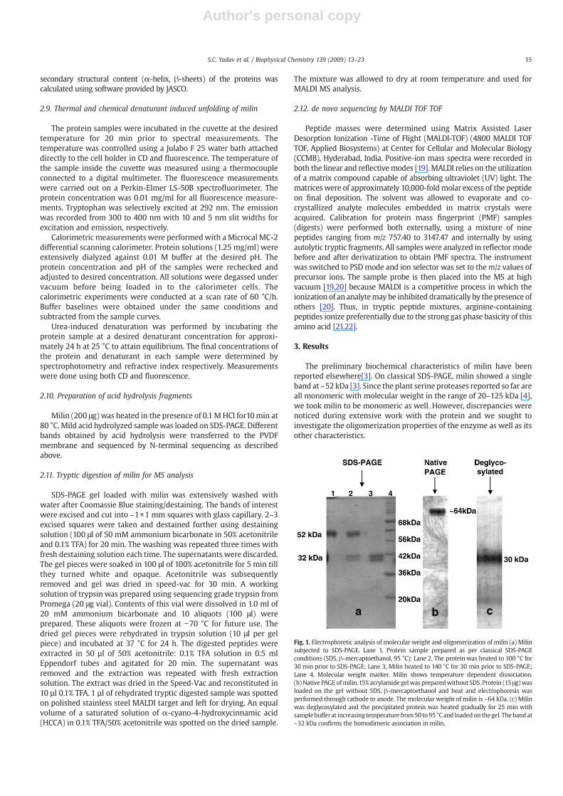
Fig. 1. Electrophoretic analysis of molecular weight and oligomerization of milin (a) Milinsubjected to SDS-PAGE. Lane 1, Protein sample prepared as per classical SDS-PAGEconditions (SDS, β-mercaptoethanol, 95 °C); Lane 2, The protein was heated to 100 °C for30 min prior to SDS-PAGE; Lane 3, Milin heated to 140 °C for 30 min prior to SDS-PAGE;Lane 4, Molecular weight marker. Milin shows temperature dependent dissociation.(b)Native PAGEofmilin.15% acrylamide gelwas preparedwithout SDS. Protein (15 μg)wasloaded on the gel without SDS, β-mercaptoethanol and heat and electrophoresis wasperformed through cathode to anode. The molecular weight of milin is ~64 kDa. (c) Milinwas deglycosylated and the precipitated protein was heated gradually for 25 min withsamplebuffer at increasing temperature from50 to95 °Cand loadedon thegel. The bandat~32 kDa confirms the homodimeric association in milin.
15S.C. Yadav et al. / Biophysical Chemistry 139 (2009) 13–23

Author's personal copy
3.1. Molecular weight of milin is 64 kDa
Initially, a molecular weight of ~52 kDa was observed for milin bySDS-PAGE under standard denaturing conditions (lane 1, Fig. 1a) [3].However, native PAGE analysis revealed that the molecular weight ofmilin is ~64 kDa andnot ~52kDa (Fig.1b). Further, themolecularweightby gel filtrationwas found to be ~64 kDa aswell (Fig. 2a), indicating thatSDS-PAGE formilin results in anomalous data. TheMSdata formilin alsoshowed two peaks indicating molecular weights of 31.593 kDaand 32.545 kDa, leading to a cumulative molecular weight of ~64 kDa(Fig. 2b). The peaks probably represent two subunits of milin.
3.2. On SDS-PAGE under harsh experimental conditions milin dissociatesinto subunits
The anomaly in molecular weight and oligomerization observed formilin on SDS-PAGE and MALDI TOF were investigated further. As seenbefore [3], milinwhen heated at 95 °C showed a single band at ~52 kDaon SDS-PAGE (lane 1, Fig. 1a). Here, the proteinwas subjected over timeto even higher temperature (140 °C) under reducing conditions prior toSDS-PAGE. As shown in lane 3, Fig. 3a, on being subjected to the highertemperature for 2min, a band also appeared at ~32 kDa. The intensity ofthe lower band increased and that of the upper band decreasedprogressivelywith time (lanes4–10, Fig. 3a), indicating thatmilin indeedcontains subunits of molecular weight ~32 kDa thus corroborating theMS data. In absence of heat, however, the low molecular weight band
was not observed (lane 2, Fig. 3a). The subunit association in milin thusseems to be based on a number of intricate interactions sincethe combination of lower temperature (95 °C) and β-mercaptoethanol(lane 1, Fig. 1a), or β-mercaptoethanol alone (lane 2, Fig. 3a) cannotdissociate the subunits. A combination of reduction (β-mercaptoetha-nol) and high temperature is needed for complete dissociation unlikemost other oligomeric proteins. Further, the negligible contribution ofdisulfide bonds to dimer association is exemplified in the appearanceof similar ~32 kDa band when milin was heated to 140 °C for 30 minunder non-reducing condition (without β-mercaptoethanol) as well(lane2, Fig. 3b). Under similar conditions, absence of heat doesnot resultindissociation (lane1, Fig. 3b). SuchanomalyonSDS-PAGEwasobservedfor a class of proteins known as “kinetically stable” proteins[23,24].These proteins are resistant to SDS denaturation and hence showanomalous migration on electrophoresis under reducing and non-reducing conditions.
The strong association between milin subunits is further demon-strated in Fig. 4. When incubated with increasing concentrations ofGuHCl (1–6 M) and subjected to SDS-PAGE without heating, at allconcentrations of the denaturant single bands at ~52 kDa wereobserved (Fig. 4a). Samples prepared similarly as above but heated to95 °C for 5min prior to SDS-PAGE showed additional bands at ~32 kDa(Fig. 4b). In 5 M GuHCl and higher, only the low molecular weightprotein band was observed indicating complete subunit dissociationat these concentrations of the denaturant when heated (Fig. 4b). Thesubunit association thus seems to be temperature dependent as seen
Fig. 2. Molecular weight and oligomerization of milin: chromatographic and mass spectrometric analysis (a) Gel filtration of milin. Milin (200 μg) was loaded under gravity onSephacryl S-200 column of bed volume 15 ml, pre-equilibrated with 50 mM phosphate buffer, pH 8 containing 100 mM NaCl. Elution was performed using the same buffer at a flowrate of 0.5 ml/min and 0.5 ml fractions were collected. The absorbance of the fractions is reported against the elution volume. Albumin, ovalbumin, chymotrypsinogen and RNasewere used as molecular weight markers under identical conditions. (b) MALDI TOF analysis. MS analysis showed two peaks at 31,593.9 and 32,545.3 Da respectively. The cumulativemolecular weight (64 kDa) coincides with that obtained by gel filtration demonstrating the dimeric nature of milin. Mass resolution was obtained in the linear modes utilizingcontinuous ion extraction at threshold laser irradiance of 25 kV accelerating potential and 50 laser pulses were averaged.
Fig. 3. Effect of high temperature (140 °C) on subunit association of milin (a) Milin (25 μg) in SDS-PAGE loading buffer, layered with mineral oil on the top of the sample to preventevaporation, was heated to 140 °C over time. Lane 1. Molecular weight marker. Lanes 2–10. Milin heated for 0, 2, 5, 8, 10, 12, 15, 20, and 25 min, respectively. (b) SDS-PAGE wasperformed under non-reducing condition (absence of β-mercaptoethanol). Lane 1. Sample loaded without heating; Lane 2. Sample heated for 30 min at 140 °C.
16 S.C. Yadav et al. / Biophysical Chemistry 139 (2009) 13–23

Author's personal copy
in both Figs. 3 and 4, and further emphasized in Fig. 1a, where nodissociation was observed at 95 °C (lane 1), partial dissociation at100 °C (lane 2), and complete dissociation at 140 °C (lane 3).
3.3. Milin is a dimeric protein
Milin showed a single band on native PAGE and single peak on gelfiltration corresponding toamolecularweightof ~64kDa (Figs.1band2a).Further, two protein peaks were observed in the mass spectrum, whoseindividual molecular weights add up to ~64 kDa, indicating that milincontains subunits with monomer molecular weights of ~32 and ~31 kDa(Fig. 2b). The MALDI laser ionization must have separated the twosubunits. All these experiments convincingly prove that milin is dimeric.Similarly, milin dissociates into subunits of molecular weight ~32 kDaunder harsh treatment on SDS-PAGE (Figs. 3 and 4). Such an oligomeriza-tion state is unprecedented for a plant serine protease to the best of ourknowledge[4]. Recently characterized serine proteases from plant latexlike cryptolepain and carnein have similar molecular weights but showsingle peaks on mass spectrometric analysis, indicating that they aremonomeric proteins[25].
3.4. Milin is homodimeric
The difference in molecular weights of the monomers as seen inMS plot suggests that milin consists of heterodimers. A closerinspection of the ~32 kDa band observed on SDS polyacrylamide
gels also seem to support such an observation, since two closelyspaced bands were actually seen (Fig. 1a, lane 3). The possibility,however, existed for differential glycosylation of the monomers, giventhe fact that milin was shown to be heavily glycosylated [3]. To verifythis possibility, milin was deglycosylated chemically (Materials andMethods), and the protein free of carbohydrate moieties, wassubjected to classical SDS-PAGE. The deglycosylated protein dissoci-ates easily into subunits and only a single band was observed at~32 kDa (Fig. 1c). This shows that milin is homodimeric and that themonomers are differentially glycosylated resulting in slight differ-ences in their molecular weight under native conditions, as observedby MS analysis. Similar difference in molecular weight (~1 kDa)between subunits was observed on SDS-PAGE due to differentialglycosylation in case of Scrapie prion protein [26,27].
The N-terminal sequence of milinwas previously determined to beDVSYVGLILETD using native pure protein [3]. We repeated theprocedure using the two closely spaced (~31.5 and 32.5 kDa) bandsobserved on SDS-PAGE under harsh conditions (Lane 3, Fig. 1a). Boththe protein bands contained the same N-terminal sequence as above,confirming the observation that milin is homodimeric.
3.5. Strong subunit association inmilin is necessary for stability, solubilityand activity
As seen in Fig.1c, deglycosylatedmilin easily dissociates into subunitsunlike native protease, which requires heating to 140 °C for complete
Fig. 4. Effect of chemical denaturant (GuHCl) on dimer integrity of milin. Milin was incubated in various concentrations of GuHCl (1–6 M) for 24 h prior to SDS-PAGE. (a) GuHClincubated protein loaded in reducing condition but without heating showed single bands at ~52 kDa. (b) Protein sample as above but heated for 5min at 95 °C before loading showedsubunits as well. The upper bands correspond to nearly dimeric molecular weight and lower bands correspond to monomeric molecular weight.
Fig. 5. CD spectra of milin. (a) Far UV CD spectra of glycosylated (○) and deglycosylated (●) milin. The samples were incubated at neutral pH for 24 h at room temperature beforethe measurements. (b) Near UV CD spectra of glycosylated (native) milin. The protein concentrations for near UV and far UV CD measurements were 1 mg/ml and 0.1 mg/ml,respectively.
17S.C. Yadav et al. / Biophysical Chemistry 139 (2009) 13–23

Author's personal copy
dissociation. This shows that deglycosylatedmilin is less stable since thedimers are probably loosely associated. The deglycosylated protein alsotends to precipitate indicating that solubility is decreased. Theprecipitated protein is soluble in 1.5 M GuHCl. Under these conditions,the deglycosylated protease showed no proteolytic activity either. Thereference protein, native milin in 1.5 M GuHCl, on the other hand,showed considerable proteolytic activity [3]. It has been shown beforethat deglycosylation does not necessarily inactivate proteins[18]. Hencethe activity loss on deglycosylation must be due to loss of proper dimerassociation. This represents that dimerization might be necessaryfor stability, solubility and activity of milin. However, the specific role
of glycosylation in dimerization of milin can be examined only after thecharacterization of the sugars bound to this plant serine protease.
3.6. Milin belongs to α/β class of proteins
Since milin seems to be a novel plant serine protease, circulardichroismwas used to get an estimation of its secondary structure andnative fold (Fig. 5). The far UV CD spectrum of native milin (Fig. 5a) istypical of α/β class of proteins [28]. The spectrum shows negativepeaks at 222 nm and 208 nm, positive peak at 195 nm and goes tonegative again above 172 nm (Fig. 5a), all characteristics of an α/β
Fig. 6. Thermal and chemical denaturant induced unfolding of Milin; (a) Differential Scanning Calorimetry of Milin. The calorimetric scans were performed with a proteinconcentration of 1.25 mg/ml at a scanning rate of 60 °C/h. The enzyme was extensively dialyzed against 0.01 M phosphate buffer, pH 7.0 before scanning. (b) Temperature inducedunfolding of milin at pH 7.0 (I) Far UV CD temperature transition curve. Inset: The entire far UV CD spectra at 40, 60 and 80 °C. (II) Temperature transition fluorescence curve. The insetshows fluorescence spectra at different temperatures. (c) Urea induced equilibrium denaturation of milin at pH 7.0. (I) Far UV CD Scan at different concentrations of urea (II)Fluorescence scan at various concentrations of urea. The protein (0.1 mg/ml) was incubated in the cuvette for 20 min at desired temperature prior to CD measurements. The resultswere expressed in mean residual ellipticity. The intrinsic fluorescence spectra of milin was obtained by excitation at 292 nm with the emission and excitation slits 10 and 5 nmrespectively. The protein (0.01 mg/ml) was incubated in the cuvette for 20 min at desired temperature prior to fluorescence measurements.
18 S.C. Yadav et al. / Biophysical Chemistry 139 (2009) 13–23

Author's personal copy
class of protein. However, deglycosylated milin shows spectrumtypical of prominently β class of proteins, with a negative peak at218 nm (Fig. 5a). The mean residue ellipticity for native milin at
222 nm was 8.5±0.20χ103° cm2 dmol−1 indicating 29% of α-helicalcontent and 38% of β-sheet content. The integrity of the three-dimensional structure of native milin was probed with near UV CD(Fig. 5b). The spectrum revealed three prominent peaks at 278, 288,and 300 nm of mean residue ellipticity 180, 180 and −50° cm2 dmol−1
respectively.
3.7. Milin is strongly resistant to thermal and chemical denaturation
Milin seems to be strongly resistant to denaturation and subunitdissociation as observed during SDS-PAGE analysis above. This wasfurther confirmed by other biophysical experiments involvingtemperature and chaotrope induced denaturation of milin. Thus, thetransition scan obtained by DSC after correction for buffer-buffertracing is incomplete at pH 7.0. (Fig. 6a). This does not allow accurateestimation of thermal stability or melting temperature (Tm). However,the apparent Tm seems to be approximately 90 °C indicating that milinis thermostable. Similar result was obtained when the effect of tem-perature on milin was probed by far UV CD measurements (Fig. 6b(I)).There was no change in ellipticity up to about 75 °C and even beyondthat temperature the changes were negligible. The constant fluores-cence emission maximum over temperature corroborates this finding(Fig. 6b(II)). The linear decrease in fluorescence intensity probablyresults from signal quenching and not due to denaturation.
Milin is resistant to chemical denaturation as well. Urea inducedunfolding of milin is also incomplete (Fig. 6c). With increasing
Fig. 7. N-terminal sequences of various peptides from milin upon acid hydrolysis Thepeptides were generated by mild acid hydrolysis in 0.1 M HCl. The molecular weight of thefragmentsand their correspondingN-terminalEdmansequenceare represented in thefigure.
Fig. 8. MALDI TOF spectra and de novo sequencing of milin (a) Peptide mass fingerprints of milin on trypsin digestion. In gel trypsin digestion was performed and MALDI TOFgenerated peptide finger print data was obtained. The mass of different peaks was searched in NCBI for identification of the molecule. (b) The TOT/TOF of 1456.8484 tryptic fragmentcontained immonium ion of Tyrosine (m/z 136). This represented high resolution of the de novo fragments of milin. Peptides with high resolution of mass were used for de novosequencing. (c) and (d) Peptide sequencing by tandemmass spectrometry. The TOF/TOF spectra of tryptic fragment of mass 1172.6883 and 2177.9521 Dawere sequenced by computerbased automated program for de novo sequencing respectively.
19S.C. Yadav et al. / Biophysical Chemistry 139 (2009) 13–23

Author's personal copy
concentration of urea neither the far UV CD spectra (Fig. 6c(I)) norfluorescence spectra (Fig. 6c(II)) change significantly. Hence Cm couldnot be determined.
3.8. N-terminal sequences of acid hydrolyzed fragments of milin areunique
Milin was hydrolyzed into seven major fragments with molecularweight ranging from 7 kDa to 32 kDa (Fig. 7). N-terminal sequences ofthe resulting fragments were determined by Edman's method. Thesepeptides, their molecular weights and corresponding N-terminalsequences were: DVSYVGLILETD for 32.5, 31.5, 23, 21 and 19 kDafragments; PSGYIIASELQVP for 13 kDa fragment; LPYEPEIVNGLR for10 kDa fragment; and VNLFHVKGIVHYG for 7 kDa fragment. The31.5 kDa fragment dissociated into 19 and 13 kDa or 21and 10 kDabands, while 32.5 kDa bands dissociated into 21and 13 kDa or 23 and10 kDa bands. Few other less intense bands that appear upon acidhydrolysis were not sequenced. The differences in the molecularweight of three bands (viz. 19, 21 and 23 kDa) were due to differentialglycosylation; the 13 and 10 kDa bands were not glycosylated. The N-terminal sequences determined do not show sequence homologyto amino terminus of known serine proteases. Homology search acrossdatabases for match at positions other than N-terminus revealedother distant possibilities with very weak scores including transcrip-tion regulator, protease domain of an ATP-independent heat shockprotease (PDB ID: JLIJ) etc.
3.9. Protein mass fingerprint (PMF) and de novo sequence of peptidesestablish milin as a novel serine protease
Since milin did not show distinct N-terminal sequence homologyto known proteins, attempt was made to identify the protein bypeptide mass fingerprinting. For this, the protein was enzymaticallydigested and resolved into a number of peptides (Fig. 8a and b). Themasses of the peptides were determined and searched againstrelevant databases in NCBI. No significant hits were obtained basedon peptide masses. Subsequently, the different peptide fragments ofmilin generated above and with good resolution were subjected to denovo sequencing by MALDI TOF/TOF. The TOF/TOF spectra ofrepresentative peptides and their de novo sequences are shown inFig. 8c and d. Sequences of several peptides thus obtained are listed inTable 1. These sequences when submitted for BLAST search in NCBIdatabase with different variables show the uniqueness of the proteinsequence. No putative domains were obtained with any confidenceusing these sequences as search query. However, weak scores wereobtainedwith similarities to esterase of theα/β hydrolase superfamilyand putative GTP binding protein.
4. Discussion
The search for stable and active biochemical components fromplant sources with novel properties and therapeutic applications hasreceived widespread attention lately. The discovery of milin withunique properties and putative medical applications [1,3] will bolstersuch effort.
4.1. Milin is a plant subtilase with unique amino acid sequence
The proteolytic activity of milin has been described before[3] andobserved in the current investigation as well. Typical cysteineprotease inhibitors or metal chelators did not inhibit the proteaseactivity of milin thus ruling out the possibility of the peptidase beinga cysteine protease ormetallic protease[3]. The proteinwas, however,strongly inhibited by phenylmethylsulphonyl fluoride (PMSF), 4-amidino-phenyl-methane-sulfonyl fluoride (APMSF) and diisopro-pylfluorophosphate (DFP) indicating that the catalysis is mechan-istically dependent on a serine residue. The serine protease familyincludes various members like trypsin, chymotrypsin, elastases,subtilases, carboxypeptidases etc. [29]. That milin was not inhibitedby soybean trypsin inhibitor (SBTI) also indicated that the serineprotease activity of milin is not trypsin-like. With syntheticsubstrates, milin showed maximal hydrolysis at the peptide bondfollowing a small hydrophobic amino acid, viz Ala[3], a propertysimilar to subtilases [30]. Trypsin prefers basic residues (Arg/Lys) andchymotrypsin prefers large hydrophobic residues (Phe/Tyr/Trp) forproteolysis unlike milin [31]. Elastases also prefer Ala/Val, however,they do not generally work on single amino acid synthetic substrates[32]. Interestingly, human herpes virus serine proteases also cleavepeptide bond between Ala and Ser[33]. Vast majority of the knownplant subtilisins like cucumisin are active over a broad pH range withan optimum at alkaline pH and have relatively high thermal stability[3]. Milin also exhibits similar properties [3] including the fact that itshares a pH optima of 8.0 with varicella-zoster virus protease [33].Milin is probably not a serine carboxypeptidase either since theseenzymes have acidic optimum pH for their activity towards peptidesubstrates [34]. Characteristic of typical bacterial subtilisins (struc-ture of plant subtilisins not yet solved), milin exhibits secondarystructure with α/β fold (Fig. 5) as ascertained by circular dichroism[35]. Three-dimensional structures have shown that subtilisinsposses α/β/α sandwich or Rossmann fold, while other serineproteases belonging to the trypsin/chymotrypsin class are predomi-nantly β-sheet protein [32,35–37]. Interestingly, human cytomega-lovirus serine protease also belongs to α/β class of proteins [6].Hence, the biochemical (inhibition, substrate specificity, pH optimumetc.) and structural evidences indicate that milin is a plant subtilase.
However, the N-terminal sequence of milin showed no homology toknown plant subtilases, either by in silico or manual search. In fact, nosignificant homologywasobservedwith anymember fromother classesof serine protease either.Milin also showed absence of the conservedG–X–S/C–G–G sequence for chymotrypsin-like and G–T–S–M/A forsubtilisin-like proteases [33] as evidenced from de novo sequences.Searchacross variousprotein databases reveal thatmilin has a uniqueN-terminus. Our added effort in sequencing N-terminal residues of acidhydrolysed fragments or de novo sequencing of tryptic digests failed toidentify any putative domains in various portions of the protein as well.It has been reported [30] that the most striking feature of members ofthe subtilisin family is the high degree of the sequence variabilitywithintheir catalytic triad; apart from the catalytic triad, all other residuesmayvirtually be replacedbyoneormoredistinct residue includingvariabilityat the N-terminus. These observations and those above indicate thatmilin is a plant subtilasewith unique amino acid sequence. Interestingly,human herpes virus serine proteases also exhibit sequence variability[16].Milin is also novel amongplant serineproteases in beingdimeric, asdiscussed below.
Table 1de novo sequences of some tryptic peptides of milin
m/z PMF Sequences Score
842.5258 VTAVSLPR 67.9688959.5275 TYHLGQLK 100.001033.5172 EFAYTKFK 83.42871172.6843 GRTYHLGQLK 88.22251172.6924 KWYHLVNGR 77.07851328.5978 AAYYANVDWKK 93.46961456.8484 SHFSYLGSVNAFK 93.80991601.3941 RLARDEMESLPER 77.12761771.9249 PSGYIIASEIQVPEIR 84.44291772.1573 PSGYLLASELQVPLER 94.41611850.8716 AVVHYGTSKT(317)DSIR 67.52282177.9521 ECLNKSCLPYEPELVDGLR 82.21662594.5638 GLNSTANSTEYSHFSYLGSVNAFK 95.3245
m/z refers to mass/charge ratio. PMF refers to Peptide mass fingerprint.
20 S.C. Yadav et al. / Biophysical Chemistry 139 (2009) 13–23

Author's personal copy
4.2. Anomalous behaviour of milin on SDS-PAGE
Contrary to our previous report [3], the molecular weight of milinwas found to be 64 kDa using a combination of gel filtration, massspectrometry and SDS-PAGE under extreme conditions (Figs. 1, 2).Unique properties of milin when subjected to standard denaturatingcondition prior to SDS-PAGE previously resulted in an anomalousmolecular weight of 52 kDa and the conclusion that the protein is amonomer. The relatively high mobility of the protein in SDS-PAGE gelprobably resulted due to the formation of some kind of compactstructure that resists subunit dissociation. The protein was probablypartially denatured forming a compact intermediate. The loss ofproteolytic activity of milin beyond 75 °C [3] excludes the possibility oflimited proteolysis resulting in the 52 kDa band under the denaturingconditions [38]. The pI of the proteinwas found to be 7.0 [3] and hencethe possibility of the anomalous migration due to high negativecharge on the protein can also be negated [38]. The presentinvestigation though provides a solution to the observed anomaly inmolecular weight and oligomerization by reporting approximately32 kDa bands on SDS-PAGE under extreme conditions (very hightemperature and presence of denaturant like GuHCl) that breaksdown the compact structure (Figs. 3, 4). The molecular weight is halfof the actual molecular weight indicating the presence of subunits andthat milin is a dimer. This conclusion was further confirmed whenmilin, a glycoprotein, was chemically deglycosylated. Deglycosylatedmilin looses its dimeric assembly and hence shows molecular weightcorresponding to monomers on SDS-PAGE under standard denaturingconditions (Fig. 1c). Most plant serine protease reported so for havebeen found to be monomeric with the molecular weight in the rangeof 30–110 kDa [4]. Thus, it is recommended to try extreme conditionsof denaturation prior to SDS PAGE before concluding the oligomericnature of such proteases by SDS-PAGE, specially the ones that arepoorly characterized biochemically and biophysically.
4.3. Dimerization of milin is novel
It is evident from the present investigation that milin is a dimericserine protease. This is the first report of a dimeric plant serineprotease to the best of our knowledge. Dimerization of milin iscertainly not dependent on disulphide bridges since the dimer can bedissociated both in the presence or absence of reducing agents asinvestigated by SDS-PAGE. It appears that temperature plays asignificant role in the dimer dissociation (Figs. 1–4 and 6). There is athreshold requirement of thermal energy necessary for the dissocia-tion of dimers in SDS-PAGE loading buffer. The threshold is lowered inpresence of high concentration of another denaturant like GuHCl,indicating that a combination of non-covalent forces probably keepsthe monomers associated. Milin was also shown to be stronglyresistant to temperature and chemical denaturation.
Some of the above observations are similar to those reported byChalut et al. (1999) for penicillin binding protein 1B in E. coli. For thisprotein, the dimer was stable at 60 °C but almost totally dissociated at80 °C and the same pattern of dissociation was observed with andwithout β-mercaptoethanol. Milin was also stable to 100 °C butdissociated when heated above this temperature prior to SDS-PAGE(Fig. 3). In the presence of GuHCl, the dissociation temperature was95 °C under similar conditions (Fig. 4).
Experimental evidences indicate that the aqueous solubility ofmilin decreases when subunits dissociate, mainly due to deglycosyla-tion. 1.5 M GuHCl is required to solubilize the resulting precipitate.Deglycosylated protein runs normally on SDS-PAGE without extrainput of thermal energy or without 6 M GuHCl and shows completedissociation with a molecular weight equivalent to milin monomer(Fig. 1c). Deglycosylated protein monomers are also proteolyticallyinactive. Since deglycosylation is known not to inactivate proteins ingeneral [18], the inactivation must be due to loss of dimer integrity. It
is possible that either the active site is close to dimer interface orstructural parts from one subunit cross over into the other requiringintact dimer for catalysis. Herpes virus serine proteases form dimerswhere such crossover between subunits has been observed in crystalstructures [33]. However, no report of glycosylation having sucheffects on serine protease dimers exist andmilin is the first report. Theimportance of this post-translational modification in maintainingoligomerization integrity needs to be examined further for milin andother proteins.
4.4. Milin seems to be a kinetically stable protein
All the general characteristics and anomalies observed for milin onSDS-PAGE is akin to kinetically stable proteins, which have infrequentoccurrence in nature [23,24]. Members of this novel class of proteinsshow differences in migration of their protein band on SDS-PAGE onheating and without heating[23,24]. They seem to be trapped by atransition state energy barrier in their highly stable native conforma-tion. The unfolding transitions are thus not easily overcome so thatrandomization of such proteins is difficult relative to other proteinsand they are resistant to harsh denaturing conditions, especially SDS[24,39,40]. Nature has favored such a unique property to allow theseproteins to maintain their activity under hostile environment. Milinseems to be amember of this class as well; especially evident from theSDS-PAGE results in which heating to a certain temperature isrequired for complete denaturation. It has been shown that majorityof the kinetically stable proteins are enzymes, belong to α/β class ofproteins and are dimeric in nature [23,24,41]. Interestingly, milin alsofollows this general trend. It is even more interesting to note thatpapain, the most widely studied plant cysteine protease from latex,also belongs to the class of kinetically stable proteins [24]. It is notdimeric in nature but contains two domains with the active site cleftsituated between them. One obvious question to ponder is whethermost proteases from latex are also members of this class, enablingthem with enhanced stability.
It has been reported that rigid protein structure is the physicalbasis of kinetic stability and resistance to SDS. Protection of surfaceresidues from the solvent, increase in rigidity and stabilization ofspecific region of a protein are potential effects of oligomerization thatmay increase kinetic stability, as seen for milin. This also supports theevolutionary pressure of forming dimers or bi-domains for proteasesfrom latex [42–49].
The extreme stability of milin is further exemplified by thermaland chemical induced denaturation studies. Temperature inducedunfolding of milin was incomplete with an apparent Tm of 90 °C.Gradual decrease of the transition start-point as a function ofchaotrope concentration is also an indication of the highly compactstructural integrity of the protein molecule. The similar shape andellipticity of the far UV CD spectra and little shift in the fluorescenceemissionmaximum on chemical or thermal denaturation is indicationof the formation of a new intermediate conformational state of themolecule which resists further denaturation.
4.5. Physiological and therapeutic importance of milin
The unique properties of milin indicate novel physiological role forthe plant protein. Latex of several plants has been found to be rich inproteolytic enzymes including mostly cysteine proteases and someserine proteases although their physiological roles are largelyunknown. They have often been indicated to play important role inplant defense, especially papain and papain-like cysteine proteases[2]. It is to be noted that cysteine proteases belonging to the papainsuperfamily all have two domains with the active site situatedbetween them. Milin, on an analogous note, has two subunits that canprobably hold the active site in between them. In doing so the activesite is shielded from thewaxy environment of the latex and large areas
21S.C. Yadav et al. / Biophysical Chemistry 139 (2009) 13–23

Author's personal copy
of protein surfaces are best protected in dimers [9]. This would explainwhy monomers of milin are inactive or less active. Thus, dimerizationcould be a way of regulating physiological function of milin orenhancing its catalytic activity. Human herpes virus serine proteasesand human cytomegalovirus proteases are also inactive in theirmonomeric form and dimerization activates them [6,14,15,33].Chymotrypsin is also known to dimerize under certain conditions [12].
Euphorbia milii is awild, tropical, xerophytic plant that has to survivehigh temperatures in summers and other extreme environmental stress.The fact that milin needs high input of thermal energy for dimerdissociation and is a kinetically stable protein could be a means for theprotein to tide over duration of high temperature by forming a compactstructure that resists thermal denaturation. The active site is protectedunder suchhostile conditionsandanymilddenaturation canbe reversedat more favourable lower temperatures [50,51]. Glycosylation can helpthe protein acquire a sheath of aqueous layer for similar purposes. Sugarresidues can also help milin in recognizing specific receptors on insectsand other pests thus triggering defense action through proteolysis.
The therapeutic importance ofmilin has already been reported [1]. Itseems to be an effectivemolluscicide that can control Schistosomiasis. Ithas been noted above that the temperature-dependent dissociation ofdimers in milin on SDS-PAGE is akin to that of penicillin binding protein1b in E. coli [52]. The presence of penicillin binding protein β-lactamasewith serine protease activity has been reported in Schistosomiasisjaponicum [53]. This, though a curious coincidence, could be related tothe usefulness of milin in treating the disease. There is every possibilitythat milin is multifunctional and just not a simple serine protease likemany other such enzymes reported in plant latex. The oligomerizationand other novel properties of milin hint at such possibilities.
Acknowledgment
The financial assistance to SCY from the Council of Scientific andIndustrial Research, Government of India, in the form of a researchfellowship and the financial assistance from UGC and DBT, Govern-ment of India, are acknowledged. Thanks are due to Prof. Rajiv Bhat,JNU for helping with DSC experiments.
References
[1] S.C. Yadav, M.V. Jagannadham, Physiological changes and molluscicidal effectsof crude latex and milin on Biomphalaria glabrata, Chemosphere 71 (2008)1295–1300.
[2] K. Konno, C. Hirayama, M. Nakamura, K. Tateishi, Y. Tamura, M. Hattori, K. Kohno,Papain protects papaya trees from herbivorous insects: role of cysteine proteasesin latex, Plant J. 37 (2004) 370–378.
[3] S.C. Yadav, M. Pande, M.V. Jagannadham, Highly stable glycosylated serine proteasefrom the medicinal plant Euphorbia milii, Phytochemistry 67 (2006) 1414–1426.
[4] C.M. Antao, F.X. Malcata, Plant serine proteases: biochemical, physiological andmolecular features, Plant Physiol. Biochem. 43 (2005) 637–650.
[5] W. Markland, R.A. Petrillo, M. Fitzgibbon, T. Fox, R. McCarrick, T. McQuaid, J.R.Fulghum, W. Chen, M.A. Fleming, J.A. Thompson, S.P. Chambers, Purification andcharacterization of NS3 serine protease domain of hepatitis C virus expressed inSaccharomyces cerevisiae, J. Gen. Virol. 78 (1997) 39–43.
[6] P. Chen, H. Tsuge, R.J. Almassy, C.L. Gribskov, S. Katoh, D.L. Vanderpool, S.A.Margosiak, C. Pinko, D.A. Matthews, C.C. Kan, Structure of the human cytomega-lovirus protease catalytic domain reveals a novel serine protease fold and catalytictriad, Cell 86 (1996) 835–843.
[7] D. Gailani, D. Ho, M.F. Sun, Q. Cheng, P.N. Walsh, Model for a factor IX activationcomplex on blood platelets: dimeric conformation of factor XIa is essential, Blood97 (2001) 3117–3122.
[8] C. Hink-Schauer, E. Estebanez-Perpina, F.C. Kurschus, W. Bode, D.E. Jenne, Crystalstructure of the apoptosis-inducing human granzyme A dimer, Nat. Struct. Biol. 10(2003) 535–540.
[9] M.W. Parker, J.T. Buckley, J.P. Postma, A.D. Tucker, K. Leonard, F. Pattus, D. Tsernoglou,Structure of the Aeromonas toxin proaerolysin in its water-soluble and membrane-channel states, Nature 367 (1994) 292–295.
[10] W.J. Garland, J.T. Buckley, The cytolytic toxin aerolysin must aggregate to disrupterythrocytes, and aggregation is stimulated by human glycophorin, Infect. Immun.56 (1988) 1249–1253.
[11] S.C. Pettit, S. Gulnik, L. Everitt, A.H. Kaplan, The dimer interfaces of protease andextra-protease domains influence the activation of protease and the specificity ofGagPol cleavage, J. Virol. 77 (2003) 366–374.
[12] K. Ikeda, S. Kunugi, N. Ise, pH dependence of the formation of dimeric alpha-chymotrypsin and its catalytic activity, J. Biochem. 92 (1982) 541–546.
[13] K. Baer, H. Al-Hasani, S. Parvaresch, T. Corona, A. Rufer, V. Nolle, E. Bergschneider, H.W.Klein, Dimerization-induced activation of soluble insulin/IGF-1 receptor kinases: analternative mechanism of activation, Biochemistry 40 (2001) 14268–14278.
[14] P.L. Darke, J.L. Cole, L. Waxman, D.L. Hall, M.K. Sardana, L.C. Kuo, Active humancytomegalovirus protease is a dimer, J. Biol. Chem. 271 (1996) 7445–7449.
[15] S.A. Margosiak, D.L. Vanderpool, W. Sisson, C. Pinko, C.C. Kan, Dimerization of thehuman cytomegalovirus protease: kinetic and biochemical characterization of thecatalytic homodimer, Biochemistry 35 (1996) 5300–5307.
[16] U. Schmidt, P.L. Darke, Dimerization and activation of the herpes simplex virustype 1 protease, J. Biol. Chem. 272 (1997) 7732–7735.
[17] S. Gras, V. Chaumont, B. Fernandez, P. Carpentier, F. Charrier-Savournin, S. Schmitt,C. Pineau, D. Flament, A. Hecker, P. Forterre, J. Armengaud, D. Housset, Structuralinsights into a new homodimeric self-activated GTPase family, EMBO. Rep. 8(2007) 569–575.
[18] A.S. Edge, Deglycosylation of glycoproteins with trifluoromethanesulphonic acid:elucidation of molecular structure and function, Biochem. J. 376 (2003) 339–350.
[19] M. Karas, M. Gluckmann, J. Schafer, Ionization in matrix-assisted laser desorption/ionization: singly chargedmolecular ions are the lucky survivors, J.Mass. Spectrom.35 (2000) 1–12.
[20] F. Hillenkamp, M. Karas, R.C. Beavis, B.T. Chait, Matrix-assisted laser desorption/ionization mass spectrometry of biopolymers, Anal. Chem. 63 (1991) 1193A–1203A.
[21] E. Krause, H. Wenschuh, P.R. Jungblut, The dominance of arginine-containingpeptides in MALDI-derived tryptic mass fingerprints of proteins, Anal. Chem. 71(1999) 4160–4165.
[22] R. Kratzer, C. Eckerskorn, M. Karas, F. Lottspeich, Suppression effects in enzymaticpeptide ladder sequencing using ultraviolet – matrix assisted laser desorption/ionization – mass spectormetry, Electrophoresis 19 (1998) 1910–1919.
[23] K. Xia, M. Manning, H. Hesham, Q. Lin, C. Bystroff, W. Colon, Identifying thesubproteome of kinetically stable proteins via diagonal 2D SDS/PAGE, Proc. Natl.Acad. Sci. U. S. A. 104 (2007) 17329–17334.
[24] M. Manning, W. Colon, Structural basis of protein kinetic stability: resistance tosodium dodecyl sulfate suggests a central role for rigidity and a bias toward beta-sheet structure, Biochemistry 43 (2004) 11248–11254.
[25] M. Pande, V.K. Dubey, S.C. Yadav, M.V. Jagannadham, A novel serine proteasecryptolepain from Cryptolepis buchanani: purification and biochemical character-ization, J. Agric. Food. Chem. 54 (2006) 10141–10150.
[26] K.J. Knaus, M. Morillas, W. Swietnicki, M. Malone, W.K. Surewicz, V.C. Yee, Crystalstructure of the human prion protein reveals a mechanism for oligomerization,Nat. Struct. Biol. 8 (2001) 770–774.
[27] M.L. Riley, C. Leucht, S. Gauczynski, C. Hundt, M. Brecelj, G. Dodson, S. Weiss, High-level expression and characterization of a glycosylated covalently linked dimer ofthe prion protein, Protein Eng. 15 (2002) 529–536.
[28] P. Manavalan, W.C. Johnson Jr., P. Modrich, Prediction of secondary structure forEco RI endonuclease, J. Biol. Chem. 259 (1984) 11666–11667.
[29] N.D. Rawlings, A.J. Barrett, Families of serine peptidases, Methods Enzymol. 244(1994) 19–61.
[30] R.J. Siezen, J.A. Leunissen, Subtilases: the superfamily of subtilisin-like serineproteases, Protein Sci. 6 (1997) 501–523.
[31] J.J. Perona, C.S. Craik, Evolutionary divergence of substrate specificity within thechymotrypsin-like serine protease fold, J. Biol. Chem. 272 (1997) 29987–29990.
[32] J.A. Bousquet, J. Duranton, Y. Mely, J.G. Bieth, Conformational change in elastasefollowing complexation with alpha1-proteinase inhibitor: a CD investigation,Biochem. J. 370 (2003) 345–349.
[33] X. Qiu, C.A. Janson, J.S. Culp, S.B. Richardson, C. Debouck, W.W. Smith, S.S. Abdel-Meguid, Crystal structure of varicella-zoster virus protease, Proc. Natl. Acad. Sci.U. S. A. 94 (1997) 2874–2879.
[34] A. Zhou, J. Li, Arabidopsis BRS1 is a secreted and active serine carboxypeptidase,J. Biol. Chem. 280 (2005) 35554–35561.
[35] H. Kano, S. Taguchi, H. Mornose, Cold adaptation of a mesophilic serine protease,subtilisin, by in vitro random mutagenesis, Appl. Microbiol. Biotechnol. 47 (1997)46–51.
[36] A. Pasternak,D. Ringe, L. Hedstrom, Comparisonof anionic and cationic trypsinogens:the anionic activation domain is more flexible in solution and differs in its mode ofBPTI binding in the crystal structure, Protein. Sci. 8 (1999) 253–258.
[37] L.M. Simon, M. Kotorman, A. Szabo, G. Garab, I. Laczko, Effects of polyethyleneglycol on stability of alpha-chymotrypsin in aqueous ethanol solvent, Biochem.Biophys. Res. Commun. 317 (2004) 610–613.
[38] S.S. Sidhu, G.B. Kalmar, L.G.Willis, T.J. Borgford, Protease evolution in Streptomycesgriseus. Discovery of a novel dimeric enzymes, J. Biol. Chem. 270 (1995) 7594–7600.
[39] J.A. Reynolds, S. Herbert, H. Polet, J. Steinhardt, The binding of divers detergentanions to bovine serum albumin, Biochemistry 6 (1967) 937–947.
[40] M.N. Jones, H.A. Skinner, E. Tipping, The interaction between bovine serumalbumin and surfactants, Biochem. J. 147 (1975) 229–234.
[41] R.E. Spelbrink, A. Kolkman, M. Slijper, J.A. Killian, B. de Kruijff, Detection andidentification of stable oligomeric protein complexes in Escherichi coli innermembranes: a proteomics approach, J. Biol. Chem. 280 (2005) 28742–28748.
[42] S.S. Jaswal, J.L. Sohl, J.H. Davis, D.A. Agard, Energetic landscape of alpha-lyticprotease optimizes longevity through kinetic stability, Nature 415 (2002) 343–346.
[43] C. Yuan, J. Li, T.L. Selby, I.J. Byeon, M.D. Tsai, Tumor suppressor INK4: comparisonsof conformational properties between p16(INK4A) and p18(INK4C), J. Mol. Biol.294 (1999) 201–211.
[44] S.M. Truhlar, E.L. Cunningham, D.A. Agard, The folding landscape of Streptomycesgriseus protease B reveals the energetic costs and benefits associatedwith evolvingkinetic stability, Protein Sci. 13 (2004) 381–390.
22 S.C. Yadav et al. / Biophysical Chemistry 139 (2009) 13–23

Author's personal copy
[45] A. Zaks, A.M. Klibanov, Enzymatic catalysis in nonaqueous solvents, J. Biol. Chem.263 (1988) 3194–3201.
[46] M. Machius, N. Declerck, R. Huber, G. Wiegand, Kinetic stabilization of Bacilluslicheniformis alpha-amylase through introduction of hydrophobic residues at thesurface, J. Biol. Chem. 278 (2003) 11546–11553.
[47] A.W. Rietveld, S.T. Ferreira, Kinetics and energetics of subunit dissociation/unfoldingof TIM: the importance of oligomerization for conformational persistence andchemical stability of proteins, Biochemistry 37 (1998) 933–937.
[48] S. Solis-Mendiola, L.H. Gutierrez-Gonzalez, A. Arroyo-Reyna, J. Padilla-Zuniga,A. Rojo-Dominguez, A. Hernandez-Arana, pH dependence of the activationparameters for chymopapain unfolding: influence of ion pairs on the kinetic stabilityof proteins, Biochim. Biophys. Acta. 1388 (1998) 363–372.
[49] R. Jaenicke, G. Bohm, The stability of proteins in extreme environments, Curr. Opin.Struct. Biol. 8 (1998) 738–748.
[50] R. Jaenicke, Protein stability and molecular adaptation to extreme conditions, Eur.J. Biochem. 202 (1991) 715–728.
[51] R. Scandurra, V. Consalvi, R. Chiaraluce, L. Politi, P.C. Engel, Protein stability inextremophilic archaea, Front. Biosci. 5 (2000) D787–795.
[52] C. Chalut, M.H. Remy, J.M. Masson, Disulfide bridges are not involved in penicillin-binding protein 1b dimerization inEscherichia coli, J. Bacteriol.181 (1999) 2970–2972.
[53] N. Peitsaro, Z. Polianskyte, J. Tuimala, I. Porn-Ares, J. Liobikas, O. Speer, D. Lindholm,J. Thompson, O. Eriksson, Evolution of a family of metazoan active-site-serineenzymes from penicillin-binding proteins: a novel facet of the bacterial legacy,BMC. Evol. Biol. 8 (2008) 26.
23S.C. Yadav et al. / Biophysical Chemistry 139 (2009) 13–23