Structural Properties of AMP-activated Protein Kinase: DIMERIZATION, MOLECULAR SHAPE, AND CHANGES...
13
Structural Properties of AMP-activated Protein Kinase DIMERIZATION, MOLECULAR SHAPE, AND CHANGES UPON LIGAND BINDING * □ S Received for publication, October 9, 2007, and in revised form, February 25, 2008 Published, JBC Papers in Press, March 27, 2008, DOI 10.1074/jbc.M708379200 Uwe Riek ‡§ , Roland Scholz ‡ , Peter Konarev ¶ , Arne Rufer**, Marianne Suter ‡ , Alexis Nazabal ‡‡ , Philippe Ringler §§ , Mohamed Chami §§ , Shirley A. Mu ¨ ller §§ , Dietbert Neumann ‡ , Michael Forstner ¶¶ , Michael Hennig**, Renato Zenobi ‡‡ , Andreas Engel §§ , Dmitri Svergun ¶ , Uwe Schlattner ‡§1,2 , and Theo Wallimann ‡1,3 From the ‡ Institute of Cell Biology, and the ‡‡ Department of Analytical Chemistry, Laboratory of Organic Chemistry, ETH Zurich, 8093 Zurich, Switzerland, § INSERM, U884, Laboratory of Fundamental and Applied Bioenergetics, University Joseph Fourier, Grenoble, France, ¶ EMBL, DESY, 22603 Hamburg, Germany, and Institute of Crystallography, Russian Academy of Sciences, 11733 Moscow, Russia, **F. Hoffmann-La Roche AG, Pharma Research Discovery Chemistry, 4070 Basel, Switzerland, the §§ M. E. Mu ¨ller Institute for Structural Biology, University of Basel, 4056 Basel, Switzerland, and the ¶¶ Zu ¨rich Financial Services, 8002 Zurich, Switzerland Heterotrimeric AMP-activated protein kinase (AMPK) is cru- cial for energy homeostasis of eukaryotic cells and organisms. Here we report on (i) bacterial expression of untagged mamma- lian AMPK isoform combinations, all containing 1 , (ii) an auto- mated four-dimensional purification protocol, and (iii) biophys- ical characterization of AMPK heterotrimers by small angle x-ray scattering in solution (SAXS), transmission and scanning transmission electron microscopy (TEM, STEM), and mass spectrometry (MS). AMPK in solution at low concentrations (< 1 mg/ml) largely consisted of individual heterotrimers in TEM analysis, revealed a precise 1:1:1 stoichiometry of the three sub- units in MS, and behaved as an ideal solution in SAXS. At higher AMPK concentrations, SAXS revealed concentration-depend- ent, reversible dimerization of AMPK heterotrimers and forma- tion of higher oligomers, also confirmed by STEM mass meas- urements. Single particle reconstruction and averaging by SAXS and TEM, respectively, revealed similar elongated, flat AMPK particles with protrusions and an indentation. In the lower AMPK concentration range, addition of AMP resulted in a sig- nificant decrease of the radius of gyration by 5% in SAXS, which indicates a conformational switch in AMPK induced by ligand binding. We propose a structural model involving a ligand-induced relative movement of the kinase domain result- ing in a more compact heterotrimer and a conformational change in the kinase domain that protects AMPK from dephos- phorylation of Thr 172 , thus positively affecting AMPK activity. Mammalian AMP-activated protein kinase (AMPK) 4 and its orthologs found in yeast, plants, insects, invertebrates, and ver- tebrates are fuel sensors of the eukaryotic cell and function as master regulators of energy metabolism (1–5). AMPK is a ser- ine/threonine protein kinase, consisting of one catalytic and two regulatory subunits ( and ), that exist as multiple iso- forms ( 1 , 2 , 1 , 2 , 1 , 2 , 3 ) and splice variants ( 2 and 3 ), thus allowing for the generation of multiple heterotrimeric iso- form combinations. Critical for activation of AMPK is its phos- phorylation at Thr 172 in the kinase domain of the -subunit by either of the two upstream kinases of AMPK, LKB1-MO25- STRAD or Ca 2 /calmodulin-dependent protein kinase kinase (CaMKK) (6 –10). In addition, AMP allosterically stimulates AMPK (10, 11) by binding to two pairs of CBS domains in the -subunits, called Bateman domains (12). These domains were reported to show high affinity for AMP (10, 13) and lower affinity for ATP (12), although a recent study indi- cates that these affinities may be similar (14). AMP is a very sensitive signal for an altered cellular energy status (13, 15, 16). Its intracellular concentration changes by 1 order of magnitude upon a 1% change in the cellular ATP concentration, due to the equilibrium reactions of adenylate kinase and creatine kinase (5, 15–18). The latter uses phosphocreatine to rapidly rephos- phorylate ADP to ATP and thus maintains a high ATP/ADP ratio (17, 18). Allosteric stimulation of AMPK by AMP increases phosphorylation at Thr 172 and simultaneously inhib- its dephosphorylation of this site by protein phosphatase 2C, thus decreasing the rate of AMPK inactivation (10, 19). The * This work was supported in part by European Union (EU) FP6 contract LSHM-CT-2004-005272 (EXGENESIS) and Swiss National Science Founda- tion (NSF) Grants 3100AO-102075 (to T. W. and U. S.), 31000-11437/1 (to T. W. and D. N.), and 501 221 (to A. E.); the French Agence Nationale de Recherche (ANR) “chaire d’excellence” (to U. S.); a graduate training fellow- ship from ETH Zurich (to U. S. and T. W.); a grant from the French Ministry of Education and Research (U. S.); EU Design Study SAXIER Contract 011934 (to D. S. and P. K.); and the Maurice E. Mu ¨ ller Foundation of Switzerland (to A. E.). The costs of publication of this article were defrayed in part by the payment of page charges. This article must therefore be hereby marked “advertisement” in accordance with 18 U.S.C. Section 1734 solely to indi- cate this fact. □ S The on-line version of this article (available at http://www.jbc.org) contains supplemental Figs. S1 and S2. 1 Both authors are considered senior authors. 2 To whom correspondence may be addressed: INSERM, U884, Laboratory of Fundamental and Applied Bioenergetics, University Joseph Fourier, BP 53, F-38041 Grenoble Cedex 9, France. E-mail: [email protected]. 3 To whom correspondence may be addressed: Inst. of Cell Biology, ETH Zurich, Schafmattstrasse 18, 8093 Zurich, Switzerland. E-mail: theo.wallimann@ cell.biol.ethz.ch. 4 The abbreviations used are: AMPK, AMP-activated protein kinase; CaMKK, Ca 2 /calmodulin-dependent protein kinase kinase; CBS, cystathionine synthase; HPLC, high pressure liquid chromatography; IMAC, immobilized metal ion affinity chromatography; ITC, isothermal titration calorimetry; LKB1, serine/threonine kinase 11 (STK11); MALDI-TOF, matrix-assisted laser desorption ionization-time of flight; MO25, mouse protein 25; MS, mass spectrometry; RS, regulatory sequence; SAMS, synthetic peptide HMR- SAMSGLHLVKRR; SAXS, small angle x-ray scattering; SNF1, carbon catabo- lite-derepressing protein kinase; STEM, scanning transmission electron microscopy; STRAD, STE20-related adaptor protein; TEM, transmission electron microscopy; Ni-IDA, nickel iminodiacetic acid resin. THE JOURNAL OF BIOLOGICAL CHEMISTRY VOL. 283, NO. 26, pp. 18331–18343, June 27, 2008 © 2008 by The American Society for Biochemistry and Molecular Biology, Inc. Printed in the U.S.A. JUNE 27, 2008 • VOLUME 283 • NUMBER 26 JOURNAL OF BIOLOGICAL CHEMISTRY 18331 at EIDGENOSSISCHE TECHNISCHE HOCHSCHULE on June 24, 2009 www.jbc.org Downloaded from http://www.jbc.org/cgi/content/full/M708379200/DC1 Supplemental Material can be found at:
Transcript of Structural Properties of AMP-activated Protein Kinase: DIMERIZATION, MOLECULAR SHAPE, AND CHANGES...

Structural Properties of AMP-activated Protein KinaseDIMERIZATION, MOLECULAR SHAPE, AND CHANGES UPON LIGAND BINDING*□S
Received for publication, October 9, 2007, and in revised form, February 25, 2008 Published, JBC Papers in Press, March 27, 2008, DOI 10.1074/jbc.M708379200
Uwe Riek‡§, Roland Scholz‡, Peter Konarev¶�, Arne Rufer**, Marianne Suter‡, Alexis Nazabal‡‡, Philippe Ringler§§,Mohamed Chami§§, Shirley A. Muller§§, Dietbert Neumann‡, Michael Forstner¶¶, Michael Hennig**,Renato Zenobi‡‡, Andreas Engel§§, Dmitri Svergun¶�, Uwe Schlattner‡§1,2, and Theo Wallimann‡1,3
From the ‡Institute of Cell Biology, and the ‡‡Department of Analytical Chemistry, Laboratory of Organic Chemistry, ETH Zurich,8093 Zurich, Switzerland, §INSERM, U884, Laboratory of Fundamental and Applied Bioenergetics, University Joseph Fourier,Grenoble, France, ¶EMBL, DESY, 22603 Hamburg, Germany, and �Institute of Crystallography, Russian Academy of Sciences, 11733Moscow, Russia, **F. Hoffmann-La Roche AG, Pharma Research Discovery Chemistry, 4070 Basel, Switzerland, the §§M. E. MullerInstitute for Structural Biology, University of Basel, 4056 Basel, Switzerland, and the ¶¶Zurich Financial Services, 8002Zurich, Switzerland
HeterotrimericAMP-activated protein kinase (AMPK) is cru-cial for energy homeostasis of eukaryotic cells and organisms.Here we report on (i) bacterial expression of untaggedmamma-lianAMPK isoformcombinations, all containing�1, (ii) an auto-mated four-dimensional purificationprotocol, and (iii) biophys-ical characterization of AMPK heterotrimers by small anglex-ray scattering in solution (SAXS), transmission and scanningtransmission electron microscopy (TEM, STEM), and massspectrometry (MS). AMPK in solution at low concentrations (<�1mg/ml) largely consisted of individual heterotrimers inTEManalysis, revealed a precise 1:1:1 stoichiometry of the three sub-units inMS, and behaved as an ideal solution in SAXS. At higherAMPK concentrations, SAXS revealed concentration-depend-ent, reversible dimerization of AMPKheterotrimers and forma-tion of higher oligomers, also confirmed by STEM mass meas-urements. Single particle reconstruction and averaging by SAXSand TEM, respectively, revealed similar elongated, flat AMPKparticles with protrusions and an indentation. In the lowerAMPK concentration range, addition of AMP resulted in a sig-nificant decrease of the radius of gyration by �5% in SAXS,which indicates a conformational switch in AMPK induced byligand binding. We propose a structural model involving aligand-induced relative movement of the kinase domain result-ing in a more compact heterotrimer and a conformational
change in the kinase domain that protects AMPK from dephos-phorylation of Thr172, thus positively affecting AMPK activity.
Mammalian AMP-activated protein kinase (AMPK)4 and itsorthologs found in yeast, plants, insects, invertebrates, and ver-tebrates are fuel sensors of the eukaryotic cell and function asmaster regulators of energy metabolism (1–5). AMPK is a ser-ine/threonine protein kinase, consisting of one catalytic � andtwo regulatory subunits (� and �), that exist as multiple iso-forms (�1, �2, �1, �2, �1, �2, �3) and splice variants (�2 and �3),thus allowing for the generation of multiple heterotrimeric iso-form combinations. Critical for activation of AMPK is its phos-phorylation at Thr172 in the kinase domain of the �-subunit byeither of the two upstream kinases of AMPK, LKB1-MO25�-STRAD� or Ca2�/calmodulin-dependent protein kinasekinase � (CaMKK�) (6–10). In addition, AMP allostericallystimulates AMPK (10, 11) by binding to two pairs of CBSdomains in the�-subunits, called Bateman domains (12). Thesedomains were reported to show high affinity for AMP (10, 13)and lower affinity for ATP (12), although a recent study indi-cates that these affinities may be similar (14). AMP is a verysensitive signal for an altered cellular energy status (13, 15, 16).Its intracellular concentration changes by 1 order of magnitudeupon a 1% change in the cellular ATP concentration, due to theequilibrium reactions of adenylate kinase and creatine kinase(5, 15–18). The latter uses phosphocreatine to rapidly rephos-phorylate ADP to ATP and thus maintains a high ATP/ADPratio (17, 18). Allosteric stimulation of AMPK by AMPincreases phosphorylation at Thr172 and simultaneously inhib-its dephosphorylation of this site by protein phosphatase 2C�,thus decreasing the rate of AMPK inactivation (10, 19). The
* This work was supported in part by European Union (EU) FP6 contractLSHM-CT-2004-005272 (EXGENESIS) and Swiss National Science Founda-tion (NSF) Grants 3100AO-102075 (to T. W. and U. S.), 3100�0-11437/1 (toT. W. and D. N.), and 501 221 (to A. E.); the French Agence Nationale deRecherche (ANR) “chaire d’excellence” (to U. S.); a graduate training fellow-ship from ETH Zurich (to U. S. and T. W.); a grant from the French Ministry ofEducation and Research (U. S.); EU Design Study SAXIER Contract 011934(to D. S. and P. K.); and the Maurice E. Muller Foundation of Switzerland (toA. E.). The costs of publication of this article were defrayed in part by thepayment of page charges. This article must therefore be hereby marked“advertisement” in accordance with 18 U.S.C. Section 1734 solely to indi-cate this fact.
□S The on-line version of this article (available at http://www.jbc.org) containssupplemental Figs. S1 and S2.
1 Both authors are considered senior authors.2 To whom correspondence may be addressed: INSERM, U884, Laboratory of
Fundamental and Applied Bioenergetics, University Joseph Fourier, BP 53,F-38041 Grenoble Cedex 9, France. E-mail: [email protected].
3 To whom correspondence may be addressed: Inst. of Cell Biology, ETH Zurich,Schafmattstrasse 18, 8093 Zurich, Switzerland. E-mail: [email protected].
4 The abbreviations used are: AMPK, AMP-activated protein kinase; CaMKK,Ca2�/calmodulin-dependent protein kinase kinase; CBS, cystathionine �synthase; HPLC, high pressure liquid chromatography; IMAC, immobilizedmetal ion affinity chromatography; ITC, isothermal titration calorimetry;LKB1, serine/threonine kinase 11 (STK11); MALDI-TOF, matrix-assisted laserdesorption ionization-time of flight; MO25, mouse protein 25; MS, massspectrometry; RS, regulatory sequence; SAMS, synthetic peptide HMR-SAMSGLHLVKRR; SAXS, small angle x-ray scattering; SNF1, carbon catabo-lite-derepressing protein kinase; STEM, scanning transmission electronmicroscopy; STRAD, STE20-related adaptor protein; TEM, transmissionelectron microscopy; Ni-IDA, nickel iminodiacetic acid resin.
THE JOURNAL OF BIOLOGICAL CHEMISTRY VOL. 283, NO. 26, pp. 18331–18343, June 27, 2008© 2008 by The American Society for Biochemistry and Molecular Biology, Inc. Printed in the U.S.A.
JUNE 27, 2008 • VOLUME 283 • NUMBER 26 JOURNAL OF BIOLOGICAL CHEMISTRY 18331
at EID
GE
NO
SS
ISC
HE
TE
CH
NIS
CH
E H
OC
HS
CH
ULE
on June 24, 2009 w
ww
.jbc.orgD
ownloaded from
http://www.jbc.org/cgi/content/full/M708379200/DC1
Supplemental Material can be found at:
precise function of the AMPK �-subunit is not entirely clear. Itcontains� and � interaction domains (20, 21), a glycogen-bind-ing domain (22, 23), and�1 additionally has anN-terminalmyr-istoylation site that may target AMPK to membranes (24).More recently, mutations of Ser108, an autophosphorylationsite, have involved the �-subunit in the allosteric activationmechanism of AMPK (73).Cellular energy stress and other signals activate AMPK by
various pathways, leading as amain consequence to compensa-tory measures that increase ATP generation and decrease ATPconsumption (16). However, the AMPK signaling pathway hasnot only been implicated in cellular energy homeostasis (13,25), but also in the regulation of whole body energy balance (2,26, 27), and more recently in energy-dependent regulation ofcell shape (4) and cancer signaling (28–32). The latter is mainlydue to the tumor suppressor properties of the upstream kinaseLKB1 (6, 8) and the downstream link to signaling by the mam-malian target of rapamycin, which is involved in proliferationcontrol (33). Mutations in the AMPK �-subunit cause severalpathologies, mainly due to accumulation of cellular glycogen(34–36). Itsmultiple downstream effects, including lowering ofblood glucose levels, have identified AMPK as a promising tar-get to treat type II diabetes mellitus and the metabolic syn-drome (37).More detailed structural information aboutmammalian het-
erotrimeric AMPK is needed to understand its complexmolec-ular architecture and function. Valuable but limited insight hasbeen provided by x-ray structures of isolated AMPK domains:the catalytic domain of the human �2-subunit (Protein DataBank code 2H6D) and its yeast ortholog Snf1 (38, 39), the gly-cogen-binding domain of the rat �-subunit (40), the Batemandomain of the �-subunit yeast ortholog Snf4 (41), and a CBS3–4-domain pair of the human �-subunit (42). More informative,but still limited in their explanatory power, are the recent x-raystructures of truncated versions of a mammalian AMPK com-plex (14) and its yeast orthologs of Schizosaccharomyces pombe(43) and Saccharomyces cerevisiae (44) (supplemental Fig. S1).For successful crystallization, large truncations were intro-duced into the mammalian �- and �-subunits or their yeasthomologs. The published core structures all lack the �-sub-unit kinase domain and a more or less large N-terminal partof the �-subunit. In the S. pombe and mammalian structures,almost 50% of the entire complex is missing. The S. cerevisiaestructure (supplemental Fig. S1) is somewhat less truncatedand contains a regulatory sequence (RS, �-subunit) and theglycogen-binding domain (�-subunit).
A major contribution of these structures is the exact defini-tion of subunit interactions. They confirm the structural con-sensus model of AMPK that had been challenged before (20,21). In addition, the larger S. cerevisiae structure reveals novelinteractions of the �-subunit homologue with both the regula-tory domain (�-subunit) and the glycogen-binding domain(�-subunit). The structures also identify the precise nucleotidebinding sites on the �-subunit (see also Fig. 9B). Although inyeast only a single AMP (or ATP) molecule is bound to the�-homolog (43, 44), probably because yeast AMPK is not acti-vated byAMP, themammalianAMPK�-subunit revealed threenucleotide binding sites (14). One site, corresponding to the
yeast binding site, contains a non-exchangeable, fixed AMP,whereas the other two allow for ATP/AMP exchange and thusprovide the AMP sensor function of AMPK. However, neitherof the AMP-containing AMPK core structures showed anappreciable structural difference as compared with ATP-con-taining or nucleotide-free structures. This observation and theabsence of the kinase domain in all known AMPK core struc-tures have so far hampered a molecular explanation for AMP-dependent activation of the holo-complex. This mechanismnecessarily involves a cross-talk between AMP binding on �and the activating Thr172 phosphorylation on the � kinasedomain, which most likely implies a conformational changeand/or a domain movement. Therefore, detailed analysis of thefull-length mammalian AMPK heterotrimer and its dynamicstructure upon activation is obviously necessary.Here we report on the molecular shape of �1-containing
AMPK holoenzyme complexes obtained using different bio-physical approaches.Wehave set up a high level expression andautomated purification protocol of untaggedAMPKprotein onthe basis of our His6-tagged tricistronic expression system (45).It yields sufficient quantities of homogeneous functional kinasecomplexes without the need of a potentially interfering purifi-cation tag. This allowed us to study AMPK in solution by smallangle x-ray scattering (SAXS) and by different electronmicros-copy techniques, including single particle reconstruction.These data characterize AMPK as an elongated, flat particlewith a large indentation and protrusions. Importantly, theydemonstrate a ligand-induced conformational change of theAMPK heterotrimer upon binding of AMP.
EXPERIMENTAL PROCEDURES
Plasmids and Expression of Proteins in Bacteria—Tricis-tronic AMPK expression plasmids were constructed asdescribed earlier (45), but encoding non-tagged versions of thefour different mammalian AMPK isoform combinations�1�1�1, �2�1�1, �1�2�1, and �2�2�1 (GenBankTM accessionnumbers U40819, Z29486, X95577, AJ224538, and X95578).Proteins were expressed in Tuner (DE3) Escherichia coli cells(Novagen, EMD Chemicals Inc., Darmstadt, Germany).Expression of AMPK in rich medium in a self-constructed fer-menter is described elsewhere (46). Preculturing and expres-sion in minimal medium was developed for full-length hetero-trimeric AMPK and was also later successfully applied for theexpression of the LKB1 complex as described recently (47). Thesame method was used here for AMPK expression in a 42-literbioreactor (MBR Switzerland), except that the growth temper-ature was kept at 34 °C, or in a 5-liter bioreactor (Minifors,Infors AG, Switzerland), except for some adaptations to thereduced vessel volume. After inoculationwith cells from400mlof pre-culture, batch growth was continued for 12 h overnightat 32 °Cwith a stirrer speed at 1000 rpm and an air flow of about4 liters/min until depletion of glucose and acetate (pO2 �100%). The feed was balanced with O2 consumption untilreaching the pO2 limitation at the maximal air flow (�7–8liters/min; to obtain such high air flow, the original air inletfilter was changed to an AcroPac 300, Pall, Switzerland) andmaximal stirrer speed (1250 rpm). Protein expression was
Structural Properties of AMPK
18332 JOURNAL OF BIOLOGICAL CHEMISTRY VOLUME 283 • NUMBER 26 • JUNE 27, 2008
at EID
GE
NO
SS
ISC
HE
TE
CH
NIS
CH
E H
OC
HS
CH
ULE
on June 24, 2009 w
ww
.jbc.orgD
ownloaded from
induced with 50 mg/liter isopropyl 1-thio-�-D-galactopyrano-side at around OD600 nm � 25 for 7.5 h at 34 °C.Bacteria were harvested by centrifugation, washed in physi-
ological NaCl solution, and immediately frozen in liquid nitro-gen. The yield per fermentation, expressed as wet weight ofbacterial pellet, was reproducibly 200–250 or 1200–1400 g inthe 5- or 42-liter bioreactor with 3 or 27 liters of medium,respectively.Protein Extraction—A 70-g aliquot of the frozen bacterial
pellet was resuspended in lysis buffer (LysB: 30%(w/v) glycerol,0.5 M sucrose, 50 mMHEPES, 2 mMMgCl2, pH 8.0, at 7 °C) to atotal volume of about 200 ml. Cells were lysed with an Emulsi-flex C5 high pressure homogenizer (Avestin, Germany), firstapplying 100–300 bar to resuspend cells, and then using 1200–1500 bar for cell lysis. The lysate was supplemented with 7�l ofBenzonase (purity grade II, Merck, Germany), stirred gently at4 °C for 1 h, and centrifuged at 23,000 � g for 1 h to pellet celldebris. The clear supernatant was used for HPLC purification.Protein Purification—A new protocol was developed based
on (i) the fact that untagged AMPK heterotrimer is able to bindto somemetal affinity matrices, and on (ii) the availability of anAkta Explorer 100 Air HPLC system (GE Healthcare) that wasmodified to allow automated multidimensional purification.Details of the machine setup will be published elsewhere.5 Thesetup used here included the following columns: (i) XK 26/40(GE Healthcare) containing 140 ml of Protino Ni-IDA (Mach-erey Nagel, Switzerland), (ii) XK 26/20 containing 45 ml withReactive Red 120 fast flow highly cross-linked 6% agarose(R-6143 Sigma), (iii) two 1-mlNi-HP columns (GEHealthcare),(iv) Superdex 200 16/60 (GE Healthcare); as well as a 10-mlSuper-loop (SL) (GEHealthcare) at the first injection valve. Thebuffers used were: elution buffer (EluB: as LysB, but with theaddition of 250 mM imidazole), Red Sepharose elution buffer(RSEluB: as LysB, butwith the addition of 600mMNaCl),Ni-HPelution buffer (HPEluB: as EluB butwith the addition of 200mMNaCl and 2 mM Tris(2-carboxyethyl)phosphine hydrochlo-ride), size exclusion buffer one (SE1: 200 mM NaCl, 50 mMHEPES, 10 mM MgCl2, 8 mM EDTA, 2 mM Tris(2-carboxyeth-yl)phosphine hydrochloride, pH 8.0, at 7 °C), and size exclusionbuffer two (SE2: as SE1, but without EDTA and with only 2 mMMgCl2).
The fully automated purification procedure was carried outat 7 °C except for the Superdex column, which was run at 25 °C.Sample tubing at S1, S2, and S3 were connected to vessels con-taining LysB, EluB, and RSEluB, respectively, and were primedmanually. The Ni-IDA column was equilibrated with LysB, theReactive Red column with EluB, and the Ni-HP column withRSEluB. Tubing and valves were flushed with the appropriatebuffers. Bacterial lysate was added to the sample vessel S1 andthe automated run was started. Lysate was then applied to theNi-IDA column at a flow rate of 4ml/min using an air sensor tostop direct load injection. The column was washed with 160mlof LysB (flow rate 5 ml/min at maximal 0.5 MPa pressure feed-back). After flushing the system and P960 with EluB, boundproteins were eluted with EluB (5 ml/min, maximum 0.5MPa).
Elution fractions between 63 and 156 ml after EluB applicationwere recovered in the S2 vessel and re-injected into theReactiveRed column at a flow rate of 1 ml/min, maximum 0.2 MPa.After washing with 94 ml of LysB at 2 ml/min (maximum 0.2MPa), the volume eluting between 20 and 95 ml after applica-tion of RSEluB was collected in the S3 vessel and re-injectedinto the Ni-HP columns (1 ml/min, maximum 0.4 MPa). Col-umnswerewashedwith 8ml of LysB (0.5ml/min,maximum0.4MPa) and eluted with EluB. A 5-ml peak fraction (starting atA
280� 0.6) was collected in the SL. The Superdex column was
equilibrated with 200 ml of SE1 buffer (1.6 ml/min, maximum0.1 MPa) and, finally, 4 ml of the SL fraction were loaded and1-ml fractions were collected.Material left in the tubing and SLwas collected later in separate fractions.For mass spectrometry (MS), STEM, transmission electron
microscopy (TEM), SAXS, crystallization, and activity experi-ments, the first part of the size exclusion peak showing thehighest specific AMPK activity was pooled, and subjected to asecond size exclusion chromatography on the same Superdexcolumn but using SE2 buffer without EDTA. Protein concen-trations were determined using the Bradford Bio-Rad microas-say (Bio-Rad) in SE2 buffer, calibrated by the 280-nmextinctioncoefficient for unfolded AMPK protein (�1�1�1, 0.911; �2�2�1,0.892 g/liter).Dynamic Light Scattering—The dynamic light scattering sig-
nal was recorded on a DynaPro molecular sizing instrument(Wyatt). Samples after elution from the second size exclusionchromatography were concentrated to 10 mg/ml in SE2 bufferand centrifuged at 20,000 � g at 4 °C for 30 min prior to themeasurements. Data were acquired using a 50-�l sample in anEppendorf UVette cuvette at 20 °C with 10-s acquisition inter-vals and maximum laser intensity and analyzed with the soft-ware Dynamics.Enzyme Activity Assay—Following full activation by
upstreamkinases, activity of purifiedAMPKwas determined byphosphorylation of the synthetic substrate SAMS in the pres-ence of saturating AMP concentrations, using a non-radioac-tive, HPLC-based method that quantifies SAMS, phospho-SAMS, AMP, ADP, and ATP, as described previously (10).Scanning Transmission Electron Microscopy (STEM)—
Cross-linking of 1 mg/ml AMPK obtained in SE2 buffer after asecond size exclusion chromatography was done at a final con-centration of 1% glutaraldehyde (Fluka, Switzerland) for 60 s onice and quenched with Tris, pH 8.0, at 4 °C. The cross-linkedAMPKwas then repurified by size exclusion on column4 in SE2to remove aggregates. An aliquot of this preparation wasdiluted 4� in SE2 buffer, and 5-�l aliquots were adsorbed for60 s to glow-discharged STEM films (thin carbon films thatspan a thick fenestrated carbon layer covering 200-mesh/inch,gold-plated copper grids). The grids were blotted, washed on 5drops of quartz double-distilled water, freeze-dried at �80 °Cand 5 � 10�8 torr overnight in the microscope. Tobaccomosaic virus particles (kindly provided by R. Diaz Avalos, Uni-versity of California, Davis, CA) were used for absolute masscalibration. These particles were similarly adsorbed to separateSTEM films, washed on 4 drops of 10 mM ammonium acetate,and air-dried. A VacuumGenerators STEMHB-5 interfaced toamodular computer system (Tietz Video and Image Processing5 U. Riek, S. Ramirez, T. Wallimann, and U. Schlattner, unpublished data.
Structural Properties of AMPK
JUNE 27, 2008 • VOLUME 283 • NUMBER 26 JOURNAL OF BIOLOGICAL CHEMISTRY 18333
at EID
GE
NO
SS
ISC
HE
TE
CH
NIS
CH
E H
OC
HS
CH
ULE
on June 24, 2009 w
ww
.jbc.orgD
ownloaded from

System GmbH, Gauting, Germany) was employed. Series of512 � 512-pixel, dark-field images were recorded from theunstained sample at an acceleration voltage of 80 kV and anominal magnification of �200,000. The recording doseranged from 474 to 945 electrons/nm2. The digital images wereevaluated using the software package IMPSYS (48). Accord-ingly, the projections were selected in circular boxes, and thetotal scattering of each region was calculated. The backgroundscattering of the carbon support film was then subtracted, andthe mass was calculated. The results were scaled according tothe mass measured for tobacco mosaic virus and corrected forbeam-induced mass loss according to the behavior of proteinsin a similar mass range (49).5 The mass values were then dis-played in histograms and described by Gaussian curves. Theoverall experimental uncertainty of the results was estimatedfrom the corresponding S.E. (S.E. � S.D./�n) and the approx-imate uncertainty of 5% in the calibration of the instrument.The number of particles giving rise to each peak, n, was esti-mated by measuring between the points of peak overlap.To aid peak assignment, galleries were created displaying
particles withmasses�20 kDa from each peak. Average imageswere calculated using the EMAN program package (50).TEMand Image Processing—AMPK samples were diluted (in
SE2) to 20 �g/ml. Aliquots of 5 �l were stained with 2% (w/v)uranyl acetate after sample adsorption onto glow discharged400-mesh carbon-coated grids. The micrographs wererecorded at an accelerating voltage of 100 kV and a magnifica-tion of �50,000, using a Hitachi 7000 electron microscope. Allmicrographs were recorded on Kodak SO-163 film. The elec-tron micrographs were digitized with a Heidelberg Primescan(Germany) scanner at 4 Å/pixel (resolution at the sample level).Manually selected particle projections were subjected to refer-ence-free alignment and classification by multivariant statisti-cal analysis using the EMAN software package (50). The classaverages with the best signal-to-noise ratio were gathered in agallery.MS—TheAMPKcomplexwas analyzed after chemical cross-
linking performed with a mixture of cross-linkers (CovalXK100 MALDI MS stabilization kit, Zurich, Switzerland). Thecross-linking chemistry and specificity has been studied anddescribed (51). 10 �l of AMPK complex at 2 �M was submittedto the cross-linking reactions and incubated on ice for 1 h toachieve complete reaction. After cross-linking, 1 �l of the sam-ple containing the stabilized complexwasmixedwith 1�l of thematrix solution of sinapic acid (10 mg/ml) in acetonitrile/deionized water 1:1 (v/v) and 0.1% trifluoroacetic acid. Aftermixing, 1 �l of the mixture was dropped onto theMALDI plateusing the dried droplet method.High mass MALDI-TOFmass spectra were obtained using a
Reflex III MALDI-TOF mass spectrometer (Bruker Daltonics,Bremen, Germany) equipped with a HM1 high-mass detectionsystem (CovalX, Zurich, Switzerland). The instrument wasoperated in linearmode by applying an accelerated voltage of 25kV. Mass spectra were acquired by averaging 200 shots. Thelaser pulse energy was adjusted slightly above threshold for ionproduction. The mass spectra were acquired after external cal-ibration with a mixture of 4 proteins covering a 6–150-kDarange. The mass spectrometric data were analyzed using Com-
plex Tracker software (CovalX AG). The software allows theoverlay and subtraction of control and cross-linked spectrumfor characterization of intact protein complexes analyzed byhigh mass MALDI-TOF mass spectrometry.SAXS—The synchrotron SAXS data were collected at DESY
(Hamburg, Germany) on the X33 camera of the EMBL (52, 53).Two isoforms of wild type AMPK (�1�1�1 and �2�2�1) in SE2with and without 1 mM AMP were analyzed at protein concen-trations ranging from 0.5 to 18 mg/ml. Data were recordedusing aMAR345 image plate detector at a sample-detector dis-tance of 2.4 m and a wavelength � � 0.15 nm, covering therange ofmomentum transfer 0.12 s 4.5 nm�1. No radiationdamage was observed during two 2-min test exposures. Thedata were reduced, processed, and the overall parameters com-puted following standard procedures of the software packagePRIMUS (54, 55).The forward scattering I(0) and the radius of gyrationRgwere
evaluated using the Guinier approximation (56) assuming thatat very small angles (s 1.3/Rg) the intensity is represented byI(s) � I(0) exp(�1/3(sRg)2). These parameters were also com-puted from the entire scattering patterns using the programGNOM (57), also yielding the maximum particle dimensionDmax. The molecular masses of the solutes were evaluated bycomparing forward scattering with that of reference solutionswith bovine serum albumin. The excluded volume of thehydrated particle (Porod volume V (58)) was computed usingEquation 1,
V � 2�2I0�/�0
�
s2Iexps�ds (Eq. 1)
(Iexp experimental data as function of momentum transfers).Prior to this analysis, an appropriate constant was subtracted
from each data point to force the s-4 decay of the intensity athigher angles following Porod law for homogeneous particles.This “shape scattering” curve was further employed to generatelow resolution ab initio models of monomeric AMPK hetero-trimers using the program DAMMIN (59), which representsthe protein by an assembly of densely packed beads. Simulatedannealing is employed to build a compact interconnected con-figuration of beads inside a sphere with the diameterDmax thatfits the experimental data Iexp(s) to minimize discrepancy,
�2 �1
n 1�j
� Iexpsj� cIcalcsj�
sj��2
(Eq. 2)
where n is the number of experimental points, c is a scalingfactor, and Icalc(sj) and (sj) are the calculated intensity and theexperimental error at the momentum transfer sj, respectively.The results from 10 runs for each type were averaged using theprogram DAMAVER (60), which superimposed the modelspairwise to generate the most typical model by retaining thecommon structural features.
RESULTS
Purification—The presence of a purification tag in a recom-binant protein may cause problems for its biophysical or bio-
Structural Properties of AMPK
18334 JOURNAL OF BIOLOGICAL CHEMISTRY VOLUME 283 • NUMBER 26 • JUNE 27, 2008
at EID
GE
NO
SS
ISC
HE
TE
CH
NIS
CH
E H
OC
HS
CH
ULE
on June 24, 2009 w
ww
.jbc.orgD
ownloaded from

chemical characterization, especially for crystallization.Attempts to remove the His tag from tagged AMPK after puri-ficationwere unsuccessful, partly due to precipitation ofAMPKafter cleavage and protein loss during IMAC employed toremove uncleaved AMPK. Further analysis revealed thatuntagged AMPK also bound to the IMAC column (data notshown). Based on this finding, a four-dimensional purificationprotocol was developed on a highly modified commercialHPLC.5 It applies first IMAC, second a nucleotide analog affin-ity chromatography (Reactive Red 120-agarose), third anotherIMAC for sample concentration, and fourth a preparative sizeexclusion column. This procedure was automated and opti-mized for minimum total run time (�18.5 h, supplemental Fig.2A), convenience, and reproducibility, rather than maximumyield (about 10–15 mg of AMPK per 70 g wet weight bacterialpellet). Several subunit isoform combinations (�1�1�1, �2�1�1,�1�2�1, and �2�2�1) were bacterially expressed and purifiedaccording to this protocol. AMPK eluted from the final cali-brated gel filtration column at an elution volume correspond-ing to�240 kDa. This is a highermolecularmass than expectedfor heterotrimers, indicating a strong non-sphericity of theseparticles. Coomassie Blue-stained SDS-PAGE gels of the peakfractions revealed the general homogeneity of the preparationswith three distinct protein bands at the expected molecularweight corresponding to the �-, �-, and �-subunits of AMPK(Fig. 1A).A final, second size exclusion chromatography run was per-
formed to remove EDTA, which was included in the buffer ofthe first size exclusion chromatography to chelate Ni2� ionsleaking from the preceding IMAC column. Functional integrityof these final AMPKpreparations was verified by activity deter-mination using CaMKK� for activation and AMP for maximalstimulation. Maximal specific activities of the elution fractions
were further increased compared with the first size exclusionstep, ranging from �4 to �6 �mol of phospho-SAMS min�1
mg�1 (supplemental Fig. 2B), which is comparable with thosereported earlier (10). However, it should be noted that specifickinase activity was always slightly higher in the first half of thesize exclusion peak.Taken together, the new automated protein purification
scheme resulted in highly purified and enzymatically functionaluntagged full-length AMPK. Either such untagged protein orthe earlier His6-tagged version (45) have been successfully usedin several studies (10, 19, 61–68).Dynamic Light Scattering—�1�1�1- and�2�2�1-AMPK sam-
ples concentrated to 10mg/ml showed a polydispersity of about20% and a hydrodynamic radius of about 5.6–7.5 nm indynamic light scattering (Fig. 1B). This results in a calculatedmolecular mass range of 210–380 kDa. Because the expectedmolecularmass of the ���AMPKheterotrimers is roughly 130kDa, this data suggests the formation of dimers or higher oli-gomers at elevated protein concentration.Scanning Transmission Electron Microscopy—For mass
determination by STEM, a solution of �2�2�1-AMPK at1 mg/ml was reacted with 1% glutaraldehyde to conserve thenative complexes prior to the dilutions required for micros-copy. The cross-linking efficiency was verified by SDS-PAGEand any large unspecific oligomers were removed by size exclu-sion chromatography as described above. The unstainedAMPK particles were imaged by STEM (Fig. 2A), manuallysorted according to their dimensions (small, medium, andlarge), and subsequently analyzed for theirmass as shown in thethree histograms (Fig. 2, B–D). The major particle population(Fig. 2, B and C) mainly contained single heterotrimers(expected molecular mass �130 kDa) including the peaks at142 (�33) kDa (n � 766; arrow in Fig. 2B) and 130 (�21) kDa(n� 124; arrow in Fig. 2C). These two peaks arise fromparticlesof different size (�11 � 8 and �8 � 7 nm in diameter; esti-mated uncertainty in longest dimension�1 nm), whichmay beinterpreted as side and top views of the heterotrimer. A minorparticle population (Fig. 2D) showed a peak at 296 (�28) kDa(n� 73; arrow in Fig. 2D), indicating the additional presence ofdimers of AMPK heterotrimers as already seen by dynamiclight scattering experiments in more concentrated solutions.The minor histogram peak at 83 (�21) kDa (n � 36; Fig. 2C) isnot significant andmay be assigned to a small number of disso-ciation products, whereas the peaks at 221 and 392 (�28) kDa(n� 52 and�17, respectively; Fig. 2D) relate to someunspecificassociation. The existence of AMPK heterotrimers and dimersthereof was corroborated by prior SDS-PAGE separations ofcross-linked AMPK material (not shown).HighMassMALDI-TOFMS—To analyze the composition of
AMPK complexes, the �2�2�1-AMPK was subjected to highmass MALDI-TOFMS. Without chemical cross-linking, threedifferent polypeptides were detected in the overlay spectrum(Fig. 3A, black trace: m/z � 62.26, 30.24, and 37.38 kDa).They very precisely correspond to the expected molecularmass of the three individual subunits �2 (62.32 kDa), �2(30.23 kDa), and �1 (37.58 kDa). To distinguish between�2�2�1 heterotrimers (130.13 kDa) and a possible (�2�1)2complex of similar size (135.56 kDa), MS of the entire cross-
FIGURE 1. SDS-PAGE and DLS of AMPK. A, Coomassie Blue-stained SDS-PAGE (12%), showing the purity of the full-length, untagged AMPK heterotri-meric isoforms purified with the four-dimensional method described herein.Note: the �2 chain shows anomalous electrophoretic migration behavior.B, dynamic light scattering of two typical samples of concentrated �2�2�1-AMPK (10 mg/ml) in SE2 buffer, showing a polydispersity of �20%.
Structural Properties of AMPK
JUNE 27, 2008 • VOLUME 283 • NUMBER 26 JOURNAL OF BIOLOGICAL CHEMISTRY 18335
at EID
GE
NO
SS
ISC
HE
TE
CH
NIS
CH
E H
OC
HS
CH
ULE
on June 24, 2009 w
ww
.jbc.orgD
ownloaded from

linked AMPK complexes was conducted (Fig. 3A, red trace).Intact complex was clearly detected with m/z � 129.95 kDa,corresponding to the �2�2�1 heterotrimer. The subtractionspectrum (Fig. 3B) confirmed the presence of the heterotri-mer ion [�2�2�1]1�, together with a peak atm/z � 64.97 kDarepresenting the doubly charged heterotrimer [�2�2�1]2�.These data corroborate the correct heterotrimeric stoichi-ometry and the absence of (��)2 complexes.Transmission Electron Microscopy—TEM of negatively
stained �2�2�1-AMPK revealed a major population of elon-gated particles with amaximum length of�11 nm (�1 nm, Fig.4). Comparison with the dimensions of the �130–140-kDaparticles analyzed by STEM, allowed their assignment as het-erotrimers. Reference-free alignment and classification of 937such particles yielded class averages corresponding to differentorientations of themolecules on the carbon film.Themoleculesappear elongated and show some cavities giving thema “cashewnut”-like appearance.Small Angle X-ray Scattering—Particle characterization in
solution with SAXS was applied to wild type AMPK of the two-subunit isoform combinations �1�1�1, and �2�2�1. The exper-imental SAXS curves for different protein concentrations areshown in Figs. 5 and 6, the corresponding integral structuraldata are presented in Table 1.First of all, the data show that the size of the scattering par-
ticle depends on the protein concentration. At high AMPK
FIGURE 2. STEM picture and mass determination of glutaraldehyde cross-linked �2�2�1-AMPK. A, section of a STEM picture used for mass determina-tion, scale bar � 50 nm. B, histogram for the main medium size population ofAMPK: 142 and 232 (�33)� kDa (n* � 766 and 154, respectively); the 142-kDaspecies corresponds to monomeric AMPK heterotrimers. C, histogram for thesmallest particles: 83,130 (�21)� kDa (n* � 36,124, respectively), correspond-ing to partially dissociated AMPK complex species (83 kDa) or heterotrimers(130 kDa). D, histogram for the largest particles: 221, 296, and 392 (�28)� kDa(n* � 52, 73, and 17, respectively), with the 296-kDa species corresponding tothe dimer of heterotrimers). *, the values of n are approximate and are meas-ured between the points of peak overlap. �, the standard deviations arewithin the size expected from background fluctuations. FIGURE 3. High mass MALDI spectra of the heterotrimeric �2�2�1-AMPK
complex and single subunits. A, overlay spectra of AMPK complex analyzedbefore cross-linking (black trace) clearly showing the mass of the single sub-units (30.24 kDa for �2 37.38 kDa for �1 and 62.26 kDa for �2) and after cross-linking (red trace) showing intact �2�2�1 complex (129.95 kDa). B, subtractionspectrum obtained by subtracting the control spectrum from the cross-linkedspectrum. It reveals the predominance of the heterotrimer complex [�2�2�1]with m/z � 129.95. Note that the peak at 64.97 kDa corresponds to the doublycharged heterotrimer ion [�2�2�1]2�.
FIGURE 4. Electron microscopy of negatively stained �2�2�1 AMPK. Thescale bar corresponds to 50 nm. The insets display the averaged images of themajor classes of a total of 937 handpicked images, representing differentorientations of the particle on the carbon support. The inset baselines eachcorrespond to 18 nm.
Structural Properties of AMPK
18336 JOURNAL OF BIOLOGICAL CHEMISTRY VOLUME 283 • NUMBER 26 • JUNE 27, 2008
at EID
GE
NO
SS
ISC
HE
TE
CH
NIS
CH
E H
OC
HS
CH
ULE
on June 24, 2009 w
ww
.jbc.orgD
ownloaded from

concentrations generally used for SAXS, an oligomerization ofAMPK occurred (Fig. 6 and Table 1). AMPK concentrated to10–20 mg/ml occasionally showed some unspecific largeaggregates, but mostly specific, larger but homogeneous parti-cles (Dmax�25–30nm) in the size range ofAMPKheterotrimerdimers. Upon dilution of such concentrated AMPK solutions, adecrease of Rg andDmax was observed, leading to values similarto those observed for samples that had always remained at lowprotein concentration. Therefore, the observed dimerization ofAMPK heterotrimers is a reversible, concentration-dependentprocess. Upon decreasing the AMPK concentration further tobelow �1 mg/ml (Fig. 5A and Table 1), the scattering signalbecame concentration-independent and no further decrease ofmaximum size and radius of gyration of AMPK particlesoccurred for both, �1�1�1- and �2�2�1-AMPK. Such ideal,monodisperse solutions (in SAXS terms) were represented by ahomogeneous population of monomers of AMPK heterotrim-ers with a maximum particle dimension Dmax of 16 � 1 nm, a
maximum distance distribution function of �5 nm, and amolecular mass of roughly 140 kDa. The larger dimensions andadditional mass of heterotrimers compared with calculateddata and to those determined in TEM/STEM can be explainedby the presence of the hydration shell and the potential flexibil-ity of the AMPK complex (69), both of which increase the sizeseen by SAXS. Furthermore, in the case of EM, an influence ofadsorption on the flexible AMPK heterotrimer cannot be
FIGURE 5. Small angle x-ray scattering curves of AMPK in solution. The log10 of the radially symmetric scattered x-ray intensity around I(0) is plotted asa function of the momentum transfer s. The large congruence of the scatter-ing curves from �1�1�1-AMPK in the diluted solution of 1.00 (blue) and 0.50mg/ml (red), after scaling to the protein concentration, indicates that no fur-ther decrease of maximum size and radius of gyration of AMPK particlesoccurs within this protein concentration range. B, diluted, monodisperseAMPK solutions of �1�1�1 (without AMP magenta; with AMP, green) and�2�2�1 (without AMP, blue; with AMP, red), showing a small but significantchange upon AMP ligand binding.
FIGURE 6. SAXS curves of AMPK isoforms and corresponding distancedistribution functions. Scattering curves in A and distance distribution func-tions in B at different concentrations show the effect of concentration-de-pendent reversible dimerization of AMPK heterotrimers. Scattering curves4 – 6 are scaled by a factor 10 for graphical comparison. 1–3, �2�2�1-AMPK: 1,high concentration (13 mg/ml); 2, low concentration (0.4 mg/ml); 3, low con-centration (0.4 mg/ml) � AMP. 4 – 6, �1�1�1-AMPK: 4, high concentration(17.6 mg/ml); 5, low concentration (0.8 mg/ml); 6, medium concentration (2.2mg/ml) � AMP.
Structural Properties of AMPK
JUNE 27, 2008 • VOLUME 283 • NUMBER 26 JOURNAL OF BIOLOGICAL CHEMISTRY 18337
at EID
GE
NO
SS
ISC
HE
TE
CH
NIS
CH
E H
OC
HS
CH
ULE
on June 24, 2009 w
ww
.jbc.orgD
ownloaded from

entirely ruled out, and interaction with the carbon film of thegrid might favor more compact conformations of the complex.The latter ideal AMPK solution, i.e. a protein concentration
below �1 mg/ml, is a precondition to study the binding of theligand AMP, or to model the heterotrimeric AMPK particlefrom SAXS data. Addition of the allosteric activator AMPinduced a noticeable change in the AMPK scattering curves(Fig. 5B) and in the derived radii of gyration (Rg, Table 1),whereas the maximum particle dimension (Dmax, Table 1)remained constant. This clearly indicates that AMP bindingleads to more compact particles (Fig. 6A). At higher, non-monodisperse protein concentrations, addition of AMP alsodecreased Rg (not shown). However, these results are ambigu-ous, because it is impossible to distinguish between effectscaused by changes in the dimerization equilibrium inducedupon ligand binding and changes in the shape of the scatteringparticles themselves.Models of the molecular shape of monodisperse heterotri-
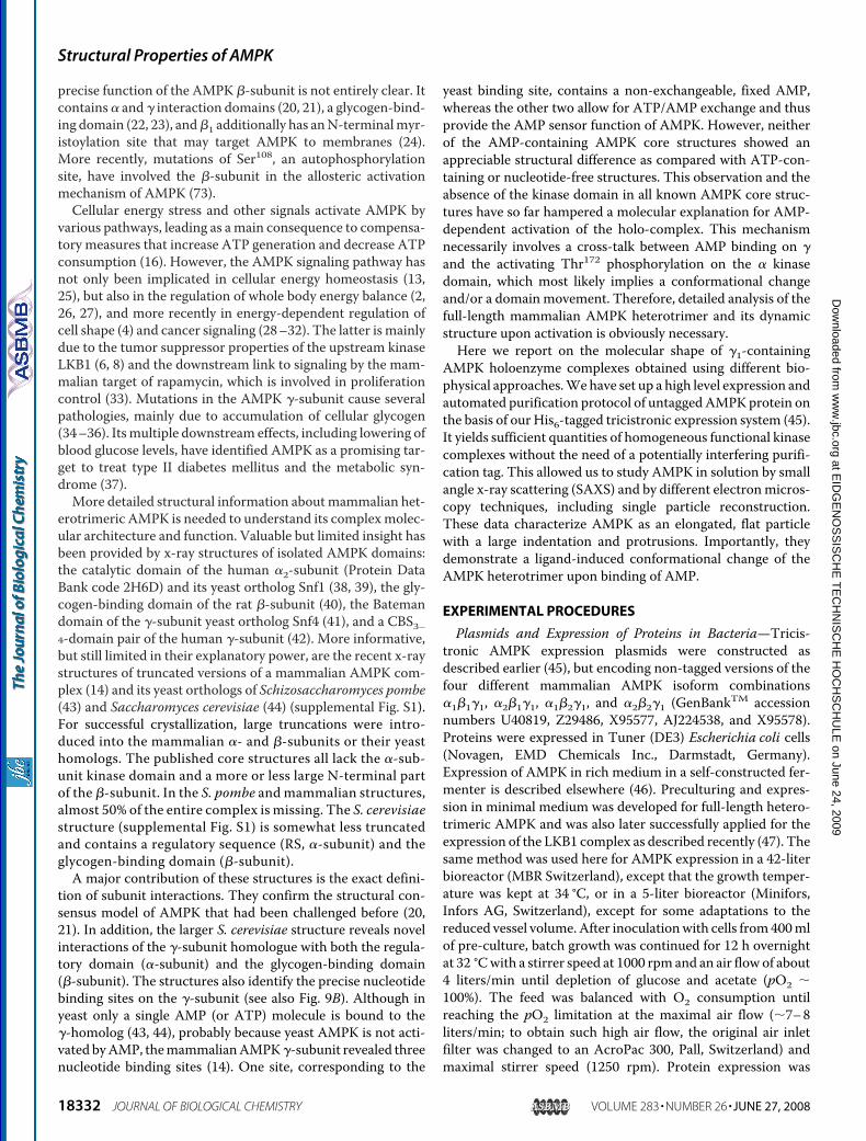
meric �1�1�1- and �2�2�1-AMPK in solution were calculatedusing the DAMMIN software in the “keep”mode and assumingP1 symmetry (no symmetry). Individual molecules were repre-sented by elongated, curved particles showing a deep indenta-tion and protrusions (Fig. 7). Some of these features are lessevident in averaged and filtered models based on 10 different,individual models each calculated from data obtained without(Fig. 8, green) or with AMP added (Fig. 8, red). Arithmetic aver-ages of the radii of gyration and the excluded model volumescalculated from these models by DAMMIN again showed aconsistent and significant change of about 5% toward a morecompact scattering particle upon AMP ligand binding.
DISCUSSION
This study provides a first detailed structural characteriza-tion of untagged, full-length and enzymatically competent het-
erotrimeric AMPK by applying a variety of independent butcomplementary biochemical and biophysical analytical tools.The quantitative results obtained suggest that AMPK hetero-trimers are not spherical but rather elongated, curved particlesthat have a strong tendency to dimerize. Most importantly,allosteric activation by AMP is shown to involve a conforma-tional change toward a more compact heterotrimer.Large Quantities of Untagged, Native AMPKAre Essential for
Biophysical Analysis—High yield bacterial expression and thenovel purification protocol for untagged AMPK were instru-mental for this study, providing the large amounts of purified,native protein required for biophysical characterization, espe-cially for SAXS or isothermal titration calorimetry (ITC).Implementation of the automated four-dimensional purifica-tion procedurewas aimed at increased reproducibility andmin-imal process time without reducing the purity of the final prep-aration. This was achieved by the use of frozen bacterial pelletaliquots from single fermentation runs and the setup of a user-modified Akta ExplorerTM.5 The latter provided reproducibleinstrumental precision, reduced the time needed for purifica-tion to about 18.5 h overnight (as compared with 1 week withmanual operation), and gave a final overall yield of about 10–15mg of AMPK per 70 g wet weight of bacterial pellet. Availabilityof freshly purified AMPK was in particular essential for meth-ods that require large amounts of material and/or that areincompatible with glycerol, like SAXS, STEM, TEM, or ITC.Otherwise, addition of 50% glycerol is necessary to stabilizeAMPK for longer time periods (10). Such purified AMPK couldbe activated byCaMKK� and further stimulated by the alloster-ic activator AMP, reaching a rather high specific activity of upto 6 �mol of phospho-SAMS min�1 mg�1. In a previous studyusing this protein (10), we could show that AMP does not affectAMPK phosphorylation by the upstream kinases LKB1/MO25�/STRAD� or CaMKK� (6–9), but inhibits AMPKdephosphorylation by protein phosphatases-2C � isoform (10).It was also shown that the combined activation effects aremorethan 1000-fold, thus much larger than thought previously (10).These data corroborate the functional integrity of the nativeAMPKpreparationswith respect to activation and deactivationmechanisms. To distinguish between the ��� heterotrimersand possible (��) dimers with very similar molecular mass, MSof the entire cross-linked AMPK complexes was conducted.The data revealed a correct heterotrimeric stoichiometry andthe absence of (��)2 contamination.Reversible Dimerization of AMPK at Higher Protein
Concentrations—High concentrations of protein (above 10mg/ml) are generally used for in solution characterization of
FIGURE 7. Gallery of selected views from an individual �1�1�1-AMPK model without AMPK ligand. The model demonstrates the similarity to the singleparticle TEM averages of AMPK. The diameter of the spheres is 8.5 Å.
TABLE 1Molecular properties of AMPK as determined by SAXS for twodifferent AMPK isoform combinations at different concentrationsThe abbreviations used are Dmax, maximal intramolocular distance; Rg, radius ofgyration;MM,molecularmass; andVPorod, particle volume according to Porod (58).
Samples c Dmax Rg MM VPorod
mg/ml nm kDa nm3
�2�2�1 0.52 16.0 4.64 140 230�2�2�1 1.92 16.0 4.84 141 254�2�2�1 13.18 28.0 7.62 215 445�2�2�1 with AMP 0.51 16.0 4.44 137 211�1�1�1 0.50 16.0 5.13 133 310�1�1�1 1.00 17.0 5.14 139 315�1�1�1 17.62 30.0 9.26 330 590�1�1�1 with AMP 0.98 16.0 4.92 136 288
Structural Properties of AMPK
18338 JOURNAL OF BIOLOGICAL CHEMISTRY VOLUME 283 • NUMBER 26 • JUNE 27, 2008
at EID
GE
NO
SS
ISC
HE
TE
CH
NIS
CH
E H
OC
HS
CH
ULE
on June 24, 2009 w
ww
.jbc.orgD
ownloaded from
protein structures by SAXS to obtain an optimal signal to noiseratio. Under these conditions, the data revealed a strong tend-ency of AMPK heterotrimers to form dimers. In addition, thefirst results with SAXS and dynamic light scattering (notshown) indicated a concentration-dependent occurrence offurther higher oligomers of the heterotrimer, as well as someunspecific aggregates or precipitates, especially upon exposi-tion of the protein to stronger shearing forces by inappropriatehandling, e.g. careless pipetting. This indicates a tendency ofAMPK to formhigher aggregates or to even denature in vitro, asalso noted during the development of the AMPK purificationprotocol. However, if handled with sufficient care, AMPKdid not just aggregate, but rather formed well defined dimersof native heterotrimers. This was confirmed independentlyby electron microscopy methods on a single molecule level.Dimers of AMPK heterotrimers were detected in smallamounts by TEM, and also by STEM, the latter allowingexact mass measurements of oligomers after the chemicalcross-linking.As shown in further SAXS experiments, the dimerization
process is a concentration-dependent and reversible process.Dilution of concentrated AMPK solutions led to a decrease inparticle size, until values were reached comparable with thosefrom AMPK samples that had been purified and maintained atsuch low protein concentrations. Samples diluted immediately(�1 min) before starting the 2-min x-ray exposure for SAXSwere no different than undiluted low concentration samples orsamples examined at a longer time after dilution. Therefore, thetime scale for the reversible concentration-dependent dimer-ization equilibrium must be much less than 3 min.Dimerization may in fact be an intrinsic, conserved property
of the AMPK kinase family and even physiologically relevant.
The catalytic domain of SNF1, theyeast ortholog of AMPK, not onlyforms crystallographic dimers by aconserved and accessible surfacemotif, but also dimerizes in solution(38, 39, 70). Mammalian AMPK isnot an abundant cellular protein,but different mechanisms may leadto higher local AMPK concentra-tions than commonly anticipated.These include compartmentationat defined subcellular loci (24, 71),e.g. in the cell nucleus or at biologi-cal membranes via the myristoylanchor, and molecular crowdingeffects. Thus, it may be entirely rea-sonable to speculate that dimeriza-tion of AMPK could occur at suchdefined subcellular loci. However,for dimerization of AMPK to occurin vivo, not only is AMPK concen-tration important, but also the con-centration of water in the cytosoland its activity coefficient(s) have tobe considered.AMP Binding to AMPK Induces a
Conformational Change—Dilution of AMPK samples below 1mg/ml did not result in a further decrease of particle size inSAXS, indicating that a thermodynamically ideal, monodis-perse protein solution had been reached. This is an absoluteprerequisite for the interpretation of SAXS data when analyz-ing ligand effects or for single particle modeling (see below). Insuch monodisperse AMPK solutions, binding of the allostericactivator AMP induced a clear and significant change in theparticle conformation as directly seen by SAXS. The radii ofgyration of both AMPK isoforms examined were reduced byabout 5%,whereas themaximumparticle dimensions remainedconstant. Comparative averaged SAXS models of AMPK withand without AMP (Fig. 8) indicate that the conformationalchange induced by ligand binding is likely to be a more radialmovement of molecular mass, involving a domain movementtangentially along the long axis of the molecule, rather than anaxially oriented shift of molecular mass. Such a change,together with the observed decrease of molecular mass byextrapolation to I(0), as well as the decrease of both the Porodvolume and the excluded DAMMIN model volume, would beconsistent with a cleft-closing model in the AMPK heterotri-mer upon binding of AMP due to loss of hydration shell water.ITC was applied in an attempt to directly measure AMP
binding constants to AMPK. Modern, state of the art micro-calorimeters require AMPK concentrations significantly above1.5 mg/ml to generate a signal that would allow analysis of thepotentially three independent accessible AMP binding sites(two exchangeable sites in the Bateman domains, one in thekinase domain). However, as shown by SAXS, in this concen-tration range the dynamic, concentration-dependent dimeriza-tion equilibriumofAMPKheterotrimers is probably influencedby AMP binding, adding a further energy parameter to the sys-
FIGURE 8. Gallery of averaged, filtered DAMMIN models of �2�2�1-AMPK. Each model was obtained byDAMAVER software, based on 10 individual models of each �AMP. Green, �2�2�1-AMPK without AMP, thecut-off volume is 2.440 � 105 Å3. Red, �2�2�1 apo-AMPK with AMP, the cut-off volume is 2.209 � 105 Å3. Note,the diameter of the spheres building the space models is 8.5 Å for green and 7.4 Å for the red model. A1 and A2,two different views of the averaged models of �2�2�1-AMPK � AMP shown superimposed, centered on thecenter of mass and oriented along their axis of inertia. B, view of �2�2�1-AMPK with AMP approximately in thesame orientation as in A2. C, view of �2�2�1 apo-AMPK without AMP approximately in the orientation as in A2.
Structural Properties of AMPK
JUNE 27, 2008 • VOLUME 283 • NUMBER 26 JOURNAL OF BIOLOGICAL CHEMISTRY 18339
at EID
GE
NO
SS
ISC
HE
TE
CH
NIS
CH
E H
OC
HS
CH
ULE
on June 24, 2009 w
ww
.jbc.orgD
ownloaded from

tem and, thus, making ITC intrinsically unsuitable for thedetermination of the exact ligand binding constants for AMPK.So far, KD values for AMP have been only determined for theBateman domains in the isolated �-subunits or the truncatedmammalian core complex, ranging from 20 to 125 �M (12, 14).Thus, with the AMP concentration of 1 mM used in our study,we clearly saturated the AMP binding sites.AMPK Heterotrimers Are Elongated, Curved Particles—In
solution SAXS and single particle TEM were applied as com-plementary techniques to analyze molecular dimensions andshape of AMPK complexes. When using SAXS and proceduresfor particle reconstruction like DAMMIN, any deviation of anideal solution has to be strictly avoided. Although heterotri-meric full-lengthAMPKcan exist asmonomers and dimers, theequilibrium between both is concentration-dependent. Amonodisperse solution (in SAXS terms) of monomers of trim-ers was obtained at protein concentrations below 1 mg/ml(Table 1). However, this reduced the signal-to-noise ratio ofSAXSmeasurements, especially at higher momentum transfer.The STEM analysis provided a first estimate of both the
mass and particle dimensions of the heterotrimers. Thisguideline ensured that the projections later selected from thenegative stain TEM images and averaged (Fig. 5) indeedarose from heterotrimers.Some differences in particle dimensions between SAXS and
single particle TEM were observed. The maximum distanceDmax inside the scattering AMPK particle, obtained by the dis-tance distribution functions in SAXS, was 16 � 1 nm, whereasparticles with a size of 11 � 1 nm were obtained by TEM withnegatively stained AMPK and by STEM with cross-linkedAMPK, respectively. Such differences may be largely due to thehydration shell and the rapid molecular dynamics of flexibleAMPK molecules observed with the SAXS in-solution method(69). In addition, it cannot be excluded thatmore compact con-formations were favored on adsorption to the EM grid addingto the size discrepancy.Despite these explainable differences, themolecular shape of
the SAXS models calculated by DAMMIN and the averagedpictures obtained by single particle TEM look strikingly similar.Both reveal large particles with an elongated, curved structureand a wider and a narrower end. This overall shape and appear-ance resembles that of a cashewnut. The SAXSmodels show anadditional protrusion emanating perpendicularly from themore planar particle, as well as a deeper indentation. The latterfeature is not seen so prominently in theTEMpictures, possiblydue to a preferred orientation of the particles on the carbon filmof the EM grid. Interestingly, the recent x-ray structure of theAMPK core complex of the S. cerevisiae AMPK homologue,SNF1, which contained most of the �-subunit, showed that thelatter protruded perpendicularly to the long axis of the mole-cule (44). Thus, the protrusion resolved here by SAXS mighteither represent the kinase domain of the �-subunit or the�-subunit.However, the large truncations in the publishedhighresolution x-ray structures (43, 44) preclude precise fitting ofthese data into the low resolution SAXS model.The presence or absence of AMP also changed the appear-
ance of the DAMMIN models of AMPK. Without AMP, thecomplex appeared very elongated with some protrusions
exposed, whereas AMP binding changed the molecule to amore compact shape preferentially at one end of the particle(compare Fig. 8, A and B). Because the heterotrimeric core ofAMPK does not change conformation upon binding of AMP(14), the conformational change observed here by SAXS musthave its structural basis in other parts of themolecule, i.e. in the�-subunit, which contains the kinase domain, or in the �-sub-unit, which contains the glycogen-binding domain. The confor-mational change upon AMP binding would thus be consistentwith a relative movement of the �- and/or �-subunit, ordomains thereof, along the long axis of themolecule toward the�-subunit. This would lead to a change inmass distribution andthus to a more compact molecule. The fact that the regulatorydomain of the �-subunit was shown to interact tightly with the�-subunit in the S. cerevisiae SNF1 structure (44) would favorsuch an interpretation.Full-lengthAMPKReveals PropertiesDifferent toAMPKCore
Complexes—Themost recent x-ray structures of severely trun-cated core complexes of mammalian AMPK (14) and itsorthologs in S. pombe (43) and S. cerevisiae (44) reveal twostructural properties that are relevant to our study: (i) the yeastenzymes occur as crystallographic dimers of trimers, and (ii) nomajor conformational differences are observed between theapoenzyme and the AMP (or ATP)-bound states.Tight dimers of yeast AMPK orthologs seem to occur readily
in crystals (39, 43, 44), but also in solution (39). However,dimerization occurs at various different interfaces: between �-and �-subunits in the S. pombe core complex (43) (whether thisinterface is accessible in the full-length holoenzyme remains anopen question), between �-subunits in the S. cerevisiae corecomplex (44), and between the kinase domains with individualSnf1 �-subunit orthologs (39). These latter interactions couldalso occur in the full-length heterotrimeric AMPK complex,but themechanism of dimer formation in solution and its puta-tive role in vivo await future clarification.No significant conformational changes upon binding of
AMP or ATPwere observed in the S. pombe complex (43), or inthe truncated mammalian AMPK (14). This may simply be dueto the large truncations (Fig. 9), in particular to the absence ofboth the autoinhibitory and kinase domains (43). However, it isalso unknown whether the yeast complexes are activated byAMP at all, thus precluding definite conclusions on the activa-tory mechanism of mammalian AMPK. AMP binding to Bate-man domains in the �-subunit of mammalian AMPK leads toactivation of the kinase domain in �, suggesting a cross-talkbetween these two subunits. The conformational changeobserved with full-length, native AMPK in our study provides apossible mechanism for this cross-talk.A Model for AMPK Activation—Combining x-ray structural
information (14, 43, 44) with known biochemical and biophys-ical data on the regulation of AMPK (10, 11) and the observedconformational change uponAMPbinding (this work), we pro-pose a new structural model for AMPK. The model is based onthe AMPK ortholog of S. cerevisiae (Fig. 9A, PBD 2QLV, Ref.44). Because only one AMPmoiety was partially resolved (as inthe S. pombe structure), the three AMP molecules observed inthe mammalian AMPK core complex (14) were fitted into thehomologous yeast sites by superposition. Turning the repre-
Structural Properties of AMPK
18340 JOURNAL OF BIOLOGICAL CHEMISTRY VOLUME 283 • NUMBER 26 • JUNE 27, 2008
at EID
GE
NO
SS
ISC
HE
TE
CH
NIS
CH
E H
OC
HS
CH
ULE
on June 24, 2009 w
ww
.jbc.orgD
ownloaded from

sentation as published in Amodeo et al. (44) (Fig. 9B and sup-plemental Fig. S1) by 90°, it is obvious that one exchangeableAMP-binding site (site 1) in the �-subunit (Fig. 9B, blue) is veryclose to the regulatory sequence of the �-subunit (Fig. 9B, RSred). The secondAMP-binding site (site 2) and the site contain-ing a non-exchangeable, fixed AMP (site 3) are somewhatmoredistant to the RS. Furthermore, the x-ray structure of the iso-lated AMPK kinase domain (Protein Data Bank code 2H6D)was positionedwith the active site pointing to the outside of thecomplex (Fig. 9B, gray). From this topology it is obvious that theRS together with a neighboring sequence, which are missing inthe crystallized structures (including a putative regulatory helix
suggested by Pang and colleagues(72)), are sandwiched between the�-subunit and the �-subunit kinasedomain. It is thus entirely conceiva-ble that a small conformationalchange and/or a change in the�-subunit surface charge uponAMP binding (as in the S. pombex-ray structure, see Fig. 2, D and F,in Ref. 43) could be transmitted viathe RS onto the kinase domain andpossibly other domains of AMPK(Fig. 9C). Involvement of the �-sub-unit Ser108 autophosphorylationsite in activation of AMPK has beenreported recently (73), suggestingthat the �-subunit may also comeclose to the kinase domain, consist-ent with our model (Fig. 9, B andC).These effects would finally lead tothe overall conformational changeof the AMPK molecule as observedby SAXS in this work. According tothis conformational switch model,the RS would not act as an autoin-hibitory pseudosubstrate sequenceinside the kinase domain loop, as forexample in PKA and other proteinkinases (see Ref. 72), but rather con-tact the kinase domain at its back-side. Upon AMP binding to the�-subunit, the RS relocates andthereby releases autoinhibition (Fig.9C). Please note that the structuralpart of the RS, which supposedlywould associate with the kinasedomain, is not resolved in any of thepublished AMPK x-ray structuresand thus structural information isnot included in Fig. 9B.Ourmodel proposes that changes
induced by AMP binding in the�-subunit and transmitted via theRS sequence to the �-subunit mayalso alter accessibility of the phos-pho-Thr172 residue. This would
make it less susceptible for dephosphorylation by protein phos-phatase 2C� (10, 19), thus prolonging AMPK activation in thepresence of AMP (Fig. 9C). The entire process of AMP sensingand transmission of the conformational changes would lead toan overall compacting of the AMPK molecule as shown bySAXS (Figs. 5 and 6) and depicted schematically in our model(Fig. 9C). The model integrating many old and recent findingsseems consistent with published data and may give some newclues for further studies on the allosteric activation of AMPK.Taken together, the quantitative results presented here shed
new light on themolecular shape of nativeAMPKheterotrimer,at the resolution attainable by negative stain TEMmethods and
FIGURE 9. Model for AMPK activation by AMP: a novel role for the regulatory sequence. The model ismainly based on the recent x-ray structure of the S. cerevisiae AMPK ortholog core complex (44). Crystallizedparts of the sequence are shown in: red, C terminus plus regulatory sequence (RS) of the �-subunit; green, Cterminus plus glycogen-binding domain (GBD) of the �-subunit; blue, complete �-subunit with the AMP-binding CBS domains. Bound AMP and ATP are in magenta or white, respectively. Sequence sections lackingcorresponding structural information are in gray: e.g. kinase domain (KD) with activation loop (AL) of the�-subunit and a significant N-terminal part of the �-subunit. A, domain organization of the S. cerevisiae AMPKortholog. Snf1, Sip2, and Snf4 correspond to �-, �-, and �-subunits, respectively, of mammalian AMPK (aminoacid numbering according to S. cerevisiae, modified using Ref. 44). B, structural model of a putative full-lengthAMPK complex. The structure of the core complex of the S. cerevisiae AMPK ortholog (PDB 2QLV) is turnedsidewise by 90° relative to the representations in Amodeo et al. (44). Three AMP molecules (one fixed, twoexchangeable) were introduced by superposition with the very homologous core structures of mammalianAMPK using PDB 2V8Q (see also supplemental Fig. 1). The �-subunit KD (Protein Data Bank code 2H6D) wasadded to its putative location (see Fig. 1B in Ref. 43), in close proximity to the �-subunit RS and with the activesite pointing to the outside of the complex. Some part of the RS, including the autoinhibitory domain with aconserved putative �-helix, is not resolved in any of the known x-ray structures. The autoinhibitory domain wasproposed to bind to the backside of the small lobe of the KD (72). Note: the RS is sandwiched between the�-subunit (blue), with its AMP binding sites, and the KD (gray). This would allow the RS to mediate a cross-talkbetween AMP binding sites and the KD or other parts of the AMPK complex as outlined in C. C, simplified modelfor the mechanism of AMPK regulation by AMP. This model is based on the putative structure of AMPK (shownin B) and incorporates data from us (this work and Ref. 10), as well as others (e.g. Refs. 43 and 72). The (P) in theKD indicates the activatory phosphorylation at Thr172 (6 –9). Left, at the low AMP/ATP ratio, binding sites 1 and2 are occupied by ATP and site 3 by non-exchangeable AMP. The Thr172-phosphate group is easily accessible toprotein phosphatase 2C� for dephosphorylation (10, 19), resulting in low AMPK activity. Right, at an increasingAMP/ATP ratio, replacement of ATP by AMP at reversible AMP-binding sites 1 and/or 2 would result in smallconformational changes and/or changes in surface potential of the �-subunit (43). Given the structure outlinedin B, our model proposes transmission and amplification of these changes by the neighboring RS and KD (smallopen arrows) leading to release of autoinhibition of the KD by retracting the RS and pulling the KD closer to thecore of the AMPK molecule. The overall compaction of the heterotrimer is interpreted in our model as amovement of RS (red arrow) and KD (black arrow), as well as a conformational change in the KD. The latter wouldprotect phospho-Thr172 against dephosphorylation, thus keeping AMPK in its phosphorylated, active form.
Structural Properties of AMPK
JUNE 27, 2008 • VOLUME 283 • NUMBER 26 JOURNAL OF BIOLOGICAL CHEMISTRY 18341
at EID
GE
NO
SS
ISC
HE
TE
CH
NIS
CH
E H
OC
HS
CH
ULE
on June 24, 2009 w
ww
.jbc.orgD
ownloaded from

in-solution SAXS techniques. Overall, the molecular shape ofAMPK particles was consistent, irrespective of the methodapplied, suggesting that it reflects the “true” structural/func-tional state ofmammalianAMPKat this resolution. The studiesrevealed a rapid concentration-dependent equilibriumbetween AMPK heterotrimers and defined dimers thereof.Finally, and most importantly, an AMP-induced conforma-tional switch is shown in the full-length AMPK complex, sug-gesting a molecular mechanism for AMPK activation by allo-steric activators. The techniques and results developed hereinare expected to stimulate new and original approaches to pur-sue high-resolution structural characterization of full-lengthAMPK, as well as to aid a more detailed study of AMPK regu-lation in vivo.
Acknowledgments—We thank Ulrich Bauer (former Institute of Bio-technology, ETH Zurich) for introducing U. R. to the art of runninglarge bioreactors, Marco Gregorini (M. E. Muller Institute for Struc-tural Biology, Basel) for contributions to TEM microscopy, and Sac-nicte Ramirez (InsermU884/LBFA, UJFGrenoble) for assistance dur-ing the SAXS measurements at DESY, Hamburg. All members of theWallimann group are acknowledged for help and discussions.
REFERENCES1. Kahn, B. B., Alquier, T., Carling, D., and Hardie, D. G. (2005) Cell Metab.
1, 15–252. Hardie, D. G. (2007) Nat. Rev. Mol. Cell Biol. 8, 774–7853. Carling, D. (2005) Biochimie (Paris) 87, 87–914. Lee, J. H., Koh, H., Kim, M., Kim, Y., Lee, S. Y., Karess, R. E., Lee, S. H.,
Shong, M., Kim, J. M., Kim, J., and Chung, J. (2007) Nature 447,1017–1020
5. Neumann, D.,Wallimann, T., Rider,M., Tokarska-Schlattner,M., Hardie,D. G., and Schlattner, U. (2007) inMolecular System Bioenergetics, Energyfor Life (Saks, V., ed) 1st Ed., pp. 303–338, Wiley-VCH, Weinheim,Germany
6. Hawley, S. A., Boudeau, J., Reid, J. L., Mustard, K. J., Udd, L., Makela, T. P.,Alessi, D. R., and Hardie, D. G. (2003) J. Biol. 2, 28
7. Shaw, R. J., Kosmatka, M., Bardeesy, N., Hurley, R. L., Witters, L. A.,DePinho, R. A., andCantley, L. C. (2004)Proc. Natl. Acad. Sci. U. S. A. 101,3329–3335
8. Woods, A., Johnstone, S. R., Dickerson, K., Leiper, F. C., Fryer, L. G.,Neumann, D., Schlattner, U., Wallimann, T., Carlson, M., and Carling, D.(2003) Curr. Biol. 13, 2004–2008
9. Hawley, S. A., Pan, D. A., Mustard, K. J., Ross, L., Bain, J., Edelman, A. M.,Frenguelli, B. G., and Hardie, D. G. (2005) Cell Metab. 2, 9–19
10. Suter, M., Riek, U., Tuerk, R., Schlattner, U., Wallimann, T., and Neu-mann, D. (2006) J. Biol. Chem. 281, 32207–32216
11. Hardie, D. G., and Carling, D. (1997) Eur. J. Biochem. 246, 259–27312. Scott, J. W., Hawley, S. A., Green, K. A., Anis, M., Stewart, G., Scullion,
G. A., Norman, D. G., and Hardie, D. G. (2004) J. Clin. Investig. 113,274–284
13. Hardie, D. G., Hawley, S. A., and Scott, J. W. (2006) J. Physiol. 574, 7–1514. Xiao, B., Heath, R., Saiu, P., Leiper, F. C., Leone, P., Jing, C., Walker, P. A.,
Haire, L., Eccleston, J. F., Davis, C. T., Martin, S. R., Carling, D., and Gam-blin, S. J. (2007) Nature 449, 496–500
15. McGilvery, R., and Murray, T. W. (1974) J. Biol. Chem. 249, 5845–585016. Hardie, D. G., and Hawley, S. A. (2001) Bioessays 23, 1112–111917. Neumann, D., Schlattner, U., and Wallimann, T. (2003) Biochem. Soc.
Trans. 31, 169–17418. Schlattner, U., and Wallimann, T. (2004) in Encyclopedia of Biological
Chemistry (Lennarz, W. J., and Lane, M. D., eds) pp. 646–651, AcademicPress, New York
19. Sanders,M. J., Grondin, P.O.,Hegarty, B.D., Snowden,M.A., andCarling,
D. (2007) Biochem. J. 403, 139–14820. Iseli, T. J., Walter, M., van Denderen, B. J., Katsis, F., Witters, L. A., Kemp,
B. E., Michell, B. J., and Stapleton, D. (2005) J. Biol. Chem. 280,13395–13400
21. Wong, K. A., and Lodish, H. F. (2006) J. Biol. Chem. 281, 36434–3644222. Hudson, E. R., Pan, D. A., James, J., Lucocq, J. M., Hawley, S. A., Green,
K. A., Baba, O., Terashima, T., and Hardie, D. G. (2003) Curr. Biol. 13,861–866
23. Polekhina, G., Gupta, A., Michell, B. J., van Denderen, B., Murthy, S., Feil,S. C., Jennings, I. G., Campbell, D. J., Witters, L. A., Parker, M. W., Kemp,B. E., and Stapleton, D. (2003) Curr. Biol. 13, 867–871
24. Warden, S.M., Richardson, C.,O’Donnell, J., Jr., Stapleton,D., Kemp, B. E.,and Witters, L. A. (2001) Biochem. J. 354, 275–283
25. Hardie, D. G., and Sakamoto, K. (2006) Physiol. (Bethesda) 21, 48–6026. Minokoshi, Y., Kim, Y. B., Peroni, O. D., Fryer, L. G., Muller, C., Carling,
D., and Kahn, B. B. (2002) Nature 415, 339–34327. Ruderman, N. B., Saha, A. K., and Kraegen, E. W. (2003) Endocrinology
144, 5166–517128. Hardie, D. G. (2005) Curr. Opin. Cell Biol. 17, 167–17329. Carretero, J., Medina, P. P., Blanco, R., Smit, L., Tang, M., Roncador, G.,
Maestre, L., Conde, E., Lopez-Rios, F., Clevers, H. C., and Sanchez-Cespedes,M. (2007)Oncogene 26, 1616–1625
30. Andersson, Y., Le, H., Juell, S., and Fodstad, O. (2006)Mol. Cancer Ther. 5,1050–1059
31. Ashrafian, H. (2006) Lancet 367, 618–62132. Luo, Z., Saha, A. K., Xiang, X., and Ruderman, N. B. (2005) Trends Phar-
macol. Sci. 26, 69–7633. Bolster, D. R., Crozier, S. J., Kimball, S. R., and Jefferson, L. S. (2002) J. Biol.
Chem. 277, 23977–2398034. Dolinsky, V.W., and Dyck, J. R. (2006) Am. J. Physiol. 291,H2557–H256935. Davies, J. K., Wells, D. J., Liu, K., Whitrow, H. R., Daniel, T. D., Grignani,
R., Lygate, C. A., Schneider, J. E., Noel, G., Watkins, H., and Carling, D.(2006) Am. J. Physiol. 290, H1942–H1951
36. Gollob, M. H. (2003) Biochem. Soc. Trans. 31, 228–23137. Hardie, D. G. (2007) Annu. Rev. Pharmacol. Toxicol. 47, 185–21038. Rudolph, M. J., Amodeo, G. A., Bai, Y., and Tong, L. (2005) Biochem.
Biophys. Res. Commun. 337, 1224–122839. Nayak, V., Zhao, K.,Wyce, A., Schwartz, M. F., Lo,W. S., Berger, S. L., and
Marmorstein, R. (2006) Structure 14, 477–48540. Polekhina, G., Feil, S. C., Gupta, A., O’Donnell, P., Stapleton, D., and
Parker, M. W. (2005) Acta Crystallogr. Sect. F Struct. Biol. Crystallogr.Commun. 61, 39–42
41. Rudolph,M. J., Amodeo, G. A., Iram, S. H., Hong, S. P., Pirino, G., Carlson,M., and Tong, L. (2007) Structure 15, 65–74
42. Day, P., Sharff, A., Parra, L., Cleasby, A., Williams, M., Horer, S., Nar, H.,Redemann, N., Tickle, I., and Yon, J. (2007) Acta Crystallogr. D Biol. Crys-tallogr. 63, 587–596
43. Townley, R., and Shapiro, L. (2007) Science 315, 1726–172944. Amodeo, G. A., Rudolph, M. J., and Tong, L. (2007)Nature 449, 492–49545. Neumann, D., Woods, A., Carling, D., Wallimann, T., and Schlattner, U.
(2003) Protein Expression Purif. 30, 230–23746. Riek, U., Tuerk, R.,Wallimann, T., Schlattner, U., andNeumann,D. (2008)
Biotechniques (in press)47. Neumann, D., Suter, M., Tuerk, R., Riek, U., and Wallimann, T. (2007)
Mol. Biotechnol. 36, 220–23148. Muller, S. A., Goldie, K. N., Burki, R., Haring, R., and Engel, A. (1992)
Ultramicroscopy 46, 317–33449. Muller, S. A., and Engel, A. (2001)Micron 32, 21–3150. Ludtke, S. J., Baldwin, P. R., and Chiu,W. (1999) J. Struct. Biol. 128, 82–9751. Nazabal, A., Wenzel, J. R., and Zenobi, R. (2006) Anal. Chem. 78,
3562–357052. Boulin, C. J., Kempf, R., Gabriel, A., and Koch, M. H. J. (1988) Nucl. In-
strum. Meth. A 269, 312–32053. Roessle, M. W., Klaering, R., Ristau, U., Robrahn, B., Jahn, D., Gehrmann,
T., Konarev, P., Round, A., Fiedler, S., Hermes, C., and Svergun, D. (2007)J. Appl. Crystallogr. 40, 190–194
54. Konarev, P. V., Volkov, V. V., Sokolova, A. V., Koch,M. H. J., and Svergun,D. I. (2003) J. Appl. Crystallogr. 36, 1277–1282
Structural Properties of AMPK
18342 JOURNAL OF BIOLOGICAL CHEMISTRY VOLUME 283 • NUMBER 26 • JUNE 27, 2008
at EID
GE
NO
SS
ISC
HE
TE
CH
NIS
CH
E H
OC
HS
CH
ULE
on June 24, 2009 w
ww
.jbc.orgD
ownloaded from
55. Konarev, P. V., Petoukhov, M. V., Volkov, V. V., and Svergun, D. I. (2006)J. Appl. Crystallogr. 39, 227–286
56. Guinier, A., and Fournet, G. (1955) Small Angle Scattering of X-rays,Wiley, New York
57. Svergun, D. I. (1992) J. Appl. Crystallogr. 25, 495–50358. Porod, G. (1982) in Small-angle X-ray Scattering (Glatter, O., and Kratky,
O., eds) pp. 17–51, Academic Press, London59. Svergun, D. I. (1999) Biophys. J. 76, 2879–288660. Volkov, V. V., and Svergun, D. I. (2003) J. Appl. Crystallogr. 36, 860–86461. Treebak, J. T., Glund, S., Deshmukh, A., Klein, D. K., Long, Y. C., Jensen,
T. E., Jorgensen, S. B., Viollet, B., Andersson, L., Neumann, D.,Wallimann,T., Richter, E. A., Chibalin, A. V., Zierath, J. R., and Wojtaszewski, J. F.(2006) Diabetes 55, 2051–2058
62. Xie, Z., Dong, Y., Zhang, M., Cui, M. Z., Cohen, R. A., Riek, U., Neumann,D., Schlattner, U., and Zou, M. H. (2006) J. Biol. Chem. 281, 6366–6375
63. Horman, S., Vertommen, D., Heath, R., Neumann, D., Mouton, V.,Woods, A., Schlattner, U., Wallimann, T., Carling, D., Hue, L., and Rider,M. H. (2006) J. Biol. Chem. 281, 5335–5340
64. Baron, S. J., Li, J., Russell, R. R., 3rd, Neumann, D., Miller, E. J., Tuerk, R.,Wallimann, T., Hurley, R. L., Witters, L. A., and Young, L. H. (2005) Circ.Res. 96, 337–345
65. Carattino, M. D., Edinger, R. S., Grieser, H. J., Wise, R., Neumann, D.,Schlattner, U., Johnson, J. P., Kleyman, T. R., and Hallows, K. R. (2005)J. Biol. Chem. 280, 17608–17616
66. Zou, M. H., Kirkpatrick, S. S., Davis, B. J., Nelson, J. S., Wiles, W. G., 4th,Schlattner, U., Neumann, D., Brownlee, M., Freeman, M. B., and Gold-man, M. H. (2004) J. Biol. Chem. 279, 43940–43951
67. Taylor, E. B., Ellingson, W. J., Lamb, J. D., Chesser, D. G., Compton, C. L.,and Winder, W. W. (2006) Am. J. Physiol. 290, E661–E669
68. Taylor, E. B., Ellingson, W. J., Lamb, J. D., Chesser, D. G., and Winder,W. W. (2005) Am. J. Physiol. 288, E1055–E1061
69. Svergun, D. I., Barberato, C., and Koch,M. H. J. (1995) J. Appl. Crystallogr.28, 768–773
70. Elbing, K., Rubenstein, E.M.,McCartney, R. R., and Schmidt,M. C. (2006)J. Biol. Chem. 281, 26170–26180
71. Mitchelhill, K. I., Michell, B. J., House, C. M., Stapleton, D., Dyck, J., Gam-ble, J., Ullrich, C.,Witters, L. A., and Kemp, B. E. (1997) J. Biol. Chem. 272,24475–24479
72. Pang, T., Xiong, B., Li, J. Y., Qiu, B. Y., Jin, G. Z., Shen, J. K., and Li, J. (2007)J. Biol. Chem. 282, 495–506
73. Sanders, M. J., Ali, Z. S., Hegarty, B. D., Heath, R., Snowden, M. A., andCarling, D. (2007) J. Biol. Chem. 282, 32539–32548
Structural Properties of AMPK
JUNE 27, 2008 • VOLUME 283 • NUMBER 26 JOURNAL OF BIOLOGICAL CHEMISTRY 18343
at EID
GE
NO
SS
ISC
HE
TE
CH
NIS
CH
E H
OC
HS
CH
ULE
on June 24, 2009 w
ww
.jbc.orgD
ownloaded from