Importin contains a COOH-terminal nucleoporin binding region important for nuclear transport
Upload
independentCategory
view
0download
0
pubs.acs.org/Biochemistry Published on Web 11/18/2009 r 2009 American Chemical Society
12180 Biochemistry 2009, 48, 12180–12190
DOI: 10.1021/bi9017076
The βγ-Crystallin Superfamily Contains a Universal Motif for Binding Calcium†,‡
Penmatsa Aravind,§ Amita Mishra,§ Shashi Kumar Suman,§ Maroor K. Jobby, ) Rajan Sankaranarayanan,*and Yogendra Sharma*
Centre for Cellular and Molecular Biology, Council of Scientific and Industrial Research, Uppal Road, Hyderabad 500007, India.§These authors contributed equally. )Present address: Department of Biochemistry, Queen’s University, King-
ston, Ontario, Canada K7L 3N6.
Received October 2, 2009; Revised Manuscript Received November 17, 2009
ABSTRACT: The βγ-crystallin superfamily consists of evolutionarily related proteins with domain topologysimilar to lens β- and γ-crystallins, formed from duplicatedGreek keymotifs. Ca2þ bindingwas found in a fewβγ-crystallin members earlier, although its prevalence and diversity as inherent molecular properties amongmembers of the superfamily are not well studied. To increase our understanding of Ca2þ binding in variousβγ-crystallins, we undertook comprehensive structural and Ca2þ-binding studies of seven members of thesuperfamily from bacteria, archaea, and vertebrates, including determination of high-resolution crystalstructures of three proteins. Our structural observations show that the determinants of Ca2þ coordinationremain conserved in the form of an N/D-N/D-#-I-S/T-S motif in all domains. However, binding of Ca2þ
elicits varied physicochemical responses, ranging from passive sequestration to active stabilization. The motifin this superfamily is modified in somemembers like lens crystallins where Ca2þ-binding abilities are partly orcompletely compromised. We show that reduction or loss of Ca2þ binding in members of the superfamily,particularly in vertebrates, is due to the selective presence of unfavorable amino acids (largely Arg) at keyCa2þ-ligation positions and that engineering of the canonical Ca2þ-binding residues can confer bindingactivity on an otherwise inactive domain. Through this work, we demonstrate that βγ-crystallins with the N/D-N/D-#-I-S/T-S motif form an extensive set of Ca2þ-binding proteins prevalent in all of the three kingdomsof life.
Ca2þ binding is a crucial step in affecting major life pro-cesses (1) with specialized protein motifs engineered by nature tobind Ca2þ with varied affinities in both the extracellular andintracellular environments (2). Proteins evolved to sense Ca2þ asa signal are commonly referred to as sensors, and a set of proteinsthat bind and undergo structural stabilization are referred to asbuffers. The allR-helical EF-hand superfamily of proteins and allβ-sheet containing C2 domains predominate sensory Ca2þ-bind-ing proteins. Proteins involved in structural binding are found inmany extracellular proteins, such as EGF domains and cadher-ins, and are involved in providing structural stability to anorganism by sequestering Ca2þ from the environment. All ofthese independent structural units have been recruited as full-length proteins or asmodules inmultidomain proteins to performCa2þ-dependent roles.
Apart from these known families of Ca2þ-binding proteins,some members of βγ-crystallins have been shown to bind Ca2þ.
The βγ-crystallin superfamily comprises proteins with β-sand-wich domains whose early characterized members, β- andγ-crystallins, are major components of the vertebrate eye lens(3-5). Individual βγ-crystallin domains are made of strand-exchanged Greek key motifs (6, 7). The wedge-like domains arecharacterized by the presence of highly ordered β-hairpin loopsbetween the first and second strands of each motif. Two loopstraverse the roof of the wedge-like domain and connect the twoopposing β-pleated sheets of the domain (6-8). Evolutionarilyrelated structural homologues of lens β- and γ-crystallins arefound in some lower eukaryotic and prokaryotic species whichtogether form the βγ-crystallin superfamily.
The Ca2þ-binding connection to the βγ-crystallin superfamilywas observed more than 2 decades ago when protein S fromMyxococcus xanthus, the first member of the superfamily, wasshown to bind Ca2þ even before the classification of theβγ-crystallin superfamily (7, 9, 10). At the same time, β-crystallin(aggregated form) from the vertebrate lens was shown to bindCa2þ albeit with low affinity in solution and with no structuralevidence for the Ca2þ-binding site (11). When the sequence ofspherulin 3a fromPhysarum polycephalumwas describedwith theputative features of βγ-crystallins as a third member, the conceptof a superfamily, the βγ-crystallin superfamily originated (12).Spherulin 3a was structurally characterized and found to havebound Ca2þ at the D/N-X-X-S motif, similar to that seen first inprotein S (13-15). Later, a fewmembers of this superfamily weredemonstrated to bind Ca2þ in solution (16-19). Protein S andspherulin 3a remained the only structurally well character-ized Ca2þ-binding members of the βγ-crystallin superfamily
†This work was supported by a Department of Science and Technol-ogy and a DBT grant to Y.S. and a Swarnajayanti Fellowship, DST,India, and an International Senior Research Fellowship, WellcomeTrust, U.K., grant to R.S. P.A. and S.K.S. are the recipients of fellow-ships from the Council of Scientific and Industrial Research, India. A.M. is supported by a postdoctoral fellowship from the Department ofBiotechnology, India.
‡The coordinates and structure factor amplitudes of M-crystallin,clostrillin (forms 1 and 2), and flavollin have been deposited in theProtein Data Bank with the IDs 3HZ2, 3I9H, 3IAJ, and 3HZB,respectively.*Corresponding authors. Y.S.: phone, þ91-40-27192544; fax, þ91-
40-27160591; e-mail, [email protected]). R.S.: phone, þ91-40-27192832; fax, þ91-40-27160591; e-mail, [email protected].
Article Biochemistry, Vol. 48, No. 51, 2009 12181
(8, 13-15, 20, 21) until the structure of Ciona crystallin, anancestral family member from urochordates that do not have alens, was solved in Ca2þ-bound form, shedding light on theevolution of lens βγ-crystallins (22). Incidentally, the Ca2þ-binding region in these three proteins was located at the homo-logous D/N-X-X-S motif.
These sporadic studies opened up several interesting questionsto be explored, particularly about the detailed description ofCa2þ binding as well as themotif of ion binding. This is necessarysince Ca2þ-bindingmotifs are well-defined supersecondary struc-tures, as in the case of the EF-hand motif. Earlier studies werelargely focused on the evolutionary aspects of crystallins, inaddition to Ca2þ binding. Therefore, first of all, an importantquestion to address is whether the sequence D/N-X-X-S forms aspecific, well-defined motif for Ca2þ binding prevalent in otherproteins of the superfamily or presents only in a few βγ-crystallinsas a unique case. One of the reasons was that a large number ofproteins have not been shown to bind Ca2þ ions (in some caseseven structurally), such as yeast killer toxins, SKLP,1 SMPI,AIM1g1, and nitrollin (23-27). In addition to this, not manymembers belonging to the βγ-crystallin superfamily were knownuntil recently. On the other hand, several proteins, such asYersinia crystallin (16), caulollins (17), AIM1 domain (28), andgeodin (19), have been shown to bind Ca2þ only in solution butwithout any structural evidence for a homologous Ca2þ-bindingmotif.
With these ambiguities, it became necessary to clarify severalquestions: (i) Does the sequence D/N-X-X-S seen in spherulin 3aand protein S form a widespread motif of Ca2þ binding in thesuperfamily? (ii) What would be the effects of sequence diversi-fication of motif on the Ca2þ-binding properties of individualproteins? (iii) If the motif is well-defined, could a gain of functionbe created in a naturally disabled protein? In order to understandand explore some of these outstanding questions pertaining toβγ-crystallins and their Ca2þ-binding properties, we undertookthis extensive structural characterization by identifying proteinsbelonging to this family from all three kingdoms of life (archaeal,bacterial, and eukaryote including vertebrate species).
Our analysis of the genomes from various species demon-strates the prevalence of the proteins with βγ-crystallin-likedomains. This Ca2þ-binding motif is widely present in close to70 independent proteins from various species across all threekingdoms of life. Our in-depth structural analysis of novelproteins, along with earlier observations of Ca2þ binding in threeβγ-crystallin homologues, suggests that the Ca2þ-binding se-quence seen earlier actually forms amotif for Ca2þ binding. Sincewe were able to create a gain of Ca2þ-binding function inthe naturally disabled protein, nitrollin, a domain-swappedβγ-crystallin from Nitrosospira multiformis (27), we suggest thatthis ancestral motif was modified and lost Ca2þ binding in somedescendant lineages. Our data comprehensively confirm thewidespread conservation of this motif for Ca2þ binding, whichhas undergone extensive modifications in several descendants,including many vertebrates.
EXPERIMENTAL PROCEDURES
Genomic DNA and Clones from Various Species. Thegenomic DNA of Flavobacterium johnsoniae, Methanosarcina
acetivorans,Reinekea sp.MED297, andN.multiformiswere kindgifts from Dr. Mark J. McBride, University of Wisconsin,Professor WilliamW.Metcalf, University of Illinois at Urbana-Champaign, Dr. Jarone Pinhassi, University of Kalmar,Sweden, and Dr. Lisa Stein, University of California, Riverside,respectively. The region of interest (500-591 amino acids) of theprotein YP_001196503 from F. johnsoniae and 36-120 aminoacids of a protein annotated as the βγ-crystallin family protein(accession number AAM05909) from M. acetivorans were am-plified by using gene-specific primers. Genomic DNA of Clos-tridium beijerincki andRhodoferax ferrireducens strains (obtainedfrom DSMZ, Germany) were isolated using the hot phenolmethod. Similarly, clostrillin (βγ-crystallin domains of118-180 amino acids) of the protein (accession numberYP_001309930) from C. beijerinckii, rhodollin (22-117 aminoacids) of the protein (accession number YP_522648) from R.ferrireducens, reinekillin (356-536 amino acids) of the protein(ZP_01115288) from Reinekea sp. D297, and nitrollin (32-140amino acids) of the protein (accession number YP_411671) fromN. multiformis were amplified. ep-37A1 (epidermal differentia-tion-specific protein) (clone BAA21832) from adult Cynopspyrrhogaster, an amphibian, was a kind gift from Dr. KazuhitoTakeshima, Nagoya University, Japan.Cloning and Overexpression of Untagged and Tagged
Proteins. PCR products of βγ-crystallin homologues fromF. johnsoniae, M. acetivorans, C. beijerinckii, R. ferrireducens,and N. multiformis were cloned in a pET21a expression vector(Novagen) at NdeI and BamHI sites. PCR fragments fromReinekea and ep37 were cloned in a pGEX-4T1 (GEHealthcare)vector at BamHI and EcoRI sites. The recombinant proteinsflavollin, M-crystallin, and nitrollin were expressed in the bacter-ial strainEscherichia coli BL21(DE3) (Invitrogen) in LBmedium(containing 100 μg of ampicillin/mL) after induction with 1 mMisopropyl thio-β-D-galactopyranoside (IPTG) for 10 h at 37 �C.The overexpression of clostrillin, reinekillin, rhodollin, and ep37was performed by growingE. coliBL21(DE3) at 18 �C for 16-18h after induction with 1 mM IPTG.Protein Purification. Proteins which formed inclusion
bodies during overexpression (flavollin, rhodollin, and nitrollin)were refolded using an on-column refolding procedure. Theinclusion bodies were washed with 1 M urea and 0.1% CHAPSand were solubilized in a buffer containing 50 mM Tris buffer,pH 8.5, or 50 mM Bis-Tris, pH 6.2, 3.5-5 M urea, and 1 mMDTT, and on-column refolding was performed on Q-Sepharosefor flavollin and on SP-Sepharose matrix for nitrollin. The yieldof flavollin and nitrollin per liter of culture was 1.8 and 0.8 mg,respectively. Rhodollin was purified using SP-Sepharose matrixin 50 mM sodium acetate, pH 5.0, 5.0 M urea, and 1 mM DTT.The proteins were eluted with a step gradient of 50 mMTris, pH8.5, or Bis-Tris, pH 5.5, 1 M NaCl, and 1 mMDTT with a finalyield of 0.5 mg/L of culture. Clostrillin was purified from thesoluble fraction on a Q-Sepharose column at pH 9.6 (yield perliter of culture was 8.5 mg). GST-tagged proteins (Reinekillin,ep37) were purified on a GST fast-flow column using prescribedprotocols (GE healthcare), and the yield was about 2 mg/L of LBculture. Final purification of flavollin, clostrillin, rhodollin, andnitrollin was performed using Superdex-75 gel filtration columns.ep37 and reinekillin were purified on a Superose-12 gel filtrationcolumn (Pharmacia). M-crystallin was purified from a solublefraction through a Bio-Gel A-1.5 m gel filtration column in50 mM Tris-HCl (pH 7.5) and 100 mM KCl, and the yield wasabout 2.5 mg/L of culture. Protein concentrations were calculated
1Abbreviations: AIM1, absent in melanoma 1; EDTA, ethylenedia-minetetraacetic acid; SKLP, Streptomyces killer toxin-like protein;SMPI, Streptomyces metalloproteinase inhibitor.
12182 Biochemistry, Vol. 48, No. 51, 2009 Aravind et al.
by measuring their absorbance at 280 nm in 6 M guanidiniumchloride using the values of the theoretical extinction coefficientcalculated by the ExPASy Proteomics Server (ProtParam).Crystallization. The purified proteins were exchanged into a
buffer containing 10 mM Tris, pH 7.2, 20 mM NaCl, 3-5 mMCaCl2, and 0.02% sodium azide. A stock solution of 10 mg/mLwas used for setting up screens in the 96-well format in Greinerplates by sitting drop vapor diffusion through mixing 1 μL ofprotein with 1 μL of reservoir solution with concentrations of 5,8, and 10 mg/mL in each of the subwells, respectively. Twocommercial screens, Crystal ScreenHT and IndexHT (HamptonResearch), were used for screening at a temperature of 4 or 20 �Cin our in-house HT facility which employs a Minstrel (Rigaku)plate incubator and imaging system. M-crystallin crystallized inthe F10 condition of Crystal Screen HT which comprises 12%PEG 20Kwith 0.1MNa-MES, pH 6.5 at 20 �C; flavollin crystalswere obtained in 30% PEG 8K with 0.1 M sodium cacodylatebuffer at pH 6.5 with 0.4 M potassium chloride. Form 1 crystalsof clostrillin crystallized in 26% PEG 3350 at pH 7.3 maintainedusing 0.1 M HEPES buffer with 0.2 M lithium sulfate; form 2crystals were obtained in 2 M ammonium sulfate at pH 6.5maintained using 0.1 M Bis-Tris.Data Collection and Processing. X-ray data were collected
in-house using a mar345dtb image-plate detector attached to aRigaku RU-H3R rotating anode generator equipped with anosmic mirror system operated at 50 kV/100 mA. Data forflavollin crystals were collected at the X11 beamline at DESY,EMBL-Hamburg. All of the data sets were processed and scaledusing DENZO and SCALEPACK (29). Conversion of reflectionformats,merging, and scaling of reflectionswere performedusingthe CCP4 suite of programs, version 6.0 (30).Structure Determination. Structures of clostrillin, M-crys-
tallin, and flavollin were solved using molecular replacementusing MOLREP and BALBES (31). Structures of Ciona crystal-lin (2BV2) and protein S (1NPS) were used as templates to solvethe structures. M-crystallin and form 1 and form 2 structures ofclostrillin were solved using the automated molecular replace-ment program MOLREP from the CCP4 package, version6.0 (30). The form 1 structure of clostrillin had two sets of fourprotomers (eight molecules/AU) related among each other by anapproximate 4-fold symmetry. The two sets of molecules wererelated by a pseudotranslation vector of (0.0, 0.34, 0.5) whichresulted in a nonorigin peak in the Pattersonmapwhose intensitywas 34%of the origin peak.MOLREP found sixmolecules in theAU, and the remaining two were positioned by using thepseudotranslation vector. The structure of flavollin was solvedin theP2 space group although the initial estimates indicated I222in which the structure could be solved but could not be refined.This disparity arose due to pseudomerohedral twinning thatoccurred as a result of the angle “β” of the monoclinic spacegroup being close to 90�. This led P2 to emulate the P222 system.A further complication occurred as the crystals had pseudobodycentering (Patterson vector 0.5, 0.5, 0.5, nonorigin peak equiva-lent to 58% of the origin peak) which led to the P222 space groupemulating the I222 lattice. Initial diagnosis and molecularreplacement were performed using BALBES which picked pro-tein S (1NPS) as a probe (31). The partially refined structure offlavollin was then used to perform the complete structure solutionand refinement. Nitrollin structure solution was performed usingmultiple isomorphous replacement as described earlier (27).Refinement and Structural Analysis. Model building in
required regions of the structure was performed using the
program “O” (32) with repeated cycles of refinement using theCNS program package (33). All of the refined structures werevalidated using PROCHECK (34) (Table 1). Distance measure-ments were performed both manually in O and using EBI-PISA (35). Structural deviations were calculated using theDALILITE server (36).Isothermal Calorimetric Titration.All of the Ca2þ-binding
experiments were carried out in a Microcal VP-ITC instrument.Protein samples and calcium chloride (10mM)were prepared in aChelex-treated 50 mM Tris, pH 7.0, and 100 mMKCl buffer. Atotal of 1.6 mL of protein solution in the concentration range of70-100 μMwas used for binding at 30 �C using CaCl2 as ligandsolution with 2 μL of injection volume for each titration. Blankswere obtained by titrating the buffer with the same concentrationof Ca2þ. Curve fitting was performed using the software Origin(version 7) supplied by MICROCAL.Fluorescence and Circular Dichroism Spectroscopy.
Fluorescence emission spectra were recorded on aF-4500Hitachifluorescence spectrophotometer with proteins in 0.1 mg/mLconcentration. Trp fluorescence was recorded between 300 to400 nm using an excitation wavelength of 295 nm in 50 mMTris-HCl, pH 7.0, and 100 mMKCl. Thermal unfolding experimentswere performed by monitoring the change in ellipticity at 218 nmon a Jasco J-815 spectropolarimeter from 25 to 85 �C in a 1 mmpath length cuvette with appropriate protein concentrations inthe presence of either EDTA or Ca2þ.Data Deposition. The coordinates and structure factor
amplitudes of M-crystallin, clostrillin (forms 1 and 2), andflavollin have been deposited in the Protein Data Bank withthe IDs 3HZ2, 3I9H, 3IAJ, and 3HZB, respectively. Crystalstructures of nitrollin wild type were deposited earlier under thePDB IDs 3ENU and 3ENT.
RESULTS
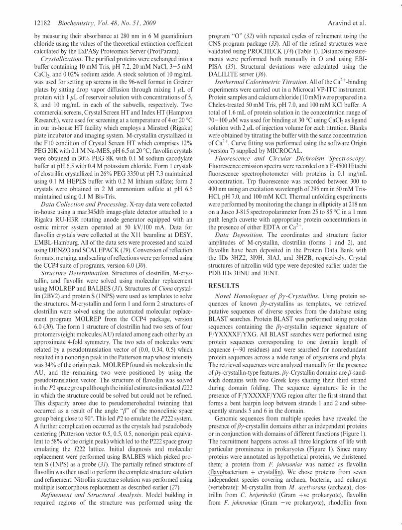
Novel Homologues of βγ-Crystallins. Using protein se-quences of known βγ-crystallins as templates, we retrievedputative sequences of diverse species from the database usingBLAST searches. Protein BLAST was performed using proteinsequences containing the βγ-crystallin sequence signature ofF/YXXXXF/YXG. All BLAST searches were performed usingprotein sequences corresponding to one domain length ofsequence (∼90 residues) and were searched for nonredundantprotein sequences across a wide range of organisms and phyla.The retrieved sequences were analyzed manually for the presenceof βγ-crystallin-type features. βγ-Crystallin domains are β-sand-wich domains with two Greek keys sharing their third strandduring domain folding. The sequence signatures lie in thepresence of F/YXXXXF/YXG region after the first strand thatforms a bent hairpin loop between strands 1 and 2 and subse-quently strands 5 and 6 in the domain.
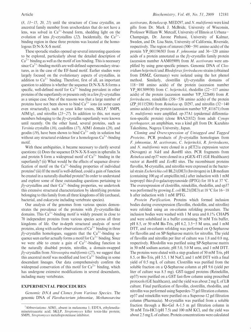
Genomic sequences from multiple species have revealed thepresence of βγ-crystallin domains either as independent proteinsor in conjunction with domains of different functions (Figure 1).The recruitment happens across all three kingdoms of life withparticular prominence in prokaryotes (Figure 1). Since manyproteins were annotated as hypothetical proteins, we christenedthem; a protein from F. johnsoniae was named as flavollin(flavobacterium þ crystallin). We chose proteins from sevenindependent species covering archaea, bacteria, and eukarya(vertebrate): M-crystallin from M. acetivorans (archaea), clos-trillin from C. beijerinckii (Gram þve prokaryote), flavollinfrom F. johnsoniae (Gram -ve prokaryote), rhodollin from
Article Biochemistry, Vol. 48, No. 51, 2009 12183
R. ferrireducens (Gram -ve prokaryote), reinekllin from Reine-kea sp. (Gram -ve prokaryote), nitrollin from N. multiformis(ammonia-oxidizing soil bacterium), and ep37 from C. pyrroga-ster (amphibian-vertebrate). A list of these proteins with theirsequence alignment with several other new members is shown inFigure 2. As seen in Figure 1, the βγ-crystallin domain in all ofthe proteins, except in nitrollin and M-crystallin, is a part of abigger protein. In such cases, only the βγ-crystallin domain wasselected for cloning and overexpression to avoid any ambiguitywhich may arise due to the role of the noncrystallin domainin Ca2þ binding. Such extensive recruitment as modules inproteins of varied functions clearly indicates that the domainsof βγ-crystallin have universal roles in the functioning of proteinsfrom multiple species.βγ-Crystallin Domain Structure: Fold and Novelties. In
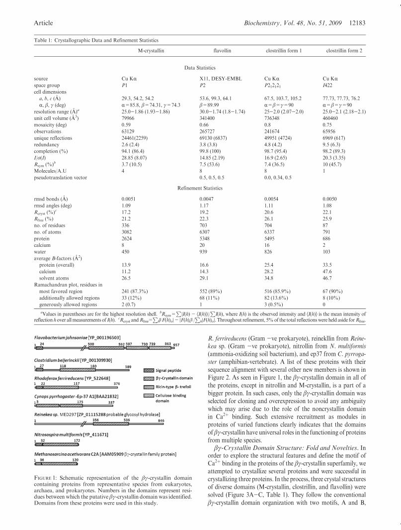
order to explore the structural features and define the motif ofCa2þ binding in the proteins of the βγ-crystallin superfamily, weattempted to crystallize several proteins and were successful incrystallizing three proteins. In the process, three crystal structuresof diverse domains (M-crystallin, clostrillin, and flavollin) weresolved (Figure 3A-C, Table 1). They follow the conventionalβγ-crystallin domain organization with two motifs, A and B,
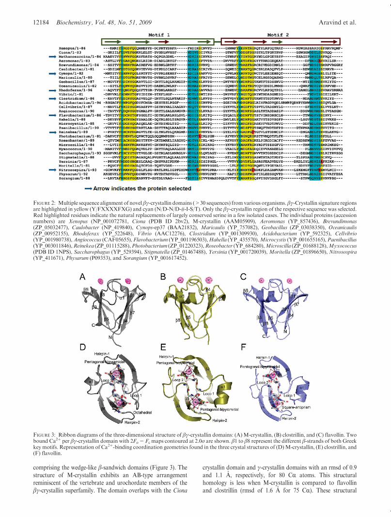
Table 1: Crystallographic Data and Refinement Statistics
M-crystallin flavollin clostrillin form 1 clostrillin form 2
Data Statistics
source Cu KR X11, DESY-EMBL Cu KR Cu KRspace group P1 P2 P212121 I422
cell dimensions
a, b, c (A) 29.3, 54.2, 54.2 53.6, 99.3, 64.1 67.5, 103.7, 105.2 77.73, 77.73, 76.2
R, β, γ (deg) R=85.8, β=74.31, γ=74.3 β=89.99 R=β=γ=90 R=β=γ=90
resolution range (A)a 25.0-1.86 (1.93-1.86) 30.0-1.74 (1.8-1.74) 25-2.0 (2.07-2.0) 25.0-2.1 (2.18-2.1)
unit cell volume (A3) 79966 341400 736348 460460
mosaicity (deg) 0.59 0.66 0.8 0.75
observations 63129 265727 241674 65956
unique reflections 24461(2259) 69130 (6837) 49951 (4724) 6969 (617)
redundancy 2.6 (2.4) 3.8 (3.8) 4.8 (4.2) 9.5 (6.3)
completion (%) 94.1 (86.4) 99.8 (100) 98.7 (95.4) 98.2 (89.3)
I/σ(I) 28.85 (8.07) 14.85 (2.19) 16.9 (2.65) 20.3 (3.35)
Rsym (%)b 3.7 (10.5) 7.5 (53.6) 7.4 (36.5) 10 (45.7)
Molecules/A.U 4 8 8 1
pseudotranslation vector 0.5, 0.5, 0.5 0.0, 0.34, 0.5
Refinement Statistics
rmsd bonds (A) 0.0051 0.0047 0.0054 0.0050
rmsd angles (deg) 1.09 1.17 1.11 1.08
Rcryst (%)c 17.2 19.2 20.6 22.1
Rfree (%) 21.2 22.3 26.1 25.9
no. of residues 336 703 704 87
no. of atoms 3082 6307 6337 791
protein 2624 5348 5495 686
calcium 8 20 16 2
water 450 939 826 103
average B-factors (A2)
protein (overall) 13.9 16.6 25.4 33.5
calcium 11.2 14.3 28.2 47.6
solvent atoms 26.5 29.1 34.8 46.7
Ramachandran plot, residues in
most favored region 241 (87.3%) 552 (89%) 516 (85.9%) 67 (90%)
additionally allowed regions 33 (12%) 68 (11%) 82 (13.6%) 8 (10%)
generously allowed regions 2 (0.7) 1 3 (0.5%) 0
aValues in parentheses are for the highest resolution shell. bRsym=P
|I(h) - ÆI(h)æ|/P
I(h), where I(h) is the observed intensity and ÆI(h)æ is the mean intensity ofreflection hover allmeasurements of I(h). cRcryst andRfree=
Ph )F(h)o|- |F(h)c )/
Ph|F(h)o|. Throughout refinement, 5%of the total reflectionswere held aside forRfree.
FIGURE 1: Schematic representation of the βγ-crystallin domaincontaining proteins from representative species from eukaryotes,archaea, and prokaryotes. Numbers in the domains represent resi-dues between which the putative βγ-crystallin domain was identified.Domains from these proteins were used in this study.
12184 Biochemistry, Vol. 48, No. 51, 2009 Aravind et al.
comprising the wedge-like β-sandwich domains (Figure 3). Thestructure of M-crystallin exhibits an AB-type arrangementreminiscent of the vertebrate and urochordate members of theβγ-crystallin superfamily. The domain overlaps with the Ciona
crystallin domain and γ-crystallin domains with an rmsd of 0.9and 1.1 A, respectively, for 80 CR atoms. This structuralhomology is less when M-crystallin is compared to flavollinand clostrillin (rmsd of 1.6 A for 75 CR). These structural
FIGURE 2: Multiple sequence alignment of novel βγ-crystallin domains (>30 sequences) fromvarious organisms. βγ-Crystallin signature regionsare highlighted in yellow (Y/FXXXXFXG) and cyan (N/D-N/D-#-I-S/T). Only the βγ-crystallin region of the respective sequence was selected.Red highlighted residues indicate the natural replacements of largely conserved serine in a few isolated cases. The individual proteins (accessionnumbers) are Xenopus (NP_001072781, Ciona (PDB ID 2bv2), M-crystallin (AAM05909), Aeromonas (YP_857436), Brevundimonas(ZP_05032477), Caulobacter (NP_419840), Cynops-ep37 (BAA21832), Maricaulis (YP_757082), Geobacillus (ZP_03038350), Oceanicaulis(ZP_00952155), Rhodoferax (YP_522648), Vibrio (AAC12276), Clostridium (YP_001309930), Acidobacterium (YP_592325), Cellvibrio(YP_001980738),Angiococcus (CAF05655), Flavobacterium (YP_001196503),Hahella (YP_435570),Microcystis (YP_001655165),Paenibacillus(YP_003011846),Reinekea (ZP_01115288),Photobacterium (ZP_01220323),Roseobacter (YP_684280),Microscilla (ZP_01688128),Myxococcus(PDB ID 1NPS), Saccharophagus (YP_529394), Stigmatella (ZP_01467488), Yersinia (YP_001720039), Moritella (ZP_01896650), Nitrosospira(YP_411671), Physarum (P09353), and Sorangium (YP_001617452).
FIGURE 3: Ribbon diagrams of the three-dimensional structure of βγ-crystallin domains: (A)M-crystallin, (B) clostrillin, and (C) flavollin. Twobound Ca2þ per βγ-crystallin domain with 2Fo- Fc maps contoured at 2.0σ are shown. β1 to β8 represent the different β-strands of both Greekkey motifs. Representation of Ca2þ-binding coordination geometries found in the three crystal structures of (D)M-crystallin, (E) clostrillin, and(F) flavollin.
Article Biochemistry, Vol. 48, No. 51, 2009 12185
differences are a result of the reversed arrangement of Greek keymotifs found in prokaryotes (BA type) as compared to archaealand vertebrate crystallins (AB type). Flavollin and clostrillinoverlap with the protein S (1NPS) structure with an rmsd of 1.0and 0.8 A, respectively, for 86 CR atoms.
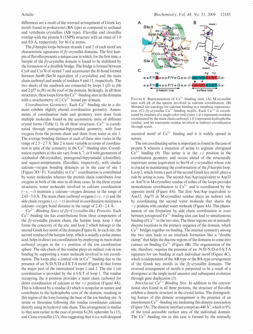
The β-hairpin loops between strands 1 and 2 of each motif arecharacteristic signatures of βγ-crystallin domains. The first hair-pin of flavollin presents a unique case inwhich, for the first time, ahairpin of the βγ-crystallin domain is found to be stabilized bythe formation of a disulfide bridge. The bridge is formed betweenCys9 and Cys70 of strand 7 and accentuates the H-bond formedbetween Ser40 (Ser34 equivalent of γ-crystallin) and the mainchain carbonyl and amide of residues 8 and 11, respectively. Thetwo sheets of the sandwich are connected by loops 1 (β3 to β4)and 2 (β7 to β8) on the roof of the domain. Strikingly, in all threestructures, these loops form theCa2þ-binding sites in the domainswith a stoichiometry of 2 Ca2þ bound per domain.Coordination Geometry. Each Ca2þ-binding site in a do-
main exhibits slightly altered coordination geometry. Assess-ments of coordination radii and geometry were done frommultiple molecules found in the asymmetric units of differentcrystal forms (Table 1). In all three structures, Ca2þ is coordi-nated through pentagonal-bipyramidal geometry, with fouroxygens from the protein chain and three from water at site 1.The average bonding distance at each of these sites varies in therange of 2.5-2.7 A. Site 2 is more variable in terms of coordina-tion in spite of the symmetry in the Ca2þ-binding sites. Coordi-nation numbers at this site vary from 6, 7, or 8 with geometries ofoctahedral (M-crystallin), pentagonal-bipyramidal (clostrillin),and square-antiprismatic (flavollin), respectively, with similarcalcium-oxygen bonding distances as in the case of site 1(Figure 3D-F). Variability in Ca2þ coordination is contributedby water molecules whereas the protein chain contributes fouroxygens at both of the sites in all three cases. In the three crystalstructures, water molecules involved in calcium coordination(-y, -z) maintain a calcium-oxygen distance in the range of2.65-3.0 A. The main chain carbonyl oxygens (þx, þy) and theside chain oxygens (þz,-x) involved in coordination maintain acalcium-oxygen bond distance in the range of 2.45-2.8 A.Ca2þ-Binding Sites of βγ-Crystallin-like Proteins. Each
Ca2þ-binding site has contributions from three components ofthe βγ-crystallin protein chain, the hairpin loop, loop 1 thatforms the concavity of the site, and loop 2 which belongs to thesecondGreek keymotif of the domain (Figure 4). In each site, thesecond residue of the hairpin loop, which is usually a polar aminoacid, helps in direct ion coordination by employing its main chaincarbonyl oxygen in the þx position of the ion coordinationsphere. The side chain of this residue plays an indirect role in ionbinding by supporting a water molecule involved in ion coordi-nation. The loops play a central role in Ca2þ binding due to thepresence of an N/D-N/D-#-I-S/T-S motif (Figure 4) that formsthe major part of the intercalated loops 1 and 2. The site 1 ioncoordination is provided by the #-I-S/T of loop 1. The residueoccupying the # position provides a main chain carbonyl fordirect coordination of calcium at the þy position (Figure 4A).This is followed by a residue (I) which is nonpolar in nature andcontributes to the hydrophobic core of the domain, resulting inthis region of the loop forming the base of the ion binding site. Aserine or threonine following this residue coordinates calciumdirectly using its hydroxyl side chain. This arrangement is similarto that seen earlier in the case of protein S (20), spherulin 3a (15),andCiona crystallin (22), thus suggesting that it is a well-designed
ancestral motif of Ca2þ binding and it is widely spread innature.
The ion coordinating serine is important as found in the case ofprotein S wherein a mutation of serine to arginine abrogatedCa2þ binding (9). This serine is at the þz position in thecoordination geometry and occurs ahead of the structurallyimportant serine (equivalent to Ser34 of γ-crystallin) whose roleis crucial in maintaining the conformation of the β-hairpin loop.Loop 2,which forms a part of the secondGreekkeymotif, plays arole by acting in trans. The secondAsn/Asp (equivalent to Asp33or Asn76 in M-crystallin) residue of either of the loops providesmonodentate coordination to Ca2þ and is coordinated by theopposite motif (Figure 4A). The first Asn/Asp (equivalent toAsn32, Asp75 in M-crystallin) residue plays an indirect roleby coordinating the second water molecule that shares the-y position with another water molecule (Figure 4A). This pheno-menon of site formation by side chain coordination in transbetween juxtaposed Ca2þ-binding sites can lead to simultaneousbinding ofCa2þ to the two sites. The three regions are atmutuallydiscrete locations in the primary sequence of the domain, whichCa2þ bridges together on binding. The internal symmetry amongthe two sites leads to an interlock formation like a “doubleclamp” that helps the discrete regions of the domains to come intocontact on binding Ca2þ (Figure 4B). The organization of thesite, therefore, requires the presence of an -N/D-N/D-#-I-S/T-Ssignature for ion binding at each individual motif (Figure 4C),which is independent of theAB-type or the BA-type arrangementof the Greek key motifs in the βγ-crystallin domains. Thereversed arrangement of motifs is purported to be a result of adivergence at the single motif ancestor and subsequent evolutionthrough gene duplication (5).Interfacial Ca2þ Binding Site. In addition to the conven-
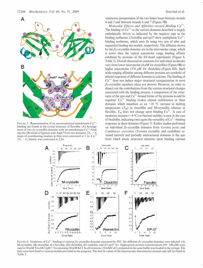
tional sites found in all three proteins, the structure of flavollinexhibits a dimeric structure in the crystal lattice. The distinguish-ing feature of this dimeric arrangement is the presence of aninterdomainCa2þ-binding sitemediating this dimeric association(Figure 5A). The dimeric interface comprises 440 A2, which is 9%of the total accessible surface area of the individual domain.The Ca2þ-binding site in this case is formed by the mutually
FIGURE 4: Representation of Ca2þ-binding sites: (A) M-crystallinseen with all of the species involved in calcium coordination. (B)Minimal site topology for calcium binding in a simplistic representa-tion. (C) βγ-crystallin Ca2þ-binding motifs. Each Ca2þ is coordi-nated by residues of a single color (red/cyan). (-#-) represents residuescoordinated by themain chain carbonyl. (-I-) represents hydrophobicresidue, and (•) represents residue involved in indirect coordinationthrough water.
12186 Biochemistry, Vol. 48, No. 51, 2009 Aravind et al.
symmetric juxtaposition of the two linker loops between strands4 and 5 and between strands 6 and 7 (Figure 5B).Molecular Effects and Affinities toward Binding Ca2þ.
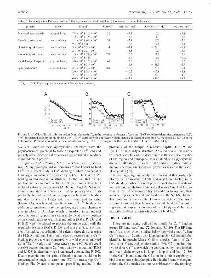
The binding of Ca2þ to the various domains described is largelyenthalpically driven as indicated by the negative sign in thebinding isotherms. Clostrillin and ep37 show multiphasic Ca2þ-binding isotherms, which were fit using two sets of sites andsequential binding site models, respectively. The affinities shownby the βγ-crystallin domains are in the micromolar range, whichis lower than the typical nanomolar range binding affinityexhibited by proteins of the EF-hand superfamily (Figure 6,Table 2). Overall dissociation constants for individual moleculesvary from lower micromolar (4 μM for clostrillin) (Figure 6B) tohigher micromolar (370 μM for rhodollin) (Figure 6D). Suchwide-ranging affinities among different proteins are symbolic ofaltered responses of different domains to calcium. The binding ofCa2þ does not induce major structural reorganization in mostβγ-crystallin members (data not shown). However, in order todissect out the contributions from the various structural changesassociated with the binding process, a comparison of the struc-tures of the apo and Ca2þ-bound forms of the proteins would berequired. Ca2þ binding evokes altered stabilization in thesedomains which manifests as an ∼18 �C increase in meltingtemperature (TM) in clostrillin and M-crystallin whereas inflavollin, TM does not change upon binding Ca2þ. A case ofmoderate increase (∼4 �C) in thermal stability is seen in the caseof rhodollin, indicating once again the versatility of Ca2þ-bindingresponses in these domains (Figure 7). Earlier studies performedon individual βγ-crystallin domains from Yersinia pestis andCaulobacter crescentus (Yersinia crystallin and caulollins) re-vealed natively and partially unstructured domains in the apoform which attain structural elements upon binding calcium
FIGURE 5: Representation of an unconventional interdomain Ca2þ-binding site found in the crystal structure of flavollin. (A) Arrange-ment of two βγ-crystallin domains with an interdomain Ca2þ-bind-ing site; (B)mode of ligationwithAsp67 from two domains. 2Fo- Fc
maps of coordinating residues in blue were contoured at 1.2σ. Ca2þ
2Fo - Fc density was contoured at 2.0σ.
FIGURE 6: Isotherms of Ca2þ binding to various βγ-crystallin domains measured by ITC. Six different βγ-crystallin domains were selected: (A)M-crystallin, (B) clostrillin, (C) flavollin, (D) rhodollin, (E) reinkllin, and (F) ep37 A1. Appropriate protein concentrations (50-100 μM) wereused in50mMTris-HCl (pH7.5) containing 50mMKCl. Inall titrations, 10mMCaCl2 prepared in the samebufferwas loaded in the syringe.Thedata were best fitted to various models provided in the program. The best fit values of the macroscopic dissociation constant andΔH are listed inTable 2.
Article Biochemistry, Vol. 48, No. 51, 2009 12187
(16, 17). Some of these βγ-crystallins, therefore, have thephysicochemical potential to sense or sequester Ca2þ ions andpossibly affect biochemical processes when recruited as modulesin multidomain proteins.Impaired Ca2þ-Binding Sites and Their Gain of Func-
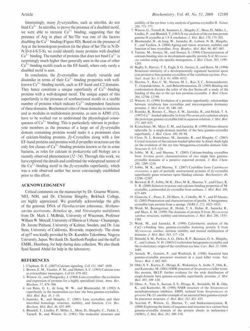
tion. Many βγ-crystallin-like domains are not known to bindCa2þ. In a recent study, a Ca2þ-binding disabled βγ-crystallinhomologue, nitrollin, was reported by us (27). The loss of Ca2þ
binding in this domain is attributed to the fact that the þzposition serines in both of the Greek key motifs have beenreplaced naturally by arginines (Arg88 and Arg133). Serine toarginine mutation is drastic as it alters polarity due to itspositively charged guanidinium group and volume of the bindingsite due to a much longer side chain compared to serine(Figure 8A), which would result in loss of Ca2þ binding. Inaddition to mutations in serine residues, site 2 has a phenylala-nine in place of Asn/Asp, which is involved in an indirectcoordination by supporting a water molecule at the -y positionof the coordination sphere. Three mutations (R88S, R133S, andF129D) were introduced to provide the amino acids with therequired side chains (R88S, R133S) and thus created an environ-ment for indirect coordination of calcium through water usingthe F129D mutation. This mutant shows a recovery in its Ca2þ-binding properties when compared with wild type as analyzedusing 45Ca2þ overlay and fluorescence (Figure 8C,D). We couldobserve weaker binding to Ca2þ only with two mutations (R88Sand R133S) in nitrollin, which is enhanced upon adding F129D.Due to precipitation, this gain-of-function mutant could not beconcentrated enough to carry out ITC for measuring Ca2þ
binding. Phe129 acts a nonpolar space-filling residue in the
proximity of the hairpin 2 residues Asp102, Glu109, andLys111 in the wild-type structure. An alteration in this residueto aspartate could lead to a disturbance in the local electrostaticsof the region and subsequent loss in stability. In βγ-crystallindomains, alterations of some of the surface residues result inmarked alterations in biophysical properties as seen in the case ofγC-crystallin (37).
Interestingly, arginine or glycine is present at this position (inplace of Ser, equivalent to Arg88 and Arg133 in nitrollin) in theCa2þ-binding motifs of several proteins, including in lens β- andγ-crystallins, mainly from vertebrates (Figures 2 and 8B), leadingto impaired Ca2þ-binding ability. In addition to arginine, thereare other replacements andmodifications in the N/D-N/D-#-I-S/T-S motif or in the vicinity. However, a detailed analysis isrequired to assess if these homologues would bind Ca2þ or not. Itsuggests that despite the presence of the fold, βγ-crystallins havenaturally disabled variants which do not bind Ca2þ.
DISCUSSION
There are not many well-defined motifs for Ca2þ binding,except EF-hand motif and C2 domains (38, 39). The EF-handmotif is a most widely studied helix-loop-helix motif whereCa2þ binds to a 12 amino acid loop region (38). C2 domains firstidentified in protein kinase C form another family of Ca2þ
sensors of β-sandwich conformation (39). C2 domains bindtwo to three Ca2þ ions which are coordinated by the side chainand main chain oxygens in loop 1, loop 3, and loop 2 (39).In the Ca2þ-bound state, the C2 domains attain a capability tobind tomembrane phospholipids. Besides the β-sandwich organi-zation, the C2 domains bear no resemblance with the topology,
Table 2: Thermodynamic Parameters of Ca2þ Binding to Various βγ-Crystallins by Isothermal Titration Calorimetry
proteins model K (mol-1) Kd (μM)a ΔH (kcal 3mol-1) ΔS (cal 3mol-13K
-1) ΔG (kcal 3mol-1)
M-crystallin (archaeal) sequential sites 7.8 � 104 ( 1.2 � 104 32 -5.3 5.0 -6.8
1.2 � 104 ( 4.9 � 102 -5.2 1.3 -5.6
flavollin (prokaryote) two set of sites 1.1 � 105 ( 6.8 � 104 33 -2.1 16.0 -7.0
9 � 103 ( 421 -6.3 -2.8 -5.4
clostrillin (prokaryote) two set of sites 3 � 104 ( 2.3 � 103 4 -43.4 -123 -6.1
2 � 106 ( 2.5 � 105 -8.5 0.77 -8.7
rhodollin (prokaryote) two set of sites 1.3 � 104 ( 9.8 � 103 370 -0.5 17.5 -5.8
5.5 � 102 ( 453 1.9 6.3 -0.02
reinikillin (prokaryote) sequential sites 5.8 � 103 ( 1.2 � 103 40 -2.0 10.5 -5.2
1.1 � 105 ( 3 � 104 1.8 -141.5 44.7
ep37 (vertebrate) sequential sites 4.1 � 104 ( 3 � 103 260 -0.2 20.4 -6.4
2.9 � 103 ( 2.3 � 102 -10.2 -17.8 -4.8
2.1 � 103 ( 1.5 � 102 6.7 37.3 -4.6
0.9 � 103 ( 0.7 � 102 -14.9 -35.5 -4.1
aKd = 1/√K1K2 (Kd represents the overall dissociation constant).
FIGURE 7: (A) Flavollinwith almost insignificant change inTm in the presence or absence of calcium. (B)Rhodollinwithmoderate increase (ΔTm
4 �C) in thermal stability upon binding Ca2þ. (C) Clostrillin with significantly high increase in thermal stability (Tm increased by 18 �C) in theholoprotein. Proteins were used in the concentration range of 0.1-0.2 mg/mL with either 0.5 mM EDTA or 1 mM CaCl2.

12188 Biochemistry, Vol. 48, No. 51, 2009 Aravind et al.
structural signatures, or the Ca2þ-binding motif of βγ-crystal-lins (40), making them a distinct set of Ca2þ-binding proteins.
The βγ-crystallin domains have varied affinities in the micro-molar range (4-250 μM) as it is common for extracellular Ca2þ-binding proteins, whereas calcium sensors of the EF-hand familyare generally high-affinity (lower micromolar to nanomolarrange) Ca2þ-binding proteins. In some cases of βγ-crystallins,particularly vertebrate homologues (such as ep37) (41), theaffinity for Ca2þ binding is much lower (Table 2). These proteinsdo not undergo major structural changes upon binding Ca2þ buthave a reduced hydrodynamic size and generally attain high tomoderate structural stabilization.
In human lens, calcium homeostasis is implicated in lenstransparency and physiology, and a disturbance in ionic home-ostasis is implicated in cataract (42). Though total Ca2þ con-centration in a human lens is up to 100 μM, and more in the caseof many cataractous lenses, only a fewmicromolar is in free formwhile the rest is in bound form (42-44). Though variousmechanisms for Ca2þ signaling and homeostasis in the lens havebeen proposed (43, 45), there are no known Ca2þ-bindingproteins. One hypothesis could be that, in an eye lens, β- andγ-crystallins appropriately suit to this function owing to their lowaffinity for Ca2þ binding complemented by their abundantpresence, although this is yet to be ascertained (46, 47). Homo-logues of β- and γ-crystallins, such as protein S fromMyxococcusxanthus and spherulin 3a from P. polycephalum, are found insome lower eukaryotic and prokaryotic species where theyhelp the organism tide over periods of stressful environment inaCa2þ-dependentmanner (10, 13). The crystal structures of threedifferent homologues of βγ-crystallins along with the threestructures (protein S, spherulin 3a, and Ciona βγ-crystallin)described earlier demonstrate the uniformity of the Ca2þ-bindingsite thereby forming a well-designed motif of Ca2þ bindingprevalent inβγ-crystallins. This is further corroborated by studieson several other proteins (ep37 from cynops, an eukarya,
rhodollin from R. ferrireducens, reinekllin from Reinekea) thathave been shown to bind Ca2þ by ITC (Figure 6). All of theseproteins have essential residues conserved at theN/D-N/D-#-I-S/T-S motif and thus would ligate Ca2þ similarly.
βγ-Crystallins, which are formed by an ancestral duplicationevent of a Greek key motif (48), have until recently been seen asstructural components of vertebrate eye lens (8). The β- andγ-crystallins themselves have low butmeasurable affinity of Ca2þ
binding owing to themodified sequence of the Ca2þ-binding sitesin some Greek key motifs (46, 47). However, one of the fourGreek key motifs (second Greek key motif in βB2-crystallin)retains canonical residues (91-TDSLSS-96) capable of bindingCa2þ, which could be a reason for observing this binding(Figure 8B). In urochordate Ciona intestinalis, a βγ-crystallinprotein is expressed in tissues (palps and otoliths) related to aprimitive light sensing system (22). The crystal structure of thisprotein (PDB ID 2BV2) is very similar to lens βγ-crystallindomains, except for the presence of canonicalN/D-N/D-#-I-S/T-S motif with bound Ca2þ (22). Urochordates do not have lenses,and thus Ciona βγ-crystallin could be considered as a closeancestor of vertebrate lens βγ-crystallins (22, 49). The presence ofthis motif in archaeal homologues, as well as in Ciona crystallin,suggests that this Ca2þ-binding motif was present in ancestors,which was modified further during evolution to have no bindingin many cases or weaker binding in lens βγ-crystallins.
Until recently, the superfamily predominantly consisted ofvertebrate lens β- and γ-crystallins and their isoforms. Recentdevelopments have yielded new insights into the βγ-crystallinmembers from nonlens tissues such as the cardiotoxic skinsecretions of Bombina maxima and a highly complex six-domain-containing protein “absent in melanoma 1” (AIM1) (50, 51).Genome sequences from multiple species are revealing newprotein sequences in which βγ-crystallin-like domains with well-conservedCa2þ-binding sites are found to be recruited as a part ofmultidomain proteins possibly to perform Ca2þ-dependent roles.
FIGURE 8: (A) Overlap of the nitrollin structure with clostrillin. Ca2þ density (2Fo - Fc) was contoured at 2.0σ. (B) Sequence alignment of fourβγ-crystallins and lensβB2-crystallin.m1andm2representmotifs 1 and2, respectively, in a domain.Nitrollin doesnot bindcalciumdue tonaturalmutation of Ser with Arg (depicted by arrows) and is selected as an example. The functional or prototype Ca2þ-bindingmotif of βB2-crystallin isshown. m1 and m2 represent the two motifs of individual domains. (C) Intrinsic Trp fluorescence titration of wild type (upper panel) and thenitrollin mutant (lower panel) with calcium. (D) Calcium-binding assay by 45Ca by the membrane overlay method. Clostrillin was used asa positive control. As seen on the membrane, the nitrollin triple mutant shows bound calcium.
Article Biochemistry, Vol. 48, No. 51, 2009 12189
Interestingly, many βγ-crystallins, such as nitrollin, do notbind Ca2þ. In nitrollin, to prove the presence of a disabled motif,we were able to recreate Ca2þ binding, suggesting that thepresence of Arg in place of Ser/Thr was one of the factorsdisabling the Ca2þ binding (Figure 8D). Based on the presence ofArg at the homologous position (in the place of Ser/Thr in N/D-N/D-#-I-S/T-S), we could identify many proteins with disabledCa2þ binding. The number of proteins that does not bind Ca2þ issurprisingly much higher than generally seen in the case of otherCa2þ-binding motifs (such as the EF-hand), where only rarely adisabled motif is seen.
In conclusion, the βγ-crystallins are clearly versatile anddissimilar in terms of their Ca2þ-binding properties with well-known Ca2þ-binding motifs, such as EF-hand and C2 domains.They hence constitute a unique superfamily of Ca2þ-bindingproteins with a well-designed motif. The unique aspect of thissuperfamily is the presence of nonfunctional motifs in a sizablenumber of proteins which indicate Ca2þ-independent functionsof these domains. Biochemical roles of these domains in isolationand as modules in multidomain proteins, as seen in AIM1 (51),have to be worked out to understand the physiological conse-quences of Ca2þ binding. This is particularly relevant in prokar-yote members as the presence of a large set of βγ-crystallindomain containing proteins would make it a prominent classof calcium-binding proteins. βγ-Crystallins along with a fewEF-hand proteins and proteins withβ-propeller structures are theonly few classes of Ca2þ-binding proteins known so far in somebacteria, as roles for calcium in prokaryotic cell biology are arecently observed phenomenon (52-54). Through this work, wehave explored the details and confirmed the widespread nature ofthe Ca2þ-binding motif in the βγ-crystallin superfamily, whichwas a role observed earlier but never convincingly establishedprior to this effort.
ACKNOWLEDGMENT
Critical comments on the manuscript by Dr. GraemeWistow,NEI, NIH, and Dr. Christine Slingsby, Birkbeck College,are highly appreciated. We gratefully acknowledge the giftsof the genomic DNA of Flavobacterium johnsoniae, Methano-sarcina acetivorans, Reinekea sp., and Nitrosospira multiformisfrom Dr. Mark J. McBride, University of Wisconsin, ProfessorWilliamW.Metcalf,University of Illinois atUrbana-Champaign,Dr. Jarone Pinhassi, University of Kalmar, Sweden, and Dr. LisaStein, University of California, Riverside, respectively. The cloneof ep37 was kindly provided by Dr. Kazuhito Takeshima, NagoyaUniversity, Japan.We thankDr. SanthoshPanjikar and the staff atEMBL, Hamburg, for help during data collection. We also thankSyed Sayeed Abdul for help in the laboratory.
REFERENCES
1. Clapham, D. E. (2007) Calcium signaling. Cell 131, 1047–1058.2. Brown, E. M., Vassilev, P. M., and Hebert, S. C. (1995) Calcium ions
as extracellular messengers. Cell 83, 679–682.3. Wistow, G., and Piatigorsky, J. (1988) Lens crystallins: the evolution
and expression of proteins for a highly specialized tissue. Annu. Rev.Biochem. 57, 479–504.
4. van Rens, G. L., de Jong, W. W., and Bloemendal, H. (1992) Asuperfamily in the mammalian eye lens: the beta/gamma-crystallins.Mol. Biol. Rep. 16, 1–10.
5. Jaenicke, R., and Slingsby, C. (2001) Lens crystallins and theirmicrobial homologs: structure, stability, and function. Crit. Rev.Biochem. Mol. Biol. 36, 435–499.
6. Blundell, T., Lindley, P., Miller, L., Moss, D., Slingsby, C., Tickle, I.,Turnell, B., and Wistow, G. (1981) The molecular structure and
stability of the eye lens: x-ray analysis of gamma-crystallin II. Nature289, 771–777.
7. Wistow,G., Turnell, B., Summers, L., Slingsby,C.,Moss,D.,Miller, L.,Lindley, P., andBlundell, T. (1983)X-ray analysis of the eye lens proteingamma-II crystallin at 1.9 A resolution. J. Mol. Biol. 170, 175–202.
8. Bloemendal, H., de Jong, W., Jaenicke, R., Lubsen, N. H., Slingsby,C., and Tardieu, A. (2004) Ageing and vision: structure, stability andfunction of lens crystallins. Prog. Biophys. Mol. Biol. 86, 407–485.
9. Teintze, M., Inouye, M., and Inouye, S. (1988) Characterization ofcalcium-binding sites in development-specific protein S of Myxococ-cus xanthus using site-specific mutagenesis. J. Biol. Chem. 263, 1199–1203.
10. Bagby, S., Harvey, T. S., Eagle, S.G., Inouye, S., and Ikura,M. (1994)Structural similarity of a developmentally regulated bacterial sporecoat protein to beta gamma-crystallins of the vertebrate eye lens.Proc.Natl. Acad. Sci. U.S.A. 91, 4308–4312.
11. Sharma, Y., Rao, C. M., Narasu, M. L., Rao, S. C., Somasundaram,T., Gopalakrishna, A., and Balasubramanian, D. (1989) Binding siteconformation dictates the color of the dye Stains-all: a study of thebinding of this dye to the eye lens proteins crystallin. J. Biol. Chem.264, 12794–12799.
12. Wistow, G. (1990) Evolution of a protein superfamily: relationshipsbetween vertebrate lens crystallins and microorganism dormancyproteins. J. Mol. Evol. 30, 140–145.
13. Rosinke, B., Renner, C., Mayr, E. M., Jaenicke, R., and Holak, T. A.(1997) Ca2þ-loaded spherulin 3a fromPhysarum polycephalum adoptsthe prototype gamma-crystallin fold in aqueous solution. J.Mol. Biol.271, 645–655.
14. Kretschmar,M.,Mayr, E.M., and Jaenicke, R. (1999) Homo-dimericspherulin 3a: a single-domain member of the beta gamma-crystallinsuperfamily. J. Biol. Chem. 380, 89–94.
15. Clout, N. J., Kretschmar, M., Jaenicke, R., and Slingsby, C. (2001)Crystal structure of the calcium-loaded spherulin 3a dimer sheds lighton the evolution of the eye lens betagamma-crystallin domain fold.Structure 9, 115–124.
16. Jobby, M. K., and Sharma, Y. (2005) Calcium-binding crystallinsfrom Yersinia pestis: characterization of two single beta gamma-crystallin domains of a putative exported protein. J. Biol. Chem.280, 1209–1216.
17. Jobby, M. K., and Sharma, Y. (2007) Caulollins from Caulobactercrescentus, a pair of partially unstructured proteins of βγ-crystallinsuperfamily gains structure upon binding calcium. Biochemistry 46,12298–12307.
18. Barnwal,R. P., Jobby,M.K.,Devi,M.K., Sharma,Y., andChary,K.V. R. (2009) Solution structure and calcium-binding properties of M-crystallin, a primordial βγ-crystallin from archaea. J. Mol. Biol. 386,675–689.
19. Giancola, C., Pizzo, E., Di Maro, A., Cubellis, M. V., and D’Alessio,G. (2005) Preparation and characterization of geodin. A betagamma-crystallin-type protein from a sponge. FEBS J. 272, 1023–1035.
20. Wenk, M., Baumgartner, R., Holak, T. A., Huber, R., Jaenicke, R.,and Mayr, E. M. (1999) The domains of protein S from Myxococcusxanthus: structure, stability and interactions. J. Mol. Biol. 286, 1533–1545.
21. Wenk, M., and Jaenicke, R. (1999) Calorimetric analysis of theCa(2þ)-binding beta gamma-crystallin homolog protein S fromMyxococcus xanthus: intrinsic stability and mutual stabilization ofdomains. J. Mol. Biol. 293, 117–124.
22. Shimeld, S.M., Purkiss, A.G., Dirks, R. P., Bateman, O.A., Slingsby,C., and Lubsen, N. H. (2005) Urochordate betagamma-crystallin andthe evolutionary origin of the vertebrate eye lens.Curr. Biol. 15, 1684–1689.
23. Antuch, W., Guntert, P., and Wuthrich, K. (1996) Ancestral betagamma-crystallin precursor structure in a yeast killer toxin. Nat.Struct. Biol. 3, 662–665.
24. Ohki, S. Y.,Kariya, E.,Hiraga,K.,Wakamiya,A., Isobe, T., Oda,K.,andKainosho,M. (2001)NMRstructure ofStreptomyces killer toxin-like protein, SKLP: further evidence for the wide distribution ofsingle-domain beta gamma-crystallin superfamily proteins. J. Mol.Biol. 305, 109–120.
25. Ohno, A., Tate, S., Seeram, S. S., Hiraga, K., Swindells, M. B., Oda,K., and Kainosho, M. (1998) NMR structure of the Streptomycesmetalloproteinase inhibitor, SMPI, isolated from Streptomyces ni-grescensTK-23: another example of an ancestral beta gamma-crystal-lin precursor structure. J. Mol. Biol. 282, 421–433.
26. Aravind, P., Wistow, G., Sharma, Y., and Sankaranarayanan, R.(2008) Exploring the limits of sequence and structure in a variant betagamma-crystallin domain of the protein absent in melanoma-1(AIM1). J. Mol. Biol. 381, 509–518.
12190 Biochemistry, Vol. 48, No. 51, 2009 Aravind et al.
27. Aravind, P., Suman, S. K., Mishra, A., Sharma, Y., and Sankaranar-ayanan, R. (2009) Three dimensional domain swapping in nitrollin, asingle-domain beta gamma-crystallin from Nitrosospira multiformis,controls protein conformation and stability but not dimerization. J.Mol. Biol. 385, 163–177.
28. Rajini, B., Graham, C., Wistow, G., and Sharma, Y. (2003) Stability,homodimerization, and calcium-binding properties of a single, var-iant betagamma-crystallin domain of the protein absent in melanoma1 (AIM1). Biochemistry 42, 4552–4559.
29. Otwinowski, Z., andMinor,W. (1997) Processing ofX-ray diffractiondata collected in oscillation mode. Methods Enzymol. 276, 307–326.
30. Collaborative Computational Project, Number 4 (1994) The CCP4suite: programs for protein crystallography.ActaCrystallogr., Sect. D50, 760–763.
31. Long, F., Vagin, A. A., Young, P., and Murshudov, G. N. (2008)BALBES: a molecular-replacement pipeline. Acta Crystallogr., Sect.D: Biol. Crystallogr. 64, 125–132.
32. Jones, T. A., Zou, J. Y., Cowan, S. W., and Kjeldgaard, M. (1991)Improved methods for building protein models in electron densitymaps and the location of errors in these models. Acta Crystallogr.,Sect. A: Found. Crystallogr. 47, 110–119.
33. Br€unger, A. T., Adams, P. D., Clore, G.M., DeLano,W. L., Gros, P.,Grosse-Kunstleve, R., Jiang, J. S., Kuszewski, J., Nilges, M., Pannu,N. S., Read, R. J., Rice, L. M., Simonson, T., and Warren, G. L.(1998) Crystallography and NMR System: a new software suite formacromolecular structure determination. Acta Crystallogr., Sect. D:Biol. Crystallogr. 54, 905–921.
34. Laskowski, R. A., MacArthur, M. W., Moss, D. S., and Thornton, J.M. (1993) PROCHECK: a program to check the stereochemicalquality of protein structures. J. Appl. Crystallogr. 26, 283–291.
35. Krissinel, E., and Henrick, K. (2007) Inference of macromolecularassemblies from crystalline state. J. Mol. Biol. 372, 774–797.
36. Holm, L., and Park, J. (2000) DaliLite workbench for proteinstructure comparison. Bioinformatics 16, 566–567.
37. Purkiss, A. G., Bateman, O.A.,Wyatt, K.,Wilmarth, P. A., David, L.L., Wistow, G. J., and Slingsby, C. (2007) Biophysical properties ofgammaC-crystallin in human and mouse eye lens: the role of molec-ular dipoles. J. Mol. Biol. 372, 205–222.
38. McPhalen, C. A., Strynadka, N. C. J., and James, M. N. G. (1991)Calcium-binding sites in proteins: a structural perspective. Adv.Protein Sci. 42, 77–144.
39. Rizo, J., and Sudhof, T. C. (1998) C2-domains, structure and functionof a universal Ca2þ-binding domain. J. Biol. Chem. 273, 15879–15882.
40. Nalefski, E. A., and Falke, J. J. (1996) The C2 domain calcium-binding motif: structural and functional diversity. Protein Sci. 5,2375–2390.
41. Ogawa, M., Takabatake, T., Takahashi, T. C., and Takeshima, K.(1997) Metamorphic change in EP37 expression: members of the βγ-crystallin superfamily in newt. Dev. Genes Evol. 206, 417–424.
42. Duncan, G., and Jacob, T. J. (1984) Calcium and the physiology ofcataract. CIBA Found. Symp. 106, 132–152.
43. Duncan, G., Williams, M. R., and Riach, R. A. (1994) Calcium, cellsignalling and cataract. Prog. Retinal Eye Res. 13, 623–652.
44. Tang,D., Borchman,D., Yappert,M.C., Vrensen,G. F., andRasi, V.(2003) Influence of age, diabetes, and cataract on calcium, lipid-calcium, and protein-calcium relationships in human lenses. Invest.Ophthalmol. Visual Sci. 44, 2059–2066.
45. Rhodes, J. D., and Sanderson, J. (2009) The mechanisms of calciumhomeostasis and signalling in the lens. Exp. Eye Res. 88, 226–234.
46. Rajini, B., Shridas, P., Sundari, C. S., Muralidhar, D., Chandani, S.,Thomas, F., and Sharma, Y. (2001) Calcium binding properties ofgamma-crystallin: calcium ion binds at the Greek key beta gamma-crystallin fold. J. Biol. Chem. 276, 38464–38471.
47. Jobby, M. K., and Sharma, Y. (2007) Calcium-binding to lens βB2-and βA3-crystallins suggests that all β-crystallins are calcium-bindingproteins. FEBS J. 274, 4135–4147.
48. D’Alessio, G. (2002) The evolution of monomeric and oligomericbetagamma-type crystallins. Facts and hypotheses. Eur. J. Biochem.269, 3122–3130.
49. Piatigorsky, J. (2006) Evolutionary genetics: seeing the light: the roleof inherited developmental cascades in the origins of vertebrate lensesand their crystallins. Heredity 96, 275–277.
50. Qian, J. Q., Liu, S. B., He, Y. Y., Lee, W. H., and Zhang, Y. (2008)Beta gamma-CAT, a non-lens betagamma-crystallin and trefoil factorcomplex from amphibian skin secretions, caused endothelium-depen-dent myocardial depression in isolated rabbit hearts.Toxicon 52, 285–292.
51. Ray, M. E., Wistow, G., Su, Y. A., Meltzer, P. S., and Trent, J. M.(1997) AIM1, a novel non-lens member of the betagamma-crystallinsuperfamily, is associated with the control of tumorigenicity in humanmalignant melanoma. Proc. Natl. Acad. Sci. U.S.A. 94, 3229–3234.
52. Michiels, J., Chuanwu, X., Verhaert, J., and Vanderleyden, J. (2002)The functions of Ca2þ in bacteria: a role for EF-hand proteins?TrendsMicrobiol. 10, 87–93.
53. Tanaka, Y., Morikawa, K., Ohki, Y., Yao, M., Tsumoto, K.,Watanabe, N., Ohta, T., and Tanaka, I. (2007) Structural andmutational analyses of Drp35 from Staphylococcus aureus: a possiblemechanism for its lactonase activity. J. Biol. Chem. 282, 5770–5780.
54. Chen, C. N., Chin, K. H., Wang, A. H., and Chou, S. H. (2008) Thefirst crystal structure of gluconolactonase important in the glucosesecondary metabolic pathways. J. Mol. Biol. 384, 604–614.
Copyright © 2022 FDOKUMEN