Ion transport in seminal and adventitious roots of cereals ...
) 63–74www.elsevier.com/locate/gene
Gene 375 (2006
Bovine seminal plasma proteins and their relatives: A new expandingsuperfamily in mammals
Jinjiang Fan a, Jasmine Lefebvre a,b, Puttaswamy Manjunath a,b,c,⁎
a Guy-Bernier Research Center, Maisonneuve-Rosemont Hospital, Montreal, Québec, Canada H1T 2M4b Department of Biochemistry, University of Montreal, Québec, Canada
c Department of Medicine, University of Montreal, Québec, Canada
Received 4 November 2005; received in revised form 10 February 2006; accepted 11 February 2006Available online 14 March 2006
Received by M. Batzer
Abstract
BSP proteins represent three major proteins of bovine seminal plasma: BSP-A1/-A2, -A3 and -30 kDa. The BSP protein signature ischaracterized by two tandemly repeated fibronectin type 2 (Fn2) domains. Although classical affinity chromatography and protein sequencing haveproven that the BSP protein homologs may be ubiquitous in mammals and functionally related to sperm capacitation, only the three bovine geneshave been reported thus far. In this study, we report three new BSP protein-related genes from bovine, as well as other BSP protein-related DNAsequences from human, chimpanzee, mouse, rat, dog, horse and rabbit. Analysis of the relationships between all Fn2 domain-containing proteinsrevealed that the Fn2 domains found in BSP-related proteins have special features that distinguish them from non-BSP-related proteins. Thesefeatures can be used to identify new BSP protein-related sequences. Further molecular evolutionary analysis of the BSP protein lineage revealed thatall BSP proteins and their related sequences can be grouped into three subfamilies: BSPH4, BSPH5 and BSPH6, which indicates that the BSPprotein family is much bigger than previously envisioned. More interestingly, the three BSP proteins in bovine within the BSPH4-subfamily wereshown to evolve rapidly. The ratio of nonsynonymous to synonymous substitutions was higher than 1. The analysis also indicated that the rate ofevolution was heterogeneous between the first and second Fn2 domains of the genes. These data may reflect that some amino acids in BSP proteinsare under a strong positive selection after gene duplication and that each BSP protein evolves rapidly, possibly to acquire new functions.© 2006 Elsevier B.V. All rights reserved.
Keywords: BSP proteins; Fibronectin type 2 domain; Phylogeny; Positive selection
1. Introduction
Bovine seminal plasma proteins (BSP-A1/-A2, -A3 and-30 kDa) and their homologs seem to be ubiquitous inmammals, as shown by classical affinity chromatography,protein sequencing, and radioimmunoassays (Leblond et al.,1993; Manjunath and Therien, 2002; Nauc and Manjunath,2000). Their crucial roles in bovine sperm capacitation and in
Abbreviations: BSP, bovine seminal plasma protein; dN, non-synonymousdivergence; dS, synonymous divergence; Fn2, fibronectin type 2; LRTs,Likelihood ratio tests; ML, Maximum likelihood; MP, Maximum parsimony;NJ, Neighbour-joining; PCR, polymerase chain reaction.⁎ Corresponding author. Centre de Recherche Guy-Bernier, Hôpital Maison-
neuve-Rosemont, 5415 Boul. de l'Assomption, Montréal, Québec, Canada, H1T2M4. Tel.: +1 514 252 3562; fax: +1 514 252 3430.
E-mail address: [email protected] (P. Manjunath).
0378-1119/$ - see front matter © 2006 Elsevier B.V. All rights reserved.doi:10.1016/j.gene.2006.02.025
cholesterol and phospholipid efflux from epididymal spermhave been established (Therien et al., 1997, 1998, 1999).Additionally, all three proteins share identical or similarbiochemical properties such as binding to gelatin, heparin,apolipoprotein A-I, glycosaminoglycans, choline phospholipidsand low-density lipoproteins (Chandonnet et al., 1990;Desnoyers and Manjunath, 1992; Manjunath et al., 1987,1989, 2002; Therien et al., 2005). Although many genomeshave been fully or almost completely sequenced hitherto, littleis known about the BSP-encoding genes, the relationshipsamong these genes and their precise roles in fertilization. Sincethe BSP protein signature is characterized by two tandemlyrepeated fibronectin type 2 (Fn2) domains, with a length ofapproximately 40 amino acids each, it is likely that we canrecognize new members and/or homologs of BSP proteinsthrough a simple comparison of the Fn2 domains. However,

64 J. Fan et al. / Gene 375 (2006) 63–74
tracing the origin of Fn2 domains and distinguishing betweenFn2 domains from the BSP protein family and those from othernon-BSP-related proteins, such as gelatinases and fibronectins,can pose a significant challenge. More than 10 different types ofnon-BSP-related proteins contain one to three Fn2 domains.Due to the fact that most reproductive proteins have been shownto evolve rapidly (Swanson and Vacquier, 2002), the compar-ison of sequence similarity alone is not enough to predict theirorigins and functions. Phylogenomics has been proposed as apowerful tool to tackle this problem (Eisen, 1998; Eisen andWu, 2002) and has actually been used for the identification ofseveral new gene families, such as DNA repair genes (Eisen andHanawalt, 1999), receptor-like proteins (Fritz-Laylin et al.,2005), meiotic genes (Ramesh et al., 2005), DsbB-like thiol-oxidoreductases (Raczko et al., 2005), potassium channel genes(Moulton et al., 2003) and many others (Eisen and Fraser, 2003;Sjolander, 2004).
Although BSP proteins have been extensively studied sincethey were identified in bovine seminal plasma some 20 yearsago (Esch et al., 1983; Manjunath, 1984), the only differencesthat have been demonstrated between the three BSP proteins aretheir relative abundance in bovine seminal plasma (approxi-mately 37% for BSP-A1/-A2, compared to 4–5% for BSP-A3and BSP-30 kDa) (Nauc and Manjunath, 2000), as well as somespecific features related to the stimulation of phospholipid andcholesterol efflux (Therien et al., 1998, 1999). Since all threeproteins stimulate in vitro capacitation to the same level undertested conditions (Therien et al., 1999), it is unclear what causesthe differences in the stimulation of phospholipid andcholesterol efflux. It is likely that these differences could berelated to other yet unrevealed biological functions, in additionto the stimulation of sperm capacitation.
Recent evolutionary analysis has shown that detectingsignals of natural selection is the prevailing approach tounderstand the functions of new genes (Yang, 2005). Naturalselection is defined as purifying, neutral or positive selectiondepending on the ratio of nonsynonymous (amino acidchanging) nucleotide substitutions to synonymous (aminoacid preserving) nucleotide substitutions (Ka/Ks or dN/dS) foreach codon in a given protein-coding DNA sequence alignment.Positive Darwinian selection is the most important driving forcein rapid evolution, with a ratio higher than 1 being the criterionfor positive selection. A positively selected amino acid site isone for which natural selection encourages the fixation ofnonsynonymous substitutions. The identification of suchpositively selected sites can be of biomedical importance; forinstance, an antigen with many positively selected sites mightbe an unsuitable vaccine candidate (de Oliveira et al., 2004). Inaddition, this approach has been used to predict the residues inglutathione transferase that are capable of driving functionaldiversification (Ivarsson et al., 2003) and to reveal thefunctional patches of primate TRIM5α in species-specificretroviral inhibition (Sawyer et al., 2005).
The functions of proteins under rapid evolution can also berevealed through comparative structural modeling, which hasbeen applied in the study of reproductive proteins in Drosophila(Mueller et al., 2004). Our research has been motivated by the
need to discover new BSP protein-related genes in severalrecently sequenced mammalian genomes, and to characterizetheir relationships and potential functions in reproduction.Given the crucial role of BSP proteins in bovine spermcapacitation and fertilization, it is of fundamental importance tocharacterize BSP-related genes and proteins from othermammals, especially humans, in order to better understandthe mechanisms underlying fertility.
2. Materials and methods
2.1. Genomic DNA sources
Bovine, mouse, rat, chimpanzee and human genomicsequences were retrieved from NCBI (http://www.ncbi.nlm.nih.gov), EMBL (http://www.ebi.ac.uk/embl) and relevantgenome sequencing projects. The sequences used for theanalysis in this study were based on the presence of one or tworepeats of Fn2 domains, each domain consisting of fourconserved cysteine residues, which form two disulfide bonds(Baker, 1985; Seidah et al., 1987). In addition, orthologousgenes containing Fn2 domains, representing different types ofenzymes/proteins, such as the 72 kDa type IV collagenase andmatrix metalloproteinase-9, were mainly selected from human,mouse and bovine. A total of 52 representative sequences wereselectively retained. Accession numbers for the genes used inthe following analysis are shown in Table 2, online.
2.2. Phylogenetic analyses
Protein and DNA sequences from GenBank (http://www.ncbi.nlm.nih.gov) were aligned using Clustal X (Thompson etal., 1997). Codon-based alignment was made manually with theintroduction of small indels and/or gaps to optimize thealignment. Unrooted neighbour-joining (NJ) trees and maxi-mum parsimony (MP) analyses were performed using theMEGA3 software with options of pairwise deletion and theDayhoff PAM matrix model (Kumar et al., 2004). Maximumlikelihood (ML) analyses were performed using Phylip 3.6 (thePhylogeny Inference Package) (Felsenstein, 2004). Bootstrapsupport values for NJ and MP trees were obtained from 1000replicates and support for ML trees was from 100 replicates.
2.3. Total RNA extraction, RT-PCR and DNA sequencing
Total RNA was extracted from tissues using the Trizolreagent (Invitrogen). 2 μg total RNA (except epididymiswhere 1.3 μg was used) was treated with DNase (NEBL) andsubjected to first-strand cDNA synthesis using the Super-script™ III First-Strand Synthesis System (Invitrogen),according to the manufacturer's instructions. The RT reactionwas performed in a PTC-100 Programmable ThermalController (MJ Research, Inc.) for 10 min at 25 °C,50 min at 50 °C and 5 min at 85 °C. 2 μL of the first-strand reaction was used as a template for the subsequentPCR amplification with the following gene-specific primers:BSPH4 (BSPH4RN: 5′-CCTGTGTTTGGGAATCTTTG-3′,

Table 1Maximum log likelihood scores, parameter estimates and likelihood ratio test (LRT) statistics of models for positive selection within BSP protein-encoding genes
Evolutionary model Parameter estimates a Positively selected sites b Log likelihood LRTc p(LRT=χ2, df=2) d
One ratio (M0) ω0=0.639, f0=1.000 None observed −1666.241Neutral (M1) ω0=0.050, f0=0.408 None allowed −1600.333 131.816 e (0)
ω1=1.000, f1=0.592Positive (M2) ω0=0.059, f0=0.391 20R, 44T −1596.678 7.310 f (2.6×10⁻²)
ω1=1.000, f1=0.353ω2=2.239, f2=0.256
Discrete (M3) ω0=0.056, f0=0.384 20R, 35S, 44T −1596.668 7.330 f (2.6×10⁻²)ω1=0.913, f1=0.320ω2=2.121, f2=0.296
Beta (M7) p=0.179, q=0.132 None allowed −1601.903Beta and ω (M8) p0=0.603, p=0.284
q=0.64111R, 13Y, 20R, 35S,44T, 59T, 69L, 71I
−1596.784 10.238 e (6.0×10⁻³)
p1=0.397, ω1=1.863
a Free parameters estimated by maximum likelihood within each model are shown for reference. ω (dN/dS) is the ratio of nonsynonymous (dN) to synonymous (dS)mutation rates; f is the proportion of sites assigned to each class of ω. The M7 model assumes a beta distribution β(p, q) within ω limited between 0 and 1. For the M8models p0 is the proportion of sites that come from the beta distribution β(p, q) and p1 is the proportion of sites under positive selection (ω>1).b Bayesian posterior probability (p) that a site is under positive selection (p≥0.95); sites in boldface have p≥0.99.c Twice the improvement in maximum log likelihood between the current and previous models: 2ΔL=2(L1−L0). L1= log likelihood of the more general model;
L0= log likelihood of the more specific model (a specific case of the general model).d p(LRT=χ2, df=2). The LRT statistic, when measured between models with nested parameters, is conservatively distributed as χ2, with degrees of freedom equal to
the number of additional parameters estimated by the larger model (Anisimova et al., 2001).e Significant at the 1% level (χ1%
2 =9.21, df=2).f Significant at the 5% level (χ5%
2 =5.99, df=2).
65J. Fan et al. / Gene 375 (2006) 63–74
BSPH4FN: 5′-ACCAAGACCTTTTAG TAAC-3′); BSPH5(BSPH5-RaceR: 5′-CTATAGAGACGGGATCTTCC-3′,BSPH5-RaceF: 5′-TGTCCTTGTTA TAATTCCGG-3′);BSPH6 (BSPH6RN: 5′-ATTTGTGTATGACGACATCG-3′,BSPH6FN: 5′-TTTCCGTCTTTGTTATAATC-3′); bE12(bE12-RaceR: 5′-ACCAGAGCCATCTACGACGG ACG-3′,bE12-RaceF: 5′-ACCAATGAGGAAATTCTGGTGTCG-3′)and the GAPDH control (btGAPDG-R: 5′-ATCCTGCCAA-CATCAAGTGG-3′, btGAPDH-F: 5′-ACCTGGTCCTCAGTGTAGCC-3′). PCR was performed in a PTC-100 Program-mable Thermal Controller (MJ Research, Inc.) with thefollowing parameters: one cycle of denaturation for 3 min at94 °C, 35 cycles of denaturation at 94 °C for 45 s, annealingat 55 °C for 45 s and elongation at 72 °C for 1 min and onecycle of final elongation for 7 min at 72 °C. PCR productswere visualized on a 1.5% agarose gel. The resulting bandswere excised from the gel, purified using a gel purificationkit (Qiagen) and sequenced in both directions to confirm theiridentity using the ABI PRISM® 3100 Genetic Analyzer(Applied Biosystems).
2.4. Estimation of positive selection
Maximum likelihood analysis using a codon-based substi-tution model was performed with the CODEML program in thePAML 3.14 software package (Yang, 1997). This was done toinfer the posterior probability that a particular codon in analignment was in a particular category (i.e., undergoing aspecific selective pressure). Generally, codon sites with P valuesof >0.95 are accepted as being significantly allocated to thatclass. The ratio of nonsynonymous (Ka or dN) to synonymous(Ks or dS) nucleotide substitution rates (ω) was estimated using
the method of Nei and Gojobori in the implementation of K-Estimator v6.1 (Comeron, 1999). Likelihood ratio tests (LRTs)of the data were performed by using different sets of site-specific (NS sites) models as follows: M0 (one-ratio) vs. M3(discrete); M1 (two-state, neutral, ω>1 disallowed) vs. M2(selection, similar to model 1 but ω>1 allowed) and M7 (fit to abeta distribution, ω>1 disallowed) vs. M8 (similar to model 7but ω>1 allowed). In all cases, permitting sites to evolve underpositive selection gave a much better fit to the data (Table 1).These analyses also identified certain amino acid residues withhigh posterior probabilities (>0.95) of having evolved underpositive selection.
2.5. Mapping of positively selected sites on a 3-D model
The deduced protein sequence of BSP-30 kDa was submittedto the SWISS-MODEL server (Automated Comparative ProteinModeling Server, Version 3.5) (Schwede et al., 2003) forcomparative protein structure modeling. All BSP-30 kDamodels were subsequently generated based on the template ofPDC-109 (PDB accession number: 1H8P) using Swiss-PdbViewer 3.7 (Guex and Peitsch, 1997; Schwede et al., 2003).
3. Results and discussion
3.1. Three new members of the BSP protein family in bovine
Available bovine genome sequences have brought about there-evaluation of the previously defined BSP protein family,which was thought to consist of three genes encoding the threemajor proteins of bovine seminal plasma (Manjunath, 1984;Manjunath et al., 1987, 1988). The BSP family seems to be
66 J. Fan et al. / Gene 375 (2006) 63–74
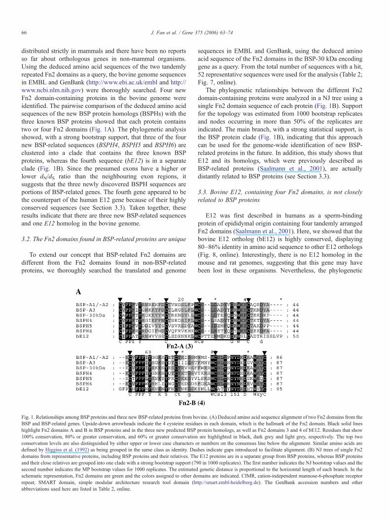
distributed strictly in mammals and there have been no reportsso far about orthologous genes in non-mammal organisms.Using the deduced amino acid sequences of the two tandemlyrepeated Fn2 domains as a query, the bovine genome sequencesin EMBL and GenBank (http://www.ebi.ac.uk/embl and http://www.ncbi.nlm.nih.gov) were thoroughly searched. Four newFn2 domain-containing proteins in the bovine genome wereidentified. The pairwise comparison of the deduced amino acidsequences of the new BSP protein homologs (BSPHs) with thethree known BSP proteins showed that each protein containstwo or four Fn2 domains (Fig. 1A). The phylogenetic analysisshowed, with a strong bootstrap support, that three of the fournew BSP-related sequences (BSPH4, BSPH5 and BSPH6) areclustered into a clade that contains the three known BSPproteins, whereas the fourth sequence (bE12) is in a separateclade (Fig. 1B). Since the presumed exons have a higher orlower dN/dS ratio than the neighbouring exon regions, itsuggests that the three newly discovered BSPH sequences areportions of BSP-related genes. The fourth gene appeared to bethe counterpart of the human E12 gene because of their highlyconserved sequences (see Section 3.3). Taken together, theseresults indicate that there are three new BSP-related sequencesand one E12 homolog in the bovine genome.
3.2. The Fn2 domains found in BSP-related proteins are unique
To extend our concept that BSP-related Fn2 domains aredifferent from the Fn2 domains found in non-BSP-relatedproteins, we thoroughly searched the translated and genome
Fig. 1. Relationships among BSP proteins and three new BSP-related proteins from boBSP and BSP-related genes. Upside-down arrowheads indicate the 4 cysteine residuhighlight Fn2 domains A and B in BSP proteins and in the three new predicted BSP100% conservation, 80% or greater conservation, and 60% or greater conservationconservation levels are also distinguished by either upper or lower case characters odefined by Higgins et al. (1992) as being grouped in the same class as identity. Dasdomains from representative proteins, including BSP proteins and their relatives. Thand their close relatives are grouped into one clade with a strong bootstrap support (79second number indicates the MP bootstrap values for 1000 replicates. The estimatedschematic representation, Fn2 domains are green and the colors assigned to other dorepeat; SMART domain, simple modular architecture research tool domain (habbreviations used here are listed in Table 2, online.
sequences in EMBL and GenBank, using the deduced aminoacid sequence of the Fn2 domains in the BSP-30 kDa encodinggene as a query. From the total number of sequences with a hit,52 representative sequences were used for the analysis (Table 2;Fig. 7, online).
The phylogenetic relationships between the different Fn2domain-containing proteins were analyzed in a NJ tree using asingle Fn2 domain sequence of each protein (Fig. 1B). Supportfor the topology was estimated from 1000 bootstrap replicatesand nodes occurring in more than 50% of the replicates areindicated. The main branch, with a strong statistical support, isthe BSP protein clade (Fig. 1B), indicating that this approachcan be used for the genome-wide identification of new BSP-related proteins in the future. In addition, this study shows thatE12 and its homologs, which were previously described asBSP-related proteins (Saalmann et al., 2001), are actuallydistantly related to BSP proteins (see Section 3.3).
3.3. Bovine E12, containing four Fn2 domains, is not closelyrelated to BSP proteins
E12 was first described in humans as a sperm-bindingprotein of epididymal origin containing four tandemly arrangedFn2 domains (Saalmann et al., 2001). Here, we showed that thebovine E12 ortholog (bE12) is highly conserved, displaying80–86% identity in amino acid sequence to other E12 orthologs(Fig. 8, online). Interestingly, there is no E12 homolog in themouse and rat genomes, suggesting that this gene may havebeen lost in these organisms. Nevertheless, the phylogenetic
vine. (A) Deduced amino acid sequence alignment of two Fn2 domains from thees in each domain, which is the hallmark of the Fn2 domain. Black solid linesprotein homologs, as well as Fn2 domains 3 and 4 of bE12. Residues that showare highlighted in black, dark grey and light grey, respectively. The top twor numbers on the consensus line below the alignment. Similar amino acids arehes indicate gaps introduced to facilitate alignment. (B) NJ trees of single Fn2e E12 proteins are in a separate group from BSP proteins, whereas BSP proteins0 in 1000 replicates). The first number indicates the NJ bootstrap values and thegenetic distance is proportional to the horizontal length of each branch. In themains are indicated. CIMR, cation-independent mannose-6-phosphate receptorttp://smart.embl-heidelberg.de). The GenBank accession numbers and other

Fig. 1 (continued).
67J. Fan et al. / Gene 375 (2006) 63–74
analysis indicates that E12 homologs form a very distinct cladefrom BSP protein homologs (Fig. 1B), indicating that theyrepresent members of a separate family of sperm-coatingproteins.
The human expressed sequence tags showed that E12 is alsowidely expressed in tissues other than epididymis and testis,such as brain (accession number: BF696501), eye (accessionnumber: BM690865, BM690040, BM663735, BM661759) andplacenta/ choriocarcinoma (accession number: BG478994,BC015598, AAH15598). E12 may therefore be involved in a
more general function such as collagen-binding or protein–protein interactions in the extracellular matrix.
3.4. The BSP protein clade is composed of three subfamilies:BSPH4, BSPH5 and BSPH6
To obtain a better resolution of the BSP protein clade in theNJ tree, a phylogenetic reconstruction was performed usingthe two tandemly repeated Fn2 domains of genes from thisgroup (Fig. 2; Fig. 9, online). Results showed that the BSP
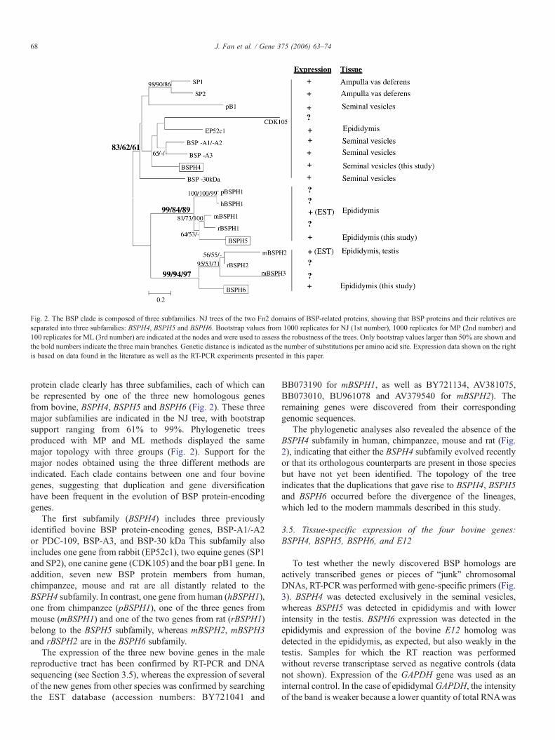
Fig. 2. The BSP clade is composed of three subfamilies. NJ trees of the two Fn2 domains of BSP-related proteins, showing that BSP proteins and their relatives areseparated into three subfamilies: BSPH4, BSPH5 and BSPH6. Bootstrap values from 1000 replicates for NJ (1st number), 1000 replicates for MP (2nd number) and100 replicates for ML (3rd number) are indicated at the nodes and were used to assess the robustness of the trees. Only bootstrap values larger than 50% are shown andthe bold numbers indicate the three main branches. Genetic distance is indicated as the number of substitutions per amino acid site. Expression data shown on the rightis based on data found in the literature as well as the RT-PCR experiments presented in this paper.
68 J. Fan et al. / Gene 375 (2006) 63–74
protein clade clearly has three subfamilies, each of which canbe represented by one of the three new homologous genesfrom bovine, BSPH4, BSPH5 and BSPH6 (Fig. 2). These threemajor subfamilies are indicated in the NJ tree, with bootstrapsupport ranging from 61% to 99%. Phylogenetic treesproduced with MP and ML methods displayed the samemajor topology with three groups (Fig. 2). Support for themajor nodes obtained using the three different methods areindicated. Each clade contains between one and four bovinegenes, suggesting that duplication and gene diversificationhave been frequent in the evolution of BSP protein-encodinggenes.
The first subfamily (BSPH4) includes three previouslyidentified bovine BSP protein-encoding genes, BSP-A1/-A2or PDC-109, BSP-A3, and BSP-30 kDa This subfamily alsoincludes one gene from rabbit (EP52c1), two equine genes (SP1and SP2), one canine gene (CDK105) and the boar pB1 gene. Inaddition, seven new BSP protein members from human,chimpanzee, mouse and rat are all distantly related to theBSPH4 subfamily. In contrast, one gene from human (hBSPH1),one from chimpanzee (pBSPH1), one of the three genes frommouse (mBSPH1) and one of the two genes from rat (rBSPH1)belong to the BSPH5 subfamily, whereas mBSPH2, mBSPH3and rBSPH2 are in the BSPH6 subfamily.
The expression of the three new bovine genes in the malereproductive tract has been confirmed by RT-PCR and DNAsequencing (see Section 3.5), whereas the expression of severalof the new genes from other species was confirmed by searchingthe EST database (accession numbers: BY721041 and
BB073190 for mBSPH1, as well as BY721134, AV381075,BB073010, BU961078 and AV379540 for mBSPH2). Theremaining genes were discovered from their correspondinggenomic sequences.
The phylogenetic analyses also revealed the absence of theBSPH4 subfamily in human, chimpanzee, mouse and rat (Fig.2), indicating that either the BSPH4 subfamily evolved recentlyor that its orthologous counterparts are present in those speciesbut have not yet been identified. The topology of the treeindicates that the duplications that gave rise to BSPH4, BSPH5and BSPH6 occurred before the divergence of the lineages,which led to the modern mammals described in this study.
3.5. Tissue-specific expression of the four bovine genes:BSPH4, BSPH5, BSPH6, and E12
To test whether the newly discovered BSP homologs areactively transcribed genes or pieces of “junk” chromosomalDNAs, RT-PCR was performed with gene-specific primers (Fig.3). BSPH4 was detected exclusively in the seminal vesicles,whereas BSPH5 was detected in epididymis and with lowerintensity in the testis. BSPH6 expression was detected in theepididymis and expression of the bovine E12 homolog wasdetected in the epididymis, as expected, but also weakly in thetestis. Samples for which the RT reaction was performedwithout reverse transcriptase served as negative controls (datanot shown). Expression of the GAPDH gene was used as aninternal control. In the case of epididymalGAPDH, the intensityof the band is weaker because a lower quantity of total RNAwas

Fig. 3. Expression analysis and tissue distribution of the newly identified bovineBSP protein homologs: BSPH4, BSPH5, BSPH6 and bE12. Total RNA wasextracted from tissues and analysed by RT-PCR, as described in the Methodssection. The expression of the GAPDH gene was used as an internal control. Allamplicons were sequenced to confirm their identities. E, epididymis; SV,seminal vesicles; T, testis; H, heart; I, intestine; K, kidney; Li, liver; Lu, lung.
69J. Fan et al. / Gene 375 (2006) 63–74
used for the RT reaction (1.3 μg instead of 2 μg). The PCRproducts, corresponding to the partial transcripts of each BSPHgene, were confirmed by DNA sequencing and pairwisesequence comparisons. These results strongly suggest that thenew BSP protein homologous genes and bE12 are activelytranscribed in male reproductive tissues, whereas no transcrip-tion was detected in non-reproductive tissues such as heart,intestine, kidney, liver and lung.
3.6. Evidence that BSP proteins are under positive selectionand that the two Fn2 domains are under heterogeneousselection pressure
The nucleotide substitution rates within the BSP proteinfamily were studied by comparing orthologous and paralogousgenes between bovine, stallion, boar, mouse, rat, chimpanzeeand human. The non-synonymous divergence (dN) betweenpairs of genes is a good indicator of their functionalconservation. Lower dN/dS ratios (<1) indicate purifyingselection, whereas higher dN/dS ratios (>1) indicate positiveselection, which means that advantageous mutations haveoccurred in the diversifying selection of the two duplicates.Similar dN/dS ratios (=1) indicate a lack of functional constraint,as synonymous substitutions are expected to be nearly neutral(Yang and Nielsen, 2000). However, different parts of a genemay have different evolutionary selection pressures, whichmake the interpretation of dN/dS ratios complicated for acomplete gene. Nevertheless, we first estimated an averagedN/dS ratio from the Nei–Gojobori method (Nei and Gojobori,1986) for each pair of BSP and BSP-related genes. The averageestimated dN/dS ratio for the BSPH4, BSPH5 and BSPH6subfamilies was 0.612±0.308, 0.194±0.087 and 0.361±0.117(mean±SD), respectively (Fig. 4A). The dN/dS ratio of theBSPH4 subfamily is significantly different from that of BSPH5(p<0.001) and BSPH6 (p=0.006). Hence, we conclude that theBSPH4 subfamily evolves faster than the other two subfamilies,indicating either a lower functional constraint or an acceleratedrate of evolution for some sites in this group. Strikingly, the only
genes under positive selection within the highly divergentBSPH4 subfamily are BSP-A1/-A2, BSP-A3 and BSP-30k Da,based on a pairwise comparison. This high divergence may becaused by positive selection of several beneficial mutations.
The regulatory genes are often divided into domains that arecharacterized by slowly and rapidly evolving sequences (Filipand Mundy, 2004). To check for heterogeneity in evolutionaryrates along BSP protein-encoding genes, the distribution ofnucleotide diversity based on nonsynonymous substitutionsalong the genes was studied using a sliding window of 130 siteswith 5 bp steps. This analysis included four bovine genes in theBSPH4 subfamily. The distribution of nonsynonymous ratesshown in Fig. 4B suggests two relatively conserved domains(Fn2 A and Fn2 B), one close to the N-terminus and one close tothe C-terminus of the protein, as shown schematically undereach graph. Although both of these domains are important forthe interaction with choline phospholipids on the bovine spermsurface (Wah et al., 2002), Fn2 domain A from the three pairs(BSP-A1/-A2 vs. BSP-A3, BSP-A1/-A2 vs. BSP-30 kDa, BSP-A3 vs. BSP-30 kDa) showed significantly higher Ka/Ks ratiosthan Fn2 domain B in all comparisons. These results suggestthat the three BSP protein-encoding genes are under strongpositive selection and that selection pressure is differentbetween these two Fn2 domains. Although the rapid evolutionof mammalian sex-related genes has been well documented, weare showing for the first time that BSP proteins are evolvingunder diversifying selection.
3.7. Five potential sites under positive selection in the BSPgenes, four of which are distributed at the edge of thephosphorycholine binding pocket in Fn2 domain A
To detect positively selected amino acid sites, we used thelikelihood ratio tests (LRT) of Goldman and Yang (1994).Synonymous substitutions in protein-encoding genes aregenerally free from natural selection and are used frequentlyas a measurement of the neutral mutation rate (Kimura, 1983).Estimation was performed by assuming either a single ratio(M0) for all branches in the BSPH4 subfamily or assumingdifferent ratios for each branch (M1, M2, M3, M7 and M8). Thelog likelihood value for the one-ratio model (M0) was−1666.241, while the strictly neutral model (M1), assuming adifferent ratio for each branch, resulted in a log likelihood of−1600.333 (Table 1). Twice the log likelihood difference wasused to test whether the free-ratio model fit the data significantlybetter than the one-ratio model. The free-ratio model resulted ina significantly better fit at the 1% level (χ1%
2 =9.21, df=2). Thisindicates that the positive Darwinian selection on BSP proteinswas not evenly distributed across the entire length of the genestested.
We next investigated models in which the dN/dS ratio isallowed to vary among different classes of sites (models M1–M3, M7 and M8; Table 1; Yang et al., 2000). The LRT statisticfor comparison of the neutral (M1), selection (M2) and discrete(M3) models rejected M1 in favour of M2 and M3. These testssuggest that sites under positive selection are present in thesegenes. Application of the discrete model (M3) suggests that

Fig. 4. Evidence of positive selection within the BSP clade. (A) The number of nonsynonymous substitutions per nonsynonymous site (dN) is plotted against thenumber of synonymous substitutions per synonymous site (dS) for the BSPH4, BSPH5 and BSPH6 lineages. The values of each pairwise comparison for the differentlineages are indicated in black, grey and white as indicated below the figure. The dotted line shows the neutral expectation of dN=dS. (B) Sliding window analysis ofsubstitution rates Ka/Ks (dN/dS) shows the heterogeneity of positive selection between Fn2 domains A and B. The grey curve illustrates that a 130-bp window wasmoved along the BSP protein-encoding sequence in 5-bp steps and the Ka/Ks ratio was computed for each window, then plotted at its median position using K-estimator 6.1 (Comeron, 1999). The dN and dS substitution rate estimates for pairwise comparisons of BSP-A1/-A2, BSP-A3, BSP-30 kDa and BSPH4 were obtainedusing the method of Nei and Gojobori as implemented in DnaSP 4.0 (Rozas et al., 2003).
70 J. Fan et al. / Gene 375 (2006) 63–74

Fig. 5. Distribution of putative positively selected sites in BSP protein sequences. (A) Amino acid sites under positive selection were detected in proteins of the BSPH4lineage. The abscissa indicates the amino acid sites counted from the positions of the amino acid alignment in the BSPH4 lineage. The ordinate indicates the value ofposterior probability for each amino acid site under model M3 above the abscissa and under model M8 below the abscissa. Dotted lines indicate the 80%, 90% and 95%significance levels. Positive selection was assigned to the amino acid sites in red, green and blue in the graph when the corresponding values exceeded that of the dottedline. The amino acid sites with the same prediction based on the two models were indicated in red (p>95% in model M8), green (p>90% in model M3) and blue(p>80% in model M3) and indicated in the sequence alignment within a red rectangle. (B) Three-dimensional models of BSP-30 kDa based on homology modelingusing BSP-A1/-A2 (also named as PDC-109) as a template (1H8P). Stick and ball structures of the side-chains of 5–6 hydrophobic amino acid residues revealed thetwo potential hydrophobic pockets in Fn2 domains A and B. (C) Codons identified as being under positive selection with high posterior probabilities (p>0.95) arehighlighted on a surface model of BSP-30 kDa. Amino acid sites inferred to be evolving under positive selection are displayed as space-filled molecules and marked byname and position. The figure was prepared with the program Swiss-PdbViewer 3.7b2.
71J. Fan et al. / Gene 375 (2006) 63–74

72 J. Fan et al. / Gene 375 (2006) 63–74
nearly 30% of sites (ω2=2.121, f2=0.296) are potentiallypositively selected sites. Over 40% of the sites detected bymodel M8 are under positive selection with ω1 (dN/dS)=1.863(Table 1).
We used the Bayes theorem to calculate the posteriorprobabilities of ω classes for each site. Sites with a highprobability of coming from the ω>1 class are likely to be underpositive selection. Five sites were predicted to be under positiveselection with a probability greater than 90% according to bothmodels M3 and M8, whereas three and eight sites werepredicted respectively by these two models with a probabilitygreater than 95% (Table 1). Of these 8 sites, 5 were located inFn2 domain A and 3 were in Fn2 domain B (Fig. 5A; Fig. 9,online). The results using PAML suggest that heterogeneousselection occurs between these two tandemly repeated domains,which is consistent with the results obtained using the moreconventional pairwise dN/dS ratios.
Since the identification of sites subjected to positiveselection may be important for the binding properties of BSPproteins, we mapped all the potential sites under positiveselection on a 3-D model of BSP-30 kDa. This allowed us toschematically illustrate their relationship to two establishedphosphorylcholine binding pockets (Desnoyers and Manju-nath, 1993; Wah et al., 2002) (Fig. 5B and C). Interestingly,the sites under positive selection in Fn2 domain A are alllocated at the edge of the phosphorylcholine-binding pocket,whereas the sites under positive selection in Fn2 domain Bare dispersed on the opposite side of the pocket (Fig. 5C).The results indicate that the positively selected sites in Fn2domain A may affect the protein's binding affinity to cholinephospholipids, whereas the positively selected sites in Fn2domain B may not be involved in the ligand-bindingproperties of the pocket. This may reflect some differencesin the features of ligand-binding affinity in the o-phosphor-ylcholine complex of PDC-109 (BSP-A1/-A2) (Wah et al.,2002), BSP-A3 and BSP-30 kDa.
Fig. 6. Proposed model for domain-specific binding activity based on the diversifyinpositively selected sites within the two Fn2 domains of BSP proteins suggests thatdomain B. Fn2 domain A could therefore be involved in binding factors for sperminteraction with choline phospholipids for adhesion purposes. The positively selectedhighlighted using a dotted red line.
3.8. A proposed model of functional diversification in BSPproteins
The location of the sites under positive selection was mappedon a homology model of BSP-30 kDa. Most predicted sitesformed a single patch around the phosphorylcholine-bindingpocket of Fn2 domain A (Fig. 6). A model was proposed toillustrate that the ligand-binding pocket in Fn2 domain A ofBSP proteins may loosely bind choline phospholipids and/orfactors involved in sperm–egg interaction. On the other hand,the binding pocket in Fn2 domain B could maintain a moreconserved function, such as the tight association withphosphorylcholine for adhesion to the surface of sperm (Fig.6). Since many reproductive proteins evolve rapidly and thedriving forces for evolution are the selection pressures, whichinclude sperm competition, cryptic female choice and sexualconflict (Swanson and Vacquier, 2002), the functional diversi-fication of BSP proteins described here may indicate somerelevant roles in these respects, which will need furtherclarification.
In summary, Fn2 domains in BSP proteins and their relativeshave special features that are different from the Fn2 domains innon-BSP-related proteins. The method established here hasproven to be powerful and efficient to identify new BSP-relatedgenes from emerging genomes. The BSP protein lineage can bedivided into three groups, which are represented by the BSPH4,BSPH5 and BSPH6 subfamilies. Expression of the three newbovine genes was confirmed in tissues of the male reproductivetract by RT-PCR and DNA sequencing. A strong evidence ofpositive selection in the BSPH4 lineage was provided.Furthermore, there seems to be heterogeneity in selectionpressures between Fn2 domains A and B. Five amino acid sitesunder positive selection were estimated using different selectionmodels and a model for the biological function of BSP proteinswas proposed. However, it remains to be determined whether ornot natural selection and/or rapid evolution of the variable Fn2
g selection between the Fn2 domains of BSP proteins. Mapping of the potentialFn2 domain A would have a more relaxed binding specificity than that of Fn2–egg interaction and capacitation, whereas Fn2 domain B would mediate thesites at the edge of the phosphorylcholine-binding pocket in Fn2 domain A are

73J. Fan et al. / Gene 375 (2006) 63–74
domain A are the main factors driving diversification of BSPprotein-encoding genes.
Acknowledgments
We thank the Canadian Institutes of Health Research forfinancial support.
Appendix A. Supplementary data
Supplementary data associated with this article can be found,in the online version, at doi:10.1016/j.gene.2006.02.025.
References
Anisimova, M., Bielawski, J.P., Yang, Z., 2001. Accuracy and power of thelikelihood ratio test in detecting adaptive molecular evolution. Mol. Biol.Evol. 18, 1585–1592.
Baker, M.E., 1985. The PDC-109 protein from bovine seminal plasma is similarto the gelatin-binding domain of bovine fibronectin and a kringle domain ofhuman tissue-type plasminogen activator. Biochem. Biophys. Res. Com-mun. 130, 1010–1014.
Chandonnet, L., Roberts, K.D., Chapdelaine, A., Manjunath, P., 1990.Identification of heparin-binding proteins in bovine seminal plasma. Mol.Reprod. Dev. 26, 313–318.
Comeron, J.M., 1999. K-Estimator: calculation of the number of nucleotidesubstitutions per site and the confidence intervals. Bioinformatics 15,763–764.
de Oliveira, T., et al., 2004. Mapping sites of positive selection and amino aciddiversification in the HIV genome: an alternative approach to vaccinedesign? Genetics 167, 1047–1058.
Desnoyers, L., Manjunath, P., 1992. Major proteins of bovine seminalplasma exhibit novel interactions with phospholipid. J. Biol. Chem. 267,10149–10155.
Desnoyers, L., Manjunath, P., 1993. Interaction of a novel class of phospholipid-binding proteins of bovine seminal fluid with different affinity matrices.Arch. Biochem. Biophys. 305, 341–349.
Eisen, J.A., 1998. Phylogenomics: improving functional predictions foruncharacterized genes by evolutionary analysis. Genome Res. 8, 163–167.
Eisen, J.A., Fraser, C.M., 2003. Phylogenomics: intersection of evolution andgenomics. Science 300, 1706–1707.
Eisen, J.A., Hanawalt, P.C., 1999. A phylogenomic study of DNA repair genes,proteins, and processes. Mutat. Res. 435, 171–213.
Eisen, J.A., Wu, M., 2002. Phylogenetic analysis and gene functionalpredictions: phylogenomics in action. Theor. Popul. Biol. 61, 481–487.
Esch, F.S., Ling, N.C., Bohlen, P., Ying, S.Y., Guillemin, R., 1983. Primarystructure of PDC-109, a major protein constituent of bovine seminal plasma.Biochem. Biophys. Res. Commun. 113, 861–867.
Felsenstein, J. PHYLIP (Phylogeny Inference Package) Version 3.6. 2004.Distributed over the World Wide Web, Seattle.
Filip, L.C., Mundy, N.I., 2004. Rapid evolution by positive Darwinian selectionin the extracellular domain of the abundant lymphocyte protein CD45 inprimates. Mol. Biol. Evol. 21, 1504–1511.
Fritz-Laylin, L.K., Krishnamurthy, N., Tor, M., Sjolander, K.V., Jones, J.D.,2005. Phylogenomic analysis of the receptor-like proteins of rice andArabidopsis. Plant Physiol. 138, 611–623.
Goldman, N., Yang, Z., 1994. A codon-based model of nucleotide substitutionfor protein-coding DNA sequences. Mol. Biol. Evol. 11, 725–736.
Guex, N., Peitsch, M.C., 1997. SWISS-MODEL and the Swiss-PdbViewer: anenvironment for comparative protein modeling. Electrophoresis 18,2714–2723.
Higgins, D.G., Bleasby, A.J., Fuchs, R., 1992. CLUSTALV: improved softwarefor multiple sequence alignment. Comput. Appl. Biosci. 8, 189–191.
Ivarsson, Y., Mackey, A.J., Edalat, M., Pearson, W.R., Mannervik, B., 2003.Identification of residues in glutathione transferase capable of driving
functional diversification in evolution. A novel approach to protein redesign.J. Biol. Chem. 278, 8733–8738.
Kimura, M., 1983. The Neutral Theory of Molecular Evolution. Cambridge Uni.Press, Cambridge, U.K.
Kumar, S., Tamura, K., Nei, M., 2004. MEGA3: integrated software formolecular evolutionary genetics analysis and sequence alignment. Brief.Bioinform. 5, 150–163.
Leblond, E., Desnoyers, L., Manjunath, P., 1993. Phosphorylcholine-bindingproteins from the seminal fluids of different species share antigenicdeterminants with the major proteins of bovine seminal plasma. Mol.Reprod. Dev. 34, 443–449.
Manjunath, P., 1984. Gonadotropin release stimulatory and inhibitory proteinsin bull seminal plasma. In: Sairam, M.R., Atkinson, L.E. (Eds.), GonadalProteins and Peptides and Their Biological Significance. World SciencePublishing, Singapore, pp. 78–84.
Manjunath, P., Therien, I., 2002. Role of seminal plasma phospholipid-bindingproteins in sperm membrane lipid modification that occurs duringcapacitation. J. Reprod. Immunol. 53, 109–119.
Manjunath, P., Sairam, M.R., Uma, J., 1987. Purification of four gelatin-bindingproteins from bovine seminal plasma by affinity chromatography. Biosci.Rep. 7, 231–238.
Manjunath, P., Baillargeon, L., Marcel, Y.L., Seidah, N.G., Chretien, M.,Chapdelaine, A., 1988. Diversity of novel proteins of gonadal fluids. In:McKerns, K.W., Chretien, M. (Eds.), Molecular Biology of Brain andEndocrine Systems. Plenum Press, New York, pp. 259–273.
Manjunath, P., Marcel, Y.L., Uma, J., Seidah, N.G., Chretien, M., Chapdelaine,A., 1989. Apolipoprotein A-I binds to a family of bovine seminal plasmaproteins. J. Biol. Chem. 264, 16853–16857.
Manjunath, P., Nauc, V., Bergeron, A., Menard, M., 2002. Major proteins ofbovine seminal plasma bind to the low-density lipoprotein fraction of hen'segg yolk. Biol. Reprod. 67, 1250–1258.
Moulton, G., Attwood, T.K., Parry-Smith, D.J., Packer, J.C., 2003. Phyloge-nomic analysis and evolution of the potassium channel gene family. Recept.Channels 9, 363–377.
Mueller, J.L., Ripoll, D.R., Aquadro, C.F., Wolfner, M.F., 2004. Comparativestructural modeling and inference of conserved protein classes inDrosophilaseminal fluid. Proc. Natl. Acad. Sci. U. S. A. 101, 13542–13547.
Nauc, V., Manjunath, P., 2000. Radioimmunoassays for bull seminalplasma proteins (BSP-A1/-A2, BSP-A3, and BSP-30-Kilodaltons), andtheir quantification in seminal plasma and sperm. Biol. Reprod. 63,1058–1066.
Nei, M., Gojobori, T., 1986. Simple methods for estimating the numbers ofsynonymous and nonsynonymous nucleotide substitutions. Mol. Biol. Evol.3, 418–426.
Raczko, A.M., Bujnicki, J.M., Pawlowski, M., Godlewska, R., Lewandowska,M., Jagusztyn-Krynicka, E.K., 2005. Characterization of new DsbB-likethiol-oxidoreductases of Campylobacter jejuni and Helicobacter pylori andclassification of the DsbB family based on phylogenomic, structural andfunctional criteria. Microbiology 151, 219–231.
Ramesh, M.A., Malik, S.B., Logsdon Jr., J.M., 2005. A phylogenomic inventoryof meiotic genes; evidence for sex in Giardia and an early eukaryotic originof meiosis. Curr. Biol. 15, 185–191.
Rozas, J., Sanchez-DelBarrio, J.C., Messeguer, X., Rozas, R., 2003. DnaSP,DNA polymorphism analyses by the coalescent and other methods.Bioinformatics 19, 2496–2497.
Saalmann, A., Munz, S., Ellerbrock, K., Ivell, R., Kirchhoff, C., 2001. Novelsperm-binding proteins of epididymal origin contain four fibronectin type II-modules. Mol. Reprod. Dev. 58, 88–100.
Sawyer, S.L., Wu, L.I., Emerman, M., Malik, H.S., 2005. Positive selection ofprimate TRIM5alpha identifies a critical species-specific retroviral restric-tion domain. Proc. Natl. Acad. Sci. U. S. A. 102, 2832–2837.
Schwede, T., Kopp, J., Guex, N., Peitsch, M.C., 2003. SWISS-MODEL: anautomated protein homology-modeling server. Nucleic Acids Res. 31,3381–3385.
Seidah, N.G., Manjunath, P., Rochemont, J., Sairam, M.R., Chretien, M., 1987.Complete amino acid sequence of BSP-A3 from bovine seminal plasma.Homology to PDC-109 and to the collagen-binding domain of fibronectin.Biochem. J. 243, 195–203.

74 J. Fan et al. / Gene 375 (2006) 63–74
Sjolander, K., 2004. Phylogenomic inference of protein molecular function:advances and challenges. Bioinformatics 20, 170–179.
Swanson, W.J., Vacquier, V.D., 2002. The rapid evolution of reproductiveproteins. Nat. Rev., Genet. 3, 137–144.
Therien, I., Soubeyrand, S., Manjunath, P., 1997. Major proteins of bovineseminal plasma modulate sperm capacitation by high-density lipoprotein.Biol. Reprod. 57, 1080–1088.
Therien, I., Moreau, R., Manjunath, P., 1998. Major proteins of bovine seminalplasma and high-density lipoprotein induce cholesterol efflux fromepididymal sperm. Biol. Reprod. 59, 768–776.
Therien, I., Moreau, R., Manjunath, P., 1999. Bovine seminal plasmaphospholipid-binding proteins stimulate phospholipid efflux from epididy-mal sperm. Biol. Reprod. 61, 590–598.
Therien, I., Bergeron, A., Bousquet, D., Manjunath, P., 2005. Isolation andcharacterization of glycosaminoglycans from bovine follicular fluid andtheir effect on sperm capacitation. Mol. Reprod. 71, 97–106.
Thompson, J.D., Gibson, T.J., Plewniak, F., Jeanmougin, F., Higgins, D.G.,1997. The CLUSTAL_X windows interface: flexible strategies for multiple
sequence alignment aided by quality analysis tools. Nucleic Acids Res. 25,4876–4882.
Wah, D.A., Fernandez-Tornero, C., Sanz, L., Romero, A., Calvete, J.J., 2002.Sperm coating mechanism from the 1.8 A crystal structure of PDC-109-phosphorylcholine complex. Structure (Camb.) 10, 505–514.
Yang, Z., 1997. PAML: a program package for phylogenetic analysis bymaximum likelihood. Comput. Appl. Biosci. 13, 555–556.
Yang, Z., 2005. The power of phylogenetic comparison in revealing proteinfunction. Proc. Natl. Acad. Sci. U. S. A. 102, 3179–3180.
Yang, Z., Nielsen, R., 2000. Estimating synonymous and nonsynonymoussubstitution rates under realistic evolutionary models. Mol. Biol. Evol. 17,32–43.
Yang, Z., Nielsen, R., Goldman, N., Pedersen, A.M., 2000. Codon-substitutionmodels for heterogeneous selection pressure at amino acid sites. Genetics155, 431–449.
Copyright © 2022 FDOKUMEN