Mannan-binding proteins from boar seminal plasma
16
Journal of Reproductive Immunology 62 (2004) 167–182 Mannan-binding proteins from boar seminal plasma Petra Jel´ ınková a , Jiˇ r´ ı Liberda b , Pavla Maˇ násková a , Helena Ryšlavá b , Vˇ era Jonáková a,∗ , Marie Tichá b a Institute of Molecular Genetics, Academy of Sciences of the Czech Republic, Flemingovo nám. 2, 166 37 Praha 6, Czech Republic b Department of Biochemistry, Charles University, Albertov 2030, 128 40 Praha 2, Czech Republic Received in revised form 10 November 2003; accepted 5 January 2004 Abstract The interaction of boar seminal plasma proteins and sperm with yeast mannan was investigated by the enzyme-linked binding assay (ELBA) and specific detection of proteins after SDS electrophoresis and blotting using biotinylated derivative of the polysaccharide. Heparin-binding proteins (especially AQN 1 and DQH proteins) and their aggregated forms showed affinity to yeast mannan. Besides that, these proteins were shown to bind to oviductal epithelium. The mannan-binding activity of boar proteins and sperm was inhibited most efficiently by ovomucoid, ovalbumin and N-glycans released from ovalbumin, but not with d-glucose, d-mannose and their phosphates. On the other hand, yeast mannan inhibited both the interaction of boar seminal plasma and sperm with heparin and the binding of these proteins to porcine oviductal epithelium. Yeast mannan immobilized to divinyl sulfone-activated Sepharose was used for the isolation of mannan-binding proteins. Proteins adsorbed to the immobilized polysaccharide were analyzed by RP-HPLC, SDS electrophoresis and N-terminal amino acid sequencing. AQN and AWN spermad- hesins and DQH protein (names are derived from the N-terminal amino acid sequence) were identified as components of the isolated fraction. The results suggest an involvement of mannan-binding proteins in the formation of the sperm oviductal reservoir in pig. The ability of these proteins to interact both the complex d-mannose- containing saccharide structures and the heparin may also play an important role in sperm release from the oviductal reservoir or the capacitation process. © 2004 Elsevier Ireland Ltd. All rights reserved. Keywords: Boar seminal plasma proteins; Mannan-binding proteins Paper presented at the 4th Congress of the European Society for Reproductive & Developmental Immunology, Rhodes, Greece, June 2003. ∗ Corresponding author. Fax: +420-2-33331274. E-mail addresses: [email protected], [email protected] (V. Jon´ akov´ a). 0165-0378/$ – see front matter © 2004 Elsevier Ireland Ltd. All rights reserved. doi:10.1016/j.jri.2004.01.007
Transcript of Mannan-binding proteins from boar seminal plasma

Journal of Reproductive Immunology62 (2004) 167–182
Mannan-binding proteins from boarseminal plasma�
Petra Jelınkováa, Jirı Liberdab, Pavla Manáskováa,Helena Ryšlaváb, Vera Jonákováa,∗, Marie Ticháb
a Institute of Molecular Genetics, Academy of Sciences of the Czech Republic,Flemingovo nám. 2, 166 37 Praha 6, Czech Republic
b Department of Biochemistry, Charles University, Albertov 2030, 128 40 Praha 2, Czech Republic
Received in revised form 10 November 2003; accepted 5 January 2004
Abstract
The interaction of boar seminal plasma proteins and sperm with yeast mannan was investigated bythe enzyme-linked binding assay (ELBA) and specific detection of proteins after SDS electrophoresisand blotting using biotinylated derivative of the polysaccharide. Heparin-binding proteins (especiallyAQN 1 and DQH proteins) and their aggregated forms showed affinity to yeast mannan. Besidesthat, these proteins were shown to bind to oviductal epithelium. The mannan-binding activity of boarproteins and sperm was inhibited most efficiently by ovomucoid, ovalbumin andN-glycans releasedfrom ovalbumin, but not withd-glucose,d-mannose and their phosphates. On the other hand, yeastmannan inhibited both the interaction of boar seminal plasma and sperm with heparin and the bindingof these proteins to porcine oviductal epithelium.
Yeast mannan immobilized to divinyl sulfone-activated Sepharose was used for the isolation ofmannan-binding proteins. Proteins adsorbed to the immobilized polysaccharide were analyzed byRP-HPLC, SDS electrophoresis and N-terminal amino acid sequencing. AQN and AWN spermad-hesins and DQH protein (names are derived from the N-terminal amino acid sequence) were identifiedas components of the isolated fraction.
The results suggest an involvement of mannan-binding proteins in the formation of the spermoviductal reservoir in pig. The ability of these proteins to interact both the complexd-mannose-containing saccharide structures and the heparin may also play an important role in sperm releasefrom the oviductal reservoir or the capacitation process.© 2004 Elsevier Ireland Ltd. All rights reserved.
Keywords:Boar seminal plasma proteins; Mannan-binding proteins
� Paper presented at the 4th Congress of the European Society for Reproductive & Developmental Immunology,Rhodes, Greece, June 2003.
∗ Corresponding author. Fax:+420-2-33331274.E-mail addresses:[email protected], [email protected] (V. Jonakova).
0165-0378/$ – see front matter © 2004 Elsevier Ireland Ltd. All rights reserved.doi:10.1016/j.jri.2004.01.007
168 P. Jelınkova et al. / Journal of Reproductive Immunology 62 (2004) 167–182
1. Introduction
Mammalian fertilization is a series of events that involve a highly coordinated sequenceof interactions between molecules located on the surface of both gametes as well as withsubstances present in the natural environment of the gametes (Töpfer-Petersen, 1999). In-teractions of the lectin type play an important role in some steps of this process (Jonákováet al., 1995; Tichá et al., 1998).
First, saccharide chains of zona pellucida glycoproteins are supposed to bind receptorspresent on the sperm surface. Although the saccharide ligands were first characterized inmice (Bleil and Wassarman, 1988) and pigs (Yurewicz et al., 1991; Yonezawa et al., 1995),the ligands of other mammals have not been fully recognized. Recently,Amari et al. (2001)described the essential role of the non-reducing terminal�-d-mannosyl residues of N-linkedsaccharide chains of bovine zona pellucida glycoproteins in sperm–egg binding.
The second type of protein–saccharide interactions seems to be involved in the forma-tion of the mammalian oviductal sperm reservoir (Töpfer-Petersen et al., 2002); proteinsattached to the sperm surface recognize oviductal epithelium glycopeptides. By initiationof capacitation, these associated proteins are shed from the sperm surface. The saccharidespecificity of the interaction of sperm surface molecules with epithelial cells appears to varyamong species (Suarez, 2001). Non-capacitated bull sperm are trapped in the reservoir bybinding tol-fucosyl residues in the oviductal epithelium (Lefebvre et al., 1997; Revah et al.,2000).l-Fucose-binding sites are lost during capacitation and, at that time,d-mannose bind-ing sites are revealed for interaction with the ovum (Revah et al., 2000; Ignotz et al., 2001).In the pig, the formation of the oviductal sperm reservoir appears to comprised-mannoseresidues (Green et al., 2001). Detailed study on the structure of oligosaccharides involvedin the carbohydrate-mediated adhesion has been described (Wagner et al., 2002).
Recent studies have shown that seminal plasma proteins participate in the formation andrearrangement of the protein coating of the sperm surface, which changes its composi-tion in different steps of the fertilization process (Dostálová et al., 1994; Töpfer-Petersen,1999; Petrunkina et al., 2000). The binding properties of the sperm surface (includingsaccharide-binding) are changed due to these events.
In a previous communication (Liberda et al., 2002), we have shown the presence ofmannan-binding proteins in bull seminal plasma. Mannan binding to seminal plasma pro-teins was inhibited byd-mannose andd-fructose, but notd-mannose-6-phosphate,d-glucose-6-phosphate, ovalbumin and ovomucoid. The aim of this study was to investi-gate the mannan-binding properties of boar seminal plasma proteins that significantly differin their physico-chemical properties and binding activities of those isolated from bull sem-inal plasma. Yeast mannan immobilized to Sepharose was used for isolation of proteinsparticipating in the studied interaction.
2. Materials and methods
2.1. Chemicals
Divinyl sulfone and 1-(3-dimethylaminopropyl)-3-ethylcarbodiimide were purchasedfrom Fluka (Buchs, Switzerland); Sepharose 4B from Pharmacia (Uppsala, Sweden);

P. Jelınkova et al. / Journal of Reproductive Immunology 62 (2004) 167–182 169
heparin, avidin-peroxidase, ABTS (2,2′-azino-bis(3-ethylbenzthiazoline-6-sulfonic acid),d-mannose- andd-glucose-6-phosphate and ovalbumin from Sigma (St. Louis, MO);Immobilon-P-membrane and 4-chloro-1-naphtol were products of Serva (Heidelberg, Ger-many);N-glycosidase F (PNGase F) (E.C.3.5.1.52) of Bio-Lab (New England, USA). Ovo-mucoid was a gift from Dr. K. Bezouška (Department of Biochemistry, Charles University,Praha, Czech Republic). SDS-PAGE standards-broad range were products of Bio-Rad (Her-cules, USA). Biotinylated polyacrylamide derivative of heparin was prepared as describedby Liberda et al. (1997).
Saccharomyces cerevisiaecell wall polysaccharide containing mannan and phospho-mannan polymers (Stewart et al., 1968) was isolated as was described byHaworth et al.(1937).
2.2. Materials
2.2.1. Porcine oviductal epithelium productsOviducts were collected from mature sows (five animals), the isthmic region of oviduct
was cut longitudinally and oviductal epithelium was gently separated. The obtained materialwas centrifuged for 20 min at 600×g to remove cellular debris, dissolved in PBS to proteinconcentration 1 mg/ml (based on A280 measurement) and frozen.
2.2.2. Boar seminal plasma and spermBoar ejaculates were obtained from the Veterinary Research Institute, Brno, Czech Re-
public. Ejaculates were centrifuged (600×g, 20 min, 5◦C) to separate plasma from sperm.Seminal plasma was directly lyophilized, sperms were suspended in phosphate-bufferedsaline (PBS, 20 mM phosphate, pH 7.4, 150 mM NaCl) and washed by centrifugation (twotimes with PBS), and sperm pellet was then lyophilized.
2.3. Preparation of ovalbumin N-glycans
Ovalbumin (5 mg) dissolved in 0.1 M Tris–HCl buffer pH 7.5 (1 ml) was incubated withPNGase F (5 U) under a toluene layer at 37◦C for 48 h. After incubation, protein wasprecipitated using ethanol and separated by centrifugation. Ethanol solution ofN-glycanswas concentrated using Speed-Vac. The saccharide content in ovalbumin after the PNGasetreatment was determined according toDubois et al. (1956).
2.4. Isolation of proteins from boar seminal plasma
Heparin-binding (H+) and non-heparin-binding (H−) proteins were obtained by affinitychromatography of boar seminal plasma on a heparin-polyacrylamide column (3 cm×15 cm). Fifty milliliters of boar seminal plasma was diluted 1:1 with PBS and applied to thecolumn (Tichá et al., 1994). H− proteins were washed out with PBS buffer, the adsorbedH+ proteins were eluted with 3 M NaCl.
Gel chromatography: boar seminal plasma (5 ml) was applied to a Sephadex G–75 SFcolumn (2.0 cm× 118.0 cm) equilibrated with 0.1 M Tris–HCl containing 0.15 M NaCl,

170 P. Jelınkova et al. / Journal of Reproductive Immunology 62 (2004) 167–182
pH 7.2 (flow rate 17.2 ml/h). Fractions, 4.3 ml, were screened for absorbance at 280 nm,pooled, desalted on Sephadex G-25 in 0.2% acetic acid and lyophilized.
Biotinylation of H+, H− and protein aggregates I–V obtained by gel chromatographywas performed as described previously (Manásková et al., 2000).
2.5. Mannan–Sepharose
The immobilized mannan was prepared by coupling of yeast mannan to divinyl sulfone-activated Sepharose as described previously (Liberda et al., 2002).
2.6. Biotinylated derivative of mannan
Biotinylated derivative of yeast mannan with coupled ethylenediamine was prepared bycombination of two procedures described previously (Novotná et al., 1996; Liberda et al.,1997). The aqueous solution of yeast mannan (50 mg in 5 ml) was mixed with 1% periodicacid (1 ml) and ethylenediamine (20�l). The mixture was shaken for 2 h and the reactionstopped by addition of ethylene glycol (4 ml). After 15 min incubation at room temperature,sodium cyanoborohydride (80 mg) was added. The reaction mixture was dialyzed againstdistilled water and lyophilized.
N-Hydroxy-succinimido-biotin (10 mg) dissolved in dimethylformamide (25�l) wasadded to the solution of mannan derivative (20 mg) in 0.1 M borate buffer, pH 8.5 (3 ml).The mixture was stirred for 30 min at room temperature and then 0.2 M ammonium chloridewas added to adjust the pH to 6.0. After dialysis against distilled water, the solution waslyophilized.
2.7. Affinity chromatography on mannan–Sepharose
Ten milligrams of H+ proteins were dissolved in 4 ml 1 mM HCl and 6 ml PBS andapplied to a mannan–Sepharose column (2.5 cm × 11 cm) pre-equilibrated with PBS.Non-mannan-binding (Man−) proteins were washed with PBS, flow rate 3.2 ml/20 min.Mannan-binding (Man+) proteins were first eluted with 3 M NaCl (Man1
+) and thenwith 4 M urea (Man2+). Fractions 4.6 ml/10 min were collected. These were pooled, de-salted on Sephadex G-25 in 0.2% acetic acid and lyophilized. The same conditions wereused for chromatography of fractions of non-heparin-binding proteins or boar seminalplasma.
2.8. Reversed phase high performance liquid chromatography (RP-HPLC)
Protein samples (Man−, Man1+, Man2
+) were subjected to the inert BiocompatibleQuaternary Gradient system of HPLC (Waters, Milford, U.S.A.). RP-HPLC was performedusing a 218 TP 104 Vydac C18 column (4.6 mm× 250 mm, 10�m particle size). One mil-ligram of the sample in 1 ml of 0.05% trifluoroacetic acid (TFA) was applied and proteinseluted with a linear gradient of 20–50% acetonitrile (ACN) in 60 min. Fractions correspond-ing to protein peaks were collected and lyophilized.

P. Jelınkova et al. / Journal of Reproductive Immunology 62 (2004) 167–182 171
2.9. Electrophoresis and blotting
Sodium dodecyl sulphate polyacrylamide gel electrophoresis (SDS-PAGE) of differentfractions of boar seminal plasma proteins and ovalbumin was carried out on 15% slabgel (Laemmli, 1970). Non-reduced samples of seminal plasma proteins and reduced pro-tein standards were applied. The relative molecular masses of the separated proteins wereestimated by comparison with relative molecular-mass protein standards run in parallel.Tris–glycine buffer (pH 9.6) with 20% (v/v) methanol was used for transfer of proteinsseparated by SDS-PAGE onto Immobilon-P-membrane or the nitrocellulose membrane forspecific detection. Electroblotting was carried out for 1.5 h at 500 mA according to thearrangement described byTowbin et al. (1979).
2.10. N-terminal amino acid sequence determination
N-terminal amino acid sequencing was performed in a Protein Sequencer LF 3600 D(Beckmann Instruments) following the Instruction Manual. Proteins isolated by affinitychromatography on immobilized mannan (fractions Man1
+ and Man2+), by RP-HPLC(fractions 1–5) and separated by SDS electrophoresis were used for the analysis. After SDSelectrophoresis, proteins were transferred onto the Immobilon-P-membrane, visualized byCoomassie Blue and subjected to N-terminal amino acid sequencing. Searches for aminoacid similarities were carried out using the protein sequence deposits in the BLAST-BASICe-mail Server Databank.
2.11. Binding studies
2.11.1. Enzyme-linked binding assay (ELBA)2.11.1.1. Mannan- and heparin-binding activity.Microtiter plates were incubatedfor 1 h at room temperature with 100�l of bovine serum albumin (BSA) solution (1%in PBS). After extensive washing with PBS, the wells were treated with 100�l of glu-taraldehyde solution (1% in distilled water) for 1 h. After thorough washing with PBS,100�l of the PBS solution of seminal plasma proteins (100�g/ml) or sperm suspen-sion (500�g/ml) were applied and incubated for 24 h at 4◦C. After extensive washingwith distilled water, wells were deactivated using 100�l of BSA solution (1% in PBS)for 1 h at room temperature. The solution of biotinylated ethylenediamine derivative ofmannan or polyacrylamide derivative of heparin (100�g/ml) was applied to each well(100�l); wells were incubated for 2 h at 37◦C and then washed with PBS.Afterwards, 100�l of avidin-peroxidase solution (0.25�g/ml) in PBS containing 1% BSAwere added to each well and incubated at 37◦C for 1 h. After washing, each well wasincubated with 250�l of substrate ABTS solution (10 mg/ml in 0.05 M phos-phate-citrate buffer, pH 5.0, containing 0.012% sodium perborate). After 30 min incu-bation at 37◦C, the reaction was stopped by adding 50�l of 1% SDS. Absorbance at405 nm was measured using a Micro-plate reader. Three parallel measurements wereperformed.
For inhibition studies, solutions of biotinylated ethylenediamine mannan or heparin-poly-acrylamide derivatives in PBS containing different concentrations of monosaccharides and

172 P. Jelınkova et al. / Journal of Reproductive Immunology 62 (2004) 167–182
their phosphates (1.5–100 mM) or polysaccharides and glycoproteins (0.02–1 mg/ml) wereapplied.
2.11.1.2. Interaction of oviductal epithelium with biotinylated proteins.Microtiter plateswere incubated for 1 h at room temperature with 100�l BSA solution (1% in PBS). Afterextensive washing with PBS, wells were activated with 100�l glutaraldehyde solution (1%in distilled water) for 1 h. After thorough washing with PBS, 100�l of the solution ofporcine oviductal epithelium (1 mg/ml PBS) were applied and incubated for 24 h at 4◦C.After extensive washing with distilled water, the wells were deactivated using 100�l ofBSA solution (1% in PBS) for 1 h at room temperature. The solution of biotinylated seminalplasma proteins (100�g/ml in PBS) containing mannan (0–2 mg/ml) was applied to eachwell (100�l); wells were incubated for 2 h at 37◦C and then washed with PBS. Afterwards,100�l of avidin-peroxidase solution (0.25�g/ml) in PBS containing 1% BSA was addedto each well and incubated at 37◦C for 1 h. After washing, peroxidase was incubated with250�l of substrate ABTS solution (10 mg/ml in 0.05 M phosphate-citrate buffer, pH 5.0,containing 0.012% sodium perborate). After 30 min incubation at 37◦C, the reaction wasstopped with 50�l of 1% SDS. Absorbance at 405 nm was measured using a Micro-platereader. As a control, biotin-labeled BSA was used.
2.11.1.3. Specific detection of proteins blotted onto nitrocellulose membrane.Nitrocel-lulose membrane with transferred proteins was washed with PBS (3× 10 min), deacti-vated with 3% BSA in PBS (overnight at 4◦C) and washed with 0.02% Tween 20 in PBS(3×10 min). The membrane was then incubated with 0.01% biotinylated mannan derivativein PBS for 2 h. After washing in PBS with Tween (3×10 min), the membrane was incubatedin the avidin-peroxidase solution in PBS (3.75�g in 10 ml) for 45 min. After washing withPBS, the membrane was developed in the following solution: 0.05% 4-chloro-1-naphtol,0.001% (w/v) CoCl2 and 0.09% hydrogen peroxide in 0.01 M Tris–HCl, pH 7.4. Afterdevelopment in the dark, the reaction was stopped by washing with distilled water.
Conditions used for the specific detection of ovalbumin blotted onto nitrocellulose mem-brane after SDS electrophoresis. The nitrocellulose membrane with transferred ovalbuminwas deactivated with 0.5% gelatin (from cold water fish skin) in PBS for 1 h. Then, the mem-brane was incubated with biotinylated aggregated protein fractions II and III obtained by gelchromatography of boar seminal plasma (40�g/1 ml PBS) for 2 h. The further procedurewas the same as in the case of detection with biotinylated mannan derivative.
3. Results
3.1. Interaction of mannan with boar seminal plasma proteins and sperm
Two methods were used to study the ability of boar seminal plasma proteins and sperm tointeract with complex saccharide structures of yeast mannan: (i) the enzyme-linked bindingassay analogous to the ELISA test; in this case, proteins or sperm were immobilized towells of a microtiter plate; (ii) detection of protein bands transferred onto nitrocellulosemembrane after separation by SDS electrophoresis. For these binding studies, biotinylated

P. Jelınkova et al. / Journal of Reproductive Immunology 62 (2004) 167–182 173
derivative of mannan with coupled ethylenediamine was prepared. Mannan was isolatedfrom S. cerevisiaeaccording toHaworth et al. (1937).
The ELBA assay showed a high mannan-binding activity of heparin-binding proteinsand sperm in comparison with that the non-heparin-binding proteins. The dependence ofmannan-binding activity on the concentration of proteins and sperm is shown inFig. 1a.After separation of boar seminal plasma by size exclusion chromatography on SephadexG-75, five fractions (I–V) with relative molecular masses of >100 000, 55 000, 45 000,30 000 and 5000–15 000 were obtained; the positive interaction with mannan was detectedonly in aggregates II and III (Fig. 1b), which have previously been described to be composedonly of H+ proteins (Jonáková et al., 2000; Manásková et al., 2000). No interaction wasobserved in the case of the main component of boar seminal plasma aggregate IV containingthe PSP I/PSP II heterodimer belonging to H− proteins. The results of ELBA tests wereconfirmed by specific detection of electroblotted proteins onto nitrocellulose membraneafter separation by SDS electrophoresis using the biotinylated derivative of mannan. Outof isolated seminal plasma proteins, spermadhesins of the AQN family and DQH proteinexhibited the mannan-binding activity (Fig. 2).
The ELBA assay was used further for inhibition studies. The mannan-binding activity ofboar seminal proteins was inhibited by ovalbumin and ovomucoid (Table 1). To prove thatthe saccharide moiety of ovalbumin is preferentially involved in the inhibition, oligosac-charide chains (N-glycans) were released from ovalbumin by theN-glycosidase F (PNGaseF) treatment and then separated. Under the conditions used, more than 90% of saccharidespresent in ovalbumin were released. To compare the inhibitory activity, the concentrationrange of separated glycans corresponded to the range of saccharide content of ovalbuminused for the testing. The ability ofN-glycans to inhibit the mannan-binding activity of boarseminal plasma proteins was slightly higher than that of intact ovalbumin (Table 1). No sig-nificant inhibition was observed in the case of monosaccharides (d-glucose andd-mannose)or their phosphates (d-glucose-6-phosphate ord-mannose-6-phosphates) and heparin.
Table 1Inhibition of mannan-binding activity of the heparin-binding fraction of boar seminal plasma proteins
Inhibitor Activitya (%)
d-Mannose 100d-Glucose 100d-Galactose 75N-Acetyl-d-glucosamine 100N-Acetyl-d-galactosamine 100d-Mannose-6-phosphate 100d-Glucose-6-phosphate 98Heparin 98Ovalbumin 35Ovomucoid 32N-Glycans of ovalbumin 30
a Inhibition of the mannan-binding activity expressed in percentage of mannan-binding activity in the presenceof an inhibitor (in concentration 3 mM in the case of monosaccharides or their derivatives or 100�g/ml formacromolecular substances) tested in the ELBA assay. The concentration of ovalbuminN-glycans was expressedas ovalbumin equivalents.

174 P. Jelınkova et al. / Journal of Reproductive Immunology 62 (2004) 167–182
0
0.05
0.1
0.15
0.2
0.25
500 250 125 63 32 16 8 0
Protein concentration (µg/ml)
Ab
sorb
ance
at
405
nm
Seminal plasma
H+
H-
Spermatozoa
(a)
0
0,05
0,1
0,15
0,2
0,25
0,3
500 250 125 63 32 16 8 0
Protein concentration (µ g/ml)
Ab
so
rban
ce a
t40
5 n
m
Fraction I
Fraction II
Fraction III
Fraction IV
Fraction V
(b)
Fig. 1. Mannan-binding activity of boar seminal plasma proteins obtained by affinity chromatography on aheparin-polyacrylamide column (a) and of protein fractions (I–V) obtained by size exclusion chromatographyon Sephadex G-75 (b). The binding activity determined by the enzyme-linked binding assay was found to bedose-dependent on the concentration of proteins and sperm. The value of absorbance at 405 nm correspondsto the formation of a product of peroxidase reaction; avidin-peroxidase was bound to the complex of proteinwith biotinylated derivative of mannan immobilized in the microtiter plate. H+: heparin-binding proteins, H−:non-heparin-binding proteins. Solutions of proteins (8–500�g/ml), biotinylated ethylenediamine derivative ofmannan (100�g/ml) and suspension of sperm (8–500�g/ml) were used. The binding activity was expressed asabsorbance at 405 nm.

P. Jelınkova et al. / Journal of Reproductive Immunology 62 (2004) 167–182 175
Fig. 2. Specific detection of AQN spermadhesins and DQH protein from boar seminal plasma with biotinylatedethylenediamine derivative of yeast mannan. Proteins were separated by SDS-PAGE and electrotransferred tonitrocellulose membrane. Prestained molecular-mass standards were used – carbonic anhydrase (35 400), soybeantrypsin inhibitor (29 000), lysozyme (21 700), and aprotinin (7300).
The ability of boar seminal plasma proteins to interact with ovalbumin is shown inFig. 3.Ovalbumin after SDS electrophoresis and blotting onto nitrocellulose membrane yieldedpositive reactions with biotinylated aggregates III and II (from the gel chromatographicseparation) which are composed only of H+ proteins.
Fig. 3. Binding of biotinylated aggregates II (a) and III (b) (obtained by gel chromatography of boar seminal plasma)with ovalbumin. Ovalbumin was separated by SDS-PAGE and electrotransferred to nitrocellulose membrane.Prestained molecular-mass standards were used: myosin (207 000),�-galactosidase (120 000), BSA (92 000),egg albumin (55 900), carbonic anhydrase (35 400), soybean trypsin inhibitor (29 000), lysozyme (21 700), andaprotinin (7300).

176 P. Jelınkova et al. / Journal of Reproductive Immunology 62 (2004) 167–182
0
20
40
60
80
100
120
0 0,2 0,4 0,6 0,8 1
Mannan concentration (mg/ml)
Act
ivit
y (%
)H+
H-
Seminal plasma
Spermatozoa
Fig. 4. Inhibition of heparin-binding activity of boar seminal plasma proteins and sperm by mannan.Heparin-binding activity was determined using ELBA. Inhibition expressed in percentage of heparin-bindingactivity in the presence of mannan. The heparin-binding activity in the absence of mannan was equal to 100%.Solutions of seminal plasma (100�g/ml) and heparin-binding proteins (H+) (100�g/ml), biotinylated polyacry-lamide derivative of heparin (100�g/ml), yeast mannan (0.03–1 mg/ml) and sperm suspension (500�g/ml) wereused.
Boar seminal plasma proteins and boar sperm that exhibit a high mannan-binding activ-ity (Fig. 1a) are characterized by their ability to interact with heparin. The heparin-bindingactivity of both the proteins and sperm was strongly inhibited by yeast mannan(Fig. 4).
3.2. Interaction of boar seminal plasma proteins with porcine oviductal epithelium
The ability of boar seminal plasma proteins to interact with porcine oviductal epitheliumwas assayed by ELBA. Oviductal epithelium adsorbed to microtiter wells interacted withbiotinylated H+ proteins and their aggregates in fractions II and III (from the gel chromato-graphic separation) (Fig. 5a). A low interaction was observed in the case of H− proteinsand fraction IV, similarly as for the mannan-binding activity.
The interaction of H+ proteins and their aggregated forms with oviductal epithelium wasinhibited by yeast mannan. The dependence of this interaction on mannan concentration isshown inFig. 5b.

P. Jelınkova et al. / Journal of Reproductive Immunology 62 (2004) 167–182 177
(a)
0,0
0,2
0,4
0,6
0,8
1,0
H+ H- I II IIII IV V
Ab
sorb
ance
at
405
nm
(b)
0
0.2
0.4
0.6
0.8
1
0 0.2 0.4 0.6 0.8 1 1.2 1.4 1.6 1.8 2
Mannan concentration (mg/ml)
Abs
orba
nce
at 4
05 n
m
fr.IV
fr.IIH-
H+
Fig. 5. Interaction of boar seminal plasma proteins with oviductal epithelium (a) and inhibition of this interactionby yeast mannan (b). The interaction was studied using ELBA. The value of absorbance at 405 nm correspondsto the formation of a product of peroxidase reaction; avidin-peroxidase was bound to the complex of oviductalepithelium with biotinylated protein fractions immobilized in the microtiter plate in the absence (a) and presence(b) of mannan. Solutions of biotinylated H+, H− and protein aggregates I–V (100 �g/ml), oviductal epithelium(1 mg/ml) and mannan (0–2 mg/ml) were used.
3.3. Isolation and characterization of mannan-binding proteins isolated from boarseminal plasma
Divinyl sulfone-activated Sepharose was used for the preparation of immobilized yeastmannan. H+ proteins of boar seminal plasma were separated on the mannan–Sepharose
178 P. Jelınkova et al. / Journal of Reproductive Immunology 62 (2004) 167–182
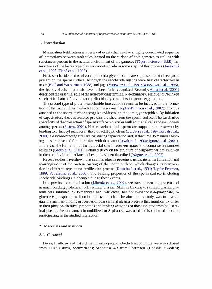
Fig. 6. RP-HPLC of mannan-binding proteins (Man+) obtained by affinity chromatography on mannan–Sepharose(a) and SDS-PAGE of separated protein fractions (b). Proteins were separated on a 218TP104 Vydac C18 columnin 0.05% TFA, with a linear gradient of 20–50% acetonitrile (60 min). Absorbance at 226 nm (—) and acetoni-trile gradient (- - -). Proteins in fractions 1–5. SDS-PAGE of protein fractions under non-reducing conditions.Molecular-mass standards – ovalbumin (45 000), carbonic anhydrase (31 000), soybean trypsin inhibitor (21 500),and lysozyme (14 400) were used.
column into three fractions: Man− fraction, which did not interact with the immobi-lized ligand, and Man+ fraction adsorbed to the affinity column and eluted with 3 MNaCl (Man1
+) and 4 M urea (Man2+). A small amount of Man1
+ proteins was elutedwith 3 M NaCl; the main portion of adsorbed proteins, Man2
+, was obtained using 4 Murea. No proteins present in the H− fraction interacted with immobilized mannan. Only asmall amount of protein was obtained from the affinity chromatography of boar seminalplasma.
Proteins adsorbed to the mannan–Sepharose column (Man1+ and Man2
+ fractions) wereseparated by RP-HPLC (Fig. 6a), by SDS electrophoresis (Fig. 6b), blotted onto PVDFmembrane and identified using N-terminal amino acid sequencing. The following proteinswere found in the mannan-binding fraction eluted from the affinity column with 3 M NaCland 4 M urea: DQH protein, differently glycosylated forms of AQN 2 and AQN 3, truncatedforms of AWN 1 and AWN 2 spermadhesins (Table 2). The composition of Man1
+ andMan2
+ fractions did not significantly differ.
P. Jelınkova et al. / Journal of Reproductive Immunology 62 (2004) 167–182 179
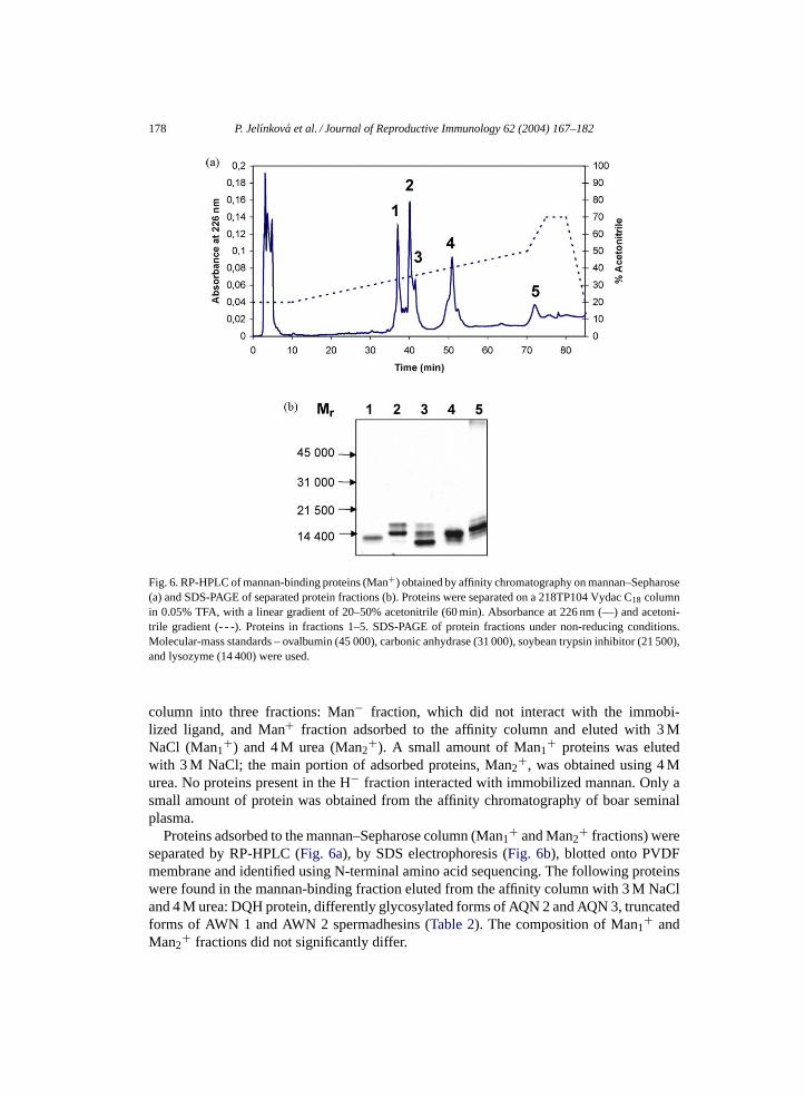
Table 2Proteins identified in the mannan-binding fraction
Fractiona Relativemolecular mass
N-terminal amino acid sequence Identified protein
1 13 000 DQHL . . . DQH protein2 14 000 AQNKGSD . . . AQN 2 spermadhesin3 12 000 AQNKGSD . . . AQN 3 spermadhesin4 14 000 RRS . . . , RSXG . . . , SRSXGVL . . . Truncated forms of
AWN 1 spermadhesin5 15 000 N-terminus blocked AWN 2 spermadhesin
a See Fig. 6.
4. Discussion
The saccharide-binding ability of sperm-associated proteins has been described in dif-ferent species (Suarez et al., 1998; Töpfer-Petersen, 1999). Many of these proteins fromseminal plasma originate in accessory sexual glands and associate with sperm during ejacu-lation. Lectin-type interactions with these proteins are an important phenomenon occurringin different phases of the fertilization process. Growing evidence suggests that such typeof interaction participates in the deposition of sperm in the isthmic region of oviduct afterits passage through uterus. The formation of the sperm reservoir in the oviduct is accom-plished due to the interaction of sperm surface proteins with the epithelial cells lining theduct (Suarez, 2001). It has been shown that the saccharide-binding specificity of proteinreceptors varies among species (Töpfer-Petersen et al., 2002). The formation of the oviduc-tal sperm reservoir in pig is the result of high-affinity sites for oligomannosyl residues andlow affinity for binding of d-galactose (Wagner et al., 2002). Ovalbumin and mannopentosewere found as the most potent inhibitors of sperm binding to the pig oviduct (Wagner et al.,2002).
In a previous communication, we have described that yeast mannan inhibited the inter-action of bull seminal plasma proteins with bovine zona pellucida (Liberda et al., 2002). Inthe present communication, we have studied the mannan-binding proteins in boar seminalplasma. Contrary to bull seminal plasma proteins (Liberda et al., 2002), the interaction ofboar proteins with mannan was inhibited most by ovalbumin, its separated N-glycans andovomucoid, slightly by d-galactose, but not by d-mannose and d-glucose. Direct bindingstudies proved an interaction of heparin-binding proteins with ovalbumin. The inhibitionstudies are in good correlation with results of inhibition of sperm binding to pig oviductalepithelium (Wagner et al., 2002). In addition to that, we have shown that boar seminalplasma heparin-binding proteins interacted with porcine oviductal epithelium and this in-teraction was inhibited by mannan. Boar seminal plasma proteins with mannan-bindingactivity are most probably responsible for formation of the porcine sperm oviductal reser-voir and thus they might participate in modulation or regulation of sperm capacitation inthe female oviduct.
The second difference between the interaction of boar seminal plasma proteins withmannan and that of bull proteins was the inhibition of heparin-binding activity of theseproteins by mannan. We have described (Liberda et al., 2001) that yeast mannan increased

180 P. Jelınkova et al. / Journal of Reproductive Immunology 62 (2004) 167–182
the heparin-binding activity of bull seminal plasma proteins. In the case of boar proteins,mannan strongly inhibited the interaction with this acidic polysaccharide. This might beexplained by different roles of proteins exhibiting the mannan-binding activity in the fertil-ization process of these two species.
The results of both methods used for studying mannan-binding activity (ELBA (Fig. 1)and specific detection of proteins after electrophoretic separation (Fig. 2)) were in anagreement. Proteins interacting with mannan belong to the heparin-binding ones. Affin-ity chromatography of boar seminal plasma proteins on immobilized mannan–Sepharoseconfirmed the results of the binding studies. Mannan-binding proteins were isolated from theheparin-binding fraction; no proteins were retained from the non-heparin-binding fraction.
The components of the mannan-binding fraction were identified according to relativemolecular-mass determination and N-terminal amino acid sequencing: the DQH protein,differently glycosylated forms of AQN, AWN 1 and AWN 2 spermadhesins. All theseproteins were found not only in seminal plasma, but also associated with ejaculated sper-matozoa (Jonákováet al., 1998; Veselský et al., 1999) and could be released from the spermsurface using acidic buffer (Jonáková et al., 1991; Dostálová et al., 1994).
These proteins adsorbed to the sperm surface might participate in the specific interactionof sperm with the saccharide moiety of the epithelium. The role of AQN spermadhesinsand not the AWN ones in the formation of the sperm oviductal reservoir has already beensuggested (Töpfer-Petersen et al., 2002). However, these suggestions are not in agreementwith the findings that a small portion of only AWN spermadhesin adsorbed to the spermsurface reaches the oviduct (Calvete et al., 1997).
The mannan-binding fraction of boar seminal plasma contains components that were alsopresent in the non-interacting fraction. The observed phenomenon may be explained by thepresence of different aggregated forms that differ in their binding properties (Jonákováet al.,2000; Manásková et al., 2000).
Acknowledgements
This work was supported by the Grant Agency of the Czech Republic, Grants Nos.303/02/0433 and 303/02/P069; by the Ministry of Education of the Czech Republic, GrantNo. MSM 1131-00001 Grant No. NJ/7463-3 from the Grant Agency of the Ministry ofHealth of the Czech Republic.
References
Amari, S., Yonezawa, N., Mitsui, K., Katsumata, T., Hamano, S., Kuwayama, M., Hashimoto, Y., Suzuki, A.,Takeda, Y., Nakano, M., 2001. Essential role of the non-reducing terminal �-d-mannosyl residues of theN-linked carbohydrate chain of bovine zona pellucida glycoproteins in sperm–egg binding. Mol. Reprod. Dev.59, 221–226.
Bleil, J.L., Wassarman, P.M., 1988. Galactose at the non-reducing terminus of O-linked oligosaccharides of mouseegg zona pellucida glycoprotein ZP3 is essential for the glycoprotein sperm receptor activity. Proc. Natl. Acad.Sci. U.S.A. 95, 6778–6782.

P. Jelınkova et al. / Journal of Reproductive Immunology 62 (2004) 167–182 181
Calvete, J.J., Ensslin, M., Mburu, J., Iborra, A., Martinez, P., Andermann, K., Waberski, D., Sanz, L.,Töpfer-Petersen, E., Weitze, K.F., Einarsson, S., Rodriguez-Martinez, H., 1997. Monoclonal antibodies againstboar sperm zona pellucida binding protein AWN 1. Characterization of continuous antigenic determinant andimmunolocalization of AWN epitopes in inseminated sows. Biol. Reprod. 57, 735–742.
Dostálová, Z., Calvete, J.J., Sanz, L., Töpfer-Petersen, E., 1994. Quantitation of boar spermadhesins in accessorysex gland fluids and on the surface of epididymial, ejaculated and capacitated spermatozoa. Biochim. Biophys.Acta 1200, 48–54.
Dubois, M., Gilles, K.A., Hamilton, J.K., Rebers, P.A., Smith, F., 1956. Anal. Chem. 18, 350–356.Green, C.E., Bredl, J., Holt, W.V., Wilson, P.F., Fazeli, A., 2001. Carbohydrate-mediation of boar sperm binding
to oviductal epithelial cells in vitro. Reproduction 122, 305–315.Haworth, W.N., Hirst, E.L., Isherwood, F.A., 1937. Polysaccharides. XXIV. Yeast Mannan. J. Chem. Soc., 784–791.Ignotz, G.G., Lo, M.C., Perez, C.L., Gwathmery, T.M., Suarez, S.S., 2001. Characterization of a fucose-binding
protein from bull sperms and seminal plasma that may be responsible for formation of oviductal sperm reservoir.Biol. Reprod. 64, 1806–1811.
Jonáková, V., Sanz, L., Calvete, J.J., Henschen, A., Cechová, D., Töpfer-Petersen, E., 1991. Isolation and bio-chemical characterization of a zona pellucida-binding glycoprotein of boar spermatozoa. FEBS Lett. 280,183–186.
Jonáková, V., Tichá, M., Kraus, M., Cechová, D., 1995. Multifuntional sperm protein in gametic interaction.Fertilität 11, 115–118.
Jonáková, V., Kraus, M., Veselský, L., Cechová, D., Bezouška, K., Tichá, M., 1998. Spermadhesins of the AQNand AWN family, DQH sperm surface protein and HNK protein in the heparin-binding fraction of boar seminalplasma. J. Reprod. Fertil. 114, 25–34.
Jonáková, V., Manásková, P., Kraus, M., Liberda, J., Tichá, M., 2000. Sperm surface proteins in mammalianfertilization. Mol. Reprod. Dev. 56, 275–277.
Laemmli, U.K., 1970. Cleavage of structural proteins during the assembly of the head of bacteriophage T4. Nature227, 680–685.
Lefebvre, R., Lo, M.C., Suarez, S.S., 1997. Bovine sperm binding to oviductal epithelium involves fucose recog-nition. Biol. Reprod. 56, 1198–1204.
Liberda, J., Ryšlavá, H., Jelınková, P., Jonáková, V., Tichá, M., 2002. Affinity chromatography of bull seminalproteins on mannan–Sepharose. J. Chromatogr. B 780, 231–239.
Liberda, J., Jonáková, V., Tichá, M., 1997. Preparation of fluorescein-labeled and biotinylated derivatives ofpolysaccharides for lectin-saccharide binding studies. Biotechnol. Tech. 11, 265–267.
Liberda, J., Kraus, M., Ryšlavá, H., Vlasáková, M., Jonáková, V., Tichá, M., 2001. d-Fructose-binding proteinsof bull seminal plasma: isolation and characterization. Folia Biol. (Praha) 47, 113–119.
Manásková, P., Liberda, J., Tichá, M., Jonáková, V., 2000. Aggregated and monomeric forms of proteins of boarseminal plasma: characterization and binding properties. Folia Biol. (Praha) 46, 143–151.
Novotná, V., Mikeš, L., Horák, P., Jonáková, V., Tichá, M., 1996. Preparation of water-soluble and water insolublepoly(acrylamide-allylamine) derivatives of polysaccharides. Int. J. Bio-Chromatogr. 2, 37–47.
Petrunkina, A.M., Harrison, E.A.P., Töpfer-Petersen, E., 2000. Only low levels of spermadhesin AWN are de-tectable on the surface of live ejaculated boar spermatozoa. Reprod. Fertil. Dev. 12, 361–371.
Revah, I., Gadella, B.M., Flesch, F.M., Colenbrander, B., Suarez, S.S., 2000. Changes in capacity of bull spermto bind fucose. Biol. Reprod. 62, 1010–1015.
Stewart, T.S., Ballou, C.E., Clinton, E., 1968. A comparison of yeast mannans and phosphomannnans. Biochemistry7, 1855–1863.
Suarez, S.S., Revah, I., Lo, M., Kolle, S., 1998. Bull sperm binding to oviductal epithelium is mediated by aCa2+-dependent lectin on sperm that recognizes Lewis-a trisaccharide. Biol. Reprod. 59, 39–44.
Suarez, S.S., 2001. Carbohydrate-mediated formation of the oviductal sperm reservoir in mammals. Cells TissuesOrgans 168, 105–112.
Tichá, M., Železná, B., Jonáková, V., Filka, K., 1994. Immobilization of heparin on polyacrylamide derivatives.J. Chromatogr. B 656, 423–426.
Tichá, M., Kraus, M., Cechová, D., Jonáková, V., 1998. Saccharide-binding properties of boar AQN spermadhesinsand DQH surface protein. Folia Biol. (Praha) 44, 15–21.
Töpfer-Petersen, E., 1999. Carbohydrate-based interactions on the route of a sperm to fertilization. Hum. Reprod.Update 5, 314–329.

182 P. Jelınkova et al. / Journal of Reproductive Immunology 62 (2004) 167–182
Töpfer-Petersen, E., Wagner, A., Friedrich, J., Petrunkina, A., Ekhlasi-Hundrieser, M., Waberski, M., Drommer,W., 2002. Function of mammalian oviductal sperm reservoir. J. Exp. Zool. 292, 210–215.
Towbin, H., Stachelin, T., Gordon, J., 1979. Electrophoretic transfer of proteins from polyacrylamide gels tonitrocellulose sheets. Procedure and some applications. Proc. Natl. Acad. Sci. U.S.A. 76, 4350–4354.
Veselský, L., Peknicová, J., Cechová, D., Kraus, M., Geussová, G., Jonáková, V., 1999. Characterization of boarspermadhesins by monoclonal and polyclonal antibodies and their role in binding to oocytes. Am. J. Reprod.Immunol. 42, 187–197.
Wagner, A., Ekhlasi-Hundrieser, M., Hettel, C., Petrunkina, A., Waberski, D., Nimtz, M., Töpfer-Petersen, E.,2002. Carbohydrate-based interactions of oviductal sperm reservoir formation, studies in pigs. Mol. Reprod.Dev. 61, 249–257.
Yonezawa, N., Aoki, H., Hatanaka, Y., Nakano, M., 1995. Involvement of N-linked carbohydrate chains of pigzona pellucida in sperm–egg binding. Eur. J. Biochem. 233, 35–41.
Yurewicz, E.C., Pack, B.A., Sacco, A.G., 1991. Isolation, composition and biological activity of sugar chains ofporcine oocyte zona pellucida 55 K glycoproteins. Mol. Reprod. Dev. 30, 126–234.