An Experimental Analysis of Data Annotation Methodologies ...
Upload
independentCategory
view
0download
0
Functional Annotation of Two New Carboxypeptidases from theAmidohydrolase Superfamily of Enzymes†
Dao Feng Xiang‡, Chengfu Xu‡, Desigan Kumaran§, Ann C. Brown£, J. Michael SauderΨ,Stephen K. BurleyΨ, Subramanyam Swaminathan§,*, and Frank M. Raushel‡,*‡ Department of Chemistry, P.O. Box 30012, Texas A&M University, College Station, Texas 77845§ Biology Department, Brookhaven National Laboratory, Upton, New York 11973£ Medgar Evers College, Brooklyn, New York 11225Ψ SGX Pharmaceuticals, 10505 Roselle Street, San Diego, California 92121
AbstractTwo proteins from the amidohydrolase superfamily of enzymes were cloned, expressed and purifiedto homogeneity. The first protein, Cc0300, was from Caulobacter crescentus CB-15 (Cc0300) whilethe second one (Sgx9355e) was derived from an environmental DNA sequence originally isolatedfrom the Sargasso Sea (gi| 44371129). The catalytic functions and the substrate profiles for the twoenzymes were determined with the aid of combinatorial dipeptide libraries. Both enzymes wereshown to catalyze the hydrolysis of L-Xaa-L-Xaa dipeptides where the amino acid at the N-terminuswas relatively unimportant. These enzymes were specific for hydrophobic amino acids at the C-terminus. With Cc0300, substrates terminating in isoleucine, leucine, phenylalanine, tyrosine, valine,methionine, and tryptophan were hydrolyzed. The same specificity was observed with Sgx9355e butthis protein was also able to hydrolyze peptides terminating in threonine. Both enzymes were ableto hydrolyze N-acetyl and N-formyl derivatives of the hydrophobic amino acids and tripeptides. Thebest substrates identified for Cc0300 were L-Ala-L-Leu with values of kcat and kcat/Km of 37 s−1 and1.1 × 105 M−1 s−1, respectively, and N-formyl-L-Tyr with values of kcat and kcat/Km of 33 s−1 and3.9 × 105 M−1 s−1, respectively. The best substrate identified for Sgx9355e was L-Ala-L-Phe willvalues of kcat and kcat/Km of 0.41 s−1 and 5.8 × 10³ M−1 s−1. The three-dimensional structure ofSgx9355e was determined to a resolution of 2.33 Å with L-methionine bound in the active site. Theα-carboxylate of the methionine is ion-paired to His-237 and also hydrogen bonded to the backboneamide groups of Val-201 and Leu-202. The α-amino group of the bound methionine interacts withAsp-328. The structural determinants for substrate recognition were identified and compared withother enzymes in this superfamily that hydrolyze dipeptides with different specificities.
Complete bacterial genomes are now being sequenced at an extraordinary rate (1). This influxof new information has provided an unprecedented view of the evolutionary relationships thatexist among enzymes that recognize different substrates and catalyze diverse functionaltransformations. The pace for the deposition of new three dimensional structures in the ProteinData Bank (PDB1) has also risen sharply in recent years (2). However, it is apparent that a
†This work was supported by the NIH (GM71790 and GM74945) and the Hackerman Advanced Research Program (010366-0034-2007).The x-ray coordinates and structure factors for Sgx9355e have been deposited in the Protein Data Bank (PDB accession code: 2qs8)*To whom correspondence may be sent: (FMR) telephone: (979)-845-3373; fax (979)-845-9452; [email protected], (SS) telephone:(631)-344-3187; fax (631)-344-3407; [email protected].ΨCurrent affiliation is Eli Lilly & Company.1Abbreviations: Protein Data Bank, PDB; American Type Culture Collection, ATCC; inductively coupled plasma emission-massspectrometry, ICP-MS;
NIH Public AccessAuthor ManuscriptBiochemistry. Author manuscript; available in PMC 2010 June 2.
Published in final edited form as:Biochemistry. 2009 June 2; 48(21): 4567–4576. doi:10.1021/bi900453u.
NIH
-PA Author Manuscript
NIH
-PA Author Manuscript
NIH
-PA Author Manuscript

significant fraction of the newly sequenced genes have an unknown, uncertain or incorrectcatalytic functional assignment (3). This reality suggests that a considerable number of newmetabolic transformations remain to be elucidated and that the annotation of function fromsequence and structural information alone is a challenging problem. Nevertheless, the vastamount of structure and sequence information provides a unique opportunity for the elucidationof the mechanisms for the evolution of new catalytic functions from pre-existing structuraltemplates.
One approach to the assignment of function for enzymes with an unknown substrate profile isthe utilization of enzyme superfamilies. Comprehensive amino acid sequence alignments andthree dimensional structural comparisons can provide valuable insights toward theidentification of the catalytic and structural motifs that dictate the substrate and reaction profiles(4). One such superfamily that has proven to be amenable to this approach is theamidohydrolase superfamily (5,6). This superfamily was first identified by Holm and Sanderwho recognized the structural and functional relationships among adenosine deaminase,urease, and phosphotriesterase (7). All enzymes from this superfamily adopt a (β/α)8-barrelstructural fold with their active sites located at the C-terminal face of the β-barrel (5). Withinthe active site is a mono- or binuclear metal center that functions to activate solvent water fornucleophilic attack and/or stabilization of the transition state. Most of the enzymes within thissuperfamily catalyze the hydrolysis of C-O, C-N, or P-O bonds (5). However, some membersat the periphery of the superfamily catalyze decarboxylation, hydration, and isomerizationreactions (8).
In this paper we have interrogated the catalytic functions for two enzymes within theamidohydrolase superfamily. The first of these enzymes, Cc0300 from Caulobactercrescentus CB15, is currently annotated by NCBI as an Xaa-Pro dipeptidase. The secondenzyme, derived from an environmental DNA sequence isolated from the Sargasso Sea andlabeled here as Sgx9355e, is 39% identical in amino acid sequence with Cc0300. We haverecently determined the catalytic functions for two other putative Xaa-Pro dipeptidases fromC. crescentus, Cc2672 and Cc3125, that are ~40% identical in sequence to Cc0300 (9). Thesetwo enzymes do not catalyze the hydrolysis of Xaa-Pro dipeptides but they do catalyze thehydrolysis of L-Xaa-LArg/Lys dipeptides and thus the current functional annotations for theseenzymes are incorrect. The X-ray structure of a catalytic homologue to Cc2672 and Cc3125has unveiled the structural motif responsible for the recognition of the cationic side chains ofarginine and lysine containing substrates in these enzymes. In Cc0300, this recognition motifis missing, suggesting that the substrate specificity for Cc0300 differs from that determinedpreviously for Cc2672 and Cc3125. The substrate profiles for Cc0300 and Sgx9355e weredetermined by screening multiple dipeptide libraries containing nearly all possiblecombinations of L-Xaa-L-Xaa dipeptides made from the 20 common amino acids. The X-raystructure of the methionine-bound form of Sgx9355e has been determined to a resolution of2.33 Å.
Materials and MethodsMaterials
Genomic DNA from C. crescentus CB15 was purchased from ATCC (American Type CultureCollection). The synthesis of oligonucleotides and DNA sequencing reactions were performedby the Gene Technology Laboratory of Texas A&M University. The pET-30a(+) expressionvector was obtained from Novagen. T4 DNA ligase and various restriction enzymes wereacquired from New England Biolabs. Platinum Pfx DNA polymerase was purchased fromInvitrogen. The Wizard Plus SV Mini-Prep DNA purification kit was obtained from Promega.Chromatographic columns and resins were from G.E. Healthcare. Chelex-100 resin waspurchased from BioRad. ICP standards were purchased from Inorganic Ventures Inc. The
Xiang et al. Page 2
Biochemistry. Author manuscript; available in PMC 2010 June 2.
NIH
-PA Author Manuscript
NIH
-PA Author Manuscript
NIH
-PA Author Manuscript

tripeptides, L-Gly-L-Phe-L-Arg and L-Gly-L-Ala-L-Tyr, were purchased from ArozTechnologies LLC. NAD and formate dehydrogenase from Candida boidinii were purchasedfrom Sigma. The N-methyl phosphonate derivative of L-leucine was synthesized according tothe method described by Xu et al. (10). The structure of this compound is presented in Scheme1. All other buffers, purification reagents and other chemicals used in this work were purchasedfrom Sigma-Aldrich, unless otherwise stated.
Synthesis of Dipeptides and Dipeptide LibrariesSyntheses of the L,L-dipeptide libraries were conducted as reported previously (9). Massspectrometric analysis (ESI, positive and negative mode) was employed to verify thecomponents of each library. The individual dipeptides L-Ala-L-Leu, L-Ala-L-Phe and L-Asp-L-Leu were prepared in a manner similar to the synthesis of the dipeptide libraries.
Gene cloningStandard molecular cloning techniques were employed throughout (11). A 1287-bp fragmentcontaining the entire open reading frame of Cc0300 was amplified from the genomic DNA ofC. crescentus CB15 using the primer pair 5′ AGAACTTCCATATGCGTAAATTGATGGCGGGCGCCTGC 3′ (forward) and 5′ACGGAATTCTTACTTGTAGACGA CGCCGCCCTTGATGAC 3′ (reverse). The forwardand reverse primers were designed to introduce an NdeI restriction site and an EcoRI restrictionsite, respectively. The PCR product, purified using a PCR clean-up system (Promega) anddigested with NdeI and EcoRI, was ligated into the same sites of the expression vector pET-30a(+). The cloned fragment was completely sequenced to verify the fidelity of the PCRamplification. Based upon the reported sequence of gi|44371129 deduced from a DNA sampleoriginally isolated from the Sargasso Sea, the New York SGX Research Center for StructuralGenomics (NYSGXRC, www.nysgxrc.org) cloned and expressed Sgx9355e as a His-taggedprotein following codon-optimization and gene synthesis by Codon Devices, Inc (Cambridge,MA). NYSGXRC will make the clone (9355e1BCt10p) available through the Protein StructureInitiative Material Repository (PSI-MR), accessible from the PSI Knowledgebase(http://kb.psi-structuralgenomics.org/KB/psi_resources.html).
Protein PurificationThe plasmid for Cc0300 was constructed for expression under control of the IPTG-inducibleT7 RNA polymerase. BL21(DE3) Star cells (Novagen) were transformed with the plasmidcontaining the gene for Cc0300. A single colony was used to inoculate a 5-mL overnight cultureof LB medium containing 50 μg/mL kanamycin. The 5-mL overnight culture was then usedto inoculate 1.0 L of LB medium containing 50 μg/mL kanamycin. The cells were grown at30 °C and induced by adding IPTG at a final concentration of 0.5 mM when an OD600 of theculture reached 0.6. Zn(OAc)2, 1.0 mM, was added to the culture and the cells harvested bycentrifugation (6,000 rpm for 15 minutes) 14 hours after IPTG induction and stored at −80 °C. To purify Cc0300, 10 g of frozen cells were thawed and resuspended in 50 mL of 50 mMHepes, pH 8.0. The cells were disrupted by sonication in an ice bath and the soluble fractionwas isolated by centrifugation. The supernatant solution was treated with 2% (w/v) protaminesulfate to precipitate nucleic acids, which were removed by centrifugation. The supernatantsolution was treated with ground ammonium sulfate until 60% saturation. The pellet from theammonium sulfate fractionation step was dissolved in a minimal volume of 50 mM Hepes, pH8.0, and then passed through a 0.2 μm pore filter prior to loading onto a pre-equilibrated HiLoad 26/60 Superdex 200 prep grade gel filtration column (Amersham Biosciences). Fractionscontaining the target protein were pooled based on SDS-PAGE analysis. The protein wasfurther purified by loading onto a Resource Q anion exchange column (Amersham Biosciences)and eluted with a linear gradient of NaCl in 20 mM Hepes, pH 8.0. The appropriate fractions
Xiang et al. Page 3
Biochemistry. Author manuscript; available in PMC 2010 June 2.
NIH
-PA Author Manuscript
NIH
-PA Author Manuscript
NIH
-PA Author Manuscript

were pooled after analysis by SDS-PAGE. Protein concentrations were determined using thecalculated extinction coefficient of 31,860 M−1cm−1 at 280 nm (12).
For purification of Sgx9355e, E. coli BL21 (DE3) Star cells were transformed with the plasmidencoding the gene for Sgx9355e. One liter of LB medium was inoculated with a 5 mL overnightculture. The inoculated culture was grown with agitation at 30 °C to an OD600 of 0.6 and thensupplemented with 1.0 mM Zn(OAc)2 and induced with IPTG at a final concentration of 0.5mM. The cells were grown at 30 °C for another 14 h and then harvested by centrifugation (6000rpm for 15 minutes). The cell pellet was resuspended in binding buffer (20 mM Hepes, pH 7.9,0.5 M NaCl and 5 mM imidazole). The cells were disrupted by sonication and the insolubledebris was removed by centrifugation (10,000 rpm for 15 minutes). The clarified cell extractwas applied to a 24 mL column of chelating Sepharose Fast Flow resin (AmershamBiosciences) charged with Ni2+ and pre-equilibrated with binding buffer. The column waswashed thoroughly with 1000 mL of binding buffer until the absorbance of the flow-throughat 280 nm did not change. The His-tagged protein, Sgx9355e, was eluted with a linear gradientof elution buffer (10 mM Hepes, pH 7.9, 0.25 M NaCl and 0.5 M imidazole). The proteinobtained was further purified by application to a Resource Q anion exchange column after theNaCl was removed by dialysis. Sgx9355e was eluted with a linear gradient of NaCl in 20 mMHepes, pH 8.0. The protein was greater than 95% pure as judged by SDS-PAGE analysis.
Amino Acid Sequence VerificationN-terminal amino acid sequence analysis of the purified Cc0300 was conducted by the ProteinChemistry Laboratory at Texas A&M University. The amino acid sequence obtained for thefirst 8 amino acid residues of Cc0300 was QATFVQAG, which indicates that the first 23 aminoacid residues of the protein were either post-translationally removed via proteolysis or theprotein was initially expressed from the 24th amino acid residue of the reported gene sequence.
Metal AnalysisApo-Sgx9355e was made by incubating the enzyme with 2.0 mM 1,10-phenanthroline for 24h at 4 °C. The chelator was removed by loading the protein/chelator mixture onto a PD-10column (GE Health care) and eluting with 50 mM metal-free Hepes buffer, pH 8.0. The metal-free Hepes buffer was made by passing through a Chelex-100 column to remove possible metalsalts present in the buffer. All glassware used for making metal-free buffer was soaked with10 mM EDTA overnight and then rinsed thoroughly with deionized water. The apo-Sgx9355ewas reconstituted with 2 equivalents of ZnCl2 in 50 mM metal-free Hepes, pH 8.0, in thepresence of 10 mM potassium bicarbonate. The metal content of as-purified and reconstitutedproteins was determined by inductively coupled plasma emission-mass spectrometry (ICP-MS) (13).
Assay Methods for Peptidase ActivityA modified colorimetric ninhydrin assay method was used to screen the dipeptidase activityof Cc0300 and Sgx9355e (14). The same method was used to measure the hydrolysis oftripeptides and the hydrolytic activity with N-acetyl- and N-formyl derivatives of L-aminoacids. The absorbance at 507 nm was recorded with a SPECTRAmax plate reader fromMolecular Devices. Quantitative analysis of the liberated amino acids was determined by theProtein Chemistry Lab at Texas A&M University.
Screening of Dipeptide LibrariesCc0300 and Sgx9355e were tested for dipeptidase activity with the entire dipeptide library(361 compounds in all). Each dipeptide library consisted of a mixture of dipeptides with anidentical L-amino acid at the N-terminus but 19 different amino acids at the C-terminus. L-
Xiang et al. Page 4
Biochemistry. Author manuscript; available in PMC 2010 June 2.
NIH
-PA Author Manuscript
NIH
-PA Author Manuscript
NIH
-PA Author Manuscript

Cysteine was not included in any of the libraries. Screening of the dipeptide libraries wasinitiated by mixing a fixed concentration of the dipeptide library (containing ~0.1 mM of eachdipeptide) and various amount of enzyme over a concentration range of 10 nM to 1000 nM.All reactions were conducted in 50 mM Hepes, pH 8.0 in 96-well microplates. The reactionswere incubated at 30 °C for a fixed time period and then quenched by adding the Cd-ninhydrinreagent. The quenched reactions were incubated for 5 minutes at 80 °C for color development.Control reactions without enzyme or without the dipeptide library were conductedsimultaneously. The relative rates were determined by a fit of the data to equation 1, where yis the change in absorbance at 507 nm, x is concentration of enzyme and b is the relative rateconstant. In these measurements the enzyme concentration was varied but the reaction timewas fixed.
(1)
Amino Acid AnalysisThe specific amino acids that were released from the dipeptide libraries were quantitativelyidentified by HPLC. The substrate specificity for either Cc0300 or Sgx9355e was determinedby assay with selected dipeptide libraries. The hydrolysis reactions were conducted in 25 mMammonium bicarbonate buffer, pH 8.0. For each dipeptide library assay, a mixture of 19dipeptides at ~0.1 mM was subjected to hydrolysis with variable concentrations of enzymeranging from 1 nM to 1000 nM. The identity of the amino acids released from the hydrolysisreactions was determined by amino acid analysis. Two dipeptide libraries (L-Ala-L-Xaa andL-Asp-L-Xaa) were assayed with Cc0300 and two dipeptide libraries (L-Ala-L-Xaa and L-Gln-L-Xaa) were used in the assays with Sgx9355e. The enzyme and dipeptide library wereincubated at 30 °C for 14 h in a 96-well microplate and then the enzyme was removed bypassage through a filter. Quantitative amino acid analysis was conducted after the sampleswere dried under reduced pressure and reconstituted with water. The relative reaction rateswere determined by fitting the change in amino acid concentration as a function of enzymeconcentration to equation 1.
Kinetic Measurements for Individual SubstratesSelected dipeptides (L-Ala-L-Leu, L-Ala-L-Phe, L-Asp-L-Leu), N-formyl-L-amino acids (N-formyl-L-Leu, N-formyl-L-Ile, N-formyl-L-Phe, N-formyl-L-Tyr, N-formyl-L-Met, N-formyl-L-Val), N-acetyl-L-amino acids (N-acetyl-L-Leu, N-acetyl-L-Ile, N-acetyl-L-Phe, N-acetyl-L-Tyr, N-acetyl-L-Met, N-acetyl-L-Val) and the tripeptide, L-Gly-L-Ala-L-Tyr, wereused as substrates for measurement of the kinetic parameters for Cc0300 and Sgx9355e. Thekinetic assays were conducted in 50 mM Hepes, pH 8.0, and the products were quantified usingthe ninhydrin-based assay. The kinetic constants, kcat, Km and kcat/Km, were determined byfitting the initial velocity data to equation 2, where ν is the initial velocity, kcat is the turnovernumber, Et is the enzyme concentration, A is the substrate concentration, and Km is theMichaelis constant.
(2)
Inhibition by Tetrahedral AnalogueThe inhibitory properties of the N-methyl phosphonate derivative of L-leucine (1) with Cc0300were determined using N-formyl-L-leucine as the substrate. The assays were conducted bymonitoring the rate of formate production using a formate dehydrogenase coupling system in
Xiang et al. Page 5
Biochemistry. Author manuscript; available in PMC 2010 June 2.
NIH
-PA Author Manuscript
NIH
-PA Author Manuscript
NIH
-PA Author Manuscript

the presence of NAD. The reaction was followed spectrophotometrically at 340 nm, using aSPECTRAmax (Molecular Devices) plate reader. All assays were conducted in a 96-well UVplate in a volume of 250 μL. The buffer used in all assays was 50 mM Hepes, pH 8.0. Theconcentrations of N-formyl-L-leucine and the N-methyl phosphonate derivative of L-leucinewere varied in the range of 50–200 μM and 1–100 μM, respectively. For data analysis, theinitial rates for substrate hydrolysis in the presence of the inhibitor at different concentrationsof the substrate were fitted to equation 3 for competitive inhibition. In this equation, νis theinitial velocity. A is the substrate concentration, I is the inhibitor concentration, kcat is theturnover number, Et is the enzyme concentration, A is the substrate concentration. Km is theMichaelis constant for the substrate, and the Ki is the competitive inhibition constant.
(3)
Crystallization and Data Collection for Sgx9355eCrystals of the selenomethionine form of Sgx9355e protein were grown using the sitting dropvapor diffusion method at 20 °C. Equal volumes of protein (10–15 mg/mL) and a solutioncontaining 30% MPD, 0.1 M Hepes, 0.2 M NaCl, pH 7.0, were mixed and equilibrated againsta 800 μL reservoir containing the same precipitant solution. A single crystal was flash frozenby adding 20% glycerol to the mother liquor before the crystal was loop mounted. SAD dataextending to 2.33 Å were collected at the selenium absorption edge (λ = 0.979 Å) from a singlecrystal at liquid nitrogen temperature using the ADSC QUANTUM 315 detector at the NationalSynchrotron Light Source (NSLS) using beamline X29. X-ray diffraction images wereprocessed with HKL2000 and the data collection statistics are given in Table 1 (15). Thecrystals belong to the monoclinic space group I4 with cell dimensions, a = b = 144.9, c = 101.0Å (Table 1). Assuming two molecules of 45,781 Da per asymmetric unit, the Matthewscoefficient is 2.0 Å3 Da−1 corresponding to an estimated solvent content of 47% by volume ofthe unit cell.
Structure Determination and Refinement of Sgx9355eThe structure was determined by the single-wavelength anomalous dispersion method. Twentytwo out of the possible twenty four selenium positions in the asymmetric unit were locatedusing SHELXD (16). These selenium positions were refined and phases calculated withSHARP followed by solvent flattening (17,18). The resulting experimental electron densitymap was of excellent quality and revealed practically all of the secondary structural elementscomprising the dimer. The automated model building program ARP/wARP was then used tobuild the model into the electron density map (19). This process built about 87% of eachprotomeric polypeptide chain and the remainder of the model was completed using the graphicsprogram ‘O’ (20,21). The structure was refined with the program Crystallography and NMRsystem (CNS) (22). Cycles of manual rebuilding and structure refinement were continued untilthe convergence of R and Rfree. Phasing and refinement statistics are provided in Table 1. Thefinal model was validated with the program PROCHECK (23). Atomic coordinates andstructure factors have been deposited in the Protein Data Bank (PDB id: 2qs8).
1. ResultsPurification of Cc0300 and Sgx9355e
Cc0300 from C. crecentus was expressed in reasonable quantities after the plasmid containingthe gene for this enzyme was transformed in BL21(DE3) Star cells and allowed to grow in anLB medium supplemented with 1.0 mM Zn(OAc)2. The enzyme was purified and SDS-PAGEanalysis showed a single protein band which was slightly smaller than the theoretical molecular
Xiang et al. Page 6
Biochemistry. Author manuscript; available in PMC 2010 June 2.
NIH
-PA Author Manuscript
NIH
-PA Author Manuscript
NIH
-PA Author Manuscript

weight of 44 kDa as calculated from the reported gene sequence. N-terminal protein sequenceanalysis of the purified Cc0300 indicated that the first 23 amino acids were missing. This resultindicates that either these residues were lost through proteolysis or that this gene has an alternateinitiation site. The as-purified protein was found to contain an average of 1.6 equivalents ofZn2+ per subunit. Sgx9355e was purified as a His-tagged protein and was found to contain 0.2equivalents of Zn per subunit. However, the Zn content could be increased to an average of1.2 equivalents per subunit by incubating apo-Sgx9355e with 2 equivalents of ZnCl2 in thepresence of 10 mM bicarbonate overnight.
Crystal Structure of Sgx9355eThe structure of Sgx9355e is shown in Figures 1 and 2. Within the crystal, the protein occursas a homo octamer with approximately 3759 Å2 buried surface area (~19% of the total surfacearea) per protein-protein interaction (Figure 1). Each subunit consists of two domains, a smallpredominantly β-stranded domain (residues 7–64 and 379–412) and a large TIM barrel domain(residues 65–370) as presented in Figure 2. The active site is located at the bottom of the cone-shaped structure of each subunit and is open to solvent.
The residues that are expected to bind the two divalent cations are His-69, His-71, His-235,His-255, Lys-194 and Asp-328 based upon the known homology with other members of theamidohydrolase superfamily (5). The relative orientations of these presumptive metal bindingresidues are presented in Figure 3. However, the enzyme crystallized without zinc and a singlemagnesium ion was found ligated to the α-metal site. The magnesium is coordinated to His-69,His-71 and Asp-328. Interestingly, methionine, from the crystallization medium, is bound inthe active site close to the position normally occupied by the more solvent exposed β-metal.The α-amino group of the methionine interacts with the side chain carboxylate of Asp-328.The α-carboxylate of the bound methionine is ion-paired with His-237 and hydrogen bondedwith the backbone amide groups of Val-201 and Ile-202 as illustrated in Figure 4. The sidechain of methionine is found within a pocket formed by the side chains of Thr-277 and Ile-309.
Screening of Dipeptide LibrariesTo determine the substrate profile of Cc0300 and Sgx9355e, a total of 19 dipeptide librarieswere tested for catalytic activity using the Cd-ninhydrin based assays. The relative preferencesfor the N-terminal amino acid residue within these dipeptide libraries are depicted in Figure 5.Cc0300 and Sgx9355e exhibit broad catalytic activity for the hydrolysis of these peptidelibraries with different N-termini but the relative rates of hydrolysis vary from library to library.These two enzymes show relatively high reaction rates with L-Trp-L-Xaa and low reactionrates with L-Ile-L-Xaa and L-Val-L-Xaa. Cc0300 shows a higher reaction rate with L-Tyr-L-Xaa and L-His-L-Xaa compared with Sgx9355e. Conversely, Sgx9355e exhibits a higherturnover rate with L-Lys-L-Xaa and L-Arg-L-Xaa compared with Cc0300.
Specificity of Cc0300 and Sgx9355eQuantitative amino acid analysis (AAA) was employed to determine the relative rates ofhydrolysis for specific dipeptides within selected dipeptide libraries. Cc0300 was assayed withthe L-Ala-L-Xaa and L-Asp-L-Xaa dipeptide libraries. The amino acid products, detected fromthe reaction mixture of Cc0300 with the L-Ala-L-Xaa library, were limited to alanine, leucine,isoleucine, phenylalanine, tryptophan, methionine, tyrosine and valine. The amino acidproducts detected from the hydrolysis reaction using the L-Asp-L-Xaa library as a substratewere aspartate, leucine, isoleucine, phenylalanine, tryptophan, methionine, tyrosine and valine.Since alanine and aspartate were only detected when libraries containing these amino acidswere at the N-terminus, it can be concluded that the C-terminal specificity of Cc0300 withdipeptides is limited to the hydrophobic amino acid residues leucine, isoleucine, phenylalanine,tryptophan, methionine, tyrosine and valine. Two libraries, L-Ala-L-Xaa and L-Gln-L-Xaa,
Xiang et al. Page 7
Biochemistry. Author manuscript; available in PMC 2010 June 2.
NIH
-PA Author Manuscript
NIH
-PA Author Manuscript
NIH
-PA Author Manuscript

were selected for quantitative analysis with Sgx9355e for specificity at the C-terminus. Aminoacid analysis showed that the C-terminal specificity of Sgx9355e with these dipeptidesubstrates is very similar to that found for Cc0300 except that Sgx9355e can hydrolyzedipeptides with threonine at the C-terminus. The relative reaction rates for Cc0300 andSgx9355e are presented in Figure 6. The relative rates of hydrolysis for the other dipeptides inthese libraries are less than 5% for both Cc0300 and Sgx9355e.
Hydrolysis of N-substituted Amino Acids and TripeptidesCc0300 and Sgx9355e were screened for catalytic activity with N-acetyl-L-Xaa and N-formyl-L-Xaa derivatives. These two enzymes showed very similar catalytic specificities with thesetwo types of substituted amino acid derivatives. The hydrolyzed substrates were limited to theN-acetyl- and N-formyl derivatives of L-hydrophobic amino acids leucine, isoleucine,phenylalanine, tryptophan, methionine, tyrosine and valine. Cc0300 and Sgx9355e were alsotested with two tripeptides: L-Gly-L-Phe-L-Arg and L-Gly-L-Ala-L-Tyr. These two enzymeswere unable to hydrolyze L-Gly-L-Phe-L-Arg but were able to hydrolyze the C-terminal L-tyrosine from L-Gly-L-Ala-L-Tyr.
Kinetic Constants for Cc0300 and Sgx9355eThe kinetic constants for Cc0300 were determined using the following substrates: L-Ala-L-Leu, L-Ala-L-Phe, L-Asp-L-Leu, N-formyl-L-Leu, N-formyl-L-Ile, N-formyl-L-Phe, N-formyl-L-Met, N-formyl-L-Tyr, N-formyl-L-Val, N-acetyl-L-Leu, N-acetyl-L-Ile, N-acetyl-L-Phe, N-acetyl-L-Met, N-acetyl-L-Tyr and N-acetyl-L-Val. Also tested was the tripeptide, L-Gly-L-Ala-L-Tyr. The kinetic data were fit to equation 2 and the values of kcat, Km, and kcat/Km for the hydrolysis of the selected substrates are presented in Table 2. The turnover numbersranged from 1.7 −1 for the hydrolysis of N-Acetyl-L-Met to 37 s−1 for the hydrolysis of L-Ala-L-Leu. The kinetic constants for the hydrolysis of selected compounds by Sgx9355e arepresented in Table 2. In general, the kinetic constants, kcat and kcat/Km, for Cc0300 are greaterthan those measured for Sgx9355e.
Inhibition by N-methyl Phosphonate Derivative (1)Compound 1 was tested as an inhibitor of Cc0300. This compound was found to be acompetitive inhibitor of this enzyme when N-formyl-L-leucine was used as the substrate. Aninhibition constant of 2.3 ± 0.2 μM was obtained from a fit of the data (not shown) to equation3.
DiscussionElucidation of the substrate specificity for any enzyme given the amino acid sequence and/orthree-dimensional structure alone is a difficult and demanding problem for most proteins. Therapid sequencing of whole bacterial genomes has provided an explosion of uncharacterizedenzymes whose functions cannot be reliably annotated based upon a traceable homology toproteins of known function. Our approach to this problem is to address an entire enzymesuperfamily simultaneously in an attempt to reveal the evolutionary lineages for the emergenceof new catalytic functions and to provide a structural framework for the deciphering of substrateprofiles from the amino acid sequences alone. Over 6,000 proteins with unique amino acidsequences have been classified as members of the amidohydrolase superfamily (24). To datemore than 30 distinct reactions have been shown to be catalyzed by members of this superfamilyusing amino acid, carbohydrate, and nucleic acid derived substrates (5,7,24).
The substrate specificity for two related enzymes was examined in this investigation. Cc0300from C. crecentus and Sgx9355e from an unknown bacterium found in the Sargasso Sea arecurrently annotated as Xaa-Pro dipeptidase in the NCBI. Sequence comparisons have
Xiang et al. Page 8
Biochemistry. Author manuscript; available in PMC 2010 June 2.
NIH
-PA Author Manuscript
NIH
-PA Author Manuscript
NIH
-PA Author Manuscript

established that both proteins belong to the amidohydrolase superfamily. In this study the twoenzymes were purified to homogeneity and the enzyme from the Sargasso Sea was crystallizedand its three dimensional structure determined. Cc0300 is a zinc metalloprotein and the activesite is likely to be populated by a binuclear metal center, similar to that found previously forurease and phosphotriesterase (25–27). In this binuclear metal center, the two metal ions arecoordinated by an aspartic acid and four histidine residues. The metal ions are also bridged bya carboxylated lysine residue and a hydroxide from solvent. The structure of Sgx9355e isconsistent with this conclusion but, unfortunately, the metal ions were lost during purificationand/or crystallization.
The substrate specificity for Cc0300 and Sgx9355e were determined using small libraries ofpotential dipeptide substrates. Contrary to initial expectations neither of these enzymes wasable to hydrolyze any peptide that contained proline at the C-terminus. However, both enzymeswere able to catalyze the hydrolysis of dipeptides that terminated in a hydrophobic amino acidbut were not specific for the amino acid at the N-terminus. N-acetyl and N-formyl derivativesof L-hydrophobic amino acids were hydrolyzed by these two enzymes. Limited experimentswith tripeptides demonstrated that hydrophobic amino acids from the C-terminus can behydrolyzed and thus these two enzymes are more accurately classified as carboxypeptidaseswith a requirement for a hydrophobic amino acid at the C-terminus.
The X-ray structure of Sgx9355e was determined with L-methionine bound in the active site.The positioning of methionine in the active site has identified those amino acid residues thatare responsible for the structural determinants of the substrate specificity for this enzyme. Inthis complex L-methionine is positioned as the product derived from the hydrolysis of the C-terminal end of an oligopeptide. The α-carboxylate of methionine is ion-paired with His-237,a residue that originates from the loop that follows β-strand 5. The α-carboxylate of L-methionine is also hydrogen bonded to the backbone amide groups of Val-201 and Leu-202.The α-amino group of the bound L-methionine interacts with Asp-328. This residue is foundat the end of β-strand 8 and is also expected to ligate the divalent metal ion in the Mα position.The positioning of the α-amino group of the C-terminal derived amino acid product withAsp-328 is consistent with this residue functioning as a general acid/base catalyst in the transferof the proton from the bridging hydroxide to the leaving group amine (28). The side chain ofthe L-methionine is found in a small pocket that is formed from Thr-277, Ala-302, Val-305,and Ile-309. Thr-277 is found in a very short loop that immediately follows β-strand 7, whereasAla-302, Val-305, and Ile-309 are found in a pair of helices that follow this loop. All of theseresidues, with the exception of Ile-309, are conserved in the amino acid sequence of Cc0300.In Cc0300 the isoleucine is substituted with a methionine.
We have previously determined the substrate profile for two other peptidases from C.crecentus, Cc2672 and Cc3125 (9). Cc2672 is specific for the hydrolysis of oligopeptides thatterminate in either L-lysine or L-arginine. The specificity of Cc3125 is more restrictive in thesense that only dipeptides (but not longer peptides) that terminate in L-lysine or L-argininerepresent productive substrates. In addition to these two proteins we have determined thestructure and substrate specificity of Sgx9359b from the Sargasso Sea (gi| 44368820). Thisenzyme is specific for the hydrolysis of oligopeptides that terminate in L-arginine. The modeof binding of L-arginine to the active site of Sgx9359b is presented in Figure 4B. In thisstructure the α-carboxylate is ion paired to His-225 and hydrogen bonded to the backboneamide groups from Val-189 and Met-190. The α-amino group interacts with the α-carboxylateof Asp-315. These residues are homologous to those residues found to interact with L-methionine in the structure of Sgx9355e (Figure 4A). As expected, the structural determinantsfor the recognition of the guanidino group of the bound L-arginine in Sgx9359b are different.The guanidino group is hydrogen bonded with the side chain amide group of Gln-296 and ionpaired with the side chain carboxylates of Glu-289 and Asp-265. These resides occur in
Xiang et al. Page 9
Biochemistry. Author manuscript; available in PMC 2010 June 2.
NIH
-PA Author Manuscript
NIH
-PA Author Manuscript
NIH
-PA Author Manuscript

positions similar to those of three amino acids that define the substrate recognition in Sgx9355e(Ile-309, Ala-302, and Thr-277).
A sequence alignment of Sgx9355e, Cc0300, Cc2672, Cc3125, and Sgx9359b is presented inFigure 7. In this alignment there are a total of 71 residues that are fully conserved among the5 proteins. All six of the amino acids expected to bind to the two divalent cations in the activesite are conserved, as is the histidine that ion pairs with the C-terminal carboxylate of peptidesubstrates. The three enzymes that recognize positively charged amino acids at the C-terminusof potential substrates (Cc2672, Cc3125, Sgx9359b) have either anionic or highly polarresidues at the binding site, indicated by black triangles in Figure 7. Conversely, the twoenzymes that recognize hydrophobic side chains at the C-terminus of potential substrates(Sgx9355e and Cc0300) have less polar amino acids at the equivalent positions.
The discovery of function for Cc0300 and Sgx9355e, coupled with the structure elucidationof Sgx9355e with a bound product, permits annotation of other proteins in the amidohydrolasesuperfamily with greater confidence. A search of the NCBI database of completely sequencedbacterial genomes finds 29 other protein sequences that are now predicted by analogy tohydrolyze short oligopeptides that terminate in hydrophobic amino acids. All of these proteinshave a conserved histidine equivalent to His-237, threonine and alanine residues equivalent toThr-277 and Ala-302, and an isoleucine, methionine, leucine, or valine residue equivalent toIle-309 in the structure of Sgx9355e. A list of these sequences is presented in Table 3.
AcknowledgmentsWe thank A. Mahmood and J. Levia for technical assistance.
References1. Wu CH, Apweiler R, Bairoch A, Natale DA, Barker WC, Boeckmann B, Ferro S, Gasteiger E, Huang
H, Lopez R, Magrane M, Martin MJ, Mazumader R, O’Donova C, Redaschi N, Suzek B. The universalprotein resource (UniProt): an expanding universe of protein information. Nucleic Acids Res2006;34:D187–D191. [PubMed: 16381842]
2. Berman HM. The protein data bank: a historical perspective. Acta Crystallogr 2008;A64:88–95.3. Gerlt JA, Babbitt PC. Can sequence determine function? Genome Biol 2000;5:1–10.4. Hermann JC, Marti-Arbona R, Fedorov AA, Fedorov EV, Almo SC, Shoichet BK, Raushel FM.
Structure-based activity prediction for an enzyme of unknown function. Nature 2007;448:775–781.[PubMed: 17603473]
5. Seibert CM, Raushel FM. Structural and catalytic diversity within the amidohydolase superfamily.Biochemistry 2005;44:6383–6391. [PubMed: 15850372]
6. Pieper U, Chiang R, Seffernick JJ, Brown SD, Glasner ME, Kelly L, Eswar N, Sauder JM, BonannoJB, Swaminathan S, Burley SK, Zheng X, Chance MR, Almo SC, Gerlt JA, Raushel FM, JacobsonMP, Babbitt PC, Sali A. Target selection and annotation for the structural genomics of theamidohydrolase and enolase superfamilies. J Struct Funct Genomics 2009;10:0000–0000.
7. Holm L, Sander C. An evolutionary treasure: unification of a broad set of amidohydrolase related tourease. Proteins 1997;28:72–82. [PubMed: 9144792]
8. Nguyen TH, Brown S, Fedorov AA, Fedorov EV, Babbitt PC, Almo SC, Raushel FM. At the peripheryof the Amidohydrolase superfamily: Bh0493 from bacillus halodurans catalyzes the isomerization ofD-galacturonate to D-tagaturonate. Biochemistry 2008;47:1194–1206. [PubMed: 18171028]
9. Xiang D, Patskovsky Y, Chengfu X, Meyer AJ, Sauder JM, Burley SK, Almo SC, Raushel FM.Functional identification of incorrectly annotated prolidases from the amidohydrolase superfamily ofenzymes. Biochemistry 2009;48:0000–0000.
10. Xu C, Hall RS, Cummings J, Raushel FM. Tight binding inhibitors of N-acyl amino sugar and N-acyl amino acid deacetylases. J Am Chem Soc 2006;128:4244–4245. [PubMed: 16568996]
Xiang et al. Page 10
Biochemistry. Author manuscript; available in PMC 2010 June 2.
NIH
-PA Author Manuscript
NIH
-PA Author Manuscript
NIH
-PA Author Manuscript

11. Russell, PJ. iGenetics: A molecular approach. San Francisco, California United States of America:Pearson education; 2005.
12. Pace CN, Vajdos F, Fee L, Grimsley G, Gray T. How to measure and predict the molar absorptioncoefficient of a protein. Protein science 1995;4:2411–2423. [PubMed: 8563639]
13. Newman A. Elements of ICPMS. Analytical chemistry 1996;68:46A–51A.14. Doi E, Shibata D, Matoba T. Modified colorimetric ninhydrin methods for peptidase assay. Analytical
Biochemistry 1981;118:173–184. [PubMed: 7039409]15. Otwinowski Z, Minor W. Processing of x-ray diffraction data collected in oscillation mode. Methods
Enzymol 1997;276:307–326.16. Sheldrick, GM.; Hauptman, HA.; Weeks, CM.; Miller, M.; Usón, I. International tables for
macromolecular crystallography. Arnold, E.; Rossmann, M., editors. Vol. F. Dordrecht: KluwerAcademic Publishers; 2001. p. 333-345.
17. Bricogne G, Vonrhein C, Flensburg C, Schiltz M, Paciorek W. Generation, representation and flowof phase information in structure determination: recent developments in and around SHARP 2.0.Acta Crystallogr D Biol Crystallogr 2003;59:2023–30. [PubMed: 14573958]
18. De-La-Fortelle E, Bricogne G. Maximum-likelihood heavy atom parameter refinement in the MIRand MAD methods. Methods Enzymol 1997;276:472–493.
19. Perrakis A, Morris R, Lamzin VS. Automated protein model building combined with iterativestructure refinement. Nature Struct Biol 1999;6:458–463. [PubMed: 10331874]
20. Jones TA. A graphics model building and refinement system for macromolecules. J Appl Crystallogr1978;11:268–272.
21. Jones TA, Zou J, Cowtan S, Kjeldgaard M. Improved methods in building protein models in electrondensity map and the location of errors in these models. Acta Crystallogr 1991;A47:110–119.
22. Brunger AT, Adams PD, Clore GM, Delano WL, Gros P, Grosse-Kunstleve RW, Jiang JS,Kuszwewski J, Nilges M, Pannu NS, Read RJ, Rice LM, Somonsom T, Warren GL. Crystallography& NMR system: a new software suite for macromolecular structure determination. Acta Crystallogr1998;D54:905–921.
23. Laskowski RA, MacArthur MW, Moss DS, Thornton JM. PROCHECK: a program to check thestereochemical quality of protein structures. J Appl Cryst 1993;26:283–291.
24. Pegg SC, Brown SD, Ojha S, Seffernick J, Menn EC, Morris JH, Chang PJ, Huang CC, Ferrin TE,Babbitt PC. Leveraging enzyme structure-function relationships for functional inference andexperimental design: the structure-function linkage database. Biochemistry 2006;45:2545–2555.[PubMed: 16489747]
25. Aubert SA, Li Y, Raushel FM. Mechanism for the hydrolysis of organophosphates by the bacterialphosphotriesterases. Biochemistry 2004;43:5707–5715. [PubMed: 15134445]
26. Jabri E, Carr MB, Hausinger RP, Karplus PA. The crystal structure of urease from Klebsiellaaerogenes. Science 1995;268:998–1004. [PubMed: 7754395]
27. Kim J, Tsai PC, Chen SL, Himo F, Almo SC, Raushel FM. Structure of diethyl phosphate bound tothe binuclear metal center of phosphotriesterases. Biochemistry 2008;47:9497–9504. [PubMed:18702530]
28. Marti-Arbona R, Fresquet V, Thoden JB, Davis ML, Holden HM, Raushel FM. Mechanism of thereaction catalyzed by isoaspartyl dipeptidase from Escherichia coli. Biochemistry 2005;44:7115–7124. [PubMed: 15882050]
29. Gouet P, Courcelle E, Stuart DI, Metoz F. ESPript: multiple sequence alignments in PostScript.Bioinformatics 1999;15:305–308. [PubMed: 10320398]
Xiang et al. Page 11
Biochemistry. Author manuscript; available in PMC 2010 June 2.
NIH
-PA Author Manuscript
NIH
-PA Author Manuscript
NIH
-PA Author Manuscript

Figure 1.Structure of Sgx9355e. Ribbon representation of the Sgx9355e homo-octamer with differentsubunits shown in green, yellow, blue, magenta, cyan, gold, brown and grey.
Xiang et al. Page 12
Biochemistry. Author manuscript; available in PMC 2010 June 2.
NIH
-PA Author Manuscript
NIH
-PA Author Manuscript
NIH
-PA Author Manuscript
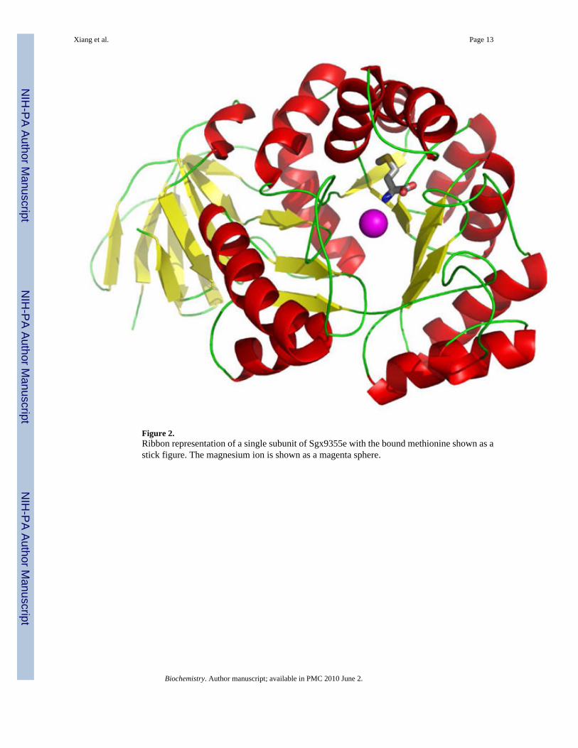
Figure 2.Ribbon representation of a single subunit of Sgx9355e with the bound methionine shown as astick figure. The magnesium ion is shown as a magenta sphere.
Xiang et al. Page 13
Biochemistry. Author manuscript; available in PMC 2010 June 2.
NIH
-PA Author Manuscript
NIH
-PA Author Manuscript
NIH
-PA Author Manuscript

Figure 3.Active site geometry of residues expected to form the binuclear metal center of Sgx9355e.
Xiang et al. Page 14
Biochemistry. Author manuscript; available in PMC 2010 June 2.
NIH
-PA Author Manuscript
NIH
-PA Author Manuscript
NIH
-PA Author Manuscript

Figure 4.(A) Structure of Sgx9355e with L-methionine bound within the active site. (B) Structure ofSgx9359b with L-arginine bound within the active site.
Xiang et al. Page 15
Biochemistry. Author manuscript; available in PMC 2010 June 2.
NIH
-PA Author Manuscript
NIH
-PA Author Manuscript
NIH
-PA Author Manuscript

Figure 5.Relative substrate activities for hydrolysis of 19 dipeptide libraries of the type L-Xaa-L-Xaawhere a single amino acid is at the N-terminus (defined on the x-axis) and 19 different aminoacids were at C-terminus (not including L-cysteine). The results for Cc0300 are presented inpanel A while those for Sgx9355e are shown in panel B. Additional details are found in thetext.
Xiang et al. Page 16
Biochemistry. Author manuscript; available in PMC 2010 June 2.
NIH
-PA Author Manuscript
NIH
-PA Author Manuscript
NIH
-PA Author Manuscript

Figure 6.Relative rates of hydrolysis for the hydrolysis of selected dipeptide libraries by Cc0300 andSgx9355e. (A) Cc0300 with L-Ala-L-Xaa (black) and L-Asp-L-Xaa (grey); (B) Sgx9355e withL-Ala-L-Xaa (black) and L-Gln-L-Xaa (grey). The relative rates were determined by a fit ofthe data to equation 1. Additional details are found in the text.
Xiang et al. Page 17
Biochemistry. Author manuscript; available in PMC 2010 June 2.
NIH
-PA Author Manuscript
NIH
-PA Author Manuscript
NIH
-PA Author Manuscript

Figure 7.Amino acid sequence comparison for Sgx9355e (gi| 44371129), Cc0300 (gi| 16124555),Cc2672 (gi| 16126907), Cc3125 (gi| 16127355) and Sgx9359b (gi|44368820). The predictedmetal ligands for the two divalent metal ions are indicated with ovals. Residues in Sgx9355e,and the equivalent residues in Cc0300, that interact with the bound L-methionine are indicatedwith triangles. Residues in Sgx9359b, and the equivalent residues in Cc2672 and Cc3125, thatinteract with the bound L-arginine are at the equivalent positions. Other fully conservedresidues in all five proteins are highlighted in red. The N-terminal residues that are removedvia proteolysis during the expression of Cc0300, Cc2672, and Cc3125 are ANA/Q, ASA/A,and AAA/Q, respectively. The figure was produced with ESPript (29).
Xiang et al. Page 18
Biochemistry. Author manuscript; available in PMC 2010 June 2.
NIH
-PA Author Manuscript
NIH
-PA Author Manuscript
NIH
-PA Author Manuscript

Scheme 1.
Xiang et al. Page 19
Biochemistry. Author manuscript; available in PMC 2010 June 2.
NIH
-PA Author Manuscript
NIH
-PA Author Manuscript
NIH
-PA Author Manuscript

NIH
-PA Author Manuscript
NIH
-PA Author Manuscript
NIH
-PA Author Manuscript
Xiang et al. Page 20
Table 1Crystal data, phasing and refinement statistics for Sgx9355eCell dimensions: a = b =144.9, c = 101.0 Å and α = β = γ = 90°; Space group I4
Data set Peak
Wavelength (Å) 0.9791
Resolution range (Å) 50.0-2.33(2.41-2.33)
Unique reflections 44,488
Completeness (%) 99.8(98.4)
Redundancy 19(12.5)
Rmerge1 0.10(0.29)
Phasing power2 (ano) 2.95
FOM3: SAD (centric/acentric) 0.18/0.55
After solvent flattening 0.95
Refinement Statistics
R-factor4/R-free 0.21/0.23
Resolution range (Å) 50.0–2.33
Number of atoms
Proteins 6165
Waters 285
Heterogen atoms 20
RMS deviation from ideality
Bonds (Å) 0.007
Angles (°) 1.3
1Rmerge = Σj(|Ih−<I>h|)/ΣIh, where <Ih> is the average intensity over symmetry equivalents
2Phasing power and
3FOM (Figure of merit) are as defined in SHARP
4R-factor =Σ|Fobs−Fcalc|/Σ|Fobs|
Biochemistry. Author manuscript; available in PMC 2010 June 2.

NIH
-PA Author Manuscript
NIH
-PA Author Manuscript
NIH
-PA Author Manuscript
Xiang et al. Page 21
Table 2Kinetic parameters for Cc0300 and Sgx9355e
enzyme substrate Kcat (s−1) Km (mM) Kcat/Km (M−1s−1)
Cc0300 L-Ala-L-Leu 37 ± 2 0.35 ± 0.04 (1.1 ± 0.1) × 105
L-Ala-L-Phe 19 ± 1 0.66 ± 0.05 (2.9 ± 0.3) × 104
L-Asp-L-Leu 29 ± 1 0.22 ± 0.04 (1.3 ± 0.2) × 105
N-acetyl-L-Leu 2.5 ± 0.1 0.15 ± 0.03 (1.6 ± 0.3) × 104
N-acetyl-L-Ile 2.6 ± 0.1 0.14 ± 0.02 (1.9 ± 0.2) × 104
N -acetyl-L-Phe 5.3 ± 0.2 0.31 ± 0.03 (1.7 ± 0.2) × 104
N -acetyl-L-Tyr 4.6 ± 0.2 0.17 ± 0.02 (2.7 ± 0.3) × 104
N -acetyl-L-Val 2.0 ± 0.2 1.1 ± 0.1 (1.8 ± 0.2) × 104
N -acetyl-L-Met 1.7 ± 0.1 0.66 ± 0.05 (2.6 ± 0.2) × 103
N -formyl-L-Leu 39 ± 2 0.13 ± 0.01 (3.0 ± 0.3) × 105
N -formyl-L-Ile 20 ± 1 0.07 ± 0.01 (2.9 ± 0.4) × 105
N-formyl-L-Phe 52 ± 2 0.26 ± 0.03 (2.0 ± 0.2) × 105
N-formyl-L-Tyr 33 ± 1 0.09 ± 0.01 (3.9 ± 0.4) × 105
N-formyl-L-Val 18 ± 1 0.08 ± 0.01 (2.2 ± 0.3) × 105
N-formyl-L-Met 12 ± 1 0.41 ± 0.03 (2.9 ± 0.2) × 104
L-Gly-L-Ala-L-Tyr 0.87 ± 0.05 0.39 ± 0.02 (2.2 ± 0.2) × 103
Sgx9355e L-Ala-L-Leu 0.34 ± 0.01 0.25 ± 0.03 (1.3 ± 0.2) × 103
L-Ala-L-Phe 0.41 ± 0.01 0.07 ± 0.01 (5.8 ± 0.6) × 103
N-acetyl-L-Leu 0.032 ± 0.01 0.11 ± 0.01 (2. 9 ± 0.4) × 102
N-formyl-L-Leu 0.072 ± 0.003 0.26 ± 0.04 (2.8 ± 0.4) × 102
L-Gly-L-Ala-L-Tyr 0.10 ± 0.01 0.30 ± 0.04 (3.3 ± 0.4) × 103
Biochemistry. Author manuscript; available in PMC 2010 June 2.

NIH
-PA Author Manuscript
NIH
-PA Author Manuscript
NIH
-PA Author Manuscript
Xiang et al. Page 22
Table 3Proteins predicted to share substrate specificity with Cc0300 and Sgx9355e.
Gi number Organism Locus tag
Identity of residues equivalent tothose in Sgx9355e(Thr277, Ala302, Val305 and Ile309)
44371129 Environmental sequence Sgx9355e P T I S A G E……R P K A A S V G P Q I S
16124555 C. crescentus CB15 CC_0300 P T L M A G D……T A K A L E A G P K M L
219674575 O. alexandrii HTCC2633 OA2633_13760 P T V M A G E……A A K A R V V G P L M L
91794386 S. denitrificans OS217 Sden_3036 P T L L A G A……R A K A I R V G A D M Q
116626946 S. usitatus Ellin6076 Acid_7923 P T L M A A E……A G K A R Q A M A A L N
114561981 S. frigidimarina NCIMB 400 Sfri_0801 P T L L A G D……K A K A I R V G G D M M
71280007 C. psychrerythraea 34H CPS_1348 P T L M A G D……S A K A I R V G G D M I
221233241 C. crescentus NA1000 CCNA_00302 P T L M A G D……T A K A L E A G P K M L
197106524 P. zucineum HLK1 PHZ_c3063 P T L M A G D……T A K A L Q A G P L M L
127513920 S. loihica PV-4 Shew_2992 P T L L A G D……K Q K A V R V G A D M T
212634704 S. piezotolerans WP3 swp_1881 P T L L A G A……K A K A I R V G A D M L
209964193 R. centenum SW RC1_0865 P T L L A G K……K R K A L T V G P M M Q
114798392 H. neptunium ATCC 15444 HNE_0934 P T V L A G A……R A K A A Q V G P L M L
196186567 B. sp. BAL3 BBAL3_2700 P T L L A G D……T A K A L E A G P K M L
148555139 S. wittichii RW1 Swit_2224 P T L L V G Q……A Q K A I A I A P M L Q
170728069 S. woodyi ATCC 51908 Swoo_3741 P T L L A G D……K A K A E R V G A D M T
83857796 C. atlanticus HTCC2559 CA2559_13368 P T I S A G K……V P K A L E I G P K I Q
56461490 I. loihiensis L2TR IL2390 P T I M A G K……R P K A R A I G P Q I Q
163753087 K. algicida OT-1 KAOT1_13042 P T I T A G K……V P K A L A V G P K I Q
163786441 F. bacterium ALC-1 FBALC1_14687 P T I S A G K……V P K A L D I G P K I Q
120434642 G. forsetii KT0803 GFO_0275 P T I T A G K……V P K A R E I G P K I Q
88711102 F. bacterium HTCC2170 FB2170_03290 P T I T A G K……A K K A A E I G P L I Q
124006459 M. marina ATCC 23134 M23134_02799 P T I S A G E……V P K A L K I G S Q V K
88803709 P. irgensii 23-P PI23P_00415 P T I T A G K……V P K A L A V G P Q I Q
89889513 F. bacterium BBFL7 BBFL7_01328 P T I T A G K……V P K A R T V G P Q I Q
86141823 L. blandensis MED217 MED217_01790 P T L S A G K……V P K A L E I G P Q L Q
86135565 P. sp. MED152 MED152_12694 P T I T A G K……V P K A L A V G P Q I Q
91215139 P. torquis ATCC 700755 P700755_12617 P T I T A G K……V P K A K A I G P K I Q
109900575 P. atlantica T6c Patl_4277 P T V M A G N……R P K A A T I G P L I L
119773229 S. amazonensis SB2B Sama_0087 P T L L A G E……R P K A A A I G P K I Q
196158836 A. macleodii ‘Deep ecotype’ MADE_04041 P T I L A G K……R P K A A A I G P L I Q
Biochemistry. Author manuscript; available in PMC 2010 June 2.
Copyright © 2022 FDOKUMEN