Importin contains a COOH-terminal nucleoporin binding region important for nuclear transport
11
The Journal of Cell Biology The Rockefeller University Press, 0021-9525/2003/08/391/11 $8.00 The Journal of Cell Biology, Volume 162, Number 3, August 4, 2003 391–401 http://www.jcb.org/cgi/doi/10.1083/jcb.200303085 JCB Article 391 Importin contains a COOH-terminal nucleoporin binding region important for nuclear transport Janna Bednenko, Gino Cingolani, and Larry Gerace Department of Cell Biology and Department of Molecular Biology, The Scripps Research Institute, La Jolla, CA 92037 roteins containing a classical NLS are transported into the nucleus by the import receptor importin , which binds to cargoes via the adaptor importin . The import complex is translocated through the nuclear pore complex by interactions of importin with a series of nucleoporins. Previous studies have defined a nucleoporin binding region in the NH 2 -terminal half of importin . Here we report the identification of a second nucleoporin binding region in its COOH-terminal half. Although the affinity of the COOH- P terminal region for nucleoporins is dramatically weaker than that of the NH 2 -terminal region, sets of mutations that perturb the nucleoporin binding of either region reduce the nuclear import activity of importin to a similar extent (50%). An importin mutant with a combination of mutations in the NH 2 - and COOH-terminal regions is completely inactive for nuclear import. Thus, importin possesses two nucleoporin binding sites, both of which are important for its nuclear import function. Introduction Nucleocytoplasmic transport occurs through nuclear pore complexes (NPCs; for reviews see Fahrenkrog et al., 2001; Rout and Aitchison, 2001; Vasu and Forbes, 2001), large proteinacious structures that span the nuclear envelope. The NPC has a central framework consisting of eight spokes surrounding a central transport channel, which is flanked by cytoplasmic and nuclear rings. Extending outward from the ring–spoke framework are eight cytoplasmic fibrils, and eight nuclear fibrils that are joined distally to form a nuclear basket. The vertebrate NPC has a mass of up to 125 MD (Reichelt et al., 1990) and consists of 30 proteins, which are each present in multiple copies (Cronshaw et al., 2002). About one third of the nucleoporins possess multiple copies of the phenylalanine-glycine (FG) sequence. The FG repeats often are part of larger GLFG or FxFG motifs and are usu- ally clustered in specific nucleoporin domains (for review see Bednenko et al., 2003). Nucleocytoplasmic transport of proteins and RNAs is commonly mediated by receptors of the importin /karyo- pherin superfamily. The transport receptors recognize NLSs or nuclear export signals (NESs) on cargoes and transfer them through the NPC by interacting with FG repeat nucle- oporins (for reviews see Görlich and Kutay, 1999; Kuersten et al., 2001; Macara, 2001; Weis, 2002). Importin , which is the best-studied nuclear import receptor, interacts with its cargos either directly or via adapters such as importin . Translocation of importin into the nucleus appears to in- volve its interaction with a series of FG repeat nucleoporins, including Nup358, which is located in the cytoplasmic fibrils (Wilken et al., 1995; Yokoyama et al., 1995; Delphin et al., 1997), Nup62 complex proteins (Nup45, Nup54, Nup58, and Nup62), which are present at the central chan- nel (Hu et al., 1996), and Nup153, which is present in the nuclear basket (Sukegawa and Blobel, 1993; Shah et al., 1998). The small GTPase Ran plays a key role in the directional- ity of karyopherin-mediated nuclear transport (for reviews see Görlich and Kutay, 1999; Kuersten et al., 2001; Macara, 2001; Weis, 2002). Due to the nucleocytoplasmic compart- mentalization of Ran regulators, RanGTP is concentrated in the nucleus and RanGDP in the cytoplasm. In the case of importin /–mediated import, RanGTP in the nucleus dissociates importin from importin , which promotes cargo unloading. RanGTP may also play an important role in dissociating importin from nucleoporins during recep- tor movement through the central channel, as well as in re- leasing the receptor from Nup153 at the end of transloca- tion (Rexach and Blobel, 1995; Görlich et al., 1996; Lyman et al., 2002). Importin is recycled to the cytoplasm in The online version of this article includes supplemental material. Address correspondence to Larry Gerace, The Scripps Research Institute, IMM-10, 10550 N. Torrey Pines Rd., La Jolla, CA 92037. Tel.: (858) 784-8514. Fax: (858) 784-9132. email: [email protected] Key words: nuclear import; importin ; nucleoporin; Ran; site-directed mutagenesis Abbreviations used in this paper: IBB, importin binding; NPC, nuclear pore complex; PTHrP, parathyroid hormone–related protein.
-
Upload
tetragenetics -
Category
Documents
-
view
1 -
download
0
Transcript of Importin contains a COOH-terminal nucleoporin binding region important for nuclear transport

The
Jour
nal o
f Cel
l Bio
logy
The Rockefeller University Press, 0021-9525/2003/08/391/11 $8.00The Journal of Cell Biology, Volume 162, Number 3, August 4, 2003 391–401http://www.jcb.org/cgi/doi/10.1083/jcb.200303085
JCB
Article
391
Importin
�
contains a COOH-terminal nucleoporin binding region important for nuclear transport
Janna Bednenko, Gino Cingolani, and Larry Gerace
Department of Cell Biology and Department of Molecular Biology, The Scripps Research Institute, La Jolla, CA 92037
roteins containing a classical NLS are transported into
the nucleus by the import receptor importin
�
, which
binds to cargoes via the adaptor importin
�
. The importcomplex is translocated through the nuclear pore complexby interactions of importin
�
with a series of nucleoporins.Previous studies have defined a nucleoporin binding region
in the NH
2
-terminal half of importin
�
. Here we report theidentification of a second nucleoporin binding region in itsCOOH-terminal half. Although the affinity of the COOH-
P
terminal region for nucleoporins is dramatically weakerthan that of the NH
2
-terminal region, sets of mutations thatperturb the nucleoporin binding of either region reduce thenuclear import activity of importin
�
to a similar extent
(
�
50%). An importin
�
mutant with a combination of
mutations in the NH
2
- and COOH-terminal regions iscompletely inactive for nuclear import. Thus, importin
�
possesses two nucleoporin binding sites, both of which areimportant for its nuclear import function.
Introduction
Nucleocytoplasmic transport occurs through nuclear porecomplexes (NPCs; for reviews see Fahrenkrog et al., 2001;Rout and Aitchison, 2001; Vasu and Forbes, 2001), largeproteinacious structures that span the nuclear envelope. TheNPC has a central framework consisting of eight spokessurrounding a central transport channel, which is flanked bycytoplasmic and nuclear rings. Extending outward from thering–spoke framework are eight cytoplasmic fibrils, andeight nuclear fibrils that are joined distally to form a nuclearbasket. The vertebrate NPC has a mass of up to 125 MD(Reichelt et al., 1990) and consists of
�
30 proteins, whichare each present in multiple copies (Cronshaw et al., 2002).About one third of the nucleoporins possess multiple copiesof the phenylalanine-glycine (FG) sequence. The FG repeatsoften are part of larger GLFG or FxFG motifs and are usu-ally clustered in specific nucleoporin domains (for review seeBednenko et al., 2003).
Nucleocytoplasmic transport of proteins and RNAs iscommonly mediated by receptors of the importin
�
/karyo-
pherin
�
superfamily. The transport receptors recognize NLSsor nuclear export signals (NESs) on cargoes and transferthem through the NPC by interacting with FG repeat nucle-oporins (for reviews see Görlich and Kutay, 1999; Kuersten
et al., 2001; Macara, 2001; Weis, 2002). Importin
�
, whichis the best-studied nuclear import receptor, interacts with itscargos either directly or via adapters such as importin
�
.Translocation of importin
�
into the nucleus appears to in-volve its interaction with a series of FG repeat nucleoporins,including Nup358, which is located in the cytoplasmicfibrils (Wilken et al., 1995; Yokoyama et al., 1995; Delphinet al., 1997), Nup62 complex proteins (Nup45, Nup54,Nup58, and Nup62), which are present at the central chan-nel (Hu et al., 1996), and Nup153, which is present in thenuclear basket (Sukegawa and Blobel, 1993; Shah et al.,1998).
The small GTPase Ran plays a key role in the directional-ity of karyopherin-mediated nuclear transport (for reviewssee Görlich and Kutay, 1999; Kuersten et al., 2001; Macara,2001; Weis, 2002). Due to the nucleocytoplasmic compart-mentalization of Ran regulators, RanGTP is concentrated inthe nucleus and RanGDP in the cytoplasm. In the case ofimportin
�
/
�
–mediated import, RanGTP in the nucleusdissociates importin
�
from importin
�
, which promotescargo unloading. RanGTP may also play an important rolein dissociating importin
�
from nucleoporins during recep-tor movement through the central channel, as well as in re-leasing the receptor from Nup153 at the end of transloca-tion (Rexach and Blobel, 1995; Görlich et al., 1996; Lymanet al., 2002). Importin
�
is recycled to the cytoplasm in
The online version of this article includes supplemental material.Address correspondence to Larry Gerace, The Scripps Research Institute,IMM-10, 10550 N. Torrey Pines Rd., La Jolla, CA 92037. Tel.: (858)784-8514. Fax: (858) 784-9132. email: [email protected] words: nuclear import; importin
�
; nucleoporin; Ran; site-directedmutagenesis
Abbreviations used in this paper: IBB, importin
�
binding; NPC, nuclearpore complex; PTHrP, parathyroid hormone–related protein.

The
Jour
nal o
f Cel
l Bio
logy
392 The Journal of Cell Biology
|
Volume 162, Number 3, 2003
complex with RanGTP, where import-competent importin
�
is regenerated upon RanGTP hydrolysis promoted byRanGAP.
Importin
�
contains 19 tandem HEAT repeats arrangedin a superhelical spiral (Cingolani et al., 1999; Fig. 1 A).Each HEAT repeat, which usually has a length of
�
40–60amino acid residues, consists of A and B helices connectedby a short turn. The A helices form the outer surface of im-portin
�
, and the B helices create the inner surface. The in-ner surface of importin
�
binds to the
�
45-residue impor-tin
�
binding (IBB) domain of importin
�
(via HEATrepeats 7–19; Cingolani et al., 1999), the NLS of parathy-roid hormone–related protein (PTHrP) (via HEAT repeats2–11; Cingolani et al., 2002), and RanGTP (via HEAT re-peats 1–8; Vetter et al., 1999). The outer surface of impor-tin
�
contacts FG repeat motifs of nucleoporins (Bayliss etal., 2000, 2002).
The major NPC binding domain in importin
�
wasmapped by deletion mutagenesis to its NH
2
-terminal regionbetween HEAT repeats 4–9 (Chi and Adam, 1997; Kose etal., 1997; Kutay et al., 1997). In addition, the importin
�
fragment containing HEAT repeats 9–19 has been shown tobind to the nuclear envelopes of digitonin-permeabilizedHeLa cells (Kutay et al., 1997), raising the possibility of ad-ditional nucleoporin binding sites in the COOH-terminalpart of importin
�
. The crystal structure of a complex be-tween an NH
2
-terminal fragment of human importin
�
(HEAT repeats 1–10) and an FxFG-rich nucleoporin frag-ment has revealed two FxFG binding sites in the NH
2
-ter-minal region of importin
�
, one formed by HEAT repeats 5and 6 and another formed by HEAT repeats 6 and 7 (Baylisset al., 2000). Each of these binding elements forms a pocketthat associates with the two phenylalanine residues of theFxFG motif by hydrophobic interactions.
In this study, we present an in-depth biochemical analysisof the interaction between importin
�
and nucleoporins.We prepared a number of point mutations in the NH
2
-ter-minal region of importin
�
previously implicated in nucle-
oporin binding. Although the mutations diminished nucle-oporin binding in a manner consistent with the predictionsof the crystal structure (Bayliss et al., 2000), they had amuch weaker effect on the level of nuclear import than re-ported previously (Bayliss et al., 2000). This led us to searchfor an additional nucleoporin binding site(s) in importin
�
.Using intramolecular structural alignment of importin
�
segments and site-directed mutagenesis, we identified anovel nucleoporin binding region in the COOH-terminalhalf of importin
�
. Although the COOH-terminal nucle-oporin binding region has a dramatically lower affinity fornucleoporins than the NH
2
-terminal region, it appears tocontribute equally to the nuclear import function of impor-tin
�
. We discuss the possibility that the presence of multi-ple nucleoporin binding regions is a general feature of nu-clear transport receptors.
Results
Effects of single point mutations in the NH
2
-terminal region of importin
�
on binding to nucleoporins
Previous studies reported that isoleucine 178 in HEAT re-peat 5 is critical for importin
�
function, because substitu-tion of isoleucine 178 with alanine, aspartate, or phenyl-alanine was observed to almost completely abolish thenucleoporin binding and nuclear import activity of impor-tin
�
(Bayliss et al., 2000, 2002). Surprisingly, when weprepared the I178A and I178D importin
�
mutants, wediscovered that these mutations had little or no effect onnuclear import (see below). Therefore, we performed a sys-tematic mutational analysis of the nucleoporin binding re-gion in HEAT repeats 5–7 to evaluate the importance ofother importin
�
residues in this region. Our goal was toidentify combinations of single point mutations that sig-nificantly reduce the ability of importin
�
to interact withnucleoporins and to support nuclear import, without di-minishing the interaction with cargo or with the transportregulator Ran.
Figure 1. Model of the importin �–IBB domain complex. (A) Rod representation of the structure of importin � complexed with the IBB domain of importin �, according to Cingolani et al. (1999). The A and B helices of the HEAT repeats are shown in red and yellow, respectively, with connecting loops and helices in gray. The IBB domain of importin � is in green. (B) An enlarged view highlighting the A helices of HEAT repeats 5–7, which were shown to be involved in binding FxFG motifs by crystallography (Bayliss et al., 2000). The enlarged image was rotated by 90� in a Y direction with respect to A. The side chains of the residues analyzed by the mutagenesis as described in Table I are highlightedin blue.
The
Jour
nal o
f Cel
l Bio
logy
Importin
�
nucleoporin binding regions |
Bednenko et al. 393
According to the crystal structure of a complex betweenan NH
2
-terminal fragment of human importin
�
(HEATrepeats 1–10, comprising amino acid residues 1–442) andan FxFG-rich fragment of the yeast nucleoporin Nsp1p (res-idues 497–608), importin
�
contains two binding sites forthe FxFG motif within HEAT repeats 5–7 (Bayliss et al.,2000). One site is formed by residues in HEAT repeats 5and 6, and a second site by residues in HEAT repeats 6 and7 (Fig. 1). The first phenylalanine residue of the FxFG motifforms a stacking interaction with either F217 on HEAT re-peat 6 (site 1) or Y255 on HEAT repeat 7 (site 2). The sec-ond phenylalanine residue of the FxFG motif interacts witha hydrophobic pocket generated by the residues of eitherHEAT repeats 5 and 6 (site 1; L174, T175, I178, E214,F217, and I218) or HEAT repeats 6 and 7 (site 2; E224,A259, and C223).
Based on predictions of the crystal structure, we madesubstitutions in residues I178, E214, F217, Y255, and I263(Fig. 1; Table I). Alanine substitutions of residues F217 andY255 are expected to abolish the stacking interactions withthe first phenylalanine of the FxFG repeat. Furthermore, themutations I178D, I178A, and E214A are expected tochange the structure of the hydrophobic pocket betweenHEAT repeats 5 and 6 and, in the case of E214A, to removea hydrogen bond with the mainchain nitrogen of glycine inthe FxFG motif (Bayliss et al., 2000). Although I263 itselfwas not predicted to form contacts with the FxFG repeat(Bayliss et al., 2000), we expected that its substitution witharginine would perturb the apolar pocket between HEATrepeats 6 and 7 located in the vicinity of I263.
We analyzed recombinant importin
�
mutants for theirinteraction with several mammalian nucleoporins implicatedin nuclear import using an ELISA-based microtiter platebinding assay to measure the apparent dissociation constants(K
d
s) (Delphin et al., 1997; Ben-Efraim and Gerace, 2001).We examined a fragment of Nup358 (residues 996–1963)that has 10 FG repeats, six occurring within an FxFG motif;full-length Nup62 that has six FG repeats, five within anFxFG; and a COOH-terminal fragment of Nup153 (resi-
dues 895–1475) that has 25 FG repeats, 16 within an FxFG(for review see Bednenko et al., 2003).
We found that importin
�
single point mutants contain-ing the substitutions I178D, E214A, F217A, Y255A, andI263R all had a decreased affinity for the Nup153 fragment,but retained wild-type affinity for RanGTP and for impor-tin
�
(Table I). Weakened nucleoporin binding was mostevident for the I178D mutant, which had an
�
40-fold de-crease in affinity for Nup153 (895–1475). In contrast, theE214A, F217A, Y255A, and I263R mutants showed a rela-tively small decrease in affinity for the Nup153 fragment(K
d
�
1.8–3.5 nM) (Table I; Fig. 2). All of these mutantshad a decreased affinity for Nup62 and Nup358 (996–1963)as well. In several cases, binding of the mutants to Nup62and Nup358 (996–1963) did not reach saturation in our as-say (Table I; Fig. 2), implying a K
d
of
��
1
�
M for the in-teraction (see Table I legend).
It was reported previously that the I178D and I178A mu-tations substantially decreased the binding of importin
�
tonucleoporins (Bayliss et al., 2000, 2002). Although we ob-served significantly weakened binding with I178D, wefound that the I178A mutation had no effect. This is consis-tent with the theoretical prediction that substitution of abulky hydrophobic residue with an alanine residue wouldnot significantly change the structure of the hydrophobicpocket. The discrepancy between our results and the previ-ously published study (Bayliss et al., 2000) may be due todifferences in the importin
�
expression and purificationconditions. Nonetheless, we observed that combining theI178A mutation with the E214A mutation resulted in sub-stantially weaker nucleoporin binding than the E214A mu-tation alone (see below; Table I).
Effects of combinations of single point mutations on importin
�
binding to nucleoporins
In an attempt to dramatically weaken or abolish the high-affinity interaction of importin
�
with Nup153 (895–1475),we examined a number of constructs that had combinationsof single point mutations within the NH
2
-terminal nucle-
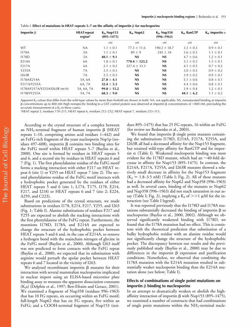
Table I.
Effect of mutations in HEAT repeats 5–7 on the affinity of importin
�
for nucleoporins
Importin
�
HEAT repeat region
a
K
d
, Nup153 (895–1475)
K
d
, Nup62 K
d
, Nup358 (996–1963)
K
d
, RanGTP K
d
, importin
�
nM nM nM nM nM
WT NA 1.1
�
0.1 77.3
�
11.6 190.2
�
18.7 3.3
�
0.3 0.9
�
0.1I178A 5A 1.1
�
0.1 89
�
9 245
�
34 3.6
�
0.3 1.1
�
0.1I178D 5A
40.1
�
9.1 NS NS
4.7 � 0.6 1.2 � 0.1E214A 6A 1.8 � 0.1 770.4 � 123.2 NS 3.1 � 0.3 1.3 � 0.1F217A 6A 2.1 � 0.2 227.4 � 33.3 NS 4.1 � 0.5 0.7 � 0.2Y255A 7A 3.5 � 0.3 NS NS 3.0 � 0.5 0.9 � 0.2I263R 7A 2.5 � 0.3 NS NS 3.9 � 0.2 0.9 � 0.1I178A/E214A 5A, 6A 27.0 � 4.1 NS NS 5.1 � 0.6 0.8 � 0.1F217A/Y255A 6A, 7A 32.4 � 5.3 NS NS 4.4 � 0.6 0.8 � 0.1I178A/F217A/Y255A/I263R (m-N) 5A, 6A, 7A 99.0 � 15.2 NS NS 3.9 � 0.4 1.2 � 0.1I178D/Y255A 5A, 7A 68.3 � 9.0 NS NS 44.1 � 6.2 1.1 � 0.2
Apparent Kd values that differ from the wild-type values by more than fivefold are shown in bold. NA, not applicable; NS, nonsaturated binding at importin� concentrations up to 800 nM (high nonspecific binding to a GST control protein was observed at importin � concentrations of �800 nM, precluding theaccurate measurement of a Kd in these cases).aHEAT repeat 5, residues 170–211; HEAT repeat 6, residues 212–252; HEAT repeat 7, residues 253–313.

The
Jour
nal o
f Cel
l Bio
logy
394 The Journal of Cell Biology | Volume 162, Number 3, 2003
oporin binding region. We found that combining the mostpotent single point mutation (I178D) with other mutationsnot only inhibited the interaction with nucleoporins, butalso resulted in considerable (�10-fold) inhibition of im-portin � binding to RanGTP (Table I; unpublished data).For example, importin � containing a combination ofI178D and Y255A had an �13-fold lower affinity forRanGTP, but showed no change in affinity for importin �(Table I). This effect suggests that certain combinations ofmutations in the A helices of HEAT repeats 5–7 alter theposition of and/or spacing between the residues in the B he-lices that are important for RanGTP binding, providing evi-dence for cross-talk between the RanGTP and nucleoporinbinding regions (Bayliss et al., 2000).
As our goal was to identify importin � mutants that havesignificantly reduced affinity for Nup153 yet retain wild-type affinity for RanGTP, we examined combinations of theother single point mutations we had tested. Combining twoalanine substitutions in importin � synergistically weakenedthe binding to Nup153 (895–1475) in the two cases we ex-amined. In one construct (I178A/E214A), the mutated resi-dues belonged to the same FxFG binding pocket, and, in asecond construct (F217A/Y255A), they belonged to the twodifferent pockets (Table I). We found that an importin �construct containing a combination of the I178A, F217A,Y255A, and I263R mutations (termed the “m-N” mutant)had an �100-fold reduced affinity for Nup153, as com-pared with wild-type importin �. At the same time, the m-Nmutant exhibited wild-type affinity for RanGTP and impor-tin � (Table I), and its circular dichroism spectrum was sim-ilar to that of wild-type importin � (unpublished data).
Considered together, these data indicate that the mutationsin m-N did not significantly alter importin � structure. In-troducing additional mutations (e.g., E214A) into the nu-cleoporin binding region of the m-N construct resulted ininhibition of its binding to RanGTP (unpublished data),and these mutants were not examined further.
Mutations in the NH2-terminal nucleoporin binding region of importin �: effects on nuclear import and translocation through the NPCTo test the functional effects of mutations in the NH2-ter-minal nucleoporin binding domain of importin �, we exam-ined the mutants in a permeabilized cell nuclear import as-say using the fluorescently labeled importin �–dependentimport cargo BSA-NLS (Adam et al., 1992). At an optimalconcentration of wild-type importin �, we observed an ap-proximately fivefold accumulation of fluorescent cargo innuclei under standard assay conditions, as compared withcontrol reactions in which ATP was depleted (Fig. 3 A, com-pare white and black columns). Examination of cells by con-focal microscopy revealed that almost all of the cell fluores-cence was in the nucleus (see Fig. S1, available at http://www.jcb.org/cgi/content/full/jcb.200303085/DC1), vali-dating the use of flow cytometry for quantification. No sig-nificant stimulation of import was observed in the absenceof recombinant importin � (Fig. 3 A). A time course of im-port revealed linear nuclear accumulation of cargo over the30-min time frame of our experiment with wild-type impor-tin � and with all the importin � mutants (unpublisheddata). This indicates that the observed variations in the level
Figure 2. Characterization of the binding of importin � to nucleoporins. Shown are binding isotherms depicting the interaction of selected importin � mutants with Nup153 (895–1475) (A), with full-length Nup62 (B), or with Nup358 (996–1963) (C). Error bars represent the standard deviation of duplicate measurements. The calculated apparent Kd values are indicated next to the binding isotherms. NS, nonsaturated binding at importin � concentrations up to 800 nM.

The
Jour
nal o
f Cel
l Bio
logy
Importin � nucleoporin binding regions | Bednenko et al. 395
of nuclear import with the various importin � constructs aredue to import rate differences.
Substitution of wild-type importin � with the I178A mu-tant did not lower the level of nuclear import, consistent withthe wild-type level of nucleoporin binding seen with this mu-tant (Fig. 3 A). Import with the F217A mutant was decreasedby 10–30% at the lower concentrations of importin � testedand was not affected at the higher importin � concentrationsexamined (Fig. 3 A). The same was true for the E214A,Y255A, and I263R mutants (unpublished data). Import ofBSA-NLS with the I178D mutant was reduced by 20–40%,depending on the concentration of the mutant in the assay,whereas import with m-N mutant was reduced by 35–50%.When we performed our assay using the experimental condi-tions of Bayliss et al. (2000), the nuclear import level was re-duced by only 20% with I178D, and no loss of activity wasobserved with I178A (see Fig. S2, available at http://www.jcb.org/cgi/content/full/jcb.200303085/DC1). Thiscontrasts with the results obtained by Bayliss et al. (2000),who observed a 76–84% loss of import with these two mu-tants. We think that this discrepancy is due to the differentmethods of preparation of the recombinant transport factors.
It has been shown that in the absence of exogenous impor-tin � and NLS cargo, importin � can move between the nu-cleus and the cytoplasm by a process that requires neitherRan nor GTP hydrolysis (Kose et al., 1997, 1999). Similar tothis previous work, we found that importin � rapidly accu-mulated in the nucleus when added to permeabilized cells inthe absence of other factors (Fig. 3 B). It quickly reached aplateau concentration, at which point its rate of nuclear entrypresumably equals its rate of nuclear exit (Fig. 3 B). To fur-ther investigate whether the decrease in cargo import activityof the I178D and m-N importin � mutants is due to a defect
in their ability to be translocated through the NPC, we mea-sured the time course of their nuclear accumulation after ad-dition to permeabilized cells. The same final concentration ofintranuclear importin � was achieved with the I178D and m-N mutants, as compared with wild-type importin � (Fig. 3B). Whereas wild-type importin � reached a plateau within�1 min, the I178D mutant took slightly longer to reach thislevel, and the m-N mutant reached a plateau only after �3min (Fig. 3 B). This suggests that the mutant importin �constructs have a decreased rate of translocation through theNPC. Moreover, their apparent rate of translocation is corre-lated with their relative nuclear import activity, with the m-N mutant being the most highly impaired.
Nucleoporin binding and NPC translocation of a COOH-terminal fragment of importin �The nucleoporin binding of the m-N protein that remainedin this quadruple mutant (Kd of �100 nM for Nup153[895–1475]) and its significant, albeit reduced, nuclear im-port activity (�50% of wild type) could have been due toresidual activity in the mutated NH2-terminal region. It wasalso possible that an additional nucleoporin binding site(s)in the COOH-terminal region of importin � contributed tonucleoporin binding and import. To systematically investi-gate these possibilities, we first examined a recombinantNH2-terminal fragment prepared from the m-N importin �mutant (HEAT repeats 1–10, residues 1–396). We observedthat this construct had a Kd of 160 nM for Nup153 (895–1475) (Table II), clearly showing that the m-N mutationsdid not inactivate all nucleoporin binding in the NH2-ter-minal portion of importin �.
We subsequently expressed a COOH-terminal fragmentof wild-type importin � that spanned HEAT repeats 8–19
Figure 3. Effect of mutations in HEAT repeats 5–7 on nuclear import activity of importin �. (A) Nuclear import assays with wild-type importin � and with importin � mutants. 40-�l samples were incubated for 30 min at 30�C, and the level of nuclear import of FITC-labeled BSA-NLS was measured by flow cyto-metry. White bars depict control reactions performed in the presence of hexokinase and glucose to deplete ATP. Black bars show the levels of fluorescence in the presence of ATP. Error bars represent the standard deviation of duplicate measure-ments. (B) Kinetics of nuclear accumu-lation of importin �. 3.4 pmol of importin � or importin � (304–876) fragment was used in each 20-�l assay. The level of intranuclear importin � was quantified by immunofluorescence and confocal microscopy in samples fixed at the various time points. Error bars represent the standard deviation of two independent experiments. The level of intranuclear wild-type importin � for the 3-min time point was set at 100%, and all other values were normalized to the 100% level.

The
Jour
nal o
f Cel
l Bio
logy
396 The Journal of Cell Biology | Volume 162, Number 3, 2003
(residues 304–876). We found that this COOH-terminalimportin � fragment bound saturably to Nup153 (895–1475) with a Kd of 152 nM (Table II). However, we werenot able to detect an interaction of the COOH-terminalfragment with Nup358 (996–1963) and Nup62, although itis possible that it interacts with these nucleoporins with anaffinity that is below the limit of sensitivity of our assay. Theaffinity of the fragment for importin � was weaker by ap-proximately threefold (Table II), but this can be ascribed tothe loss of several amino residues in HEAT repeat 7 that in-teract with the importin � IBB domain (Cingolani et al.,1999). The fact that the COOH-terminal importin � frag-ment is still capable of high-affinity binding to importin �suggests that it preserves most of its native structure whenexpressed as a fragment.
Interestingly, when the COOH-terminal fragment of im-portin � (304–876) was added to permeabilized cells, it ac-cumulated in the nucleus and reached the same concentra-tion as full-length importin �, although its rate of entry was
significantly reduced as compared with the wild-type protein(Fig. 3 B). In addition, importin � (304–876) supported invitro nuclear import of BSA-NLS in the presence of impor-tin � (see Fig. S3, available at http://www.jcb.org/cgi/content/full/jcb.200303085/DC1). Nuclear import mediated by im-portin � (304–876) was inhibited by WGA (Fig. S3), sug-gesting that it was a nucleoporin-mediated process. How-ever, the level of import obtained with the importin �(304–876) fragment was only 30–35% as compared withwild-type importin �, which may be due to the absenceof the NH2-terminal nucleoporin binding domain, theRanGTP binding domain, or both.
Involvement of the COOH-terminal nucleoporin binding region in importin �–mediated nuclear importTo further analyze the nucleoporin binding site containedwithin the COOH-terminal fragment of importin �, weattempted to identify the COOH-terminal amino acid resi-dues involved in nucleoporin binding. Initially, we per-formed an intramolecular structural alignment of the NH2-terminal (HEAT repeats 1–10) and the COOH-terminal(HEAT repeats 11–19) fragments of importin � (Fig. 4, Aand B). The two polypeptide chains were aligned using thecombinatorial extension (CE) method, which determines anoptimal alignment between fragment pairs (Shindyalov andBourne, 1998). Interestingly, we found that the importin �regions containing residues 1–445 and 446–876 were struc-turally similar and could be superimposed with a root meansquare deviation for the � carbon chains of �3.4 Å (Fig. 4B). The central HEAT repeats (4–7 and 13–16) presentedthe highest structural homology (root mean square deviationfor the � carbon chains of �2.1 Å), whereas the HEAT re-peats located to the periphery of these core regions showedthe highest divergence (Fig. 4 B).
Using this structural alignment, we identified three aminoacid residues in HEAT repeats 14 and 16 (L612, F688, and
Figure 4. Structural alignment of the NH2- and COOH-terminal segments of importin �. (A) Worm representation of importin �. Residues 1–445 are highlighted in gray and residues 446–876 are in green. (B) Alignment of the NH2- and COOH-terminal segments of importin �. Residues 1–445 (HEAT repeats 1–10) are in gray and residues 446–876 (HEAT repeats 11–19) are in green. (C) The side chains of importin � residues I178, Y255, and I263 (yellow) closely align with those of L612, F688, and L695 (red), respectively. The enlarged image in C was rotated 90� in a Y direction and 30� in a Z direction with respect to B.
Table II. Binding of importin � fragments and mutants to Nup153 (895–1475) and to importin �
Importin � Kd, Nup153 (895–1475) Kd, importin �
nM nM
WT 1.0 � 0.1 0.9 � 0.11–396 1.2 � 0.1 NDm-N (1–396) 160.1 � 25.3 ND304–876 152.2 � 17.5 2.5 � 0.2m-C (304–876) NS 4.3 � 0.5m-N 91.3 � 11.8 1.1 � 0.1L612D/F688A (m-C) 1.2 � 0.1 2.2 � 0.3m-N/m-C 83.2 � 9.1 2.1 � 0.2
The binding reactions with importin � were performed in PBS containing30 mg/ml BSA (see Materials and methods). NS, nonsaturated binding atimportin � concentrations up to 800 nM.

The
Jour
nal o
f Cel
l Bio
logy
Importin � nucleoporin binding regions | Bednenko et al. 397
L695) that matched residues in the NH2-terminal nucle-oporin binding site in HEAT repeats 5 and 7 (I178, Y255,and I263, respectively; Fig. 4 C). To test whether L612,F688, and L695 are important for the interaction of theCOOH-terminal fragment of importin � with nucleopor-ins, we performed a mutational analysis of the three residues.The mutation L612D (analogous to I178D) strongly de-creased the affinity of the importin � fragment for Nup153(895–1475), such that the Kd was not measurable, whereasF688A (analogous to Y255A) decreased the affinity by ap-proximately twofold (Fig. 5 A). The mutation L695R (anal-ogous to I263R) had no detectable effect on the binding ofthe importin � fragment to Nup153 (unpublished data). Amutant importin � fragment containing a combination ofL612D and F688A showed an even lower level of nonsat-urated binding for Nup153 (895–1475) than did L612Dalone (Fig. 5 A). The L612D and F688A mutations did notsignificantly affect importin � folding, as only an approxi-mately twofold decrease in the affinity of the mutant proteinfragments for importin � was observed (Table II).
We then analyzed the effect of L612D and F688A in thecontext of full-length importin �. Importin � containingthe mutations L612D and F688A (m-C mutant) displayed
wild-type affinity for the three nucleoporins tested in thisstudy (Table II; unpublished data). This is expected, as thepresence of the high-affinity NH2-terminal nucleoporinbinding region in full-length importin � would mask muta-tional inactivation of the low-affinity COOH-terminal re-gion. We then introduced the L612D/F688A mutationsinto the m-N mutant to create the m-N/m-C mutant. Theaffinity of the latter for Nup153 was similar to the affinity ofthe m-N mutant alone (Table II). This can be explained bythe fact that the NH2-terminal nucleoporin binding regionin m-N is capable of binding to Nup153 independently ofthe COOH-terminal nucleoporin binding region (Table II).Both the m-C and the m-N/m-C mutants retained a highaffinity for importin � (only approximately twofold weakerthan wild type; Table II) and did not exhibit any major al-terations in secondary structure, as determined by circulardichroism spectroscopy (unpublished data).
To test the effect of the mutations in the COOH-terminalnucleoporin binding region on importin � function, we firstexamined the ability of the m-C mutant to accumulate inthe nucleus of permeabilized cells. Interestingly, the rate ofnuclear entry of the m-C mutant was substantially lowerthan that of wild-type importin �, but the m-C mutant
Figure 5. Effects of mutations in the COOH-terminal segment of importin � on nucleoporin binding and nuclear import. (A) Binding isotherms showing the interaction of the importin � fragment comprising residues 304–876 (with or without the mutations L612D and/or F688A) to Nup153 (895–1475). Error bars represent the standard deviation of duplicate measurements. The numbers next to the binding curves indicate apparent dissociation constants. NS, nonsaturated binding at importin � fragment concentrations of up to 800 nM. (B) Kinetics of nuclear accumulation of wild-type (WT), m-C, and m-N/m-C full-length importin � proteins. 3.4 pmol of each importin � construct was used in each 20-�l assay. The nuclear fluorescence was measured by immunofluorescence and confocal microscopy after 20–600 s incubation at room temperature, as indicated. Error bars represent the standard deviation of two independent experiments. The level of intranuclear wild-type importin � for the 3-min time point was set at 100%, and all other values were normalized as a fraction of the 100% level. (C) Nuclear import assays with the wild-type (WT), m-N, m-C, and m-N/m-C full-length importin � proteins. 40-�l reactions were incubated for 30 min at 30�C, and the level of nuclear import was measured by flow cytometry. White bars designate control reactions performed in the presence of hexokinase and glucose to deplete ATP. Black bars show the level of fluorescence in the presence of ATP. Error bars represent the standard deviation of duplicate measurements.

The
Jour
nal o
f Cel
l Bio
logy
398 The Journal of Cell Biology | Volume 162, Number 3, 2003
eventually accumulated to similar levels as the wild-type pro-tein (Fig. 5 B). The reduced rate of nuclear entry suggeststhat the COOH-terminal nucleoporin binding region is im-portant for translocation of importin � through the NPC.Furthermore, the m-C mutant was clearly deficient in nu-clear import of BSA-NLS, which was decreased by 45–55%as compared with wild-type importin �, depending on theconcentration of importin � in the assay (Fig. 5 C). Thetwofold decrease in the affinity of m-C for importin � is notpredicted to affect the level of nuclear import, as the concen-tration of importin � in the assay is 390 nM (see Materialsand methods).
Examination of the m-N/m-C mutant revealed that it didnot accumulate significantly in the nucleus when added topermeabilized cells over the 10-min time course of our assay,indicating that it is highly deficient in translocation throughthe NPC. Moreover, the m-N/m-C mutant did not supportsignificant nuclear import of BSA-NLS at all receptor con-centrations tested (Fig. 5 C), even though its affinity for im-portin � was only twofold weaker than wild type and thebinding to RanGTP was nearly normal (4.4 vs. 3.3 nM forthe wild type) (Table II; unpublished data). Considered to-gether, these data demonstrate that both the NH2- andCOOH-terminal nucleoporin binding regions of importin� are important for translocation through the NPC in nu-clear import.
DiscussionImportin � contains a COOH-terminal nucleoporin binding region in addition to the previously described NH2-terminal regionBiochemical and functional analysis of a large number ofimportin � mutants allowed us to characterize the interac-tions between importin � and several FG repeat nucleopor-ins. Our data indicate that importin � has a second, previ-ously uncharacterized nucleoporin binding region in itsCOOH-terminal region (in HEAT repeats 14–16), in addi-tion to the NH2-terminal nucleoporin binding area inHEAT repeats 5–7 that was characterized by crystallo-graphic analysis (Bayliss et al., 2000). Interestingly, the twonucleoporin binding areas are located on the opposite side ofthe protein (Fig. 6 A), and both regions appear to contrib-ute, to a similar extent, to nuclear import (discussed later).
The results of our quantitative nucleoporin binding assaysinvolving a number of point mutants in HEAT repeats 5–7support the predictions of the crystal structure by Bayliss etal. (2000). However, in our hands, the functional effects ofmutations in the single residue that was analyzed previously(I178; Bayliss et al., 2000) were significantly weaker than re-ported before. Our most severe mutant involving the NH2-terminal nucleoporin binding region that still retained wild-type binding for RanGTP and importin � was the quadru-ple point mutant m-N. Although the affinity of the m-Nmutant for Nup153 was decreased by �100-fold, its importactivity was decreased by only 35–50%. Our finding thatm-N and other importin � mutants having a tremendous de-crease in binding affinity for nucleoporins can still supportsubstantial levels of nuclear import indicates that importin �can employ low-affinity interactions to move through theNPC. This is analogous to another nuclear import receptor,transportin, which is estimated to have a Kd of �4 �M fornucleoporins (Ribbeck and Görlich, 2001). We identifiedthe COOH-terminal nucleoporin binding region in impor-tin � (located in HEAT repeats 14–16) by using a structuralalignment of the NH2- and COOH-terminal halves of im-portin �, combined with site-directed mutagenesis. Thiswork identified two key residues in the A helices of HEATrepeats 14 and 16 (L612 and F688) that appear to be analo-gous to two residues of HEAT repeats 5 and 7 (I178 andY255, respectively), which are important for nucleoporinbinding.
We found that a COOH-terminal fragment of importin� (comprising HEAT repeats 8–19) binds saturably toNup153, although it has a much weaker affinity (�150-fold) than a fragment containing the NH2-terminal region(HEAT repeats 1–10). Nonetheless, the weak nucleoporinbinding is sufficient to mediate the translocation of theCOOH-terminal fragment into the nucleus. The mutationsL612D and F688A inhibit the binding of the COOH-ter-minal importin � fragment to nucleoporins, but have no de-tectable effect on nucleoporin binding when they are intro-duced into full-length importin �, due to the presence of thehigh-affinity NH2-terminal region. However, the integrityof the COOH-terminal nucleoporin binding region is cru-cial for the high-level importin �–mediated nuclear import,as the import activity of an importin � mutation containingboth L612D and F688A (the m-C mutant) is reduced by
Figure 6. Model depicting the involvement of two nucleoporin binding regions in importin � in its translocation through the NPC. (A) The two nucleoporin binding regions are illustrated in a ribbon representation of importin �. The NH2-terminal region is shown in blue, and the COOH-terminal region in green. (B) One possible model for importin � translocation through the NPC. The model depicts only the central channel region, but movement between the peripheral fibrils of the NPC and the central channel could be conceptually similar. The two nucleoporin binding regions are shown in blue and green as in A, and the FG-rich nucleoporin regions are depicted as flexible filaments. Importin � could use its NH2- and COOH-terminal regions simultaneously or in succession to promote movement through the NPC.

The
Jour
nal o
f Cel
l Bio
logy
Importin � nucleoporin binding regions | Bednenko et al. 399
�50%. An importin � construct containing both the m-Nand the m-C mutations was almost completely inactive innuclear import, even though it maintained nearly wild-typeaffinity for RanGTP and for importin �. Considered to-gether, our results indicate that both the NH2- and COOH-terminal regions of importin � play an important role inbinding to FG repeat nucleoporins to direct importin �translocation through the NPC.
In previous work, we found that an NH2-terminal frag-ment of importin � containing HEAT repeats 1–11 showsan �50% reduction in the nuclear import of PTHrP ascompared with full-length importin �, despite the fact thatthis fragment binds PTHrP NLS and RanGTP with wild-type affinity (Cingolani et al., 2002). In light of the presentstudy, the decrease in the efficiency of PTHrP import withthe NH2-terminal importin � fragment is likely to be due tothe absence of the COOH-terminal nucleoporin binding re-gion. This further supports the notion that both the NH2-and COOH-terminal nucleoporin binding regions of im-portin � have an important functional role.
Although the structure of the nucleoporin binding site(s)in the COOH-terminal region of importin � is uncertain,by analogy with the structurally homologous NH2-terminalregion, hydrophobic pockets between HEAT repeats 14–15and 15–16 probably are responsible for FG repeat binding.Because importin � consists entirely of tandem HEAT re-peats, it is possible that it contains additional hydrophobicbinding pockets in regions adjacent to HEAT repeats 5–7and 14–16, which can form weak contacts with FG repeatsof nucleoporins. In this scenario, the nucleoporin bindingregions in HEAT repeats 5–7 and 14–16 would serve asnucleating sites that would allow initial positioning of a nu-cleoporin FG repeat region on importin �. Upon formingthe initial contacts, the interactions could propagate toneighboring HEAT repeats. This might explain the high af-finity of importin � for certain nucleoporins, such asNup153, which is characterized by a high density of FG re-peats in its COOH-terminal region (for review see Bed-nenko et al., 2003).
Two nucleoporin binding sites can enhance translocation of receptors through the NPCA crucial step in the translocation of cargo–receptor com-plexes across the NPC involves movement through the cen-tral channel, which provides the major barrier to the free dif-fusion of the macromolecules across the NPC (Feldherr andAkin, 1997). The structure of the central channel is obscure,but it is estimated that the concentration of FG repeat mo-tifs in this region may be very high (50 mM; Bayliss et al.,1999). FG-rich nucleoporin regions have been shown to beflexible and largely unstructured (Denning et al., 2002,2003), which could allow importin � to contact FG motifsin many nucleoporins in its vicinity as it is translocatedthrough the NPC.
Based on our data, we propose that importin � utilizes itstwo nucleoporin binding regions coordinately as it translo-cates through the NPC. A hypothetical view of receptormovement through the diffusionally restricted central chan-nel is illustrated in Fig. 6 B. According to this model, bind-
ing of one of the importin � regions to an FG repeat nucle-oporin would help to locally tether the receptor andpromote its interaction with another nearby nucleoporin. Ineffect, this could mean that importin � is continuouslybound to the NPC as it progresses from one FG repeat nu-cleoporin to another. It is possible that the two nucleoporinbinding regions on importin � communicate with eachother, such that the interaction of one binding region withan FG repeat induces a change in importin � conformationand modulates the interaction of the second binding regionwith the other FG repeat. Alternatively, the activities of thetwo nucleoporin binding sites may be independent. As thebinding of RanGTP to importin � promotes the movementof large cargoes through the central channel (Lyman et al.,2002), it will be interesting to determine if transientRanGTP binding to the NH2 terminus of importin � selec-tively weakens FG repeat binding to one of the sites (e.g.,the high-affinity, NH2-terminal site) and not to the other(e.g., the low-affinity, COOH-terminal site).
It is intriguing that two other nuclear transport receptorswhose nucleoporin interactions have been analyzed by crys-tallography, the mRNA nuclear export factor TAP/NXF1and the Ran import factor NTF2, also contain two nucle-oporin binding sites (for review see Bednenko et al., 2003).Similar to importin �, mutations in either nucleoporinbinding site of TAP reduce its mRNA export activity,whereas mutations in both sites completely abolish it (Fri-bourg et al., 2001). In addition, both NH2- and COOH-terminal fragments of exportin-t, a member of the importin�/karyopherin � family, have been shown to bind to certainnucleoporins and to be able to move through the NPC(Kuersten et al., 2002). It is therefore plausible that the pres-ence of two nucleoporin binding sites is a general feature ofnuclear transport receptors.
Structural evolution of importin �Receptors of the importin � superfamily are proposed tohave evolved from an ancient importin, which resembled theNH2-terminal fragment of importin � (Malik et al., 1997;Cingolani et al., 2002). This model originally was based on aphylogenetic analysis of a number of importin �– and im-portin �–like transport factors (Malik et al., 1997). It wassupported by the observation that the NH2-terminal frag-ment of importin � containing HEAT repeats 1–11 canserve as a minimal nuclear import receptor for PTHrP (Cin-golani et al., 2002). Our demonstration here of the struc-tural homology and functional overlap between the NH2-terminal and the COOH-terminal halves of importin � of-fers further support for this model.
Importin � could have evolved from this prototypical im-port receptor by obtaining additional HEAT repeats, possi-bly by gene duplication, which allowed it to broaden its sub-strate specificity by providing binding sites for adapters suchas importin �. In addition, the acquisition of the COOH-terminal nucleoporin binding region by importin � wouldhave increased the efficiency of nuclear import. Structuralanalysis of complexes between FG repeat nucleoporins andother members of the importin � superfamily should pro-vide further insight into this fundamental question related tonuclear transport receptor evolution.

The
Jour
nal o
f Cel
l Bio
logy
400 The Journal of Cell Biology | Volume 162, Number 3, 2003
Materials and methodsPlasmidsExpression clones for importin � and importin � fragments were con-structed by PCR amplification of the 6� his-S–tagged importin � DNA (Chiet al., 1996) and insertion of the PCR products into the NdeI and NotI sitesof the pTYB2 vector (New England Biolabs, Inc.). Site-directed mutagene-sis of importin � was performed by overlap extension PCR (Ho et al.,1989). All constructs were verified by DNA sequencing.
Protein expression and purificationWild-type importin � and its point mutants and fragments were expressedas fusion proteins containing COOH-terminal intein and chitin binding do-main tags in the Escherichia coli ER2566 strain (New England Biolabs,Inc.) under the following conditions. Cultures were inoculated with freshlytransformed bacteria and grown at 37�C to an A600 of 0.4. Importin � ex-pression was induced by the addition of 0.5 mM IPTG and incubation for4 h at 22�C. Bacteria were collected and stored at 80�C. For protein puri-fication, cells were resuspended in lysis buffer (50 mM Tris, pH 8.0, 500mM NaCl, 2 mM MgCl2, 10 mM CHAPS, 10 mM thioglycolic acid, and aprotease inhibitor cocktail containing 1 �g/ml each of pepstatin, leupeptin,and aprotinin). The suspension was sonicated three times for 30 s and cen-trifuged at 100,000 g for 30 min. Importin � fusions were purified on chitinbeads (New England Biolabs, Inc.) and induced to undergo an intein-medi-ated self-cleavage and release from the beads by overnight incubation at4�C in the presence of 30 mM DTT. The purified proteins were dialyzedagainst transport buffer (20 mM Hepes, pH 7.4, 110 mM KOAc, 2 mMMgOAc, 2 mM DTT, and protease inhibitors) and stored at 80�C. 1 literof E. coli yielded �1 mg of �95% pure protein; the importin � mutationswe analyzed did not have an effect on the protein yield. All of the importin� mutants used in this study were expressed and purified at least two sepa-rate times. Different preparations of the same mutant displayed the samenucleoporin binding properties and nuclear import activity. GST-taggednucleoporins and transport factors were expressed and purified as de-scribed previously (Ben-Efraim and Gerace, 2001), except that a shorterCOOH-terminal fragment of Nup153 (residues 895–1475) was used in thisstudy (Nakielny et al., 1999). FITC-labeled BSA-NLS was prepared as de-scribed previously (Melchior et al., 1995).
Solid phase binding assayThe solid phase binding assay was performed on microtiter plates (Nunc)essentially as previously described (Delphin et al., 1997). Proteins wereadsorbed to microtiter plates by incubating 200 ng of GST-tagged nucle-oporin, GST–RanGTP, importin �, or GST with each well. Except as notedbelow, importin � binding reactions were conducted in PBS containing0.1% Tween at 4�C for 1.5 h. Importin � was detected using a polyclonalanti–S tag antibody and HRP-coupled secondary antibody. The colorimet-ric reaction, which was performed using tetramethylbenzidine (Calbio-chem), was stopped by the addition of 125 nM HCl, and the signal wasmeasured at 450 nm. The values obtained with GST were subtracted fromthe values obtained with GST-tagged nucleoporin or GST–RanGTP to cor-rect for nonspecific binding. The apparent Kd values were determined byfitting the data into a binding equilibrium equation using nonlinear regres-sion (KaleidaGraph software). The Kd values varied slightly from experi-ment to experiment, and variation came mostly from different concentra-tions of primary and secondary antibodies in different experiments.Therefore, for a given importin � binding partner, we assayed different im-portin � mutants in the same experiment. We found that the presence of0.1% Tween in the binding buffer significantly decreased the affinity of theimportin � mutants L612D and F688A for importin �. These mutants, how-ever, had wild-type affinity for importin � when assayed in PBS withoutdetergent, PBS containing 30 mg/ml BSA, or PBS containing 30 mg/ml BSAand 0.1% Tween. We concluded that this effect was due to differentialbinding of Tween to L612D and F688A mutants, which interfered with im-portin � binding. The L612D and F688A mutations reduced the affinity ofthe interaction between the COOH-terminal fragment of importin � (304–876) and Nup153 in all the buffers tested.
Nuclear import assayAnalysis of nuclear import in digitonin-permeabilized HeLa cells grown insuspension was performed essentially as described previously (Adam et al.,1992), except that cells were treated with 0.4 �M RanQ69L and 0.8 �MRanBP1 for 15 min at 30�C to deplete endogenous importin � and Ran(Ben-Efraim and Gerace, 2001). Each 40-�l import reaction contained 3 �105 HeLa cells, 1–3.4 pmol of importin �, 15.6 pmol of importin �, 10pmol of Ran, 25 pmol of NTF2, 8 pmol of BSA-NLS, and an ATP regenera-
tion system (0.5 mM ATP, 0.8 mg/ml creatine phosphate, 20 U/ml creatinephosphate kinase) in transport buffer. Import reactions were incubated for30 min at 30�C, and the level of nuclear import was quantified by flow cy-tometry (Paschal and Gerace, 1995). Control reactions contained 13.8 U/mlhexokinase and 25 �M glucose to deplete ATP. Analysis of nuclear importin adherent cells was performed as described by Bayliss et al. (2000), ex-cept that the cells were incubated in transport buffer for 15 min at 30�C be-fore import to deplete endogenous importin �.
Importin � nuclear migration assay3.4 pmol of importin � in 20 �l was added to digitonin-permeabilized ad-herent HeLa cells, and the cells were incubated at room temperature forvarious times. The cells were quickly washed with cold PBS and fixed byaddition of 4% formaldehyde in PBS. Importin � was detected by immu-nofluorescence using anti–S tag polyclonal antibody and FITC-conjugatedsecondary antibody. The cells were examined by confocal microscopy us-ing the same detection settings for all samples. The level of intranuclearimportin � was determined using NIH Image program.
Structural alignment of importin �Intramolecular structural alignment of importin � was performed using thecombinatorial extension (CE) method as implemented at http://cl.sdsc.edu/ce/ce_align.html (Shindyalov and Bourne, 1998). Figures were generatedwith BOBSCRIPT (Esnouf, 1999) and Rasted3D (Merritt and Bacon, 1997).
Online supplemental materialThe supplemental material (Figs. S1–S3) is available at http://www.jcb.org/cgi/content/full/jcb.200303085/DC1. The supplemental material containsconfocal images of nuclear import assays with wild-type importin � andimportin � mutants (Figs. S1 and S2) as well as with the COOH-terminalimportin � fragment (Fig. S3).
We thank the members of our lab for many helpful suggestions.This work was supported by a National Institutes of Health grant to L.
Gerace (GM41955). Postdoctoral fellowship support came from The Leu-kemia and Lymphoma Society (to J. Bednenko) and the Human FrontierScience Program (to G. Cingolani).
Submitted: 13 March 2003Accepted: 13 June 2003
ReferencesAdam, S.A., R. Sterne-Marr, and L. Gerace. 1992. Nuclear protein import using
digitonin-permeabilized cells. Methods Enzymol. 219:97–110.Bayliss, R., K. Ribbeck, D. Akin, H.M. Kent, C.M. Feldherr, D. Görlich, and M.
Stewart. 1999. Interaction between NTF2 and xFxFG-containing nucle-oporins is required to mediate nuclear import of RanGDP. J. Mol. Biol. 293:579–593.
Bayliss, R., T. Littlewood, and M. Stewart. 2000. Structural basis for the interac-tion between FxFG nucleoporin repeats and importin � in nuclear traffick-ing. Cell. 102:99–108.
Bayliss, R., T. Littlewood, L.A. Strawn, S.R. Wente, and M. Stewart. 2002. GLFGand FxFG nucleoporins bind to overlapping sites on importin �. J. Biol.Chem. 277:50597–50606.
Bednenko, J., G. Cingolani, and L. Gerace. 2003. Nucleocytoplasmic transport:navigating the channel. Traffic. 4:127–135.
Ben-Efraim, I., and L. Gerace. 2001. Gradient of increasing affinity of importin � fornucleoporins along the pathway of nuclear import. J. Cell Biol. 152:411–418.
Chi, N.C., and S.A. Adam. 1997. Functional domains in nuclear import factorp97 for binding the nuclear localization sequence receptor and the nuclearpore. Mol. Biol. Cell. 8:945–956.
Chi, N.C., E.J. Adam, G.D. Visser, and S.A. Adam. 1996. RanBP1 stabilizes the in-teraction of Ran with p97 nuclear protein import. J. Cell Biol. 135:559–569.
Cingolani, G., C. Petosa, K. Weis, and C.W. Muller. 1999. Structure of importin� bound to the IBB domain of importin �. Nature. 399:221–229.
Cingolani, G., J. Bednenko, M.T. Gillespie, and L. Gerace. 2002. Molecular basisfor the recognition of a non-classical nuclear localization signal by importin�. Mol. Cell. 10:1345–1353.
Cronshaw, J.M., A.N. Krutchinsky, W. Zhang, B.T. Chait, and M.J. Matunis.2002. Proteomic analysis of the mammalian nuclear pore complex. J. CellBiol. 158:915–927.
Delphin, C., T. Guan, F. Melchior, and L. Gerace. 1997. RanGTP targets p97 to

The
Jour
nal o
f Cel
l Bio
logy
Importin � nucleoporin binding regions | Bednenko et al. 401
RanBP2, a filamentous protein localized at the cytoplasmic periphery of thenuclear pore complex. Mol. Biol. Cell. 8:2379–2390.
Denning, D.P., S.S. Patel, A.L. Fink, and M. Rexach. 2002. The Saccharomyces cere-visiae nucleoporin Nup2p is a natively unfolded protein. J. Biol. Chem. 277:33447–33455.
Denning, D.P., V. Uversky, and M. Rexach. 2003. Disorder in the nuclear porecomplex: the FG repeat regions of nucleoporins are natively unfolded. Proc.Natl. Acad. Sci. USA. 100:2450–2455.
Esnouf, R.M. 1999. Further additions to MolScript version 1.4, including readingand contouring of electron-density maps. Acta Crystallogr. D Biol. Crystal-logr. 55:938–940.
Fahrenkrog, B., D. Stoffler, and U. Aebi. 2001. Nuclear pore complex architectureand functional dynamics. Curr. Top. Microbiol. Immunol. 259:95–117.
Feldherr, C.M., and D. Akin. 1997. The location of the transport gate in the nu-clear pore complex. J. Cell Sci. 110:3065–3070.
Fribourg, S., I.C. Braun, E. Izaurralde, and E. Conti. 2001. Structural basis for therecognition of a nucleoporin FG repeat by the NTF2-like domain of theTAP/p15 mRNA nuclear export factor. Mol. Cell. 8:645–656.
Görlich, D., and U. Kutay. 1999. Transport between the cell nucleus and the cyto-plasm. Annu. Rev. Cell Dev. Biol. 15:607–660.
Görlich, D., N. Pante, U. Kutay, U. Aebi, and F.R. Bischoff. 1996. Identificationof different roles for RanGDP and RanGTP in nuclear protein import.EMBO J. 15:5584–5594.
Ho, S., H.D. Hunt, R.M. Horton, J.K. Pullen, and L.R. Pease. 1989. Site-directedmutagenesis by overlap extension using the polymerase chain reaction. Gene.77:51–59.
Hu, T., T. Guan, and L. Gerace. 1996. Molecular and functional characterizationof the p62 complex, an assembly of nuclear pore complex glycoproteins. J.Cell Biol. 134:589–601.
Kose, S., N. Imamoto, T. Tachibana, T. Shimamoto, and Y. Yoneda. 1997. Ran-unassisted nuclear migration of a 97-kD component of nuclear pore–target-ing complex. J. Cell Biol. 139:841–849.
Kose, S., N. Imamoto, T. Tachibana, M. Yoshida, and Y. Yoneda. 1999. �-Subunitof nuclear pore-targeting complex (importin �) can be exported from thenucleus in a Ran-independent manner. J. Biol. Chem. 274:3946–3952.
Kuersten, S., M. Ohno, and I.W. Mattaj. 2001. Nucleocytoplasmic transport: Ran,� and beyond. Trends Cell Biol. 11:497–503.
Kuersten, S., G.J. Arts, T.C. Walther, L. Englmeier, and I.W. Mattaj. 2002.Steady-state nuclear localization of exportin-t involves RanGTP binding andtwo distinct nuclear pore complex interaction domains. Mol. Cell. Biol. 22:5708–5720.
Kutay, U., E. Izaurralde, F.R. Bischoff, I.W. Mattaj, and D. Görlich. 1997. Domi-nant-negative mutants of importin � block multiple pathways of import andexport through the nuclear pore complex. EMBO J. 16:1153–1163.
Lyman, S.K., T. Guan, J. Bednenko, H. Wodrich, and L. Gerace. 2002. Influenceof cargo size on Ran and energy requirements for nuclear protein import. J.Cell Biol. 159:55–67.
Macara, I.G. 2001. Transport into and out of the nucleus. Microbiol. Mol. Biol.Rev. 65:570–594.
Malik, H.S., T.H. Eickbush, and D.S. Goldfarb. 1997. Evolutionary specialization ofthe nuclear targeting apparatus. Proc. Natl. Acad. Sci. USA. 94:13738–13742.
Melchior, F., D.J. Sweet, and L. Gerace. 1995. Analysis of Ran/TC4 function innuclear protein import. Methods Enzymol. 257:279–291.
Merritt, E.A., and D.J. Bacon. 1997. Raster3D: photorealistic molecular graphics.Methods Enzymol. 277:505–524.
Nakielny, S., S. Shaikh, B. Burke, and G. Dreyfuss. 1999. Nup153 is an M9-con-taining mobile nucleoporin with a novel Ran-binding domain. EMBO J. 18:1982–1995.
Paschal, B.M., and L. Gerace. 1995. Identification of NTF2, a cytosolic factor fornuclear import that interacts with nuclear pore complex protein p62. J. CellBiol. 129:925–937.
Reichelt, R., A. Holzenburg, E.K. Buhle, Jr., M. Jarnik, A. Engel, and U. Aebi.1990. Correlation between structure and mass distribution of the nuclearpore complex and of distinct pore complex components. J. Cell Biol. 110:883–894.
Rexach, M., and G. Blobel. 1995. Protein import into nuclei: association and dis-sociation reactions involving transport substrate, transport factors, and nu-cleoporins. Cell. 83:683–692.
Ribbeck, K., and D. Görlich. 2001. Kinetic analysis of translocation through nu-clear pore complexes. EMBO J. 20:1320–1330.
Rout, M.P., and J.D. Aitchison. 2001. The nuclear pore complex as a transportmachine. J. Biol. Chem. 276:16593–16596.
Shah, S., S. Tugendreich, and D. Forbes. 1998. Major binding sites for the nuclearimport receptor are the internal nucleoporin Nup153 and the adjacent nu-clear filament protein Tpr. J. Cell Biol. 141:31–49.
Shindyalov, I.N., and P.E. Bourne. 1998. Protein structure alignment by incre-mental combinatorial extension (CE) of the optimal path. Protein Eng. 11:739–747.
Sukegawa, J., and G. Blobel. 1993. A nuclear pore complex protein that containszinc finger motifs, binds DNA, and faces the nucleoplasm. Cell. 72:29–38.
Vasu, S.K., and D.J. Forbes. 2001. Nuclear pores and nuclear assembly. Curr.Opin. Cell Biol. 13:363–375.
Vetter, I.R., A. Arndt, U. Kutay, D. Görlich, and A. Wittinghofer. 1999. Struc-tural view of the Ran-importin � interaction at 2.3 Å resolution. Cell. 97:635–646.
Weis, K. 2002. Nucleocytoplasmic transport: cargo trafficking across the border.Curr. Opin. Cell Biol. 14:328–335.
Wilken, N., J.L. Senecal, U. Scheer, and M.C. Dabauvalle. 1995. Localization ofthe Ran-GTP binding protein RanBP2 at the cytoplasmic side of the nuclearpore complex. Eur. J. Cell Biol. 68:211–219.
Yokoyama, N., N. Hayashi, T. Seki, N. Pante, T. Ohba, K. Nishii, K. Kuma, T.Hayashida, T. Miyata, U. Aebi, et al. 1995. A giant nucleopore protein thatbinds Ran/TC4. Nature. 376:184–188.