Mast cells aggravate sepsis by inhibiting peritoneal macrophage phagocytosis
Upload
independentCategory
view
2download
0
MOLECULAR AND CELLULAR BIOLOGY, May 2011, p. 2111–2121 Vol. 31, No. 100270-7306/11/$12.00 doi:10.1128/MCB.01159-10Copyright © 2011, American Society for Microbiology. All Rights Reserved.
Importin Beta Plays an Essential Role in the Regulationof the LysRS-Ap4A Pathway in Immunologically
Activated Mast Cells�
Irit Carmi-Levy,1 Alex Motzik,1 Yifat Ofir-Birin,1 Zohar Yagil,1 Christopher Maolin Yang,2David Michael Kemeny,2 Jung Min Han,3 Sunghoon Kim,4 Gillian Kay,1
Hovav Nechushtan,5 Ryo Suzuki,6 Juan Rivera,6 and Ehud Razin1*Department of Biochemistry and Molecular Biology, The Institute for Medical Research—Israel-Canada, The HebrewUniversity-Hadassah Medical School, Jerusalem 91120, Israel1; Immunology Program and Department of Microbiology,
Centre for Life Sciences, National University of Singapore, Singapore 117597, Republic of Singapore2; Center forMedicinal Protein Network and Systems Biology and the Research Institute of Pharmaceutical Sciences,
College of Pharmacy, Seoul National University, Seoul 151-742, South Korea3; National CreativeResearch Initiatives Center for ARS Network, College of Pharmacy, Seoul National University,Seoul 151-742, South Korea4; Oncology Department, Hadassah Hebrew University Medical Center,
POB 12272, Jerusalem 91120, Israel5; and Laboratory of Molecular Immunogenetics,National Institute of Arthritis and Musculoskeletal and Skin Diseases,
National Institutes of Health, Bethesda, Maryland 20892-36756
Received 3 October 2010/Returned for modification 9 November 2010/Accepted 1 March 2011
We recently reported that diadenosine tetraphosphate hydrolase (Ap4A hydrolase) plays a critical role ingene expression via regulation of intracellular Ap4A levels. This enzyme serves as a component of our newlydescribed lysyl tRNA synthetase (LysRS)-Ap4A biochemical pathway that is triggered upon immunologicalchallenge. Here we explored the mechanism of this enzyme’s translocation into the nucleus and found itsimmunologically dependent association with importin beta. Silencing of importin beta prevented Ap4A hydro-lase nuclear translocation and affected the local concentration of Ap4A, which led to an increase in micro-phthalmia transcription factor (MITF) transcriptional activity. Furthermore, immunological activation of mastcells resulted in dephosphorylation of Ap4A hydrolase, which changed the hydrolytic activity of the enzyme.
Diadenosine tetraphosphatase (Ap4A hydrolase) is a Nudixtype 2 (nudt2) gene product. This enzyme hydrolyzes Ap4Ainto AMP and ATP (Ap4A3 ATP � AMP). Ap4A hydrolasebelongs to the Nudix hydrolase (or MutT) family, which is agroup of enzymes containing the Nudix consensus motif GX5
EX5[UA]XREX2EEXGU, where X is any amino acid and U isa bulky aliphatic residue (25). Other than the Nudix signature,the enzymes share another common feature—they all hydro-lyze nucleoside diphosphate linked to another moiety, X.
In order for Ap4A to be established unambiguously as asecond messenger, several criteria had to be fulfilled, includingthe presence of a metabolizing enzyme in the context of astimulus. In a previous work, we provided evidence that Ap4Ahydrolase is responsible for Ap4A degradation following im-munological activation of mast cells (6). We further describedthe critical role in transcriptional regulation played by Ap4Ahydrolase as a component of our newly described lysyl tRNAsynthetase (LysRS) biochemical pathway, which is activatedupon immunological challenge. Knockdown of Ap4A hydro-lase modulated Ap4A accumulation, resulting in changes in theexpression of the microphthalmia transcription factor (MITF)
and upstream stimulating factor 2 (USF2) target genes. Thus,we provided concrete evidence establishing Ap4A as a secondmessenger in the regulation of gene expression (6).
In addition to the increased interest in Ap4A hydrolase asthe main hydrolyzer of this novel second messenger, a recentpaper emphasized the value of this protein not only as anenzyme but also as a potent prognostic factor in human breastcarcinoma associated with cell proliferation (29).
In Ehrlich ascites tumor cells, it was found that more than75% of the whole cellular content of Ap4A hydrolase’s sub-strate (Ap4A) is localized in the nucleus (37). In G1-phase andearly S-phase cells of synchronized BHK fibroblast cultures,approximately 90% of the intracellular Ap4A pool is confinedto the nuclear compartment. In contrast, Ap4A is distributednearly equally between the cytoplasm and the nucleus duringmid-S phase (37). In light of these findings, the intracellularlocation of Ap4A hydrolase is of major importance.
Despite extensive studies of eukaryotic Ap4A hydrolase, todate there are only two previous studies—using tomato (14)and Drosophila (39) cells—indicating that Ap4A hydrolase islocated in the nucleus. The Caenorhabditis elegans NUDT2orthologue, Ndx-4, on the other hand, appears to be ribosomeassociated, similar to Escherichia coli YgdP (23). There are noreports in the literature on the subcellular localization of mam-malian Ap4A hydrolase.
Nuclear transport of proteins is mediated by a superfamily oftransport receptors known collectively as karyopherins (im-portins and exportins) (reviewed in reference 30). In the first
* Corresponding author. Mailing address: Department of Biochem-istry and Molecular Biology, The Institute for Medical Research—Israel-Canada, The Hebrew University-Hadassah Medical School,POB 12272, Jerusalem 91120, Israel. Phone: 972 2 675 8288. Fax: 9722 675 7379. E-mail: [email protected].
� Published ahead of print on 14 March 2011.
2111

and best-described mechanism for nuclear import, proteinsdestined for transport into the nucleus contain amino acidtargeting sequences called nuclear localization signals (NLSs)(reviewed in reference 21). The best-characterized transportsignal is the classical NLS for nuclear protein import, whichconsists of either one (monopartite) or two (bipartite)stretches of basic amino acids (19, 32).
Importin beta (karyopherin beta) is a helicoidal moleculeconstructed of 19 HEAT repeats. Many nuclear pore proteinscontain Phe-Gly (FG) sequence repeats that can bind toHEAT repeats within importins, which is important for impor-tin beta-mediated transport (2, 16).
Members of the importin beta family either can bind ortransport cargo themselves or can form heterodimers with im-portin alpha. As part of a heterodimer, importin beta mediatesinteractions with the nuclear pore complex, while importinalpha acts as an adaptor protein to bind the NLS on the cargo.Most importin beta proteins, however, bind directly to cargoesand therefore do not rely on an adapter (10, 38). NLSs recog-nized by importin beta are more difficult to identify (30).
As mentioned above, LysRS, which produces Ap4A, canbind directly to the transcription factor where Ap4A is ex-pected to act as a second messenger in a local environment.Therefore, it seems that local regulation of Ap4A levels isextremely important for its efficient activity. Indeed, it waspreviously shown that local concentration of another secondmessenger, such as cyclic AMP (cAMP), is essential for itsactivity. This is achieved mainly through compartment-specificactivity of the phosphodiesterase degrading cAMP (15).
In this study, Ap4A hydrolase was found to translocate intothe nuclei of mast cells following immunological activation, asdo LysRS and MITF. Using a database of putative interactingprotein domains, the Nudix motif of Ap4A hydrolase was pre-dicted to interact with importin beta of the karyopherin family.We then obtained evidence that Ap4A hydrolase and importinbeta interact in the cell and, moreover, do so in an IgE-antigen(Ag) activation-dependent manner. The Ap4A hydrolase se-quence does not contain any predicted NLS. Here we describea noncanonical mechanism by which this translocation occurs.
In addition, we found that immunological challenge led todephosphorylation of Ap4A hydrolase, which affected the hy-drolytic activity of the enzyme. Thus, by means of nonconven-tional, Ag-induced regulation, Ap4A hydrolase modulates thetranscriptional activity of MITF.
MATERIALS AND METHODS
Cell culture. RBL-2H3 cells were maintained in RPMI 1640 medium as pre-viously described (31). Bone marrow was isolated from 5- to 6-week-old mice,and bone marrow-derived mast cells (BMMC) were cultured in RPMI mediumsupplemented with 20 ng/ml interleukin-3 (IL-3) and 20 ng/ml SCF as previouslydescribed (33). Cells were generally grown for a minimum of 4 weeks and usedwhen �90% of the population expressed FcεRI. BMMC and rat basophilicleukemia (RBL) cells were sensitized first with anti-DNP IgE monoclonal anti-body (SPE-7; Sigma-Aldrich Corp., St. Louis, MO) and then challenged withDNP (Sigma-Aldrich Corp.). IgE antibody was centrifuged (18,000 � g, 5 min)before use to remove aggregates. Cell activation was verified by demonstratingan increased phosphorylation of extracellular signal-regulated kinase (ERK)following the Ag challenge.
Ap4A hydrolase activity assay. The assay mixture contained 35 mM HEPES-KOH (pH 7.8), 5 mM magnesium acetate, 10 �M Ap4A (Sigma), 15 �l recon-stituted ATP monitoring reagent (Perkin-Elmer), and 15 �l of extract and was
incubated at room temperature. The initial rate of increase in luminescence wasmeasured using a luminometer as previously described (1).
Gel electrophoresis and Western blotting. Protein concentration was deter-mined using a Tecan microplate reader (Tecan, Research Triangle Park, NC).Equal amounts of protein from each sample were resolved by either 10% or 15%SDS-PAGE under reducing conditions and transferred to polyvinylidene di-fluoride (PVDF) membranes. Visualization of reactive proteins was performedby enhanced chemiluminescence.
The antibody to Ap4A hydrolase was custom-made using a specially designeddeterminant (FKEMKATLQEGHQFLC) (Hy Laboratories Ltd., Israel). Anti-body against importin beta was purchased from Abcam (Cambridge, MA). An-tibody against the Myc tag was purchased from Santa Cruz Biotechnology.
Immunoprecipitation. Immunoprecipitation of specific proteins from RBLcells was carried out as previously described (22).
siRNA. Cells were transfected with a small interfering RNA (siRNA) duplexconsisting of two complementary 21-nucleotide RNA strands with 3�-dTdT over-hangs (Qiagen Inc., CA) in order to downregulate Ap4A hydrolase, importinbeta, importin 5, and importin 7. siRNAs were designed to be complementary tonucleotide sequences found in rat mRNA for Ap4A hydrolase, and a nontarget-ing nucleotide sequence was used as the control (NT siRNA).
Transfection. Amaxa Nucleofector technology (Amaxa, Cologne, Germany)was used to transfect cells. A total of 2 � 106 cells were transfected with 3 �g ofthe selected siRNA oligonucleotide according to the manufacturer’s protocol.Briefly, the cells were resuspended in 100 �l Ingenio solution (Mirus, WI), eitherDNA (plasmid) or RNA was added, and the mixture was transferred to anelectroporation cuvette. Electroporation was performed using the T-20 program.
Real-time quantitative PCR. MITF-responsive gene transcription was mea-sured using real-time quantitative PCR. mRNAs of the target genes were quan-tified by SYBR green incorporation (ABgene SYBR green ROX mix; ABgene).Real-time PCR was performed on a Rotor-Gene sequence detection system(Corbett, Australia). The genes whose mRNA levels were quantified by real-timePCR were the rat Kit, granzyme B (GrB), and �-actin genes.
TC-FlAsh-based protein detection. TC-FlAsH in-cell tetracysteine tag (TCtag) detection uses biarsenical labeling reagents to bind and detect proteinscontaining a tetracysteine motif (11). The biarsenical labeling reagents are non-fluorescent until they bind the tetracysteine motif, at which time they becomehighly fluorescent. The FlAsH-EDT2 labeling reagent binds the TC tag throughfour covalent bond formations—the two arsenic groups of the FlAsH-EDT2reagent each bind two thiols in the TC tag sequence. Upon binding, the FlAsH-EDT2 reagent is converted to a highly fluorescent state that can be detected atthe appropriate emission peak.
The TC tag (Cys-Cys-Pro-Gly-Cys-Cys) is encoded in the pcDNA6.2/TC-Tag-DEST vector. When fused to a gene of interest, the TC tag allows the expressedfusion protein to be recognized specifically by a biarsenical labeling reagent.
Cloning of the gene of interest into the vector is performed using Gatewaytechnology, which is a universal cloning method that takes advantage of thesite-specific recombination properties of bacteriophage lambda.
Briefly, BMMC were transiently transfected with the TC tag expression con-struct and plated on the day prior to the experiment at �90% confluence in achambered coverslip. On the day of the experiment, growth medium was aspi-rated, replaced with Opti-MEM I containing 2 �M FlAsH, and incubated for 1 h.After incubation, cells were washed 3 times with BAL buffer (supplied by Invit-rogen in the labeling kit).
ImageStream flow cytometry. An ImageStream instrument automatically ac-quires up to six different spatially registered images (bright-field, dark-field, andfour fluorescent images) per cell at rates on the order of thousands of objects perminute, using a digital charge-coupled device (CCD) camera. The digital imageryobtained is analyzed using the IDEAS statistical image analysis program, whichprovides tools for the objective numerical scoring and discrimination of cellsbased on the characteristics of their imagery. The ability to numerically scorelarge numbers of automatically acquired images is ideally suited to the analysisof nuclear translocation within primary immune system cells.
2D electrophoresis. Two-dimensional (2D) electrophoresis was performed aspreviously described (13).
Cells were solubilized in 2D lysis buffer {7 M urea, 2 M thiourea, 4% (wt/vol)3-[(3-cholamidopropyl)-dimethylammonio]-1-propanesulfonate (CHAPS), 100mM dithiothreitol (DTT)}. Cell lysates were loaded onto immobilized pH gra-dient (IPG) strip gels (linear pH gradient of pH 3 to 10; 7 cm). Isoelectricfocusing (IEF) was performed at 4,000 V until the total volt-hours reached 10kV-h, using a Protean IEF cell (Bio-Rad). Following two-step equilibration with375 mM Tris-HCl (pH 8.8), 6 M urea, 2% SDS, 20% glycerol, 2% DTT, and2.5% iodoacetamide, the IPG strips were embedded on top of 8% SDS-PAGE
2112 CARMI-LEVY ET AL. MOL. CELL. BIOL.

gels and sealed with 2% agarose. Proteins were separated based on their mo-lecular weight.
Nuclear protein extraction. Subcellular protein fractionation was performedusing a ProteoJET cytoplasmic and nuclear protein extraction kit (Fermentas)following the procedures suggested by the manufacturer. Briefly, 10 volumes ofcell lysis buffer (containing protease inhibitors, phosphatase inhibitors, andDTT) was added to 1 volume of packed cells. Cells were vortexed for 10 s andthen set on ice for 10 min. The cytoplasmic fraction was separated from nuclei bycentrifugation at 500 � g for 7 min at 4°C. Isolated nuclear pellets were washedtwice with 500 �l of nuclear washing buffer. Nuclear pellets were resuspendedwith ice-cold nuclear storage buffer (containing protease inhibitors, phosphataseinhibitors, and DTT), and a 1/10 volume of nuclear lysis reagent was added to thesuspension. Separation of the nucleus and the cytosol was verified using anti-tubulin as a cytoplasmic marker and antilamin as a nuclear marker.
Statistical analysis. Analysis of variance (ANOVA) was performed whenappropriate, with differences being considered significant at P values of �0.05.
RESULTS
Ap4A hydrolase translocates into the nuclei of BMMC. Werecently showed that LysRS, the main enzyme that producesAp4A, translocates into the nucleus in activated mast cells (40).We hypothesized that Ap4A production occurs in the nuclei ofactivated mast cells, with concurrent Ap4A accumulation andLysRS nuclear localization. Evidence has previously been pre-sented for a predominantly nuclear location of Ap4A itself(37). Since we have previously shown that Ap4A hydrolase ispart of the transcriptional network regulating MITF andUSF2, we examined whether aggregation of FcεRI by IgE-Ag
causes translocation of Ap4A hydrolase from the cytosol to thenuclear compartment.
In order to determine whether Ap4A hydrolase nucleartranslocation occurs in activated mast cells, murine Ap4A hy-drolase was cloned into pcDNA6.2/TC-Tag-DEST, which en-codes the six-amino-acid TC tag, by using the unique Gatewayrecombination system. By means of the FlAsh labeling system,as described in detail in Materials and Methods, we deter-mined the localization of Ap4A hydrolase-TC tag in BMMC.The FlAsh reagent remained nonfluorescent in control cellsbut gained significant fluorescence in cells which were trans-fected with the Ap4A hydrolase-TC tag vector (Fig. 1A).
BMMC were transiently transfected with Ap4A hydro-lase-TC tag, plated on chambered coverslips, and stained withthe biarsenical dye FlAsH. Distinct localization patterns wereobserved for Ap4A hydrolase-TC-FlAsh (green), as can beseen in Fig. 1B. Ap4A hydrolase was found solely in the cyto-plasm in quiescent cells and in cells activated for 5 min,whereas 30 min following activation, Ap4A hydrolase was alsofound in the nucleus (Fig. 1B). While the subcellular localiza-tion of Ap4A hydrolase was cytoplasmic both in quiescent cellsand in cells activated for 5 min, different distribution patternswere observed. In cells activated for 5 min, Ap4A hydrolasewas closer to the cell membrane and nonuniformly distributed,with accumulations at specific foci.
In order to verify the entrance of the Ap4A hydrolase into
FIG. 1. Ap4A hydrolase translocates into the nuclei of immunologically activated BMMC. (A) BMMC were transfected with an Ap4Ahydrolase-TC tag-expressing vector. Transfected cells exhibited fluorescence at the predicted wavelength, while no fluorescence was detected innontransfected cells. (B) BMMC were transfected with Ap4A hydrolase-TC tag. The cells were immunologically activated for specific times, platedon chambered coverslips, and labeled with 2 �M FlAsH. The cell nuclei were stained with Hoechst stain. Confocal microscopy was used to evaluateAp4A hydrolase subcellular localization. One representative field of three is presented. (C) BMMC were transfected with Ap4A hydrolase-TC tagand activated for 30 min. z-Stack analysis of Ap4A hydrolase was performed using confocal microscopy to obtain sequential z-axis images.
VOL. 31, 2011 Ap4A HYDROLASE REGULATION OF MITF TRANSCRIPTION 2113

FIG. 2. Identification of Ap4A hydrolase nuclear translocation in RBL cells following IgE-Ag activation. (A) RBL cells were either quiescentor sensitized with IgE and then were challenged with antigen for 20 or 60 min. Immunostaining was performed with anti-Ap4A hydrolase. The cellswere analyzed by confocal laser scanning microscopy. Images for one representative experiment of three are presented. (B) One representative
2114 CARMI-LEVY ET AL. MOL. CELL. BIOL.
the nuclei of activated mast cells, 12 sequential z-stack imagesof the same field are presented. It is clear that 30 min afteractivation, Ap4A hydrolase was present within the nuclei ofBMMC rather than surrounding the nuclei from the outside(Fig. 1C).
Similarly, Ap4A hydrolase was found to translocate into thenucleus in immunologically activated RBL cells (Fig. 2A andB). In order to quantitatively assess this nuclear internalizationof Ap4A hydrolase, ImageStream imaging flow cytometry wasused. The ImageStream instrument has been utilized previ-ously by others to demonstrate quantitative measurement ofnuclear translocation events with regard to NF-B (12), IRF-7(12), RAF1 (35), and others.
In this study, cell nuclei were stained with propidium iodide(PI) while Ap4A hydrolase was stained with anti-Ap4A hydro-lase, using a fluorescent Cy3-conjugated antibody as a second-ary antibody. To assess Ap4A hydrolase nuclear translocation,the localization of Ap4A hydrolase and PI was measured andcompared (giving a similarity score) for each RBL cell, withareas positively stained for PI considered nuclear. The “Hydtranslocation” gate represents positive correlation for the PI-Cy3 similarity score, and the relative population size of cellsthat fall within this gate is indicated in the upper right cornerof the plots (Fig. 2C). Representative images of cells that fallwithin and to the left of the gate are shown (Fig. 2D). As shownin Fig. 2C, the localization of Ap4A hydrolase in the nuclei wasincreased 2-fold 15 min following activation.
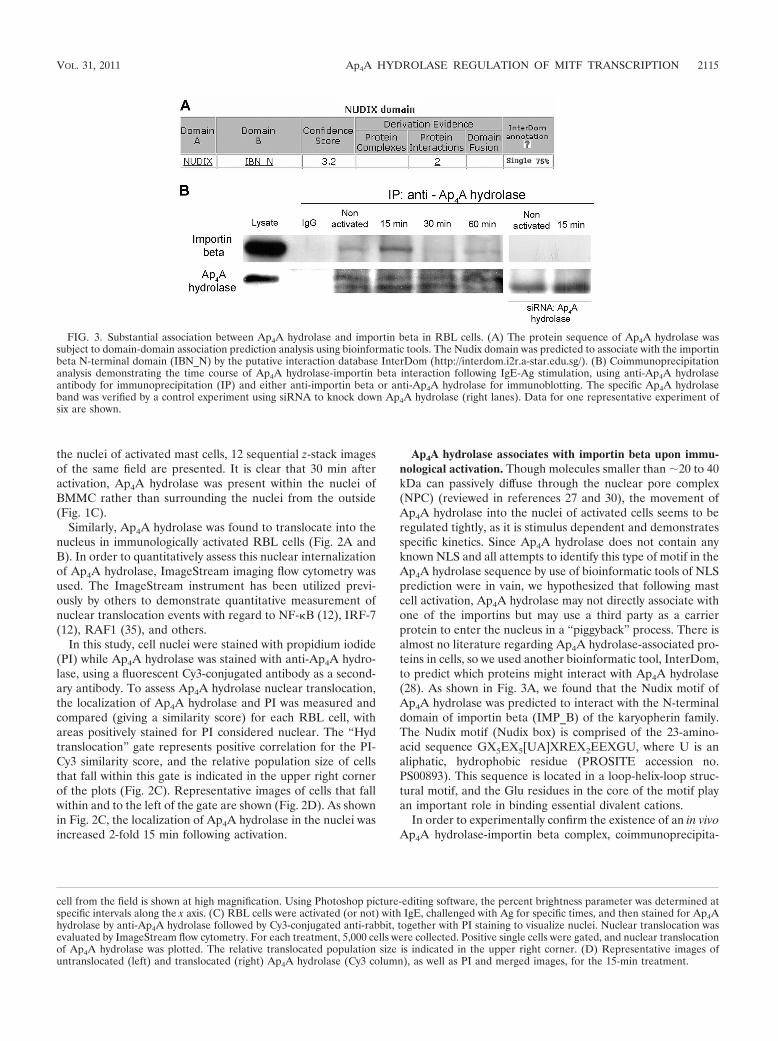
Ap4A hydrolase associates with importin beta upon immu-nological activation. Though molecules smaller than �20 to 40kDa can passively diffuse through the nuclear pore complex(NPC) (reviewed in references 27 and 30), the movement ofAp4A hydrolase into the nuclei of activated cells seems to beregulated tightly, as it is stimulus dependent and demonstratesspecific kinetics. Since Ap4A hydrolase does not contain anyknown NLS and all attempts to identify this type of motif in theAp4A hydrolase sequence by use of bioinformatic tools of NLSprediction were in vain, we hypothesized that following mastcell activation, Ap4A hydrolase may not directly associate withone of the importins but may use a third party as a carrierprotein to enter the nucleus in a “piggyback” process. There isalmost no literature regarding Ap4A hydrolase-associated pro-teins in cells, so we used another bioinformatic tool, InterDom,to predict which proteins might interact with Ap4A hydrolase(28). As shown in Fig. 3A, we found that the Nudix motif ofAp4A hydrolase was predicted to interact with the N-terminaldomain of importin beta (IMP_B) of the karyopherin family.The Nudix motif (Nudix box) is comprised of the 23-amino-acid sequence GX5EX5[UA]XREX2EEXGU, where U is analiphatic, hydrophobic residue (PROSITE accession no.PS00893). This sequence is located in a loop-helix-loop struc-tural motif, and the Glu residues in the core of the motif playan important role in binding essential divalent cations.
In order to experimentally confirm the existence of an in vivoAp4A hydrolase-importin beta complex, coimmunoprecipita-
cell from the field is shown at high magnification. Using Photoshop picture-editing software, the percent brightness parameter was determined atspecific intervals along the x axis. (C) RBL cells were activated (or not) with IgE, challenged with Ag for specific times, and then stained for Ap4Ahydrolase by anti-Ap4A hydrolase followed by Cy3-conjugated anti-rabbit, together with PI staining to visualize nuclei. Nuclear translocation wasevaluated by ImageStream flow cytometry. For each treatment, 5,000 cells were collected. Positive single cells were gated, and nuclear translocationof Ap4A hydrolase was plotted. The relative translocated population size is indicated in the upper right corner. (D) Representative images ofuntranslocated (left) and translocated (right) Ap4A hydrolase (Cy3 column), as well as PI and merged images, for the 15-min treatment.
FIG. 3. Substantial association between Ap4A hydrolase and importin beta in RBL cells. (A) The protein sequence of Ap4A hydrolase wassubject to domain-domain association prediction analysis using bioinformatic tools. The Nudix domain was predicted to associate with the importinbeta N-terminal domain (IBN_N) by the putative interaction database InterDom (http://interdom.i2r.a-star.edu.sg/). (B) Coimmunoprecipitationanalysis demonstrating the time course of Ap4A hydrolase-importin beta interaction following IgE-Ag stimulation, using anti-Ap4A hydrolaseantibody for immunoprecipitation (IP) and either anti-importin beta or anti-Ap4A hydrolase for immunoblotting. The specific Ap4A hydrolaseband was verified by a control experiment using siRNA to knock down Ap4A hydrolase (right lanes). Data for one representative experiment ofsix are shown.
VOL. 31, 2011 Ap4A HYDROLASE REGULATION OF MITF TRANSCRIPTION 2115

tion of Ap4A hydrolase with importin beta was performed inquiescent and activated cells (Fig. 3B). The recovered andresolved immune complexes showed that Ap4A hydrolase wasassociated with importin beta in an IgE-Ag activation-depen-dent manner. The observed association between the two pro-teins peaked 15 to 60 min after stimulation. Knockdown ofAp4A hydrolase by an RNA interference (RNAi) silencingapproach (siRNA) abolished coimmunoprecipitation of im-portin beta.
Nuclear translocation of Ap4A hydrolase in activated mastcells is mediated by importin beta. In order to verify theessential role played by importin beta in nuclear translocationof Ap4A hydrolase, we used an siRNA approach to targetexpression of importin beta. Cells were transfected with vari-
ous siRNA oligonucleotides, each targeting a different mem-ber of the importin family, and the nuclear extracts from therespective cells were analyzed for the presence of Ap4A hy-drolase by using anti-Ap4A hydrolase. While silencing of eitherimportin 5 or 7 as well as transfection with a nontargeting (NT)siRNA did not interfere with translocation to the nucleus,Ap4A hydrolase was not detected in nuclei of cells that weretreated with siRNA against importin beta (Fig. 4A).
Nuclear translocation of Ap4A hydrolase via associationwith importin beta plays a role in the regulation of MITFtranscriptional activity. We previously provided evidence thatAp4A hydrolase is responsible for Ap4A degradation in immu-nologically activated mast cells and, moreover, that Ap4A hy-drolase plays a critical role in the transcriptional regulation of
FIG. 4. Importin beta-mediated Ap4A hydrolase nuclear translocation is involved in MITF transcriptional activity regulation. (A and B) RBLcells were transfected with siRNA oligonucleotides against importin beta, 5, and 7. NT siRNA was used as a control. Nuclear fractions were isolatedfrom cells activated with IgE and antigen for 30 min. Nuclear extracts (A) and cytosolic extracts (B) were analyzed by Western blotting withanti-Ap4A hydrolase antibody. Silencing of importin beta was verified by Western blotting with anti-importin beta. Each lane represents anindependent experiment. (C) RBL cells were transfected with either importin beta siRNA or NT siRNA. Control and transfected cells wereactivated with IgE-Ag. Twenty-four hours later, cells were lysed and mRNA quantitation for GrB and Kit was performed by SYBR greenincorporation into real-time PCR with RBL cells. Expression levels were normalized to those of the �-actin housekeeping gene. Results arepresented as relative quantities, with the untreated cell level arbitrarily set to 1. The means and standard errors of the means for three experimentsare shown.
2116 CARMI-LEVY ET AL. MOL. CELL. BIOL.

MITF (6). Here we further explored the effect of Ap4A hydro-lase nuclear translocation on MITF transcriptional activity.
The transcript levels of two MITF target genes, encodingGrB (17) and Kit (36), were measured in immunologicallyactivated RBL cells in which importin beta was knocked downby siRNA. A significant accumulation of GrB mRNA and amoderate increase in Kit mRNA were observed in RBL cells,concurrent with reduced expression of importin beta and re-duced nuclear translocation of Ap4A hydrolase (Fig. 4B).Thus, nuclear translocation of Ap4A hydrolase via its associa-tion with importin beta is clearly involved in the regulation ofMITF transcriptional activity.
Ap4A hydrolase is subject to dephosphorylation upon im-munological activation. Recently, we found that LysRS trans-locates into the nuclei of activated mast cells in a serine phos-phorylation-dependent manner (40) via the mitogen-activatedprotein kinase (MAPK)/ERK pathway. In order to determinewhether the phosphorylation status of Ap4A hydrolase changesupon IgE-Ag activation, we used 2D gel electrophoresis. Ap4Ahydrolase was immunoprecipitated from nonstimulated orIgE-Ag-activated mast cells, and its phosphorylation was iden-tified by a more acidic pI compatible with phosphorylation (asseen by a shift to the left of the gel). This species was detectedin unstimulated cells and cells that were activated for shortperiods (up to 15 min), whereas in cells activated with IgE-Ag
for 30 min the enzyme appeared to be dephosphorylated (Fig.5A). This phosphorylation was totally blocked by the presenceof alkaline phosphatase (AP) (Fig. 5A). Thus, Ap4A hydrolaseis dephosphorylated upon immunological activation.
We then determined whether the dephosphorylation ofAp4A hydrolase (Fig. 5A) correlated with the enzyme’s nucleartranslocation. Nuclear and cytoplasmic fractions were isolatedfrom IgE-Ag-activated RBL cells. The subcellular extractswere analyzed by 2D gel electrophoresis with anti-Ap4A hy-drolase antibody. No significant difference in phosphorylationstatus was detected in the cytosolic and nuclear fractions. Sim-ilar to the case with whole-cell lysates, phosphorylation wasdetected in both the cytosol and nuclei of nonactivated cells aswell as in cells that were activated for less than 30 min, whilethe dephosphorylated form of the hydrolase was detected inboth the cytosol and nuclei of cells 30 and 60 min followingactivation (Fig. 5B).
We subsequently determined the amino acid residue(s) sub-ject to the dephosphorylation process. Immunoprecipitation ofpotential phosphorylated proteins with phospho-specific anti-bodies and immunoblot analysis with anti-Ap4A hydrolasedemonstrated dephosphorylation of Ap4A hydrolase on serineresidues, but not on threonine residues, 60 min after cell acti-vation.
FIG. 5. Ap4A hydrolase is subject to dephosphorylation upon IgE-Ag activation. (A) Lysates from IgE-Ag-activated and nonactivated RBLcells were subjected to 2D electrophoresis on a pH 3 to 10 gradient in an 8% polyacrylamide gel. The gel was blotted with anti-Ap4A hydrolaseantibody. WB, Western blot. (B) Nuclear and cytoplasmic fractions were isolated from RBL cells activated with IgE and antigen. The subcellularextracts were analyzed by 2D electrophoresis with anti-Ap4A hydrolase antibody.
VOL. 31, 2011 Ap4A HYDROLASE REGULATION OF MITF TRANSCRIPTION 2117

Ser108 phosphorylation of Ap4A hydrolase increases its en-zymatic activity. In order to specifically predict which serineresidues may be subject to phosphorylation, the web-based toolNetPhos was used (3). Only one serine residue, S108, in thehuman Ap4A (hAp4A) hydrolase sequence was significantlypredicted to be phosphorylated (Fig. 6A). Based on this ob-servation, fused Myc-tagged constructs of mutated hAp4A hy-drolase were constructed (S108A and S108D mutants). Each ofthe constructs was administered to the cells after the endoge-nous Ap4A hydrolase was silenced by siRNA. Under theseconditions, no loss of the exogenous hAp4A hydrolase oc-curred (Fig. 6B). Assessment of Ap4A hydrolytic activity wascarried out on extracts derived from immunologically activatedRBL cells that were transfected with each of the constructs. Asshown in Fig. 6C, the S108A mutant hAp4A hydrolase, inwhich the residue at position 108 was not phosphorylated,exhibited reduced Ap4A degradation in these transfected RBLcells, suggesting that phosphorylation of this residue is criticalfor Ap4A hydrolase enzymatic activity.
DISCUSSION
We previously demonstrated that LysRS forms a complexwith MITF and its repressor Hint-1, which is released from thecomplex by its binding to the LysRS product Ap4A, enablingMITF to transcribe its target genes (40).
Recently, we also provided evidence that Ap4A hydrolase isresponsible for Ap4A degradation following immunological ac-tivation of mast cells and thus established Ap4A as a novelsecond messenger (6). In that study, we described the key roleplayed by Ap4A hydrolase in regulation of both MITF andUSF2 transcriptional activity.
The intracellular compartmentalization of Ap4A in mamma-lian cells during various growth and cell cycle stages has beenstudied, and the majority of cellular Ap4A is localized in thenuclei, especially during G1 and early S phase (37). As men-tioned above, we previously reported the nuclear function ofAp4A in the transcription regulation network in activated mastcells. Moreover, we showed that the nuclear translocation ki-netics of the main Ap4A producer, LysRS, corresponded withthe synthesis of this nucleotide (40). Therefore, we hypothe-sized that at least some of the Ap4A hydrolase, which in thiscontext is the major degrading enzyme of Ap4A, also changesits subcellular localization upon immunological challenge.
Though bioinformatic analysis did not predict an NLS forAp4A hydrolase, in the present work we characterized themechanism of its nuclear import. We found that the 17-kDaAp4A hydrolase translocates to the nucleus following immu-nological activation of mast cells (Fig. 1 and 2). Proteinssmaller than 40 kDa have been proposed to diffuse freelythrough the nuclear pores. However, we showed in mast cellsthat Ap4A hydrolase actively translocates to the nucleus in astimulus-dependent manner rather than passively diffusing intothis compartment.
Stimulus-dependent nuclear translocation of signaling pro-teins is critical for time-regulated processes such as cell cycleand transcription. In the first and best-described mechanismfor nuclear import, proteins destined for transport into thenucleus contain amino acid targeting sequences called nuclearlocalization signals (NLSs) (reviewed in reference 21), which
consist of either one (monopartite) or two (bipartite) stretchesof basic amino acids (19, 32).
Unexpectedly, however, many of the proteins involved in theaforementioned functions, including cyclins (26), ERKs (41),SMAD transcription factors (24), and Ap4A hydrolase, do notcontain the canonical NLS. ERKs, MEKs, and SMADs wererecently found to contain a specific 3-amino-acid motif, SPS,which was termed the nuclear translocation sequence (NTS).Upon stimulation, this motif is phosphorylated, resulting innuclear translocation of these proteins in an importin 7-depen-dent manner (8). Though the sequence of Ap4A hydrolasedoes not contain either SPS or the similarly functioning TPTmotif, the description of the above-mentioned mechanism ledus to hypothesize that it was possible that Ap4A hydrolasetranslocated in a nonclassical (non-NLS) manner via an im-portin family member.
As shown in Fig. 3, the Nudix motif of Ap4A hydrolase wasindeed predicted to interact directly with the N-terminal do-main of importin beta. This association was verified using co-immunoprecipitation of the two proteins, and moreover, si-lencing of Ap4A hydrolase prevented coimmunoprecipitationof importin beta (Fig. 3B) and silencing of importin beta pre-vented Ap4A hydrolase nuclear translocation (Fig. 4A).
Importin beta has previously been described to mediate thenuclear translocation of numerous components of cyclin-de-pendent kinase (Cdk)/cyclin complexes, such as cyclin B1 (26)and Cdc7 protein (20). Cyclin B1 performs cardinal roles in theeukaryotic cell division cycle by phosphorylating key cellularsubstrates, and Cdc7 is essential for the initiation of DNAreplication (4). It has been reported that importin beta bindsdirectly to Cdc7, which does not have a classical NLS, via thekinase insert II domain, promoting its nuclear import. Immu-nodepletion of importin beta, but not importin alpha, abro-gated Cdc7 nuclear import (20).
In addition to interfering with Ap4A hydrolase nucleartranslocation, silencing of importin beta led to an increase inMITF transcriptional activity (Fig. 4). This finding addsstrength to the notion that the LysRS-Ap4A pathway is centralin the regulation of MITF transcriptional activity.
Posttranslational modifications, e.g., the phosphorylation ofnuclear import substrates, is required for optimal interactionwith importin alpha (7, 9). ERK phosphorylation at the SPSmotif was found to be critical for its association with importin7 (8). Additionally, for Trx, it was demonstrated that specificS-nitros(yl)ation of the Cys residue is critical for its nuclearimport (34).
We recently showed that LysRS nuclear translocation oc-curred as a result of serine 207 phosphorylation via the MAPK/ERK pathway. Furthermore, phosphorylation of the serine 207residue was found to be a prerequisite for Ap4A synthesis, asa LysRS S207A variant that was linked to a strong NLS did notcause induction of Ap4A synthesis, despite its nuclear localiza-tion (40).
Here we hypothesized that Ap4A hydrolase may also besubject to a posttranslational modification, such as phosphor-ylation, which modulates its intracellular location and/or activ-ity. We found that Ap4A hydrolase was phosphorylated inresting mast cells, while dephosphorylation was observed inactivated cells (Fig. 5). Moreover, the phosphorylated form ofAp4A hydrolase was found to have a higher Ap4A hydrolysis
2118 CARMI-LEVY ET AL. MOL. CELL. BIOL.

FIG. 6. Phosphorylation status of serine 108 in Ap4A hydrolase is involved in the enzymatic activity of Ap4A metabolism. (A) Putative serine,threonine, and tyrosine phosphorylation sites in the Ap4A hydrolase protein sequence, as predicted by the NetPhos 2.0 program (www.cbs.dtu.dk/services/NetPhos/). (B) RBL cells were transfected with rat Ap4A hydrolase siRNA. Twenty-four hours later, cells were transfected with humanAp4A hydrolase S108A and S108D variants. Next, the cells were incubated with IgE and challenged with DNP for 30 min. The cell extracts wereanalyzed by Western blotting with anti-Myc or anti-Ap4A hydrolase antibodies. (C) RBL cell lysates from panel B were subjected to an enzymaticactivity assay. Ap4A hydrolase hydrolytic activity was determined as the rate of synthetic Ap4A degradation, which was evaluated by chemilumi-nescence detection of ATP as described in Materials and Methods. Data for one representative experiment of three are shown.
VOL. 31, 2011 Ap4A HYDROLASE REGULATION OF MITF TRANSCRIPTION 2119

rate than the nonphosphorylated form, suggesting that its de-phosphorylation is involved in the maintenance of the Ap4Alevel in cells upon activation. It is important that although theS108A mutant of Ap4A hydrolase also exhibited hydrolyticactivity, it was observed to be 2-fold lower than that of theS108D mutant, suggesting that phosphorylation of Ser108 onlypartially affects the enzyme’s activity and that it may be regu-lated by additional modifications which have yet to be investi-gated.
Ap4A hydrolase phosphorylation seems to peak 15 min afteractivation. The positive correlation between phosphorylationstatus and Ap4A hydrolysis rate is in accordance with the Ap4Alevel, which drops rapidly 15 min after exposure to Ag (6).
In summary, this study demonstrates nonconventional, Ag-induced regulation of Ap4A hydrolase (Fig. 7). As the impor-tance of Ap4A as a second messenger and as a signaling mol-ecule involved in gene transcription is uncovered, a betterunderstanding of the mechanisms regulating its cellular levelsis important to shed light on its function(s). The findings re-ported herein may also contribute to future emphasis on themanipulation of Ap4A levels in diseases.
ACKNOWLEDGMENTS
This work was supported by the United States Binational ScienceFoundation (E.R.), the Israeli Academy of Science (E.R.), the Ger-man-Israel Foundation for Scientific Research and Development(E.R.), DKFZ-MOST cooperation (E.R.), the National Medical Re-
search Council of Singapore (D.M.K. and E.R.), the Morasha Foun-dation Fund (H.N.), the Acceleration Research of KOSEF (S.K.), the21st Frontier Functional Proteomics Research (S.K.), and theCREATE project of the National Research Foundation of Singa-pore (E.R. and M.K.). I.C.-L. and Z.Y. were supported by theCanadian Friends of Hebrew University.
We have no conflicting financial interests.
REFERENCES
1. Abdelghany, H. M., et al. 2001. Cloning, characterisation and crystallisationof a diadenosine 5�,5-P(1),P(4)-tetraphosphate pyrophosphohydrolasefrom Caenorhabditis elegans. Biochim. Biophys. Acta 1550:27–36.
2. Bayliss, R., T. Littlewood, L. A. Strawn, S. R. Wente, and M. Stewart. 2002.GLFG and FxFG nucleoporins bind to overlapping sites on importin-beta.J. Biol. Chem. 277:50597–50606.
3. Blom, N., S. Gammeltoft, and S. Brunak. 1999. Sequence and structure-based prediction of eukaryotic protein phosphorylation sites. J. Mol. Biol.294:1351–1362.
4. Bousset, K., and J. F. Diffley. 1998. The Cdc7 protein kinase is required fororigin firing during S phase. Genes Dev. 12:480–490.
5. Reference deleted.6. Carmi-Levy, I., N. Yannay-Cohen, G. Kay, E. Razin, and H. Nechushtan.
2008. Diadenosine tetraphosphate hydrolase is part of the transcriptionalregulation network in immunologically activated mast cells. Mol. Cell. Biol.28:5777–5784.
7. Chook, Y. M., and G. Blobel. 2001. Karyopherins and nuclear import. Curr.Opin. Struct. Biol. 11:703–715.
8. Chuderland, D., A. Konson, and R. Seger. 2008. Identification and charac-terization of a general nuclear translocation signal in signaling proteins. Mol.Cell 31:850–861.
9. Conti, E., M. Uy, L. Leighton, G. Blobel, and J. Kuriyan. 1998. Crystallo-graphic analysis of the recognition of a nuclear localization signal by thenuclear import factor karyopherin alpha. Cell 94:193–204.
10. Fried, H., and U. Kutay. 2003. Nucleocytoplasmic transport: taking an in-ventory. Cell. Mol. Life Sci. 60:1659–1688.
FIG. 7. Proposed model for involvement of Ap4A hydrolase in transcription regulation. Upon immunological activation, Ap4A hydrolaseassociates with importin beta and is translocated into the nucleus when Ap4A levels are elevated. Ap4A binds to Hint-1, dissociating it from MITF.With the repression removed, transcription factors are free to transcribe their target genes. The presence of Ap4A hydrolase in the nucleus leadsto hydrolysis of the accumulated Ap4A into AMP and ATP, decreasing its levels to that found in resting cells. When Ap4A levels decrease, Hint-1reassociates with MITF.
2120 CARMI-LEVY ET AL. MOL. CELL. BIOL.

11. Gaietta, G., et al. 2002. Multicolor and electron microscopic imaging ofconnexin trafficking. Science 296:503–507.
12. George, T. C., P. J. Morrissey, C. Cui, S. Singh, and P. Fitzgerald Bocarsly.2009. Measurement of cytoplasmic to nuclear translocation. Curr. Protoc.Cytom. 2009:Unit 9.28.
13. Han, J. M., et al. 2008. AIMP2/p38, the scaffold for the multi-tRNA synthe-tase complex, responds to genotoxic stresses via p53. Proc. Natl. Acad. Sci.U. S. A. 105:11206–11211.
14. Hause, B., K. Feussner, and C. Wasternack. 1997. Nuclear location of adiadenosine 5�,5-P1,P4-tetraphosphate (Ap4A) hydrolase in tomato cellsgrown in suspension cultures. Bot. Acta 110:452–457.
15. Houslay, M. D. 2010. Underpinning compartmentalised cAMP signallingthrough targeted cAMP breakdown. Trends Biochem. Sci. 35:91–100.
16. Isgro, T. A., and K. Schulten. 2007. Association of nuclear pore FG-repeatdomains to NTF2 import and export complexes. J. Mol. Biol. 366:330–345.
17. Ito, A., et al. 1998. Systematic method to obtain novel genes that are regu-lated by mi transcription factor: impaired expression of granzyme B andtryptophan hydroxylase in mi/mi cultured mast cells. Blood 91:3210–3221.
18. Reference deleted.19. Kalderon, D., B. L. Roberts, W. D. Richardson, and A. E. Smith. 1984. A
short amino acid sequence able to specify nuclear location. Cell 39:499–509.20. Kim, B. J., and H. Lee. 2006. Importin-beta mediates Cdc7 nuclear import by
binding to the kinase insert II domain, which can be antagonized by impor-tin-alpha. J. Biol. Chem. 281:12041–12049.
21. Lange, A., et al. 2007. Classical nuclear localization signals: definition, func-tion, and interaction with importin alpha. J. Biol. Chem. 282:5101–5105.
22. Levy, C., H. Nechushtan, and E. Razin. 2002. A new role for the STAT3inhibitor, PIAS3: a repressor of microphthalmia transcription factor. J. Biol.Chem. 277:1962–1966.
23. Li, S., et al. 2004. A map of the interactome network of the metazoan C.elegans. Science 303:540–543.
24. Massague, J., J. Seoane, and D. Wotton. 2005. Smad transcription factors.Genes Dev. 19:2783–2810.
25. McLennan, A. G. 2006. The Nudix hydrolase superfamily. Cell. Mol. Life Sci.63:123–143.
26. Moore, J. D., J. Yang, R. Truant, and S. Kornbluth. 1999. Nuclear import ofCdk/cyclin complexes: identification of distinct mechanisms for import ofCdk2/cyclin E and Cdc2/cyclin B1. J. Cell Biol. 144:213–224.
27. Mosammaparast, N., and L. F. Pemberton. 2004. Karyopherins: from nu-clear-transport mediators to nuclear-function regulators. Trends Cell Biol.14:547–556.
28. Ng, S. K., Z. Zhang, S. H. Tan, and K. Lin. 2003. InterDom: a database ofputative interacting protein domains for validating predicted protein inter-actions and complexes. Nucleic Acids Res. 31:251–254.
29. Oka, K., et al. 2011. Nudix-type motif 2 (NUDT2) in human breast carci-noma: a potent prognostic factor associated with cell proliferation. Int. J.Cancer 128:1770–1782.
30. Pemberton, L. F., and B. M. Paschal. 2005. Mechanisms of receptor-medi-ated nuclear import and nuclear export. Traffic 6:187–198.
31. Razin, E., et al. 1999. Suppression of microphthalmia transcriptional activityby its association with protein kinase C-interacting protein 1 in mast cells.J. Biol. Chem. 274:34272–34276.
32. Robbins, J., S. M. Dilworth, R. A. Laskey, and C. Dingwall. 1991. Twointerdependent basic domains in nucleoplasmin nuclear targeting sequence:identification of a class of bipartite nuclear targeting sequence. Cell 64:615–623.
33. Saitoh, S., et al. 2000. LAT is essential for FceRI-mediated mast cell acti-vation. Immunity 12:525–535.
34. Schroeder, P., R. Popp, B. Wiegand, J. Altschmied, and J. Haendeler. 2007.Nuclear redox-signaling is essential for apoptosis inhibition in endothelialcells—important role for nuclear thioredoxin-1. Arterioscler. Thromb. Vasc.Biol. 27:2325–2331.
35. Smith, J., et al. 2009. Retinoic acid induces nuclear accumulation of Raf1during differentiation of HL-60 cells. Exp. Cell Res. 315:2241–2248.
36. Tsujimura, T., et al. 1996. Involvement of transcription factor encoded bythe mi locus in the expression of c-kit receptor tyrosine kinase in culturedmast cells of mice. Blood 88:1225–1233.
37. Weinmann-Dorsch, C., and F. Grummt. 1986. Diadenosine tetraphosphate(Ap4A) is compartmentalized in nuclei of mammalian cells. Exp. Cell Res.165:550–554.
38. Weis, K. 2003. Regulating access to the genome: nucleocytoplasmic trans-port throughout the cell cycle. Cell 112:441–451.
39. Winward, L., W. G. Whitfield, T. J. Woodman, A. G. McLennan, and S. T.Safrany. 2007. Characterisation of a bis(5�-nucleosyl)-tetraphosphatase(asymmetrical) from Drosophila melanogaster. Int. J. Biochem. Cell Biol.39:943–954.
40. Yannay-Cohen, N., et al. 2009. LysRS serves as a key signaling molecule inthe immune response by regulating gene expression. Mol. Cell 34:603–611.
41. Yoon, S., and R. Seger. 2006. The extracellular signal-regulated kinase:multiple substrates regulate diverse cellular functions. Growth Factors 24:21–44.
VOL. 31, 2011 Ap4A HYDROLASE REGULATION OF MITF TRANSCRIPTION 2121
Copyright © 2022 FDOKUMEN