Restoration of p53 function for selective Fas-mediated apoptosis in human and rat glioma cells in...
10
2006;5:20-28. Mol Cancer Ther Patrick B. Senatus, Yin Li, Christopher Mandigo, et al. by a p53 COOH-terminal peptide in vivo and in vitro apoptosis in human and rat glioma cells Restoration of p53 function for selective Fas-mediated Updated version http://mct.aacrjournals.org/content/5/1/20 Access the most recent version of this article at: Cited Articles http://mct.aacrjournals.org/content/5/1/20.full.html#ref-list-1 This article cites by 24 articles, 10 of which you can access for free at: Citing articles http://mct.aacrjournals.org/content/5/1/20.full.html#related-urls This article has been cited by 4 HighWire-hosted articles. Access the articles at: E-mail alerts related to this article or journal. Sign up to receive free email-alerts Subscriptions Reprints and . [email protected] Department at To order reprints of this article or to subscribe to the journal, contact the AACR Publications Permissions . [email protected] Department at To request permission to re-use all or part of this article, contact the AACR Publications Research. on November 9, 2014. © 2006 American Association for Cancer mct.aacrjournals.org Downloaded from Research. on November 9, 2014. © 2006 American Association for Cancer mct.aacrjournals.org Downloaded from
Transcript of Restoration of p53 function for selective Fas-mediated apoptosis in human and rat glioma cells in...
2006;5:20-28. Mol Cancer Ther Patrick B. Senatus, Yin Li, Christopher Mandigo, et al. by a p53 COOH-terminal peptide
in vivo and in vitroapoptosis in human and rat glioma cells Restoration of p53 function for selective Fas-mediated
Updated version
http://mct.aacrjournals.org/content/5/1/20
Access the most recent version of this article at:
Cited Articles
http://mct.aacrjournals.org/content/5/1/20.full.html#ref-list-1
This article cites by 24 articles, 10 of which you can access for free at:
Citing articles
http://mct.aacrjournals.org/content/5/1/20.full.html#related-urls
This article has been cited by 4 HighWire-hosted articles. Access the articles at:
E-mail alerts related to this article or journal.Sign up to receive free email-alerts
Subscriptions
Reprints and
To order reprints of this article or to subscribe to the journal, contact the AACR Publications
Permissions
To request permission to re-use all or part of this article, contact the AACR Publications
Research. on November 9, 2014. © 2006 American Association for Cancermct.aacrjournals.org Downloaded from
Research. on November 9, 2014. © 2006 American Association for Cancermct.aacrjournals.org Downloaded from
Restoration of p53 function for selective Fas-mediatedapoptosis in human and rat glioma cells in vitro andin vivo by a p53 COOH-terminal peptide
Patrick B. Senatus,1 Yin Li,2 Christopher Mandigo,1
Gwen Nichols,2 Gaetan Moise,1 Yuehua Mao,2
Melandee D. Brown,1 Richard C. Anderson,1
Andrew T. Parsa,1 Paul W. Brandt-Rauf,3
Jeffrey N. Bruce,1 and Robert L. Fine2
1Department of Neurological Surgery; 2Experimental TherapeuticsProgram, Division of Medical Oncology, Department of Medicine;and 3Department of Environmental Health Sciences, MailmanSchool of Public Health, Columbia University Medical Center,New York, New York
AbstractWe have shown that a COOH-terminal peptide of p53(amino acids 361–382, p53p), linked to the truncatedhomeobox domain of Antennapedia (Ant) as a carrierfor transduction, induced rapid apoptosis in human pre-malignant and malignant cell lines. Here, we report thathuman and rat glioma lines containing endogenous mutantp53 or wild-type (WT) p53 were induced into apoptosisby exposure to this peptide called p53p-Ant. The peptidewas comparatively nontoxic to proliferating nonmalig-nant human and rat glial cell lines containing WT p53 andproliferating normal human peripheral marrow blood stemcells. Degree of sensitivity to the peptide correlateddirectly with the level of endogenous p53 expressionand mutant p53 conformation. Apoptosis induction byp53p-Ant was quantitated by terminal deoxynucleotidyltransferase–mediated dUTP nick end labeling assayand Annexin V staining in human glioma cells in vitroand in a syngeneic orthotopic 9L glioma rat model using
convection-enhanced delivery in vivo. The mechanismof cell death by this peptide was solely through theFas extrinsic apoptotic pathway. p53p-Ant induced a3-fold increase in extracellular membrane Fas expressionin glioma cells but no significant increase in nonmalig-nant glial cells. These data suggest that p53 functionfor inducing Fas-mediated apoptosis in gliomas, whichexpress sufficient quantities of endogenous mutant orWT p53, may be restored or activated, respectively, bya cell-permeable peptide derived from the p53 COOH-terminal regulatory domain (p53p-Ant). p53p-Ant mayserve as a prototypic model for the development of newanticancer agents with unique selectivity for gliomacancer cells and it can be successfully delivered in vivointo a brain tumor by a convection-enhanced deliverysystem, which circumvents the blood-brain barrier. [MolCancer Ther 2006;5(1):20–8]
Introductionp53, known as the ‘‘guardian of the genome,’’ is asequence-specific transcription factor that binds DNA andtransactivates cellular proteins involved in growth and cellcycle regulation (1–6). p53 is the most frequently mutatedgene in human neoplasms (7). Major transcriptional targetsof functional p53 are p21WAF/Cip1, which mediates cellcycle arrest; GADD45, which mediates growth arrest andDNA repair; and the proteins Bax, Bak, Bcl-2, and Fas/Apo-1, which mediate programmed cell death (4, 8–11). Inaddition, functional p53 can induce activation of Fas/Apo-1-mediated apoptosis without transcriptional/translationalchanges, although this is not completely understood.
The p53 molecule contains three major domains: NH2-terminal transactivation domain, central sequence-specificDNA-binding domain, and a COOH-terminal negativeregulatory domain involved in modulating p53 function(1, 12, 13). Fifty percent of all human malignancies andup to 65% of human gliomas have missense or nonsensemutations of p53 . The great majority of these missensemutations have been mapped to the central DNA-bindingregion of the protein. The sequence-specific DNA-bindingactivity of p53 is negatively regulated by its 30–amino acidCOOH-terminal sequence (amino acids 363–393) in con-cert with its NH2-terminal proline-rich region locatedbetween amino acids 80 and 93. Synthetic peptides derivedfrom the COOH-terminal sequence bind directly to wild-type (WT) and mutant p53 in vitro , and binding activitywas dependent on the presence of both the proline-richNH2-terminal amino acids 80 to 93 sequence and theCOOH-terminal amino acids 363 to 393 sequence in the p53protein (13). We have calculated computational dockingmodels of low-energy conformations of COOH-terminal
Received 6/7/05; revised 9/19/05; accepted 10/14/05.
Grant support: NIH grant RO1 CA 82528, Columbia University CancerCenter Translational Award, and Vinzinni Brain Cancer Award (R.L. Fine),NIH grant RO1 CA 89395 (J.N. Bruce), NIH grant RO1 OH 07590 (P.W.Brandt-Rauf), Catherine O’Rourke/American Brain Tumor AssociationFellowship (P.B. Senatus), Reza Khatib Fellowship (C. Mandigo), AmericanBrain Tumor Association Medical Student Fellowship (G. Moise), andGeorge Becker Fellowship (M.D. Brown).
The costs of publication of this article were defrayed in part by thepayment of page charges. This article must therefore be hereby markedadvertisement in accordance with 18 U.S.C. Section 1734 solely toindicate this fact.
Note: P.B. Senatus and Y. Li contributed equally to the work. J.N. Bruceand R.L. Fine are senior authors.
Requests for reprints: Robert L. Fine, Experimental Therapeutics Program,Division of Medical Oncology, Department of Medicine, ColumbiaUniversity Medical Center, 650 West 168th Street, BB 20-05,New York, NY 10032. Phone: 212-305-1168; Fax: 212-305-7348.E-mail: [email protected]
Copyright C 2006 American Association for Cancer Research.
doi:10.1158/1535-7163.MCT-05-0181
20
Mol Cancer Ther 2006;5(1). January 2006
Research. on November 9, 2014. © 2006 American Association for Cancermct.aacrjournals.org Downloaded from
and NH2-terminal p53-derived sequences that predict aunique low-energy, favorable, and stable complex betweenthe two domains (14). Addition of exogenous COOH-terminal p53 peptide restored sequence-specific DNA-binding function to many mutant p53 molecules, includingp53-273 (Arg-to-His) in vitro . This mutation is the secondmost common p53 mutation in human cancers. Moreover,introduction of COOH-terminal p53 peptide, via micro-injection or Antennapedia (Ant) transduction protein, hasrestored transactivation of a p53-responsive reporterconstruct and induced apoptosis in SW480 colon carci-noma cells with mutant p53-273 (Arg-to-His; refs. 15, 16).COOH-terminal p53 peptide alone cannot traverse thecell membrane because of its multiple basic chargedamino acids unless it is linked to a carrier peptide, suchas Ant.
We reported that p53 COOH-terminal peptide (p53p-Ant) induced rapid apoptosis in breast cancer cell linescarrying either endogenous p53 mutations or overex-pressed WT p53. The peptide was not toxic to nonmalig-nant breast or colon cells and null p53 human breast orcolon cancer cells (14, 17, 18). The mechanism of apoptosisby p53p-Ant in breast cancer cells was through the Fas/Apo-1 pathway involving an increased surface expressionof Fas/Apo-1 and Fas ligand due to increased ‘‘flipping’’ ofthe Fas intracellular membrane receptor to the extracellularmembrane milieu without transcriptional or translationalchanges (14, 18). We studied the three-dimensionalstructure of p53p-Ant by proton nuclear magnetic reso-nance spectroscopy and found that it did not have any rigidsecondary or tertiary structure; rather, it was flexible andable to conform to multiple mutant p53 conformations (19).Because various p53 mutations are prevalent in humangliomas, restoration of p53 function in malignant gliomamay present a novel approach for therapy. In addition, theprolonged half-life of mutant p53 compared with WT p53,due to decreased ubiquitination-proteosomic degradation,leads to mutant p53 accumulation, which increases thetarget and selectivity of the COOH-terminal p53 peptide.This also occurs with WT p53 that is overexpressed butinactive because of accumulation in the cytoplasm insteadof the nucleus, such as in neuroblastoma and some breastcancers. Thus, cells with either mutant p53 or overex-pressed WT p53 present more p53 target protein for thepeptide and explain why these cells are selectively moresensitive to the peptide. Conversely, normal cells have verylow or nondetectable levels of WT p53 in Western blots andare relatively insensitive to the peptide (14, 17, 18, 20).Thus, our hypothesis for selective apoptosis induced by thep53 peptide is that it is regulated mainly by the status(mutant > WT) and the level of p53 expression: the morep53, the more potential for inducing cell death, especially ifit is mutant p53. This unique characteristic for the peptidehypothetically allows it to be more selective for mutant andoverexpressed WT p53 tumors (18, 20). We tested thishypothesis in this article with human and rat glioma andnonmalignant glial cell lines with different levels of p53expression and p53 status (mutant and WT).
Materials andMethodsCell Lines andTissue CultureHuman glioma cancer cell lines U138 (mutant p53) and
U87 (WT p53) and rat glioma cancer cell lines 9L (mutantp53), D74 (mutant p53), and F98 (mutant p53) werepurchased from American Type Culture Collection(Rockville, MD) and maintained according to their cellculture guidelines. The nonmalignant human glial cell lineRAV (WT p53) was derived from normal human braintissue of an anterior temporal lobe resection for intractableseizures. The normal rat glial cell line NL (WT p53) washarvested from normal brain cortices of Fischer rats. All ofthe malignant glioma and the nonmalignant glial cell lineswere cultured in DMEM supplemented with 10% FCS,2 mmol/L L-glutamine, and 100 Ag/mL penicillin/strepto-mycin. Assessment of cell proliferation in glial and gliomacell lines was done by 3-(4,5-dimethylthiazol-2-yl)-2,5-diphenyltetrazolium bromide (MTT) assays.
PeptidesThe COOH-terminal p53 peptide (amino acids 361–382,
p53p), Ant, and p53p-Ant peptide were chemically synthe-sized by Research Genetics (Huntsville, AL) as describedpreviously (14). All peptides were high-performanceliquid chromatography purified to >95%. Peptide stocks(4 mmol/L) were prepared in sterile distilled water andstored in aliquots at �80jC.
p53p: N-GSRAHSSHLKSKKGQSTSRHKK-C (22 aminoacids, 361–382 of p53),
Ant: KKWKMRRNQFWVKVQRG (17 amino acids), andp53p-Ant: N-GSRAHSSHLKSKKGQSTSRHKKWKMR-
RNQFWVKVQRG-C (37 amino acids).
The chimeric peptide has 37 amino acids instead of 39amino acids, because KK was eliminated from the NH2
terminus of the Ant sequence because p53p has a KK at theCOOH terminus.
AntibodiesAnti-human p53 monoclonal antibody DO-1 (epitope:
amino acids 11–25) and anti-human and anti-rat p53polyclonal antibody PAb-240 (epitope: amino acids 212–217) were obtained from Santa Cruz Biotechnology (SanDiego, CA). FITC-conjugated mouse anti-human Fas/CD95monoclonal antibody (clone DX2) was obtained fromPharMingen (San Diego, CA). Specific caspase-8 inhibitor(Z-IETD-AFC) and caspase-9 inhibitor (Z-LEHD-FMK)were obtained from MBL International Corp. (Watertown,MA). Anti-a-tubulin monoclonal antibody was obtainedfrom Sigma (St. Louis, MO).
DNASequencing of p53 in F98 and D74 Cell LinesTotal RNA was extracted from rat glioma D74 and F98
cells by using RNeasy Mini kit (Qiagen, Valencia, CA) andits cDNA was reverse transcribed with SuperScript IIReverse Transcriptase kit (Invitrogen, Carlsbad, CA).The PCR product was amplified by using 5V primer 5-ATGGAGGATTCACAGTCGGATATGAGCAT-3 and 3Vprimer 5-TCAGTCTGAGTCAGGCCCCACTTTCTT-3 withPlatinum Pfx DNA Polymerase. After running the gel, the
Molecular Cancer Therapeutics 21
Mol Cancer Ther 2006;5(1). January 2006
Research. on November 9, 2014. © 2006 American Association for Cancermct.aacrjournals.org Downloaded from
PCR product with the corresponding band was purifiedand sequenced with the primers as above at the ColumbiaUniversity DNA Sequencing Core Laboratory. Sequenc-ing for all of the p53 exons (1–11) were done for p53mutations.
MTTAssayCells (3,000) were seeded per well in 96-well plates
overnight for 18 hours and then treated with variousconcentrations of peptide for 12 hours. The assay wasdone with a Cell Proliferation Kit I (MTT; Roche Diag-nostics, Indianapolis, IN) according to the manufacturer’sinstructions.
Western Blot AnalysisCell lysates were prepared in 1 mL lysis buffer [20
mmol/L Tris-HCl (pH 7.6), 1 mmol/L EDTA (pH 8.0),150 mmol/L NaCl, 1% Triton X-100, 10 Ag/mL aprotinin,5 mmol/L benzamidine, 50 Ag/mL leupeptin, 10 Ag/mLpepstatin A, and 1 mmol/L phenylmethylsulfonyl fluoride]for 15 minutes on ice and centrifuged at 10,000 � g for30 minutes. Equal amounts of lysates (40 Ag) weredetermined by the Bradford method and then boiled inSDS sample buffer and loaded on SDS-PAGE. Aftertransferring, immunoreactive products were detected bythe enhanced chemiluminescence system (Amersham,Piscataway, NJ). a-Tubulin staining was used for a secondloading control in the gels. p53 expression was quantitatedby densitometry and the cell line with the lowestexpression band for p53 was given the arbitrary numberof 1 (i.e., NL glial line). Cell lines expressing more p53 bydensitometry than the arbitrary number of 1 were givennumbers that equal the x-fold increase over the NL glialline, which has a densitometric p53 expression value of 1.
Terminal Deoxynucleotidyl Transferase ^MediateddUTPNick End Labeling Assay
Cells were treated with 0, 30, or 50 Amol/L p53p-Ant for6 hours. After washing with PBS, cells were fixed in 4%paraformaldehyde for 1 hour. Terminal deoxynucleotidyltransferase–mediated dUTP nick end labeling (TUNEL)assay was done with a MEBSTAIN Apoptosis Kit Direct(MBL, Nagoya, Japan) according to the manufacturer’sinstructions. TUNEL-positive cells were measured from thefluorescence intensity of 5,000 cells using FACSCaliburflow cytometry.
AnnexinVAssayCells were treated with 30 Amol/L Ant, p53p, or
p53p-Ant for 2 hours. Annexin V assays were doneaccording to the manufacturer’s protocol (PharMingen).Annexin V–positive cells were measured from the fluo-rescence intensity of 10,000 cells using FACSCalibur flowcytometry.
Membrane-Bound FASExpressionHuman U138 glioma and RAV glial cells were treated
with either saline or 30 Amol/L Ant alone, p53p alone,or p53p-Ant for 6 hours. For cell surface Fas analysis, theFITC-conjugated mouse anti-human monoclonal Fas/CD95antibody (clone DX2) was used by measuring theirfluorescent intensity in FACSCalibur flow cytometryaccording to the manufacturer’s protocol.
Collection of Stem Cells and Colony-Forming Unitsfor Granulocyte, Erythroid, Monocyte, and Macro-phage Cells Assay
Human peripheral blood stem cells from normal volun-teers were collected after informed consent by standardapheresis in acid-citrate-dextrose formula A solution.Permission for use of these stem cells in our experimentswas obtained from each volunteer. This protocol followedHealth Insurance Portability and Accountability Act regu-lations and was institutional review board approved.Aliquots were separated by Ficoll-Hypaque and washedonce in PBS and centrifuged 1,000 � g for 5 minutes. Cellswere resuspended in PBS and added to the methylcellulosemedium MethoCult GF H4434 (StemCell Technologies,Vancouver, British Columbia, Canada) containing growthfactors for colony-forming units for granulocyte, erythroid,monocyte, and macrophage cells (CFU-GEMM; CD34+
stem cells). For the CFU-GEMM assay, the cells wereseeded in four-well plates (100,000 per well), treated witheither 30 Amol/L Ant, p53p, or p53p-Ant, and incubatedat 37jC with 5% CO2 for 2 weeks. Cell colonies containing>50 cells per colony were counted as positive and scoredafter a 14-day incubation by an inverted microscope. CFU-GEMM assays were done three separate times each intriplicate from three separate normal donors.
Convection-Enhanced DeliveryMale Fischer 344 rats (ages 12–14 weeks, 250 g) were
anesthetized and a 20-gauge plastic guide cannula wascemented into their skulls. After allowing for a 48-hourrecovery, a catheter was stereotactically inserted into theright caudate nucleus and 105 syngeneic 9L glioma cells in avolume of 5 AL were injected. The catheter was thenwithdrawn and the guide cannula was sealed with adummy stylet. The tumor cells were allowed to grow for10 days to an average size of 2 � 2 mm. In vivo p53 peptidedoses for convection-enhanced delivery were determinedpreviously in the laboratory (J.N.B.) in in vitro doseescalation toxicity trials (21, 22). Either 100 Amol/L Ant,p53p, p53p-Ant, or saline (30 AL) was constantly infused byconvection-enhanced delivery over a 6-hour period andrepeated once after a 12-hour hiatus between infusions.Animals were monitored for adverse symptoms, such asataxia, lethargy, or seizure, and sacrificed 12 hours after thesecond infusion of peptide. Thus, the total exposure time topeptide was over a 36-hour period for the experiment.Brains were immediately fixed, then sectioned, and stainedby the TUNEL method for apoptotic cells.
ResultsGrowth Rates and Transduction of p53 COOH-
Terminal Peptide into Normal Glial and MalignantGliomaCells
We initially assessed whether growth rates and peptidetransduction into the cell lines were similar to ensure thatthe proliferation status and the ability of the peptides toenter the various cell lines were not different (humanglioma U138 and rat glioma F98 versus human glial RAV
Selective Induction of Apoptosis in Glioma Cells22
Mol Cancer Ther 2006;5(1). January 2006
Research. on November 9, 2014. © 2006 American Association for Cancermct.aacrjournals.org Downloaded from
and rat glial NL). These control experiments werenecessary to ensure that any differential toxicities fromthe peptide in nonmalignant glial cells versus malignantglioma cells were not due to differences in growth ratesor differential transduction of peptide. Growth rates, asmeasured by MTT assays between the cell lines, showeda mean F SD doubling rate in the glioma cell lines of30 F 6 hours, and for glial cell lines, it was 36 F 7 hours(n = 3). Because the mean F SD of doubling timesoverlapped, we considered the lines to have no majorsignificant differences in growth rates. To test for differ-ences in peptide transduction across cell membranes, weused p53-Ant linked to rhodamine B (30 Amol/L p53p-Ant-RhoB) in the human glioma U138 and human glial RAV celllines as assessed by fluorescence microscopy. p53-Ant-RhoB quickly entered both cell lines by 1 minute andmaximized at 10 minutes after peptide exposure. Thepeptide entered cytoplasmic and nuclear compartmentsequally in both cell lines (data not shown). Our previouswork with p53-Ant-RhoB in normal and malignant colonand breast cell lines also showed equal peptide transduc-tion (14, 18).
Inhibition of Cell Proliferation by p53 COOH-TerminalPeptide
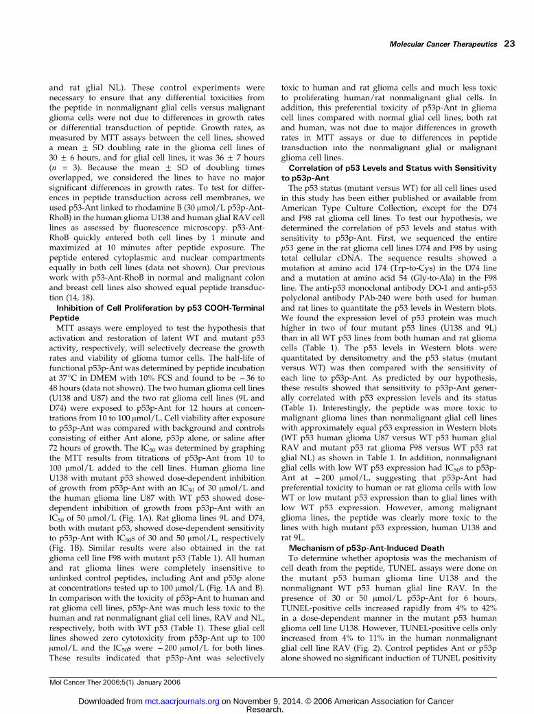
MTT assays were employed to test the hypothesis thatactivation and restoration of latent WT and mutant p53activity, respectively, will selectively decrease the growthrates and viability of glioma tumor cells. The half-life offunctional p53p-Ant was determined by peptide incubationat 37jC in DMEM with 10% FCS and found to be f36 to48 hours (data not shown). The two human glioma cell lines(U138 and U87) and the two rat glioma cell lines (9L andD74) were exposed to p53p-Ant for 12 hours at concen-trations from 10 to 100 Amol/L. Cell viability after exposureto p53p-Ant was compared with background and controlsconsisting of either Ant alone, p53p alone, or saline after72 hours of growth. The IC50 was determined by graphingthe MTT results from titrations of p53p-Ant from 10 to100 Amol/L added to the cell lines. Human glioma lineU138 with mutant p53 showed dose-dependent inhibitionof growth from p53p-Ant with an IC50 of 30 Amol/L andthe human glioma line U87 with WT p53 showed dose-dependent inhibition of growth from p53p-Ant with anIC50 of 50 Amol/L (Fig. 1A). Rat glioma lines 9L and D74,both with mutant p53, showed dose-dependent sensitivityto p53p-Ant with IC50s of 30 and 50 Amol/L, respectively(Fig. 1B). Similar results were also obtained in the ratglioma cell line F98 with mutant p53 (Table 1). All humanand rat glioma lines were completely insensitive tounlinked control peptides, including Ant and p53p aloneat concentrations tested up to 100 Amol/L (Fig. 1A and B).In comparison with the toxicity of p53p-Ant to human andrat glioma cell lines, p53p-Ant was much less toxic to thehuman and rat nonmalignant glial cell lines, RAV and NL,respectively, both with WT p53 (Table 1). These glial celllines showed zero cytotoxicity from p53p-Ant up to 100Amol/L and the IC50s were f200 Amol/L for both lines.These results indicated that p53p-Ant was selectively
toxic to human and rat glioma cells and much less toxicto proliferating human/rat nonmalignant glial cells. Inaddition, this preferential toxicity of p53p-Ant in gliomacell lines compared with normal glial cell lines, both ratand human, was not due to major differences in growthrates in MTT assays or due to differences in peptidetransduction into the nonmalignant glial or malignantglioma cell lines.
Correlation of p53 Levels and Status with Sensitivityto p53p-Ant
The p53 status (mutant versus WT) for all cell lines usedin this study has been either published or available fromAmerican Type Culture Collection, except for the D74and F98 rat glioma cell lines. To test our hypothesis, wedetermined the correlation of p53 levels and status withsensitivity to p53p-Ant. First, we sequenced the entirep53 gene in the rat glioma cell lines D74 and F98 by usingtotal cellular cDNA. The sequence results showed amutation at amino acid 174 (Trp-to-Cys) in the D74 lineand a mutation at amino acid 54 (Gly-to-Ala) in the F98line. The anti-p53 monoclonal antibody DO-1 and anti-p53polyclonal antibody PAb-240 were both used for humanand rat lines to quantitate the p53 levels in Western blots.We found the expression level of p53 protein was muchhigher in two of four mutant p53 lines (U138 and 9L)than in all WT p53 lines from both human and rat gliomacells (Table 1). The p53 levels in Western blots werequantitated by densitometry and the p53 status (mutantversus WT) was then compared with the sensitivity ofeach line to p53p-Ant. As predicted by our hypothesis,these results showed that sensitivity to p53p-Ant gener-ally correlated with p53 expression levels and its status(Table 1). Interestingly, the peptide was more toxic tomalignant glioma lines than nonmalignant glial cell lineswith approximately equal p53 expression in Western blots(WT p53 human glioma U87 versus WT p53 human glialRAV and mutant p53 rat glioma F98 versus WT p53 ratglial NL) as shown in Table 1. In addition, nonmalignantglial cells with low WT p53 expression had IC50s to p53p-Ant at f200 Amol/L, suggesting that p53p-Ant hadpreferential toxicity to human or rat glioma cells with lowWT or low mutant p53 expression than to glial lines withlow WT p53 expression. However, among malignantglioma lines, the peptide was clearly more toxic to thelines with high mutant p53 expression, human U138 andrat 9L.
Mechanism of p53p-Ant-Induced DeathTo determine whether apoptosis was the mechanism of
cell death from the peptide, TUNEL assays were done onthe mutant p53 human glioma line U138 and thenonmalignant WT p53 human glial line RAV. In thepresence of 30 or 50 Amol/L p53p-Ant for 6 hours,TUNEL-positive cells increased rapidly from 4% to 42%in a dose-dependent manner in the mutant p53 humanglioma cell line U138. However, TUNEL-positive cells onlyincreased from 4% to 11% in the human nonmalignantglial cell line RAV (Fig. 2). Control peptides Ant or p53palone showed no significant induction of TUNEL positivity
Molecular Cancer Therapeutics 23
Mol Cancer Ther 2006;5(1). January 2006
Research. on November 9, 2014. © 2006 American Association for Cancermct.aacrjournals.org Downloaded from
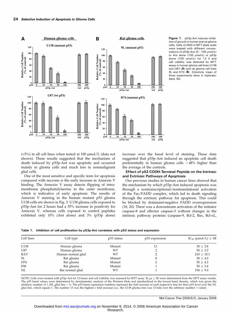
(<5%) in all cell lines when tested at 100 Amol/L (data notshown). These results suggested that the mechanism ofdeath induced by p53p-Ant was apoptotic and occurredmainly in glioma cells and much less in nonmalignantglial cells.
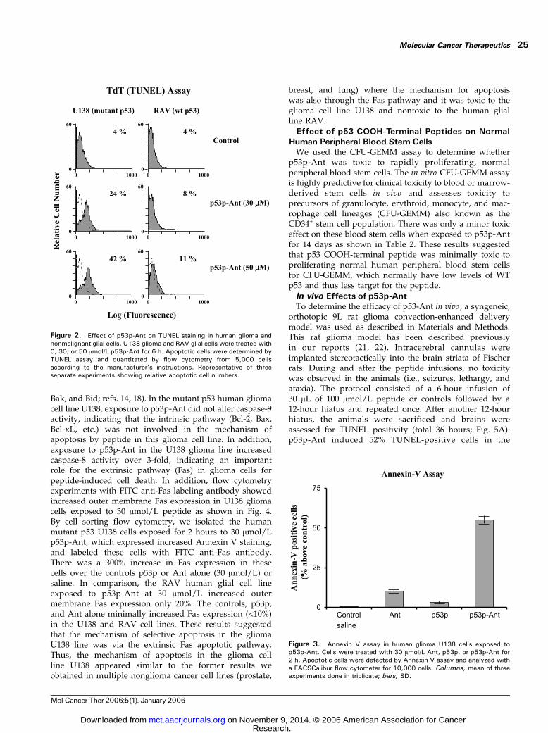
One of the most sensitive and specific tests for apoptosiscompared with necrosis is the early increase in Annexin Vbinding. The Annexin V assay detects flipping of intra-membrane phosphatidylserine to the outer membrane,which is indicative of early apoptosis. The results ofAnnexin V staining in the human mutant p53 gliomaU138 cells are shown in Fig. 3. U138 glioma cells exposed top53p-Ant for 2 hours had a 55% increase in positivity forAnnexin V, whereas cells exposed to control peptidesexhibited only 10% (Ant alone) and 3% (p53p alone)
increase over the basal level of staining. These datasuggested that p53p-Ant induced an apoptotic cell deathpreferentially in human glioma cells f48% higher thanthe average of the controls.
Effect of p53 COOH-Terminal Peptide on the Intrinsicand Extrinsic Pathways of Apoptosis
Our previous studies in human cancer lines showed thatthe mechanism by which p53p-Ant induced apoptosis wasthrough a nontranscriptional/nontranslational activationof the Fas/FADD complex, which led to death signalingthrough the extrinsic pathway for apoptosis. This couldbe blocked by dominant-negative FADD overexpression(18, 20). There was a downstream activation of the initiatorcaspase-8 and effector caspase-3 without changes in theintrinsic pathway proteins (caspase-9, Bcl-2, Bax, Bcl-xL,
Table 1. Inhibition of cell proliferation by p53p-Ant correlates with p53 status and expression
Cell lines Cell type p53 status p53 expression IC50 (Amol/L) F SE
U138 Human glioma Mutant 13 30 F 2.8U87 Human glioma WT 2 50 F 3.2RAV Human normal glial WT 2 210 F 10.19L Rat glioma Mutant 8 30 F 4.0D74 Rat glioma Mutant 2 50 F 4.2F98 Rat glioma Mutant 1 50 F 3.4NL Rat normal glial WT 1 194 F 9.0
NOTE: Cells were treated with p53p-Ant for 12 hours and cell viability was assessed by MTT assay. IC50s F SE were determined from the MTT assay results.The p53 band values were determined by densitometric analysis of the Western blots and standardized to the lowest band density, which was given thearbitrary number of 1 (NL glial line = 1). The p53 band expression numbers represent the fold increase of each respective line for their p53 level over the NLglial line, which equals 1. The number 13 was the highest x -fold increase (i.e., the U138 glioma line was 13-fold over the arbitrary number 1 value).
Figure 1. p53p-Ant induces inhibi-tion of growth in human and rat gliomacells. Cells (3,000) in MTT plate wellswere treated with different concen-trations of p53p-Ant (0–100 Amol/L)or Ant alone (100 Amol/L) or p53palone (100 Amol/L) for 12 h andcell viability was detected by MTTassay in human glioma cell lines U138and U87 (A) and rat glioma cell lines9L and D74 (B). Columns, mean ofthree experiments done in triplicate;bars, SD.
Selective Induction of Apoptosis in Glioma Cells24
Mol Cancer Ther 2006;5(1). January 2006
Research. on November 9, 2014. © 2006 American Association for Cancermct.aacrjournals.org Downloaded from
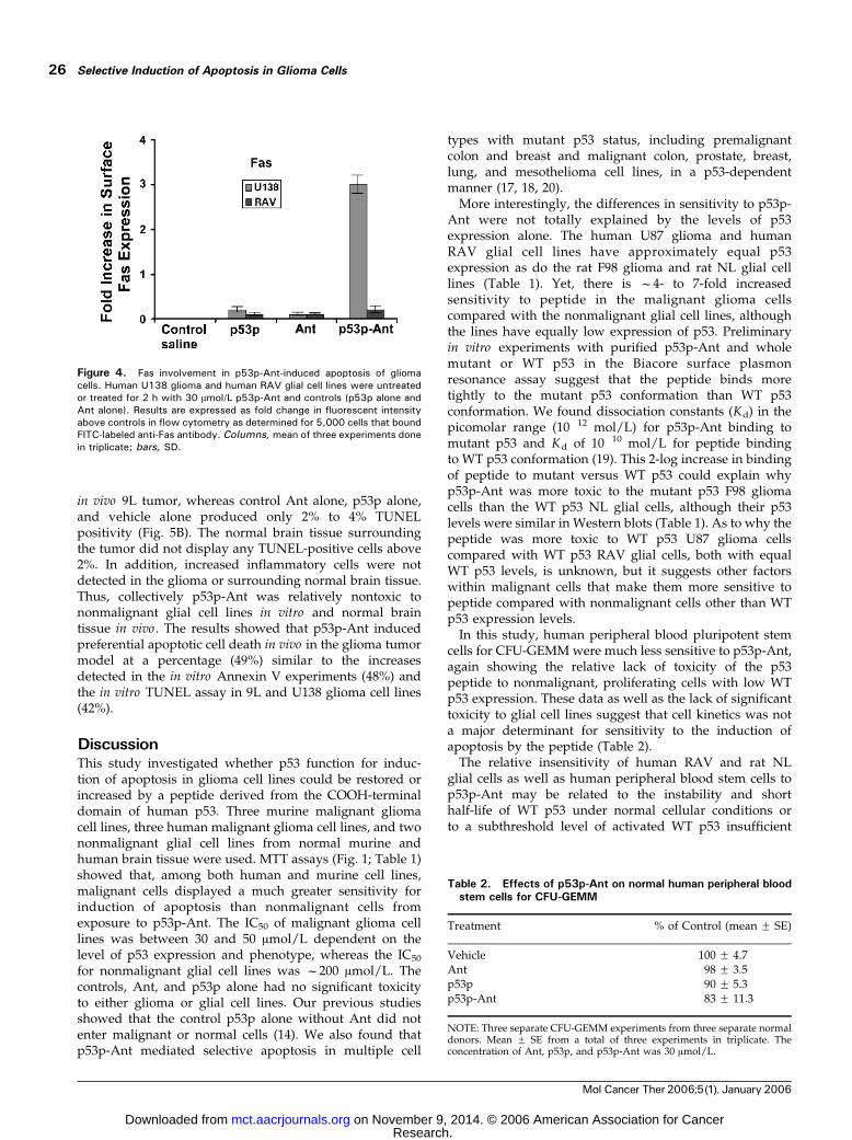
Bak, and Bid; refs. 14, 18). In the mutant p53 human gliomacell line U138, exposure to p53p-Ant did not alter caspase-9activity, indicating that the intrinsic pathway (Bcl-2, Bax,Bcl-xL, etc.) was not involved in the mechanism ofapoptosis by peptide in this glioma cell line. In addition,exposure to p53p-Ant in the U138 glioma line increasedcaspase-8 activity over 3-fold, indicating an importantrole for the extrinsic pathway (Fas) in glioma cells forpeptide-induced cell death. In addition, flow cytometryexperiments with FITC anti-Fas labeling antibody showedincreased outer membrane Fas expression in U138 gliomacells exposed to 30 Amol/L peptide as shown in Fig. 4.By cell sorting flow cytometry, we isolated the humanmutant p53 U138 cells exposed for 2 hours to 30 Amol/Lp53p-Ant, which expressed increased Annexin V staining,and labeled these cells with FITC anti-Fas antibody.There was a 300% increase in Fas expression in thesecells over the controls p53p or Ant alone (30 Amol/L) orsaline. In comparison, the RAV human glial cell lineexposed to p53p-Ant at 30 Amol/L increased outermembrane Fas expression only 20%. The controls, p53p,and Ant alone minimally increased Fas expression (<10%)in the U138 and RAV cell lines. These results suggestedthat the mechanism of selective apoptosis in the gliomaU138 line was via the extrinsic Fas apoptotic pathway.Thus, the mechanism of apoptosis in the glioma cellline U138 appeared similar to the former results weobtained in multiple nonglioma cancer cell lines (prostate,
breast, and lung) where the mechanism for apoptosiswas also through the Fas pathway and it was toxic to theglioma cell line U138 and nontoxic to the human glialline RAV.
Effect of p53 COOH-Terminal Peptides on NormalHuman Peripheral Blood Stem Cells
We used the CFU-GEMM assay to determine whetherp53p-Ant was toxic to rapidly proliferating, normalperipheral blood stem cells. The in vitro CFU-GEMM assayis highly predictive for clinical toxicity to blood or marrow-derived stem cells in vivo and assesses toxicity toprecursors of granulocyte, erythroid, monocyte, and mac-rophage cell lineages (CFU-GEMM) also known as theCD34+ stem cell population. There was only a minor toxiceffect on these blood stem cells when exposed to p53p-Antfor 14 days as shown in Table 2. These results suggestedthat p53 COOH-terminal peptide was minimally toxic toproliferating normal human peripheral blood stem cellsfor CFU-GEMM, which normally have low levels of WTp53 and thus less target for the peptide.
In vivo Effects of p53p-AntTo determine the efficacy of p53-Ant in vivo , a syngeneic,
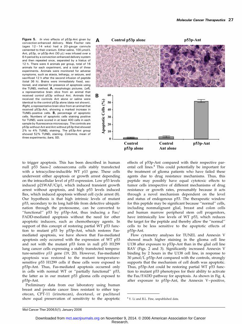
orthotopic 9L rat glioma convection-enhanced deliverymodel was used as described in Materials and Methods.This rat glioma model has been described previouslyin our reports (21, 22). Intracerebral cannulas wereimplanted stereotactically into the brain striata of Fischerrats. During and after the peptide infusions, no toxicitywas observed in the animals (i.e., seizures, lethargy, andataxia). The protocol consisted of a 6-hour infusion of30 AL of 100 Amol/L peptide or controls followed by a12-hour hiatus and repeated once. After another 12-hourhiatus, the animals were sacrificed and brains wereassessed for TUNEL positivity (total 36 hours; Fig. 5A).p53p-Ant induced 52% TUNEL-positive cells in the
Figure 2. Effect of p53p-Ant on TUNEL staining in human glioma andnonmalignant glial cells. U138 glioma and RAV glial cells were treated with0, 30, or 50 Amol/L p53p-Ant for 6 h. Apoptotic cells were determined byTUNEL assay and quantitated by flow cytometry from 5,000 cellsaccording to the manufacturer’s instructions. Representative of threeseparate experiments showing relative apoptotic cell numbers.
Figure 3. Annexin V assay in human glioma U138 cells exposed top53p-Ant. Cells were treated with 30 Amol/L Ant, p53p, or p53p-Ant for2 h. Apoptotic cells were detected by Annexin V assay and analyzed witha FACSCalibur flow cytometer for 10,000 cells. Columns, mean of threeexperiments done in triplicate; bars, SD.
Molecular Cancer Therapeutics 25
Mol Cancer Ther 2006;5(1). January 2006
Research. on November 9, 2014. © 2006 American Association for Cancermct.aacrjournals.org Downloaded from
in vivo 9L tumor, whereas control Ant alone, p53p alone,and vehicle alone produced only 2% to 4% TUNELpositivity (Fig. 5B). The normal brain tissue surroundingthe tumor did not display any TUNEL-positive cells above2%. In addition, increased inflammatory cells were notdetected in the glioma or surrounding normal brain tissue.Thus, collectively p53p-Ant was relatively nontoxic tononmalignant glial cell lines in vitro and normal braintissue in vivo . The results showed that p53p-Ant inducedpreferential apoptotic cell death in vivo in the glioma tumormodel at a percentage (49%) similar to the increasesdetected in the in vitro Annexin V experiments (48%) andthe in vitro TUNEL assay in 9L and U138 glioma cell lines(42%).
DiscussionThis study investigated whether p53 function for induc-tion of apoptosis in glioma cell lines could be restored orincreased by a peptide derived from the COOH-terminaldomain of human p53. Three murine malignant gliomacell lines, three human malignant glioma cell lines, and twononmalignant glial cell lines from normal murine andhuman brain tissue were used. MTT assays (Fig. 1; Table 1)showed that, among both human and murine cell lines,malignant cells displayed a much greater sensitivity forinduction of apoptosis than nonmalignant cells fromexposure to p53p-Ant. The IC50 of malignant glioma celllines was between 30 and 50 Amol/L dependent on thelevel of p53 expression and phenotype, whereas the IC50
for nonmalignant glial cell lines was f200 Amol/L. Thecontrols, Ant, and p53p alone had no significant toxicityto either glioma or glial cell lines. Our previous studiesshowed that the control p53p alone without Ant did notenter malignant or normal cells (14). We also found thatp53p-Ant mediated selective apoptosis in multiple cell
types with mutant p53 status, including premalignantcolon and breast and malignant colon, prostate, breast,lung, and mesothelioma cell lines, in a p53-dependentmanner (17, 18, 20).
More interestingly, the differences in sensitivity to p53p-Ant were not totally explained by the levels of p53expression alone. The human U87 glioma and humanRAV glial cell lines have approximately equal p53expression as do the rat F98 glioma and rat NL glial celllines (Table 1). Yet, there is f4- to 7-fold increasedsensitivity to peptide in the malignant glioma cellscompared with the nonmalignant glial cell lines, althoughthe lines have equally low expression of p53. Preliminaryin vitro experiments with purified p53p-Ant and wholemutant or WT p53 in the Biacore surface plasmonresonance assay suggest that the peptide binds moretightly to the mutant p53 conformation than WT p53conformation. We found dissociation constants (Kd) in thepicomolar range (10�12 mol/L) for p53p-Ant binding tomutant p53 and Kd of 10�10 mol/L for peptide bindingto WT p53 conformation (19). This 2-log increase in bindingof peptide to mutant versus WT p53 could explain whyp53p-Ant was more toxic to the mutant p53 F98 gliomacells than the WT p53 NL glial cells, although their p53levels were similar in Western blots (Table 1). As to why thepeptide was more toxic to WT p53 U87 glioma cellscompared with WT p53 RAV glial cells, both with equalWT p53 levels, is unknown, but it suggests other factorswithin malignant cells that make them more sensitive topeptide compared with nonmalignant cells other than WTp53 expression levels.
In this study, human peripheral blood pluripotent stemcells for CFU-GEMM were much less sensitive to p53p-Ant,again showing the relative lack of toxicity of the p53peptide to nonmalignant, proliferating cells with low WTp53 expression. These data as well as the lack of significanttoxicity to glial cell lines suggest that cell kinetics was nota major determinant for sensitivity to the induction ofapoptosis by the peptide (Table 2).
The relative insensitivity of human RAV and rat NLglial cells as well as human peripheral blood stem cells top53p-Ant may be related to the instability and shorthalf-life of WT p53 under normal cellular conditions orto a subthreshold level of activated WT p53 insufficient
Figure 4. Fas involvement in p53p-Ant-induced apoptosis of gliomacells. Human U138 glioma and human RAV glial cell lines were untreatedor treated for 2 h with 30 Amol/L p53p-Ant and controls (p53p alone andAnt alone). Results are expressed as fold change in fluorescent intensityabove controls in flow cytometry as determined for 5,000 cells that boundFITC-labeled anti-Fas antibody. Columns, mean of three experiments donein triplicate; bars, SD.
Table 2. Effects of p53p-Ant on normal human peripheral bloodstem cells for CFU-GEMM
Treatment % of Control (mean F SE)
Vehicle 100 F 4.7Ant 98 F 3.5p53p 90 F 5.3p53p-Ant 83 F 11.3
NOTE: Three separate CFU-GEMM experiments from three separate normaldonors. Mean F SE from a total of three experiments in triplicate. Theconcentration of Ant, p53p, and p53p-Ant was 30 Amol/L.
Selective Induction of Apoptosis in Glioma Cells26
Mol Cancer Ther 2006;5(1). January 2006
Research. on November 9, 2014. © 2006 American Association for Cancermct.aacrjournals.org Downloaded from
to trigger apoptosis. This has been described in humannull p53 Saos-2 osteosarcoma cells stably transfectedwith a tetracycline-inducible WT p53 gene. These cellsunderwent either apoptosis or growth arrest dependingon the intracellular level of p53 expression. Low p53 levelsinduced p21WAF/Cip1, which induced transient growtharrest without apoptosis, and high p53 levels inducedBax, which induced apoptosis without cell cycle arrest (8).Our hypothesis is that high intrinsic levels of mutantp53, secondary to its long half-life from defective ubiquiti-nation through the proteosome, can be converted to‘‘functional’’ p53 by p53p-Ant, thus inducing a Fas/FADD-mediated apoptosis without the need for otherapoptotic inducers, such as chemotherapy agents. Insupport of this concept of restoring partial WT p53 func-tion to mutant p53 by p53p-Ant, which restores Fas-mediated apoptosis, we have shown that Fas-mediatedapoptosis only occurred with the expression of WT p53and not with the mutant p53 form in null p53 H1299lung cancer cells expressing a stably transfected tempera-ture-sensitive p53 gene (19, 20). However, Fas-mediatedapoptosis was restored to the mutant temperature-sensitive p53 H1299 cells if these cells were exposed top53p-Ant. Thus, Fas-mediated apoptosis occurred onlyin cells with normal WT or ‘‘partially functional’’ p53,the latter as in our mutant p53 glioma cells exposed top53p-Ant.
Preliminary data from our laboratory using humanbreast and prostate cancer lines resistant to either top-otecan, CPT-11 (irinotecan), docetaxel, or paclitaxelshow equal preservation of sensitivity to the apoptotic
effects of p53p-Ant compared with their respective par-ental cell lines.4 This could potentially be important forthe treatment of glioma patients who have failed theseagents due to drug resistance mechanisms. Thus, thispeptide may possibly have equal cytotoxic effects totumor cells irrespective of different mechanisms of drugresistance or growth rates, presumably because it actsthrough a novel mechanism dependent on the leveland status of endogenous p53. The therapeutic windowfor this peptide may be significant because ‘‘normal’’ cells,including nonmalignant glial, breast and colon cellsand human marrow peripheral stem cell progenitors,have intrinsically low levels of WT p53, which reducesthe target for the peptide and thereby allow the ‘‘normal’’cells to be less sensitive to the apoptotic effects ofp53p-Ant.
Flow cytometry analyses for TUNEL and Annexin Vshowed much higher staining in the glioma cell lineU138 after exposure to p53p-Ant than in the glial cell lineRAV (Figs. 2 and 3). Significantly increased Annexin Vbinding by 2 hours in the U138 cell line, in response to30 Amol/L p53p-Ant compared with the controls, stronglysupports that the mechanism of cell death was apoptotic.Thus, p53p-Ant could be restoring partial WT p53 func-tion to mutant p53 phenotypes for their ability to activatethe Fas/FADD pathway for apoptosis. As shown in Fig. 4,after exposure to p53p-Ant, the Annexin V–positive,
Figure 5. In vivo effects of p53p-Ant given byconvection-enhanced delivery. Male Fischer rats(ages 12 – 14 wks) had a 20-gauge cannulacemented to their cranium. Either saline, 100 Amol/LAnt, p53p, or p53p-Ant (30 AL) was infused over a6-h period by a convection-enhanced delivery systemand then repeated once, separated by a hiatus of12 h. There were 4 animals per group, total of 16animals for each experiment, and a total of threeexperiments. Animals were monitored for adversesymptoms, such as ataxia, lethargy, or seizure, andsacrificed 12 h after the second infusion of peptide(total 36 h). Brains were immediately fixed, sec-tioned, and stained for presence of apoptosis usingthe TUNEL method. A, morphologic pictures. Left,a representative brain slice from an animal thatreceived control p53p without Ant. Animals thatreceived the controls Ant alone or saline wereidentical to the control p53p alone (data not shown).Right, a representative brain slice from an animal thatreceived p53p-Ant, showing a marked increase inTUNEL-positive cells. B, percentage of apoptoticcells. Numbers of apoptotic cells staining positivefor TUNEL were scored in at least 400 cells in eachsample by fluorescencemicroscopy. The controls arep53pwithout Ant andAntwithout p53p that showed2% to 4% TUNEL staining. The p53p-Ant groupshowed 52% TUNEL staining. Columns, mean ofthree experiments; bars, SD.
4 Y. Li and R.L. Fine, unpublished data.
Molecular Cancer Therapeutics 27
Mol Cancer Ther 2006;5(1). January 2006
Research. on November 9, 2014. © 2006 American Association for Cancermct.aacrjournals.org Downloaded from

mutant p53 U138 malignant glioma cells had increasedsurface Fas expression above control (300%). In compari-son, Fas expression did not increase significantly abovecontrol in the RAV human glial cell line (20%) on expo-sure to the same concentration of p53p-Ant. As in ourprevious breast and lung cell line studies, increasedouter membrane Fas expression (extrinsic pathway) wasthe only death pathway induced in glioma cells withoutchanges in the intrinsic caspase-9 pathway for apoptosis(23). Thus, in glioma cell lines, in accordance with ourhypothesis, the extrinsic pathway (Fas) seems to be themajor mechanism for peptide-induced apoptosis and theability of the peptide to cause apoptosis seems, in general,related to the endogenous levels of p53, mutant more sothan WT.
Induction of apoptosis in the syngeneic, orthotopic rat9L glioma model by convection-enhanced delivery of p53p-Ant suggested that in vitro results could be translated tothe in vivo model. In vivo , p53p-Ant induced 52% apoptosisin the 9L glioma by TUNEL staining, whereas the controlsp53p and Ant alone induced only 2% to 4% apoptosis.Histochemical analysis of the normal rat brain surround-ing the 9L glioma did not display TUNEL positivity oralteration in the number of inflammatory cells. This wasof concern because Ant is derived from Drosophila .However, the time window for the convection-enhanceddelivery experiments was probably too short to examinethis fully (36 hours). A preliminary survival study withcontrols versus p53p-Ant in the above in vivo 9L gliomamodel has shown an increase in survival from a medianof 21 to 37 days, respectively, translating to a 76% increasein median survival from p53p-Ant.5 This increase byKaplan-Meier survival analysis was statistically signifi-cant (P < 0.001). However, further studies are needed tocorroborate this preliminary experiment. Long-term sur-vival studies in this syngeneic, orthotopic rat glioma modelare in progress.
Investigation into synthetic small molecules, whichpartially restore p53 function to mutant p53 tumor cells,is also an active area of research by many laboratories. Wehave shown recently that the small-molecule PRIMA-1,which is cell permeable without a carrier, induced apo-ptosis selectively in premalignant and malignant mutantp53 colon and breast cells through the c-Jun NH2-terminalkinase pathway without affecting the intrinsic or extrinsicpathways of apoptosis (24).
In total, this work supports the potential clinical utility ofp53p-Ant as a selective novel therapeutic agent for gliomasand the clinical advantages of convection-enhanced deliv-ery as a system to circumvent the blood-brain barrier fordelivery of large and/or charged therapeutic moieties, suchas p53 peptides.
References
1. Levine AJ. p53, the cellular gatekeeper for growth and division. Cell1997;88:323–31.
2. Hollstein M, Sidransky D, Vogelstein B, Harris CC. p53 mutations inhuman cancers. Science 1991;253:49–53.
3. Cho Y, Gorina S, Jeffrey PD, Pavletich NP. Crystal structure of a p53tumor suppressor-DNA complex: understanding tumorigenic mutations.Science 1994;265:346–55.
4. Kastan MB, Zhan Q, El-Deiry WS, et al. A mammalian cell cyclecheckpoint pathway utilizing p53 and GADD45 is defective in ataxia-telangiectasia. Cell 1992;71:587–97.
5. Livingstone LR, White A, Sprouse J, Livanos E, Jacks T, Tlsty TD.Altered cell cycle arrest and gene amplification potential accompany lossof wild-type p53. Cell 1992;70:923–35.
6. Yin Y, Tainsky MA, Bischoff FZ, Strong LC, Wahl GM. Wild-type p53restores cell cycle control and inhibits gene amplification in cells withmutant p53 alleles. Cell 1992;70:937–48.
7. Friend S. p53: a glimpse at the puppet behind the shadow play.Science 1994;265:334–5.
8. Ko LJ, Prives C. p53: puzzle and paradigm. Gene Dev 1996;10:1054–72.
9. El-Deiry WS, Tokino T, Velculescu VE, et al. WAF1, a potentialmediator of p53 tumor suppression. Cell 1993;75:817–25.
10. Oltvai ZN, Milliman CL, Korsmeyer SJ. Bcl-2 heterodimerizes in vivowith a conserved homolog, Bax, that accelerates programmed cell death.Cell 1993;74:609–19.
11. Owen-Schaub LB, Zhang W, Cusack JC, et al. Wild-type human p53and a temperature-sensitive mutant induce Fas/APO-1 expression. MolCell Biol 1995;15:3032–40.
12. Hupp TR, Lane DP. Allosteric activation of latent p53 tetramers. CurrBiol 1994;4:865–75.
13. Muller-Tiemann BF, Halazonetis TD, Elting JJ. Identification of anadditional negative regulatory region for p53 sequence specific DNAbinding. Proc Natl Acad Sci U S A 1998;95:6079–84.
14. Kim AL, Raffo AJ, Brandt-Rauf PW, et al. Conformational andmolecular basis for induction of apoptosis by a p53 C-terminal peptide inhuman cancer cells. J Biol Chem 1999;274:34924–31.
15. Abarzua P, LoSardo JE, Gubler ML, et al. Restoration of thetranscription activation function to mutant p53 in human cancer cells.Oncogene 1996;13:2477–82.
16. Selivanova G, Iotsova V, Okan I, et al. Restoration of the growthsuppression function of mutant p53 by a synthetic peptide derived fromthe p53 C-terminal domain. Nat Med 1997;3:632–8.
17. Li Y, Rosal RV, Brandt-Rauf PW, Fine RL. Correlation betweenhydrophobic properties and efficiency of carrier-mediated membranetransduction and apoptosis of a p53 C-terminal peptide. Biochem BiophysRes Commun 2002;298:439–49.
18. Li Y, Mao Y, Rosal RV, et al. Selective induction of apoptosis throughthe FADD/caspase-8 pathway by a p53 C-terminal peptide in human pre-malignant and malignant cells. Int J Cancer 2005;115:55–64.
19. Rosal RV, Brandt-Rauf P, Pincus MR, et al. The role of a helicalstructure in p53 peptides as a determinant for their mechanism of celldeath: necrosis versus apoptosis. Adv Drug Deliv Rev 2005;57:653–60.
20. Li Y, Raffo AJ, Drew L, et al. Fas-mediated apoptosis is dependent onwild-type p53 status in human cancer cells expressing a temperature-sensitive p53 mutant alanine-143. Cancer Res 2003;63:1527–33.
21. Bruce JN, Falavigna A, Johnson JP, et al. Intracereral lysis in a ratglioma model. Neurosurgery 2000;46:683–91.
22. Kaiser MG, Parsa A, Fine RL, Hall JS, Chakrabarti I, Bruce JN. Tissuedistribution and antitumor activity of topotecan delivered by intracerebrallysis in a rat glioma model. Neurosurgery 2000;47:1391–9.
23. Kluck RM, Bossy-Wetzel E, Green DR, Newmeyer DD. The release ofcytochrome c from mitochondria: a primary site for Bcl-2 regulation ofapoptosis. Science 1997;275:1132–6.
24. Li Y, Mao Y, Brandt-Rauf P, Williams A, Fine RL. Selective induction ofapoptosis in mutant p53 premalignant and malignant cancer cells byPRIMA-1 through the c-JUN-NH2-kinase pathway. Mol Cancer Ther2005;4:901–9.5 J.N. Bruce and R.L. Fine, unpublished data.
Selective Induction of Apoptosis in Glioma Cells28
Mol Cancer Ther 2006;5(1). January 2006
Research. on November 9, 2014. © 2006 American Association for Cancermct.aacrjournals.org Downloaded from