Novel role for a Saccharomyces cerevisiae nucleoporin, Nup170p, in chromosome segregation
Upload
independentCategory
view
3download
0
Christopher J. Rivard and Tomas BerlAna Andres-Hernando, Miguel A. Lanaspa, NucleusEnhancer-binding Protein (TonEBP) in thethe Transcription Factor Tonicity
RetainHypertonic Stress in Kidney Cells to Nucleoporin 88 (Nup88) Is Regulated byMechanisms of Signal Transduction:
doi: 10.1074/jbc.M802381200 originally published online July 7, 20082008, 283:25082-25090.J. Biol. Chem.
10.1074/jbc.M802381200Access the most updated version of this article at doi:
.JBC Affinity SitesFind articles, minireviews, Reflections and Classics on similar topics on the
Alerts:
When a correction for this article is posted•
When this article is cited•
to choose from all of JBC's e-mail alertsClick here
Supplemental material:
http://www.jbc.org/jbc/suppl/2008/07/08/M802381200.DC1.html
http://www.jbc.org/content/283/36/25082.full.html#ref-list-1
This article cites 35 references, 19 of which can be accessed free at
at Univ C
olorado - Denison M
emorial L
ibrary on July 22, 2014http://w
ww
.jbc.org/D
ownloaded from
at U
niv Colorado - D
enison Mem
orial Library on July 22, 2014
http://ww
w.jbc.org/
Dow
nloaded from

Nucleoporin 88 (Nup88) Is Regulated by Hypertonic Stress inKidney Cells to Retain the Transcription Factor TonicityEnhancer-binding Protein (TonEBP) in the Nucleus*□S
Received for publication, March 27, 2008, and in revised form, July 3, 2008 Published, JBC Papers in Press, July 7, 2008, DOI 10.1074/jbc.M802381200
Ana Andres-Hernando, Miguel A. Lanaspa, Christopher J. Rivard, and Tomas Berl1
From the Division of Renal Diseases and Hypertension, School of Medicine, University Colorado Health Sciences Center,Denver, Colorado 80262
Antibody microarray technology identified Nup88 (nucleo-porin 88) as ahighly up-regulatedprotein in response toosmoticstress in inner medullary collecting duct (IMCD3) cells.Changes in expression were verified byWestern blot and quan-titative PCR for protein and message expression. In mouse andhuman kidney, Nup88 expression was substantial in the papilla,whereas it was nearly absent in the cortex. Furthermore, theexpression of Nup88 increased 410.4 � 22% in the papilla ofmice after 36 h of thirsting.Nup88 protein expression in IMCD3cells was significantly up-regulated in the first 8 h followingexposure to acute osmotic stress, indicating that Nup88 is anearly response protein. To define the function of Nup88 in theosmotic stress response, the transcription factor associatedwithhypertonicity, tonicity enhancer-binding protein (TonEBP),was cloned upstream of the green fluorescent protein. Employ-ing this construct, we demonstrate that silencing Nup88 inIMCD3 cells acutely stressed to hypertonic conditions reducesnuclear retention of TonEBP, resulting in a substantial bluntingin transcription of important osmotic stress response targetgenes and reduced cell viability. Finally, we show that in IMCD3cells, nuclear export of TonEBP under isotonic conditionsinvolves CRM-1 but under hypertonic stress is CRM1-inde-pendent. Our data, therefore, suggest that Nup88 is up-regu-lated in response to hypertonic stress and acts to retain TonEBPin the nucleus, activating transcription of critical osmoprotec-tive genes.
The cells that inhabit the hypertonic environment of theinner medulla of the kidney possess a number of adaptativemechanisms that allow them to survive in this environment(1–5). The classical osmotic stress response involves theprompt transcription of several target genes by the tonicityenhancer-binding protein (TonEBP),2 also known as NFAT5
(6–9). Under isotonic conditions (300 mosmol/kg H2O),TonEBP is mainly present in the cytosol with only minor local-ization in the nucleus. However, under hypertonic stress,TonEBP is translocated to the nucleus, where it enhances thetranscription of genes that are important in the early osmoticstress response. These genes includes aldose reductase (AR)(10), the sodium-myoinositol transporter (10), the BGT1 (beta-ine/GABA transporter 1) (11), the taurine transporter (TauT)(12), and heat shock protein 70 (Hsp70) (13) among others.Expression of these target genes results in the accumulation ofa number of compatible organic osmolytes (mainly sorbitol,myoinositol, betaine, and taurine) that allow the cell to com-pensate the extracellular osmotic gradient and, hence, adapt tohypertonic stress (see Refs. 3 and 6–9 for excellent reviews).One of the main mechanisms involved in the regulation ofTonEBP activity under hypertonic stress is nucleocytoplasmictrafficking (7, 9, 14, 15).Our laboratory has employed several proteomic approaches,
including two-dimensional difference gel electrophoresis (16)and antibodymicroarray (17) technologies to evaluate hyperto-nicity-induced up-regulation of important proteins in innermedullary collecting duct (IMCD3) cells. Here, we describe themarked up-regulation of Nup88 (nucleoporin 88) under hyper-tonic stress. Nup88was first identified as an interacting partnerof Nup214 (nucleoporin 214) (18). These two proteins are com-ponents of a nuclear pore complex in the nuclear membraneand are involved in the nucleocytoplasmic trafficking of differ-ent molecules, including transcription factors (19–22).It was previously demonstrated in Drosophila that Nup88
does not interfere in the nuclear import of NF-�B and insteadacts as an inhibitor of CRM1-mediated protein export (20, 22).In experiments with mutated (inactive) Nup88, CRM1 expres-sion is mislocalized in the nucleus and is not present in thenuclear membrane. Interestingly, this mislocalization resultedin a continuous shuttling of the NF-�B Rel-like transcriptionfactorDorsal in and out of the nucleus (22). These data suggestthat CRM1 and, therefore, Nup88 expression are necessary forregulating Dorsal nuclear export. The mechanism by whichNup88 functions is by sequestering CRM1 at the nuclear poreand thereby avoiding its recycling back to the nucleus foranother round of export. Similarly, another Rel-like transcrip-tion factor, Dif, seems to be regulated by Nup88 expression in
* This work was supported, in whole or in part, by National Institutes of HealthGrants DK-19928 and DK-66544 (to T. B.). The costs of publication of thisarticle were defrayed in part by the payment of page charges. This articlemust therefore be hereby marked “advertisement” in accordance with 18U.S.C. Section 1734 solely to indicate this fact.
□S The on-line version of this article (available at http://www.jbc.org) containssupplemental Tables 1–3 and Figs. 1 and 2.
1 To whom correspondence should be addressed: University of ColoradoHealth Sciences Center, Division of Renal Diseases and Hypertension,School of Medicine, 4200 E. 9th Ave., Denver, CO 80262. Tel.: 303-315-7204;Fax: 303-315-4852; E-mail: [email protected].
2 The abbreviations used are: TonEBP, tonicity enhancer-binding protein; AR,aldose reductase; TauT, taurine transporter; QPCR, quantitative PCR; DAPI,
4�,6-diamidino-2-phenylindole; GFP, green fluorescent protein; LMB, lep-tomycin B.
THE JOURNAL OF BIOLOGICAL CHEMISTRY VOL. 283, NO. 36, pp. 25082–25090, September 5, 2008© 2008 by The American Society for Biochemistry and Molecular Biology, Inc. Printed in the U.S.A.
25082 JOURNAL OF BIOLOGICAL CHEMISTRY VOLUME 283 • NUMBER 36 • SEPTEMBER 5, 2008
at Univ C
olorado - Denison M
emorial L
ibrary on July 22, 2014http://w
ww
.jbc.org/D
ownloaded from

Drosophila (20). Likewise, other members of the Rel-like tran-scription factor class, including TonEBP (10), may be con-trolled by the expression of Nup88.The up-regulation of Nup88 under hypertonic stress in the
kidney has not been previously described. The present workwas undertaken to confirm the observations made by antibodymicroarray analysis and further characterize the osmotic regu-lation of expression, half-life, cellular distribution, and in vivoexpression. Furthermore, we identify a potential role forNup88in the nuclear retention of TonEBP in hypertonically stressedcells.
EXPERIMENTAL PROCEDURES
Materials—Cell culture medium, FCS, and antibiotics werefrom Invitrogen. Antibodies to Nup88 andGS15 were obtainedfrom Clontech, anti-AR antibody was purchased from SantaCruz Biotechnology, Inc. (Santa Cruz, CA), anti-Hsp70 wasfrom Stressgen (Ann Arbor, MI), and anti-�-actin was fromCell Signaling (Danvers, MA). All other chemicals were pur-chased from Sigma.Antibody Microarray—Antibody microarrays and reagents
were purchased from Clontech. Microarrays were processed asper the manufacturer’s protocol and as previously described(17). Antibody microarrays were analyzed using a ProScan-Array HT scanner (PerkinElmer Life Sciences).Cell Culture—The established IMCD3 cell line originally
developed by Rauchman et al. (23) was provided by Steve Gul-lans (Rx Gen, Hamden, CT). IMCD3 cells chronically adaptedto 600 and 900 mosmol/kg H2O were previously established inour laboratory (24, 25) and were compared with cells grown atisotonic conditions. In experiments involving hypertonicstress, themedia in culture dishes were exchanged for that withadded NaCl to the specified osmolality, depending on theexperiment. Osmolality was determined with a microosmom-eter (model 3300; Advanced Instruments, Norwood, MA).Cell Viability and Growth Experiments—Cell viability in tis-
sue culture stress experiments was determined by cell countsfollowing exposure to sublethal osmotic stress. Experimentswere initiated once cells reached confluence at isotonic condi-tions in 24-well flat bottom tissue culture plates (35-3047; Fal-con BD Labware, Franklin Lakes, NJ), with each experimentaltime point after exposure to the sublethal osmotic stress per-formed in triplicate. In cell growth studies, cell counts wereinitiated when cultures had reached �90% confluence and fol-lowed for an additional 80-h period with counts at 24 and 48 h.Growth curves were fit to linear regression using the Prism 4.0software, and the slopes of the curves were compared for rela-tive differences in growth rate.Stable Cell Lines Silenced for Nup88 Expression—IMCD3
cultures were transfected with the pSM2 empty vector(v2MM_173601) or the short hairpin RNA vector pSM2-Nup88 (v2HS_152407; Open Biosystems) using Lipofectamine2000 (Invitrogen) as described by the manufacturer. Stabletransfectants (clones) were selected from colonies growing inplates from a 10-fold dilution series in media prepared with 10�g/ml puromycin antibiotic (Sigma). The absence of Nup88expression in silenced clones was verified by Western blot of
cell lysates obtained from these clones grown inmedia adjustedto 500 mosmol/kg H2O for 48 h.Cloning and Transfection of the Construct TonEBP-GFP—
The first 800 bpofmouseTonEBP, including the nuclear exportsignal, the auxiliary export domain, and the nuclear localizationsignal, was obtained by PCR using the following primers, whichcontained, respectively, XhoI and EcoRI restriction sites (bothunderlined) for directional cloning into pAcGFP-N1 vector(Clontech): sense, 5�-GAC CTC GAG ATG CCC TCG GACTTC ATC TCA TTG CTC-3�; antisense, 5�-GAC GAA TTCGAG GTG CTT TGG CAC TGT CGG CAT CAA-3�. Trans-fection into IMCD3 cells or into the stable cell lines expressingpSM2-Nup88 vector or pSM2-Empty vector control was per-formed using Lipofectamine 2000.Mouse Kidney Tissues—C57/B6 mice were obtained from
Jackson Laboratories (BarHarbor,ME).Mice were subjected tofood and ad libitumwater or deprived for water for 36 h. Urinesampleswere collected from the bladder for osmolality analysis.Mice were harvested by cervical dislocation, and kidneys wereremoved. Papilla and cortex tissues were dissected and homog-enized with a glass tissue grinder on ice withmitogen-activatedprotein kinase lysis buffer for protein and analyzed as described(24).Human Kidney Tissues—Human kidney tissues from cortex
and papilla were obtained under the Colorado Multiple Insti-tutional Review Board and National Institutes of Health GrantU19A10636030 from a kidney that was not suitable for trans-plantation. Tissues were processed for protein as describedabove.RNA Extraction, Analysis, andMessage Quantification—Cy-
tosolic RNAwas isolated from confluent cell cultures using theRNeasy kit (Qiagen, Valencia, CA) as per the manufacturer’sprotocol. Before quantitative PCR (QPCR), sample RNA con-centration and integrity was assessed by UV spectrometry(absorbance at 260 nm) and by capillary electrophoresis using abioanalyzer (model 210, Agilent, Foster City, CA; using the 28 Sto 18 S rRNA ratio), respectively. RNAwas converted to cDNAusing the iScript cDNA synthesis kit (Bio-Rad). QPCRwas per-formed using primer pairs identified in supplemental Table 3,designed using Beacon Designer 7.0 (Premier Biosoft, PaloAlto, CA). QPCR runs were performed using the SYBR greenJumpStart Taq Readymix QPCR kit (Sigma) on an I-Cycler(Bio-Rad). QPCR runs were analyzed by agarose gel electro-phoresis andmelt curve to verify that the correct amplicon wasproduced. �-Actin RNA was used as internal control in allQPCRs, and the amount of RNAwas calculated by the compar-ative CT method.Protein Extraction and Western Blotting—Cell protein
lysates were obtained from confluent cell culture dishes as pre-viously described (24). Protein concentration was determinedby the BCA protein assay (Pierce). Seventy micrograms of totalprotein was loaded per lane for SDS-PAGE (7.5%, w/v) analysisand then transferred to polyvinylidene difluoride membranes.Membranes were incubated with primary antibody and visual-ized by using an alkaline phosphatase secondary antibody andLumi-Phos reagent (Pierce) as described by the manufacturer.Chemiluminescence was recorded with an Image Station440CF (Kodak Digital Science), and results were analyzed with
Nup88 Is Regulated by Hypertonic Stress
SEPTEMBER 5, 2008 • VOLUME 283 • NUMBER 36 JOURNAL OF BIOLOGICAL CHEMISTRY 25083
at Univ C
olorado - Denison M
emorial L
ibrary on July 22, 2014http://w
ww
.jbc.org/D
ownloaded from

1D Image Software (Kodak Digital Science). Blots were alsoanalyzed for �-actin as a loading control.Confocal Fluorescence Microscopy—IMCD3 cells were
seeded in 8-well chambers (Nunc) and stained using an anti-Nup88 antibody (1:50 dilution in phosphate-buffered saline;Clontech) or anti-TonEBP antibody (1:400 dilution in PBS)kindly provided by Dr. H.Moo Kwon (University ofMaryland).Inmunofluorescence was carried out as previously described(17). Preparations and TonEBP-GFP fusion protein transfec-tions were imaged with a �40 water immersion objective usinga laser-scanning confocal microscope (model LSM510; Zeiss,Thornwood, NY). Data were analyzed with LSM Image ana-lyzer postacquisition software (Zeiss).Statistics and Data Analysis—All data are presented as the
mean � S.E. Data graphics and statistical analysis were per-formed using Instat (version 3.0) and Prism 4 (both fromGraphPad, San Diego, CA). Independent replicates for eachdata point (n) are identified in figure legends. p � 0.05 wasrecognized as statistically significant.
RESULTS
Nup88 Expression Is Up-regulated under Hypertonic Stress—Employing antibody microarray proteomics (Clontech, Moun-tain View, CA), we found that 5% of the 512 proteins analyzed
were up-regulated (17) in IMCD3cells chronically adapted to hyper-tonic stress (900 mosmol/kg H2O)as compared with cells grown underisotonic conditions. As shown inFig. 1A, the Golgi vesicle solubleN-ethylmaleimide-sensitive factorattachment protein receptor pro-tein (GS15) is representative of thegreater than 90% of the proteinswhose expression did not changeunder hypertonic conditions. Incontrast, Nup88 demonstrated asubstantial increase in expression inhypertonically adapted cells. A sta-tistical analysis of four spots foreach protein (from two arrays; i.e.dye swap (Fig. 1B)) showed an inter-nal normalization ratio for Nup88signal intensity of 1.65 (p� 0.001) ascompared with 1.05 for GS15. Vali-dation of antibody microarray datawas performed by QPCR andWest-ern blot. Data shown in Fig. 1C indi-cate a substantial increase in Nup88message levels (7-fold; p � 0.001) incells adapted to 900 mosmol/kgH2O as compared with isotonicconditions (300 mosmol/kg H2O).This up-regulation was also sub-stantial for protein expression(12.9-fold; p � 0.001) under thesame conditions. For comparison,Western blots prepared with anti-
GS15 demonstrated equal protein expression in IMCD3 cells atisotonic and hypertonic conditions (Fig. 1D).Expression of Nup88 in Renal Cortex and Medulla from
Rodents and Human Kidney—To assess whether the changesseen in cultured cells are also observed in vivo, we examined therenal tissues of mice and a human kidney. Western blot datashown in Fig. 2A reveal a near absence ofNup88 protein expres-sion in the cortex of both species. In contrast, substantialNup88 protein expression was determined in the papilla forboth mice and human. These determinations were made inmice on ad libitumwater intake. The difference between cortexand papilla is quantitatively depicted in Fig. 2B. We also exam-ined the kidney tissues of mice subjected to 36 h of thirsting(urine osmolality increased from 1,424 � 211 to 3,105 � 524mosmol/kg H2O; n � 6). As shown in Fig. 2B, thirstingincreased Nup88 expression by 2-fold (107 � 12%, p � 0.01) inthe cortex and 5-fold (410 � 22%, p � 0.01) in the papillatissues.Immunocytochemical Localization of Nup88 in Cells
Adapted to Hypertonicity—Immunofluorescence microscopystudies were undertaken to assess the presence and localizationof Nup88 protein in IMCD3 cells at isotonic conditions andchronically adapted to hypertonicity (900 mosmol/kg H2O).Fig. 3 shows that Nup88 expression (green) is substantial in
FIGURE 1. Identification of Nup88 as an up-regulated protein in IMCD3 cells in response to osmotic stressby antibody microarray proteomics. A, individual spot signals were analyzed with ScanArrayGx/ProScanar-ray software. Selected antibody pairs for array 1 are compared with the dye swap array 2 and display pseudo-colors for Cy3 (green) and Cy5 (red). Greater than 90% of the array spots demonstrated equal signal intensity(yellow in the composite) as illustrated by the response for Golgi vesicle soluble N-ethylmaleimide-sensitivefactor attachment protein receptor (SNARE) (GS15) protein. In contrast, spots corresponding to the nucleo-porin Nup88 showed a substantial increase in signal intensity from hypertonic adapted cells, as shown in thecomposite spots for both arrays. B, channel intensity from both arrays was used to calculate the internalnormalization ratio for GS15 representing no change (value between 0.82 and 1.15) as compared with a Nup88internal normalization ratio value of 1.65 (p � 0.001). C, QPCR was performed using specific primers for Nup88and demonstrated a 7-fold increase in message for IMCD3 cells adapted to 900 versus 300 mosmol/kg H2O (p �0.001). D, Western blot data reveal a 12-fold increase in Nup88 protein expression in IMCD3 cells chronicallyadapted to hypertonicity as compared with cells maintained at isotonic conditions. Data represent the aver-age � S.E. for three independent experiments performed in triplicate (n � 9). Both QPCR and Western blot datawere normalized using �-actin message and protein expression, respectively. A representative Western blot isshown including GS15 as a loading control.
Nup88 Is Regulated by Hypertonic Stress
25084 JOURNAL OF BIOLOGICAL CHEMISTRY VOLUME 283 • NUMBER 36 • SEPTEMBER 5, 2008
at Univ C
olorado - Denison M
emorial L
ibrary on July 22, 2014http://w
ww
.jbc.org/D
ownloaded from

IMCD3 cells chronically adapted to 900 mosmol/kg H2O ascompared with isotonic conditions where no Nup88 signal isfound. The presence of Nup88 in the nuclearmembrane is con-firmed by colocalization with the nuclear marker DAPI.Kinetics of Nup88Message and Protein Expressionwith Acute
Exposure to Hypertonicity—Nup88 protein expression inIMCD3 cells subjected to acute sublethal osmotic stress wasevaluated to determine the timing of expression. To this end,we performed Western blot analysis for protein at numeroustime points after exposure to acute sublethal hypertonicity (550mosmol/kg H2O). Data shown in Fig. 4 demonstrate a signifi-cant increase inNup88protein expression after 4–6hof hyper-tonic stress (p � 0.01). To assess the half-life for Nup88 mes-sage and protein, IMCD3 cells were returned to isotonicconditions. Data shown in Fig. 5 were curve-fit for exponentialdecay, and the half-life was calculated to be 2.8 and 18.4 h formessage and protein, respectively. Separately, cells were treatedwith actinomycin D or cyclohexamide to evaluate potentialchanges in the stability of Nup88 message and protein. Thesedata were essentially identical to the data shown in Fig. 5 indi-cating no significant changes in the stability of Nup88 messageor protein (data not shown).Studies on the Osmotic and Ionic Mediators of the Up-Regu-
lation of Nup88 Expression to Hypertonic Stress—To assesswhether the effects observed with increasing NaCl levels wereunique to this solute, Nup88 protein expression was measuredafter exposure to other mediators added to the medium to
reach the same osmolality (550 mosmol/kg H2O). As depictedin Fig. 6, replacement of sodium by choline or chloride by ace-tate does not affect the response (p � 0.01 versus isotonic).Other solutes, including sucrose, mannitol, and, interestingly,urea, also caused a marked increase in Nup88 protein expres-sion (p � 0.01 versus isotonic).Development and Use of a TonEBP-GFP Construct to Study
Trafficking in IMCD3 Cells under Changing Osmotic Stress—Primers for the N terminus of human TonEBP (amino acids1–267) that include the nuclear export signal, auxiliary exportdomain, and nuclear localization signal were designed toamplify an 800-bp fragment from cDNA of murine IMCD3cells chronically adapted at 900 mosmol/kg H2O. Sequencedata for the amplicon and its translation are shown as supple-mental data (supplemental Tables 1 and 2, respectively). Theamplicon was cloned into pAcGFP-N1 (Clontech) upstream ofthe green fluorescent protein (GFP). To validate the utility of
FIGURE 2. Nup88 expression in mouse and human kidney tissues. Mousekidney tissues (cortex and papilla) were harvested from ad libitum water mice(n � 3) and following 36 h of water restriction (thirst, n � 3). Expression ofNup88 protein was substantial in kidney papilla as compared with little or noexpression in cortex tissues (A, top). Nup88 protein expression in human cor-tex and papilla tissues was similar to that observed in mouse tissues as shownin A (bottom). Water restriction in mice led to a 2-fold increase in Nup88 pro-tein expression in the cortex and a 5-fold increase in protein expression inpapilla (p � 0.01) (B). Data represent the mean � S.E. from three Western blotsperformed in triplicate (n � 9).
FIGURE 3. Cellular expression of Nup88 in IMCD3 cells chronicallyadapted to hypertonicity. Confocal immunohistochemistry analysis ofIMCD3 cells grown under isotonic (left) and hypertonic (right) conditions forNup88 protein expression (300 versus 900 mosmol/kg H2O). Cells grownunder isotonic conditions demonstrated little or no expression of Nup88(green) as compared with cells chronically adapted to hypertonicity. Counter-staining of the nucleus with DAPI demonstrates the localization of Nup88 atthe nuclear border. Scale bars, 20 �m.
Nup88 Is Regulated by Hypertonic Stress
SEPTEMBER 5, 2008 • VOLUME 283 • NUMBER 36 JOURNAL OF BIOLOGICAL CHEMISTRY 25085
at Univ C
olorado - Denison M
emorial L
ibrary on July 22, 2014http://w
ww
.jbc.org/D
ownloaded from

the TonEBP-GFP construct, a comparison was made with anti-body labeling of fixed cells. Fig. 7 shows the results obtainedfrom IMCD3 cells transfected with the TonEBP-GFP construct(columns 2 and 4) as compared with immunohistochemistryanalysis (columns 1 and 3). In both cases, cells maintained atisotonic conditions (300 mosmol/kg H2O) demonstrated thatthemajority of the TonEBP signal is present in the cytosol (withsome nuclear staining), whereas in cells acutely stressed to 550mosmol/kg H2O for 30 min, TonEBP is rapidly shifted to the
nucleus. Analysis of confocal images reveals that the TonEBP-GFP construct cells demonstrate a more profound and quanti-fiable image as compared with immunostained cells. The appli-cation of the TonEBP-GFP construct also, and moreimportantly, allows for live imaging of TonEBP trafficking inIMCD3 cells in response to changes in acute osmotic stress.Wetherefore employed the TonEBP-GFP construct in furtherexperiments.TonEBP Is Exported from the Nucleus under Hypertonic
Stress in Cells Silenced for Nup88 Expression—IMCD3 cellswere silenced for expression ofNup88 employing a commercialvector pSM2-Nup88 (Open Biosystems). BLAST and align-ment analysis were performed to ascertain the selectivity of thesilencing sequence with other genes that may induce off-targetresponses. Analysis reveals a �70% homology of the silencingsequencewithwell known nucleoporins and exportins. In addi-tion, message levels for nucleoporins and exportins reported tobe involved with Nup88, including Nup153 (26), Nup358 (27),Nup214 (28), and CRM1 (18), did not change significantly inNup88-silenced cells as compared with empty vector controls(data not shown). Stable silenced clones using puromycin (10�g/ml)were validated as depicted in supplemental Fig. 1A. Twoclones (clones 3 and 6) were selected for further experiments,since they showed nearly complete silencing of Nup88 expres-sion under hypertonic stress. Fig. 8Adepicts a stable control cellline expressing pSM2-Empty Vector (left) or pSM2-Nup88clone 3 (right) and transiently transfected with the TonEBP-GFP construct. In cells expressing the empty vector control,TonEBP (green) is present in the nucleus after 8 h of acutesublethal stress (550 mosmol/kg H2O), as demonstrated by itscolocalizationwithDAPI (blue). In contrast, inNup88-silenced
FIGURE 4. Effect of acute sublethal osmotic stress (550 mosmol/kg H2O)on Nup88 protein expression in IMCD3 cells. Cell lysates (0 – 48 h) wereanalyzed by Western blot. A, data depict the mean � S.E. from three Westernblots performed in duplicate (n � 6). B, a representative Western blot isshown.
FIGURE 5. Estimation of half-life for Nup88 mRNA and protein in IMCD3cells. A, QPCR and Western blot analysis were from three independent exper-iments performed in duplicate (n � 6). B, a representative Western blotincluding a �-actin loading control is shown.
FIGURE 6. Effects of acute osmotic stress using various solutes on MUPP1protein expression in IMCD3 cells. Solutes were added to increase mediumtonicity to 550 mosmol/kg H2O, and cells were exposed for 48 h. Cell lysateswere analyzed by Western blot. Data depict the mean � S.E. from four West-ern blots performed in duplicate (n � 8). A representative Western blot,including a �-actin loading control is shown.
Nup88 Is Regulated by Hypertonic Stress
25086 JOURNAL OF BIOLOGICAL CHEMISTRY VOLUME 283 • NUMBER 36 • SEPTEMBER 5, 2008
at Univ C
olorado - Denison M
emorial L
ibrary on July 22, 2014http://w
ww
.jbc.org/D
ownloaded from
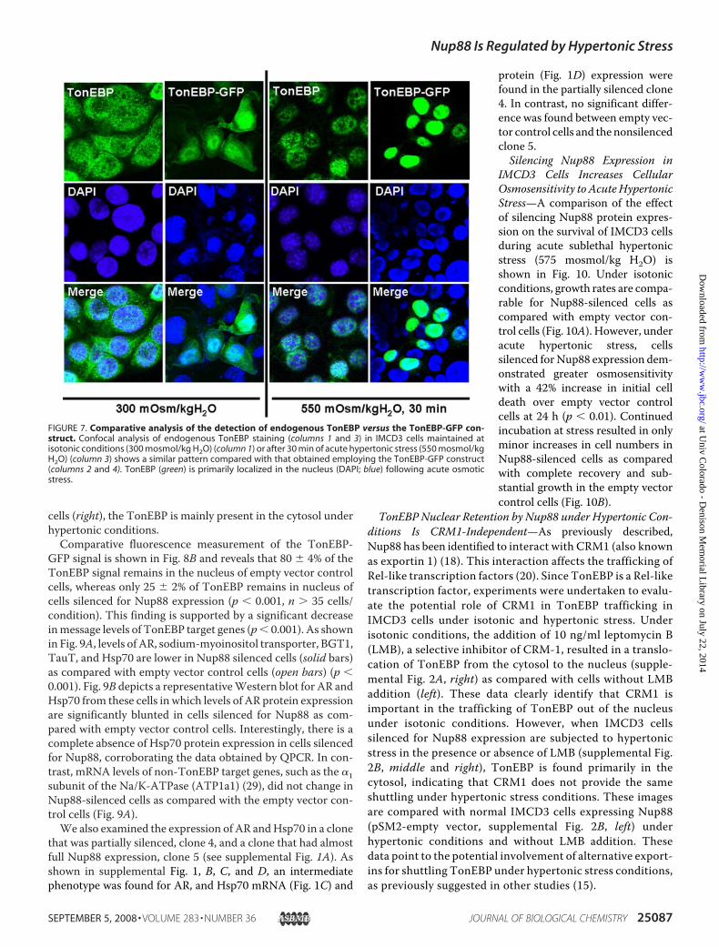
cells (right), the TonEBP is mainly present in the cytosol underhypertonic conditions.Comparative fluorescence measurement of the TonEBP-
GFP signal is shown in Fig. 8B and reveals that 80 � 4% of theTonEBP signal remains in the nucleus of empty vector controlcells, whereas only 25 � 2% of TonEBP remains in nucleus ofcells silenced for Nup88 expression (p � 0.001, n � 35 cells/condition). This finding is supported by a significant decreaseinmessage levels of TonEBP target genes (p� 0.001). As shownin Fig. 9A, levels of AR, sodium-myoinositol transporter, BGT1,TauT, and Hsp70 are lower in Nup88 silenced cells (solid bars)as compared with empty vector control cells (open bars) (p �0.001). Fig. 9B depicts a representativeWestern blot for AR andHsp70 from these cells in which levels of AR protein expressionare significantly blunted in cells silenced for Nup88 as com-pared with empty vector control cells. Interestingly, there is acomplete absence of Hsp70 protein expression in cells silencedfor Nup88, corroborating the data obtained by QPCR. In con-trast, mRNA levels of non-TonEBP target genes, such as the �1subunit of the Na/K-ATPase (ATP1a1) (29), did not change inNup88-silenced cells as compared with the empty vector con-trol cells (Fig. 9A).We also examined the expression of AR andHsp70 in a clone
that was partially silenced, clone 4, and a clone that had almostfull Nup88 expression, clone 5 (see supplemental Fig. 1A). Asshown in supplemental Fig. 1, B, C, and D, an intermediatephenotype was found for AR, and Hsp70 mRNA (Fig. 1C) and
protein (Fig. 1D) expression werefound in the partially silenced clone4. In contrast, no significant differ-ence was found between empty vec-tor control cells and the nonsilencedclone 5.Silencing Nup88 Expression in
IMCD3 Cells Increases CellularOsmosensitivity to AcuteHypertonicStress—A comparison of the effectof silencing Nup88 protein expres-sion on the survival of IMCD3 cellsduring acute sublethal hypertonicstress (575 mosmol/kg H2O) isshown in Fig. 10. Under isotonicconditions, growth rates are compa-rable for Nup88-silenced cells ascompared with empty vector con-trol cells (Fig. 10A). However, underacute hypertonic stress, cellssilenced forNup88 expression dem-onstrated greater osmosensitivitywith a 42% increase in initial celldeath over empty vector controlcells at 24 h (p � 0.01). Continuedincubation at stress resulted in onlyminor increases in cell numbers inNup88-silenced cells as comparedwith complete recovery and sub-stantial growth in the empty vectorcontrol cells (Fig. 10B).
TonEBP Nuclear Retention by Nup88 under Hypertonic Con-ditions Is CRM1-Independent—As previously described,Nup88 has been identified to interact with CRM1 (also knownas exportin 1) (18). This interaction affects the trafficking ofRel-like transcription factors (20). Since TonEBP is a Rel-liketranscription factor, experiments were undertaken to evalu-ate the potential role of CRM1 in TonEBP trafficking inIMCD3 cells under isotonic and hypertonic stress. Underisotonic conditions, the addition of 10 ng/ml leptomycin B(LMB), a selective inhibitor of CRM-1, resulted in a translo-cation of TonEBP from the cytosol to the nucleus (supple-mental Fig. 2A, right) as compared with cells without LMBaddition (left). These data clearly identify that CRM1 isimportant in the trafficking of TonEBP out of the nucleusunder isotonic conditions. However, when IMCD3 cellssilenced for Nup88 expression are subjected to hypertonicstress in the presence or absence of LMB (supplemental Fig.2B, middle and right), TonEBP is found primarily in thecytosol, indicating that CRM1 does not provide the sameshuttling under hypertonic stress conditions. These imagesare compared with normal IMCD3 cells expressing Nup88(pSM2-empty vector, supplemental Fig. 2B, left) underhypertonic conditions and without LMB addition. Thesedata point to the potential involvement of alternative export-ins for shuttling TonEBP under hypertonic stress conditions,as previously suggested in other studies (15).
FIGURE 7. Comparative analysis of the detection of endogenous TonEBP versus the TonEBP-GFP con-struct. Confocal analysis of endogenous TonEBP staining (columns 1 and 3) in IMCD3 cells maintained atisotonic conditions (300 mosmol/kg H2O) (column 1) or after 30 min of acute hypertonic stress (550 mosmol/kgH2O) (column 3) shows a similar pattern compared with that obtained employing the TonEBP-GFP construct(columns 2 and 4). TonEBP (green) is primarily localized in the nucleus (DAPI; blue) following acute osmoticstress.
Nup88 Is Regulated by Hypertonic Stress
SEPTEMBER 5, 2008 • VOLUME 283 • NUMBER 36 JOURNAL OF BIOLOGICAL CHEMISTRY 25087
at Univ C
olorado - Denison M
emorial L
ibrary on July 22, 2014http://w
ww
.jbc.org/D
ownloaded from

DISCUSSION
Cellular changes in the genome and proteome are requiredfor cells of the renal inner medulla to adapt to extreme changesin their hypertonic environment. Although a large body ofinformation already exists for changes in expression of proteinsinvolved in the osmotic response (1, 3–5, 30), it is expected thata larger set of genes and proteins yet unknown are also involvedthat have wide ranging effects of critical importance.In an approach to elucidate proteins involved in this osmotic
stress response, our laboratory is using genomic and proteomictools, including gene arrays, two-dimensional differential gelelectrophoresis (16), and antibody microarrays (17). Althoughtwo-dimensional difference gel electrophoresis is a powerfulproteomic discovery technique, antibody microarray technol-ogy is an excellent complementary approach to two-dimen-sional difference gel electrophoresis, because it allows for thediscovery of changes in expression of hydrophobic as well aslow abundance proteins.Themouse IMCD3 cell line provides a unique tool for study-
ing the adaptative changes in the collecting duct of the kidney.Unlike mouse papilla, IMCD3 cells may be examined underisotonic growth conditions and acutely challenged or chroni-cally adapted to the hypertonic setting (24, 25). In the currentwork, we have employed antibody microarray proteomics andidentified Nup88 as highly up-regulated in IMCD3 cells underhypertonic stress.
Previous researchers have dem-onstrated that Nup88 associateswith nucleoporin 214 to form partof a nuclear pore complex at thenuclear membrane (18). Nuclearpore complexes are involved in thetrafficking of several molecules,including RNAs and proteins. Ofspecific interest are data indicatingthat Nup88 inhibits the nuclearexport of several Rel-like transcrip-tion factors through anchoringCRM1 also referred to as exportin 1,to the nuclear membrane, therebyavoiding its return to the nucleus fora new round of export (20). How-ever, other authors (19) propose anopposite role for Nup88, indicatingthat it enhances nuclear export. Wepropose that up-regulation ofNup88 in IMCD3 cells in responseto hypertonicity acts to retainthe Rel-like transcription factorTonEBP in the nucleus. TonEBP isessential for adaptation to hyper-tonic stress via transcription of tar-get genes necessary for compatibleosmolyte accumulation, ion trans-port, and DNA repair (11, 13,30–32).The up-regulation of Nup88 in
IMCD3 cells determined by anti-body microarray was further validated by both QPCR for mes-sage and Western blot for protein. Nup88 protein expressionincreased rapidly upon exposure to cells to an acute sublethalosmotic stress in a time period (4–6 h after hypertonic stress)similar to early response proteins. Under isotonic conditions,IMCD3 cells were found to express low levels ofNup88 protein,which markedly increased during acute or chronic exposure tohypertonicity. The expression of Nup88 protein in response tohypertonicity was also demonstrated in mice and human kid-ney tissues with substantial Nup88 protein levels at the hyper-tonic papilla tissues andnearly absent at the isotonic cortex. It isof interest that Nup88 expression in papilla frommice deprivedof water for 36 h was markedly up-regulated as compared withmice that received ad libitum water. Nup88 message and pro-tein half-lives were determined by returning cells chronicallyadapted to 900mosmol to isotonic conditions and are relativelyshort (2.8 and 18.4 h, respectively), possibly due to its proposedrole as an inhibitor of TonEBP nuclear export, since it has beendemonstrated that TonEBP is rapidly exported out of thenucleus after cells are returned to isotonic conditions (15).High osmolality induced by ionic solutes as well as mannitol,
sucrose, and urea resulted in near equal up-regulation ofNup88. In this regard, up-regulation ofNup88with ureamay beexplained by the fact that urea transporter A is a target gene forTonEBP under urea conditions, as shown previously by others(33, 34). Localization of Nup88 protein in IMCD3 cells by con-
B
Nuclear Cytosolic0
25
50
75
100
125
TonE
BP
fluor
esce
nt s
igna
l
(ar
bitr
ary
units
)
pSM2-Empty Vector
Nuclear Cytosolic0
25
50
75
100
125pSM2-Nup88
TonE
BP
fluor
esce
nt s
igna
l
(a
rbitr
ary
units
)
A
pSM2-Empty Vector pSM2-Nup88
Tonicity (550 mOsm/kgH 2O, 8 hours)
TonEBP-GFP
DAPI
Merge
TonEBP-GFP
DAPI
Merge
FIGURE 8. Nup88 retains TonEBP in the nucleus under hypertonic stress. A, the left panel shows the stablecell line obtained by transfection with the pSM2 empty vector control. After 8 h of acute hypertonic stress,TonEBP (green) is substantially present in the nucleus, as demonstrated by colocalization with DAPI (blue).Alternatively, the right panel shows the stable cell line in which Nup88 expression is silenced. TonEBP is pri-marily present in the cytosol under osmotic stress conditions. B, fluorometry intensity analysis for cellsdepicted in A (n � 39/6 wells for pSM2-empty vector cells and n � 36/6 wells for pSM2-Nup88 cells from threedifferent experiments). Fluorescent data indicate that 80% of the signal is present in the nucleus of emptyvector control cells under hypertonic stress as compared with 25% for the Nup88-silenced cells.
Nup88 Is Regulated by Hypertonic Stress
25088 JOURNAL OF BIOLOGICAL CHEMISTRY VOLUME 283 • NUMBER 36 • SEPTEMBER 5, 2008
at Univ C
olorado - Denison M
emorial L
ibrary on July 22, 2014http://w
ww
.jbc.org/D
ownloaded from

focal fluorescence microscopy demonstrated its presenceexclusively at the nuclear membrane, consistent with the pro-tein’s potential involvement in nuclear pore complexes (18). Inorder to better define whether Nup88 is involved in the reten-tion of TonEBP under hypertonic stress, we amplified a frag-ment of the mouse N terminus of TonEBP containing itsnuclear localization signal, its auxiliary export domain, and itsnuclear export signal.We cloned this fragment upstream of theGFP to obtain TonEBP-GFP expression. Results obtained withthis construct correlate with those obtained from analysis ofendogenous TonEBP using a specific antibody. Confocal dataindicated essentially identical localization, thereby validatingthe TonEBP-GFP construct as a useful tool to track the nucle-ocytoplasmic traffic of TonEBP under hypertonic stress in liv-ing cells. We show that TonEBP is present in the cytosol ofIMCD3 cells at isotonic conditions and is rapidly shifted to thenucleus in only 30 min of acute sublethal stress. Confocal anal-ysis of TonEBP fluorescent signal showed that 90% of TonEBPis internalized in the nucleus in the first hour of hypertonicstress. As shown in Fig. 4, Nup88 is barely expressed in thesecells after 30 min of osmotic stress, indicating that TonEBPimport to the nucleus occurs in a Nup88-independent way.This result is confirmed using the stable cell line silenced forNup88 expression (data not shown).After 8 h of acute hypertonic stress, the majority of TonEBP
is present in the cytosol in cells silenced for Nup88 expression,as compared with empty vector control cells that retainmost ofthe transcription factor in the nucleus. GFP analysis revealedthat �75% of the TonEBP signal is located in the cytosol ofNup88 silenced cells; in contrast, 80% of the TonEBP is presentin the nucleus of nonsilenced cells. The significance of thisobservation is reflected in the fact that themessage levels ofwellestablished TonEBP target genes (AR, sodium-myoinositoltransporter, BGT1, TauT, and Hsp70) (10, 13, 31, 32) are
blunted under acute hypertonicstress in cells silenced for Nup88 ascompared with empty vector con-trol cells expressing Nup88. Inter-estingly, this blunting of mRNA lev-els of TonEBP target genes underhypertonic stress is similar to thatreported in the kidney papilla fromTonEBP knock-out mice (35). Asconfirmation, protein expressionfor AR and Hsp70 was examinedand found to be substantiallyreduced (66 and 95%, respectively;p � 0.001) in Nup88-silenced cellsas compared with control cellsunder hypertonic stress with anintermediate expression in partiallysilenced clones. This blunting ofexpression of critical osmotic stressresponse genes leads to an increasedosmosensitivity in Nup88-silencedcells as compared with controls. Infact, over a 5-day period, little if anycell recovery is determined when
0 1 2 3 4 5 60
25
50
75
100
125
150pSM2-Empty VectorpSM2-Nup88
575 mOsm/kgH2O
Time (Days)
Perc
enta
ge in
itial
cel
l num
ber
A B
0 1 2 3 40
50100150200250300350400450
pSM2-Empty VectorpSM2-Nup88
300 mOsm/kgH2O
Time (Days)
Perc
enta
ge in
itial
cel
l num
ber
FIGURE 10. Effect of silencing Nup88 expression on IMCD3 cell survival under hypertonic conditions.A, comparison of cell growth rates of IMCD3 clones expressing the empty vector or silencer for Nup88 underisotonic conditions. Data indicate no significant difference in the growth rates over a 72-h period. B, compar-ison of cell survival and growth for empty vector and Nup88-silenced cells under sublethal hypertonic stressconditions (575 mosmol/kg H2O). A substantial difference in immediate cell viability at 24 h was determined forNup88-silenced cells as compared with empty vector controls (42%, p � 0.001). In addition, whereas emptyvector control cells demonstrated recovery and cell growth over the ensuing 4 days, in Nup88-silenced cul-tures, no significant cell growth was determined (p � 0.001 between controls and silenced cells). Data repre-sent the mean � S.E. for three independent experiments performed in triplicate (n � 9).
FIGURE 9. Effect of silencing Nup88 on TonEBP target gene expressionunder acute hypertonic stress in IMCD3 cells. A, message levels of TonEBPtarget genes (AR, BGT1, TauT, and Hsp70) are decreased in the absence ofNup88 under acute sublethal hypertonic stress (550 mosmol/kg H2O, 24 h).Open bars, relative message levels in empty vector control stable cells; solidbars, relative message levels in the stable Nup88-silenced cells. A comparisonwith message levels for the �1 subunit of Na/K-ATPase (a non-TonEBP target)is shown. B, a representative Western blot for AR and Hsp70 protein expres-sion in empty vector and Nup88-silenced cells treated as above is shown. A�-actin loading control is included.
Nup88 Is Regulated by Hypertonic Stress
SEPTEMBER 5, 2008 • VOLUME 283 • NUMBER 36 JOURNAL OF BIOLOGICAL CHEMISTRY 25089
at Univ C
olorado - Denison M
emorial L
ibrary on July 22, 2014http://w
ww
.jbc.org/D
ownloaded from

Nup88-silenced IMCD3 cells are exposed to sublethal hyper-tonic stress.Previous research points to a role for exportin 1 (CRM1) in
the localization of Rel-like transcription factors, such asTonEBP. Our data employing the specific inhibitor LMB dem-onstrate that in IMCD3 cells, CRM1 appears to be involved inthe nuclear export of TonEBP under isotonic conditions. Incontrast, under hypertonic stress, CRM1 is not involved, sinceinhibition of CRM1 with LMB did not prevent the loss ofTonEBP from the nucleus in Nup88-silenced cells. Theseresults are similar to those previously described by Tong et al.(15).In conclusion, our data are the first to demonstrate that the
nucleoporin protein, Nup88, is up-regulated both in vitro andin vivo in the kidney in response to hypertonic stress. Thesedata also suggest that Nup88 expression serves to retainTonEBP in the nucleus, thus allowing the transcription of targetgenes essential for cell adaptation to osmotic stress.
REFERENCES1. Burg, M. B., Kwon, E. D., and Kultz, D. (1997) Annu. Rev. Physiol. 59,
437–4552. Burg, M. B., Kwon, E. D., and Kultz, D. (1996) FASEB J. 10, 1598–16063. Ferraris J. D., and Burg, M. B. (2006) Contrib. Nephrol. 152, 125–1414. Handler, J. S., and Kwon, H. M. (1996) Kidney Int. 49, 1682–16835. Handler, J. S., and Kwon, H. M. (2001) Nephron 87, 106–1106. Handler, J. S., and Kwon, H. M. (2001) Kidney Int. 60, 408–4117. Jeon, U. S., Kim, J. A., Sheen, M. R., and Kwon, H. M. (2006) Acta Physiol.
(Oxf.) 187, 241–2478. Woo, S. K., and Kwon, H. M. (2002) Int. Rev. Cytol. 215, 189–2029. Woo, S. K., Lee, S. D., andKwon,H.M. (2002)Pflugers Arch. 444, 579–58510. Miyakawa, H., Woo, S. K., Dahl, S. C., Handler, J. S., and Kwon, H. M.
(1999) Proc. Natl. Acad. Sci. U. S. A. 96, 2538–254211. Miyakawa,H.,Woo, S. K., Chen, C. P., Dahl, S. C., Handler, J. S., andKwon,
H. M. (1998) Am. J. Physiol. 274, F753–F76112. Ito, T., Fujio, Y., Hirata, M., Takatani, T., Matsuda, T., Muraoka, S., Taka-
hashi, K., and Azuma, J. (2004) Biochem. J. 382, 177–18213. Woo, S. K., Lee, S. D., Na, K. Y., Park, W. K., and Kwon, H. M. (2002)Mol.
Cell. Biol. 22, 5753–576014. Cha, J. H., Woo, S. K., Han, K. H., Kim, Y. H., Handler, J. S., Kim, J., and
Kwon, H. M. (2001) J. Am. Soc. Nephrol. 12, 2221–2230
15. Tong, E. H., Guo, J. J., Huang, A. L., Liu, H., Hu, C. D., Chung, S. S., and Ko,B. C. (2006) J. Biol. Chem. 281, 23870–23879
16. Rivard, C. J., Brown, L. M., Almeida, N. E., Maunsbach, A. B., Pihakaski-Maunsbach, K., Andres-Hernando, A., Capasso, J. M., and Berl, T. (2007)J. Biol. Chem. 282, 6644–6652
17. Lanaspa, M. A., Almeida, N. E., Andres-Hernando, A., Rivard, C. J.,Capasso, J. M., and Berl, T. (2007) Proc. Natl. Acad. Sci. U. S. A. 104,13672–13677
18. Fornerod, M., van Deursen, J., van Baal, S., Reynolds, A., Davis, D., Murti,K. G., Fransen, J., and Grosveld, G. (1997) EMBO J. 16, 807–816
19. Hutten, S., and Kehlenbach, R. H. (2006)Mol. Cell. Biol. 26, 6772–678520. Roth, P., Xylourgidis, N., Sabri, N., Uv, A., Fornerod, M., and Samakovlis,
C. (2003) J. Cell Biol. 163, 701–70621. Uv, A. E., Roth, P., Xylourgidis, N., Wickberg, A., Cantera, R., and Sama-
kovlis, C. (2000) Genes Dev. 14, 1945–195722. Xylourgidis, N., Roth, P., Sabri, N., Tsarouhas, V., and Samakovlis, C.
(2006) J. Cell Sci. 119, 4409–441923. Rauchman, M. I., Nigam, S. K., Delpire, E., and Gullans, S. R. (1993)Am. J.
Physiol. 265, F416–F42424. Capasso, J. M., Rivard, C. J., and Berl, T. (2001) Am. J. Physiol. 280,
F768–F77625. Capasso, J. M., Rivard, C. J., Enomoto, L.M., and Berl, T. (2003)Ann. N. Y.
Acad. Sci. 986, 410–41526. Kodiha, M., Tran, D., Qian, C., Morogan, A., Presley, J. F., Brown, C. M.,
and Stochaj, U. (2008) Biochim. Biophys. Acta 1783, 405–41827. Bernad, R., van der Velde, H., Fornerod,M., and Pickersgill, H. (2004)Mol.
Cell. Biol. 24, 2373–238428. Bernad, R., Engelsma, D., Sanderson, H., Pickersgill, H., and Fornerod, M.
(2006) J. Biol. Chem. 281, 19378–1938629. Capasso, J. M., Rivard, C., and Berl, T. (2001) Proc. Natl. Acad. Sci. U. S. A.
98, 13414–1341930. Dmitrieva, N. I., and Burg, M. B. (2005)Mutat. Res. 569, 65–7431. Na, K. Y., Woo, S. K., Lee, S. D., and Kwon, H. M. (2003) J. Am. Soc.
Nephrol. 14, 283–28832. Zhang, Z., Ferraris, J. D., Brooks, H. L., Brisc, I., and Burg, M. B. (2003)
Am. J. Physiol. 285, F688–F69333. Lim, S. W., Ahn, K. O., Sheen, M. R., Jeon, U. S., Kim, J., Yang, C. W., and
Kwon, H. M. (2007) J. Am. Soc. Nephrol. 18, 421–42934. Nakayama, Y., Peng, T., Sands, J. M., and Bagnasco, S. M. (2000) J. Biol.
Chem. 275, 38275–3828035. Lopez-Rodrıguez, C., Antos, C. L., Shelton, J. M., Richardson, J. A., Lin, F.,
Novobrantseva, T. I., Bronson, R. T., Igarashi, P., Rao, A., and Olson, E. N.(2004) Proc. Natl. Acad. Sci. U. S. A. 101, 2392–2397
Nup88 Is Regulated by Hypertonic Stress
25090 JOURNAL OF BIOLOGICAL CHEMISTRY VOLUME 283 • NUMBER 36 • SEPTEMBER 5, 2008
at Univ C
olorado - Denison M
emorial L
ibrary on July 22, 2014http://w
ww
.jbc.org/D
ownloaded from
Copyright © 2022 FDOKUMEN