Infliximab for severe, treatment-resistant psoriasis: a prospective, open-label study
Upload
rockefellerCategory
view
3download
0
Resolved psoriasis lesions retain expression of a subset ofdisease-related genes
Mayte Suárez-Fariñas1,2, Judilyn Fuentes-Duculan1, Michelle A. Lowes1, and James G.Krueger11 The Laboratory for Investigative Dermatology, The Rockefeller University, 1230 York Ave, NewYork, NY, 100652 Center for Clinical and Translational Science, The Rockefeller University, 1230 York Ave, NewYork, NY, 10065
AbstractPsoriasis is a complex inflammatory disease that usually heals without visible scarring.Histological evaluation often suggests complete resolution, but reversal of genomic disease-associated alterations has not yet been defined. Gene expression profiling was used to determinethe extent to which the psoriasis genes were reversed after 3 months of etanercept in patients whoresponded to treatment. We reviewed the histology, leukocyte counts, and PCR data forinflammatory genes, to compare recovery of these parameters and the genomic studies. Manycellular markers do return close to non-lesional levels, although five inflammatory genes did notimprove >75% (IL-12p35, MX1, IL-22, IL-17, IFNγ). Psoriasis-related genes with <75%improvement were defined as comprising a “residual disease genomic profile”, composed of 248probesets. Genes of interest in psoriasis tissue that did not return to baseline included LYVE-1,WNT5A, RAB31 and AQP9. It appears that even though the epidermal reaction in psoriasis isfully resolved, inflammation, as defined by expression of key cytokines and chemokines, is notcompletely resolved in treated lesions. We also found that structural cells of the skin continued toexpress molecular alterations, and that some subtle features of skin structure, e.g., lymphatics,were not fully normalized with treatment.
IntroductionPsoriasis is a complex inflammatory disease with a characteristic clinical and histologicalphenotype. The non-affected skin of psoriasis patients does not appear to have any distinctclinical features, and effective treatment returns lesional skin (LS) to a non-lesional (NL)state, i.e. skin that appears virtually normal. Our past studies with several different therapiesfor psoriasis suggest that successful treatment is correlated with reduced epidermal thicknessand reductions in inflammatory cellular infiltrates and gene expression (Chamian et al.,2005; Zaba et al., 2007). We have shown that in patients responding to therapy, the cellularinfiltrate and expression of selected inflammatory genes in affected LS skin return to NLlevels at the end of treatment. The clinical appearance, combined with histological andmolecular studies, indicated that psoriasis skin lesions were reversed without obvious
Corresponding author: James G. Krueger, The Rockefeller University, Laboratory for Investigative Dermatology, 1230 York Avenue,Box 178, NY, NY 10065, Ph: 212-327-7730, Fax: 212-327-8232, [email protected] authorMayte Suárez-Fariñas, The Rockefeller University, Laboratory for Investigative Dermatology, 1230 York Avenue, Box 178, NY, NY10065, Ph: 212-327-8213, Fax: 212-327-8353, [email protected]
NIH Public AccessAuthor ManuscriptJ Invest Dermatol. Author manuscript; available in PMC 2012 February 1.
Published in final edited form as:J Invest Dermatol. 2011 February ; 131(2): 391–400. doi:10.1038/jid.2010.280.
NIH
-PA Author Manuscript
NIH
-PA Author Manuscript
NIH
-PA Author Manuscript

differences to NL skin. Thus psoriasis can be viewed as “reversible” when the inflammationis removed from the skin.
Although psoriasis may recur in new areas, it is well recognized that often recurs in the samelocations on the body, which may suggest that the skin where psoriasis lesions have been isnot fully “healed”. There may be some residual changes in treated lesions that predispose torecurrence of lesions in the same locations. However, histopathology does not give a goodindication of cell function, and PCR studies have been conducted on a small group ofselected genes. The introduction of microarray profiling has given a more comprehensiveplatform to study disease-specific genes, and how they respond to treatment.
We wanted to determine the extent to which the genomic psoriasis phenotype, as defined bythe differentially expressed genes (DEGs) between LS and NL skin, was reversed byetanercept treatment in responding patients. At the end of the three-month clinical trialperiod, we analyzed how closely the psoriasis transcriptome returned to a NL state inresponders. We were interested to see if psoriasis was fully reversible at the molecular levelwhen the skin was clinically clear of disease. Although many DEGs were normalized inpost-treatment biopsies, 248 genes did not return to baseline (NL) after three months oftreatment. We have termed this residual genomic expression that did not return to NL levels“residual disease genomic profile (RDGP)”. This gene-set hasimportant implications for thebiology and treatment of psoriasis, and it could serve as a means to study progressivealterations in skin structure and function as a consequence of temporal progression orseverity of skin lesions.
ResultsResolution of psoriasis lesions
Much of clinical disease pathology in psoriasis is related to altered epidermal growth anddifferentiation. In this analysis, we sought to relate epidermal improvement to modulation ofdisease-related genes by etanercept. In the group of patients that respond to etanercept, theepidermis was almost fully recovered (Table 1). The epidermal thickness improved 92% inaverage, Ki67 (a marker of cellular proliferation) improved 119%, K16 mRNA improved by100%, and all cases were K16 negative by immunohistochemistry (Zaba et al., 2007).Hence, to a pathologist using an H&E stain or growth-related markers, the epidermis wouldappear histologically “normal”.
In order to determine the extent to which the inflammatory infiltrate and specificinflammatory genes improved with treatment, we calculated the mean improvement in cellcounts (Table 1). CD3+ T-cells almost completely returned to baseline (95.5%improvement); CD8+ T-cells were 77% improved; CD11c+ myeloid dendritic cells (DCs)were 88% improved; and CD83+ mature DCs were 121% improved; while CD163+
macrophages were 97% improved. CD206 is on both DCs and macrophages and improved84% and DCLAMP+ mature DCs were 100% improved. Overall, resolution of inflammatorycell populations in the epidermis was often higher than the dermis, although both wereimproved greater than 75% for all populations studied, except dermal CD8+ cells, whichimproved only 64%.
In Table 1, improvement of selected genes by RT-PCR expression in responding patients ispresented. Five genes that did not improve by greater than 75% were important pro-inflammatory genes, IL-12p35, MX1, IL-22, IL-17, IFN-γ (range of improvement was 62–67%). Hence, while within resolved skin pathologic T-cells were more than 95% reducedbased on CD3+ cell counts, dermal CD8+ cells were not fully resolved, and some T-cellactivation may be ongoing within residual infiltrates. Overall, we interpret these findings to
Suárez-Fariñas et al. Page 2
J Invest Dermatol. Author manuscript; available in PMC 2012 February 1.
NIH
-PA Author Manuscript
NIH
-PA Author Manuscript
NIH
-PA Author Manuscript

indicate that the epidermis can improve almost completely even in the context of someresidual inflammatory gene expression.
Resolution of psoriasis disease-related genesOur first publication of the genomic data from this group of patients (n=15) described thegenomic effects of etanercept in responders and non-responders (Zaba et al., 2009b). Wehave also defined the psoriasis transcriptome using these patients, and compared with otherpublished psoriasis DEG lists (Suárez-Fariñas et al., 2010). Our psoriasis-related DEGscontained 732 up-regulated probesets (579 known unique genes) and 890 down-regulatedprobesets (703 genes) (Figure 1a). Here, we considered the psoriasis phenotype asgenomically defined by this DEG and used them to determine the extent to which thepsoriasis phenotype was reversed after 3 months of etanercept in 11 patients who respondedto treatment. Indeed, our analysis showed that a large set of disease-related genes wasimproved by >90%, and some genes had improvement even beyond NL levels. A selectionof improved psoriasis genes is presented in Table 2, and full list in Supplementary Table 1.Many inflammatory genes such as granzyme B, IL-8 and S100A8 improved by 91%.Structural genes, such as keratin 6B and 16, as well as epidermal protein lipocalin 2 (LCN2)also improved more than 96%.
Resolution of gene-sets representing specific cells (Guttman-Yassky et al., 2009; Haider etal., 2007; Zaba et al., 2010) could also be evaluated using a gene-set approach (Table 3). Forexample up-regulated keratinocyte genes were improved over 105%, and up-regulated T-cellgenes around 88%. Sets representing myeloid cell populations, including immature andmature DCs, inflammatory myeloid DCs, and macrophages, all improved over 89%.
Defining “residual disease genomic profile” in treated psoriasisGenes with less than 75% improvement were defined as comprising the RDGP. From thepsoriasis up-regulated genes, 76 probesets (64 genes) did not improve above 75% withtreatment, and among disease down-regulated genes, 206 probesets (184 genes) were below75% improvement (Figure 1a). A selection of potentially interesting genes that did notreturn to NL levels is presented in Table 4, for both up and down-regulated psoriasis-genes(See Supplementary Table 1 for complete RDGP). These genes identify a set of disease-related genes with incomplete reversal even after effective therapy. Unfortunately, manyprimary cytokines such as IFNγ, IL-17, IL-22, are not reliably detected by Affymetrix chipsdue to low mRNA abundance (Suárez-Fariñas et al., 2010). Since our definition of RDGP isbased on Affymetrix expression data, those genes are not in Supplementary Table 1.However, since we have assessed their improvement by PCR (Table 1), they could beconsidered as residual genes in a broader definition of RDGP.
Within this RDGP, we consider two sets of effects. First, there was continued expression ofmany inflammation-related genes, possibly indicating incomplete suppression ofinflammatory circuits by etanercept. Genes such as lymphotoxin and TCRβ1 were up-regulated in psoriasis and improved less than 75% (66% and 43% improvement,respectively). This is in keeping with our observations described above at the PCR level,that epidermal improvement can occur even in the setting of some continued inflammatorygene expression.
Structural changes in the skinThe second major effect that we considered within the RDGP was the altered expression ofgenes associated with “structural” cell types of the skin. This suggests altered physiology ortissue structure in resolved lesions, in some sense, a “molecular scar”. Many of the genesthat did not return to NL levels were for structural cutaneous components. There were genes
Suárez-Fariñas et al. Page 3
J Invest Dermatol. Author manuscript; available in PMC 2012 February 1.
NIH
-PA Author Manuscript
NIH
-PA Author Manuscript
NIH
-PA Author Manuscript

that were down-regulated during psoriasis inflammation (LS<NL) that were not completelyresolved by week 12 (e.g. LYVE-1 and AQP9). There were up-regulated genes that did notreturn to NL (e.g. RAB31 and WNT5A). Gene expression for these four genes is shown inFigure 1b, and compared to STAT1, an up-regulated gene that does return to NL. We havealso performed immunohistochemstry for these four gene products from both down and up-regulated groups over the treatment period (Figure 2).
LYVE-1, a well-recognized marker of lymphatic endothelial cells, showed only 21%maximal improvement at week 12. To determine the significance of persistent down-modulation of LYVE-1 at the protein level, we performed standard immunohistochemistryof LYVE-1 in normal skin, NL and LS skin, and at 12 weeks of treatment (Figure 2a). Innormal skin the lymphatic vessels identified by this antibody were mostly in the upperreticular dermis and had a discernible (wide) lumen. In LS skin the lymphatic vessels weremore collapsed and were observed to be closer to the dermo-epidermal junction compared toNL skin and week 12. Hence LYVE-1+ cells showed a different pattern in normal skincompared to psoriatic lesions, and even after treatment, it did not return to the pattern ofexpression of normal skin.
WNT5A (wingless-related MMTV integration site 5A) regulates epidermal differentiation inadult skin and acts in synergistic induction of type-I IFN target genes (Romanowska et al.,2009). This gene has been previously reported to show increased expression in psoriaticplaques (Reischl et al., 2007). WNT5A was only improved 54% at the genomic level. Byimmunohistochemistry, WNT5A showed the greatest expression in the epidermis of LSskin, with some dermal staining (Figure 2b). There was a persistent expression by week 12,greater than NL and minimal staining of WNT5A in normal skin.
RAB31 was another gene of interest that is increased in psoriasis tissue but did not return toNL levels. RAB31, a member of RAS oncogene family that plays an essential role in vesicleand granule targeting (Bao et al., 2002), showed 34% improvement at week 12.Immunohistochemistry was performed in normal skin, NL and LS skin, as well as after 12weeks of treatment (Figure 2c). There were many RAB31 positive cells in the papillarydermis of LS skin close to the dermo-epidermal junction, with fewer positive cells in NLskin and at week 12. There was minimal RAB31 expression in normal skin. In LS skin,RAB31 was expressed on CD45+ cells, and also some CD11c+ myeloid DCs mainly in thepapillary dermis and few CD163+ macrophages, (Figure S1).
Aquaporin 9 (AQP9) was another interesting down-regulated gene with maximalimprovement of 26%. Mammalian aquaporins are a family of 13 membrane proteins thatform water channels across cell membranes, and may transport small solutes such asglycerol (Boury-Jamot et al., 2006). Aquaporin 9 has been described in human skin,although its role there is not fully understood. In normal and NL skin there was someepidermal expression, with subtle loss of expression in LS skin, and some recovery at week12, although not to NL levels (Figure 2d).
Canonical pathways do not fully resolveTo obtain a global view of the RDGP in terms of pathways rather than gene by gene, wequantified the average improvement for a number of canonical pathways. Of 358 canonicalpathways that were represented in the transcriptome several of them showed less than 75%improvement (selected pathways shown in Table 4). For example disease-genes in theLeptin pathway improved only 56%. This could be important as leptin is considered to bepartially responsible for obesity (Kelesidis et al.), and recently there has been an associationwith psoriasis and metabolic syndrome (Azfar and Gelfand, 2008). The failure of thispathway to improve 100% may be related to ongoing risk for obesity and the metabolic
Suárez-Fariñas et al. Page 4
J Invest Dermatol. Author manuscript; available in PMC 2012 February 1.
NIH
-PA Author Manuscript
NIH
-PA Author Manuscript
NIH
-PA Author Manuscript
syndrome, even with clinical improvement in the skin. Myocyte AD pathway only improved52%, and Wnt-pathway was 66% improved, both of which could relate to structuralcomponents in the skin or other organs.
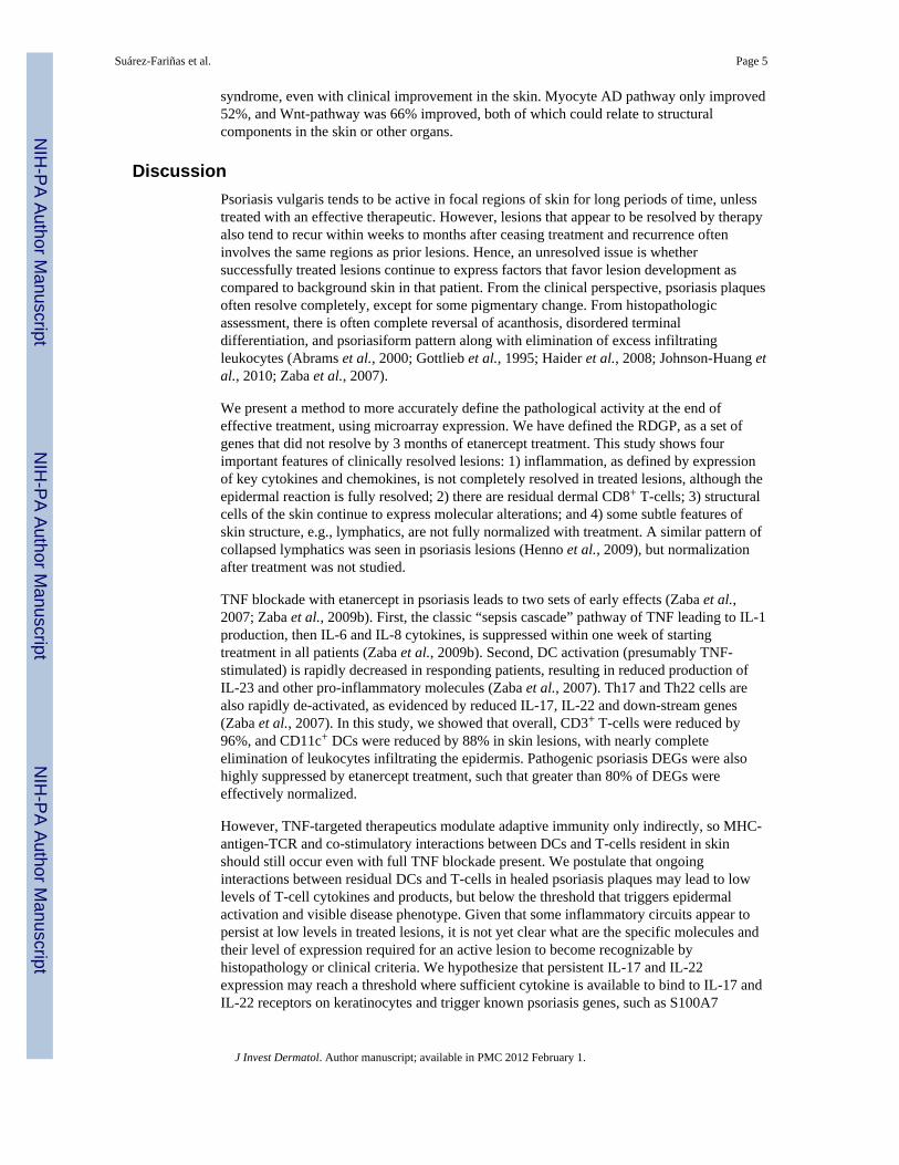
DiscussionPsoriasis vulgaris tends to be active in focal regions of skin for long periods of time, unlesstreated with an effective therapeutic. However, lesions that appear to be resolved by therapyalso tend to recur within weeks to months after ceasing treatment and recurrence ofteninvolves the same regions as prior lesions. Hence, an unresolved issue is whethersuccessfully treated lesions continue to express factors that favor lesion development ascompared to background skin in that patient. From the clinical perspective, psoriasis plaquesoften resolve completely, except for some pigmentary change. From histopathologicassessment, there is often complete reversal of acanthosis, disordered terminaldifferentiation, and psoriasiform pattern along with elimination of excess infiltratingleukocytes (Abrams et al., 2000; Gottlieb et al., 1995; Haider et al., 2008; Johnson-Huang etal., 2010; Zaba et al., 2007).
We present a method to more accurately define the pathological activity at the end ofeffective treatment, using microarray expression. We have defined the RDGP, as a set ofgenes that did not resolve by 3 months of etanercept treatment. This study shows fourimportant features of clinically resolved lesions: 1) inflammation, as defined by expressionof key cytokines and chemokines, is not completely resolved in treated lesions, although theepidermal reaction is fully resolved; 2) there are residual dermal CD8+ T-cells; 3) structuralcells of the skin continue to express molecular alterations; and 4) some subtle features ofskin structure, e.g., lymphatics, are not fully normalized with treatment. A similar pattern ofcollapsed lymphatics was seen in psoriasis lesions (Henno et al., 2009), but normalizationafter treatment was not studied.
TNF blockade with etanercept in psoriasis leads to two sets of early effects (Zaba et al.,2007; Zaba et al., 2009b). First, the classic “sepsis cascade” pathway of TNF leading to IL-1production, then IL-6 and IL-8 cytokines, is suppressed within one week of startingtreatment in all patients (Zaba et al., 2009b). Second, DC activation (presumably TNF-stimulated) is rapidly decreased in responding patients, resulting in reduced production ofIL-23 and other pro-inflammatory molecules (Zaba et al., 2007). Th17 and Th22 cells arealso rapidly de-activated, as evidenced by reduced IL-17, IL-22 and down-stream genes(Zaba et al., 2007). In this study, we showed that overall, CD3+ T-cells were reduced by96%, and CD11c+ DCs were reduced by 88% in skin lesions, with nearly completeelimination of leukocytes infiltrating the epidermis. Pathogenic psoriasis DEGs were alsohighly suppressed by etanercept treatment, such that greater than 80% of DEGs wereeffectively normalized.
However, TNF-targeted therapeutics modulate adaptive immunity only indirectly, so MHC-antigen-TCR and co-stimulatory interactions between DCs and T-cells resident in skinshould still occur even with full TNF blockade present. We postulate that ongoinginteractions between residual DCs and T-cells in healed psoriasis plaques may lead to lowlevels of T-cell cytokines and products, but below the threshold that triggers epidermalactivation and visible disease phenotype. Given that some inflammatory circuits appear topersist at low levels in treated lesions, it is not yet clear what are the specific molecules andtheir level of expression required for an active lesion to become recognizable byhistopathology or clinical criteria. We hypothesize that persistent IL-17 and IL-22expression may reach a threshold where sufficient cytokine is available to bind to IL-17 andIL-22 receptors on keratinocytes and trigger known psoriasis genes, such as S100A7
Suárez-Fariñas et al. Page 5
J Invest Dermatol. Author manuscript; available in PMC 2012 February 1.
NIH
-PA Author Manuscript
NIH
-PA Author Manuscript
NIH
-PA Author Manuscript
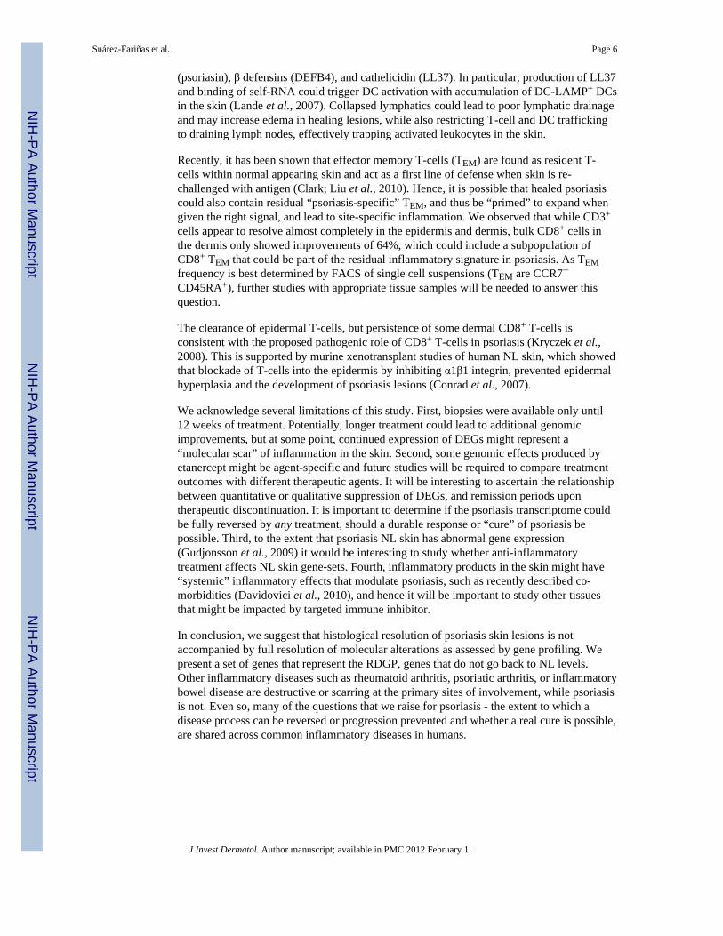
(psoriasin), β defensins (DEFB4), and cathelicidin (LL37). In particular, production of LL37and binding of self-RNA could trigger DC activation with accumulation of DC-LAMP+ DCsin the skin (Lande et al., 2007). Collapsed lymphatics could lead to poor lymphatic drainageand may increase edema in healing lesions, while also restricting T-cell and DC traffickingto draining lymph nodes, effectively trapping activated leukocytes in the skin.
Recently, it has been shown that effector memory T-cells (TEM) are found as resident T-cells within normal appearing skin and act as a first line of defense when skin is re-challenged with antigen (Clark; Liu et al., 2010). Hence, it is possible that healed psoriasiscould also contain residual “psoriasis-specific” TEM, and thus be “primed” to expand whengiven the right signal, and lead to site-specific inflammation. We observed that while CD3+
cells appear to resolve almost completely in the epidermis and dermis, bulk CD8+ cells inthe dermis only showed improvements of 64%, which could include a subpopulation ofCD8+ TEM that could be part of the residual inflammatory signature in psoriasis. As TEMfrequency is best determined by FACS of single cell suspensions (TEM are CCR7−CD45RA+), further studies with appropriate tissue samples will be needed to answer thisquestion.
The clearance of epidermal T-cells, but persistence of some dermal CD8+ T-cells isconsistent with the proposed pathogenic role of CD8+ T-cells in psoriasis (Kryczek et al.,2008). This is supported by murine xenotransplant studies of human NL skin, which showedthat blockade of T-cells into the epidermis by inhibiting α1β1 integrin, prevented epidermalhyperplasia and the development of psoriasis lesions (Conrad et al., 2007).
We acknowledge several limitations of this study. First, biopsies were available only until12 weeks of treatment. Potentially, longer treatment could lead to additional genomicimprovements, but at some point, continued expression of DEGs might represent a“molecular scar” of inflammation in the skin. Second, some genomic effects produced byetanercept might be agent-specific and future studies will be required to compare treatmentoutcomes with different therapeutic agents. It will be interesting to ascertain the relationshipbetween quantitative or qualitative suppression of DEGs, and remission periods upontherapeutic discontinuation. It is important to determine if the psoriasis transcriptome couldbe fully reversed by any treatment, should a durable response or “cure” of psoriasis bepossible. Third, to the extent that psoriasis NL skin has abnormal gene expression(Gudjonsson et al., 2009) it would be interesting to study whether anti-inflammatorytreatment affects NL skin gene-sets. Fourth, inflammatory products in the skin might have“systemic” inflammatory effects that modulate psoriasis, such as recently described co-morbidities (Davidovici et al., 2010), and hence it will be important to study other tissuesthat might be impacted by targeted immune inhibitor.
In conclusion, we suggest that histological resolution of psoriasis skin lesions is notaccompanied by full resolution of molecular alterations as assessed by gene profiling. Wepresent a set of genes that represent the RDGP, genes that do not go back to NL levels.Other inflammatory diseases such as rheumatoid arthritis, psoriatic arthritis, or inflammatorybowel disease are destructive or scarring at the primary sites of involvement, while psoriasisis not. Even so, many of the questions that we raise for psoriasis - the extent to which adisease process can be reversed or progression prevented and whether a real cure is possible,are shared across common inflammatory diseases in humans.
Suárez-Fariñas et al. Page 6
J Invest Dermatol. Author manuscript; available in PMC 2012 February 1.
NIH
-PA Author Manuscript
NIH
-PA Author Manuscript
NIH
-PA Author Manuscript

Materials and MethodsPatients
Twenty adult patients with moderate to severe psoriasis were treated with etanercept 50 mgsubcutaneously twice weekly for 12 weeks (clinical trial no. NCT00116181). Writteninformed consent was obtained from all patients. The clinical and histological response ofthese 20 patients was previously published (Zaba et al., 2007). Patients were definedhistologically as “responders” if the serial biopsies of an index plaque showed return ofepidermal thickness and keratin 16 (K16) immunohistochemistry to NL levels.
Gene Expression DataRNA from skin biopsies of 15 patients was hybridized to Affymetrix HGU133a2 chips asdescribed (Zaba et al., 2009b). Microarray is deposited at the Gene Expression Omnibusrepository (GSE11903). Probeset annotation was obtained with Bioconductor’shgu133a2.db package (version 2.3.5).
Statistical AnalysisIn a prior publication (Zaba et al., 2009b), a mixed-effect linear model with “patient” asrandom effect was used to model gene expression using interaction TimexResponse. Fromthis model, the treatment effect at 12 weeks was estimated for responders using limmapackage form Bioconductor.
Psoriasis-related genes (Suárez-Fariñas et al., 2010) were analyzed at the end of treatment toevaluate return to NL. For each disease-gene, we quantified improvement after 12 weeks oftreatment as:
where XLS XNL X12 are the expression values at LS, NL and 12 weeks of treatmentrespectively. RDGP is defined as the genes with improvement below 75%.
We used Gene-Set approach to quantify the average improvement for a collection ofpathways (Table 3). We included the canonical pathways (C2 CP) from MDigDB(http://www.broadinstitute.org/gsea/msigdb) and several gene-sets developed by our group(Haider et al., 2007); (Guttman-Yassky et al., 2009).
RT-PCR, Immunohistochemistry and ImmunofluorescenceSkin biopsies for leukocyte markers were stained and counted, and PCR conducted, both ina standard manner (Zaba et al., 2007). Most of these results had been published (Zaba et al.,2007), and are re-analyzed here to determine mean improvement after 12 weeks oftreatment. Standard procedures were followed for immunohistochemistry (n=6),immunofluorescence (n=3) and CD8+ cell counts (n=6) as previously described (Zaba et al.,2009a). Antibodies used for immunohistochemistry and immunofluorescence are listed inSupplementary Table 2. Images were acquired using appropriate filters of a Zeiss Axioplan2 widefield fluorescence microscope with Plan Neofluar 20×0.7 numerical aperture lens andHamamatsu Orca ER-cooled charge-coupled device camera, controlled by METAVUEsoftware (MDS Analytical Technologies, Downington, PA). Immunohistochemistry wasconducted in batches for paired samples and representative staining is shown.
Suárez-Fariñas et al. Page 7
J Invest Dermatol. Author manuscript; available in PMC 2012 February 1.
NIH
-PA Author Manuscript
NIH
-PA Author Manuscript
NIH
-PA Author Manuscript

Supplementary MaterialRefer to Web version on PubMed Central for supplementary material.
AcknowledgmentsResearch was supported by National Institutes of Health (NIH) grant UL1 RR024143 from the National Center forResearch Resources (NCRR) and the Milstein Program in Medical Research. MSF is partially supported by NIHgrant UL1 RR024143; MAL is supported by 1 K23 AR052404-01A1 and The Doris Duke Charitable Foundation.We thank I. Novitskaya for technical assistance during the revision of the manuscript, and Kristine Nograles forcritical reading of the manuscript.
Abbreviations
DEG differentially expressed genes
FCH fold change
FDR false discovery rate
LS lesional
NL non-lesional
RDGP residual disease genomic profile
ReferencesAbrams JR, Kelley SL, Hayes E, Kikuchi T, Brown MJ, Kang S, et al. Blockade of T lymphocyte
costimulation with cytotoxic T lymphocyte-associated antigen 4-immunoglobulin (CTLA4Ig)reverses the cellular pathology of psoriatic plaques, including the activation of keratinocytes,dendritic cells, and endothelial cells. J Exp Med 2000;192:681–694. [PubMed: 10974034]
Azfar RS, Gelfand JM. Psoriasis and metabolic disease: epidemiology and pathophysiology. Curr OpinRheumatol 2008;20:416–422. [PubMed: 18525354]
Bao X, Faris AE, Jang EK, Haslam RJ. Molecular cloning, bacterial expression and properties ofRab31 and Rab32. Eur J Biochem 2002;269:259–271. [PubMed: 11784320]
Boury-Jamot M, Sougrat R, Tailhardat M, Le Varlet B, Bonte F, Dumas M, et al. Expression andfunction of aquaporins in human skin: Is aquaporin-3 just a glycerol transporter? Biochim BiophysActa 2006;1758:1034–1042. [PubMed: 16872579]
Chamian F, Lowes MA, Lin SL, Lee E, Kikuchi T, Gilleaudeau P, et al. Alefacept reduces infiltratingT cells, activated dendritic cells, and inflammatory genes in psoriasis vulgaris. Proc Natl Acad SciU S A 2005;102:2075–2080. [PubMed: 15671179]
Clark RA. Skin-resident T cells: the ups and downs of on site immunity. J Invest Dermatol 130:362–370. [PubMed: 19675575]
Conrad C, Boyman O, Tonel G, Tun-Kyi A, Laggner U, de Fougerolles A, et al. Alpha1beta1 integrinis crucial for accumulation of epidermal T cells and the development of psoriasis. Nat Med2007;13:836–842. [PubMed: 17603494]
Davidovici BB, Sattar N, Jorg PC, Puig L, Emery P, Barker JN, et al. Psoriasis and SystemicInflammatory Diseases: Potential Mechanistic Links between Skin Disease and Co-MorbidConditions. Journal of Investigative Dermatology. 2010 Advance online publication 6 May 2010.
Gottlieb SL, Gilleaudeau P, Johnson R, Estes L, Woodworth TG, Gottlieb AB, et al. Response ofpsoriasis to a lymphocyte-selective toxin (DAB389IL-2) suggests a primary immune, but notkeratinocyte, pathogenic basis. Nat Med 1995;1:442–447. [PubMed: 7585092]
Gudjonsson JE, Ding J, Li X, Nair RP, Tejasvi T, Qin ZS, et al. Global gene expression analysisreveals evidence for decreased lipid biosynthesis and increased innate immunity in uninvolvedpsoriatic skin. J Invest Dermatol 2009;129:2795–2804. [PubMed: 19571819]
Suárez-Fariñas et al. Page 8
J Invest Dermatol. Author manuscript; available in PMC 2012 February 1.
NIH
-PA Author Manuscript
NIH
-PA Author Manuscript
NIH
-PA Author Manuscript

Guttman-Yassky E, Suarez-Farinas M, Chiricozzi A, Nograles KE, Shemer A, Fuentes-Duculan J, etal. Broad defects in epidermal cornification in atopic dermatitis identified through genomicanalysis. J Allergy Clin Immunol 2009;124:1235–1244. e1258. [PubMed: 20004782]
Haider AS, Lowes MA, Suarez-Farinas M, Zaba LC, Cardinale I, Blumenberg M, et al. CellularGenomic Maps Help Dissect Pathology in Human Skin Disease. J Invest Dermatol 2007;128:606–615. [PubMed: 17928892]
Haider AS, Lowes MA, Suarez-Farinas M, Zaba LC, Cardinale I, Khatcherian A, et al. Identificationof Cellular Pathways of “Type 1,” Th17 T Cells, and TNF- and Inducible Nitric Oxide Synthase-Producing Dendritic Cells in Autoimmune Inflammation through Pharmacogenomic Study ofCyclosporine A in Psoriasis. J Immunol 2008;180:1913–1920. [PubMed: 18209089]
Henno A, Blacher S, Lambert C, Colige A, Seidel L, Noel A, et al. Altered expression of angiogenesisand lymphangiogenesis markers in the uninvolved skin of plaque-type psoriasis. Br J Dermatol2009;160:581–590. [PubMed: 18945297]
Johnson-Huang LM, Suarez-Farinas M, Sullivan-Whalen M, Gilleaudeau P, Krueger JG, Lowes MA.Effective Narrow-Band UVB Radiation Therapy Suppresses the IL-23/IL-17 Axis in NormalizedPsoriasis Plaques. J Invest Dermatol. 2010
Kelesidis T, Kelesidis I, Chou S, Mantzoros CS. Narrative review: the role of leptin in humanphysiology: emerging clinical applications. Ann Intern Med 2010;152:93–100. [PubMed:20083828]
Kryczek I, Bruce AT, Gudjonsson JE, Johnston A, Aphale A, Vatan L, et al. Induction of IL-17+ Tcell trafficking and development by IFN-gamma: mechanism and pathological relevance inpsoriasis. J Immunol 2008;181:4733–4741. [PubMed: 18802076]
Lande R, Gregorio J, Facchinetti V, Chatterjee B, Wang YH, Homey B, et al. Plasmacytoid dendriticcells sense self-DNA coupled with antimicrobial peptide. Nature 2007;449:564–569. [PubMed:17873860]
Liu L, Zhong Q, Tian T, Dubin K, Athale SK, Kupper TS. Epidermal injury and infection duringpoxvirus immunization is crucial for the generation of highly protective T cell-mediated immunity.Nat Med 2010;16:224–227. [PubMed: 20081864]
Reischl J, Schwenke S, Beekman JM, Mrowietz U, Sturzebecher S, Heubach JF. Increased expressionof Wnt5a in psoriatic plaques. J Invest Dermatol 2007;127:163–169. [PubMed: 16858420]
Romanowska M, Evans A, Kellock D, Bray SE, McLean K, Donandt S, et al. Wnt5a exhibits layer-specific expression in adult skin, is upregulated in psoriasis, and synergizes with type 1 interferon.PLoS One 2009;4:e5354. [PubMed: 19399181]
Suárez-Fariñas M, Lowes MA, Zaba LC, Krueger JG. Evaluation of the psoriasis transcriptome acrossdifferent studies by Gene Set Enrichment Analysis (GSEA). PLoS ONE. 2010
Zaba LC, Cardinale I, Gilleaudeau P, Sullivan-Whalen M, Farinas MS, Fuentes-Duculan J, et al.Amelioration of epidermal hyperplasia by TNF inhibition is associated with reduced Th17responses. J Exp Med 2007;204:3183–3194. [PubMed: 18039949]
Zaba LC, Fuentes-Duculan J, Eungdamrong NJ, Abello MV, Novitskaya I, Pierson KC, et al. PsoriasisIs Characterized by Accumulation of Immunostimulatory and Th1/Th17 Cell-Polarizing MyeloidDendritic Cells. J Invest Dermatol 2009a;129:79–88. [PubMed: 18633443]
Zaba LC, Fuentes-Duculan J, Eungdamrong NJ, Johnson-Huang LM, Nograles KE, White TR, et al.Identification of TRAIL and other molecules that distinguish inflammatory DCs from residentDCs in psoriasis. J Allergy Clin Immunol. 2010
Zaba LC, Suarez-Farinas M, Fuentes-Duculan J, Nograles KE, Guttman-Yassky E, Cardinale I, et al.Effective treatment of psoriasis with etanercept is linked to suppression of IL-17 signaling, notimmediate response TNF genes. J Allergy Clin Immunol 2009b;124:1022–1010. e1021–1395.[PubMed: 19895991]
Suárez-Fariñas et al. Page 9
J Invest Dermatol. Author manuscript; available in PMC 2012 February 1.
NIH
-PA Author Manuscript
NIH
-PA Author Manuscript
NIH
-PA Author Manuscript

Figure 1. Residual Disease Genomic Profilea) Treatment response evaluated in the set of psoriasis genes (at fold change greater than 2and a false discovery rate of 0.05). Genes that improved > 75% after 12 weeks of treatmentwere considered part of “molecular resolution” while those with < 75% form the “molecularremnant”. b) Example of treatment response of several genes. The x-axis refers to timeduring the clinical trial, with NL considered at 0, LS levels showing increase or decreasecompared to NL levels, and genes that at week 12 attain this line have an improvement of100%. Up-regulated psoriasis genes WNT5, TCRβ1 and RAB31 improved by < 75% (redlines), while AQP9 and LYVE1 were down-regulated that improved < 75% (green lines).STAT1, showed a complete remission of pathological expression (black line).
Suárez-Fariñas et al. Page 10
J Invest Dermatol. Author manuscript; available in PMC 2012 February 1.
NIH
-PA Author Manuscript
NIH
-PA Author Manuscript
NIH
-PA Author Manuscript

Figure 2. Immunohistochemistry for select genes in residual disease genomic profileRepresentative staining in normal, NL, LS and after 12 weeks of etanecept treatment. a)LYVE-1 showed discernible lymphatic lumen in normal and NL skin compared to collapsedlymphatic vessels in LS skin and after 12 weeks of treatment. b) WNT5A showed increasedexpression in LS skin and even after 12 weeks of treatment compared to NL and normalskin. c) RAB31 identified many positive dermal cells in LS skin, which decreased after 12weeks of treatment but did not completely return to NL levels. d) AQP9 showed decreasedexpression in LS skin compared to NL and normal skin and after 12 weeks of treatment.Bar= 100μm.
Suárez-Fariñas et al. Page 11
J Invest Dermatol. Author manuscript; available in PMC 2012 February 1.
NIH
-PA Author Manuscript
NIH
-PA Author Manuscript
NIH
-PA Author Manuscript

NIH
-PA Author Manuscript
NIH
-PA Author Manuscript
NIH
-PA Author Manuscript
Suárez-Fariñas et al. Page 12
Table 1
Improvement of Cell Counts and RT-PCR data for 11 responding patients.
Symbol LS vs NL W12 vs 0 % Improvement
EPIDERMIS
Thickness.Original 245.67 −224.51 91.39
Ki67 cell counts 122.13 −145.00 118.7
K16 mRNA 5.06 −5.05 99.86
K16 histology 100% were K16−
CELLS
CD3 (T-cells)
Total 145.12 −138.59 95.5
Epidermis 51.27 −54.29 105.91
Dermis 80.87 −84.29 104.24
CD8 (Cytotoxic T-cells)
Total 126.83 −96.50 76.80
Epidermis 60.33 −54.67 90.80
Dermis 65.67 −41.83 63.71
CD11c (Myeloid DCs)
Total 216.59 −197.47 88.4
Epidermis 47.69 −47.41 99.42
Dermis 168.88 −144.06 85.31
CD83 (Mature DCs)
Total 7.24 −8.76 121.14
Epidermis 1.76 −2.35 133.33
Dermis 5.47 −6.41 117.20
CD163 (Macrophages)
Total 68.94 −66.82 96.93
Epidermis
Dermis 64.13 −66.82 104.19
CD206 (DCs, macs)
Total 167.12 −140.41 84.02
Epidermis 9.87 −8.76 88.83
Dermis 145.07 −131.65 90.75
DCLAMP (Mature DC)
Total 48.59 −48.71 100.24
Epidermis 5.13 −4.65 90.53
Dermis 42 −44.06 104.90
GENES
LTA.1 0.37 −2.34 623.75
IL-4 −1.15 1.80 156.66
CCL3 3.17 −4.75 149.87
CCL4 2.94 −3.92 133.26
AREG 2.16 −2.86 132.56
IL-19 5.35 −6.11 114.27
IL-6 2.88 −3.13 108.61
IL-1β 4.04 −4.08 101.03
p40 3.83 −3.55 92.82
J Invest Dermatol. Author manuscript; available in PMC 2012 February 1.

NIH
-PA Author Manuscript
NIH
-PA Author Manuscript
NIH
-PA Author Manuscript
Suárez-Fariñas et al. Page 13
Symbol LS vs NL W12 vs 0 % Improvement
IL-8 6.75 −6.07 89.82
iNOS 6.37 −5.54 87.00
IL-20 3.72 −3.13 84.22
Beta.defensin 4.08 −3.34 81.81
IL23p19 3.09 −2.49 80.77
CCL20 3.31 −2.67 80.72
IL12p35 −1.58 1.07 67.43
MX-1 3.67 −2.42 66.06
IL-22 3.60 −2.34 65.09
IL-17 6.16 −3.92 63.66
IFN-γ 2.26 −1.41 62.22
*cell counts were summarized across patients for the total, epidermal, and dermal counts so the average of the total does not add up to the average
of the epidermis and dermis.
J Invest Dermatol. Author manuscript; available in PMC 2012 February 1.

NIH
-PA Author Manuscript
NIH
-PA Author Manuscript
NIH
-PA Author Manuscript
Suárez-Fariñas et al. Page 14
Table 2
Selected Disease Genes that have more than 90% Improvement.
Symbol Description LS vs NL W12 vs 0 Improvement (%)
GZMB Granzyme B 1.75 −1.83 91.27
IL8* Interleukin 8 5.85 −6.02 91.87
S100A8 S100 Calcium binding protein A8 1.73 −1.51 91.96
CCL4 Chemokine (C-C motif) ligand 4 1.38 −1.62 92.25
CCL18 Chemokine (C-C motif) ligand 18 1.48 −1.79 92.43
IL19 Interleukin 19 1.43 −2.08 92.50
IL1RN Interleukin 1 receptor antagonist 1.89 −1.94 94.55
S100A9 S100 Calcium binding protein A9 3.90 −3.59 94.59
IL1B* Interleukin 1, beta 2.61 −3.22 95.94
IFI16* Interferon, gamma-inducible protein 16 1.08 −0.90 96.31
KRT6B Keratin 6B 1.52 −1.74 96.68
KRT16 Keratin 16 3.69 −4.04 96.74
CCL20 Chemokine (C-C motif) ligand 20 2.79 −3.71 96.81
SELE Selectin E 1.80 −2.39 98.53
LAMP3 Lysosomal associated membrane protein 3 1.97 −2.19 99.95
LCN2 Lipocalin 2 5.65 −5.82 102.35
STAT1 Signal transducer and activator of transcription 1 1.41 −1.35 102.62
TNFSF10 Tumor necrosis factor (ligand) superfamily 10 1.41 −1.14 102.73
CCNF Cyclin F 1.17 −1.25 104.33
MKi67* Antigen identified by monoclonal antibody 1.66 −1.78 105.43
ILF9 Interleukin 1 family, member 9 4.73 −5.09 106.97
CXCL9 Chemokine (C-X-C motif) ligand 9 1.85 −2.04 107.97
CDK5R1 Cyclin-dependent kinase 5, regulatory subunit 2.41 −2.55 109.19
TNFRSF21 Tumor necrosis factor receptor superfamily 21 2.00 −2.37 109.55
TK1 Thymidine kinase 1, soluble 1.18 −1.34 110.19
CCNB1 Cyclin B1 2.55 −2.89 110.80
CCNB2 Cyclin B2 1.49 −1.86 111.88
STAT2 Signal transducer and activator of transcription 2 1.59 −1.36 115.37
S100A7 S100 Calcium binding protein A7 1.78 −1.57 115.68
STAT3 Signal transducer and activator of transcription 3 1.40 −1.69 121.52
*when multiple probesets are present, the smallest improvement is reported.
J Invest Dermatol. Author manuscript; available in PMC 2012 February 1.

NIH
-PA Author Manuscript
NIH
-PA Author Manuscript
NIH
-PA Author Manuscript
Suárez-Fariñas et al. Page 15
Table 3
Resolution of cellular gene-sets and canonical pathways at the end of treatment.
CELLULAR GENE-SETS
Name Mean n Description Reference
Keratinocytes 105.14 85 Up-regulated in Keratinocytes Haider’sCell Maps
(Haider et al., 2007).
Terminal Differentiation Genes 93.78 17 Genes representing epidermaldifferentiation complex and cornified
envelope
(Guttman-Yassky et al., 2009)
Keratinocytes + TNF 98.37 179 Up-regulated in TNF-treatedkeratinocytes at 1.5-fold-change and
FDR<0.1
(Zaba et al., 2010)
Tcells 87.73 25 Up-regulated in Tcells Haider’s CellMaps
(Haider et al., 2007)
Immature DCs 89.36 8 Up-regulated in immature DCs Haider’sCell Maps
(Haider et al., 2007)
Mature DCs
97.61 19 Up-regulated in mature DCs Haider’sCell Maps
(Haider et al., 2007)
Inflammatory myeloid DCs 97.30 41 Up-regulated in BDCA1− cells (vsBDCA1+) from psoriasis lesions at 2-
fold-change and FDR<0.1
(Zaba et al., 2010)
Macrophages 91.10 14 Up-regulated in macrophages Haider’sCell Maps
(Haider et al., 2007)
CANONICAL PATHWAYS (C2 CP collection from MSigDB)
Name Mean n Description Genes in pathway(%improvement)
ST_MYOCYTE_AD_PATHWAY 51.70 6 Cardiac myocytes have a variety ofadrenergic receptors that induce
subtype-specific signaling effects.
TPR3(41.37), ITPR3(65.9),ITPR2(49.19), ITPR2(70.8),
ITPR2(38.44), EPHB2(44.51)
LEPTIN PATHWAY 55.62 5 Leptin is secreted by adipose tissue thatin skeletal muscle promotes fatty acid
oxidation and insulin sensitivity,decreases lipid content.
LEP (110.95), LEPR (69.68),LEPR (26.49), LEPR (43.29),
LEPR (27.71),
ST_WNT_CA2_CYCLIC_GMP_PATHWAY 65.61 7 Some Wnt glycoprotein/Frizzledreceptor interactions increase
intracellular calcium and decreasecGMP.
ITPR3(41.37), ITPR3(65.9),ITPR2(49.19), ITPR2(70.8),
ITPR2(38.44),ITPKB(100.93),NFAT5(92.66)
TCYTOTOXIC PATHWAY 76.22 4 Cytotoxic T-cells release perforin andgranzyme to lyse foreign cell targets and
express Fas ligand to promote Fas-induced apoptosis.
CD2(48.72), PTPRC(87.43),PTPRC(59.08),PTPRC(109.66)
J Invest Dermatol. Author manuscript; available in PMC 2012 February 1.

NIH
-PA Author Manuscript
NIH
-PA Author Manuscript
NIH
-PA Author Manuscript
Suárez-Fariñas et al. Page 16
Table 4
Selected Disease Genes that have less than 75% Improvement.
Symbol Description LS vs NL W12 vs LS Improvement (%)
UP Genes that have less than 75% improvement
RAB31* Member RAS oncogene family 1.14 −0.31 33.80
TRCB1* T-cell receptor beta constant 1 1.82 −0.86 42.62
MMP 9 Matrix metallopeptidase 9 (gelatinase B, 92kDa type IV collagenase) 1.54 −0.83 46.42
CD2 CD2 molecule 1.51 −0.75 48.72
LCK Lymphocyte-specific tyrosine kinase 1.23 −0.77 52.59
WNT5A* Wingless-type MMTV integration site family 2.83 −1.55 54.24
CD48 CD48 molecule 1.04 −0.69 57.69
IF144* Interferon-induced protein 44 2.86 −1.46 61.06
SYK Spleen tyrosine kinase 1.09 −0.79 62.04
GZMA Granzyme A (granzyme 1, cytotoxic T-lymphocyte 1.73 −0.97 64.34
LTB Lymphotoxin beta (TNF superfamily, member) 1.30 −1.01 66.30
CD36* CD36 molecule (thrombospondin receptor) 1.89 −0.67 68.20
IRF1 Interferon regulatory factor 1 1.13 −0.90 68.66
HLA-DPA1 Major histocompatibility comples, class II 1.40 −0.79 68.73
IFIT3 Interferon-induced protein with tetratricope 1.80 −1.20 70.86
CCL2 Chemokine (C-C motif) ligand 2 1.36 −1.33 73.06
IFI27 Interferon, alpha-inducible protein 27 2.24 −1.73 73.42
CXCR4 Chemokine (C-X-C motif) receptor 4 3.05 −2.40 73.54
CCL18 Chemokine (C-C motif) ligand 18 2.35 −2.13 73.86
ISG20 Interferon stimulated exonuclease gene 20 1.59 −1.05 74.87
DOWN Genes that have less than 75% improvement
MUC7 Mucin 7, secreted −2.27 0.37 15.51
LYVE1* Lymphatic vessel endothelial hyaluronan receptor 1 −1.38 0.4 21.45
AQP9 Aquaporin 9 −1.85 0.43 25.85
CLDN10 Claudin 10 −1.38 0.43 26.01
LEPR* Leptin receptor −1.26 0.28 26.49
CLDN8 Claudin 8 −3.44 1.52 44.95
AQP5 Aquaporin 5 −1.52 0.85 52.72
MMP7 Matrix metallopeptidase 7 (matrilysin, uterine) −1.17 0.68 54.83
DSG2 Desmoglein 2 −1.23 0.93 56.53
*when multiple probesets are present, the smallest improvement is reported.
J Invest Dermatol. Author manuscript; available in PMC 2012 February 1.
Copyright © 2022 FDOKUMEN