Against deficiency-based typologies: Manner-alternation parameters in Italian and English

Ovarian normal and tumor-associated fibroblasts retain in vivostromal characteristics in a 3-D matrix-dependent manner
Roderick M. Quiros1,2,3, Matthildi Valianou1,2, Youngjoo Kwon3, Kimberly M. Brown2,3,Andrew K. Godwin3, and Edna Cukierman2,4
2Basic Science, Fox Chase Cancer Center, Philadelphia, PA.
3Medical Science Divisions, Fox Chase Cancer Center, Philadelphia, PA.
AbstractObjective—Due to a lack of experimental systems, little is known about ovarian stroma. Here, weintroduce an in vivo-like 3-D system of mesenchymal stromal progression during ovariantumorigenesis to support the study of stroma permissiveness in human ovarian neoplasias.
Methods—To sort 3-D cultures into ‘normal,’ ‘primed’ and ‘activated’ stromagenic stages, 29fibroblastic cell lines from 5 ovarian tumor samples (tumor ovarian fibroblasts, TOFs) and 14 celllines from normal prophylactic oophorectomy samples (normal ovarian fibroblasts, NOFs) wereharvested and characterized for their morphological, biochemical and 3-D culture features.
Results—Under 2-D conditions, cells displayed three distinct morphologies: spread, spindle, andintermediate. We found that spread and spindle cells have similar levels of α-SMA, a desmoplasticmarker, and consistent ratios of pFAKY397/totalFAK. In 3-D intermediate cultures, α-SMA levelswere virtually undetectable while pFAKY397/totalFAK ratios were low. In addition, we used confocalmicroscopy to assess in vivo-like extracellular matrix topography, nuclei morphology and α-SMAfeatures in the 3-D cultures. We found that all NOFs presented ‘normal’ characteristics, while TOFspresented both ‘primed’ and ‘activated’ features. Moreover, immunohistochemistry analysesconfirmed that the 3-D matrix-dependent characteristics are reminiscent of those observed in invivo stromal counterparts.
Conclusions—We conclude that primary human ovarian fibroblasts maintain in vivo-like (staged)stromal characteristics in a 3-D matrix dependent manner. Therefore, our stromal 3-D system offersa tool that can enhance the understanding of both stromal progression and stroma-induced ovariantumorigenesis. In the future, this system could also be used to develop ovarian stroma-targetedtherapies.
KeywordsExtracellular matrix; 3-D culture; tumor stroma; tumor-associated fibroblasts
4Corresponding author: Edna Cukierman, PhD Basic Science Division Fox Chase Cancer Center 333 Cottman Avenue Philadelphia, PA19111-2497 Phone: (215) 214-4218 Fax: (215) 728-3616 Email: [email protected] contributedPublisher's Disclaimer: This is a PDF file of an unedited manuscript that has been accepted for publication. As a service to our customerswe are providing this early version of the manuscript. The manuscript will undergo copyediting, typesetting, and review of the resultingproof before it is published in its final citable form. Please note that during the production process errorsmaybe discovered which couldaffect the content, and all legal disclaimers that apply to the journal pertain.
NIH Public AccessAuthor ManuscriptGynecol Oncol. Author manuscript; available in PMC 2009 July 1.
Published in final edited form as:Gynecol Oncol. 2008 July ; 110(1): 99–109. doi:10.1016/j.ygyno.2008.03.006.
NIH
-PA Author Manuscript
NIH
-PA Author Manuscript
NIH
-PA Author Manuscript

BACKGROUNDOvarian cancer is the most lethal among gynecological malignancies in the United States.About 22,430 new cases and 15,280 deaths from ovarian cancer were estimated for 2007 [1].More than 70% of patients with epithelial ovarian cancer are diagnosed at an advanced stagewhen the disease has already spread throughout the peritoneal cavity. Although there has beenan improvement in the five-year survival rate for patients diagnosed with advanced, stage IIIdisease, the long-term survival rate is only 15-25% [2]. Although about 75% of patients initiallyrespond to primary therapy, the majority of patients eventually progress and require additionalchemotherapy. Unfortunately, second and third-line (and beyond) therapies are aimed atpalliative care to prolong time of progression and improve quality of life. Uncontrolledmetastases account for the majority of the morbidity and death associated with ovarian cancer.A better understanding of the development and progression of the disease will most likely resultin new approaches to treat this aggressive cancer.
One poorly studied aspect of ovarian tumorigenesis is the tumor-stromal interaction. It hasbecome clear that the tumor microenvironment (including stromal fibroblasts, infiltratingimmune cells, blood and lymphatic vascular network, and the extracellular matrix (ECM)) isan integral part of the carcinogenic process promoting cell growth and metastases [3-6]. Benignepithelial tumors are constrained by a surrounding stroma [7] that contains fibroblastic cellsand a fibrillar three-dimensional (3-D) ECM. These ‘normal’ fibroblasts and ECMs assist inmaintaining a homeostatic equilibrium, exerting an inhibitory effect on the epithelial cellsundergoing malignant changes [8,9]. However, tumor-generated paracrine signaling alters andovercomes this stromal barrier, inducing changes that promote rather than impede tumorprogression [10-13].
Changes in the stroma accompanying tumor progression include emergence of discontinuitiesin the basement membrane surrounding the growing tumor, severe immune responses, and theformation of new blood vessels (angiogenesis). Among these host responses, additionalalterations to the mesenchymal connective tissue appear in the vicinity of the tumor [14-21].These changes resemble the tissue responses observed during wound healing or fibrosis [22,23]. In the course of this parallel progression, differentiated myofibroblastic stromal cells(termed tumor-or carcinoma-associated fibroblasts) begin to increase expression of a set ofproteins including type I collagen, smooth muscle α-actin (α-SMA) [23], and others [24], thusgrossly altering the protein constituents and architecture of both mesenchymal cells and ECM.During this later ‘activated’ stroma phase, the tumor becomes invasive and metastatic [23,25,26].
Many human tumors are characterized by the progression of their stroma to an activated stage[27-29]. Desmoplasia, the development of an activated (fibrous or desmoplastic) tumor-promoting stroma, occurs in association with invasive cancers of the ovary, breast, prostate,pancreas, gastrointestinal tract, lung, squamous cell carcinomas, and others [3,14,15,23,29,30]. This stroma often represents more than 50% of the tumor mass, and is associated with apoor prognosis [9,31,32] marked by increased cancer progression to metastasis [24,33-35].The study of fibroblasts and their role in tumorigenesis has been proposed with the aim ofdeveloping treatment strategies to target tumor stroma [36-38], i.e., the tumor-associatedfibroblasts or their self-modified microenvironment [36,39]. Successful inhibition of pro-tumorigenic signals from such fibroblasts could replace or complement therapies directed atthe malignant cells themselves. In order to develop such therapies, it is essential to use rigorousbiochemical methods to characterize tumor-specific fibroblastic stroma to minimize unwantedside effects to host bystander tissue.
Quiros et al. Page 2
Gynecol Oncol. Author manuscript; available in PMC 2009 July 1.
NIH
-PA Author Manuscript
NIH
-PA Author Manuscript
NIH
-PA Author Manuscript

At present, we know very little about ovarian fibroblastic stromal activation due to the lack ofa suitable, physiologically relevant experimental system. We have previously described thedevelopment of a murine in vivo-like 3-D stromal system derived from squamous cellcarcinoma-associated fibroblasts, which effectively recapitulate their in vivo counterparts in aself-derived 3-D matrix dependent manner [40]. Extending this approach in this study, humanovarian fibroblasts were harvested from fresh surgical normal or cancerous ovaries. Theseharvested human ovarian fibroblasts formed self-derived matrices that effectively resulted inthe production of an in vitro 3-D matrix system reminiscent of in vivo ovarian tumor-associatedstroma. We suggest that 3-D matrix-dependent characteristics of harvested fibroblasts can beused to effectively sort these cells as ‘normal’, ‘primed’ or ‘activated’ ovarian fibroblasts. Webelieve that an in vivo-like ovarian stroma 3-D system will enable researchers to study ovarianstromal progression, and to begin to uncover mechanisms that promote ovarian tumordevelopment, progression, and metastasis.
MATERIALS & METHODSAntibodies
Mouse anti-α-SMA (1:4000, Western blot; 1:300, immunofluorescence; 1:200,immunohistochemistry) and rabbit anti-human fibronectin (1:100, immunofluorescence) wereobtained from Sigma-Aldrich (St. Louis, MO), mouse anti-total FAK (1:1000, Western blot)from Millipore (Billerica, MA) and rabbit anti-pFAKY397 (1:500, Western blot; 1:50,immunohistochemistry) from Invitrogen (Carlsbad, CA). Rabbit anti-Vimentin (1:3000,Western blot) was obtained from Biovision (Mountain View, CA), mouse anti-pan Keratin(1:5000, western blot) from Abcam (Cambridge, MA) and mouse anti-GAPDH (1:10,000,Western blot) from Millipore. Goat anti-mouse IRDye 680 and goat anti-rabbit IRDye 800from LI-COR biosciences (Lincoln, NE) were used for infrared Western blot detection at1:10,000. Anti-mouse Rhodamine red and anti-rabbit Cy5, conjugated affinity purified donkeyF(ab')2 fragments (1:100, immunofluorescence) were obtained from Jackson ImmunoresearchLaboratories (West Grove, PA). Anti-rabbit and anti-mouse, Zymax goat biotinylated IgG(1:1000, immunohistochemistry) were obtained from Invitrogen.
TissueOvarian specimens were obtained with IRB approval from Fox Chase Cancer Center'sBiosample Repository. Five tumor and fourteen normal ovarian samples were obtained. Thetumor samples were surgically removed from ovarian tumor sites whereas the controls camefrom non-cancerous prophylactic oophorectomy specimens. Twenty mine (29) tumor and 14normal ovarian primary fibroblastic cell lines (TOFs and NOFs, respectively) were derivedfrom the above. All the normal and 8 tumor-derived cell lines were selected for further study,according to their morphology. Paraffin-embedded, 5 μm thick, tissue sections correspondingto the fresh tissue specimens were used for immunohistochemical analysis (seeImmunohistochemistry below).
Isolation of Primary FibroblastsFibroblasts were isolated as described [40,41]. Specifically, under sterile conditions, tissueculture treated 12-well plates were scratched in hashed patterns, rinsed with Phosphate-Buffered Saline (PBS) to remove plastic shavings, and tumor pieces of approximate 1mm3 sizewere placed into the wells on the scratched areas, one piece per well. A small amount, 250μlapproximately, of medium comprised of high glucose Dulbecco's modified Eagle's medium(Mediatech Inc., Manassas, VA) containing 10% fetal bovine serum (Hyclone, South Logan,UT), 100 U/ml penicillin, and 100 μg/ml streptomycin (DMEM complete), was added to eachwell and plates were incubated in a humidified 37°C, 10% CO2 incubator for 2 hours tofacilitate tissue adherence. Once the tissue appeared to be attached, more medium (1ml total
Quiros et al. Page 3
Gynecol Oncol. Author manuscript; available in PMC 2009 July 1.
NIH
-PA Author Manuscript
NIH
-PA Author Manuscript
NIH
-PA Author Manuscript

volume) was added gently to the tissue pieces and the plates were returned to the incubator.Specimens were inspected daily for the emergence of fibroblasts and media were changedevery 3 days. Fibroblasts were transferred to larger tissue culture vessels once they had reachedconfluency, after approximately 2 weeks. All fibroblasts used for this study were betweenpassages 3 and 5.
Production of fibroblast-derived unextracted 3-D matricesIsolated primary fibroblasts were induced to secrete and organize their own 3-D in vivo-likematrices as previously described [40-42]. Briefly, 250,000 cells/ml were plated on chemicallycross-linked gelatin-coated tissue culture dishes, or coverslips, and maintained in a state ofconfluency for 6-8 days, supplemented every 48 hours with 50 μg/ml fresh L-ascorbic acid(Sigma-Aldrich). At the end of this period, the resultant “unextracted 3-D matrix” cultureswere either lysed for Western blot or stained for immunofluorescence analyses (see Indirectimmunofluorescence). For 2-D cultures, isolated fibroblasts were plated at 100,000 cells/mldensity and cultured overnight in DMEM complete (see Isolation of Primary Fibroblasts)on tissue culture treated plastic astic dishes before they were further characterized.
Indirect immunofluorescenceUnextracted fibroblast-derived 3-D matrices cultured on glass coverslips were fixed,permeabilized and stained according to standard procedures [40,41,43]. Briefly, blocking wasperformed using M.O.M (“Mouse on Mouse”) mouse IgG blocking reagent (M.O.M. kitBMK-2202, Vector Laboratories, Burlingame, CA) supplemented with 20% normal donkeyserum (Jackson Immunoresearch Laboratories) in PBS, inside a humidified chamber for 1 hourat 37°C. After washing, an incubation at room temperature for 5 minutes with 8% M.O.M.protein concentrate (M.O.M. kit BMK-2202, Vector Laboratories) and 5% normal donkeyserum in PBS (antibody incubation media), was followed by staining with rabbit anti-humanfibronectin (1:100) and mouse anti-α-SMA (1:300) in antibody incubation media. Afterwashing, coverslips were incubated with anti-rabbit Cy5 and anti-mouse Rhodamine Redconjugated donkey F(ab')2 fragments (1:100) and SYBR Green nuclear stain (1:50,000,Invitrogen) in 10% normal donkey serum PBS-Tween 20 (Sigma-Aldrich) for 30 minutes atroom temperature. Coverslips were then washed and mounted using Prolong gold (Invitrogen)anti-fading reagent.
Image Acquisition and ReconstitutionThree-dimensional immunofluorescently stained pre-extracted matrix samples (unextracted 3-D matrices) were imaged using an Ultraview spinning-disc confocal (Perkin-Elmer LifeSciences, Boston, MA) paired to a Nikon TE-2000U microscope (Optical Apparatus Co.,Ardmore, PA). Sequential images, at three different wavelengths, were captured at 0.5 μm-thick Z-slices using the company's software. Maximum reconstruction of planes was performedusing MetaMorph offline 7.0r1 imaging analysis softwareftware (Molecular Devices,Downingtown, PA).
Two-dimensional cultures were imaged using a Nikon TE-2000U wide field invertedmicroscope (Optical Apparatus Co., Ardmore, PA) with a Roper Scientific Cool Snap HQcamera. Captures were taken using MetaMorph offline 6.2r1 imaging analysis software.
Quantitative Western blottingCells from both 2-D and 3-D cultures were lysed and the lysates were resolved by SDS-PAGE(sodium dodecyl sulfate-polyacrylamide electrophoresis) and transferred, as previouslydescribed [40,41]. Blocking was performed overnight, at 4°C, using Odyssey Blocking Buffer.Blots were then incubated with a combination of primary antibodies against α-SMA (1:4000),
Quiros et al. Page 4
Gynecol Oncol. Author manuscript; available in PMC 2009 July 1.
NIH
-PA Author Manuscript
NIH
-PA Author Manuscript
NIH
-PA Author Manuscript

total FAK (1:1000), pFAKY397 (1:500), Vimentin (1:3000), pan-Keratin (1:5000) and GAPDH(1:10,000) in Odyssey Blocking Buffer with 0.05% Tween-20, for 2 hours at room temperature.After three 15-minute washes in Tris-Buffered Saline Tween-20 (TBST), the blots wereincubated with secondary antibodies, IRDye 680 goat anti-mouse and IRDye 800 goat anti-rabbit (1:10,000, LI-COR) in Odyssey Blocking Buffer supplemented with 0.05% Tween-20and 0.01% SDS (Sigma-Aldrich), for 2 hours at room temperature. After three 10-minutewashes in TBST, blots were transferred in TBS and scanned using the Odyssey InfraredImaging System (LI-COR) following the Odyssey User Guide's (version 2.1) instructions formembranes. A minimum of two independent analyses were performed for each fibroblasticcell line. Results were quantified using the Odyssey 2.1.12 application software, with medianbackground subtraction and 3 points selected border width. Ratios were calculated andhistograms created using MS-Excel 2002 (Microsoft Corp., Redmond, WA).
ImmunohistochemistryParaffin-embedded sections were deparaffinized in xylene and rehydrated in a graded seriesof ethanol and water. For antigen retrieval, sections were boiled in 10 mM sodium citrate-buffer (Thermo Fisher Scientific, Waltham, MA) pH 6.0, for 8 min. Endogenous peroxidaseactivity was blocked with 3% hydrogen peroxide (Sigma-Aldrich) in PBS for 30 min at roomtemperature. Nonspecific background was eliminated incubating the tissue with 5% normalgoat serum (Sigma-Aldrich) in PBS for 30 min at room temperature, followed by streptavidinand biotin blocking according to the manufacturer's instructions (Vector Laboratories).Sections were then incubated in a humidified chamber with rabbit anti-pFAKY397 (1:50) ormouse anti-α-sma (1:200) antibodies overnight at 4°C. Zymax goat anti-rabbit and anti-mousebiotinylated IgG secondary antibodies (Invitrogen) were used at 1:1000 dilution. The sampleswere then labeled with HRP-conjugated streptavidin (Invitrogen) and the chromogenic reactiondeveloped using Liquid DAB Substrate Pack (Biogenex, San Ramon, CA) according to themanufacturer's instructions. Sections were counterstained using Harris modified hematoxylin(Thermo Fisher Scientific). Negative controls included omission of the primary antibodies andsubstitution of the primary antibodies with normal rabbit and mouse IgG (Sigma-Aldrich).
RESULTSPrimary ovarian fibroblasts cultured under 2-D conditions, present three distinctmorphological phenotypes
Table 1 lists 14 normal ovarian fibroblast (NOF) cell lines derived from 14 postmenopausalnon-cancerous ovaries (from patients aged 55.7 +/− 8 years) that were incidentally resectedduring laparotomies from patients with high risks of developing ovarian cancers (i.e., basedon BRCA1/2 mutational status). In addition, Table 1 also lists 29 tumor ovarian fibroblast(TOF) cell lines from 5 distinct surgical ovarian tumor samples (from patients aged 57.4 +/−17 years), which were harvested as previously described (see Materials and Methods in [40,41]). When cultured under traditional 2-D conditions, these fibroblasts presented threedistinguishable morphologies. The first morphology was elongated, and almost needle-like.This was seen in three cell lines from the same patient, and was designated as ‘spindle.’ Thesecond, seen in one cell line, was more elliptical in shape, and was classified as ‘intermediate.’The third was broad and flat, and was designated ‘spread’. In sum, the TOFs had (3) spindle,(25) spread, and (1) intermediate cell-line morphologies (Figure 1 and Table 1). In contrast,all NOFs were ‘spread’, and were morphologically indistinguishable from each other.
Two spindle (1a and 1b), the single intermediate (2a) and 4 spread (2b, 2c, 3a and 3b) TOFs,together with the 14 NOFs (6-20), were selected for further characterization. In order to confirmthat the harvested cells were indeed fibroblasts, cells were cultured on conventional 2-Dconditions overnight, and cell lysates subsequently analyzed by Western blot, using antibodies
Quiros et al. Page 5
Gynecol Oncol. Author manuscript; available in PMC 2009 July 1.
NIH
-PA Author Manuscript
NIH
-PA Author Manuscript
NIH
-PA Author Manuscript

recognizing vimentin as fibroblastic and keratin as epithelial cell markers. The human primaryfibroblastic cell line, WI38 was used as positive control, while the epithelial human tumorigenicMCF7 line was used as negative control. One cell line, (2c), was positive for both vimentinand keratin expression, and therefore was not characterized further. Figure 2 showsrepresentative fibroblastic and non fibroblastic cell line identifications.
ECM-dependent biochemical differences between NOFs and TOFs mark the distinctmorphological classes
Fibroblasts were maintained for 8 days at confluent cell concentrations under matrix-stabilizingconditions [41,42]. At the end of the 8-day period, unextracted 3-D cultures, where cells growon a substrate made of their own secreted ECM, were obtained [40,43]. As controls, allfibroblasts were also cultured under conventional 2-D conditions (see Materials and Methods).Western blot analysis of cell lysates from 2-D- and unextracted 3-D-cultured cells wasperformed with antibodies to α-SMA, focal adhesion kinase (FAK), and pFAKY397.
α-SMA is a marker of myofibroblastic differentiation, and is expressed in vivo by activated(e.g., desmoplastic) tumor- or carcinoma-associated fibroblasts [23,44,45]. We have recentlyshown (using a murine squamous cell carcinoma system) that levels of α-SMA expression are3-D matrix dependent [40]. Proto-myofibroblastic cells are known to be ‘primed’ or pre-myofibroblastic cells that down-regulate α-SMA expression before differentiation intomyofibroblastic cells [45]. Further, expression of α-SMA in normal ovarian fibroblast has beenpreviously reported in some parts of the normal ovary [46]. Separately, we and others haveshown that while total levels of FAK expression remain unchanged in fibroblastic 2-D vs. 3-D cultures, the levels of tyrosine phosphorylated (activated) pFAKY397 is greater in 2-D cultureconditions [43,47-55]. The housekeeping protein glyceraldehyde 3-phosphate dehydrogenase(GAPDH) was used as a loading control.
We calculated the averages of α-SMA 287 expression levels (normalized to GAPDH) and thepFAK/tFAK ratios for the NOFs and TOFs. Figure 3A shows that there were no significantdifferences between 2-D and unextracted 3-D cultures, or between the NOF and TOFpopulations, in expression of both α-SMA and pFAK/tFAK ratios. Next, we sorted the NOFsin to BRCA1/2 mutant vs. wild type groups (see Table 1). Again, no differences between thesepopulations were observed (Figure 3B). We then sorted all fibroblasts (NOFs and TOFs) basedon their morphology (Table 1) and again compared them as above. Figure 3C shows that‘spread’ (including both NOF and TOF samples) and ‘spindle’ (TOFs) populations are similarto each other in expressing high levels of α- SMA, which correlated with high pFAK/tFAKratios, paralleling studies of myofibroblastic differentiation in other systems [56-58].Interestingly, the single TOF line that was morphologically intermediate showed 3-D matrix-dependent low α-SMA expression and pFAK/tFAK ratios corresponding to our previouslyreported primed 3-D cultures [40, 41, 43, 47]. These results are summarized in Table 2 whilerepresentative Western blots are shown in Figure 4.
Confocal microscopy analyses of unextracted 3-D cultures are necessary to characterizeprimary ovarian fibroblasts as ‘normal,’ ‘primed’ or ‘activated ’
To test whether the observed morphological features of cells, cultured under conventional 2-D conditions, are sufficient (or not) to predict whether the assorted TOFs are normal, primed,or activated (e.g., desmoplastic and myofibroblastic), we conducted indirectimmunofluorescence analyses. In this assay, we simultaneously scanned triple labeled imagesusing a spinning disc confocal microscope equipped with lasers to excite and emit threeindividual wavelengths (see Materials and Methods). We have reported that unextractednormal and primed cultures present disorganized ECMs, and dense cultures of cells withrelatively round nuclei, while activated (e.g., desmoplastic or myofibroblastic) unextracted
Quiros et al. Page 6
Gynecol Oncol. Author manuscript; available in PMC 2009 July 1.
NIH
-PA Author Manuscript
NIH
-PA Author Manuscript
NIH
-PA Author Manuscript
cultures produce signature parallel 3-D matrix patterns while their nuclei are condensed,elliptical and organized in the same orientation as their ECMs [40,41]. In addition, it has beenreported that while α-SMA expression is heterogeneously expressed in normal ovarian tissues[46], primed unextracted cultures down regulate α-SMA expression levels [40,41], similarlyto proto-myofibroblastic cells [45,59]. In contrast, activated myofibroblasts (e.g.,desmoplastic), homogenously express α-SMA in an intense stress fiber-like pattern [59,60].Therefore, in our analysis we simultaneously labeled ECM (fibronectin), nuclei (DNA), andα-SMA. Figure 5 shows three individual populations, which were positively sorted due to theirmatrix, nuclei and α-SMA characteristics as ‘normal,’ ‘primed’ or ‘activated’ (e.g.,myofibroblastic or desmoplastic).
Next, we asked if the morphological and biochemical features correlated with the indirectimmunofluorescence patterns. We observed that while NOFs were sorted as ‘normal,’ the TOFsthat originally presented spread and spindle morphologies were indistinguishable from eachother yet very different from all NOFs, in unextracted 3-D cultures and, thus, sorted as‘activated.’ Figure 6 depicts the biochemical analyses using ‘normal,’ ‘primed’ or ‘activated’sorting.
Matrix-dependent characteristics of ovarian ‘normal,’ ‘primed’ and ‘activated’ fibroblastic 3-D cultures are reminiscent of their in vivo counterparts
To extend our observations and assess the physiological and pathological relevance ofunextracted cultures, we tested paraffin-embedded samples of some of the normal andcancerous tissues from which the fibroblast cell lines were harvested. Samples correspondingto numbers 1, 2 and 8 (see Table 1), were immunohistochemically labeled to identify thelocalization and intensities of pFAKY397 and α-SMA, and counterstained using hematoxylin(see Materials and Methods).
Figure 7 shows that in vivo mesenchymal ovarian tissue with high α-SMA expression (e.g.,normal and activated or myofibroblastic stroma) correlated with high levels of pFAKY397,while low α-SMA levels (indicating primed or proto-myofibroblastic stroma) correlated withlow pFAKY397 levels. Table 3 summarizes these observations, which suggest that conventional2-D culture morphological features, as well as biochemical analyses are sufficient to identifythe intermediate stage ‘primed’ fibroblasts; while indirect immunofluorescence analyses of theunextracted 3-D cultures confirmed the ‘primed’ phenotype and effectively sorted ‘normal’away from ‘activated’ 3-D cultures.. These observations confirmed that ‘normal,’ ‘primed’ and‘activated’ fibroblasts in vitro maintain their in vivo characteristics in a 3-D matrix dependentmanner. Therefore, we believe that results from the immunohistochemical analyses validatethis staged stromal 3-D approach as a physiologically relevant in vivo-like system.
DISCUSSIONIt is increasingly recognized that interactions between cancer cells and their surrounding stromaare critical for promoting the growth and invasiveness of tumors [10,11,61-63]. For example,cancer cells alter the topography and molecular composition of stromal ECM by paracrineregulation of fibroblastic stromal cells [64] during early tumor development. These physicaland biochemical alterations of the stroma, in turn, profoundly affect the properties of the cancercells [10,55,65]. The vital interplay between tumor and stroma means that, in principle, it ispossible to target signaling pathways that regulate tumor-induced stroma progression and, inthis way, contain tumorigenesis [36,39,66-70]. To achieve this, an in vivo-like, progressivestromal model system in ovarian malignancies is needed to facilitate the study of mechanismsthat support and induce metastatic behavior and are responsible for stroma permissiveness.This study, for the first time, reports the development of such an in vivo-like 3-D stromal modelsystem.
Quiros et al. Page 7
Gynecol Oncol. Author manuscript; available in PMC 2009 July 1.
NIH
-PA Author Manuscript
NIH
-PA Author Manuscript
NIH
-PA Author Manuscript

Our data suggest that although fibroblasts harvested from postmenopausal normal andneoplastic ovarian samples present three clear morphologies, biochemical analyses comparing2-D and 3-D cultures, as well as confocal studies of unextracted 3-D cultures are needed inorder to sort these as ‘normal,’ ‘primed’ or ‘activated’ fibroblasts. For example, fibroblastspresenting spread morphology were sorted as ‘normal’ or ‘activated,’ while all spindle cellswere sorted as ‘activated’ and the single intermediate morphology cell-line was characterizedas ‘primed.’ Note that this study does not rule out the possibility that the single intermediate-morphology cell characterized as ‘primed,’ is in fact representative of an additional stromalphenotype (e.g., oncofetal [71-73] as opposed to desmoplastic). The reason for characterizingthe single intermediate cell type as ‘primed’ (as opposed to oncofetal ‘activated’), is that thesame tumor sample, #2, rendered additional cells presenting a spread morphology (Table 1),which were sorted as desmoplastic ‘activated’ and, to our knowledge, the literature, has notidentified desmoplastic and oncofetal stromas to coincide at a single tumor location [3,14].Moreover, attempts to confirm oncofetal marker expression such as oncofetal fibronectin werenot successful (not shown). Nevertheless, there is no question that this cell type indeedrepresents a discrete stromal setting that differs from both normal and desmoplastic (‘active’or myofibroblastic) microenvironments and, as such, it might represent the early predisposedstromal stage identified in vivo as ‘primed stroma’.
Moreover, it is important to underline that the goal of this study is not to provide statisticsregarding the confidence of sorting cells by their morphology, biochemistry and unextracted3-D topography; instead, the study aims at stressing the importance of establishing a reliable3-D system model that effectively mimics progressive stages of human ovarian cancer-associated stroma in vivo. Historically, harvesting and culturing of fibroblasts (and additionalcells) has taken place in conventional tissue culture plates (or flasks). Therefore, fibroblastsplated onto plastic dishes represent a 2-D system resulting in an artificially polarized culturewith distinctive dorsal and ventral divergence [47,49,54,74-77]. We, and others, have shownthat cells grown on 2-D cultures have both biochemical and morphological altered phenotypesthat greatly differ from their in vivo counterparts [6,47,49,55,78-81]. Given that cell-matrixinteractions are different in 2-D and 3-D systems, we have developed a model system wherefresh ovarian tissue samples are used to harvest normal- and/or tumor-associated fibroblasts.These fibroblasts, in turn, derive an in vitro, but still physiologically relevant, 3-D scaffold(matrix) that effectively mimics a number of in vivo ovarian stromal microenvironmentalsettings, as shown herein by the immunohistochemistry analyses.
Fibroblasts, within the stroma, have been shown to undergo both structural and biochemicalalterations based on their relationship to cancer cells [14,15,23]. Among the goals of this studywas to determine if f fibroblasts could be harvested from postmenopausal yet normal orpathologically identified as non-cancerous ovaries (from individuals considered to be at a highrisk for ovarian cancer) and ovarian tumor specimens, to characterize these fibroblasts andquestion whether they can reconstitute a progressive staged 3-D model system that recapitulatesits in vivo counterparts. Once the in vivo-like relevance is established, this model system canbegin to be used for characterization of the progressive stromal changes that occur in the ovariantissue during tumor development. Characterizing stroma, particularly stroma directly adjacentto tumor and presumably permissive or primed to promote tumor growth, may facilitate theuse of chemotherapeutic agents directed against it. Use of the 3-D matrix model system wouldallow for a more in vivo-like representation of both stromal fibroblast and stroma-inducedcancer cell behaviors. In theory, such a technique is attractive in the sense that therapy can be(in the future) tailored and, thus, directed towards patient-specific cell phenotypes. Althoughmuch study is needed in order to use this model system for diagnosis or prognosis, we believethat it constitutes the basis for the future development of such tools.
Quiros et al. Page 8
Gynecol Oncol. Author manuscript; available in PMC 2009 July 1.
NIH
-PA Author Manuscript
NIH
-PA Author Manuscript
NIH
-PA Author Manuscript

ACKNOWLEDGMENTSWe thank Dr. A. Klein-Szanto for assistance in pathological observations of immunohistochemistry assays, Ms. C.M. Slater for her expertise in tissue culture, Mrs. O. Villamar for assistance on immunohistochemistry techniques,Drs. R. Castelló-Cros and G. P. Adams, for critical comments, Ms. R. Roth for technical assistance and Mrs. K.Buchheit for proofreading. This study was assisted by the following Fox Chase Cancer Center facilities: BiosampleRepository, Histopathology Facility, Cell Culture, Glass-Washing and the Talbot Research Library. In addition, fundssupporting this work were obtained from the American Association of Cancer Research (the department specificallydisclaims responsibility for any analyses, interpretations, or conclusions), the WW Smith Charitable Trust, the OvarianCancer Research Fund, the Ovarian Cancer SPORE at FCCC (P50 CA083638), grants RO1-CA113451 and CA-06927from the NCI, as well as an appropriation from the Commonwealth of Pennsylvania.
REFERENCES1. Jemal A, Siegel R, Ward E, Murray T, Xu J, Thun MJ. Cancer statistics, 2007. CA Cancer J Clin
2007;57(1):43–66. [PubMed: 17237035]2. Ozols RF, Bookman MA, Connolly DC, Daly MB, Godwin AK, Schilder RJ, et al. Focus on epithelial
ovarian cancer. Cancer Cell 2004;5(1):19–24. [PubMed: 14749123]3. Beacham DA, Cukierman E. Stromagenesis: The changing face of fibroblastic microenvironments
during tumor progression. Semin Cancer Biol 2005;15(5):329–41. [PubMed: 15970443]4. Albini A, Sporn MB. The tumour microenvironment as a target for chemoprevention. Nat Rev Cancer
2007;7(2):139–47. [PubMed: 17218951]5. Li H, Fan X, Houghton J. Tumor microenvironment: The role of the tumor stroma in cancer. J Cell
Biochem 2007;101(4):805–15. [PubMed: 17226777]6. Bissell MJ. Modelling molecular mechanisms of breast cancer and invasion: Lessons from the normal
gland. Biochem Soc Trans 2007;35(Pt 1):18–22. [PubMed: 17212581]7. Parrott JA, Nilsson E, Mosher R, Magrane G, Albertson D, Pinkel D, et al. Stromal-epithelial
interactions in the progression of ovarian cancer: Influence and source of tumor stromal cells. MolCell Endocrinol 2001;175(12):29–39. [PubMed: 11325514]
8. Kuperwasser C, Chavarria T, Wu M, Magrane G, Gray JW, Carey L, et al. Reconstruction offunctionally normal and malignant human breast tissues in mice. Proc Natl Acad Sci 2004;101(14):4966–71. [PubMed: 15051869]
9. Bissell MJ, Radisky D. Putting tumours in context. Nat Rev Cancer 2001;1(1):46–54. [PubMed:11900251]
10. Bhowmick NA, Moses HL. Tumor-stroma interactions. Curr Opin Genet Dev 2005;15(1):97–101.[PubMed: 15661539]
11. Glick AB, Yuspa SH. Tissue homeostasis and the control of the neoplastic phenotype in epithelialcancers. Semin Cancer Biol 2005;15(2):75–83. [PubMed: 15652452]
12. Maffini MV, Soto AM, Calabro JM, Ucci AA, Sonnenschein C. The stroma as a crucial target in ratmammary gland carcinogenesis. J Cell Sci 2004;117(8):1495–502. [PubMed: 14996910]
13. Kalluri R, Zeisberg M. Fibroblasts in cancer. Nat Rev Cancer 2006;6(5):392–401. [PubMed:16572188]
14. Kunz-Schughart LA, Knuechel R. Tumor-associated fibroblasts (part i): Active stromal participantsin tumor development and progression? Histol Histopathol 2002;17(2):599–621. [PubMed:11962761]
15. Kunz-Schughart LA, Knuechel R. Tumor-associated fibroblasts (part ii): Functional impact on tumortissue. Histol Histopathol 2002;17(2):623–37. [PubMed: 11962762]
16. Thomas GJ, Hart IR, Speight PM, Marshall JF. Binding of tgf-beta1 latency associated peptide (lap)to alpha(v)beta6 integrin modulates behaviour of squamous carcinoma cells. Br J Cancer 2002;87(8):859–67. [PubMed: 12373600]
17. Hauptmann S, Siegert A, Berger S, Denkert C, Kobel M, Ott S, et al. Regulation of cell growth andthe expression of extracellular matrix proteins in colorectal adenocarcinoma: A fibroblast-tumor cellcoculture model to study tumor-host interactions in vitro. Eur J Cell Biol 2003;82(1):1–8. [PubMed:12602943]
Quiros et al. Page 9
Gynecol Oncol. Author manuscript; available in PMC 2009 July 1.
NIH
-PA Author Manuscript
NIH
-PA Author Manuscript
NIH
-PA Author Manuscript

18. Kaarteenaho-Wiik R, Soini Y, Pollanen R, Paakko P, Kinnula VL. Over expression of tenascin-c inmalignant pleural mesothelioma. Histopathology 2003;42(3):280–91. [PubMed: 12605648]
19. Slater MD, Lauer C, Gidley-Baird A, Barden JA. Markers for the development of early prostatecancer. J Pathol 2003;199(3):368–77. [PubMed: 12579539]
20. De Santis R, Anastasi AM, D'Alessio V, Pelliccia A, Albertoni C, Rosi A, et al. Novel antitenascinantibody with increased tumour localisation for pretargeted antibody guided radioimmunotherapy(pagrit(r)). Br J Cancer 2003;88(7):996–1003. [PubMed: 12671694]
21. Kalembeyi I, Inada H, Nishiura R, Imanaka-Yoshida K, Sakakura T, Yoshida T. Tenascin-cupregulates matrix metalloproteinase-9 in breast cancer cells: Direct and synergistic effects withtransforming growth factor beta1. Int J Cancer 2003;105(1):53–60. [PubMed: 12672030]
22. Dvorak HF. Tumors: Wounds that do not heal. Similarities between tumor stroma generation andwound healing. N Engl J Med 1986;315(26):1650–59. [PubMed: 3537791]
23. Desmouliere A, Guyot C, Gabbiani G. The stroma reaction myofibroblast: A key player in the controlof tumor cell behavior. Int J Dev Biol 2004;48(56):509–17. [PubMed: 15349825]
24. Binkley CE, Zhang L, Greenson JK, Giordano TJ, Kuick R, Misek D, et al. The molecular basis ofpancreatic fibrosis: Common stromal gene expression in chronic pancreatitis and pancreaticadenocarcinoma. Pancreas 2004;29(4):254–63. [PubMed: 15502640]
25. Chung LW, Baseman A, Assikis V, Zhau HE. Molecular insights into prostate cancer progression:The missing link of tumor microenvironment. J Urol 2005;173(1):10–20. [PubMed: 15592017]
26. Hsu MY, Meier F, Herlyn M. Melanoma development and progression: A conspiracy between tumorand host. Differentiation 2002;70(910):522–36. [PubMed: 12492494]
27. Cunha GR, Matrisian LM. It's not my fault, blame it on my microenvironment. Differentiation 2002;70(910):469–72. [PubMed: 12492489]
28. Olumi AF, Grossfeld GD, Hayward SW, Carroll PR, Tlsty TD, Cunha GR. Carcinoma-associatedfibroblasts direct tumor progression of initiated human prostatic epithelium. Cancer Res 1999;59(19):5002–11. [PubMed: 10519415]
29. Omary MB, Lugea A, Lowe AW, Pandol SJ. The pancreatic stellate cell: A star on the rise in pancreaticdiseases. J Clin Invest 2007;117(1):50–59. [PubMed: 17200706]
30. Tuxhorn JA, Ayala GE, Smith MJ, Smith VC, Dang TD, Rowley DR. Reactive stroma in humanprostate cancer: Induction of myofibroblast phenotype and extracellular matrix remodeling. ClinCancer Res 2002;8(9):2912–23. [PubMed: 12231536]
31. Cheng JD, Weiner LM. Tumors and their microenvironments: Tilling the soil. Commentary re: A.M. Scott et al., a phase i dose-escalation study of sibrotuzumab in patients with advanced or metastaticfibroblast activation protein-positive cancer. Clin. Cancer res., 9: 1639-1647, 2003. Clin Cancer Res2003;9(5):1590–95. [PubMed: 12738710]
32. Barsky SH, Green WR, Grotendorst GR, Liotta LA. Desmoplastic breast carcinoma as a source ofhuman myofibroblasts. Am J Pathol 1984;115(3):329–33. [PubMed: 6329001]
33. Tuxhorn JA, Ayala GE, Rowley DR. Reactive stroma in prostate cancer progression. J Urol 2001;166(6):2472–83. [PubMed: 11696814]
34. Edlund M, Sung SY, Chung LW. Modulation of prostate cancer growth in bone microenvironments.J Cell Biochem 2004;91(4):686–705. [PubMed: 14991761]
35. Hasebe T, Sasaki S, Imoto S, Ochiai A. Highly proliferative fibroblasts forming fibrotic focus governmetastasis of invasive ductal carcinoma of the breast. Mod Pathol 2001;14(4):325–37. [PubMed:11301349]
36. Liotta LA, Kohn EC. The microenvironment of the tumour-host interface. Nature 2001;411(6835):375–79. [PubMed: 11357145]
37. Sung SY, Chung LW. Prostate tumor-stroma interaction: Molecular mechanisms and opportunitiesfor therapeutic targeting. Differentiation 2002;70(910):506–21. [PubMed: 12492493]
38. Mersmann M, Schmidt A, Rippmann JF, Wuest T, Brocks B, Rettig WJ, et al. Human antibodyderivatives against the fibroblast activation protein for tumor stroma targeting of carcinomas. Int JCancer 2001;92(2):240–8. [PubMed: 11291052]
39. Simberg D, Duza T, Park JH, Essler M, Pilch J, Zhang L, et al. Biomimetic amplification ofnanoparticle homing to tumors. Proc Natl Acad Sci USA 2007;104(3):932–36. [PubMed: 17215365]
Quiros et al. Page 10
Gynecol Oncol. Author manuscript; available in PMC 2009 July 1.
NIH
-PA Author Manuscript
NIH
-PA Author Manuscript
NIH
-PA Author Manuscript

40. Amatangelo MD, Bassi DE, Klein-Szanto AJ, Cukierman E. Stroma-derived three-dimensionalmatrices are necessary and sufficient to promote desmoplastic differentiation of normal fibroblasts.Am J Pathol 2005;167(2):475–88. [PubMed: 16049333]
41. Castelló-Cros, R.; Cukierman, E. Stromagenesis during tumorigenesis: Characterization of tumor-associated fibroblasts and stroma-derived 3d matrices. In: Even-Ram, S., editor. Methods mol biol /extracellular matrix protocols. Totowa, NJ: Humana Press; 2008. in press
42. Beacham, DA.; Amatangelo, MD.; Cukierman, E. Preparation of extracellular matrices produced bycultured and primary fibroblasts. In: Bonifacino, JS.; Dasso, M.; Lippincott-Schwartz, J.; Harford,JB.; Yamada, KM., editors. Curr protocols cell biol. John K. Wiley & Sons: 2006. p.10.09.01-10.09.21.
43. Cukierman E, Pankov R, Stevens DR, Yamada KM. Taking cell-matrix adhesions to the thirddimension. Science 2001;294(5547):1708–12. [PubMed: 11721053]
44. Yazhou C, Wenlv S, Weidong Z, Licun W. Clinicopathological significance of stromal myofibroblastsin invasive ductal carcinoma of the breast. Tumour Biol 2004;25(56):290–95. [PubMed: 15627894]
45. Gabbiani G. The myofibroblast in wound healing and fibrocontractive diseases. J Pathol 2003;200(4):500–03. [PubMed: 12845617]
46. Santini D, Ceccarelli C, Leone O, Pasquinelli G, Piana S, Marabini A, et al. Smooth muscledifferentiation in normal human ovaries, ovarian stromal hyperplasia and ovarian granulosa-stromalcells tumors. Mod Pathol 1995;8(1):25–30. [PubMed: 7731938]
47. Cukierman E, Pankov R, Yamada KM. Cell interactions with three-dimensional matrices. Curr OpinCell Biol 2002;14(5):633–40. [PubMed: 12231360]
48. Yamada KM, Pankov R, Cukierman E. Dimensions and dynamics in integrin function. Braz J MedBiol Res 2003;36(8):959–66. [PubMed: 12886449]
49. Yamada K, M. Cukierman E. Modeling tissue morphogenesis and cancer in 3d. Cell 2007;130(4):601–10. [PubMed: 17719539]
50. Li S, Lao J, Chen BPC, Li Y-S, Zhao Y, Chu J, et al. Genomic analysis of smooth muscle cells in 3-dimensional collagen matrix. FASEB J 2003;17(1):97–99. [PubMed: 12475912]
51. Wozniak MA, Desai R, Solski PA, Der CJ, Keely PJ. Rock-generated contractility regulates breastepithelial cell differentiation in response to the physical properties of a three-dimensional collagenmatrix. J Cell Biol 2003;163(3):583–95. [PubMed: 14610060]
52. Mitra SK, Hanson DA, Schlaepfer DD. Focal adhesion kinase: In command and control of cellmotility. Nature Reviews Molecular Cell Biology Nat Rev Mol Cell Biol 2005;6(1):56–68.
53. Winters BS, Raj BKM, Robinson EE, Foty RA, Corbett SA. Three-dimensional culture regulatesraf-1 expression to modulate fibronectin matrix assembly. Mol Biol Cell 2006;17(8):3386–96.[PubMed: 16707572]
54. Rhee S, Jiang H, Ho CH, Grinnell F. Microtubule function in fibroblast spreading is modulatedaccording to the tension state of cell-matrix interactions. Proc Natl Acad Sci U S A 2007;104(13):5425–3540. [PubMed: 17369366]
55. Paszek MJ, Zahir N, Johnson KR, Lakins JN, Rozenberg GI, Gefen A, et al. Tensional homeostasisand the malignant phenotype. Cancer Cell 2005;8(3):241–54. [PubMed: 16169468]
56. Greenberg RS, Bernstein AM, Benezra M, Gelman IH, Taliana L, Masur SK. Fakdependent regulationof myofibroblast differentiation. FASEB J 2006;20(7):1006–08. [PubMed: 16585062]
57. Asano Y, Ihn H, Yamane K, Jinnin M, Tamaki K. Increased expression of integrin alphavbeta5 inducesthe myofibroblastic differentiation of dermal fibroblasts. Am J Pathol 2006;168(2):499–510.[PubMed: 16436664]
58. Mimura Y, Ihn H, Jinnin M, Asano Y, Yamane K, Tamaki K. Constitutive phosphorylation of focaladhesion kinase is involved in the myofibroblast differentiation of scleroderma fibroblasts. J InvestDermatol 2005;124(5):886–92. [PubMed: 15854026]
59. Hinz B, Gabbiani G. Mechanisms of force generation and transmission by myofibroblasts. Curr OpinBiotechnol 2003;14:538–46. [PubMed: 14580586]
60. Hinz B. Formation and function of the myofibroblast during tissue repair. J Invest Dermatol 2007;127(3):526–37. [PubMed: 17299435]
61. Campisi J. Senescent cells, tumor suppression, and organismal aging: Good citizens, bad neighbors.Cell 2005;120(4):513–22. [PubMed: 15734683]
Quiros et al. Page 11
Gynecol Oncol. Author manuscript; available in PMC 2009 July 1.
NIH
-PA Author Manuscript
NIH
-PA Author Manuscript
NIH
-PA Author Manuscript

62. Mueller MM, Fusenig NE. Friends or foes - bipolar effects of the tumour stroma in cancer. Nat RevCancer 2004;4(11):839–49. [PubMed: 15516957]
63. Li G, Satyamoorthy K, Meier F, Berking C, Bogenrieder T, Herlyn M. Function and regulation ofmelanoma-stromal fibroblast interactions: When seeds meet soil. Oncogene 2003;22(20):3162–31671. [PubMed: 12789292]
64. Bhowmick NA, Neilson EG, Moses HL. Stromal fibroblasts in cancer initiation and progression.Nature 2004;432(7015):332–37. [PubMed: 15549095]
65. Bhowmick NA, Chytil A, Plieth D, Gorska AE, Dumont N, Shappell S, et al. Tgfbeta signaling infibroblasts modulates the oncogenic potential of adjacent epithelia. Science 2004;303(5659):848–51. [PubMed: 14764882]
66. Kenny PA, Bissell MJ. Tumor reversion: Correction of malignant behavior by microenvironmentalcues. Int J Cancer 2003;107(5):688–95. [PubMed: 14566816]
67. Soto AM, Sonnenschein C. Emergentism as a default: Cancer as a problem of tissue organization. JBiosci 2005;30(1):103–18. [PubMed: 15824446]
68. Baker SG, Kramer BS. Paradoxes in carcinogenesis: New opportunities for research directions. BMCCancer 2007;7:151. [PubMed: 17683619]
69. Kaspar M, Zardi L, Neri D. Fibronectin as target for tumor therapy. Int J Cancer 2006;118(6):1331–39. [PubMed: 16381025]
70. Fidler IJ. The pathogenesis of cancermetastasis: The ‘seed and soil’ hypothesis revisited. NatureReviews Cancer 2003;3(6):453–58.
71. Loridon-Rosa B, Vielh P, Matsuura H, Clausen H, Cuadrado C, Burtin P. Distribution of oncofetalfibronectin in human mammary tumors: Immunofluorescence study on histological sections. CancerRes 1990;50(5):1608–12. [PubMed: 2406016]
72. Matsuura H, Hakomori S. The oncofetal domain of fibronectin defined by monoclonal antibody fdc-6:Its presence in fibronectins from fetal and tumor tissues and its absence in those from normal adulttissues and plasma. Proc Natl Acad Sci U S A 1985;82(19):6517–21. [PubMed: 2995969]
73. Menzin AW, Loret de Mola JR, Bilker WB, Wheeler JE, Rubin SC, Feinberg RF. Identification ofoncofetal fibronectin in patients with advanced epithelial ovarian cancer: Detection in ascitic fluidand localization to primary sites and metastatic implants. cancer 1998;82(1):152–58. [PubMed:9428492]
74. Ahmed I, Ponery AS, Nur EKA, Kamal J, Meshel AS, Sheetz MP, et al. Morphology, cytoskeletalorganization, and myosin dynamics of mouse embryonic fibroblasts cultured on nanofibrillarsurfaces. Mol Cell Biochem 2007;301(12):241–49. [PubMed: 17294137]
75. Grinnell F, B. Rocha L, Iucu C, Rhee S, Jiang H. Nested collagen matrices: A new model to studymigration of human fibroblast populations in three dimensions. Experimental Cell Research2006;312(1):86–94. [PubMed: 16256985]
76. Tamariz E, Grinnell F. Modulation of fibroblast morphology and adhesion during collagen matrixremodeling. Mol Biol Cell 2002;13(11):3915–29. [PubMed: 12429835]
77. Jiang H, Grinnell F. Cell-matrix entanglement and mechanical anchorage of fibroblasts in three-dimensional collagen matrices. Mol Biol Cell 2005;16(11):5070–76. [PubMed: 16107563]
78. Gospodarowicz D, Greenburg G, Birdwell CR. Determination of cellular shape by the extracellularmatrix and its correlation with the control of cellular growth. Cancer Res 1978;38(11 Pt 2):4155–71.[PubMed: 359133]
79. Fischbach C, Chen R, Matsumoto T, Schmelzle T, Brugge JS, Polverini PJ, et al. Engineering tumorswith 3d scaffolds. Nat Methods 2007;4(10):855–60. [PubMed: 17767164]
80. Larsen M, Artym VV, Green JA, Yamada KM. The matrix reorganized: Extracellular matrixremodeling and integrin signaling. Current Opinion in Cell Biology 2006;18(5):463–71. [PubMed:16919434]
81. Pedersen JA, Swartz MA. Mechanobiology in the third dimension. Ann Biomed Eng 2005;33(11):1469–90. [PubMed: 16341917]
Quiros et al. Page 12
Gynecol Oncol. Author manuscript; available in PMC 2009 July 1.
NIH
-PA Author Manuscript
NIH
-PA Author Manuscript
NIH
-PA Author Manuscript

Figure 1. Fibroblasts harvested from human ovarian tissue present, spindle, intermediate or spreadmorphologies under 2-D conditionsPrimary fibroblasts, harvested from fresh ovarian tissue samples (see Materials and Methods),were cultured under traditional 2-D conditions to assess their morphological features.Representative cultures displaying spindle (A), intermediate (B) and spread (C) morphologiesare shown.
Quiros et al. Page 13
Gynecol Oncol. Author manuscript; available in PMC 2009 July 1.
NIH
-PA Author Manuscript
NIH
-PA Author Manuscript
NIH
-PA Author Manuscript

Figure 2. Positive identification of human ovarian fibroblastsPrimary fibroblasts, harvested from fresh ovarian tissue samples (see Materials and Methods),were subjected to Western blot analyses. Cell lysates obtained from epithelial (e) andfibroblastic (f) cells were compared to sample (s and s') cells in question. Antibodies againstvimentin (vim), shown in green, and pan keratin (ker), shown in red, were used to probe eachsample and identify cell types as fibroblastic or epithelial, respectively. Cells expressingvimentin but not keratin (A) were selected for further characterization while double vimentin/keratin positive cells (B) were not used in this study. See Materials and Methods for additionaldetails.
Quiros et al. Page 14
Gynecol Oncol. Author manuscript; available in PMC 2009 July 1.
NIH
-PA Author Manuscript
NIH
-PA Author Manuscript
NIH
-PA Author Manuscript

Figure 3. Biochemical characteristics of primary ovarian fibroblastic cellsCell lysates from 2-D (white bars) and unextracted 3-D cultures (shaded bars) were subjectedto Western blot analyses (see Materials and Methods). Levels of total (tFAK) and activatedpFAKY397 (pFAK), as well as α-SMA expression normalized to GAPDH, were calculated byfluorescent intensity measurements using the Licor Odyssey apparatus and its analysissoftware. Bar graphs depict specific FAK activities (p/t FAK) and α-SMA expression levels(normalized to GAPDH levels) of primary ovarian fibroblasts comparing NOFs vs. TOFs(A), mutant vs. wild type NOFs (B), or all fibroblasts sorted by their morphological 2-D featuresas spread, intermediate or spindle (C). Note that the main observed differences are in panelC, where ‘spread’ and ‘spindle’ cells present clear FAK activity and even more α-SMAexpression differences when compared with intermediate.
Quiros et al. Page 15
Gynecol Oncol. Author manuscript; available in PMC 2009 July 1.
NIH
-PA Author Manuscript
NIH
-PA Author Manuscript
NIH
-PA Author Manuscript

Figure 4. Cells characterized by their 2D morphology as ‘spread’ and ‘spindle’ differ in theirspecific FAK activity and α-SMA expression from ‘intermediate’ TOFs2-D and unextracted 3-D lysates of primary fibroblastic cells, originally sorted as spread(spr), intermediate (interm) and spindle (spind), were subjected to Western blot analyseswhile 2-D cultures of fibroblastic WI38 (f) and epithelial MCF7 (e) cells were used as controls.All lysates were probed using antibodies against activated (pFAKY397) and total FAK or α-SMA and GAPDH followed by secondary fluorophore-labeled antibodies (see materials andMethods). Images were acquired using the Odyssey apparatus from Licor. Representativemonochromic images are shown. Note that while spread and spindle cells show an increase inFAK activity when comparing 2-D vs. 3-D cultures, intermediate cells appear to down regulateboth FAK activity and α-SMA expression in a 3-D dependent manner (see Table 2 forcomplementary data).
Quiros et al. Page 16
Gynecol Oncol. Author manuscript; available in PMC 2009 July 1.
NIH
-PA Author Manuscript
NIH
-PA Author Manuscript
NIH
-PA Author Manuscript
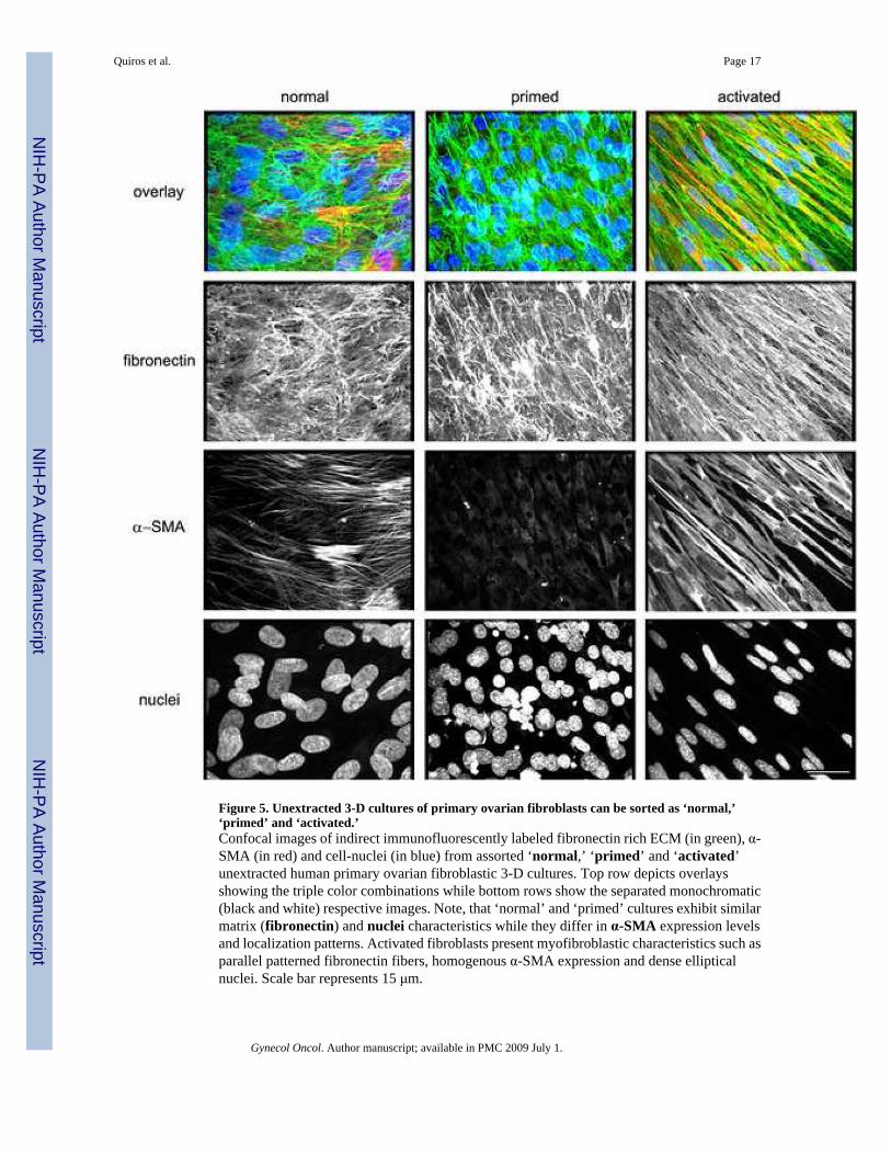
Figure 5. Unextracted 3-D cultures of primary ovarian fibroblasts can be sorted as ‘normal,’‘primed’ and ‘activated.’Confocal images of indirect immunofluorescently labeled fibronectin rich ECM (in green), α-SMA (in red) and cell-nuclei (in blue) from assorted ‘normal,’ ‘primed’ and ‘activated’unextracted human primary ovarian fibroblastic 3-D cultures. Top row depicts overlaysshowing the triple color combinations while bottom rows show the separated monochromatic(black and white) respective images. Note, that ‘normal’ and ‘primed’ cultures exhibit similarmatrix (fibronectin) and nuclei characteristics while they differ in α-SMA expression levelsand localization patterns. Activated fibroblasts present myofibroblastic characteristics such asparallel patterned fibronectin fibers, homogenous α-SMA expression and dense ellipticalnuclei. Scale bar represents 15 μm.
Quiros et al. Page 17
Gynecol Oncol. Author manuscript; available in PMC 2009 July 1.
NIH
-PA Author Manuscript
NIH
-PA Author Manuscript
NIH
-PA Author Manuscript

Figure 6. Biochemical characteristics of primary ovarian fibroblasts sorted by stromal stages‘normal,’ ‘primed’ and ‘activated.’Cell lysates from 2-D (white bars) and unextracted 3-D cultures (shaded bars) were subjectedto Western blot analyses (see Materials and Methods). Levels of total (tFAK) and activatedpFAKY397 (pFAK), as well as α-SMA expression normalized to GAPDH, were calculated byfluorescent intensity measurements using the Licor Odyssey apparatus and its analysissoftware. Graphs depict specific FAK activities (left panel p/t FAK) and α-SMA (right panel)expression levels of primary ovarian fibroblasts comparing cells sorted, by immunofluorescentanalyses of the 3-D cultures (see Figure 5) as ‘normal’, ‘primed’ or ‘activated.’ Note thatthis biochemical analyses clearly separate ‘primed’ from both ‘normal’ and ‘activated’fibroblasts.
Quiros et al. Page 18
Gynecol Oncol. Author manuscript; available in PMC 2009 July 1.
NIH
-PA Author Manuscript
NIH
-PA Author Manuscript
NIH
-PA Author Manuscript

Figure 7. Ovarian ‘normal,’ ‘primed’ and ‘activated’ phenotypes in vivo are reminiscent of invitro unextracted 3-D culturesImmunohistochemistry showing activated FAK (pFAKY397) and α-SMA levels,counterstained with Harris hematoxylin nucleic stain, of representative cancerous (A) andnormal (B) human ovarian tissues. Normal epithelium (E) and tumor areas (T), as well asnormal (N), primed (P) and activated (Ac) stroma are marked. Note the correlation ofpFAKY397 and α-SMA in normal and tumor-associated (e.g., activated) stroma in comparisonto primed stromal tissue. Scale bars represent, 50 μm.
Quiros et al. Page 19
Gynecol Oncol. Author manuscript; available in PMC 2009 July 1.
NIH
-PA Author Manuscript
NIH
-PA Author Manuscript
NIH
-PA Author Manuscript

NIH
-PA Author Manuscript
NIH
-PA Author Manuscript
NIH
-PA Author Manuscript
Quiros et al. Page 20Ta
ble
1Li
st o
f hum
an o
varia
n tis
sue
sam
ples
and
thei
r har
vest
ed c
ell l
ines
.
Sam
ple
His
topa
thol
ogy
BR
CA
1B
RC
A2
mut
atio
nst
atus
Tot
allin
esyi
elde
d
Mor
phol
ogy
in 2
D c
ultu
res
spre
adin
term
edia
tesp
indl
e
1En
dom
etrio
id a
deno
carc
inom
aN
D3
00
32
Sex-
cord
stro
mal
tum
orN
D13
121
03
Sero
us a
deno
carc
inom
aN
D9
90
04
Muc
inou
s cys
t-ade
noca
rcin
oma
ND
33
00
5C
lear
cel
l ade
noca
rcin
oma
ND
11
00
6N
orm
al o
vary
11
10
07
Nor
mal
ova
ry1
11
00
8N
orm
al o
vary
11
10
09
Nor
mal
ova
ryW
T1
10
010
Nor
mal
ova
ryW
T1
10
011
Nor
mal
ova
ryW
T1
10
012
Nor
mal
ova
ry1
11
00
13N
orm
al o
vary
21
10
014
Nor
mal
ova
ry2
11
00
15N
orm
al o
vary
11
10
016
Nor
mal
ova
ryW
T1
10
017
Nor
mal
ova
ryW
T1
10
018
Nor
mal
ova
ry2
11
00
19N
orm
al o
vary
11
10
020
Nor
mal
ova
ryW
T1
10
0Fr
esh
hum
an o
varia
n sa
mpl
es w
ere
obta
ined
and
num
bere
d as
show
n in
the
Tabl
e. M
utat
ion
stat
uses
and
his
topa
thol
ogy
info
rmat
ion
for t
he sa
mpl
es is
pro
vide
d. T
otal
num
ber o
f har
vest
ed c
ell
lines
per
sam
ple
is li
sted
und
er ‘l
ines
yie
lded
’. Fu
rther
sorti
ng a
ccor
ding
to th
eir m
orph
olog
y in
2-D
cul
ture
s is r
epor
ted
unde
r ‘sp
read
,’ ‘in
term
edia
te’ o
r ‘sp
indl
e’ c
ateg
orie
s. (W
T), p
atie
nt te
sted
nega
tive
for m
utat
ions
in b
oth
BR
CA
1 an
d B
RC
A2,
(1),
BR
CA
1 ge
rmlin
e m
utat
ion
carr
iers
, (2)
, BR
CA
2 ge
rmlin
e m
utat
ion
carr
iers
, and
(ND
), no
t det
erm
ined
. Not
e th
at w
hile
all
norm
al o
varia
nsa
mpl
es (6
-20)
yie
lded
spre
ad m
orph
olog
ies,
tum
or sa
mpl
es (1
-5) y
ield
ed m
ore
than
one
mor
phol
ogic
cel
l typ
e, a
nd sa
mpl
e ‘2
’ yie
lded
the
only
cel
l lin
e w
ith in
term
edia
te m
orph
olog
y. C
ultu
res
iden
tifie
d as
fibr
obla
stic
cel
ls fr
om sa
mpl
es 1
-3 a
nd 5
-20
and
thei
r der
ived
3-D
mat
rices
wer
e su
bmitt
ed to
furth
er a
naly
ses.
Gynecol Oncol. Author manuscript; available in PMC 2009 July 1.

NIH
-PA Author Manuscript
NIH
-PA Author Manuscript
NIH
-PA Author Manuscript
Quiros et al. Page 21Ta
ble
2FA
K sp
ecifi
c ac
tivity
and
α-S
MA
exp
ress
ion
in 2
-D v
s. un
extra
cted
3-D
cul
ture
sSa
mpl
ep/
t FA
K (a
.u)
α-SM
A (a
.u)
mor
phol
ogy
stro
mal
stag
e2-
D3-
D2-
D3-
D1a
3.51
0688
3.66
4565
4.76
8442
2.72
9572
spin
dle
activ
ated
1b2.
4930
752.
9239
324.
9913
084.
1433
42sp
indl
eac
tivat
ed2a
1.99
1342
1.67
0815
0.30
2325
0.00
7041
inte
rmed
iate
prim
ed2b
2.65
5383
3.87
4818
13.1
047
6.31
0094
spre
adac
tivat
ed3a
1.23
5808
1.58
3466
1.38
9722
6.55
9197
spre
adac
tivat
ed3b
1.04
0733
1.60
9456
2.09
4714
5.19
7083
spre
adac
tivat
ed6
1.00
5451
1.53
3515
1.74
9649
4.15
7934
spre
adno
rmal
71.
1674
31.
2340
64.
8721
038.
7632
13sp
read
norm
al8
0.81
2585
1.18
693
4.54
5376
5.86
3946
spre
adno
rmal
91.
5916
672.
4171
522.
9191
577.
6923
73sp
read
norm
al10
1.76
9155
1.77
4064
3.77
0505
5.06
9575
spre
adno
rmal
110.
9042
861.
1479
193.
7506
123.
9718
65sp
read
norm
al12
2.02
8826
0.76
9688
3.95
4337
3.14
0526
spre
adno
rmal
130.
8355
711.
2632
546.
3981
264.
5742
51sp
read
norm
al14
1.21
7967
1.79
0383
4.35
9564
5.31
6091
spre
adno
rmal
151.
0401
91.
1326
963.
1447
396.
8113
27sp
read
norm
al17
0.88
7134
1.34
5262
4.73
4823
7.24
62sp
read
norm
al18
0.81
8529
1.07
0927
3.23
6699
3.86
2374
spre
adno
rmal
190.
8542
952.
0089
293.
6455
093.
8600
75sp
read
norm
al20
1.83
1502
2.31
746
23.7
383
12.9
0775
spre
adno
rmal
Cel
l lys
ates
from
2-D
and
une
xtra
cted
3-D
cul
ture
s wer
e su
bjec
ted
to W
este
rn b
lot a
naly
ses (
see
Mat
eria
ls a
nd M
etho
ds).
Spec
ific
FAK
act
iviti
es (p
/t FA
K) a
nd le
vels
of α
-SM
A/G
PAD
H e
xpre
ssio
nw
ere
obta
ined
by
fluor
esce
nt in
tens
ity m
easu
rem
ents
usi
ng th
e Li
cor O
dyss
ey a
ppar
atus
and
its a
naly
sis s
oftw
are
(see
Mat
eria
ls a
nd M
etho
ds).
Sam
ples
wer
e no
rmal
ized
, arb
itrar
y un
it=1
(a.u
), to
the
activ
ities
and
leve
ls o
btai
ned
from
con
trol 2
-D fi
brob
last
ic (W
I38)
cul
ture
s. Sh
aded
are
as d
epic
t cel
l lin
es h
arve
sted
from
can
cero
us a
s opp
osed
to n
orm
al o
varie
s. C
orre
spon
ding
2-D
cul
ture
dce
ll m
orph
olog
ies a
nd fi
nal s
ortin
g of
cor
resp
ondi
ng st
rom
al-s
tage
are
pro
vide
d.
Gynecol Oncol. Author manuscript; available in PMC 2009 July 1.

NIH
-PA Author Manuscript
NIH
-PA Author Manuscript
NIH
-PA Author Manuscript
Quiros et al. Page 22Ta
ble
3So
rting
cha
ract
eris
tics o
f hum
an o
varia
n st
rom
al st
ages
; ‘no
rmal
,’ ‘p
rimed
’ and
‘act
ivat
ed.’
stro
mal
stag
esa
mpl
enu
mbe
r
Res
ults
in v
itro
in v
ivo
mor
phol
ogy
WB
IFIH
C
activ
ated
1asp
indl
eH
ighe
st F
AK
expr
essi
on. F
AK
activ
ity h
ighe
r in
3-D
.Lo
wer
than
‘nor
mal
’α-
SMA
yet
sim
ilar i
n2-
D a
nd 3
-D.
Myo
fibro
blas
ticel
liptic
al n
ucle
i and
hom
ogen
ous s
tress
fiber
-like
α-S
MA
expr
essi
onor
gani
zed
insi
gnat
ure
mat
chin
gpa
ralle
l EC
Mfib
rous
pat
tern
s.
α-SM
A a
nd a
ctiv
eFA
K in
desm
opla
stic
stro
ma
show
ing
typi
cal
myo
fibro
blas
ticst
ring-
like
feat
ures
.
1bsp
indl
e2b
spre
ad3a
spre
ad
3bsp
read
prim
ed2a
inte
rmed
iate
FAK
act
ivity
low
in3-
D. L
owes
t α-S
MA
;de
crea
sing
eve
nm
ore
in 3
-D.
Rou
nd n
ucle
i, lo
wα-
SMA
and
diso
rgan
ized
EC
M.
Low
α-S
MA
corr
elat
es w
ith lo
wac
tive
FAK
inst
rom
a.
norm
al6-
20sp
read
Low
est F
AK
with
high
er a
ctiv
ity in
3-D
.H
ighe
st α
-SM
A w
ith3-
D in
crea
se.
Rou
nd n
ucle
i,he
tero
gene
ous
stre
ss fi
ber-
like α-
SMA
and
diso
rgan
ized
EC
M.
Nor
mal
post
men
opau
sal
stro
ma
expr
esse
sα-
SMA
and
som
eFA
K a
ctiv
ity.
Sum
mar
y of
obs
erva
tions
des
crib
ing
thre
e st
rom
al st
ages
(act
ivat
ed, p
rim
ed a
nd n
orm
al).
The
tabl
e lis
ts th
e as
sorte
d ce
ll nu
mbe
r des
igna
tions
(sam
ple
num
ber)
, the
ir ob
serv
ed in
vitr
o 2-
Dm
orph
olog
y, W
este
rn b
lot a
naly
ses (
WB
) of 2
-D a
nd 3
-D c
ultu
res,
indi
rect
imm
unof
luor
esce
nce
(IF)
of u
next
ract
ed 3
-D c
ultu
res a
nd c
orre
spon
ding
in v
ivo
char
acte
ristic
s of i
mm
unoh
isto
chem
ical
stai
ning
(IH
C) o
f rep
rese
ntat
ive
hum
an o
varia
n ca
ncer
ous a
nd n
orm
al ti
ssue
s.
Gynecol Oncol. Author manuscript; available in PMC 2009 July 1.
Copyright © 2022 FDOKUMEN