Importin-α3 Is Required at Multiple Stages of Drosophila Development and Has a Role in the...
16
Importin-a3 Is Required at Multiple Stages of Drosophila Development and Has a Role in the Completion of Oogenesis Endre Ma ´ the ´,* ,1 Helen Bates,² ,1 Hella Huikeshoven,² Pe ´ter Dea ´ k,* David M. Glover,* and Sue Cotterill² ,2 *Cell Cycle Genetics Group, Department of Genetics, University of Cambridge, Cambridge CB2 3EH, United Kingdom; and ²Department of Biochemistry and Immunology, St. Georges Hospital Medical School, Cranmer Terrace, London SW17 0RE, United Kingdom The Drosophila importin-a3 gene was isolated through its interaction with the large subunit of the DNA polymerase a in a two-hybrid screen. The predicted protein sequence of Importin-a3 is 65– 66% identical to those of the human and mouse importin-a3 and a4 and 42.7% identical to that of Importin-a2 (Oho31/Pendulin), the previously reported Drosophila homologue. Both Importin-a3 and Importin-a2 interact with similar subsets of proteins in vitro, one of which is Ketel, the importin-b homologue of Drosophila. importin-a3 is an essential gene, whose encoded protein is expressed throughout development. During early embryogenesis, Importin-a3 accumulates at the nuclear membrane of cleavage nuclei, whereas after blastoderm formation it is characteristically found within the interphase nuclei. Nuclear localisation is seen in several tissues throughout subsequent development. During oogenesis its concentration within the nurse cell nuclei increases during stages 7–10, concomitant with a decline in levels in the oocyte nucleus. Mutation of importin-a3 results in lethality throughout pupal development. Surviving females are sterile and show arrest of oogenesis at stages 7–10. Thus, Importin-a3-mediated nuclear transport is essential for completion of oogenesis and becomes limiting during pupal development. Since they have different expression patterns and subcellular localisation profiles, we suggest that the two importin-a homologues are not redundant in the context of normal Drosophila development. © 2000 Academic Press Key Words: Importin-a; Karyopherin-a; nuclear transport; oogenesis; embryogenesis; Drosophila. INTRODUCTION Nucleocytoplasmic transport is a complex phenomenon, ensuring not only nuclear import of a variety of different proteins, but also the export of other proteins, tRNA, rRNA, mRNA, and ribosomal subunits into the cytoplasm (for re- view see Ullman et al., 1997; Go ¨ rlich, 1998; Mattaj and Englmeier, 1998; Stutz and Rosbach, 1998; Stochaj and Rother, 1999). The best-characterised nucleocytoplasmic transport mechanism is the nuclear localisation signal (NLS)- dependent nuclear protein import by means of an importin- a/b heterodimer. The basic-amino-acid-rich NLS plays a de- terminative role in the substrate specificity of this process (Koepp and Silver, 1996; Go ¨ rlich, 1997; Dingwall and Laskey, 1998). Most of our knowledge of this system arises from in vitro nuclear import reconstitution experiments that make use of selectively permeabilized mammalian cells (Nigg, 1997; Go ¨ rlich, 1998). These studies suggest that importin-a binds through its Armadillo (Arm) motifs to the NLS protein (Adam and Adam, 1994; Conti et al., 1998) and through its importin-b binding domain (IBB) to importin-b (Moroianu et al., 1995a; Go ¨ rlich et al., 1996; Weis et al., 1996). Importin-b interacts with the nuclear pore complex (NPC) and the NLS protein/importin-a/importin-b trimeric complex is translo- cated into the nucleus in an energy-dependent manner (Go ¨ r- lich et al., 1995; Moroianu et al., 1995b). Inside the nucleus, in an environment with high RanGTP concentration, the direct binding of RanGTP to importin-b terminates the transloca- tion of the trimeric complex, which then disassembles (Rex- ach and Blobel, 1995; Chi et al., 1996; Go ¨ rlich et al., 1996). Consequently, the NLS protein is released from importin-a. Importin-b is probably exported to the cytoplasm as a complex 1 Both authors contributed equally to the paper. 2 To whom correspondence should be addressed. Fax: 44 208 725 3549. E-mail: [email protected]. Developmental Biology 223, 307–322 (2000) doi:10.1006/dbio.2000.9743, available online at http://www.idealibrary.com on 0012-1606/00 $35.00 Copyright © 2000 by Academic Press All rights of reproduction in any form reserved. 307
-
Upload
independent -
Category
Documents
-
view
4 -
download
0
Transcript of Importin-α3 Is Required at Multiple Stages of Drosophila Development and Has a Role in the...
Di
a
h
atdtIdi
Developmental Biology 223, 307–322 (2000)doi:10.1006/dbio.2000.9743, available online at http://www.idealibrary.com on
Importin-a3 Is Required at Multiple Stages ofrosophila Development and Has a Role
n the Completion of Oogenesis
Endre Mathe,*,1 Helen Bates,†,1 Hella Huikeshoven,† Peter Deak,*David M. Glover,* and Sue Cotterill†,2
*Cell Cycle Genetics Group, Department of Genetics, University of Cambridge, CambridgeCB2 3EH, United Kingdom; and †Department of Biochemistry and Immunology, St. GeorgesHospital Medical School, Cranmer Terrace, London SW17 0RE, United Kingdom
The Drosophila importin-a3 gene was isolated through its interaction with the large subunit of the DNA polymerase a intwo-hybrid screen. The predicted protein sequence of Importin-a3 is 65–66% identical to those of the human and mouse
importin-a3 and a4 and 42.7% identical to that of Importin-a2 (Oho31/Pendulin), the previously reported Drosophilaomologue. Both Importin-a3 and Importin-a2 interact with similar subsets of proteins in vitro, one of which is Ketel, the
importin-b homologue of Drosophila. importin-a3 is an essential gene, whose encoded protein is expressed throughoutdevelopment. During early embryogenesis, Importin-a3 accumulates at the nuclear membrane of cleavage nuclei, whereasfter blastoderm formation it is characteristically found within the interphase nuclei. Nuclear localisation is seen in severalissues throughout subsequent development. During oogenesis its concentration within the nurse cell nuclei increasesuring stages 7–10, concomitant with a decline in levels in the oocyte nucleus. Mutation of importin-a3 results in lethalityhroughout pupal development. Surviving females are sterile and show arrest of oogenesis at stages 7–10. Thus,mportin-a3-mediated nuclear transport is essential for completion of oogenesis and becomes limiting during pupalevelopment. Since they have different expression patterns and subcellular localisation profiles, we suggest that the twomportin-a homologues are not redundant in the context of normal Drosophila development. © 2000 Academic Press
Key Words: Importin-a; Karyopherin-a; nuclear transport; oogenesis; embryogenesis; Drosophila.
1vuGtaia
cl
INTRODUCTION
Nucleocytoplasmic transport is a complex phenomenon,ensuring not only nuclear import of a variety of differentproteins, but also the export of other proteins, tRNA, rRNA,mRNA, and ribosomal subunits into the cytoplasm (for re-view see Ullman et al., 1997; Gorlich, 1998; Mattaj andEnglmeier, 1998; Stutz and Rosbach, 1998; Stochaj andRother, 1999). The best-characterised nucleocytoplasmictransport mechanism is the nuclear localisation signal (NLS)-dependent nuclear protein import by means of an importin-a/b heterodimer. The basic-amino-acid-rich NLS plays a de-terminative role in the substrate specificity of this process(Koepp and Silver, 1996; Gorlich, 1997; Dingwall and Laskey,
1 Both authors contributed equally to the paper.2 To whom correspondence should be addressed. Fax: 44 208 725
3549. E-mail: [email protected].
0012-1606/00 $35.00Copyright © 2000 by Academic PressAll rights of reproduction in any form reserved.
998). Most of our knowledge of this system arises from initro nuclear import reconstitution experiments that makese of selectively permeabilized mammalian cells (Nigg, 1997;orlich, 1998). These studies suggest that importin-a binds
hrough its Armadillo (Arm) motifs to the NLS protein (Adamnd Adam, 1994; Conti et al., 1998) and through itsmportin-b binding domain (IBB) to importin-b (Moroianu etl., 1995a; Gorlich et al., 1996; Weis et al., 1996). Importin-b
interacts with the nuclear pore complex (NPC) and the NLSprotein/importin-a/importin-b trimeric complex is translo-ated into the nucleus in an energy-dependent manner (Gor-ich et al., 1995; Moroianu et al., 1995b). Inside the nucleus, in
an environment with high RanGTP concentration, the directbinding of RanGTP to importin-b terminates the transloca-tion of the trimeric complex, which then disassembles (Rex-ach and Blobel, 1995; Chi et al., 1996; Gorlich et al., 1996).Consequently, the NLS protein is released from importin-a.
Importin-b is probably exported to the cytoplasm as a complex307
Caa
R1i
ctiw
hcae1le
amMtifo
IdI
Cagp
rmc
s
l
k(ppatit
w
BnAcIr
GIU
pd
s
308 Mathe et al.
with RanGTP and subsequently released from the complex byRanBP1 and RanGAP1 (Izaurralde et al., 1997). The export ofimportin-a to the cytoplasm is mediated by the RanGTP/
AS/importin-a trimeric complex in which the CAS (cellularpoptosis susceptibility) protein interacts with NPC (Kutay etl., 1997; Herold et al., 1998). In the cytoplasm RanBP1 causes
the dissociation of RanGTP from the RanGTP/CAS/importin-a complex, and reassociation is prevented by
anGAP1-triggered GTP hydrolysis (Bischoff and Gorlich,997). The RanGTP-free CAS is now in a low-affinity form formportin-a binding and importin-a is released. In the cyto-
plasm, where the RanGTP concentration is low, theimportin-a and b will combine to reform the nuclear import-ompetent heterodimer. Despite the functional characterisa-ion of these key components, several aspects of NLS-proteinmport are still poorly understood. It is not clear for examplehether the multiple isoforms of importin a and b present in
many species have specific functions or whether they arefunctionally redundant. However, the high degree of identityobserved in the case of importin-a and b proteins in differentmodel organisms suggests the evolutionarily conserved natureof the NLS-protein import.
Drosophila genes encoding importin-a and b homologuesave already been reported. The oho31/pendulin gene en-odes an importin-a2 subfamily member, which is presentt high levels in early embryos and is rapidly degraded at thend of embryogenesis (Kussel and Frasch, 1995; Torok et al.,995). This protein shows cell cycle-dependent nuclearocalisation, accumulating in the nucleus at the prophase ofmbryonic divisions. The ketel gene encodes an importin-b
homologue required for viability throughout development(Erdelyi et al., 1997).
In this study, we report the isolation of the importin-a3(imp-a3) gene of Drosophila through its interaction withthe NLS-containing protein DNA polymerase a in a yeasttwo-hybrid screen. The predicted sequence of Importin-a3shows the characteristic IBB domain, Arm motifs, andC-terminal acidic region. Importin-a3 protein is expressedt almost constant levels throughout Drosophila develop-ent and during interphase accumulates in nuclei, while in-phase is cytoplasmic. The imp-a3 mutant exhibits le-
hality in late larval to pharate adult stages, underlining themportance of nuclear transport during development. Aemale sterile phenotype characterised by the degenerativevaries of the imp-a3 mutants also suggests a role for this
importin-a during oogenesis. The subcellular localisation ofmportin-a3 is discussed in the light of the Drosophilaevelopment and compared to the previously reportedmportin-a2.
MATERIALS AND METHODS
Fly Strains and Culture Conditions
Wild-type and mutant strains were maintained and mated onstandard yeast–agar–cornmeal medium and all experiments were
performed at 25°C. All genetic markers and mutations used are gCopyright © 2000 by Academic Press. All right
described in Lindsley and Zimm (1992). Deficiency stocksDf(3R)by10 and Df(3R)by62 were obtained from the Umea Stock
enter. The imp-a31 line was originally designated 335/13 (Deak etl., 1997). The TM3, Sb, ry, [D2-3, ry1]/Df(3R)C7, ry507 stock wasiven by Janos Gausz. All the stocks utilised were isogenised andut on a w1118 background.
Mutant Phenotype Determination
The homozygous and hemizygous imp-a31-associated mutantphenotypes were determined on a TM6b, Tb or a TM6c, Tb, Sbbackground as described by Deak et al. (1997). Individual sterilitytests were performed for homozygous and hemizygous imp-a31
females by crossing them to wild-type Canton S males.
Remobilization of the P-lacW Element
In order to revert the imp-a31 mutation the P-lacW element wasemobilised under dysgenic conditions. About 250 jump-starterales of genotype w1118/Y; imp-a31/TM3, Sb, ry, [D2-3, ry1] were
rossed individually to w1118/w1118; TM3, Sb, Ser/TM6b, Tb virgins.From their progeny the imp-a31/TM3 and imp-a31/TM6b flies werecored for w2 or modified w1 expression compared to original w1
expression level seen in the eyes of imp-a31 flies. For each jump-starter male, only one fly was selected showing the w2 or themodified w1 phenotype and for these revertant strains were estab-ished over TM6b, Tb.
Cloning and Sequencing
Yeast two-hybrid screening was carried out as described (Rudenet al., 1991; Gyuris et al., 1993; Zervos et al., 1993). Roger Brent
indly provided the yeast strain EGY48, the lacZ reporter plasmidpSH18-34), the LexA fusion control plasmid (RFHM1), and thelasmid library (Drosophila ovarian cDNA in pJG4-5–RFLY3). Thelasmid used to make the LexA fusion bait was based on pRS313nd pV44ER-LEX. The SacI–KpnI fragment of pV44ER-LEX, con-aining the galactose-inducible LexA protein and a multiple clon-ng site, was ligated into SacI–KpnI-cut pRS313 in order to changehe selectable marker from Trp to His. The Drosophila DNA
polymerase-a p180 subunit-coding region was then cloned in frameith the LexA protein.DNA sequencing was carried out using the Sequenase kit (US
iochemicals, Cleveland, OH) or was sent to Advanced Biotech-ology Centre, Charing Cross and Westminster Medical School, forBI sequencing. Nucleotide sequence analysis and amino acidomparisons were performed using Geneworks (IntelliGenetics,nc.) for the Macintosh and the BLAST programs at NIH. The 59egion of imp-a3 was amplified by PCR from the RFLY3 library
using one primer from the pJG4-5 vector, 59-GCCTC-CTACCCTTATGATGTGCCAG-39, and another, 59-GAATGGT-GGCTGTCGGGGGAGCCGGATCCTT-39, from within theimp-a3 coding region. The Drosophila cDNA clones GM01016,
M05410, and GM06753 were obtained from Genome Systems,nc., and they were sequenced in the Department of Genetics,niversity of Cambridge.The P-lacW-mediated plasmid rescue and the salivary gland
olytene chromosome in situ hybridisation were carried out asescribed by Deak et al. (1997).
Both Southern and Northern blots were carried out according totandard procedures (Sambrook et al., 1989). For Southerns,
enomic DNA was extracted from wild-type and mutant imp-a3s of reproduction in any form reserved.
u
cti
mo5ma(N
wcTftas1
ple(u
ca
waBM
i
o
O4awCr
309The Importin-a3 Gene of Drosophila
lines and then restriction digestions were performed. Poly(A)1
RNA from all developmental stages of Drosophila was purifiedsing the Oligotex mRNA kit from QIAgen.
Drosophila Germ-Line Transformation
A 9-kb genomic fragment containing the entire imp-a3 gene wasloned into pCasper4 and the resulting pR4 construct together withhe transposase encoding the pP25.7WC plasmid were injectednto w1118 flies. Six independent transgenic lines were obtained and
among them the P[imp-a31]42 homozygous viable, second-chromosome insertion line was used to rescue the imp-a3-associated mutant phenotypes. A defective construct, pRD4.1, wascreated by deleting a BsiWI–SwaI fragment (Fig. 1). Followinggerm-line transformation one P[imp-a32]411 homozygous viable,first chromosome insertion line was established.
Production of Importin-a3 Antibody
The region of imp-a3 coding for amino acids 227–514 was clonedinto pQE31 (QIAgen) and the His-tagged protein expressed andpurified according to the manufacturer’s manual. An amount of 0.8mg of protein was sent to Neosystem Laboratoire for antibodyproduction in two rabbits. The polyclonal antiserum of these tworabbits was affinity purified and used on Western blots.
Preparation of Drosophila Protein Extracts
To make cytoplasmic and nuclear extracts, dechorionated wild-type Drosophila embryos were homogenised in the presence ofprotease inhibitors, with a loose pestle to maintain the integrity ofthe nuclei. The homogenate was filtered through two layers ofMiracloth to remove cell debris and centrifuged at 25K for 20 minin a Beckman TL100. The resulting supernatant was the cytoplas-mic fraction. The nuclear pellet was washed five times in TP3buffer (10 mM Hepes, pH 7.9, 250 mM sucrose, 2.5 mM MgCl2, 50
M KCl, 1 mM DTT, protease inhibitors). Nuclei were brokenpen by adding an equal volume of Buffer A (50 mM Hepes, pH 7.9,0 mM NaCl, 10% glycerol, 0.1% Triton X-100, 1 mM EDTA) andixing at 4°C for 30 min. This was repeated to give fractions T1
nd T2. Proteins bound to nuclei were released with low-saltBuffer A 1 50 mM NaCl) followed by high-salt (Buffer A 1 2 M
aCl) extractions. The remaining pellet was sonicated.Total extracts for immunoprecipitations and GST pull-downs
ere prepared in a similar manner except that homogenisation wasarried out in IP buffer (10 mM Tris–HCl, pH 7.5, 50 mM KCl, 0.1%ween 20, protease inhibitors) at 1 g/ml embryos, with a loose
ollowed by a tight pestle to break open nuclei. After filtrationhrough Miracloth, nuclear debris was removed by centrifugationt 4°C at low speed (6.5 K) in a bench-top microcentrifuge. Theupernatant was either used immediately or mixed with glycerol at0% and frozen as droplets in liquid nitrogen.
Developmental Western Blot Analysis
Total protein extracts corresponding to the different develop-mental stages of Drosophila were made into standard 23 SDSrotein loading buffer. Equal amounts of protein samples wereoaded onto 10% polyacrylamide gels and after electrophoresislectroblotted onto Hybond ECL (Amersham) or Immobilon-PMillipore) membranes. a-Tubulin was used as a loading standard
sing monoclonal a-tubulin antibody (Amersham). The wCopyright © 2000 by Academic Press. All right
peroxidase-labelled anti-rabbit and anti-mouse secondary antibod-ies were purchased from Amersham or Jackson ImmunoresearchLaboratories, Inc. The blots were developed using ECL kit (Amer-sham) or SuperSignal Substrate Western Blotting (Pierce). Quanti-tation of ECL blots was carried out by comparing the sample totitrated amounts of overproduced protein on the same blot. Totalovarian protein extracts were prepared from 15 either wild-type orimp-a31/imp-a31 females. Equal volumes of ovarian proteinsamples were analysed on Western blots as described above.
Western Analysis of Drosophila Cell Extracts
Full-length open reading frames of imp-a3 and oho31 wereloned in frame into pGEX2TK. Proteins were induced, purified,nd labelled with [g-32P]ATP according to manufacturer instruc-
tions. Total Drosophila extracts (cytoplasmic, high-salt nuclearash, and nuclear pellet) were electrophoresed on 12% SDS–PAGE
nd blotted onto nitrocellulose. Blots were incubated overnight inB buffer (20 mM Hepes, pH 7, 10% glycerol, 50 mM KCl, 10 mMgCl2, 1 mM DTT, 0.1% Tween 20) plus 5% milk powder at 4°C
to block and also to allow the proteins to renature. Probes wereincubated with the renatured blots for 1 h at 4°C, washed five timeswith BB buffer, and exposed to film.
GST Pull-downs
One millilitre of 0- to 5-h embryo extracts (see above) wasincubated at 4°C for 1 h with an equal amount of GST orGST–Importin-a1 or GST–Importin-a3 fusion protein bound toglutathione agarose (Pharmacia). The beads were then washedextensively in IP buffer before elution of bound proteins with IPbuffer containing 100, 200, and 500 mM NaCl. Proteins eluted inhigh salt were directly visualised by silver staining.
Immunoprecipitations
Antibodies were crosslinked to protein A–Sepharose beads usingdimethylpimelimidate. An amount of 40 ml crosslinked beads wasncubated for 1 h at 4°C with 200 ml Drosophila 0- to 4-h
whole-embryo extracts. Beads were then washed 10 times with IPbuffer plus 100 mM NaCl (1 ml per wash) and resuspended directlyinto 40 ml 23 SDS–PAGE loading buffer.
Immunocytochemistry and Confocal Microscopy
Cytological preparations of third-instar larval brains were madeaccording to Sunkel and Glover (1988). For each genotype 10microscopic fields per brain from 10 brains were analysed, usingthe phase-contrast Neofluar 603 oil Nikon objective, the 103culars, and the Optovar set at 1.253.
Ovaries were dissected in 23 PBT (137 mM NaCl, 2.7 mM KCl,10 mM Na2HPO4, 1.8 mM K2HPO4, 0.2% Triton X-100, pH 7.5) andfixed with 5% formaldehyde or paraformaldehyde in PBS for 20min. A second fixation was carried out in absolute methanol for 30min and then the samples were rehydrated in 23 PBT for 1 h.
vernight incubations were done with the primary antibodies at°C and the incubations in the secondary antibodies lasted for 4 ht room temperature. The fixation and immunostaining of embryosere performed according to Mathe et al. (1998) or Maldonado-odina and Glover (1992). The Importin-a2 was detected with the
abbit anti-Pendulin antibody. The microtubules were detected
ith YL1/2 rat monoclonal anti-a-tubulin antibody (Sera Lab, Inc.),s of reproduction in any form reserved.
hsp1Is
e
yNtfswdtaircnlA
cctA(ttfifrttIs
cnTpsmi
va
sIt
fft
IApcG
t
310 Mathe et al.
while the T47 mouse monoclonal anti-lamin antibody was used tovisualise the nuclear lamina (Paddy et al., 1990). The Alexa 488anti-rat or anti-mouse and Texas red anti-rabbit secondary antibod-ies were obtained from Jackson Immunoresearch Laboratories, Inc.Digital images of optical sections were collected with a Bio-Rad1024 confocal microscope.
RESULTS
Structural Characteristics of the Importin-a3Protein
Two proteins showing significant homology to theimportin-a protein family were identified by a yeast two-
ybrid screen as proteins that interact with the largeubunit of DNA polymerase a. One of these had beenreviously reported as Oho31/Pendulin (Kussel and Frasch,995; Torok et al., 1995), which we will refer to asmportin-a2 based on its homology to the importin-a2ubfamily. The second is a new importin-a homologue of
Drosophila, which we named Importin-a3, similarly basedon its homology to the importin-a3 protein subfamily—seebelow. Both of these also showed interactions with DNApolymerase a in immunoprecipitates from crude yeastxtracts (data not shown).The original cDNA clone of imp-a3 isolated from the
east two-hybrid screen was found to be truncated at the-terminus, starting 622 bases into the coding region. We
herefore used PCR to isolate the upstream coding regionrom the same cDNA library used for the initial two-hybridcreen. A clone of 2.2 kb containing the full-length cDNAas then constructed by splicing together the upstream andownstream regions by means of a unique BamHI site inhe primer region (Fig. 1). The EMBL Database Libraryccession number of the imp-a3 cDNA and promoter regions AJ237997. BLAST searches with this cDNA sequenceevealed three Drosophila ESTs, and the correspondingDNA clones GM01016, GM05410, and GM06753 (Ge-ome Systems, Inc.) were actually identical to our full-ength cDNA (GenBank Accession Nos. AF230871,F230872, and AF230873).Analysis of the nucleotide sequence of the 2.2-kb cDNA
lone showed that there are two possible start codons verylose together, but only the first is immediately preceded byhe Drosophila translation start consensus sequenceCAAA (Cavener, 1987). Moreover, according to Kozak
1995), when there are two ATG start codons very closeogether the first codon is usually the one at which initia-ion occurs. We therefore chose to assign the open readingrame from the first ATG to the TAA stop codons such thatt encoded a protein of 514 amino acids. The open readingrame is followed by a 524-nucleotide-long untranslatedegion containing several close, but not identical, matcheso the AATAAAA poly(A)-addition signal before the start ofhe poly(A) tract. The calculated molecular mass ofmportin-a3 is 57 kDa, which is in agreement with our
ubsequent Western blot analysis (Fig. 4). The most highly iCopyright © 2000 by Academic Press. All right
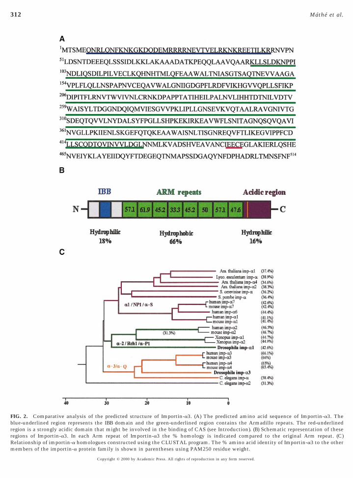
onserved central region of the Importin-a3 is predomi-antly hydrophobic and is composed of eight Arm motifs.he N- and C-termini are both hydrophilic and containreviously characterised motifs. The N-terminus contains atretch of amino acids (residues 6–45) which is an absoluteatch for the IBB domain, shown in the case of other
mportin-a homologues to be essential for importin-b bind-ing and nuclear import (Weis et al., 1996; Gorlich et al.,1996; Conti et al., 1998) (Figs. 2A and 2B). The C-terminushas a strongly acidic amino acid stretch (residues 430–514),which was shown for the human importin-a2 to be in-olved in CAS binding (Herold et al., 1998). Multiplelignment analysis of Importin-a3 with other members of
the importin-a protein family revealed that its closestrelatives (65–66% identity) are the mouse and humanimportin-a3 and a4 homologues (the mouse homologueswere also described as importin a-Q1 and a-Q2; Tsuji et al.,1997) (Fig. 2C). This is a higher homology than that ob-served between Importin-a3 and the other reported Dro-ophila homologue Importin-a2 (42.7%). The Drosophilamportin-a2 is in turn more closely related to members ofhe importin-a2/Rch1/a-P1 subfamily from other species.
Importin-a3 and Importin-a2 Interact with aSimilar Subset of Proteins in Vitro
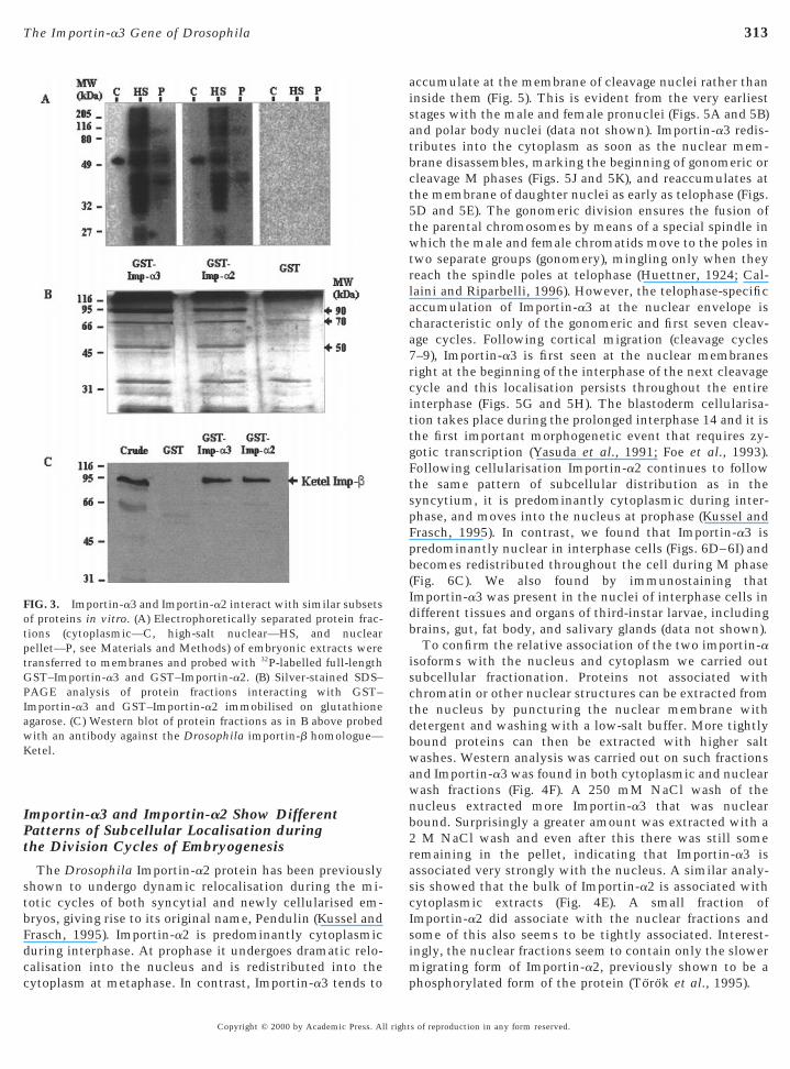
To compare proteins interacting with Importin-a3 andImportin-a2 we prepared nuclear and cytoplasmic proteinractions of a Drosophila embryonic extract. The proteinractions were separated by electrophoresis, blotted, andhen probed with 32P-labelled full-length GST fusion pro-
teins (Fig. 3A). Importin-a3 and Importin-a2 recognise simi-lar patterns of interacting proteins in all types of extract,whereas GST alone showed no interactions. A greaternumber of interacting proteins were detected in the high-salt nuclear wash fraction compared with the pellet and thecytoplasmic fraction, which showed only one protein ofapproximately 50 kDa. Since some proteins may not rena-ture correctly during the electroblotting procedure we alsolooked for proteins within embryonic extracts that wouldinteract directly with GST–Importin-a2 or GST–mportin-a3 immobilised on glutathione beads (Fig. 3B).fter the beads were washed with 200 mM NaCl, the boundroteins were eluted with 500 mM NaCl. From Fig. 3B itan be again seen that similar sets of proteins interact withST–Importin-a2 and GST–Importin-a3, but not with the
control GST alone, the most prominent being of 90, 70, and50 kDa. The 90-kDa band corresponds to Ketel, the Dro-sophila homologue of the importin-b protein, as shown byWestern blotting of the eluted proteins with antibodiesagainst the Ketel protein (a kind gift from Janos Szabad).The identity of the other two bands has not been deter-mined.
Immunoprecipitations from 0- to 5-h embryonic extractsusing anti-Importin-a3 antibodies also revealed interac-ions with proteins of 90 and 70 kDa (data not shown). The
nteraction of the 50-kDa protein could not, however, bes of reproduction in any form reserved.
ice
oe
mrIttwors
T
.
311The Importin-a3 Gene of Drosophila
confirmed by this method due to the masking of this regionof the gel by bands from the antibody. Although we cannotrole out differences in the strengths of the interaction foreach protein, it seems that Importin-a2 and Importin-a3nteract with similar sets of proteins in vitro that mightorrespond to other members of the NLS-transport machin-ry yet unidentified in Drosophila.
Importin-a3 Is Present throughout Development
Probes from either the full-length cDNA or thegenomic region of imp-a3 detect a single transcript of 2.2kb that was present in poly(A)1 RNA throughout devel-
pment, being particularly abundant in female flies and
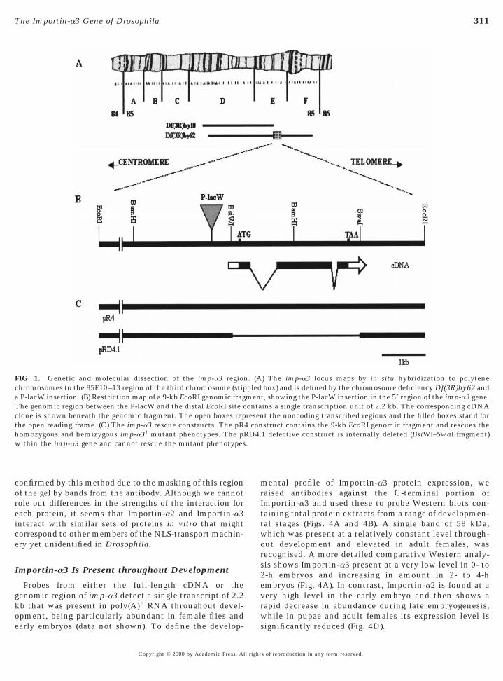
FIG. 1. Genetic and molecular dissection of the imp-a3 regionchromosomes to the 85E10–13 region of the third chromosome (stipa P-lacW insertion. (B) Restriction map of a 9-kb EcoRI genomic frag
he genomic region between the P-lacW and the distal EcoRI site cclone is shown beneath the genomic fragment. The open boxes repthe open reading frame. (C) The imp-a3 rescue constructs. The pRhomozygous and hemizygous imp-a31 mutant phenotypes. The pRwithin the imp-a3 gene and cannot rescue the mutant phenotypes
arly embryos (data not shown). To define the develop-
Copyright © 2000 by Academic Press. All right
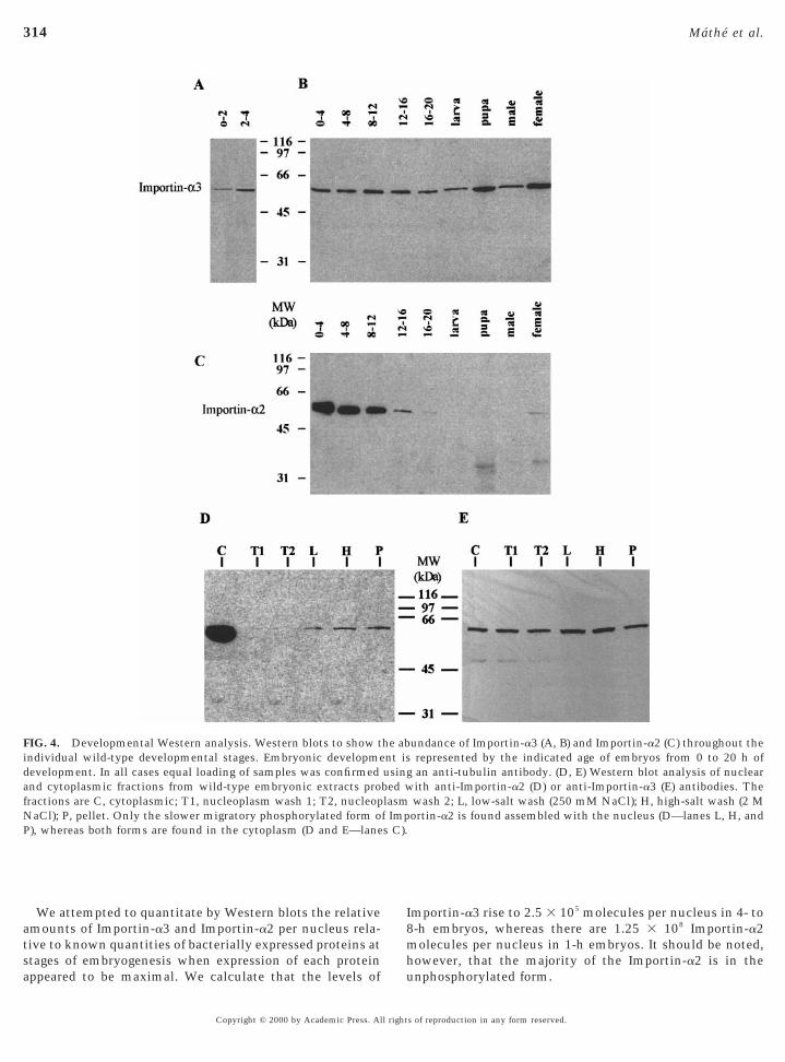
ental profile of Importin-a3 protein expression, weaised antibodies against the C-terminal portion ofmportin-a3 and used these to probe Western blots con-aining total protein extracts from a range of developmen-al stages (Figs. 4A and 4B). A single band of 58 kDa,hich was present at a relatively constant level through-ut development and elevated in adult females, wasecognised. A more detailed comparative Western analy-is shows Importin-a3 present at a very low level in 0- to
2-h embryos and increasing in amount in 2- to 4-hembryos (Fig. 4A). In contrast, Importin-a2 is found at avery high level in the early embryo and then shows arapid decrease in abundance during late embryogenesis,while in pupae and adult females its expression level is
) The imp-a3 locus maps by in situ hybridization to polytenebox) and is defined by the chromosome deficiency Df(3R)by62 and
t, showing the P-lacW insertion in the 59 region of the imp-a3 gene.ins a single transcription unit of 2.2 kb. The corresponding cDNAt the noncoding transcribed regions and the filled boxes stand forstruct contains the 9-kb EcoRI genomic fragment and rescues the1 defective construct is internally deleted (BsiWI–SwaI fragment)
. (Apledmenontaresen4 con
D4.
significantly reduced (Fig. 4D).
s of reproduction in any form reserved.
312 Mathe et al.
FIG. 2. Comparative analysis of the predicted structure of Importin-a3. (A) The predicted amino acid sequence of Importin-a3. Theblue-underlined region represents the IBB domain and the green-underlined region contains the Armadillo repeats. The red-underlinedregion is a strongly acidic domain that might be involved in the binding of CAS (see Introduction). (B) Schematic representation of theseregions of Importin-a3. In each Arm repeat of Importin-a3 the % homology is indicated compared to the original Arm repeat. (C)Relationship of importin-a homologues constructed using the CLUSTAL program. The % amino acid identity of Importin-a3 to the other
members of the importin-a protein family is shown in parentheses using PAM250 residue weight.Copyright © 2000 by Academic Press. All rights of reproduction in any form reserved.
aisatbct5twtrlaca7
db
b2ras
sim
GPIawK
313The Importin-a3 Gene of Drosophila
Importin-a3 and Importin-a2 Show DifferentPatterns of Subcellular Localisation duringthe Division Cycles of Embryogenesis
The Drosophila Importin-a2 protein has been previouslyshown to undergo dynamic relocalisation during the mi-totic cycles of both syncytial and newly cellularised em-bryos, giving rise to its original name, Pendulin (Kussel andFrasch, 1995). Importin-a2 is predominantly cytoplasmicduring interphase. At prophase it undergoes dramatic relo-calisation into the nucleus and is redistributed into the
FIG. 3. Importin-a3 and Importin-a2 interact with similar subsetsof proteins in vitro. (A) Electrophoretically separated protein frac-tions (cytoplasmic—C, high-salt nuclear—HS, and nuclearpellet—P, see Materials and Methods) of embryonic extracts weretransferred to membranes and probed with 32P-labelled full-length
ST–Importin-a3 and GST–Importin-a2. (B) Silver-stained SDS–AGE analysis of protein fractions interacting with GST–mportin-a3 and GST–Importin-a2 immobilised on glutathionegarose. (C) Western blot of protein fractions as in B above probedith an antibody against the Drosophila importin-b homologue—etel.
cytoplasm at metaphase. In contrast, Importin-a3 tends to
Copyright © 2000 by Academic Press. All right
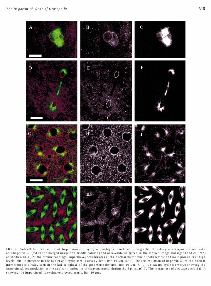
ccumulate at the membrane of cleavage nuclei rather thannside them (Fig. 5). This is evident from the very earliesttages with the male and female pronuclei (Figs. 5A and 5B)nd polar body nuclei (data not shown). Importin-a3 redis-ributes into the cytoplasm as soon as the nuclear mem-rane disassembles, marking the beginning of gonomeric orleavage M phases (Figs. 5J and 5K), and reaccumulates athe membrane of daughter nuclei as early as telophase (Figs.D and 5E). The gonomeric division ensures the fusion ofhe parental chromosomes by means of a special spindle inhich the male and female chromatids move to the poles in
wo separate groups (gonomery), mingling only when theyeach the spindle poles at telophase (Huettner, 1924; Cal-aini and Riparbelli, 1996). However, the telophase-specificccumulation of Importin-a3 at the nuclear envelope isharacteristic only of the gonomeric and first seven cleav-ge cycles. Following cortical migration (cleavage cycles–9), Importin-a3 is first seen at the nuclear membranes
right at the beginning of the interphase of the next cleavagecycle and this localisation persists throughout the entireinterphase (Figs. 5G and 5H). The blastoderm cellularisa-tion takes place during the prolonged interphase 14 and it isthe first important morphogenetic event that requires zy-gotic transcription (Yasuda et al., 1991; Foe et al., 1993).Following cellularisation Importin-a2 continues to followthe same pattern of subcellular distribution as in thesyncytium, it is predominantly cytoplasmic during inter-phase, and moves into the nucleus at prophase (Kussel andFrasch, 1995). In contrast, we found that Importin-a3 ispredominantly nuclear in interphase cells (Figs. 6D–6I) andbecomes redistributed throughout the cell during M phase(Fig. 6C). We also found by immunostaining thatImportin-a3 was present in the nuclei of interphase cells inifferent tissues and organs of third-instar larvae, includingrains, gut, fat body, and salivary glands (data not shown).To confirm the relative association of the two importin-a
isoforms with the nucleus and cytoplasm we carried outsubcellular fractionation. Proteins not associated withchromatin or other nuclear structures can be extracted fromthe nucleus by puncturing the nuclear membrane withdetergent and washing with a low-salt buffer. More tightlybound proteins can then be extracted with higher saltwashes. Western analysis was carried out on such fractionsand Importin-a3 was found in both cytoplasmic and nuclearwash fractions (Fig. 4F). A 250 mM NaCl wash of thenucleus extracted more Importin-a3 that was nuclearound. Surprisingly a greater amount was extracted with aM NaCl wash and even after this there was still some
emaining in the pellet, indicating that Importin-a3 isssociated very strongly with the nucleus. A similar analy-is showed that the bulk of Importin-a2 is associated with
cytoplasmic extracts (Fig. 4E). A small fraction ofImportin-a2 did associate with the nuclear fractions andome of this also seems to be tightly associated. Interest-ngly, the nuclear fractions seem to contain only the slower
igrating form of Importin-a2, previously shown to be a
phosphorylated form of the protein (Torok et al., 1995).s of reproduction in any form reserved.
I
314 Mathe et al.
We attempted to quantitate by Western blots the relativeamounts of Importin-a3 and Importin-a2 per nucleus rela-tive to known quantities of bacterially expressed proteins atstages of embryogenesis when expression of each protein
FIG. 4. Developmental Western analysis. Western blots to show tindividual wild-type developmental stages. Embryonic developmedevelopment. In all cases equal loading of samples was confirmedand cytoplasmic fractions from wild-type embryonic extracts profractions are C, cytoplasmic; T1, nucleoplasm wash 1; T2, nucleopNaCl); P, pellet. Only the slower migratory phosphorylated form oP), whereas both forms are found in the cytoplasm (D and E—lane
appeared to be maximal. We calculate that the levels of u
Copyright © 2000 by Academic Press. All right
mportin-a3 rise to 2.5 3 105 molecules per nucleus in 4- to8-h embryos, whereas there are 1.25 3 108 Importin-a2molecules per nucleus in 1-h embryos. It should be noted,however, that the majority of the Importin-a2 is in the
undance of Importin-a3 (A, B) and Importin-a2 (C) throughout therepresented by the indicated age of embryos from 0 to 20 h ofan anti-tubulin antibody. (D, E) Western blot analysis of nuclearith anti-Importin-a2 (D) or anti-Importin-a3 (E) antibodies. Thewash 2; L, low-salt wash (250 mM NaCl); H, high-salt wash (2 Mortin-a2 is found assembled with the nucleus (D—lanes L, H, and
he abnt isusingbed wlasmf Imps C).
nphosphorylated form.
s of reproduction in any form reserved.
al
s
315The Importin-a3 Gene of Drosophila
FIG. 5. Subcellular localisation of Importin-a3 in syncytial embryos. Confocal micrographs of wild-type embryos stained withanti-Importin-a3 (red in the merged image and middle column) and anti-a-tubulin (green in the merged image and right-hand column)ntibodies. (A–C) At the pronuclear stage, Importin-a3 accumulates at the nuclear membrane of both female and male pronuclei at highevels, but its presence in the nuclei and cytoplasm is also evident. Bar, 10 mm. (D–F) The accumulation of Importin-a3 at the nuclear
membranes is already seen in the late telophase of the gonomeric division. Bar, 10 mm. (G–L) A cleavage cycle 8 embryo showing theImportin-a3 accumulation at the nuclear membranes of cleavage nuclei during the S phase (G–I). The metaphase of cleavage cycle 8 (J–L)
howing the Importin-a3 is exclusively cytoplasmic. Bar, 10 mm.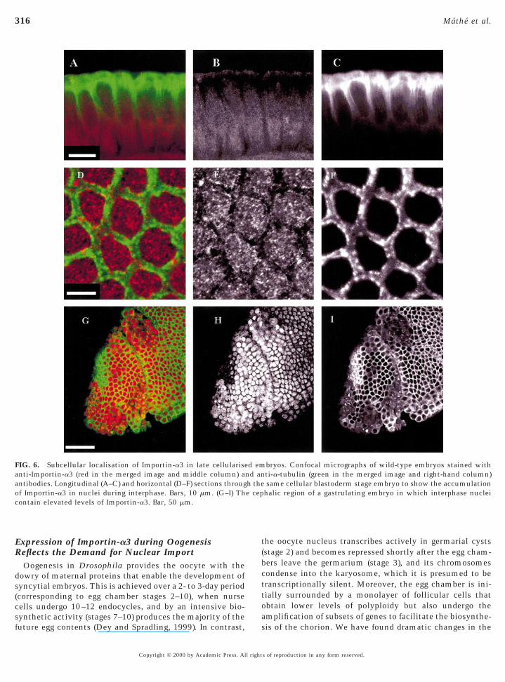
ds(csf
t(bcttoa
aoc
316 Mathe et al.
Expression of Importin-a3 during OogenesisReflects the Demand for Nuclear Import
Oogenesis in Drosophila provides the oocyte with theowry of maternal proteins that enable the development ofyncytial embryos. This is achieved over a 2- to 3-day periodcorresponding to egg chamber stages 2–10), when nurseells undergo 10–12 endocycles, and by an intensive bio-ynthetic activity (stages 7–10) produces the majority of the
FIG. 6. Subcellular localisation of Importin-a3 in late cellulariseanti-Importin-a3 (red in the merged image and middle column) antibodies. Longitudinal (A–C) and horizontal (D–F) sections througf Importin-a3 in nuclei during interphase. Bars, 10 mm. (G–I) Thontain elevated levels of Importin-a3. Bar, 50 mm.
uture egg contents (Dey and Spradling, 1999). In contrast, s
Copyright © 2000 by Academic Press. All right
he oocyte nucleus transcribes actively in germarial cystsstage 2) and becomes repressed shortly after the egg cham-ers leave the germarium (stage 3), and its chromosomesondense into the karyosome, which it is presumed to beranscriptionally silent. Moreover, the egg chamber is ini-ially surrounded by a monolayer of follicular cells thatbtain lower levels of polyploidy but also undergo themplification of subsets of genes to facilitate the biosynthe-
bryos. Confocal micrographs of wild-type embryos stained withti-a-tubulin (green in the merged image and right-hand column)same cellular blastoderm stage embryo to show the accumulation
halic region of a gastrulating embryo in which interphase nuclei
d emnd anh thee cep
is of the chorion. We have found dramatic changes in the
s of reproduction in any form reserved.
cla
sh
aAcwf
ci
317The Importin-a3 Gene of Drosophila
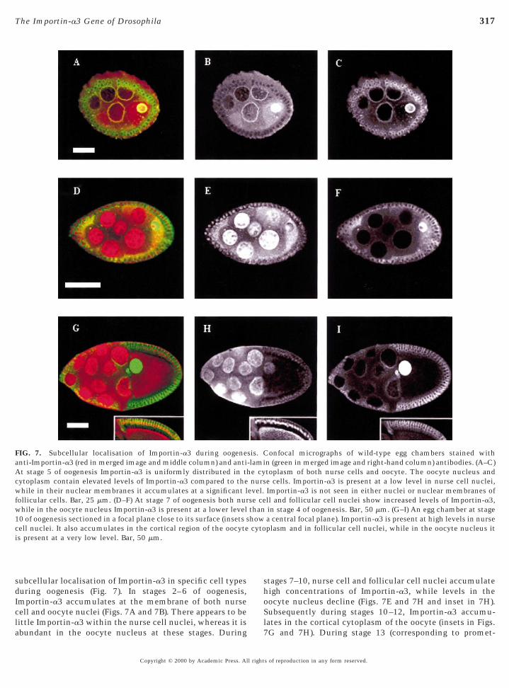
subcellular localisation of Importin-a3 in specific cell typesduring oogenesis (Fig. 7). In stages 2–6 of oogenesis,Importin-a3 accumulates at the membrane of both nurseell and oocyte nuclei (Figs. 7A and 7B). There appears to beittle Importin-a3 within the nurse cell nuclei, whereas it is
FIG. 7. Subcellular localisation of Importin-a3 during oogenenti-Importin-a3 (red in merged image and middle column) and antit stage 5 of oogenesis Importin-a3 is uniformly distributed in th
ytoplasm contain elevated levels of Importin-a3 compared to thehile in their nuclear membranes it accumulates at a significant l
ollicular cells. Bar, 25 mm. (D–F) At stage 7 of oogenesis both nurwhile in the oocyte nucleus Importin-a3 is present at a lower level10 of oogenesis sectioned in a focal plane close to its surface (insets sell nuclei. It also accumulates in the cortical region of the oocytes present at a very low level. Bar, 50 mm.
bundant in the oocyte nucleus at these stages. During
Copyright © 2000 by Academic Press. All right
tages 7–10, nurse cell and follicular cell nuclei accumulateigh concentrations of Importin-a3, while levels in the
oocyte nucleus decline (Figs. 7E and 7H and inset in 7H).Subsequently during stages 10–12, Importin-a3 accumu-lates in the cortical cytoplasm of the oocyte (insets in Figs.
onfocal micrographs of wild-type egg chambers stained withn (green in merged image and right-hand column) antibodies. (A–C)toplasm of both nurse cells and oocyte. The oocyte nucleus ande cells. Importin-a3 is present at a low level in nurse cell nuclei,Importin-a3 is not seen in either nuclei or nuclear membranes ofll and follicular cell nuclei show increased levels of Importin-a3,in stage 4 of oogenesis. Bar, 50 mm. (G–I) An egg chamber at stagea central focal plane). Importin-a3 is present at high levels in nurseplasm and in follicular cell nuclei, while in the oocyte nucleus it
sis. C-lamie cynurs
evel.se cethanhowcyto
7G and 7H). During stage 13 (corresponding to promet-
s of reproduction in any form reserved.
2ow(
WDtifltfi
iwdt
meo
nrbeotrg
twpPscr
lgpttdm
pi
wp
afi
318 Mathe et al.
aphase of meiosis I) and stage 14 (corresponding to meta-phase of meiosis I), Importin-a3 persists in the corticalcytoplasm of matured eggs (data not shown). The follicularcell nuclei show little Importin-a3 during oogenetic stages–6 (Figs. 7A and 7B), but this increases markedly through-ut stages 7–12 (Figs. 7D and 7E and inset in 7H), decliningith the degeneration of the follicular cells from stage 13
data not shown).
The imp-a3 Mutants Are Poorly Viable and FemaleSterile
In order to gain some insight into the functions ofImportin-a3 we sought to identify mutations in its gene.
e identified a P-lacW mutant line from the collection ofeak et al. (1997) in which the transposon was inserted into
he cytological interval to which we had mapped themp-a3 gene by in situ hybridisation. Rescue of the P-lacWanking genomic sequences from this line indicated thathe P-lacW element was inserted 550 bp upstream of therst ATG codon in the imp-a3 open reading frame (Fig. 1).
We therefore tentatively designated this mutant allele imp-a31. Homozygous imp-a31/imp-a31 and hemizygous imp-a31/Df(3R)by62 individuals show pharate adult lethalityand female sterility. The cytological interval in which themutation lies was narrowed to 85E10–F16 since the imp-a31/Df(3R)by10 individuals were viable and fertile, indicat-ng that the gene lies in the interval 85E10–F16, consistentith the insertion site of the P-lacW element (Fig. 1). Toemonstrate that the P-lacW element was associated withhe partially lethal mutation we placed the imp-a31 mutant
line under dysgenic conditions and selected revertants inwhich the P-lacW element had been remobilised. Sixty-tworevertants were isolated that were viable and female fertilewhen homozygous and had w2 eyes in a w2 background,indicating the complete reversion of the imp-a31-associated
utant phenotypes and the precise excision of the P-lacWlement. These complete revertants crossed back to theriginal imp-a31 allele or the Df(3R)by62 gave viable and
fertile offspring, strongly suggesting that the P-lacW inser-tion is responsible for the imp-a31-associated mutant phe-
otype. These reversion tests also generated 10 other partialevertants whose mutant phenotypes can be accounted fory imprecise excision and/or local hopping of the P-lacWlement. The partial revertants gave viable and fertileffspring over Df(3L)by62, while under homozygous condi-ions they were lethal, indicating that remobilisation hadesulted in mutations in other complementation groups orenes.In order to define the imp-a31 mutant allele, we under-
ook germ-line transformation experiments to determinehether the cloned imp-a3 gene would rescue the mutanthenotypes. We screened the European Drosophila Genomeroject cosmid library using the P-lacW flanking chromo-omal sequences and cDNA clones. We identified cosmidsarrying the gene from which we selected a 9-kb EcoRI
estriction fragment for insertion into the P-element germ-Copyright © 2000 by Academic Press. All right
ine transformation vector (Fig. 1). This fragment ofenomic DNA (pR4, Fig. 1) was able to rescue all mutanthenotypes associated with the imp-a31 allele. As a nega-ive control we used a variant of the germ-line transforma-ion construct in which most of the imp-a3 gene waseleted (pRD4, Fig.1) and which was unable to rescue theutant phenotypes.We have been unable to pinpoint a specific cause of
harate adult lethality in homozygous and hemizygousmp-a31 mutants. Examination of orcein-stained prepara-
tions of brains from mature third-instar larvae failed toreveal any mitotic defects and in fact the mitotic index wasidentical to that seen in wild-type brains (data not shown),suggesting there are no generalised defects in cell prolifera-tion.
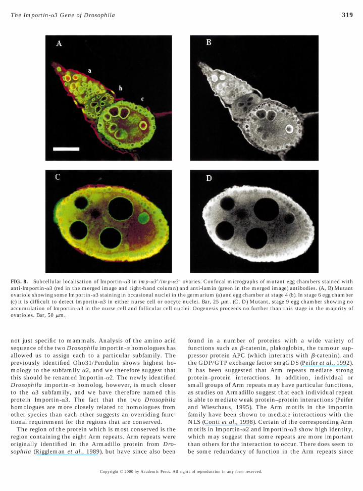
The imp-a31 allele seems to be hypomorphic since theWestern blot of ovarian total protein extracts shows areduced level of Importin-a3 in imp-a31/imp-a31 femalescompared to wild-type (Fig. 4C). The sterile homozygousimp-a31 mutant females have defective ovaries that showfewer ovarioles (4–10 per ovary) and degenerated egg cham-bers (Fig. 8). Whereas some nuclei within the germariumcontain Importin-a3 (region a in Figs. 8A and 8B), incontrast to the wild-type stage 7–10 egg chambers, neithernurse cell nor follicular cell nuclei accumulate detectableamounts of Importin-a3 (Figs. 8C and 8D, compare withFigs. 7D and 7E). Typically the egg chambers in suchmutant ovaries degenerate at stage 7–10 of oogenesis al-though in some ovaries one or two eggs may reach matu-rity, exhibiting normal chorional appendages (data notshown), but they are never released from the ovaries. Stage7–10 of oogenesis is a time at which the nurse cells shouldhave reached their maximum ploidy in readiness to under-take the biosynthetic activity necessary to provide theoocyte with its maternal dowry.
DISCUSSION
Isolation of importin-a Homologues fromDrosophila melanogaster
In this study we have identified two karyopherin-a orimportin-a homologues from Drosophila as proteins thatinteracted in a yeast two-hybrid screen with the largesubunit of DNA polymerase-a (p180). This is perhaps notsurprising since Drosophila p180 shows homology to aregion in the Schizosaccharomyces pombe DNApolymerase-a, which has been shown to contain a func-tional bipartite NLS (Bouvier and Baldacci, 1995). It ishighly likely that the two importin-a genes of Drosophila
ere detected in our screen through the interaction of theirroteins with the NLS of the bait protein.It has been known for some time that mammals (mouse
nd human) have several importin-a homologues, whichall into three subfamilies. The isolation of a secondmportin-a from Drosophila now suggests that this phe-
nomenon is widespread among multicellular organisms and
s of reproduction in any form reserved.
apmt
p
mwt
o
o
319The Importin-a3 Gene of Drosophila
not just specific to mammals. Analysis of the amino acidsequence of the two Drosophila importin-a homologues hasllowed us to assign each to a particular subfamily. Thereviously identified Oho31/Pendulin shows highest ho-ology to the subfamily a2, and we therefore suggest that
his should be renamed Importin-a2. The newly identifiedDrosophila importin-a homolog, however, is much closerto the a3 subfamily, and we have therefore named thisrotein Importin-a3. The fact that the two Drosophila
homologues are more closely related to homologues fromother species than each other suggests an overriding func-tional requirement for the regions that are conserved.
The region of the protein which is most conserved is theregion containing the eight Arm repeats. Arm repeats wereoriginally identified in the Armadillo protein from Dro-
FIG. 8. Subcellular localisation of Importin-a3 in imp-a31/imp-a3anti-Importin-a3 (red in the merged image and right-hand columnvariole showing some Importin-a3 staining in occasional nuclei in
(c) it is difficult to detect Importin-a3 in either nurse cell or oocyaccumulation of Importin-a3 in the nurse cell and follicular cell nvarioles. Bar, 50 mm.
sophila (Riggleman et al., 1989), but have since also been b
Copyright © 2000 by Academic Press. All right
found in a number of proteins with a wide variety offunctions such as b-catenin, plakoglobin, the tumour sup-pressor protein APC (which interacts with b-catenin), andthe GDP/GTP exchange factor smgGDS (Peifer et al., 1992).It has been suggested that Arm repeats mediate strongprotein–protein interactions. In addition, individual orsmall groups of Arm repeats may have particular functions,as studies on Armadillo suggest that each individual repeatis able to mediate weak protein–protein interactions (Peiferand Wieschaus, 1995). The Arm motifs in the importinfamily have been shown to mediate interactions with theNLS (Conti et al., 1998). Certain of the corresponding Arm
otifs in Importin-a2 and Importin-a3 show high identity,hich may suggest that some repeats are more important
han others for the interaction to occur. There does seem to
aries. Confocal micrographs of mutant egg chambers stained withanti-lamin (green in the merged image) antibodies. (A, B) Mutantermarium (a) and egg chamber at stage 4 (b). In stage 6 egg chamberclei. Bar, 25 mm. (C, D) Mutant, stage 9 egg chamber showing no. Oogenesis proceeds no further than this stage in the majority of
1 ov) andthe g
te nuuclei
e some redundancy of function in the Arm repeats since
s of reproduction in any form reserved.

isrigat
aI
brbkci
spoIppdoIapItstt
tTwan
320 Mathe et al.
the initial Importin-a3 clone, missing the N-terminal por-tion of the protein up to the third Arm motif, was stillapparently capable of NLS interactions (data not shown).This is also consistent with results from Prieve et al. (1996),who showed that a truncated form of human importin-a1(hSrp1) missing the first two Arm motifs could still interactwith an NLS-protein.
Analysis of Cellular Functions of Importin-aIsoforms
The presence of conserved multiple importin-a isoformsn many species suggests that each of them may have apecific function. It is possible that each homologue isesponsible for the transport of a unique subset of proteinsn a controlled spatial and temporal pattern as was sug-ested for human (Kohler et al., 1997) and mouse (Tsuji etl., 1997; Kamei et al., 1999). In this case we might expecto see each importin-a isoform showing different substrate
specificities. Alternatively, it is possible that some of theisoforms are functionally redundant by featuring overlap-ping substrate specificities. In this case the importin-aisoforms would be expected to have similar substrate affini-ties, and any specificity in the system would be introducedby the binding properties of individual substrates and thelevels of the proteins that are available. The differentexpression levels and/or subcellular distributions of theimportin-a isoforms during development could thereforerepresent a requirement for the nuclear import of specificproteins and/or levels of generally increased nuclear proteinimport. Therefore the phenotypes of mutants in each caseshould also reflect stages in development or cell cycle atwhich there is a specific requirement for each importin-aisoform. In this case the phenotype of the mutant mightalso help to define the role of the importin-a isoformspecific to a particular developmental or cell cycle stage,whereas a less defined developmental arrest would suggestpleiotropic function.
Importin-a3 and Importin-a2 Interact with theSame Range of Proteins in Vitro
Our molecular and biochemical studies suggest that theprotein interactions made by Importin-a3 and Importin-a2re very similar under in vitro conditions. The GST-taggedmportin-a3 and Importin-a2 bound similar profiles of
proteins both in solution and immobilised following elec-troblotting. Furthermore similar patterns of potentiallyinteracting proteins were identified within immunoprecipi-tates. A particularly large number of interacting proteinswere found in washes of high-salt nuclear preparations. It islikely that the more tightly interacting bands representcomponents of the NLS-dependent nuclear transport ma-chinery. This is confirmed by the identification of the90-kDa band as the Drosophila homologue of theimportin-b protein. Identification of the 50- and 70-kDa
bands has not been possible since Drosophila homologuesCopyright © 2000 by Academic Press. All right
of other components of the NLS-dependent nuclear trans-port machinery have not yet been reported. The weakerinteracting bands may be additional components of thetransport machinery or transport substrates.
Although our results argue that the two importin-aisoforms are very similar in terms of the contacts that theymake in vitro, we remain cautious as to whether theyreflect interactions in vivo since the proteins are perhapsforced into unusual interactions due to the nonphysiologi-cal conditions used. Moreover, we might be seeing only asubset of the interactions taking place, for example, withthe transport machinery and are therefore missing crucialdifferences between substrates.
It is also possible that phosphorylation might play somerole in controlling the binding affinities of the twoimportin-a isoforms of Drosophila. The protein used for ourinding studies was not phosphorylated since it is of bacte-ial origin, but it may become phosphorylated during incu-ation with the embryonic extracts. However, we do notnow whether this occurs or the extent to which thereould be a preference for nonphosphorylated forms by thenteracting proteins. A phosphorylated form of Importin-a2
can be detected in early embryos and in ovaries (Torok etal., 1995). However, we have not been able to detect aphosphorylated form of Importin-a3 at any stage of devel-opment. Since phosphorylated Importin-a2 is the only formeen in the nucleus it is tempting to speculate that phos-horylation is necessary for activity. This could be a meansf regulating the utilisation of large maternal input ofmportin-a2 to early embryos. It is also possible that phos-horylation could control substrate recognition or therophase-specific interaction of Importin-a2 with the NLS-ependent nuclear transport machinery. One intriguingbservation with respect to this alternative is thatmportin-a2 has a potential Cdc2 phosphorylation sitedjacent to the IBB domain. If it were necessary to phos-horylate this site in order to get interaction withmportin-b it is easy to see how this would help to controlhe activity of the protein. Importin-a3 in contrast has nouch phosphorylation site, but has instead a run of nega-ively charged residues at a similar location, which alterna-ively may provide the cell cycle control of its function.
Importin-a3 and Importin-a2 Show DifferentDevelopmental Profiles
Importin-a3 and Importin-a2 show dramatically differentiming of expression during the life cycle of Drosophila.he appearance of Importin-a2 in the embryos coincidesith the time of intense mitotic activity, which presum-
bly imposes specific demands upon traffic to and from theucleus. On the other hand levels of Importin-a3 in the
syncytial embryos are very low and do not increase untilafter cellularisation, following which they appear to remainapparently at a constant level throughout development.The simplest rationalisation of the expression patterns is
that Importin-a3 functions as the main “housekeeping”s of reproduction in any form reserved.
sp1
iti
rwpifTttawisIn
C
G
G
G
G
G
321The Importin-a3 Gene of Drosophila
importin-a with Importin-a2 playing an additional or morepecialised role during embryogenesis consistent with itsrophase-specific nuclear accumulation (Kussel and Frasch,995; Torok et al., 1995). In contrast to the predominant
distribution of Importin-a2 in the interphase cytoplasm,Importin-a3 is principally nuclear in interphase althoughts intensity of nuclear staining also increases in prophaseo become more dispersed at metaphase. However, it isntriguing that although Importin-a3 is present mainly
within the nucleus at most developmental stages, it isdistinctly associated with the nuclear envelope in the earlyembryos before its levels reach their maximum and also inthe endoreduplicating nurse cells before they reach theirmaximal ploidy during oogenesis. The significance of this isnot clear, but the association with the nuclear envelopemay anticipate a requirement for a higher amount ofImportin-a3 prior to it filling the entire nucleus.
Importin-a3 Is Required to Complete PupalDevelopment and Oogenesis
The differences that we have observed in the develop-mental and subcellular localisation profiles of Importin-a3and Importin-a2 indicate that the two proteins are notedundant in the context of normal development. However,hether this reflects differential control of expression ofroteins or particular substrate specificity for each proteins uncertain. It will require a detailed analysis of the cellularunctions of both proteins at critical stages of development.he pleiotropy of imp-a31 mutant phenotypes suggests that
he Importin-a3 has a general house keeping function andhat the developmental processes affected reflect the pointt which the levels of the protein become rate limiting. Weere unable to pinpoint the precise nature of these defects
n the pupal stages. However, oogenesis is interruptedpecifically at stage 7–10 a time at which large amounts ofmportin-a3 would normally be present in the wild-typeurse cell nuclei. It therefore seems likely that Importin-a3
is required at this stage for transport of NLS proteins intothe nurse cell nuclei. This precedes a crucial transition inoogenesis when at stage 10, the nurse cell contents aredramatically transported into the oocyte cytoplasm (Sprad-ling, 1993).
ACKNOWLEDGMENTS
We thank Hiro Ohkura for his contribution to some of the earlyscreens of P-element mutant collections. We thank Carmo Avidesfor providing RNA samples for developmental Northern blots. Weare also grateful to Istvan Torok and Manfred Frasch for antibodiesthat recognise Drosophila Importin-a2. Antibodies against Ketel,the Importin-b homologue, were kindly provided by Janos Szabad.Roger Brent kindly provided the two-hybrid libraries and plasmids.The work was supported by the Marie Curie Memorial Foundation
and the Cancer Research Campaign.Copyright © 2000 by Academic Press. All right
REFERENCES
Adam, E. J. H., and Adam, S. A. (1994). Identification of cytosolicfactors required for nuclear location sequence-mediated bindingto the nuclear envelope. J. Cell Biol. 125, 547–555.
Bischoff, F. R., and Gorlich, D. (1997). RanBP1 is crucial for therelease of RanGTP from importin-b related nuclear transportfactors. FEBS Lett. 419, 249–254.
Bouvier, D., and Baldacci, D. (1995). The N-terminus of fissionyeast DNA polymerase alpha contains a basic pentapeptide thatacts in vivo as a nuclear localisation signal. Mol. Biol. Cell 6,1697–1705.allaini, G., and Riparbelli, M. G. (1996). Fertilization in Drosoph-ila melanogaster: Centrosome inheritance and organization ofthe first mitotic spindle. Dev. Biol. 176, 199–208.
Cavener, D. R. (1987). Comparison of the consensus sequenceflanking translational start sites in Drosophila and vertebrates.Nucleic Acids Res. 15, 1353–1361.
Chi, N. C., Adam, E. J. H., Visser, G. D., and Adam, S. A. (1996).RanBP1 stabilises the interaction of Ran with p97 in nuclearprotein import. J. Cell Biol. 135, 559–569.
Conti, E., Uy, M., Leighton, L., Blobel, G., and Kuriyan, J. (1998).Crystallographic analysis of the recognition of a nuclear local-ization signal by the nuclear import factor karyopherin alpha.Cell 94, 193–204.
Deak, P., Omar, M. M., Saunders, R. D. C., Pal, M., Komonyi, O.,Szidonya, J., Maroy, P., Zhang, Y., Ashburner, M., Benos, P.,Savakis, C., Siden-Kiamos, I., Louis, C., Bolshakov, V., Kafatos,F. C., Madueno, E., Modelell, J., and Glover, D. M. (1997).P-element insertion alleles of essential genes on the third chro-mosome of Drosophila melanogaster: Correlation of physicaland cytogenetic maps in chromosomal region 86E–87F. Genetics147, 1697–1722.
Dey, J. K., and Spradling, A. (1999). The endocycle controls nursecell polytene chromosome structure during Drosophila oogen-esis. Development 126, 293–303.
Dingwall, C., and Laskey, R. A. (1998). Nuclear import: A tale oftwo sites. Curr. Biol. 8, 922–924.
Erdelyi, M., Mathe, E., and Szabad, J. (1997). Genetic and develop-mental analysis of Ketel mutant alleles that identify the Dro-sophila importin-b homologue. Acta Biol. Hung. 48, 323–338.
Foe, E. V., Odell, G. M., and Edgar, B. A. (1993). Mitosis andmorphogenesis in the Drosophila embryo: Point and counter-point. In “The Development of Drosophila melanogaster” (M.Bate and A. Martinez Arias, Eds.), pp. 149–300. Cold SpringHarbor Laboratory Press, New York.orlich, D., Kotska, S., Kraft, R., Dingwall, C., Laskey, R. A.,Hartmann, E., and Prehn, S. (1995). Two different subunits ofimportin cooperate to recognize nuclear localization signals andbind them to the nuclear envelope. Curr. Biol. 5, 383–392.orlich, D., Henklein, P., Laskey, R. A., and Hartmann, E. (1996).A 41 amino acid motif in importin-a confers binding toimportin-b and hence transit into the nucleus. EMBO J. 15,1810–1817.orlich, D. (1997). Nuclear protein import. Curr. Opin. Cell Biol. 9,412–419.orlich, D. (1998). Transport into and out of the cell nucleus.EMBO J. 17, 2721–2727.yuris, J., Golemis, E., Chertkov, H., and Brent, R. (1993). Cdi1, ahuman G1 and S phase protein phosphatase that associates with
Cdk2. Cell 75, 791–803.s of reproduction in any form reserved.
H
I
K
K
K
K
K
L
M
M
M
M
M
N
P
P
P
P
R
R
R
S
S
S
S
S
T
T
322 Mathe et al.
Herold, A., Truant, R., Wiegand, H., and Cullen, B. R. (1998).Determination of the functional domain organisation of theimportin a nuclear import factor. J. Cell Biol. 143, 309–318.uettner, A. F. (1924). Maturation and fertilization in Drosophilamelanogaster. J. Morphol. Physiol. 39, 249–265.
zaurralde, E., Kutay, U., von Kobbe, U., Mattaj, I. W., and Gorlich,D. (1997). The asymmetric distribution of the constituents of theRan system is essential for transport into and out of the nucleus.EMBO J. 16, 6535–6547.amei, Y., Yuba, S., Nakayama, T., and Yoneda, Y. (1999). Threedistinct classes of the alpha-subunit of the nuclear pore-targetingcomplex (importin-alpha) are differentially expressed in adultmouse tissues. J. Histochem. Cytochem. 47, 363–372.oepp, D. M., and Silver, P. A. (1996). A GTPase controllingnuclear trafficking: Running the right way or walking randomly?Cell 87, 1–4.ozak, M. (1995). Adherence to the first-AUG rule when a secondAUG codon follows closely upon the first. Proc. Natl. Acad. Sci.USA 92, 7134.ohler, M., Ansieau, S., Prehn, S., Leutz, A., Haller, H., Haller, H.,and Hartmann, E. (1997). Cloning of two novel humanimportin-a subunits and analysis of the expression pattern of theimportin-a protein family. FEBS Lett. 417, 104–108.
ussel, P., and Frasch, M. (1995). Pendulin, a Drosophila proteinwith cell cycle-dependent nuclear localization, is required fornormal cell proliferation. J. Cell Biol. 129, 1491–1507.
Kutay, U., Bischoff, F. R., Kostka, S., Kraft, R., and Gorlich, D.(1997). Export of importin-a from the nucleus is mediated by aspecific nuclear transport factor. Cell 90, 1061–1071.
indsley, D. L., and Zimm, G. G. (1992). “The Genome of Drosoph-ila melanogaster.” Academic Press, San Diego/London.aldonado-Codina, G., and Glover, D. M. (1992). Cyclin A and Bassociate with chromatin and the polar regions of spindles,respectively, and do not undergo a complete degradation atanaphase in syncytial Drosophila embryos. J. Cell Biol. 116,967–976.athe, E., Boros, I., Josvay, K., Kaijun L., Puro, J., Kaufman, T. C.,and Szabad, J. (1998). The Tomaj mutant alleles of aTubulin67Creveal a requirement for the encoded maternal specific tubulinisoform in the sperm aster, the cleavage spindle apparatus andneurogenesis during embryonic development in Drosophila.J. Cell Sci. 111, 887–896.attaj, I. W., and Engelmeier, L. (1998). Nucleocytoplasmic trans-port: The soluble phase. Annu. Rev. Biochem. 67, 265–306.oroianu, J., Blobel, G., and Radu, A. (1995a). Previously identifiedprotein of uncertain function is karyopherin alpha and togetherwith karyopherin beta docks import substrate at nuclear porecomplexes. Proc. Natl. Acad. Sci. USA 92, 2008–2011.oroianu, J., Hijikata, M., Blobel, G., and Radu, A. (1995b).Mammalian karyopherin a1b and a2b heterodimers: a1 or a2subunit binds nuclear localisation sequence and b subunit inter-acts with peptide repeat containing nucleoporins. Proc. Natl.Acad. Sci. USA 92, 6532–6536.igg, E. A. (1997). Nucleocytoplasmic transport: Signals, mecha-nisms and regulation. Nature 386, 779–787.
addy, M. R., Belmont, A. S., Saumweber, H., Agard, D. A., andSedat, J. (1990). Interphase nuclear envelope lamins form adiscontinuous network that interacts with only a fraction of thechromatin in the nuclear periphery. Cell 62, 89–106.
eifer, M., McCrea, P. D., Green, K. J., Wieschaus, E., and Gum-
biner, B. M. (1992). The vertebrate adhesive junction proteinsCopyright © 2000 by Academic Press. All right
beta-catenin and plakoglobin and the Drosophila segment polar-ity gene armadillo form a multigene family with similar proper-ties. J. Cell Biol. 118, 681–691.
eifer, M., and Wieshaus, E. (1995). The segment polarity genearmadillo encodes a functionally modular protein that is theDrosophila homolog of human plakoglobulin. Cell 63, 1167–1176.
rieve, M. G., Guttridge, K. L., Munguia, J. E., and Waterman, M. L.(1996). The nuclear localization signal of lymphoid enhancerfactor-1 is recognized by two differentially expressed Srp1-nuclear localization sequence receptor proteins. J. Biol. Chem.271, 7654–7658.exach, M., and Blobel, G. (1995). Protein import into nuclei:Association and dissociation reactions involving transport sub-strate, transport factors, and nucleoporins. Cell 83, 683–692.
iggleman, B., Wieschaus, E., and Schedl, P. (1989). Molecularanalysis of the armadillo locus: Uniformly distributed tran-scripts and a protein with novel internal repeats are associatedwith a Drosophila segment polarity gene. Genes Dev. 3, 96–113.uden, D. M., Ma, J., Li, Y., Wood, K., and Ptashne, M. (1991).Generating yeast transcriptional activators containing no yeastprotein sequences. Nature 350, 250–252.
ambrook, J., Fritsch, E. F., and Maniatis, T. (1989). “MolecularCloning: A Laboratory Manual.” Cold Spring Harbor LaboratoryPress, Cold Spring Harbor, NY.
pradling, A. C. (1993). Developmental genetics of oogenesis. In“The Development of Drosophila melanogaster” (M. Bate and A.Martinez Arias, Eds.), pp. 1–70. Cold Spring Harbor LaboratoryPress, New York.
tochaj, U., and Rother, K. L. (1999). Nucleocytoplasmic traffickingof proteins: With or without ran? BioEssays 21, 579–589.
tutz, F., and Rosbash, M. (1998). Nuclear RNA export. Genes Dev.12, 3303–3319.
unkel, C. E., and Glover, D. M. (1988). Polo, a mitotic mutant ofDrosophila displaying abnormal spindle poles. J. Cell Sci. 89,25–38.orok, I., Strand, D., Schmitt, G., Tick, T., Torok, T., Kiss, I., andMechler, B. M. (1995). The overgrown hematopoietic organs-31tumor suppressor gene of Drosophila encodes an Importin-likeprotein accumulating in the nucleus at the onset of mitosis.J. Cell Biol. 129, 1473–1489.suji, L., Takumi, T., Imamoto, N., and Yoneda, Y. (1997). Identi-fication of novel homologues of mouse importin-a, the a subunitof nuclear pore-targetting complex, and their tissue-specificexpression. FEBS Lett. 416, 30–34.
Ullman, K. S., Powers, M. A., and Forbes, D. J. (1997). Nuclearexport receptors: From importin to exportin. Cell 90, 967–970.
Weis, K., Ryder, U., and Lamond, A. I. (1996). The conserved aminoterminal domain of hSRP1a is essential for nuclear proteinimport. EMBO J. 15, 1818–1825.
Yasuda, G. K., Baker, J., and Schubinger, G. (1991). Temporalregulation of gene expression in the blastoderm Drosophilaembryo. Gen. Dev. 5, 1800–1812.
Zervos, A. S., Gyuris, J., and Brent, R. (1993). Mxi1, a protein thatspecifically interacts with Max to bind Myc–Max recognitionsites. Cell 72, 223–232.
Received for publication November 11, 1999Revised April 10, 2000
Accepted April 10, 2000
s of reproduction in any form reserved.