The adenosine A1 receptor agonist adenosine amine congener exerts a neuroprotective effect against...
12
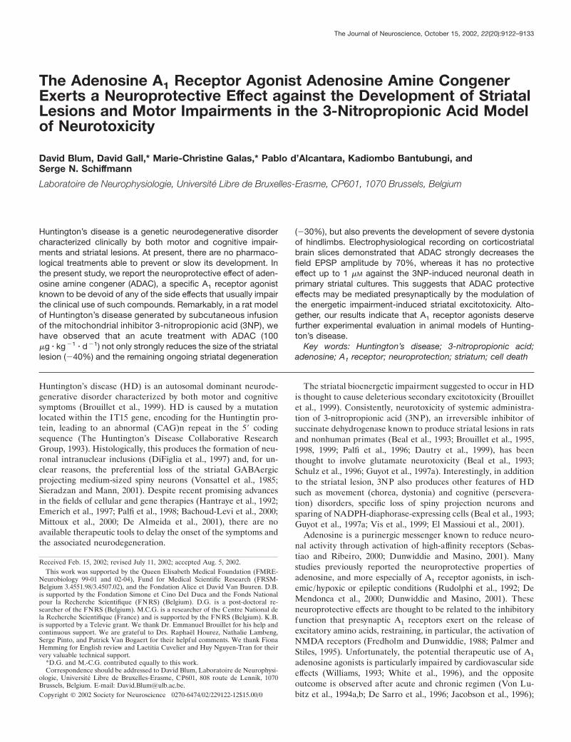
The Adenosine A 1 Receptor Agonist Adenosine Amine Congener Exerts a Neuroprotective Effect against the Development of Striatal Lesions and Motor Impairments in the 3-Nitropropionic Acid Model of Neurotoxicity David Blum, David Gall,* Marie-Christine Galas,* Pablo d’Alcantara, Kadiombo Bantubungi, and Serge N. Schiffmann Laboratoire de Neurophysiologie, Universite ´ Libre de Bruxelles-Erasme, CP601, 1070 Brussels, Belgium Huntington’s disease is a genetic neurodegenerative disorder characterized clinically by both motor and cognitive impair- ments and striatal lesions. At present, there are no pharmaco- logical treatments able to prevent or slow its development. In the present study, we report the neuroprotective effect of aden- osine amine congener (ADAC), a specific A 1 receptor agonist known to be devoid of any of the side effects that usually impair the clinical use of such compounds. Remarkably, in a rat model of Huntington’s disease generated by subcutaneous infusion of the mitochondrial inhibitor 3-nitropropionic acid (3NP), we have observed that an acute treatment with ADAC (100 g kg 1 d 1 ) not only strongly reduces the size of the striatal lesion (40%) and the remaining ongoing striatal degeneration (30%), but also prevents the development of severe dystonia of hindlimbs. Electrophysiological recording on corticostriatal brain slices demonstrated that ADAC strongly decreases the field EPSP amplitude by 70%, whereas it has no protective effect up to 1 M against the 3NP-induced neuronal death in primary striatal cultures. This suggests that ADAC protective effects may be mediated presynaptically by the modulation of the energetic impairment-induced striatal excitotoxicity. Alto- gether, our results indicate that A 1 receptor agonists deserve further experimental evaluation in animal models of Hunting- ton’s disease. Key words: Huntington’s disease; 3-nitropropionic acid; adenosine; A 1 receptor; neuroprotection; striatum; cell death Huntington’s disease (HD) is an autosomal dominant neurode- generative disorder characterized by both motor and cognitive symptoms (Brouillet et al., 1999). HD is caused by a mutation located within the IT15 gene, encoding for the Huntingtin pro- tein, leading to an abnormal (CAG)n repeat in the 5 coding sequence (The Huntington’s Disease Collaborative Research Group, 1993). Histologically, this produces the formation of neu- ronal intranuclear inclusions (DiFiglia et al., 1997) and, for un- clear reasons, the preferential loss of the striatal GABAergic projecting medium-sized spiny neurons (Vonsattel et al., 1985; Sieradzan and Mann, 2001). Despite recent promising advances in the fields of cellular and gene therapies (Hantraye et al., 1992; Emerich et al., 1997; Palfi et al., 1998; Bachoud-Levi et al., 2000; Mittoux et al., 2000; De Almeida et al., 2001), there are no available therapeutic tools to delay the onset of the symptoms and the associated neurodegeneration. The striatal bioenergetic impairment suggested to occur in H D is thought to cause deleterious secondary excitotoxicity (Brouillet et al., 1999). Consistently, neurotoxicity of systemic administra- tion of 3-nitropropionic acid (3NP), an irreversible inhibitor of succinate dehydrogenase known to produce striatal lesions in rats and nonhuman primates (Beal et al., 1993; Brouillet et al., 1995, 1998, 1999; Palfi et al., 1996; Dautry et al., 1999), has been thought to involve glutamate neurotoxicity (Beal et al., 1993; Schulz et al., 1996; Guyot et al., 1997a). Interestingly, in addition to the striatal lesion, 3NP also produces other features of HD such as movement (chorea, dystonia) and cognitive (persevera- tion) disorders, specific loss of spiny projection neurons and sparing of NADPH-diaphorase-expressing cells (Beal et al., 1993; Guyot et al., 1997a; Vis et al., 1999; El Massioui et al., 2001). Adenosine is a purinergic messenger known to reduce neuro- nal activity through activation of high-affinity receptors (Sebas- tiao and Ribeiro, 2000; Dunwiddie and Masino, 2001). Many studies previously reported the neuroprotective properties of adenosine, and more especially of A 1 receptor agonists, in isch- emic/hypoxic or epileptic conditions (Rudolphi et al., 1992; De Mendonca et al., 2000; Dunwiddie and Masino, 2001). These neuroprotective effects are thought to be related to the inhibitory function that presynaptic A 1 receptors exert on the release of excitatory amino acids, restraining, in particular, the activation of NMDA receptors (Fredholm and Dunwiddie, 1988; Palmer and Stiles, 1995). Unfortunately, the potential therapeutic use of A 1 adenosine agonists is particularly impaired by cardiovascular side effects (Williams, 1993; White et al., 1996), and the opposite outcome is observed after acute and chronic regimen (Von Lu- bitz et al., 1994a,b; De Sarro et al., 1996; Jacobson et al., 1996); Received Feb. 15, 2002; revised July 11, 2002; accepted Aug. 5, 2002. This work was supported by the Queen Elisabeth Medical Foundation (FMRE- Neurobiology 99-01 and 02-04), Fund for Medical Scientific Research (FRSM- Belgium 3.4551.98/3.4507.02), and the Fondation Alice et David Van Buuren. D.B. is supported by the Fondation Simone et Cino Del Duca and the Fonds National pour la Recherche Scientifique (FNRS) (Belgium). D.G. is a post-doctoral re- searcher of the FNRS (Belgium). M.C.G. is a researcher of the Centre National de la Recherche Scientifique (France) and is supported by the FNRS (Belgium). K.B. is supported by a Televie grant. We thank Dr. Emmanuel Brouillet for his help and continuous support. We are grateful to Drs. Raphae ¨l Hourez, Nathalie Lambeng, Serge Pinto, and Patrick Van Bogaert for their helpful comments. We thank Fiona Hemming for English review and Laetitia Cuvelier and Huy Nguyen-Tran for their very valuable technical support. *D.G. and M.-C.G. contributed equally to this work. Correspondence should be addressed to David Blum, Laboratoire de Neurophysi- ologie, Universite ´ Libre de Bruxelles-Erasme, CP601, 808 route de Lennik, 1070 Brussels, Belgium. E-mail: [email protected]. Copyright © 2002 Society for Neuroscience 0270-6474/02/229122-12$15.00/0 The Journal of Neuroscience, October 15, 2002, 22(20):9122–9133
-
Upload
independent -
Category
Documents
-
view
4 -
download
0
Transcript of The adenosine A1 receptor agonist adenosine amine congener exerts a neuroprotective effect against...
The Adenosine A1 Receptor Agonist Adenosine Amine CongenerExerts a Neuroprotective Effect against the Development of StriatalLesions and Motor Impairments in the 3-Nitropropionic Acid Modelof Neurotoxicity
David Blum, David Gall,* Marie-Christine Galas,* Pablo d’Alcantara, Kadiombo Bantubungi, andSerge N. Schiffmann
Laboratoire de Neurophysiologie, Universite Libre de Bruxelles-Erasme, CP601, 1070 Brussels, Belgium
Huntington’s disease is a genetic neurodegenerative disordercharacterized clinically by both motor and cognitive impair-ments and striatal lesions. At present, there are no pharmaco-logical treatments able to prevent or slow its development. Inthe present study, we report the neuroprotective effect of aden-osine amine congener (ADAC), a specific A1 receptor agonistknown to be devoid of any of the side effects that usually impairthe clinical use of such compounds. Remarkably, in a rat modelof Huntington’s disease generated by subcutaneous infusionof the mitochondrial inhibitor 3-nitropropionic acid (3NP), wehave observed that an acute treatment with ADAC (100�g � kg�1 � d�1) not only strongly reduces the size of the striatallesion (�40%) and the remaining ongoing striatal degeneration
(�30%), but also prevents the development of severe dystoniaof hindlimbs. Electrophysiological recording on corticostriatalbrain slices demonstrated that ADAC strongly decreases thefield EPSP amplitude by 70%, whereas it has no protectiveeffect up to 1 �M against the 3NP-induced neuronal death inprimary striatal cultures. This suggests that ADAC protectiveeffects may be mediated presynaptically by the modulation ofthe energetic impairment-induced striatal excitotoxicity. Alto-gether, our results indicate that A1 receptor agonists deservefurther experimental evaluation in animal models of Hunting-ton’s disease.
Key words: Huntington’s disease; 3-nitropropionic acid;adenosine; A1 receptor; neuroprotection; striatum; cell death
Huntington’s disease (HD) is an autosomal dominant neurode-generative disorder characterized by both motor and cognitivesymptoms (Brouillet et al., 1999). HD is caused by a mutationlocated within the IT15 gene, encoding for the Huntingtin pro-tein, leading to an abnormal (CAG)n repeat in the 5� codingsequence (The Huntington’s Disease Collaborative ResearchGroup, 1993). Histologically, this produces the formation of neu-ronal intranuclear inclusions (DiFiglia et al., 1997) and, for un-clear reasons, the preferential loss of the striatal GABAergicprojecting medium-sized spiny neurons (Vonsattel et al., 1985;Sieradzan and Mann, 2001). Despite recent promising advancesin the fields of cellular and gene therapies (Hantraye et al., 1992;Emerich et al., 1997; Palfi et al., 1998; Bachoud-Levi et al., 2000;Mittoux et al., 2000; De Almeida et al., 2001), there are noavailable therapeutic tools to delay the onset of the symptoms andthe associated neurodegeneration.
The striatal bioenergetic impairment suggested to occur in HDis thought to cause deleterious secondary excitotoxicity (Brouilletet al., 1999). Consistently, neurotoxicity of systemic administra-tion of 3-nitropropionic acid (3NP), an irreversible inhibitor ofsuccinate dehydrogenase known to produce striatal lesions in ratsand nonhuman primates (Beal et al., 1993; Brouillet et al., 1995,1998, 1999; Palfi et al., 1996; Dautry et al., 1999), has beenthought to involve glutamate neurotoxicity (Beal et al., 1993;Schulz et al., 1996; Guyot et al., 1997a). Interestingly, in additionto the striatal lesion, 3NP also produces other features of HDsuch as movement (chorea, dystonia) and cognitive (persevera-tion) disorders, specific loss of spiny projection neurons andsparing of NADPH-diaphorase-expressing cells (Beal et al., 1993;Guyot et al., 1997a; Vis et al., 1999; El Massioui et al., 2001).
Adenosine is a purinergic messenger known to reduce neuro-nal activity through activation of high-affinity receptors (Sebas-tiao and Ribeiro, 2000; Dunwiddie and Masino, 2001). Manystudies previously reported the neuroprotective properties ofadenosine, and more especially of A1 receptor agonists, in isch-emic/hypoxic or epileptic conditions (Rudolphi et al., 1992; DeMendonca et al., 2000; Dunwiddie and Masino, 2001). Theseneuroprotective effects are thought to be related to the inhibitoryfunction that presynaptic A1 receptors exert on the release ofexcitatory amino acids, restraining, in particular, the activation ofNMDA receptors (Fredholm and Dunwiddie, 1988; Palmer andStiles, 1995). Unfortunately, the potential therapeutic use of A1
adenosine agonists is particularly impaired by cardiovascular sideeffects (Williams, 1993; White et al., 1996), and the oppositeoutcome is observed after acute and chronic regimen (Von Lu-bitz et al., 1994a,b; De Sarro et al., 1996; Jacobson et al., 1996);
Received Feb. 15, 2002; revised July 11, 2002; accepted Aug. 5, 2002.This work was supported by the Queen Elisabeth Medical Foundation (FMRE-
Neurobiology 99-01 and 02-04), Fund for Medical Scientific Research (FRSM-Belgium 3.4551.98/3.4507.02), and the Fondation Alice et David Van Buuren. D.B.is supported by the Fondation Simone et Cino Del Duca and the Fonds Nationalpour la Recherche Scientifique (FNRS) (Belgium). D.G. is a post-doctoral re-searcher of the FNRS (Belgium). M.C.G. is a researcher of the Centre National dela Recherche Scientifique (France) and is supported by the FNRS (Belgium). K.B.is supported by a Televie grant. We thank Dr. Emmanuel Brouillet for his help andcontinuous support. We are grateful to Drs. Raphael Hourez, Nathalie Lambeng,Serge Pinto, and Patrick Van Bogaert for their helpful comments. We thank FionaHemming for English review and Laetitia Cuvelier and Huy Nguyen-Tran for theirvery valuable technical support.
*D.G. and M.-C.G. contributed equally to this work.Correspondence should be addressed to David Blum, Laboratoire de Neurophysi-
ologie, Universite Libre de Bruxelles-Erasme, CP601, 808 route de Lennik, 1070Brussels, Belgium. E-mail: [email protected] © 2002 Society for Neuroscience 0270-6474/02/229122-12$15.00/0
The Journal of Neuroscience, October 15, 2002, 22(20):9122–9133

however, some A1 receptor agonists, devoid of any deleteriouscardiovascular effects, have been described recently (Bischof-berger et al., 1997). Interestingly, among them it was shown thatlow concentrations of adenosine amine congener (ADAC) (VonLubitz et al., 1996a,b) can efficiently protect hippocampal neu-rons against cerebral ischemia after both acute and chronic treat-ments (Von Lubitz et al., 1999). This attractive compound thusdeserves evaluation, especially in HD models for which A1 re-ceptor agonists have never been evaluated.
Recently, it was reported that Lewis rats, unlike the SpragueDawley strain, respond homogeneously to 3NP and develop to-pologically reproducible striatal lesions, providing a suitable HDmodel for neuroprotective studies (Ouary et al., 2000; Blum et al.,2001, 2002; Mittoux et al., 2002). In the present work, we thusaimed to determine the potential neuroprotective effect of eitherchronic or acute treatment with the A1 receptor agonist ADACagainst the striatal lesions induced by 3NP in Lewis rats.
MATERIALS AND METHODSAnimalsAdult male Lewis rats (IFFA Credo, Reims, France), 12 weeks of age,weighing 320–380 gm were used in this study. Animals were housed threeper cage and maintained in a temperature- and humidity-controlled roomon a 12 hr light /dark cycle with food and water ad libitum. The numberof animals was kept to the minimum, and all efforts to avoid animalsuffering were made in accordance with the standards of the InstitutionalEthical Committee of the School of Medicine of Universite Libre deBruxelles.
Surgery and 3NP treatmentRats were anesthetized with a mixture containing xylazine hydrochloride(Rompun, Bayer; 4.5 mg/kg) and ketamine hydrochloride (Imalgene,Merial; 90 mg/kg). In the 3NP-treated animals, an incision was madebelow the base of the neck and a 2mL1 Alzet osmotic minipump (deliv-ering 10 �l /hr for 7 d; IFFA Credo) containing 3-nitropropionic acid(Fluka) was positioned under the skin. 3NP was dissolved in 0.1 M PBS,pH 7.4, adjusted to pH 7.3–7.4 with 5N NaOH. The final concentrationof 3NP in the pump was adjusted to the weight of the rats on the day ofimplantation to exactly deliver 56 mg � kg �1 � d �1. Sham rats and animalstreated with ADAC alone underwent all of the surgical procedures(without minipump implantation).
All rats were killed after 5 d of 3NP subcutaneous infusion. It isnoteworthy that the time they were killed was chosen according to theknown kinetics of striatal lesion occurrence in this particular model,because it was determined previously that macroscopic lesions inducedby 3NP are detected 5 d after the beginning of the toxic treatment(Ouary et al., 2000; Blum et al., 2001).
ADAC treatmentTwo separate experiments were performed (Fig. 1). In the first one, 28rats (sham, n � 7; vehicle/3NP, n � 7; ADAC�8/5, n � 7; ADAC�8/5/3NP, n � 7) were used to study the potential protective effect of chronicadministration of ADAC against the neurotoxic effects of 3NP. In thisprotocol, ADAC injections began 8 d before the onset of 3NP intoxica-tion and were continued until the animals were killed (13 d in all; lastinjection 6 hr before animals were killed). ADAC solution was preparedas follows: 6 mg of ADAC (Sigma) were first dissolved in 200 �l of 1NHCl and then added to 120 ml of 0.1 M PBS, pH 7.4, to reach a finalconcentration of 50 �g/ml. Before each injection, the solution was heatedto 37°C for 15–30 min to ensure complete dissolution. Vehicle solution(200 �l of 1N HCl in 120 ml of 0.1 M PBS) was processed similarly. Thefinal pH of all solutions was 7.3–7.4. The volume of the ADAC solutioninjected intraperitoneally was adjusted to the weight of rats to deliverexactly 100 �g � kg �1 � d �1 (200 �l /100 gm of weight), a supramaximaldose that has been shown previously to be protective against cerebralischemia after both chronic and acute administration (Von Lubitz et al.,1999) without any cardiovascular side effects (Von Lubitz et al., 1996b).
In the second experiment, 50 rats were used to study the potentialneuroprotective effects of acute treatments with ADAC (sham, n � 7;3NP/vehicle, n � 9; ADAC alone injected at days 3, 4, and 5 � ADAC3/5
group, n � 7; 3NP-treated rats injected with ADAC at days 3, 4 and 5 �3NP/ADAC3/5, n � 10; ADAC alone injected at days 4 and 5 � ADAC4/5group, n � 7; 3NP-treated rats injected with ADAC at days 4 and 5 �3NP/ADAC4/5, n � 10). ADAC injections were performed as describedabove (last injection 6 hr before animals were killed).
Tissue post-processingAll rats were killed by decapitation, and their brains were quicklyremoved. The two cerebral hemispheres were separated. One was frozenin 2-methylbutane cooled by dry ice (�40°C), and the other was embed-ded in paraffin. The frozen tissue was cut at 16 �m thickness on a cryostat(Leitz), and the serial coronal sections were mounted onto poly-L-lysineor gelatin-coated slides and stored at �20°C until use. Paraffin-embeddedbrains were cut at 10 �m using a microtome (Historange), and thecoronal sections were mounted on gelatin-coated slides in glutaminealbumin (BDH Chemicals, Poole, UK) for immunohistochemistry.
Behavioral analysisControls and 3NP-treated animals were evaluated every day for bothweight loss and motor impairment. For the latter, we used a quantitativeneurological scale as described previously (Guyot et al., 1997a; Ouary etal., 2000; Blum et al., 2001, 2002; Mittoux et al., 2002). Briefly, behavioralabnormalities were determined according to the presence and severity ofmotor symptoms consisting of dystonia (intermittent dystonia of onehindlimb, score � 1; intermittent dystonia of two hindlimbs, score � 2;permanent dystonia of hindlimbs, score � 3), gait abnormalities consist-ing mainly of an uncoordinated and wobbling gait (score � 1), andrecumbency (animals lying on one side but showing uncoordinatedmovements when stimulated, score � 1; near-death recumbency charac-terized by almost complete paralysis with rapid breathing, score � 2).Additionally, the capabilities of animals to grasp a cage grid with fore-paws (unable � 1) or to remain on a small platform (9 � 5 cm) for �10sec (unable � 1) were determined. The final neurological score wasassessed as the sum of the above individual scores (minimum � 0, normalanimal; score � 8, animal showing near-death recumbency).
NeuN immunohistochemistryParaffin sections were successively dipped in toluol and 100% alcohol.After quenching of endogenous peroxidases (0.3% hydrogen peroxide inmethanol for 30 min), sections were rehydrated by 90 and 70% ethanoland then water. After a 10 min microwave treatment in citrate buffer(0.01 M, pH 6), slides were rinsed in PBS and incubated for 10 min in 10%normal horse serum (Invitrogen). Sections were then incubated overnightat 4°C with mouse monoclonal anti-NeuN antibody [1:300 in 1% normalhorse serum (Chemicon, Temecula, CA) MAB377]. After two washes,they were incubated further for 15 min with 10% normal horse serumand then for 30 min with biotinylated donkey anti-mouse secondaryantibody (1:200 in 1% normal horse serum; Jackson ImmunoResearch,West Grove, PA). After two washes, the signal was revealed by the ABCmethod (Vector Laboratories) and diaminobenzidine (Dako).
Figure 1. Schematic drawing representing the experimental protocolsused for chronic and acute administrations of ADAC in the control and3NP-treated rats.
Blum et al. • ADAC-Induced Protection against 3NP Neurotoxicity J. Neurosci., October 15, 2002, 22(20):9122–9133 9123

Terminal deoxynucleotidyl transferase-mediated biotinylatedUTP nick end labeling stainingDetection of nuclei presenting DNA-strand breaks was obtained by theterminal deoxynucleotidyl transferase-mediated biotinylated UTP nickend labeling (TUNEL) method. Frozen sections mounted on poly-L-lysine-coated slides were postfixed for 30 min in 4% paraformaldehyde,rinsed three times in PBS, and successively treated at room temperatureby a 0.1% citrate and 1% Triton X-100 buffer for 5 min, rinsed in PBS,and incubated for an additional 5 min with proteinase K (10 �g/ml inPBS, pH 7.4). TUNEL reaction (30 min at 37°C) was performed using acommercial kit according to the manufacturer’s instructions (Roche).Positive-labeled cells were observed under epifluorescence using a Zeissmicroscope connected to an acquisition system. Quantification ofTUNEL-positive cells was performed at 40� magnification on fourdigitalized fields located in the dorsolateral part of the striatum at thelevel of bregma approximately �0.48 mm according to the atlas ofPaxinos and Watson (1990) (Fig. 2) [region of interest 2 (ROI 2)].
Semiquantitative measurement of succinatedehydrogenase activityMeasurement of succinate dehydrogenase (SDH) activity in control and3NP-treated rats was performed as described previously (Brouillet et al.,1998). Frozen sections mounted on poly-L-lysine-coated slides were airdried and then incubated for 15 min in 0.1 M PBS (pH 7.4, 0.9% NaCl)at 37°C followed by incubation in 0.3 mM nitroblue tetrazolium (Sigma),0.05 M sodium succinate (Sigma), and 0.05 M phosphate buffer, pH 7.6,for 30 min at 37°C. Finally, sections were rinsed successively for 5 min incold PBS and deionized water and dried at room temperature. The imageof each section was acquired, and the quantification was performed asdescribed previously (Brouillet et al., 1998; Blum et al., 2001) using NIHimage software according to the ROIs (1–3 and 5) presented in Figure 2.Results were expressed as the percentage mean � SEM of mean shamvalue.
Enkephalin mRNA in situ hybridizationThe hybridization technique was adapted from previous reports from ourlab (Schiffmann and Vanderhaeghen, 1993; Dassesse et al., 1999). Thesections mounted on RNase free poly-L-lysine-coated slides were fixed in4% freshly prepared paraformaldehyde for 30 min and rinsed in 0.1 MMPBS. All sections were dehydrated and dipped for 3 min in chloroform.After air drying, the sections were incubated overnight at 42°C with0.35 � 10 6 cpm per section of 35S-labeled probes diluted in hybridizationbuffer, which consisted of 50% formamide, 4� SSC (1� SSC: 0.15 MNaCl, 0.015 M sodium citrate, pH 7.4), 1� Denhardt’s solution (0.02%each of polyvinylpyrrolidone, bovine serum albumin, Ficoll), 1% sarco-syl, 0.02 M sodium phosphate at pH 7.4, 10% dextran sulfate, yeast tRNAat 500 �g/ml, salmon sperm DNA at 100 �g/ml, and 60 mM dithiothreitol.Compounds were provided by Sigma. After hybridization, the sectionswere rinsed for 4 � 15 min in 1� SSC at 55°C, dehydrated, and coveredwith Hyperfilm-�max film (Amersham) for 2 or 3 weeks. The oligonu-cleotide enkephalin probe (5�-GTGTGCATGCCAGGAAGTTGATGT-
CGCCGGGACGTACCAGGCGG-3�) was synthesized on an AppliedBiosystems 381A DNA synthesizer. It was labeled with �- 35S dATP(DuPont NEN) at its 3� end by terminal DNA deoxynucleotidylexotrans-ferase (Invitrogen) and purified with a G50 column (Pharmacia Bio-sciences) according to the manufacturer’s instructions.
In vitro receptor autoradiographyA1 receptor binding autoradiography was performed as described previ-ously (Dassesse et al., 2001) using a single concentration of radioligand.The gelatin-coated slides, stored at �20°C until use, were brought toroom temperature 30 min before the autoradiographic experiments.
The sections were preincubated for 30 min at 37°C in preincubationbuffer [170 mM Tris-HCl, pH 7.4, 1 mM EDTA, 2 UI/ml adenosinedeaminase (ADA)] and incubated for 2 hr at room temperature in buffercontaining 170 mM Tris-HCl, pH 7.4, 1 mM MgCl2, 2 UI/ml ADA, and0.5 nM of the A1 antagonist [ 3H]-8-cyclopentyl-1,3-dipropylxanthine(DPCPX) (120.0 Ci /mmol; DuPont NEN). Nonspecific binding of the3H-labeled ligand was assessed by the addition of 20 �M R(�)N 6-2-phenylisopropyladenosine (R-PIA). Slides were washed three times for15 min in ice-cold 170 mM Tris-HCl, pH 7.4 buffer, dipped in ice-colddistilled water, dried under a stream of cold air, and exposed to 3H-Hyperfilms (Amersham) for 6 weeks.
Image analysisDigitalized images with 256 gray levels were generated from the autora-diograms with the public domain NIH image 1.61 program (NationalInstitutes of Health, Bethesda, MD), a Power Macintosh G3, and a CCDvideo camera (Dage-MTI) with fixed gain and black level. For thequantification of in situ hybridization and binding, depending on themarker studied, the average optical densities were measured on x-ray filmautoradiograms in three different striatal regions (ROIs 1, 2, and 3) (Fig.2) and in the overlying cortex (ROI 6) (Fig. 2). For in situ hybridizationanalysis, on each section an averaged optical density of the backgroundlevel was subtracted from that of the measured areas to obtain correctedvalues. For quantification of binding autoradiography, specific bindingwas determined by subtracting the nonspecific from the total binding.Analyses were performed at the transverse level �0.48 mm rostral tobregma, according to the rat brain atlas of Paxinos and Watson (1990).Results were expressed as the percentage mean � SEM of mean shamvalue.
ElectrophysiologyElectrophysiological experiments were performed as described previ-ously (D’Alcantara et al., 2001). Wistar rats (15–30 d old) were used (n �5). Neostriatal slices were prepared as follows. Animals were anesthe-tized with ether and decapitated. The brain was immediately removed,transferred to ice-cold modified Krebs’ solution, and cut in coronalblocks that were then glued to the stage of a Vibratome (Leica) withcyanoacrylate glue. The composition of the solution was (in mM): 124NaCl, 3 KCl, 1.2 NaH2PO4, 1 MgCl2, 2 CaCl2, 10 glucose, and 26NaHCO3, and it was gassed continuously with a 95% O2, 5% CO2mixture. Coronal slices (300 �m) taken �0.48 mm rostral to the bregmaaccording to the brain atlas of Paxinos and Watson (1990) were cut fromthese tissue blocks. Slices were allowed to recover for �1 hr in the sameoxygenated solution at 32–34°C before recording. A single slice wastransferred to a recording chamber (3–4 ml volume) and submerged in acontinuously flowing extracellular solution (21–24°C, 2–3 ml/min) gassedwith a 95% O2, 5% CO2 mixture. For recording, this modified Krebs’solution also contained 25 �M picrotoxin, a GABA receptor antagonist,to isolate excitatory potentials. The slices were allowed to further recoverfor 30–60 min at room temperature in the picrotoxin-containing solutionbefore the recording procedure was started. Standard field potentialrecording techniques were used. Electrodes (2–8 M) pulled out fromborosilicate capillaries were filled with the same solution without picro-toxin. Extracellular potentials were amplified using a World PrecisionInstrument DAM 80 amplifier, displayed on an oscilloscope, and digi-tized using the Bio-logic LM-200 interface. Slices were stimulated by a50 �sec current pulse delivered through bipolar tungsten electrodes andcontrolled by a Master 8 pulse generator (AMPI). The stimulationelectrode was located in the dorsolateral striatum close to the recordingelectrode (0.2–1.5 mm). For all experiments, data were filtered at 1 kHz,digitized at 6–7 kHz, and collected using the Bio-logic Acquis1 acquisi-tion program, which provided an on-line analysis of the amplitude of therising phase of the field EPSP (fEPSP). Stimuli were given at 0.1 Hz, andpoints on the illustrated figures represent the means of data collected
Figure 2. Schematic drawing showing the ROIs (1-6 ) delimited forquantification: 1, whole striatum; 2, dorsolateral striatum; 3, dorsomedialstriatum; 4, ventrolateral striatum; 5, outer layers of the cortex; 6, over-lying cortex.
9124 J. Neurosci., October 15, 2002, 22(20):9122–9133 Blum et al. • ADAC-Induced Protection against 3NP Neurotoxicity

from all traces in bins of 1 min. In electophysiological protocols, theinput stimulation was calibrated to obtain half-maximal fEPSP. Nu-merical data were expressed as mean � SEM. Depending on experi-mental protocols, the extracellular solution was modified by addition ofADAC (diluted at the same concentration as for in vivo experiments,i.e., 50 �g /ml, �86 �M).
Cell culture, treatments, and viability assayPrimary cultures of striatal neurons were obtained from 17- to 18-d-oldWistar rat embryos and prepared as described (Schiffmann et al., 1998).Cells were cultured in Neurobasal medium supplemented with B27containing 200 mM glutamine. Cells were treated with 3NP at 7 DIV.ADAC treatment (10 � 2 M stock solution in DMSO, diluted in culturemedium) was performed 60 min before the addition of 3NP. Cellviability was assessed by MTT assay, 3 d after 3NP treatment as follows.Cells were cultured for 4 hr in the presence of MTT (5 mg/ml; Sigma).The reaction was stopped by adjunction of DMSO. The optical densitywas measured at a wavelength of 540 nm on a Titertek MultiskanMCC/340 (ICN Biomedicals, Costa Mesa, CA).
Determination of protein kinase A activityIn vivo treatments. Lewis rats (12 weeks old) were treated either byvehicle (n � 3) or by one daily injection of ADAC for 3 d (n � 3). As acontrol for desensitization, one rat was chronically treated with ADACfor 13 d. At the end of treatments, animals were killed by decapitation.Their striata were dissected out and homogenized with a glass tissueblender in 10 vol of ice-cold extraction buffer (25 mM Tris-HCl, pH 7.4,1 mM EDTA, 1 mM DTT) containing a protease inhibitor mixture(Complete, Roche Molecular Biochemicals, Mannheim, Germany). For-skolin (10 � 4 M) was incubated with striatal homogenates for 10 min at30°C. ADAC (10 � 5 M) treatment of the homogenates was started 10 minbefore incubation with forskolin.
In vitro treatments. Striatal neurons were treated with forskolin (10 � 4
M) for 1 hr at 37°C in a 5% CO2 atmosphere. ADAC (10 � 6 M) treatmentwas started 30 min before this incubation. Cells were then washed withwarm PBS and homogenized in lysis buffer (M-PER, Pierce, Rockford,IL; 6 � 10 6 cells in 400 �l) containing a protease inhibitor mixture(Complete, Roche Molecular Biochemicals).
Protein kinase A assay. Soluble extracts from rat striata or striatalneurons were separated by centrifugation (15,000 � g, 10 min, 4°C), andthe protein concentration was determined using MicroBCA ProteinAssay (Pierce). PepTag assay for nonradioactive detection of cAMP-dependent protein kinase was performed consistently following the man-ufacturer’s instructions (Promega, Madison, WI). Briefly, all reactioncomponents were combined on ice, and protein kinase A (PKA) activitywas assayed at 30°C for 30 min, in a final volume of 25 �l of the followingmixture: 5 �l of reaction buffer, 5 �l (0.4 �g/�l) f-kemptide, 1 �l ofanti-protease solution, and sample striatal extract (2 �g protein) orsample neuron lysate (10 �g protein). For each condition, reactions wereperformed in triplicate. Reactions were stopped after 30 min by placingthe tubes in a water bath at 95°C for 10 min and then either immediatelyloaded for electrophoresis or frozen at �20°C until use. Samples wereloaded on a 0.8% agarose gel prepared in 50 mM Tris buffer, pH 8.0. Theelectrophoresis was run at 100 V for 30 min. Resulting separation wasobserved under UV light and acquired using a CDD camera. Opticaldensity of the bands was measured using NIH image software, and theresults were expressed as the ratio of phosphorylated over nonphospho-rylated peptide.
Electrophoresis and immunoblottingProtein electrophoresis was performed as described previously (Galas etal., 2000). Briefly, cells were homogenized in lysis buffer (M-PER,Pierce) containing a protease inhibitor mixture (Complete, Roche Mo-lecular Biochemicals) (6 � 10 6 cells in 500 �l). Samples were stored at�20°C until they were analyzed. Protein concentration was determinedusing MicroBCA Protein Assay (Pierce). Equal amounts of proteins (10�g) were denaturated in 2� Laemmli buffer at 100°C for 5 min and thenseparated on 10% SDS-polyacrylamide gels. Proteins were transferred tonitrocellulose (Bio-Rad, Hercules, CA) at 250 mA for 90 min at 4°C. Themembrane was blocked with 5% BSA, 0.1% Tween 20 in PBS buffercontaining 2% goat serum and then incubated with the primary antibody(anti-A1 receptor, A1R11-A, 1:1000; Alpha Diagnostic, San Antonio,TX) overnight at 4°C. After washing in PBS buffer containing 0.1%Tween 20, the membrane was incubated with the HRP-labeled secondaryantibody (goat anti-rabbit IgG; DuPont NEN, Boston, MA) at a concen-
tration of 0.1 �g/ml for 60 min at room temperature. Immunoreactivebands were visualized by chemiluminescent ECL Plus Western blottingdetection reagents (Amersham, Buckinghamshire, UK). A control exper-iment performed without the primary antibody demonstrated the ab-sence of signal at the molecular weight corresponding to A1 receptor(data not shown).
Analysis and statisticsResults were expressed as means � SEM. Depending on the parameterstudied, comparisons among groups were made using either Mann–Whitney or Kruskal–Wallis/Dunn nonparametric tests, the unpaired ttest, the Fisher test with Yates correction, or one-way ANOVA followedby a Newman–Keuls post hoc test. Electrophysiological data were ana-lyzed by a paired t test (GraphPad Software).
RESULTSEffects of ADAC on 3NP-induced motor deficitsChronic treatmentThe first 2 d after minipump implantation, animals did notpresent any behavioral changes (Fig. 3A). Disabilities began thethird day, and the evolution of motor symptomatology was similarin the vehicle/3NP and the ADAC�8/5/3NP groups (Fig. 3A). Atday 3, three of seven and four of seven rats were clinically affectedin the vehicle/3NP and the ADAC�8/5/3NP groups, respectively,with animals presenting slight intermittent dystonia of one hind-limb and gait abnormalities. The following day (day 4), motor
Figure 3. Evolution of neurological score in rats treated with 3NP aloneor with ADAC and 3NP. A, Behavioral changes for both 3NP andADAC�8/5 /3NP rats in the chronic experiment. B, Behavioral changes ofrats treated with either 3NP alone or 3NP with acute administration ofADAC (3NP/ADAC3/5 and 3NP/ADAC4/5 groups). ***p 0.001;Kruskal–Wallis/Dunn test versus rats treated with 3NP alone.
Blum et al. • ADAC-Induced Protection against 3NP Neurotoxicity J. Neurosci., October 15, 2002, 22(20):9122–9133 9125

impairments worsened. In both groups, all animals were clinicallyaffected and shared gait abnormalities. Rats presented eitherpronounced dystonia of one hindlimb or intermittent dystonia ofboth hindlimbs. After 5 d of subcutaneous infusion of 3NP, all ofthe rats were alive and homogeneously clinically affected withstrong impairment of locomotor activity (Fig. 3A, Table 1). Per-manent dystonia of both hindlimbs was observed in six of seven orfive of seven rats in the vehicle/3NP or the ADAC�8/5/3NPgroups, respectively. In both groups, inability to remain on the
platform for a short time was observed in six of seven rats; amongthem, two were recumbent. Neurological symptoms were accom-panied with pronounced weight loss, reaching 17–18% at day 5(Table 1). Therefore, motor disabilities as well as weight loss weresimilar in the 3NP group of rats chronically treated or not withADAC (Fig. 3A, Table 1). The neurological score of the shamanimals as well as the rats chronically treated with ADAC alonewas always of zero.
Acute treatmentsBehavioral changes observed in the 3NP/vehicle-treated ratswere essentially similar to those described above (Fig. 3B, Table1). At day 3, five of nine rats were clinically affected, with animalspresenting gait abnormalities. At day 4, all animals were clinicallyaffected and shared gait abnormalities. Seven of nine rats pre-sented intermittent dystonia of both hindlimbs. At day 5, all rats(nine of nine) were alive (Fig. 3B, Table 1). Permanent dystoniaof both hindlimbs and inability to remain on the platform wereobserved in all rats, and seven of nine were recumbent (Table 1).3NP rats acutely treated by ADAC 4 and 5 d after minipumpimplantation were relatively spared (3NP/ADAC4/5 group) (Fig.3B, Table 1). Indeed, although evolution of motor symptomatol-ogy was similar to that of 3NP/vehicle group from days 1–4, atday 5 all of these rats (10 of 10) had a neurological score of 3 (Fig.3B, Table 1), and none of them (0 of 10) presented eitherpermanent dystonia of both hindlimbs or recumbency (Table 1).They were all (10 of 10) able to remain on the platform for �10
Figure 4. Histological evaluation of the neuroprotection induced by ADAC in 3NP-treated rats. A, Typical hematoxylin staining of section for a rattreated by 3NP alone or after a chronic treatment with ADAC (ADAC�8/5 /3NP group). B, Typical hematoxylin staining of a section from a rat treatedwith 3NP alone or after acute treatments with ADAC (3NP/ADAC3/5 and 3NP/ADAC4/5 groups). C, D, Quantification of the striatal lesion size at the�0.48 mm stereotaxic plane represented in A and B, respectively. **p 0.01 using Newman–Keuls ANOVA post hoc test versus 3NP-treated rats.ŒŒŒ p 0.001 using Newman–Keuls ANOVA post hoc test versus 3NP/ADAC3/5-treated rats.
Table 1. Behavioral and weight changes observed after 5 d in ratstreated by 3NP alone or in animals cotreated with chronic or acuteADAC administration
Score atday 5
Permanentdystoniaof twohindlimbs Recumbency
% ofweight loss
Vehicle/3NP 5.0 � 0.3 6/7 2/7 17.9 � 1.1ADAC�8/5/3NP 5.0 � 0.7 5/7 2/7 16.9 � 2.1
3NP/Vehicle 5.5 � 0.2 9/9 7/9 18.8 � 0.73NP/ADAC3/5 5.1 � 0.5 6/10 6/10 15.8 � 0.8**3NP/ADAC4/5 3.0 � 0ooo 0/10§§§ 0/10§§§ 13.4 � 2.1***,Œ
ooop 0.001 using non-parametric Kruskall–Wallis/Dunn test versus 3NP-treated rats. §§§p 0.001 using Fisher–Yates correction test versus 3NP-treatedrats. **p 0.01, ***p 0.001 using Newman-Keuls ANOVA post hoc test versus3NP-treated rats. Œp 0.05 using Newman-Keuls ANOVA post hoc test versus3NP/ADAC3/5-treated rats.
9126 J. Neurosci., October 15, 2002, 22(20):9122–9133 Blum et al. • ADAC-Induced Protection against 3NP Neurotoxicity

sec, and their motor impairment consisted only of intermittentdystonia of both hindlimbs. Additionally, the mean weight loss of3NP/ADAC4/5 group was lower than for the 3NP/vehicle animals(Table 1). Conversely, although only 6 of 10 animals presentedpermanent dystonia of both hindlimbs and the weight loss wasslightly lower than for 3NP/vehicle animals, behavioral alter-ations observed at day 5 in the 3NP/ADAC3/5 group were notsignificantly different from the 3NP/vehicle group (Fig. 3B, Table1). As in the first experiment, ADAC alone did not alter bodyweight or motor behavior (data not shown).
Effects of ADAC on 3NP-induced striatal lesionChronic treatmentIn the 3NP-treated rats of the first experiment, macroscopicanalysis of hematoxylin-stained sections confirmed the presenceof a large striatal pale area topologically restricted to the lateralstriatum (surface of 8.2 � 0.6 mm2 at the stereotaxic planeapproximatively �0.48 mm) (Fig. 4A,C). The surface of thisstriatal lesion (7.95 � 0.45 mm2) was not modified in rats chron-ically treated with ADAC (ADAC�8/5/3NP group) (Fig. 4A,C).
Acute treatmentsIn the 3NP-treated rats of the second experiment, the surface ofthe striatal lesion was 11.3 � 0.6 mm2 at the stereotaxic planeapproximatively �0.48 mm (Fig. 4B,D). The surface of the lesioncore was significantly reduced by 41.2 � 7.1% in the 3NP/ADAC4/5 group (Fig. 4B,D). Conversely, it was not modified inthe 3NP/ADAC3/5 group (Fig. 4B,D). Neither chronic nor acuteinjections of ADAC alone induced striatal histological alterations(data not shown). It is noteworthy that in the protected group, thelesion core was less extended to the ventral part of the striatumwhen compared with 3NP animals (Fig. 4B). Similar results werefound when we measured the volume of the lesion core in theanterior part of the striatum (bregma approximately �1.4 mm tobregma approximately �0.2 mm using Cavalieri’s principle onsections separated by 320 �m intervals). Indeed, we found a valueof 15.2 � 0.5 mm3 for the 3NP-treated rats. The volume of thelesion core was significantly reduced by 30 � 6% in the 3NP/ADAC4/5 group (10.6 � 1 mm3; p 0.05; Newman–Keuls posthoc test vs 3NP group). Conversely, it was not modified in the3NP/ADAC3/5 group (17.3 � 1.7 mm3; NS; Newman–Keuls posthoc test vs 3NP group).
Effects of ADAC on 3NP-induced succinatedehydrogenase inhibitionBecause SDH is a well known irreversible target for 3NP (Brouil-let et al., 1998, 1999), we aimed to determine whether ADACtreatment could interfere with the 3NP-induced complex II inhi-bition. Semiquantitative histochemical measurements showedthat 3NP greatly decreased SDH activity in both the striatum(ROI 1) (Fig. 2) and the outer cortical layers (ROI 5) (Fig. 2),with the higher alteration in the former (Fig. 5A,B). In the3NP/ADAC4/5 group, striatal enzymatic activity was slightly butsignificantly increased when compared with 3NP/vehicle animals,whereas 3NP-induced SDH inhibition was similar in the cortex(Fig. 5B). Striatal and cortical SDH activity impairment was notmodified by a chronic treatment with ADAC or within the 3NP/ADAC3/5 group (Fig. 5A,B). It should also be noted that ADACalone did not modify basal SDH activity in any of the conditionstested (data not shown).
Striatal and cortical effects of ADAC on the density ofA1 receptor binding sitesTo determine whether the lack of striatal protection observedafter chronic administration or in the 3NP/ADAC3/5 group wascaused by downregulation of the adenosine A1 receptors, bindingexperiments were performed in rats treated with the agonistalone (ADAC�8/5, ADAC3/5, and ADAC4/5 groups). As shown inFigure 6, chronic ADAC treatment dramatically decreased thedensity of [3H]-DPCPX binding sites, reaching 60.9 � 10.6 and73.9 � 4.9% in the striatum (ROI 1) and the overlying cortex(ROI 6), respectively (Fig. 6A,B). It is noteworthy that the[ 3H]-DPCPX binding signal observed in the ADAC�8/5 group ishigher than the nonspecific binding level, itself indistinguishablefrom the film background (Fig. 6A). In opposition to chronictreatment, in acute conditions ADAC did not significantly modifythe density of [3H]-DPCPX binding sites (Fig. 6C,D).
To determine whether the lack of protective effects of ADACin the 3NP/ADAC3–5 group was not caused by a functionaldesensitization of the A1 receptor, we have tested the efficacy ofA1 receptor activation to inhibit the forskolin-induced activationof PKA in brain homogenates from rats treated with vehicle, withADAC for 3 d, or, as a control of desensitization, with ADAC for13 d. We found that in vehicle rats, forskolin induced PKAactivation to 155.2 � 9.4% of the control value ( p 0.05 vscontrol; Newman–Keuls post hoc test). This increase was reducedto 100.4 � 18.6% of the control value in the presence of ADAC
Figure 5. Determination of succinate dehydrogenase activity in the stri-atum and the cortex of 3NP-treated rats and animals receiving chronic(A) or acute ( B) treatment with ADAC. ***p 0.001, **p 0.01 usingNewman–Keuls ANOVA post hoc test versus sham rats. ŒŒŒ p 0.001using Newman–Keuls ANOVA post hoc test versus 3NP/Vehicle or3NP/ADAC3/5-treated rats.
Blum et al. • ADAC-Induced Protection against 3NP Neurotoxicity J. Neurosci., October 15, 2002, 22(20):9122–9133 9127

( p 0.05 vs forskolin and NS vs control; Newman–Keuls post hoctest), although the agonist alone did not share a significant effectby itself in the absence of forskolin (104.9 � 5% of the control;NS; Newman–Keuls post hoc test). In accordance with the bind-ing experiment, after a chronic treatment with the agonist, wefound that ADAC was unable to reduce the forkolin-inducedPKA activation (data not shown). Similar results were found inrats treated with ADAC for 3 d. Indeed, in these rats, althoughforskolin induced PKA activity by 179 � 38% ( p 0.01 vscontrol; Newman–Keuls post hoc test), in the presence of theagonist, PKA activity did not return to the basal level because itwas still activated by 199 � 23% as compared with the controlcondition (NS vs forskolin condition; Newman–Keuls post hoctest). Consequently, it appears that a 3 d treatment with ADAC issufficient to induce a functional desensitization of the A1
receptor.
Histological and functional effects of acute ADAC4/5treatment on the striatal alterations induced by 3NPNeuN immunoreactivity and TUNEL stainingUsing NeuN immunohistochemistry, we observed neuronal lossafter 3NP treatment within both the dorsolateral and ventrolat-eral striatum (ROIs 2 and 4) (Fig. 7C,D). In accordance with thelimited extension of the lesion core toward the ventrolateralstriatum (Fig. 4B), the neurons of 3NP/ADAC4/5-treated ratslocated within this area appeared healthy (Fig. 7F). UsingTUNEL staining, we aimed to determine whether the remainingcells located within the lesion core (ROI 2) (Fig. 2) underwent
ongoing DNA damage. In 3NP animals, the density of TUNEL-positive cells was measured as 640 � 46 cells per millimeterssquared. This density was significantly reduced by 29.7 � 7.7% inthe lesion core of the 3NP/ADAC4/5 group ( p 0.01 vs 3NPgroup; unpaired t test) (Fig. 8). ADAC is thus able to reduce notonly the lesion size but also the ongoing striatal degeneration.
Enkephalin mRNA expressionUsing in situ hybridization, we also quantified the expression ofenkephalin mRNA, which is expressed by a subpopulation of theaffected spiny neurons. As shown in Figure 9, ADAC4/5 reducedthe loss of enkephalin mRNA expression observed after the 3NPtreatment (Fig. 9A,B). Indeed, 3NP alone decreased the en-kephalin mRNA level by 65 � 5.4%, whereas this reduction waslowered to 42 � 5.7% in the 3NP/ADAC4/5 group (Fig. 9B). It isnoteworthy that the striatal surface presenting enkephalin mRNAdepletion was strongly reduced in the 3NP/ADAC4/5 group (ste-reotaxic plane approximatively �0.48 mm) as compared with the3NP rats and that ADAC alone did not modify basal enkephalinmRNA expression (data not shown).
Effect of ADAC on corticostriatal synaptic transmissionBecause glutamate was suggested to be, at least in part, a com-ponent of striatal vulnerability to 3NP (Beal et al., 1993; Guyot etal., 1997b), we aimed to determine whether ADAC, which hasnever before been evaluated by electrophysiology, would be ableto modify glutamatergic corticostriatal transmission. In agree-ment with previous results obtained with other A1 receptor ago-
Figure 6. Striatal and cortical density of A1 receptor binding sites in vehicle- or ADAC-treated rats. A, [ 3H]-DPCPX binding in rats chronically treatedwith ADAC. C, [ 3H]-DPCPX binding in rats acutely treated with ADAC. B, D, Quantification of autoradiograms represented in A and C. ***p 0.001using unpaired t test versus vehicle rats.
9128 J. Neurosci., October 15, 2002, 22(20):9122–9133 Blum et al. • ADAC-Induced Protection against 3NP Neurotoxicity

nists (Flagmeyer et al., 1997; D’Alcantara et al., 2001), the fEPSPamplitude was markedly decreased after addition of ADAC (33 �6% of the control value; n � 5; p 0.001) (Fig. 10), and this effectwas totally reversed by washing out this compound from theexternal solution.
Effects of ADAC on 3NP-induced striatal cell deathin vitroWe finally aimed to determine whether the protective effect ofADAC could be related to an intrinsic effect involving A1 recep-tors located on striatal cells. As shown in Figure 11A, striatalneurons from primary cultures express A1 receptors, confirmingRT-PCR experiments (data no shown). Treatment of striatal cellswith 75 �M 3NP led to a significant reduction of neuronal viability(Fig. 11B). The 3NP concentration was chosen according to thedose–effect experiments that we performed, and we gave 75 �M
as the ED50 in our cellular system (data not shown). ADAC alonedid not significantly alter cellular viability (data not show), andthis A1 agonist did not modify the 3NP-induced cell death what-ever the concentration (Fig. 11B).
Because it was reported previously that in immature rat brainthe lack of a neuroprotective effect of ADAC may be caused by afunctional uncoupling of the A1 receptor (Aden et al., 2001), wemeasured the functional state of the latter in our culture system.We found that A1 receptors were functionally and negativelycoupled to adenylyl cyclase because ADAC, by itself, reducedbasal PKA activation by 31.7 � 4.2% ( p 0.001 vs untreatedcontrol; Newman–Keuls post hoc test). In the presence of fors-kolin, PKA activity was increased to 228.4 � 10.8% of theuntreated control ( p 0.05 vs untreated control; Newman–Keuls
post hoc test), and this activation was reduced by 52.71 � 9.8% inthe presence of ADAC ( p 0.001 vs forskolin-activated cells;Newman–Keuls post hoc test).
DISCUSSIONAt present, there are no pharmacological treatments for Hunting-ton’s disease despite first attempts with compounds such as ri-luzole or coenzyme Q10 (Beal et al., 1994; Guyot et al., 1997b;Palfi et al., 1997; The Huntington’s Disease Study Group, 2001)and more recent promising studies evaluating dichloroacetateand creatine (Andreassen et al., 2001a,b; Tarnopolsky and Beal,2001). The most striking experimental and preclinical advances inthe treatment of this neurodegenerative disorder have been ob-tained recently using cellular and gene therapy strategies (Hant-raye et al., 1992; Emerich et al., 1997; Palfi et al., 1998; Bachoud-Levi et al., 2000; Mittoux et al., 2000, 2002; De Almeida et al.,2001). However, considering the technical complexity of suchapproaches, it remains important to test new drugs susceptible todisplay neuroprotective properties in this neurodegenerativedisease.
In the present study, we report that treatment with a low dose
Figure 7. Micrographs showing NeuN immunoreactivity in the dorsolat-eral (A, C, E) and ventrolateral (B, D, F ) striatum of sham rats (A–B) inanimals treated with 3NP (C, D) or in animals from the 3NP/ADAC4/5group (E, F ). Scale bar, 25 �m.
Figure 8. A, Micrographs showing TUNEL labeling in the dorsolateralstriatum of animals treated with 3NP or in rats from the 3NP/ADAC4/5group. B, Quantification of the density of TUNEL-positive cells repre-sented in A. **p 0.001 unpaired t test versus 3NP rats. Scale bar, 25 �m.
Blum et al. • ADAC-Induced Protection against 3NP Neurotoxicity J. Neurosci., October 15, 2002, 22(20):9122–9133 9129

of the A1 receptor agonist ADAC protects Lewis rats from3NP-induced striatal degeneration and motor disabilities. In par-ticular, our results show that acute administration of ADAC onthe day preceding the lesion (day 4) (Ouary et al., 2000; Blum etal., 2002) not only significantly decreases the size of the striatallesion but also delays the ongoing cellular degeneration thatoccurs in the remaining lesion core. Also of great interest, ADACefficiently slowed the worsening of motor disturbances, notably
dystonia. To our knowledge, this is the first report suggesting thepotential use of an A1 receptor agonist in a model of HD,opening new possibilities for adenosinergic compounds.
The beneficial potential of adenosine, and particularly of A1
receptor agonists, in neurodegenerative disorders has alreadybeen suggested, especially for ischemia and epilepsy (Connickand Stone, 1989; Rudolphi et al., 1992; De Mendonca et al., 2000;Dunwiddie and Masino, 2001; Huber et al., 2001). Nevertheless,their clinical implementation was mitigated by cardiovascular andhypotensive side effects (Williams, 1993; White et al., 1996).Recently, new classes of A1 agonists, devoid of such disturbingissues, have been disclosed (Knutsen et al., 1995; Bischofbergeret al., 1997). Among them, ADAC has been studied extensively asa potential candidate for the treatment of ischemia (Von Lubitzet al., 1996a,b, 1999).
Our electophysiological data clearly demonstrate that ADACpowerfully inhibits striatal fEPSPs generated by the stimulationof cortical afferent fibers. This is in agreement with previousfindings showing that presynaptic activation of A1 receptors in-terferes with glutamate neurotransmission, thereby reducingAMPA- and NMDA-mediated field potentials (Malenka andKocsis, 1988; De Mendonca et al., 1995; Flagmeyer et al., 1997;D’Alcantara et al., 2001). It can thus be suggested that ADAClikely prevents striatal excitotoxicity generated by 3NP (Brouilletet al., 1999), without generating any behavioral side effects. Thishypothesis is supported by several studies demonstrating theprotective potency of NMDA antagonists against striatal neuro-
Figure 9. A, Striatal enkephalin mRNA expression in sham rats, after a5 d subcutaneous infusion of 3NP or in 3NP/ADAC4/5 animals. B,Quantification of the mRNA expression represented in A. ***p 0.001,**p 0.01, *p 0.05 using Newman–Keuls ANOVA post hoc test versussham rats. ŒŒ p 0.01 using Newman–Keuls ANOVA post hoc test versus3NP/vehicle-treated rats.
Figure 10. Effect of A1 receptor activation on the corticostriatal fEPSPamplitude in rat brain slices. Inset, Superimposed fEPSP traces obtainedbefore (E) and 30 min after (F) ADAC addition. Calibration: horizontalbar, 5 msec; vertical bar, 0.1 mV.
Figure 11. A, Western blotting analysis of A1 receptor expression (79kDa) in a primary culture of striatal cells. B, Effect of various concentra-tions of ADAC against deleterious effects of 3NP (75 �M) on striatal cellsin vitro. ADAC was added 60 min before 3NP treatment. Cell viabilitywas determined 3 d thereafter. Each value represents mean � SEM offour measurements from a representative experiment. Similar resultswere obtained in three separate experiments. ***p 0.001 versus un-treated control, Newman–Keuls post hoc test.
9130 J. Neurosci., October 15, 2002, 22(20):9122–9133 Blum et al. • ADAC-Induced Protection against 3NP Neurotoxicity

pathology induced by quinolinic acid, malonate, or 3NP (Beal etal., 1993; Schulz et al., 1995, 1996; Jenkins et al., 1996). At acellular level, ADAC could then indirectly counteract theNMDA-mediated neuronal calcium overload induced by 3NP(Brouillet et al., 1999), thereby depressing the deleterious activa-tion of detrimental enzymes such as nitric oxide synthase orcalpain (Nishino et al., 1996; Bizat et al., 2001). Activation ofneuronal postsynaptic A1 receptors could also directly (Mogul etal., 1993) and indirectly (Trussell and Jackson, 1985) depresscalcium influx. However, the lack of protection provided byADAC against 3NP-induced striatal cell death in vitro, despitethe presence of functional A1 receptors, argues against this latterpossibility. It has to be mentioned that electrophysiological andcell culture experiments have been performed on Wistar insteadof Lewis rats. Although very unlikely, an effect of the straindifference therefore may not be ruled out.
Striatal 3NP toxicity also involves dopaminergic neurotrans-mission. Indeed, this neurotoxin increases the striatal dopamineoverflow (Nishino et al., 1997; Johnson et al., 2000) that isresponsible, at least in part, for the rise in intracellular calcium(Nishino et al., 1997). Enhancing striatal dopamine release usingmethamphetamine or sulpiride potentiates 3NP neurotoxicity(Reynolds et al., 1998; Nishino et al., 2000), whereas nigral lesionsprevent it (Reynolds et al., 1998). Given that stimulation ofpresynaptic A1 receptors results in an inhibition of striatal dopa-mine release (Wood et al., 1989; Zetterstrom and Fillenz, 1990),ADAC-mediated neuroprotection could also be attributed to A1
receptor regulation at the nigrostriatal terminals.A possible interaction between ADAC and 3NP limiting the
access of the toxin to SDH was ruled out because our resultsindicate that both chronic and acute treatments with ADAC donot alleviate the 3NP-induced SDH inhibition in either the stri-atum or the cortex. The slight increase in SDH activity observedwithin the whole striatum in the 3NP/ADAC4/5 group is likelyattributable to the reduction of the lesion size rather than to adirect interaction between ADAC and 3NP, because we showedpreviously that the inhibition of SDH activity was greater withinthe lesion core after a 5 d subcutaneous infusion of 3NP (Blum etal., 2001, 2002).
We did not observe any neuroprotective effects of ADAC in thechronic ADAC�8/5/3NP or acute 3NP/ADAC3/5 groups. It ap-pears that a sustained administration of the A1 receptor agonist(chronic condition) but also a 3 d treatment (ADAC3–5) leads toa functional desensitization of the A1 receptors. This phenome-non has already been described after chronic exposure to variousA1 receptor agonists such as R-PIA and N6-cyclopentyladenosine(Abbracchio et al., 1992; Lee et al., 1993). Additionally, suchstimulations can also result in a desensitization characterized bya loss of Gi�-protein expression (Longabaugh et al., 1989). Sur-prisingly, Von Lubitz et al. (1999) reported a protective effect ofADAC against ischemia in gerbils after chronic treatment for60 d. This suggests that under their conditions, ADAC did notdownregulate or desensitize the A1 receptors. The reasons forthis discrepancy with our data are elusive. It remains possible,however, that the pharmacokinetics of ADAC or the mechanismsof A1 receptor desensitization may be species dependent. Never-theless, despite profound A1 receptor downregulation, thechronic treatment with ADAC did not increase the neurotoxiceffects of 3NP. Therefore, ADAC does not produce any regimen-dependent inversion of the beneficial effect as reported previouslyfor other A1 agonists (Von Lubitz et al., 1994a,b; Jacobson et al.,1996).
The fact that the first injection of ADAC in the 3NP/ADAC3–5
group did not reduce the neurological score of the animals ascompared with the 3NP/vehicle group suggests also that thedevelopment of motor symptoms 4 d after minipump implanta-tion is probably not caused by a modulation of glutamate ordopamine release within the striatum. It could likely reflect in-trinsic, striatal, metabolic alterations as suggested by the earlydecrease in striatal N-acetylaspartate level and zif-268 mRNAexpression induced by 3NP (Dautry et al., 2000; Blum et al.,2002).
In conclusion, our results provide the first demonstration thatA1 receptor activation is able to delay the development of striatallesions as well as the worsening of motor disabilities in a ratmodel of Huntington’s disease. This is of particular interest giventhe lack of pharmacological therapy for this neurological affec-tion. Although the absence of a beneficial effect of ADAC aftermore than two injections precludes a direct application at thetherapeutic level, the present strategy clearly deserve furtherevaluation to assess whether (1) post-lesional administration ofADAC or spaced short treatments at different times after theonset of degeneration would also be beneficial and (2) ADAC orrelated compounds may have functional effects in transgenic micemodels of HD.
REFERENCESAbbracchio MP, Fogliatto G, Paoletti AM, Rovati GE, Cattabeni F
(1992) Prolonged in vitro exposure of rat brain slices to adenosineanalogues: selective desensitization of adenosine A1 but not A2 recep-tors. Eur J Pharmacol 227:317–324.
Aden U, Leverin AL, Hagberg H, Fredholm BB (2001) Adenosine A(1)receptor agonism in the immature rat brain and heart. Eur J Pharmacol426:185–192.
Andreassen OA, Dedeoglu A, Ferrante RJ, Jenkins BG, Ferrante KL,Thomas M, Friedlich A, Browne SE, Schilling G, Borchelt DR, HerschSM, Ross CA, Beal MF (2001a) Creatine increases survival and de-lays motor symptoms in a transgenic animal model of Huntington’sdisease. Neurobiol Dis 8:479–491.
Andreassen OA, Ferrante RJ, Huang HM, Dedeoglu A, Park L, FerranteKL, Kwon J, Borchelt DR, Ross CA, Gibson GE, Beal MF (2001b)Dichloroacetate exerts therapeutic effects in transgenic mouse modelsof Huntington’s disease. Ann Neurol 50:112–117.
Bachoud-Levi AC, Remy P, Nguyen JP, Brugieres P, Lefaucheur JP,Bourdet C, Baudic S, Gaura V, Maison P, Haddad B, Boisse MF,Grandmougin T, Jeny R, Bartolomeo P, Dalla BG, Degos JD, LisovoskiF, Ergis AM, Pailhous E, Cesaro P, Hantraye P, Peschanski M (2000)Motor and cognitive improvements in patients with Huntington’s dis-ease after neural transplantation. Lancet 356:1975–1979.
Beal MF, Brouillet E, Jenkins BG, Ferrante RJ, Kowall NW, Miller JM,Storey E, Srivastava R, Rosen BR, Hyman BT (1993) Neurochemicaland histologic characterization of striatal excitotoxic lesions pro-duced by the mitochondrial toxin 3-nitropropionic acid. J Neurosci13:4181– 4192.
Beal MF, Henshaw DR, Jenkins BG, Rosen BR, Schulz JB (1994)Coenzyme Q10 and nicotinamide block striatal lesions produced by themitochondrial toxin malonate. Ann Neurol 36:882–888.
Bischofberger N, Jacobson KA, Von Lubitz DK (1997) Adenosine A1receptor agonists as clinically viable agents for treatment of ischemicbrain disorders. Ann NY Acad Sci 825:23–29.
Bizat C, Creminon JM, Hermel F, Conde F, Boyer S, Ouary S, KrajewskiJC, Reed P, Hantraye P, Brouillet E (2001) In vivo study of theapoptotic machinery in the preferential neurodegeneration of the stri-atum due to chronic energy compromise. Soc Neurosci Abstr 31:432.6.
Blum D, Gall D, Cuvelier L, Schiffmann SN (2001) Topological analysisof striatal lesions induced by 3-nitropropionic acid in the Lewis rat.NeuroReport 12:1769–1772.
Blum D, Galas MC, Gall D, Cuvelier L, Schiffmann SN (2002) Striataland cortical neurochemical changes induced by a chronic metaboliccompromise in the 3-nitropropionic model of Huntington’s disease.Neurobiol Dis 10:410–426.
Brouillet E, Hantraye P, Ferrante RJ, Dolan R, Leroy-Willig A, KowallNW, Beal MF (1995) Chronic mitochondrial energy impairment pro-duces selective striatal degeneration and abnormal choreiform move-ments in primates. Proc Natl Acad USA 92:7105–7109.
Brouillet E, Guyot MC, Mittoux V, Altairac S, Conde F, Palfi S, HantrayeP (1998) Partial inhibition of brain succinate dehydrogenase by
Blum et al. • ADAC-Induced Protection against 3NP Neurotoxicity J. Neurosci., October 15, 2002, 22(20):9122–9133 9131

3-nitropropionic acid is sufficient to initiate striatal degeneration in rat.J Neurochem 70:794–805.
Brouillet E, Conde F, Beal MF, Hantraye P (1999) Replicating Hunting-ton’s disease phenotype in experimental animals. Prog Neurobiol59:427–468.
Connick JH, Stone TW (1989) Quinolinic acid neurotoxicity: protectionby intracerebral phenylisopropyladenosine (PIA) and potentiation byhypotension. Neurosci Lett 101:191–196.
D’Alcantara P, Ledent C, Swillens S, Schiffmann SN (2001) Inactivationof adenosine A2A receptor impairs long term potentiation in the ac-cumbens nucleus without altering basal synaptic transmission. Neuro-science 107:455–464.
Dassesse D, Vanderwinden JM, Goldberg I, Vanderhaeghen JJ, Schiff-mann SN (1999) Caffeine-mediated induction of c-fos, zif-268 and arcexpression through A1 receptors in the striatum: different interactionswith the dopaminergic system. Eur J Neurosci 11:3101–3114.
Dassesse D, Massie A, Ferrari R, Ledent C, Parmentier M, Arckens L,Zoli M, Schiffmann SN (2001) Functional striatal hypodopaminergicactivity in mice lacking adenosine A(2A) receptors. J Neurochem78:183–198.
Dautry C, Conde F, Brouillet E, Mittoux V, Beal MF, Bloch G, HantrayeP (1999) Serial 1H-NMR spectroscopy study of metabolic impairmentin primates chronically treated with the succinate dehydrogenase in-hibitor 3-nitropropionic acid. Neurobiol Dis 6:259–268.
Dautry C, Vaufrey F, Brouillet E, Bizat N, Henry PG, Conde F, Bloch G,Hantraye P (2000) Early N-acetylaspartate depletion is a marker ofneuronal dysfunction in rats and primates chronically treated with themitochondrial toxin 3-nitropropionic acid. J Cereb Blood Flow Metab20:789–799.
De Almeida LP, Zala D, Aebischer P, Deglon N (2001) Neuroprotec-tive effect of a CNTF-expressing lentiviral vector in the quinolinic acidrat model of Huntington’s disease. Neurobiol Dis 8:433–446.
De Mendonca A, Sebastiao AM, Ribeiro JA (1995) Inhibition ofNMDA receptor-mediated currents in isolated rat hippocampal neu-rones by adenosine A1 receptor activation. NeuroReport 6:1097–1100.
De Mendonca A, Sebastiao AM, Ribeiro JA (2000) Adenosine: does ithave a neuroprotective role after all? Brain Res Brain Res Rev33:258–274.
De Sarro G, Donato DP, Falconi U, Ferreri G, De Sarro A (1996)Repeated treatment with adenosine A1 receptor agonist and antagonistmodifies the anticonvulsant properties of CPPene. Eur J Pharmacol317:239–245.
DiFiglia M, Sapp E, Chase KO, Davies SW, Bates GP, Vonsattel JP,Aronin N (1997) Aggregation of huntingtin in neuronal intranuclearinclusions and dystrophic neurites in brain. Science 277:1990–1993.
Dunwiddie TV, Masino SA (2001) The role and regulation of adenosinein the central nervous system. Annu Rev Neurosci 24:31–55.
El Massioui N, Ouary S, Cheruel F, Hantraye P, Brouillet E (2001)Perseverative behavior underlying attentional set shifting deficit in ratschronically treated with the neurotoxin 3-nitropropionic acid. ExpNeurol 172:172–181.
Emerich DF, Winn SR, Hantraye PM, Peschanski M, Chen EY, Chu Y,McDermott P, Baetge EE, Kordower JH (1997) Protective effect ofencapsulated cells producing neurotrophic factor CNTF in a monkeymodel of Huntington’s disease. Nature 386:395–399.
Flagmeyer I, Haas HL, Stevens DR (1997) Adenosine A1 receptor-mediated depression of corticostriatal and thalamostriatal glutamater-gic synaptic potentials in vitro. Brain Res 778:178–185.
Fredholm BB, Dunwiddie TV (1988) How does adenosine inhibit trans-mitter release? Trends Pharmacol Sci 9:130–134.
Galas MC, Chasserot-Golaz S, Dirrig-Grosch S, Bader MF (2000) Pres-ence of dynamin-syntaxin complexes associated with secretory granulesin adrenal chromaffin cells. J Neurochem 75:1511–1519.
Guyot MC, Hantraye P, Dolan R, Palfi S, Maziere M, Brouillet E (1997a)Quantifiable bradykinesia, gait abnormalities and Huntington’s disease-like striatal lesions in rats chronically treated with 3-nitropropionic acid.Neuroscience 79:45–56.
Guyot MC, Palfi S, Stutzmann JM, Maziere M, Hantraye P, Brouillet E(1997b) Riluzole protects from motor deficits and striatal degenerationproduced by systemic 3-nitropropionic acid intoxication in rats. Neuro-science 81:141–149.
Hantraye P, Riche D, Maziere M, Isacson O (1992) Intrastriatal trans-plantation of cross-species fetal striatal cells reduces abnormal move-ments in a primate model of Huntington disease. Proc Natl Acad SciUSA 89:4187–4191.
Huber A, Padrun V, Deglon N, Aebischer P, Mohler H, Boison D (2001)Grafts of adenosine-releasing cells suppress seizures in kindling epi-lepsy. Proc Natl Acad Sci USA 98:7611–7616.
Jacobson KA, Von Lubitz DK, Daly JW, Fredholm BB (1996) Adeno-sine receptor ligands: differences with acute versus chronic treatment.Trends Pharmacol Sci 17:108–113.
Jenkins BG, Brouillet E, Chen YC, Storey E, Schulz JB, Kirschner P,Beal MF, Rosen BR (1996) Non-invasive neurochemical analysis offocal excitotoxic lesions in models of neurodegenerative illness usingspectroscopic imaging. J Cereb Blood Flow Metab 16:450–461.
Johnson JR, Robinson BL, Ali SF, Binienda Z (2000) Dopamine toxicityfollowing long term exposure to low doses of 3-nitropropionic acid(3-NPA) in rats. Toxicol Lett 116:113–118.
Knutsen LJS, Lau J, Sheardown MJ, Eskesen K, Thomsen C, Weis JU,Judge ME, Klitgaard H (1995) Anticonvulsivant actions of novel andreference adenosine agonists. In: Adenine and adenosine nucleotides:From molecular biology to integrative physiology (Belardinelli L, Pel-leg A, eds), pp 479–488. Boston: Kluwer.
Lee HT, Thompson CI, Hernandez A, Lewy JL, Belloni FL (1993)Cardiac desensitization to adenosine analogues after prolonged R-PIAinfusion in vivo. Am J Physiol 265:H1916–H1927.
Longabaugh JP, Didsbury J, Spiegel A, Stiles GL (1989) Modification ofthe rat adipocyte A1 adenosine receptor-adenylate cyclase system dur-ing chronic exposure to an A1 adenosine receptor agonist: alterations inthe quantity of GS alpha and Gi alpha are not associated with changesin their mRNAs. Mol Pharmacol 36:681–688.
Malenka RC, Kocsis JD (1988) Presynaptic actions of carbachol andadenosine on corticostriatal synaptic transmission studied in vitro.J Neurosci 8:3750–3756.
Mittoux V, Joseph JM, Conde F, Palfi S, Dautry C, Poyot T, Bloch J,Deglon N, Ouary S, Nimchinsky EA, Brouillet E, Hof PR, PeschanskiM, Aebischer P, Hantraye P (2000) Restoration of cognitive and mo-tor functions by ciliary neurotrophic factor in a primate model ofHuntington’s disease. Hum Gene Ther 11:1177–1187.
Mittoux V, Ouary S, Monville C, Lisovoski F, Poyot T, Conde F, EscartinC, Robichon R, Brouillet E, Peschanski M, Hantraye P (2002) Corti-costriatopallidal neuroprotection by adenovirus-mediated ciliary neu-rotrophic factor gene transfer in a rat model of progressive striataldegeneration. J Neurosci 22:4478–4486.
Mogul DJ, Adams ME, Fox AP (1993) Differential activation of adeno-sine receptors decreases N-type but potentiates P-type Ca 2� current inhippocampal CA3 neurons. Neuron 10:327–334.
Nishino H, Fujimoto I, Shimano Y, Hida H, Kumazaki M, Fukuda A(1996) 3-Nitropropionic acid produces striatum selective lesions ac-companied by iNOS expression. J Chem Neuroanat 10:209–212.
Nishino H, Kumazaki M, Fukuda A, Fujimoto I, Shimano Y, Hida H,Sakurai T, Deshpande SB, Shimizu H, Morikawa S, Inubushi T (1997)Acute 3-nitropropionic acid intoxication induces striatal astrocytic celldeath and dysfunction of the blood-brain barrier: involvement of do-pamine toxicity. Neurosci Res 27:343–355.
Nishino H, Hida H, Kumazaki M, Shimano Y, Nakajima K, Shimizu H,Ooiwa T, Baba H (2000) The striatum is the most vulnerable region inthe brain to mitochondrial energy compromise: a hypothesis to explainits specific vulnerability. J Neurotrauma 17:251–260.
Ouary S, Bizat N, Altairac S, Menetrat H, Mittoux V, Conde F, HantrayeP, Brouillet E (2000) Major strain differences in response to chronicsystemic administration of the mitochondrial toxin 3-nitropropionicacid in rats: implications for neuroprotection studies. Neuroscience97:521–530.
Palfi S, Ferrante RJ, Brouillet E, Beal MF, Dolan R, Guyot MC, Peschan-ski M, Hantraye P (1996) Chronic 3-nitropropionic acid treatment inbaboons replicates the cognitive and motor deficits of Huntington’sdisease. J Neurosci 16:3019–3025.
Palfi S, Riche D, Brouillet E, Guyot MC, Mary V, Wahl F, Peschanski M,Stutzmann JM, Hantraye P (1997) Riluzole reduces incidence of ab-normal movements but not striatal cell death in a primate model ofprogressive striatal degeneration. Exp Neurol 146:135–141.
Palfi S, Conde F, Riche D, Brouillet E, Dautry C, Mittoux V, Chibois A,Peschanski M, Hantraye P (1998) Fetal striatal allografts reverse cog-nitive deficits in a primate model of Huntington disease. Nat Med4:963–966.
Palmer TM, Stiles GL (1995) Adenosine receptors. Neuropharmacology34:683–694.
Paxinos G, Watson C (1990) The rat brain in stereotaxic coordinates.New York: Academic.
Reynolds DS, Carter RJ, Morton AJ (1998) Dopamine modulates thesusceptibility of striatal neurons to 3-nitropropionic acid in the ratmodel of Huntington’s disease. J Neurosci 18:10116–10127.
Rudolphi KA, Schubert P, Parkinson FE, Fredholm BB (1992) Neuro-protective role of adenosine in cerebral ischaemia. Trends PharmacolSci 13:439–445.
Schiffmann SN, Vanderhaeghen JJ (1993) Adenosine A2 receptors reg-ulate the gene expression of striatopallidal and striatonigral neurons.J Neurosci 13:1080–1087.
Schiffmann SN, Desdouits F, Menu R, Greengard P, Vincent JD, Vander-haeghen JJ, Girault JA (1998) Modulation of the voltage-gated so-dium current in rat striatal neurons by DARPP-32, an inhibitor ofprotein phosphatase. Eur J Neurosci 10:1312–1320.
Schulz JB, Matthews RT, Jenkins BG, Ferrante RJ, Siwek D, HenshawDR, Cipolloni PB, Mecocci P, Kowall NW, Rosen BR, Beal MF(1995) Blockade of neuronal nitric oxide synthase protects againstexcitotoxicity in vivo. J Neurosci 15:8419–8429.
Schulz JB, Matthews RT, Henshaw DR, Beal MF (1996) Neuroprotec-tive strategies for treatment of lesions produced by mitochondrial
9132 J. Neurosci., October 15, 2002, 22(20):9122–9133 Blum et al. • ADAC-Induced Protection against 3NP Neurotoxicity
toxins: implications for neurodegenerative diseases. Neuroscience71:1043–1048.
Sebastiao AM, Ribeiro JA (2000) Fine-tuning neuromodulation byadenosine. Trends Pharmacol Sci 21:341–346.
Sieradzan KA, Mann DM (2001) The selective vulnerability of nervecells in Huntington’s disease. Neuropathol Appl Neurobiol 27:1–21.
Tarnopolsky MA, Beal MF (2001) Potential for creatine and other ther-apies targeting cellular energy dysfunction in neurological disorders.Ann Neurol 49:561–574.
The Huntington’s Disease Collaborative Research Group (1993) Anovel gene containing a trinucleotide repeat that is expanded andunstable on Huntington’s disease chromosomes. Cell 72:971–983.
The Huntington’s Disease Study Group (2001) A randomized, placebo-controlled trial of coenzyme Q10 and remacemide in Huntington’sdisease. Neurology 57:397–404.
Trussell LO, Jackson MB (1985) Adenosine-activated potassium con-ductance in cultured striatal neurons. Proc Natl Acad Sci USA82:4857–4861.
Vis JC, Verbeek MM, De Waal RM, Ten Donkelaar HJ, Kremer HP(1999) 3-Nitropropionic acid induces a spectrum of Huntington’sdisease-like neuropathology in rat striatum. Neuropathol Appl Neuro-biol 25:513–521.
Von Lubitz DK, Paul IA, Ji XD, Carter M, Jacobson KA (1994a)Chronic adenosine A1 receptor agonist and antagonist: effect on recep-tor density and N-methyl-D-aspartate induced seizures in mice. EurJ Pharmacol 253:95–99.
Von Lubitz DK, Lin RC, Melman N, Ji XD, Carter MF, Jacobson KA(1994b) Chronic administration of selective adenosine A1 receptor
agonist or antagonist in cerebral ischemia. Eur J Pharmacol256:161–167.
Von Lubitz DK, Beenhakker M, Lin RC, Carter MF, Paul IA, Bischof-berger N, Jacobson KA (1996a) Reduction of postischemic brain dam-age and memory deficits following treatment with the selective adeno-sine A1 receptor agonist. Eur J Pharmacol 302:43–48.
Von Lubitz DK, Lin RC, Paul IA, Beenhakker M, Boyd M, Bischof-berger N, Jacobson KA (1996b) Postischemic administration of aden-osine amine congener (ADAC): analysis of recovery in gerbils. EurJ Pharmacol 316:171–179.
Von Lubitz DK, Lin RC, Bischofberger N, Beenhakker M, Boyd M,Lipartowska R, Jacobson KA (1999) Protection against ischemic dam-age by adenosine amine congener, a potent and selective adenosine A1receptor agonist. Eur J Pharmacol 369:313–317.
Vonsattel JP, Myers RH, Stevens TJ, Ferrante RJ, Bird ED, RichardsonEP Jr (1985) Neuropathological classification of Huntington’s disease.J Neuropathol Exp Neurol 44:559–577.
White PJ, Rose’Meyer RB, Hope W (1996) Functional characterizationof adenosine receptors in the nucleus tractus solitarius mediating hy-potensive responses in the rat. Br J Pharmacol 117:305–308.
Williams M (1993) Purinergic drugs: opportunities in the 1990s. DrugRes Dev 28:438–441.
Wood PL, Kim HS, Boyar WC, Hutchison A (1989) Inhibition of nigro-striatal release of dopamine in the rat by adenosine receptor agonists:A1 receptor mediation. Neuropharmacology 28:21–25.
Zetterstrom T, Fillenz M (1990) Adenosine agonists can both inhibitand enhance in vivo striatal dopamine release. Eur J Pharmacol 180:137–143.
Blum et al. • ADAC-Induced Protection against 3NP Neurotoxicity J. Neurosci., October 15, 2002, 22(20):9122–9133 9133