Redox Regulation of a Novel Plastid-Targeted beta-Amylase of Arabidopsis
Upload
independentCategory
view
1download
0
10.1128/JB.187.17.6197-6205.2005.
2005, 187(17):6197. DOI:J. Bacteriol. Charles Gerday, Laura Giaquinto and Ricardo CavicchioliKhawar S. Siddiqui, Georges Feller, Salvino D'Amico,
-AmylaseαMultidomain Cold-Adapted Structure in the Unfolding Pathway of a The Active Site Is the Least Stable
http://jb.asm.org/content/187/17/6197Updated information and services can be found at:
These include:
REFERENCEShttp://jb.asm.org/content/187/17/6197#ref-list-1This article cites 24 articles, 6 of which can be accessed free at:
CONTENT ALERTS more»articles cite this article),
Receive: RSS Feeds, eTOCs, free email alerts (when new
http://journals.asm.org/site/misc/reprints.xhtmlInformation about commercial reprint orders: http://journals.asm.org/site/subscriptions/To subscribe to to another ASM Journal go to:
on Septem
ber 22, 2014 by guesthttp://jb.asm
.org/D
ownloaded from
on S
eptember 22, 2014 by guest
http://jb.asm.org/
Dow
nloaded from
JOURNAL OF BACTERIOLOGY, Sept. 2005, p. 6197–6205 Vol. 187, No. 170021-9193/05/$08.00�0 doi:10.1128/JB.187.17.6197–6205.2005Copyright © 2005, American Society for Microbiology. All Rights Reserved.
The Active Site Is the Least Stable Structure in the Unfolding Pathwayof a Multidomain Cold-Adapted �-Amylase
Khawar S. Siddiqui,1 Georges Feller,2 Salvino D’Amico,2 Charles Gerday,2Laura Giaquinto,1 and Ricardo Cavicchioli1*
School of Biotechnology and Biomolecular Sciences, University of New South Wales, Sydney, 2052 New South Wales, Australia,1
and Laboratory of Biochemistry, University of Liege, Institute of Chemistry B6, B-4000 Liege-Sart Tilman, Belgium2
Received 17 March 2005/Accepted 31 May 2005
The cold-active �-amylase from the Antarctic bacterium Pseudoalteromonas haloplanktis (AHA) is the largestknown multidomain enzyme that displays reversible thermal unfolding (around 30°C) according to a two-statemechanism. Transverse urea gradient gel electrophoresis (TUG-GE) from 0 to 6.64 M was performed undervarious conditions of temperature (3°C to 70°C) and pH (7.5 to 10.4) in the absence or presence of Ca2� and/orTris (competitive inhibitor) to identify possible low-stability domains. Contrary to previous observations bystrict thermal unfolding, two transitions were found at low temperature (12°C). Within the duration of theTUG-GE, the structures undergoing the first transition showed slow interconversions between different con-formations. By comparing the properties of the native enzyme and the N12R mutant, the active site was shownto be part of the least stable structure in the enzyme. The stability data supported a model of cooperativeunfolding of structures forming the active site and independent unfolding of the other more stable proteindomains. In light of these findings for AHA, it will be valuable to determine if active-site instability is a generalfeature of heat-labile enzymes from psychrophiles. Interestingly, the enzyme was also found to refold andrapidly regain activity after being heated at 70°C for 1 h in 6.5 M urea. The study has identified fundamentalnew properties of AHA and extended our understanding of structure/stability relationships of cold-adaptedenzymes.
Cold-adapted enzymes have been isolated and characterizedfrom a diverse range of organisms living in permanently coldenvironments (11) and are characterized by high activity, lowactivation enthalpy, and low stability at low and moderatetemperatures (2, 5, 10). These properties have provided signif-icant opportunities for exploitation of cold-active enzymes in arange of industries (3, 11).
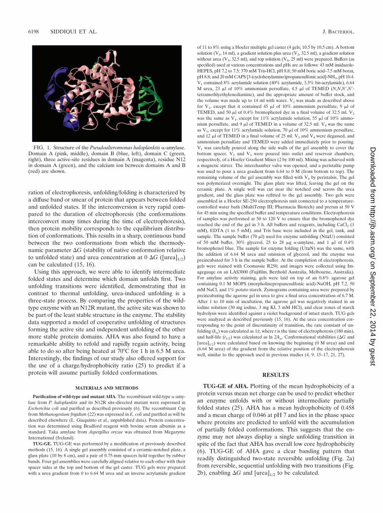
The cold-adapted �-amylase from Pseudoalteromonas halo-planktis (AHA) was the first cold-adapted enzyme to have anX-ray structure solved (1). This revealed that the enzyme con-sists of three domains (Fig. 1). Domain A is the N-terminaldomain which is composed of a (�/�)8-barrel forming one sideof the catalytic cleft (residues 1 to 86 and 130 to 356) and isable to bind a Tris molecule in the active site. Domain B(residues 87 to 129) protrudes from domain A and forms theother side of the active cleft. Domain C is the C-terminaldomain consisting of residues 357 to 448. A Ca2� ion is boundbetween domains A and B. The enzyme has evolved a lowconformational stability and high flexibility through a reduc-tion in the number and type of inter- and intramolecular in-teractions (12, 13), and the enhanced flexibility has resulted ina decreased enthalpy of activation with a concomitant im-provement in kcat (6, 8).
The lower stability of cold-adapted enzymes has been linkedto either a general reduction in “weak interactions” causingglobal flexibility or weakened interactions of specific regions
generating local flexibility. Site-directed mutagenesis studies ofAHA have revealed a global weakening of intramolecular in-teractions leading to an overall decrease in thermostability (6,14). In contrast, citrate synthase and chitobiase from Ar-throbacter species, phosphoglycerate kinase from Pseudomonasspecies, and lactate dehydrogenases from Antarctic fish havebeen suggested to possibly contain distinct heat-labile andheat-stable domains (14, 19).
AHA is the largest multidomain protein (�50 kDa) that hasbeen shown to unfold/refold reversibly by a cooperative two-state mechanism (7, 8, 12). Biophysical measurements of un-folding/refolding have been performed by differential scanningcalorimetry, circular dichroism, and fluorescence spectropho-tometry (6, 12), and the spectrophotometric methods haveclearly revealed cooperative unfolding through a single transi-tion (12).
In the case of mesophilic and thermophilic �-amylases, theoptimal growth temperature for activity closely corresponds tothe transition temperature, illustrating that the loss in activityis due to structural unfolding (8). In contrast, AHA loses�50% of its enzyme activity before reaching its melting tem-perature (Tm) (44°C), suggesting that a local rather than globalmechanism of unfolding may be involved (8). To further iden-tify the structures that may first unfold and to examine localversus global stability, we developed transverse urea gradient(TUG) gel electrophoresis (TUG-GE) (15, 16) for the cold-active �-amylase. TUG-GE can be used to distinguish confor-mational states of a protein caused by different structures un-folding/folding independently at different urea concentrations,thereby identifying multistep, sequential unfolding/foldingtransitions. If conformations interconvert slowly within the du-
* Corresponding author. Mailing address: School of Biotechnologyand Biomolecular Sciences, University of New South Wales, Sydney,2052 New South Wales, Australia. Phone: 61-2-93853516. Fax: 61-2-93852742. E-mail: [email protected].
6197
on Septem
ber 22, 2014 by guesthttp://jb.asm
.org/D
ownloaded from
ration of electrophoresis, unfolding/folding is characterized bya diffuse band or smear of protein that appears between foldedand unfolded states. If the interconversion is very rapid com-pared to the duration of electrophoresis (the conformationsinterconvert many times during the time of electrophoresis),then protein mobility corresponds to the equilibrium distribu-tion of conformations. This results in a sharp, continuous bandbetween the two conformations from which the thermody-namic parameter �G (stability of native conformation relativeto unfolded state) and urea concentration at 0 �G ([urea]1/2)can be calculated (15, 16).
Using this approach, we were able to identify intermediatefolded states and determine which domain unfolds first. Twounfolding transitions were identified, demonstrating that incontrast to thermal unfolding, urea-induced unfolding is athree-state process. By comparing the properties of the wild-type enzyme with an N12R mutant, the active site was shown tobe part of the least stable structure in the enzyme. The stabilitydata supported a model of cooperative unfolding of structuresforming the active site and independent unfolding of the othermore stable protein domains. AHA was also found to have aremarkable ability to refold and rapidly regain activity, beingable to do so after being heated at 70°C for 1 h in 6.5 M urea.Interestingly, the findings of our study also offered support forthe use of a charge/hydrophobicity ratio (25) to predict if aprotein will assume partially folded conformations.
MATERIALS AND METHODS
Purification of wild-type and mutant AHA. The recombinant wild-type �-amy-lase from P. haloplanktis and its N12R site-directed mutant were expressed inEscherichia coli and purified as described previously (6). The recombinant Cspfrom Methanogenium frigidum (22) was expressed in E. coli and purified as will bedescribed elsewhere (L. Giaquinto et al., unpublished data). Protein concentra-tion was determined using Bradford reagent with bovine serum albumin as astandard. Taka amylase from Aspergillus oryzae was obtained from MegazymeInternational (Ireland).
TUG-GE. TUG-GE was performed by a modification of previously describedmethods (15, 16). A single gel assembly consisted of a ceramic-notched plate, aglass plate (10 by 8 cm), and a pair of 0.75-mm spacers held together by rubberbands. Four gel assemblies were carefully aligned relative to each other with theirspacer sides at the top and bottom of the gel caster. TUG gels were preparedwith a urea gradient from 0 to 6.64 M urea and an inverse acrylamide gradient
of 11 to 8% using a Hoefer multiple gel caster (4 gels; 10.5 by 10.5 cm). A bottomsolution (V1, 14 ml), a gradient solution plus urea (V2, 32.5 ml), a gradient solutionwithout urea (V3, 32.5 ml), and top solution (V4, 25 ml) were prepared. Buffers (asspecified) used at various concentrations and pHs are as follows: 43 mM imidazole-HEPES, pH 7.2 to 7.5; 370 mM Tris-HCl, pH 8.8; 50 mM boric acid–7.5 mM borax,pH 8.8; and 20 mM CAPS [3-(cyclohexylamino)propanesulfonic acid]-NH3, pH 10.4.V1 contained 8% acrylamide solution (40% acrylamide, 3.3% bis-acrylamide), 6.64M urea, 23 �l of 10% ammonium persulfate, 4.5 �l of TEMED (N,N,N�,N�-tetramethlyethylenediamine), and the appropriate amount of buffer stock, andthe volume was made up to 14 ml with water. V2 was made as described abovefor V1, except that it contained 45 �l of 10% ammonium persulfate, 9 �l ofTEMED, and 50 �l of 0.4% bromophenol dye in a final volume of 32.5 ml. V3
was the same as V1, except for 11% acrylamide solution, 55 �l of 10% ammo-nium persulfate, and 9 �l of TEMED in a volume of 32.5 ml. V4 was the sameas V1, except for 11% acrylamide solution, 70 �l of 10% ammonium persulfate,and 12 �l of TEMED in a final volume of 25 ml. V3 and V4 were degassed, andammonium persulfate and TEMED were added immediately prior to pouring.V1 was carefully poured along the side walls of the gel assembly to cover thebottom spacer. V2 and V3 were poured into outlet and reservoir chambers,respectively, of a Hoefer Gradient Mixer (2 by 100 ml). Mixing was achieved witha magnetic stirrer. The interchamber valve was opened, and a peristaltic pumpwas used to pour a urea gradient from 6.64 to 0 M (from bottom to top). Theremaining volume of the gel assembly was filled with V4 by peristalsis. The gelwas polymerized overnight. The glass plate was lifted, leaving the gel on theceramic plate. A single well was cut near the notched end across the ureagradient, and the glass plate was refitted to the gel assembly. Two gels wereassembled in a Hoefer SE-250 electrophoresis unit connected to a temperature-controlled water bath (MultiTemp III; Pharmacia Biotech) and prerun at 50 Vfor 45 min using the specified buffer and temperature conditions. Electrophoresisof samples was performed at 50 to 120 V to ensure that the bromophenol dyereached the end of the gel in 3 h. All buffers and reagents, including CaCl2 (1mM), EDTA (1 to 5 mM), and Tris base were included in the gel, tank, andsample. The sample buffer (70 �l) used for enzyme unfolding (NuU) consistedof 50 mM buffer, 30% glycerol, 25 to 28 �g �-amylase, and 1 �l of 0.4%bromophenol blue. The sample for enzyme folding (UuN) was the same, withthe addition of 6.64 M urea and omission of glycerol, and the enzyme waspreincubated for 3 h in the sample buffer. At the completion of electrophoresis,gels were stained with Coomassie R250, and images were collected using Im-agegauge on an LAS3000 (Fujifilm, Berthold Australia, Melbourne, Australia).For amylase activity staining, gels were laid on top of an 0.8% agarose gelcontaining 0.1 M MOPS (morpholinepropanesulfonic acid)-NaOH, pH 7.2, 50mM NaCl, and 1% potato starch. Zymograms containing urea were prepared bypreincubating the agarose gel in urea to give a final urea concentration of 6.7 M.After 1 to 10 min of incubation, the agarose gel was negatively stained in aniodine solution (30 mg iodine, 3 mg KI, 5 mM HCl), and clear zones of starchhydrolysis were identified against a violet background of intact starch. TUG gelswere analyzed as described previously (15, 16). At the urea concentration cor-responding to the point of discontinuity of transition, the rate constant of un-folding (ku) was calculated as 1/t, where t is the time of electrophoresis (180 min),and half-life (t1/2) was calculated as ln 2/ku. Conformational stabilities (�G and[urea]1/2) were calculated based on knowing the beginning (0 M urea) and end(6.64 M urea) of the gradient from the relative position of the electrophoresiswell, similar to the approach used in previous studies (4, 9, 15–17, 21, 27).
RESULTS
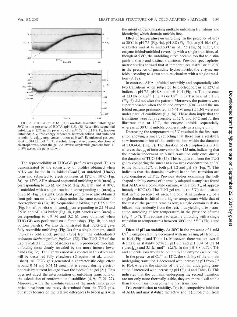
TUG-GE of AHA. Plotting of the mean hydrophobicity of aprotein versus mean net charge can be used to predict whetheran enzyme unfolds with or without intermediate partiallyfolded states (25). AHA has a mean hydrophobicity of 0.458and a mean charge of 0.046 at pH 7 and lies in the phase spacewhere proteins are predicted to unfold with the accumulationof partially folded conformations. This suggests that the en-zyme may not always display a single unfolding transition inspite of the fact that AHA has overall low core hydrophobicity(6). TUG-GE of AHA gave a clear banding pattern thatreadily distinguished two-state reversible unfolding (Fig. 2a)from reversible, sequential unfolding with two transitions (Fig.2b), enabling �G and [urea]1/2 to be calculated.
FIG. 1. Structure of the Pseudoalteromonas haloplanktis �-amylase.Domain A (pink, middle), domain B (blue, left), domain C (green,right), three active-site residues in domain A (magenta), residue N12in domain A (green), and the calcium ion between domains A and B(red) are shown.
6198 SIDDIQUI ET AL. J. BACTERIOL.
on Septem
ber 22, 2014 by guesthttp://jb.asm
.org/D
ownloaded from
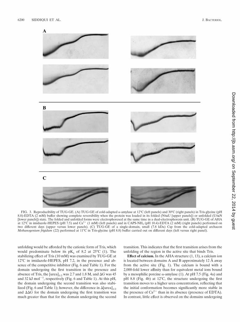
The reproducibility of TUG-GE profiles was good. This isdemonstrated by the consistency of profiles obtained whenAHA was loaded in its folded (NuU) or unfolded (UuN)form and subjected to electrophoresis at 12°C or 30°C (Fig.3a). At 12°C, AHA showed sequential unfolding with [urea]1/2
corresponding to 1.3 M and 3.6 M (Fig. 3a, left), and at 30°C,it unfolded with a single transition corresponding to [urea]1/2
of 2.5 M (Fig. 3a, right). Consistent profiles were also obtainedfrom gels run on different days under the same conditions ofelectrophoresis (Fig. 3b). Sequential unfolding in pH 7.5 buffer(Fig. 3b, left panels) with [urea]1/2 corresponding to 2.1 M and3.5 M and pH 10.4 buffer (Fig. 3b, right panels) with [urea]1/2
corresponding to 0.9 M and 3.2 M were obtained whenTUG-GE was performed on different days (Fig. 3b, top andbottom panels). We also demonstrated that TUG-GE gavefully reversible unfolding (Fig. 3c) for a single domain, small(7.8-kDa) cold shock protein (Csp) from the cold-adaptedarchaeon Methanogenium frigidum (22). The TUG-GE of theCsp revealed a number of isomers with reproducible two-stateunfolding most clearly revealed by the more intense lowerband (Fig. 3c). The Csp was used as a control in this study andwill be described fully elsewhere (Giaquinto et al., unpub-lished). All TUG gels generated a characteristic edge effectaround 0 M and 6.64 M urea that is caused during electro-phoresis by current leakage down the sides of the gel (21). Thisdoes not affect the interpretation of unfolding transitions orthe calculation of conformational stabilities (4, 9, 17, 21, 27).Moreover, while the absolute values of thermodynamic prop-erties have been accurately determined from the TUG gels,our study focuses on the relative differences between gels with
the intent of demonstrating multiple unfolding transitions andidentifying which domain unfolds first.
Effect of temperature on unfolding. In the presence of ureaat 30°C in pH 7.5 (Fig. 4a), pH 8.8 (Fig. 4b), or pH 10.4 (Fig.4c) buffer and at 42 and 53°C in pH 7.5 (Fig. 5) buffer, theenzyme folded/unfolded reversibly with a single transition, al-though at 53°C, the unfolding curve became too flat to distin-guish a sharp and distinct transition. Previous spectrophoto-metric studies showed that at temperatures �40°C or at 20°Cin the presence of guanidine hydrochloride, the enzyme un-folds according to a two-state mechanism with a single transi-tion (8, 12).
In contrast, AHA unfolded reversibly and sequentially withtwo transitions when subjected to electrophoresis at 12°C inbuffers at pH 7.5, pH 8.8, and pH 10.4 (Fig. 4). The presenceof EDTA or Ca2� (Fig. 4) or Ca2� plus Tris base at pH 7.2(Fig. 6) did not alter the pattern. Moreover, the patterns weresuperimposable when the folded enzyme (NuU) and the un-folded enzyme preincubated in 6.64 M urea (UuN) were rununder parallel conditions (Fig. 3a). These data imply that thetransitions were fully reversible at 12°C and 30°C and furtherillustrate that at 12°C, the enzyme unfolds sequentially,whereas at 30°C, it unfolds cooperatively as a single unit.
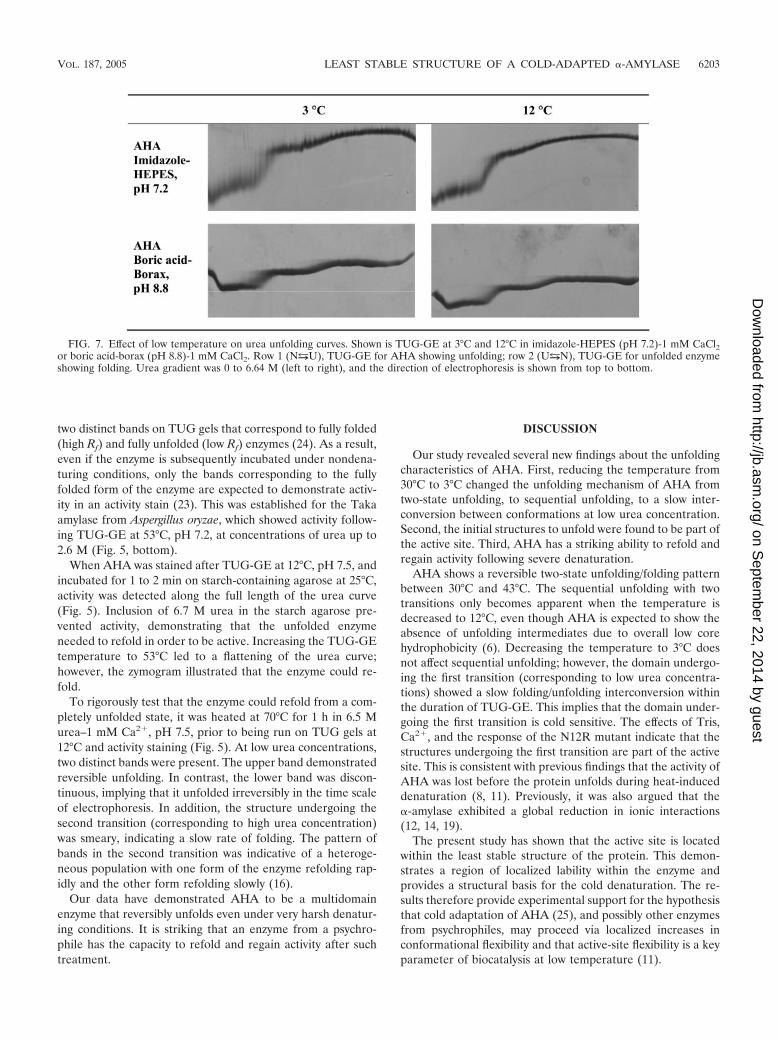
Decreasing the temperature to 3°C resulted in the first tran-sition showing a smear, reflecting that there was a relativelyslow interconversion of the conformations within the durationof TUG-GE (Fig. 7). The duration of electrophoresis is 3 h,whereas the t1/2 of interconversion is �125 min, indicating thatthe protein underwent an NuU transition only once duringthe duration of TUG-GE (15). This is apparent from the TUGgel by comparing the smear at a low urea concentration at 3°Cto the band at 12°C at both pH 7.2 and pH 8.8 (Fig. 7). Thisindicates that the domains involved in the first transition arecold denatured at 3°C. Previous studies examining the bell-shaped stability curves of thermally adapted �-amylases foundthat AHA was a cold-labile enzyme, with a low Tm of approx-imately �10°C (8). The TUG gel results (at 3°C) demonstratethat in the presence of urea, the cold denaturation Tm of asingle domain is shifted to a higher temperature while that ofthe rest of the protein remains low; a single domain is desta-bilized independently from the rest, thus yielding a two-tran-sition unfolding at low temperature in the presence of urea(Fig. 4 to 7). This contrasts to enzyme unfolding with a singletransition at temperatures between 30°C and 53°C (Fig. 4 and5).
Effect of pH on stability. At 30°C in the presence of 1 mMCa2�, enzyme stability decreased with increasing pH from 7.5to 10.4 (Fig. 4 and Table 1). Moreover, there was an overalldecrease in stability between pH 7.5 and pH 10.4 of 0.2 M([urea]1/2) and 3.1 kJ mol�1 (�G). In the pH 8.8 buffer, Trisand chloride ions would be bound by the enzyme (see below).
In the presence of Ca2� at 12°C, the stability of the domainundergoing transition 1 decreased with increasing pH from 7.5to 10.4, whereas the stability of the domain undergoing tran-sition 2 increased with increasing pH (Fig. 4 and Table 1). Thisindicates that the domains undergoing the second transitionare not only more thermally stable, they are more alkali stablethan the domain undergoing the first transition.
Tris contribution to stability. Tris is a competitive inhibitorof AHA and may stabilize the active site (1). Protection from
FIG. 2. TUG-GE of AHA. (A) Two-state reversible unfolding at30°C in the presence of EDTA (pH 8.8). (B) Reversible sequentialunfolding at 12°C in the presence of 1 mM Ca2�, pH 8.8. Fu, fractionunfolded; �G, free-energy difference between folded and unfoldedprotein; [urea]1/2, urea concentration at 0 �G; R, universal gas con-stant (8.314 kJ mol�1); T, absolute temperature; arrow, direction ofelectrophoresis down the gel. An inverse acrylamide gradient from 11to 8% across the gel is shown.
VOL. 187, 2005 LEAST STABLE STRUCTURE OF A COLD-ADAPTED �-AMYLASE 6199
on Septem
ber 22, 2014 by guesthttp://jb.asm
.org/D
ownloaded from
unfolding would be afforded by the cationic form of Tris, whichwould predominate below its pKa of 8.2 at 25°C (1). Thestabilizing effect of Tris (10 mM) was examined by TUG-GE at12°C in imidazole-HEPES, pH 7.2, in the presence and ab-sence of the competitive inhibitor (Fig. 6 and Table 1). For thedomain undergoing the first transition in the presence andabsence of Tris, the [urea]1/2 was 2.7 and 1.8 M, and �G was 45and 32 kJ mol�1, respectively (Fig. 6 and Table 1). At this pH,the domain undergoing the second transition was also stabi-lized (Fig. 6 and Table 1); however, the difference in �[urea]1/2
and ��G for the domain undergoing the first transition wasmuch greater than that for the domain undergoing the second
transition. This indicates that the first transition arises from theunfolding of the region in the active site that binds Tris.
Effect of calcium. In the AHA structure (1, 13), a calcium ionis located between domains A and B approximately 12 Å awayfrom the active site (Fig. 1). The calcium is bound with a2,000-fold lower affinity than for equivalent metal ions boundby a mesophilic porcine �-amylase (1). At pH 7.5 (Fig. 4a) andpH 8.8 (Fig. 4b) at 12°C, the structure undergoing the firsttransition moves to a higher urea concentration, reflecting thatthe initial conformation becomes significantly more stable inthe presence of Ca2� than in its absence (presence of EDTA).In contrast, little effect is observed on the domains undergoing
FIG. 3. Reproducibility of TUG-GE. (A) TUG-GE of cold-adapted �-amylase at 12°C (left panels) and 30°C (right panels) in Tris-glycine (pH8.8)-EDTA (2 mM) buffer showing complete reversibility when the protein was loaded in its folded (NuU [upper panels]) or unfolded (UuN[lower panels]) state. The folded and unfolded forms were electrophoresed at the same time in a dual-electrophoresis unit. (B) TUG-GE of AHAat 12°C in imidazole-HEPES (pH 7.5) and Ca2� (1 mM) (left panels) and in CAPS-NH3 (pH 10.4)-EDTA (2 mM) (right panels) performed ontwo different days (upper versus lower panels). (C) TUG-GE of a single-domain, small (7.8 kDa) Csp from the cold-adapted archaeonMethanogenium frigidum (22) performed at 11°C in Tris-glycine (pH 8.8) buffer carried out on different days (left versus right panel).
6200 SIDDIQUI ET AL. J. BACTERIOL.
on Septem
ber 22, 2014 by guesthttp://jb.asm
.org/D
ownloaded from
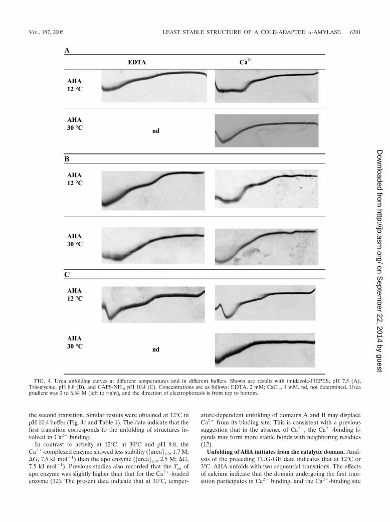
the second transition. Similar results were obtained at 12°C inpH 10.4 buffer (Fig. 4c and Table 1). The data indicate that thefirst transition corresponds to the unfolding of structures in-volved in Ca2� binding.
In contrast to activity at 12°C, at 30°C and pH 8.8, theCa2�-complexed enzyme showed less stability ([urea]1/2, 1.7 M;�G, 7.5 kJ mol�1) than the apo enzyme ([urea]1/2, 2.5 M; �G,7.5 kJ mol�1). Previous studies also recorded that the Tm ofapo enzyme was slightly higher than that for the Ca2�-loadedenzyme (12). The present data indicate that at 30°C, temper-
ature-dependent unfolding of domains A and B may displaceCa2� from its binding site. This is consistent with a previoussuggestion that in the absence of Ca2�, the Ca2�-binding li-gands may form more stable bonds with neighboring residues(12).
Unfolding of AHA initiates from the catalytic domain. Anal-ysis of the preceding TUG-GE data indicates that at 12°C or3°C, AHA unfolds with two sequential transitions. The effectsof calcium indicate that the domain undergoing the first tran-sition participates in Ca2� binding, and the Ca2�-binding site
FIG. 4. Urea unfolding curves at different temperatures and in different buffers. Shown are results with imidazole-HEPES, pH 7.5 (A),Tris-glycine, pH 8.8 (B), and CAPS-NH3, pH 10.4 (C). Concentrations are as follows: EDTA, 2 mM; CaCl2, 1 mM. nd, not determined. Ureagradient was 0 to 6.64 M (left to right), and the direction of electrophoresis is from top to bottom.
VOL. 187, 2005 LEAST STABLE STRUCTURE OF A COLD-ADAPTED �-AMYLASE 6201
on Septem
ber 22, 2014 by guesthttp://jb.asm
.org/D
ownloaded from
is known to be located near the active site (1). The Tris-induced thermodynamic stabilization of the domain undergo-ing the first transition also indicates that the active site is thestructure which initially unfolds. In order to prove that the firsttransition corresponds to unfolding of structures from domainA, which bears the catalytic residues, we applied TUG-GE toan N12R mutant of the �-amylase (6). The N12R mutation isin domain A (Fig. 1), which results in N12R forming an extrasalt bridge with Asp15, thereby decreasing the unfolding co-operativity of the enzyme (6, 7). Differential scanning calorim-etry has shown that the N12R mutant exhibits a predominantlyirreversible pattern of unfolding (6, 7). In fact, this mutant hasbeen used in lieu of wild-type AHA to study kinetic stabilitydue to its irreversible unfolding (8).
The mutant AHA run at 12°C showed an unfolding patternsimilar to that of the wild-type enzyme (unpublished results);therefore, drastic TUG-GE conditions were chosen to enablethe structural differences between the wild-type and mutantproteins to become evident. The TUG gel was run at 12°C for
45 min for the separation of the domains undergoing sequen-tial transitions and then shifted to 47°C for 90 min to modifythe unfolding mechanism and facilitate the split in the curvedue to irreversible denaturation (Fig. 8, top). The break, whichoccurred at 1 M urea, was caused by low mobility resultingfrom the increased hydrodynamic volume of the irreversiblyunfolded form of the enzyme (Fig. 8, top). This demonstratesthat an extra salt bridge in domain A produces an irreversibleunfolding within the duration of TUG-GE that corresponds tothe first transition. It can therefore be concluded that cold-adapted �-amylase has two stability domains. The cold-labiledomain contains the active site (effect of Tris), the Ca2�-binding site (that is part of the active site), and the N-terminalpart of domain A, which contains the catalytic acid residues(effect of both Tris and N12R), whereas the more stable do-mains (part of domain A and domain C) correspond to thestructural elements undergoing the second transition.
A high level of unfolding reversibility at high temperatures.Multidomain enzymes typically unfold irreversibly to produce
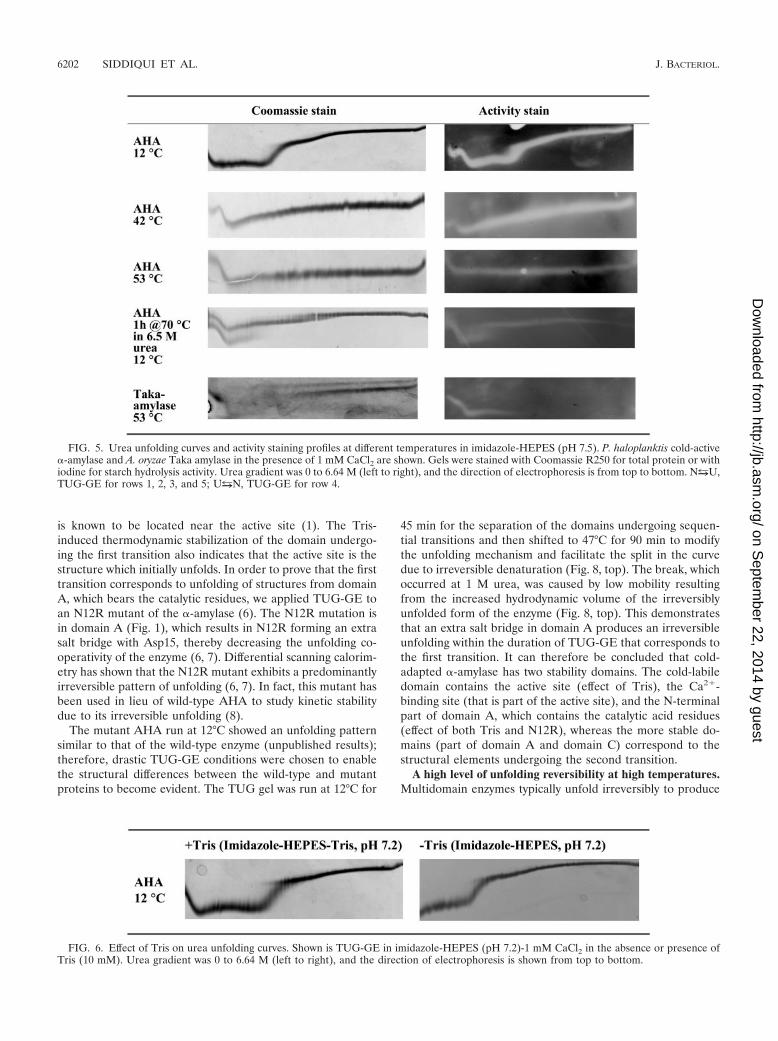
FIG. 5. Urea unfolding curves and activity staining profiles at different temperatures in imidazole-HEPES (pH 7.5). P. haloplanktis cold-active�-amylase and A. oryzae Taka amylase in the presence of 1 mM CaCl2 are shown. Gels were stained with Coomassie R250 for total protein or withiodine for starch hydrolysis activity. Urea gradient was 0 to 6.64 M (left to right), and the direction of electrophoresis is from top to bottom. NuU,TUG-GE for rows 1, 2, 3, and 5; UuN, TUG-GE for row 4.
FIG. 6. Effect of Tris on urea unfolding curves. Shown is TUG-GE in imidazole-HEPES (pH 7.2)-1 mM CaCl2 in the absence or presence ofTris (10 mM). Urea gradient was 0 to 6.64 M (left to right), and the direction of electrophoresis is shown from top to bottom.
6202 SIDDIQUI ET AL. J. BACTERIOL.
on Septem
ber 22, 2014 by guesthttp://jb.asm
.org/D
ownloaded from
two distinct bands on TUG gels that correspond to fully folded(high Rf) and fully unfolded (low Rf) enzymes (24). As a result,even if the enzyme is subsequently incubated under nondena-turing conditions, only the bands corresponding to the fullyfolded form of the enzyme are expected to demonstrate activ-ity in an activity stain (23). This was established for the Takaamylase from Aspergillus oryzae, which showed activity follow-ing TUG-GE at 53°C, pH 7.2, at concentrations of urea up to2.6 M (Fig. 5, bottom).
When AHA was stained after TUG-GE at 12°C, pH 7.5, andincubated for 1 to 2 min on starch-containing agarose at 25°C,activity was detected along the full length of the urea curve(Fig. 5). Inclusion of 6.7 M urea in the starch agarose pre-vented activity, demonstrating that the unfolded enzymeneeded to refold in order to be active. Increasing the TUG-GEtemperature to 53°C led to a flattening of the urea curve;however, the zymogram illustrated that the enzyme could re-fold.
To rigorously test that the enzyme could refold from a com-pletely unfolded state, it was heated at 70°C for 1 h in 6.5 Murea–1 mM Ca2�, pH 7.5, prior to being run on TUG gels at12°C and activity staining (Fig. 5). At low urea concentrations,two distinct bands were present. The upper band demonstratedreversible unfolding. In contrast, the lower band was discon-tinuous, implying that it unfolded irreversibly in the time scaleof electrophoresis. In addition, the structure undergoing thesecond transition (corresponding to high urea concentration)was smeary, indicating a slow rate of folding. The pattern ofbands in the second transition was indicative of a heteroge-neous population with one form of the enzyme refolding rap-idly and the other form refolding slowly (16).
Our data have demonstrated AHA to be a multidomainenzyme that reversibly unfolds even under very harsh denatur-ing conditions. It is striking that an enzyme from a psychro-phile has the capacity to refold and regain activity after suchtreatment.
DISCUSSION
Our study revealed several new findings about the unfoldingcharacteristics of AHA. First, reducing the temperature from30°C to 3°C changed the unfolding mechanism of AHA fromtwo-state unfolding, to sequential unfolding, to a slow inter-conversion between conformations at low urea concentration.Second, the initial structures to unfold were found to be part ofthe active site. Third, AHA has a striking ability to refold andregain activity following severe denaturation.
AHA shows a reversible two-state unfolding/folding patternbetween 30°C and 43°C. The sequential unfolding with twotransitions only becomes apparent when the temperature isdecreased to 12°C, even though AHA is expected to show theabsence of unfolding intermediates due to overall low corehydrophobicity (6). Decreasing the temperature to 3°C doesnot affect sequential unfolding; however, the domain undergo-ing the first transition (corresponding to low urea concentra-tions) showed a slow folding/unfolding interconversion withinthe duration of TUG-GE. This implies that the domain under-going the first transition is cold sensitive. The effects of Tris,Ca2�, and the response of the N12R mutant indicate that thestructures undergoing the first transition are part of the activesite. This is consistent with previous findings that the activity ofAHA was lost before the protein unfolds during heat-induceddenaturation (8, 11). Previously, it was also argued that the�-amylase exhibited a global reduction in ionic interactions(12, 14, 19).
The present study has shown that the active site is locatedwithin the least stable structure of the protein. This demon-strates a region of localized lability within the enzyme andprovides a structural basis for the cold denaturation. The re-sults therefore provide experimental support for the hypothesisthat cold adaptation of AHA (25), and possibly other enzymesfrom psychrophiles, may proceed via localized increases inconformational flexibility and that active-site flexibility is a keyparameter of biocatalysis at low temperature (11).
FIG. 7. Effect of low temperature on urea unfolding curves. Shown is TUG-GE at 3°C and 12°C in imidazole-HEPES (pH 7.2)-1 mM CaCl2or boric acid-borax (pH 8.8)-1 mM CaCl2. Row 1 (NuU), TUG-GE for AHA showing unfolding; row 2 (UuN), TUG-GE for unfolded enzymeshowing folding. Urea gradient was 0 to 6.64 M (left to right), and the direction of electrophoresis is shown from top to bottom.
VOL. 187, 2005 LEAST STABLE STRUCTURE OF A COLD-ADAPTED �-AMYLASE 6203
on Septem
ber 22, 2014 by guesthttp://jb.asm
.org/D
ownloaded from
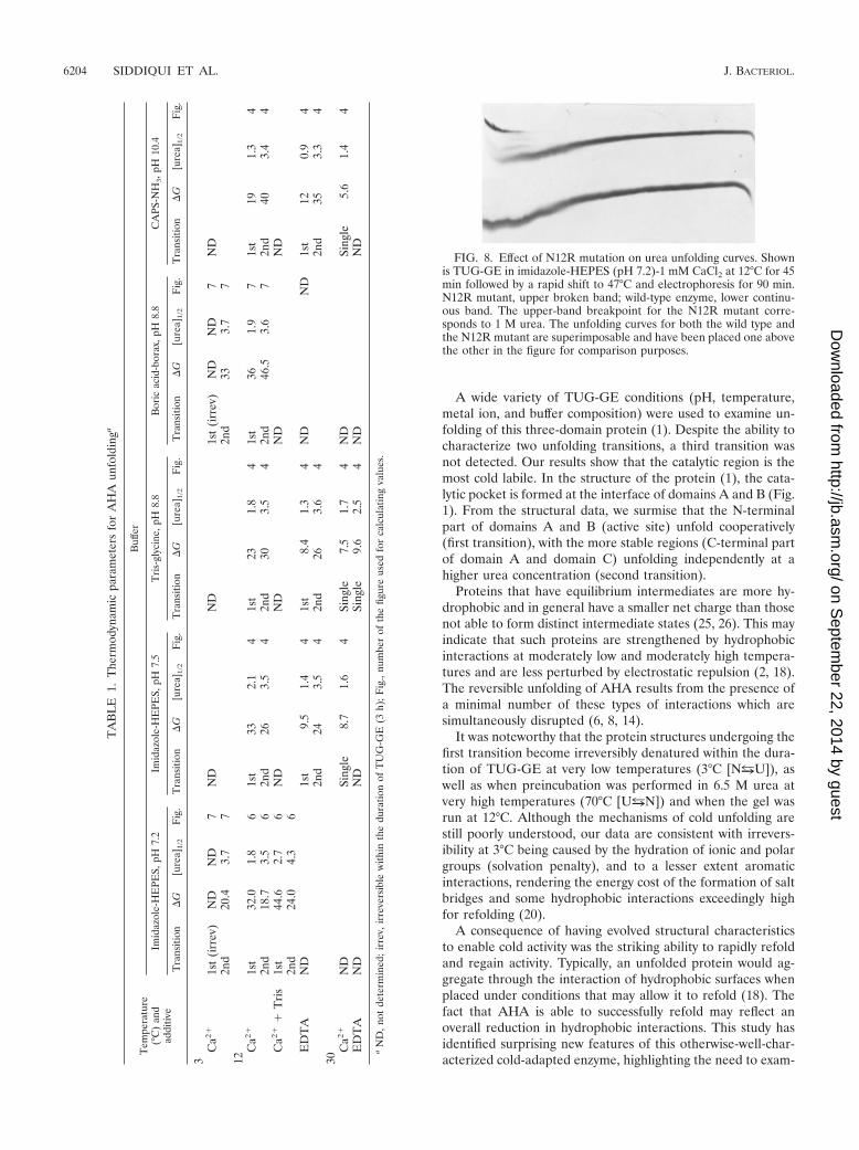
A wide variety of TUG-GE conditions (pH, temperature,metal ion, and buffer composition) were used to examine un-folding of this three-domain protein (1). Despite the ability tocharacterize two unfolding transitions, a third transition wasnot detected. Our results show that the catalytic region is themost cold labile. In the structure of the protein (1), the cata-lytic pocket is formed at the interface of domains A and B (Fig.1). From the structural data, we surmise that the N-terminalpart of domains A and B (active site) unfold cooperatively(first transition), with the more stable regions (C-terminal partof domain A and domain C) unfolding independently at ahigher urea concentration (second transition).
Proteins that have equilibrium intermediates are more hy-drophobic and in general have a smaller net charge than thosenot able to form distinct intermediate states (25, 26). This mayindicate that such proteins are strengthened by hydrophobicinteractions at moderately low and moderately high tempera-tures and are less perturbed by electrostatic repulsion (2, 18).The reversible unfolding of AHA results from the presence ofa minimal number of these types of interactions which aresimultaneously disrupted (6, 8, 14).
It was noteworthy that the protein structures undergoing thefirst transition become irreversibly denatured within the dura-tion of TUG-GE at very low temperatures (3°C [NuU]), aswell as when preincubation was performed in 6.5 M urea atvery high temperatures (70°C [UuN]) and when the gel wasrun at 12°C. Although the mechanisms of cold unfolding arestill poorly understood, our data are consistent with irrevers-ibility at 3°C being caused by the hydration of ionic and polargroups (solvation penalty), and to a lesser extent aromaticinteractions, rendering the energy cost of the formation of saltbridges and some hydrophobic interactions exceedingly highfor refolding (20).
A consequence of having evolved structural characteristicsto enable cold activity was the striking ability to rapidly refoldand regain activity. Typically, an unfolded protein would ag-gregate through the interaction of hydrophobic surfaces whenplaced under conditions that may allow it to refold (18). Thefact that AHA is able to successfully refold may reflect anoverall reduction in hydrophobic interactions. This study hasidentified surprising new features of this otherwise-well-char-acterized cold-adapted enzyme, highlighting the need to exam-
FIG. 8. Effect of N12R mutation on urea unfolding curves. Shownis TUG-GE in imidazole-HEPES (pH 7.2)-1 mM CaCl2 at 12°C for 45min followed by a rapid shift to 47°C and electrophoresis for 90 min.N12R mutant, upper broken band; wild-type enzyme, lower continu-ous band. The upper-band breakpoint for the N12R mutant corre-sponds to 1 M urea. The unfolding curves for both the wild type andthe N12R mutant are superimposable and have been placed one abovethe other in the figure for comparison purposes.
TA
BL
E1.
The
rmod
ynam
icpa
ram
eter
sfo
rA
HA
unfo
ldin
ga
Tem
pera
ture
(°C
)an
dad
ditiv
e
Buf
fer
Imid
azol
e-H
EPE
S,pH
7.2
Imid
azol
e-H
EPE
S,pH
7.5
Tri
s-gl
ycin
e,pH
8.8
Bor
icac
id-b
orax
,pH
8.8
CA
PS-N
H3,p
H10
.4
Tra
nsiti
on�
G[u
rea]
1/2
Fig
.T
rans
ition
�G
[ure
a]1/2
Fig
.T
rans
ition
�G
[ure
a]1/2
Fig
.T
rans
ition
�G
[ure
a]1/2
Fig
.T
rans
ition
�G
[ure
a]1/2
Fig
.
3C
a2�1s
t(i
rrev
)N
DN
D7
ND
ND
1st
(irr
ev)
ND
ND
7N
D2n
d20
.43.
77
2nd
333.
77
12C
a2�1s
t32
.01.
86
1st
332.
14
1st
231.
84
1st
361.
97
1st
191.
34
2nd
18.7
3.5
62n
d26
3.5
42n
d30
3.5
42n
d46
.53.
67
2nd
403.
44
Ca2�
�T
ris
1st
44.6
2.7
6N
DN
DN
DN
D2n
d24
.04.
36
ED
TA
ND
1st
9.5
1.4
41s
t8.
41.
34
ND
ND
1st
120.
94
2nd
243.
54
2nd
263.
64
2nd
353.
34
30C
a2�N
DSi
ngle
8.7
1.6
4Si
ngle
7.5
1.7
4N
DSi
ngle
5.6
1.4
4E
DT
AN
DN
DSi
ngle
9.6
2.5
4N
DN
D
aN
D,n
otde
term
ined
;irr
ev,i
rrev
ersi
ble
with
inth
edu
ratio
nof
TU
G-G
E(3
h);F
ig.,
num
ber
ofth
efig
ure
used
for
calc
ulat
ing
valu
es.
6204 SIDDIQUI ET AL. J. BACTERIOL.
on Septem
ber 22, 2014 by guesthttp://jb.asm
.org/D
ownloaded from
ine the folding/unfolding properties of other cold-adapted en-zymes using a similar approach.
ACKNOWLEDGMENTS
The work was supported by the Australian Research Council.Thanks to Lily Ting for careful reading of the manuscript and Paul
Curmi and Anne Poljak for helpful discussions. S.D. is a postdoctoralresearcher from the Fonds National de la Recherche Scientifique(FNRS, Belgium). We express our gratitude to the reviewers for theirparticular insight and constructive comments.
REFERENCES
1. Aghajari, N., G. Feller, C. Gerday, and R. Haser. 1998. Crystal structure ofthe psychrophilic �-amylase from Alteromonas haloplanctis in its native formand complexed with an inhibitor. Protein Sci. 7:564–572.
2. Cavicchioli, R., and K. S. Siddiqui. 2004. Cold-adapted enzymes, p. 615–638.In A. Pandey, C. Webb, C. R. Soccol, and C. Larroche (ed.), Enzymetechnology. AsiaTech Publishers, New Delhi, India.
3. Cavicchioli, R., K. S. Siddiqui, D. Andrews, and K. R. Sowers. 2002. Low-temperature extremophiles and their applications. Curr. Opin. Biotechnol.13:253–261.
4. Creighton, T. E., and R. H. Paine. 1980. Unfolding and refolding of Staph-ylococcus aureus penicillinase by urea-gradient electrophoresis. J. Mol. Biol.137:431–436.
5. D’Amico, S., P. Claverie, C. Collins, D. Georlette, E. Gratia, A. Hoyoux,M.-A. Meuwis, G. Feller, and C. Gerday. 2002. Molecular basis of coldadaptation. Philos. Trans. R. Soc. Lond. B 357:917–925.
6. D’Amico, S., C. Gerday, and G. Feller. 2001. Structural determinants of coldadaptation and stability in a large protein. J. Biol. Chem. 276:25791–25796.
7. D’Amico, S., C. Gerday, and G. Feller. 2003. Temperature adaptation ofproteins: engineering mesophilic-like activity and stability in a cold-adapted�-amylase. J. Mol. Biol. 332:981–988.
8. D’Amico, S., J. C. Marx, C. Gerday, and G. Feller. 2003. Activity-stabilityrelationship in extremophilic enzymes. J. Biol. Chem. 278:7891–7896.
9. Evans. R. W., and J. Williams. 1980. The electrophoresis of transferrins inurea/polyacrylamide gels. Biochem. J. 189:541–546.
10. Feller, G. 2003. Molecular adaptations to cold in psychrophilic enzymes.Cell. Mol. Life Sci. 60:648–662.
11. Feller, G., and C. Gerday. 2003. Psychrophilic enzymes: hot topics in coldadaptation. Nat. Rev. Microbiol. 1:200–208.
12. Feller, G., D. D’Amico, and C. Gerday. 1999. Thermodynamic stability of acold-active �-amylase from Antarctic bacterium Alteromonas haloplanctis.Biochemistry 38:4613–4619.
13. Feller, G., F. Payan, F. Theys, M. Qian, R. Hasser, and C. Gerday. 1994.
Stability and structural analysis of �-amylase from the Antarctic psychrophileAlteromonas haloplanctis A23. Eur. J. Biochem. 222:441–447.
14. Georlette, D., V. Blaise, T. Collins, S. D’Amico, E. Gratia, A. Hoyoux, J.-C.Marx, G. Sonan, G. Feller, and C. Gerday. 2004. Some like it cold: bioca-talysis at low temperatures. FEMS Microbiol. Rev. 28:25–42.
15. Goldenberg, D. P. 1989. Analysis of protein conformation by gel electro-phoresis, p. 225–250. In T. E. Creighton (ed.), Protein structure: a practicalapproach. IRL Press, Oxford, United Kingdom.
16. Goldenberg, D. P., and T. E. Creighton. 1984. Gel electrophoresis in studiesof protein conformation and folding. Anal. Biochem. 138:1–18.
17. Kashiwagi, K., K. Shiba, K. Fukami-Kobayashi, T. Noda, K. Nishikawa, andH. Noguchi. 2003. Characterization of folding pathways of the type-1 andtype-2 periplasmic binding proteins MglB and ArgT. J. Biochem. 133:371–376.
18. Kumar, S., J.-G. Tsai, and R. Nussinov. 2002. Maximal stabilities of revers-ible two-state proteins. Biochemistry 41:5359–5374.
19. Lonhienne, T., C. Gerday, and G. Feller. 2000. Psychrophilic enzymes: re-visiting the thermodynamic parameters of activation may explain local flex-ibility. Biochim. Biophys. Acta 1543:1–10.
20. Makhtadze, G. I., and P. L. Privalov. 1994. Hydration effects in proteinunfolding. Biophys. Chem. 51:291–309.
21. Mast, A. E., J. J. Enghild, S. V. Pizzo, and G. Salvesen. 1991. Analysis of theplasma elimination kinetics and conformational stabilities of native, protein-ase-complexed, and reactive site cleaved serpins: comparison of �1-protein-ase inhibitor, �1-antichymotrypsin, antithrombin III, �2-antiplasmin, angio-tensinogen, and ovalbumin. Biochemistry 30:1723–1730.
22. Saunders, N., T. Thomas, P. M. G. Curmi, J. S. Mattick, E. Kuczek, R. Slade,J. Davis, P. D. Franzmann, D. Boone, K. Rusterholtz, R. Feldman, C. Gates,S. Bench, K. Sowers, K. Kadner, A. Aerts, P. Dehal, C. Detter, T. Glavina, S.Lucas, P. Richardson, F. Larimer, L. Hauser, M. Land, and R. Cavicchioli.2003. Mechanisms of thermal adaptation revealed from the genomes of theAntarctic Archaea, Methanogenium frigidum and Methanococcoides burtonii.Genome Res. 13:1580–1588.
23. Siddiqui, K. S. 1990. Proteolytic nicking and carboxyl group modification ofArthrobacter D-xylose isomerase. Ph.D. thesis. London University, London,United Kingdom.
24. Siddiqui, K. S., M. Rangarajan, B. S. Hartley, A. Kitmitto, M. Panico, I. P.Blench, and H. R. Morris. 1993. Arthrobacter D-xylose isomerase: partialproteolysis with thermolysin. Biochem. J. 289:201–208.
25. Uversky, V. N. 2002. Cracking the folding code. Why do some proteins adoptpartially folded conformations, whereas others don’t? FEBS Lett. 514:181–183.
26. Uversky, V. N., J. R. Gillespie, and A. J. Fink. 2000. Why are “nativelyunfolded” proteins unstructured under physiologic conditions? Proteins 41:415–427.
27. Yamashita, H., T. Nakatsuka, and M. Hirose. 1995. Structural and functionalcharacteristics of partially disulfide-reduced intermediates of ovotransferrinN lobe. J. Biol. Chem. 270:29806–29812.
VOL. 187, 2005 LEAST STABLE STRUCTURE OF A COLD-ADAPTED �-AMYLASE 6205
on Septem
ber 22, 2014 by guesthttp://jb.asm
.org/D
ownloaded from
Copyright © 2022 FDOKUMEN