Salivary α-Amylase As a Measure of Endogenous Adrenergic Activity
Upload
khangminh22Category
view
0download
0
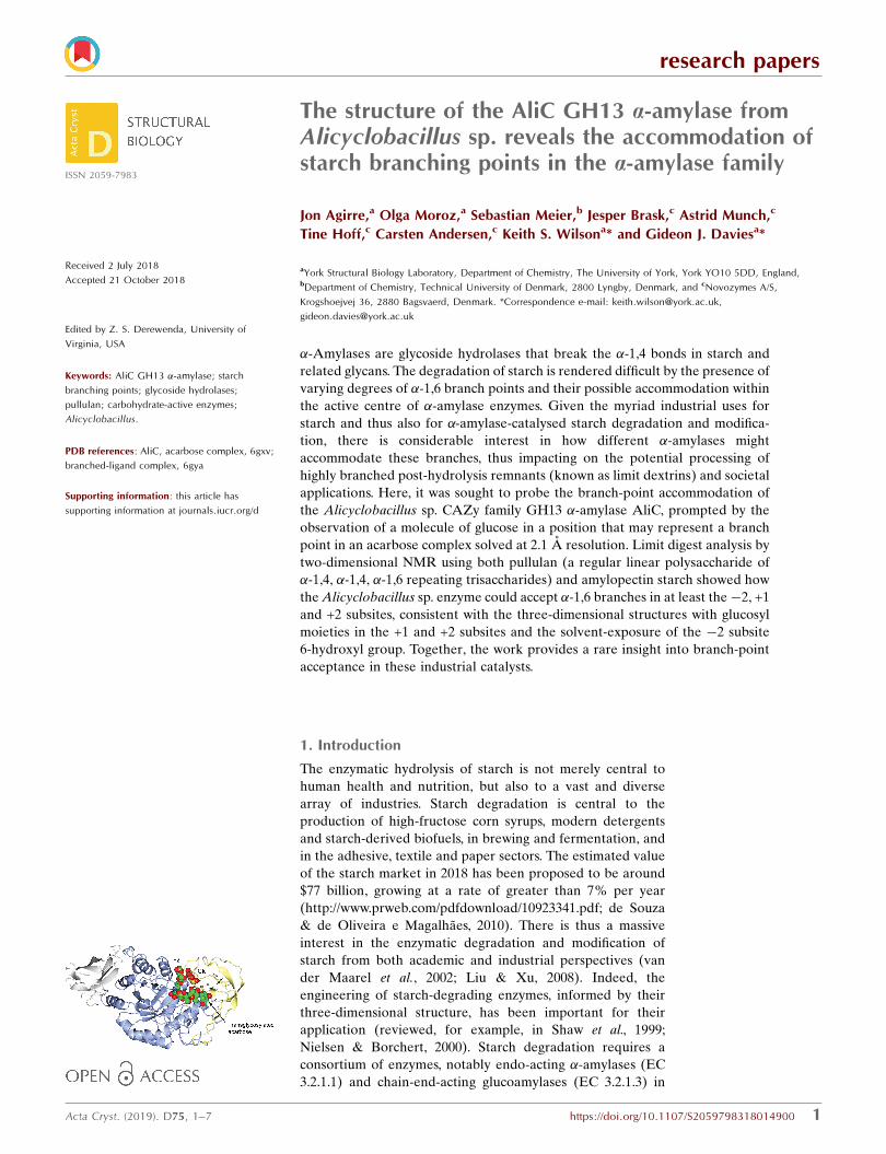
This is a repository copy of The structure of the AliC GH13 α-amylase from Alicyclobacillussp. reveals the accommodation of starch branching points in the α-amylase family.
White Rose Research Online URL for this paper:https://eprints.whiterose.ac.uk/141278/
Version: Published Version
Article:
Agirre, Jon orcid.org/0000-0002-1086-0253, Moroz, Olga, Meier, Sebastian et al. (6 more authors) (2019) The structure of the AliC GH13 α-amylase from Alicyclobacillus sp. revealsthe accommodation of starch branching points in the α-amylase family. Acta crystallographica. Section D, Structural biology. pp. 1-7. ISSN 2059-7983
https://doi.org/10.1107/S2059798318014900
[email protected]://eprints.whiterose.ac.uk/
Reuse
Items deposited in White Rose Research Online are protected by copyright, with all rights reserved unless indicated otherwise. They may be downloaded and/or printed for private study, or other acts as permitted by national copyright laws. The publisher or other rights holders may allow further reproduction and re-use of the full text version. This is indicated by the licence information on the White Rose Research Online record for the item.
Takedown
If you consider content in White Rose Research Online to be in breach of UK law, please notify us by emailing [email protected] including the URL of the record and the reason for the withdrawal request.
research papers
Acta Cryst. (2019). D75, 1–7 https://doi.org/10.1107/S2059798318014900 1
Received 2 July 2018
Accepted 21 October 2018
Edited by Z. S. Derewenda, University of
Virginia, USA
Keywords: AliC GH13 �-amylase; starch
branching points; glycoside hydrolases;
pullulan; carbohydrate-active enzymes;
Alicyclobacillus.
PDB references: AliC, acarbose complex, 6gxv;
branched-ligand complex, 6gya
Supporting information: this article has
supporting information at journals.iucr.org/d
The structure of the AliC GH13 a-amylase fromAlicyclobacillus sp. reveals the accommodation ofstarch branching points in the a-amylase family
Jon Agirre,a Olga Moroz,a Sebastian Meier,b Jesper Brask,c Astrid Munch,c
Tine Hoff,c Carsten Andersen,c Keith S. Wilsona* and Gideon J. Daviesa*
aYork Structural Biology Laboratory, Department of Chemistry, The University of York, York YO10 5DD, England,bDepartment of Chemistry, Technical University of Denmark, 2800 Lyngby, Denmark, and cNovozymes A/S,
Krogshoejvej 36, 2880 Bagsvaerd, Denmark. *Correspondence e-mail: [email protected],
�-Amylases are glycoside hydrolases that break the �-1,4 bonds in starch and
related glycans. The degradation of starch is rendered difficult by the presence of
varying degrees of �-1,6 branch points and their possible accommodation within
the active centre of �-amylase enzymes. Given the myriad industrial uses for
starch and thus also for �-amylase-catalysed starch degradation and modifica-
tion, there is considerable interest in how different �-amylases might
accommodate these branches, thus impacting on the potential processing of
highly branched post-hydrolysis remnants (known as limit dextrins) and societal
applications. Here, it was sought to probe the branch-point accommodation of
the Alicyclobacillus sp. CAZy family GH13 �-amylase AliC, prompted by the
observation of a molecule of glucose in a position that may represent a branch
point in an acarbose complex solved at 2.1 A resolution. Limit digest analysis by
two-dimensional NMR using both pullulan (a regular linear polysaccharide of
�-1,4, �-1,4, �-1,6 repeating trisaccharides) and amylopectin starch showed how
theAlicyclobacillus sp. enzyme could accept �-1,6 branches in at least the�2, +1
and +2 subsites, consistent with the three-dimensional structures with glucosyl
moieties in the +1 and +2 subsites and the solvent-exposure of the �2 subsite
6-hydroxyl group. Together, the work provides a rare insight into branch-point
acceptance in these industrial catalysts.
1. Introduction
The enzymatic hydrolysis of starch is not merely central to
human health and nutrition, but also to a vast and diverse
array of industries. Starch degradation is central to the
production of high-fructose corn syrups, modern detergents
and starch-derived biofuels, in brewing and fermentation, and
in the adhesive, textile and paper sectors. The estimated value
of the starch market in 2018 has been proposed to be around
$77 billion, growing at a rate of greater than 7% per year
(http://www.prweb.com/pdfdownload/10923341.pdf; de Souza
& de Oliveira e Magalhaes, 2010). There is thus a massive
interest in the enzymatic degradation and modification of
starch from both academic and industrial perspectives (van
der Maarel et al., 2002; Liu & Xu, 2008). Indeed, the
engineering of starch-degrading enzymes, informed by their
three-dimensional structure, has been important for their
application (reviewed, for example, in Shaw et al., 1999;
Nielsen & Borchert, 2000). Starch degradation requires a
consortium of enzymes, notably endo-acting �-amylases (EC
3.2.1.1) and chain-end-acting glucoamylases (EC 3.2.1.3) in
ISSN 2059-7983

microbes. In recent times these two players have been
accompanied by copper-dependent lytic polysaccharide
monooxygenases that break down starch, including highly
recalcitrant forms, through an oxidative mechanism (Vu et al.,
2014; Lo Leggio et al., 2015).
The majority of endo-acting �-amylases in industrial starch-
degradation processes are CAZy (http://www.cazy.org; see
Lombard et al., 2014) family GH13 enzymes. GH13 is one of
the most well studied glycoside hydrolase families (reviewed
in CAZypedia at http://www.cazypedia.org/index.php/
Glycoside_Hydrolase_Family_13; The CAZypedia Consor-
tium, 2018). Over 111 different three-dimensional structures
of GH13 enzymes are now known (see http://www.cazy.org/
GH13_structure.html). One particularly important subset of
GH13 enzymes are the ‘Termamyl’-like �-amylases, histori-
cally named after an enzyme from Bacillus licheniformis.
These enzymes typically feature a three-domain ‘A, B, C’
arrangement with a C-terminal �-sheet domain and with
domain B being a protrusion from the (�/�)8 fold of domain A.
The catalytic centre is placed in domain A, whilst the A–B
interface forms the substrate-binding cleft. Many three-
dimensional structures of ‘Termamyl’-like �-amylases are
known. Some notable members include that from B. licheni-
formis (Machius et al., 1995), a chimeric B. licheniformis/
B. amyloliquefaciens enzyme (Brzozowski et al., 2000), an
enzyme from Geobacillus stearothermophilus (Suvd et al.,
2001) and an enzyme from B. halmapalus (Davies et al., 2005).
Notably, as well as having stabilizing Ca2+ ions in various
domains, a characteristic Ca2+–Na+–Ca2+ triad is observed at
the A/B-domain interface (for the historical context, see
Machius et al., 1998; Brzozowski et al., 2000).
Currently, the CAZy classification lists over 100 different
three-dimensional structures of �-amylases from family
GH13. Remarkably, to our knowledge only one of these, the
Bacteroides thetaiotaomicron SusG protein, contains a
branched substrate within its active centre. In this case,
following elegant work by the Koropatkin and Brumer groups
(Arnal et al., 2018), an �-1,6 branch was observed in the +1
subsite. The GlgE protein from Streptomyces coelicolor (PDB
entry 5lgw; Syson et al., 2016) also contains a branched
oligosaccharide, but this ligand is bound far from the active
centre and is instead located on a distal starch-binding
domain. Here, we report the three-dimensional structure
of a ‘Termamyl’-like �-amylase, the AliC �-amylase from
Alicyclobacillus sp. 18711. An initial ligand-bound structure
with a transglycosylated acarbose-derived oligosaccharide at a
resolution of 2.1 A revealed a noncovalently linked glucose
moiety, hinting at a putative branch-accommodation site
around the +2/+3 subsites. A subsequent lower resolution
(approximately 3 A) analysis revealed the binding of a bran-
ched ligand in the +1/+2 subsites with an �-1,6-linked glucose
branch bound to the +1 subsite sugar. Motivated by these
observations, two-dimensional NMR was used to map the
subsite branch preferences on the basis of the structures of the
observed limit dextrin products, highlighting how the AliC
�-amylase can accommodate amylopectin and pullulan
substrates.
2. Methods
2.1. Crystallization
Alicyclobacillus sp. 18711 �-amylase (GenBank MH533021)
was a kind gift from Novozymes A/S (Bagsvaerd, Denmark),
where it had been cloned in a strain variant of B. subtilis
PL1801 from Alicyclobacillus sp. 18711 isolated from a Danish
forest floor. A two-amino-acid deletion (T182*G183*) was
introduced by SOE PCR (Higuchi et al., 1988) using synthetic
oligonucleotides purchased from Invitrogen, and the �-
amylase variant was expressed by fermenting at 37�C for four
days in a soy- and starch-based broth.
The fermentation supernatant was filtrated through a
0.45 mm filter followed by filtration through a 0.2 mm filter.
After the addition of 1M ammonium sulfate and adjustment
of the pH to pH 8, the supernatant was applied onto a 69 ml
Butyl TOYOPEARL column. Prior to loading, the column
had been equilibrated in three column volumes (CV) of
25 mM borate pH 8, 2 mM CaCl2, 1 M ammonium sulfate. In
order to remove unbound material, the column was washed
with 3 CV of 25 mM borate pH 8, 2 mM CaCl2, 1M ammo-
nium sulfate. Elution of the target protein was obtained with a
decreasing salt gradient from 1 to 0M ammonium sulfate in
25 mM borate pH 8, 2 mM CaCl2, followed by 3 CV of 100%
25 mM borate pH 8, 2 mM CaCl2. The flow rate was
10 ml min�1. Relevant fractions were selected and pooled
based on the chromatogram and on SDS–PAGE analysis. The
amylase activity of the purified enzymes was confirmed using
the AMYL liquid amylase assay (Roche/Hitachi system).
2.1.1. Acarbose complex. For the acarbose complex, co-
crystallization screening was carried out using sitting-drop
research papers
2 Agirre et al. � AliC GH13 �-amylase Acta Cryst. (2019). D75, 1–7
Table 1Crystallization.
Acarbose complex Branched-ligand complex
Method Vapour diffusion, sitting drop Vapour diffusion, hanging dropPlate type Swissci 96-well Linbro 24-wellTemperature (K) 293 293Protein concentration (mg ml�1) 20 20Buffer composition of protein solution 25 mM borate, 2 mM CaCl2 pH 8 + 40 mM acarbose 25 mM borate, 2 mM CaCl2 pH 8 + 20 mM GMTComposition of reservoir solution 0.2 M sodium citrate, 0.1M BTP pH 6.5, 20% PEG 3350
(PACT G11)20%(w/v) PEG 3350, 0.1 M BTP pH 8.5, 0.2%(w/v)sodium sulfate
Volume and ratio of drop 300 nl total, 1:1 ratio 1 ml total, 1:1 ratioVolume of reservoir (ml) 54 1000

vapour diffusion with drops set up using a Mosquito Crystal
liquid-handling robot (TTP LabTech, UK) with 150 nl protein
solution plus 150 nl reservoir solution in 96-well format plates
(MRC 2-well crystallization microplates; Swissci, Switzerland)
equilibrated against 54 ml reservoir solution. Initial crystal-
lization experiments were carried out at room temperature
using a number of commercial screens. Diffraction-quality
crystals were obtained in PACT screen condition G11 [0.2 M
sodium citrate, 0.1 M bis-tris propane (BTP) pH 6.5, 20% PEG
3350]. The crystals were tested in-house prior to being sent to
the synchrotron. Crystallization conditions are given in Table 1.
2.1.2. Branched-ligand complex. Crystals of the complex
with 20 mM 63-�-d-glucosyl-maltotriose (GMT; a branched
ligand) were obtained by manual optimization in a 24-well
Linbro tray (hanging drops) in 20% PEG 3350, 0.1 M BTP pH
8.5, 0.2% sodium sulfate with seeding. The initial seeding stock
was prepared by crushing crystals of the acarbose complex,
adding 50 ml mother liquor and vortexing the mixture for
1 min using a Seed Bead (Hampton Research), based on the
protocol described in D’Arcy et al. (2014). Different seed
dilutions were screened; the final crystals grew using a 1:1000
seed dilution.
Details of the crystallization experiments are given in
Table 1.
2.2. Data collection and processing, structure solution and
refinement
Computations were carried out using programs from the
CCP4 suite (Winn et al., 2011) unless otherwise stated. For the
structure of the acarbose complex, data were collected to
2.1 A resolution on beamline I04 at Diamond Light Source
(DLS). The crystal belonged to space group P41212, with unit-
cell parameters a = b = 180.90, c = 77.85 A. The data were
processed with xia2 (Winter et al., 2013). The structure was
solved using MOLREP (Vagin & Teplyakov, 2010) with the
maltohexaose-producing amylase from alkalophilic Bacillus
sp. 707 as a search model (PDB entry 1wp6; Kanai et al., 2004).
For the branched-ligand complex, data were collected to
2.95 A resolution on beamline I04 at DLS. The crystals
belonged to space group P61, with unit-cell parameters
a = b = 212.18, c= 172.22 A. The data were processed with xia2
(Winter et al., 2013). The structure was solved by MOLREP
using the acarbose complex (minus all ligands) as a search
model. Data-collection statistics are given in Table 2.
Both structures were refined by REFMAC (Murshudov et
al., 2011) iterated with manual model correction using Coot
(Emsley et al., 2010). Those monosaccharides that were
expected to be in their minimal energy conformation (4C1 for
d-glucopyranose) were additionally restrained to adopt
torsional values consistent with such a conformation. This was
performed using a dictionary containing unimodal dihedral
restraints produced by Privateer (Agirre et al., 2015). Including
these restraints in the refinement caused the Rfree values to
decrease for both structures. The final R and Rfree are 0.138
and 0.176 for the acarbose ligand complex and 0.156 and 0.183
for the branched-ligand complex, respectively. Validation was
performed using MolProbity (Chen et al., 2010), EDSTATS
(Tickle, 2012) and Privateer (Agirre et al., 2015) through the
use of the CCP4i2 interface (Potterton et al., 2018). During
research papers
Acta Cryst. (2019). D75, 1–7 Agirre et al. � AliC GH13 �-amylase 3
Table 2Data collection and processing.
Values in parentheses are for the outer shell.
Acarbose complexBranched-ligandcomplex
Diffraction source I04, DLS I04, DLSWavelength (A) 0.98 0.98Temperature (K) 100 100Detector ADSC Quantum 315 PILATUS 6M
Prosport+Crystal-to-detector distance
(mm)254.8 475.8
Rotation range per image (�) 0.5 0.2Total rotation range (�) 180 180Exposure time per image (s) 0.5 0.2Space group P41212 P61a, b, c (A) 180.9, 180.9, 77.85 212.18, 212.18, 172.22�, �, � (�) 90, 90, 90 90, 90, 120Mosaicity (�) 0.20 0.13Resolution range (A) 57.21–2.07 (2.12–2.07) 66.9–2.95 (3.03–2.95)Total No. of reflections 1147294 (85223) 921282 (71482)No. of unique reflections 78951 (5774) 92496 (6812)Completeness (%) 99.6 (100) 100 (100)Multiplicity 14.5 (14.8) 10.0 (10.5)CC1/2† 1.0 (0.907) 0.998 (0.914)hI/�(I)i 16.7 (4.5) 13.4 (2.7)Rmerge (%) 17.3 (74.5) 12.6 (74.8)Rr.i.m.‡ (%) 18.4 (79.9) 14.1 (83.1)Overall B factor from
Wilson plot (A2)15.4 50.9
† CC1/2 values for Imean are calculated by splitting the data randomly into half datasets. ‡ Estimated Rr.i.m. = Rmerge [N/(N � 1)]1/2, where N is the data multiplicity andRmerge is defined as
Phkl
Pi jIiðhklÞ � hIðhklÞij=
Phkl
Pi IiðhklÞ, where I(hkl) is the
intensity of the reflection.
Table 3Structure solution and refinement.
Acarbose complexBranched-ligand
complex
PDB code 6gxv 6gyaResolution range (A) 57.21–2.07 66.86–2.95No. of reflections, working set 78885 92448No. of reflections, test set 3965 4492Final Rcryst 0.138 0.156Final Rfree 0.176 0.183Cruickshank DPI 0.14 0.37No. of non-H atoms
Protein 7770 15151Ion 8 16Ligand 272 312Water 1092 224Total 9142 15703
R.m.s. deviationsBonds (A) 0.012 0.016Angles (�) 1.5 1.8
Average B factors† (A2)Protein 18 (6) 61 (13)Ion 18 (8) 60 (9)Ligand 34 (16) 92 (26)Water 33 (13) 48 (10)
Ramachandran plotMost favoured (%) 920 (96.03%) 1830 (95.51%)Allowed (%) 33 (3.44%) 76 (3.97%)Outliers (%) 5 (0.52%) 10 (0.52%)
† Values in parentheses are standard deviations.

this work, a MolProbity graphical interface for CCP4i2 was
developed. Aside from supporting the usual reporting, the
functionality of the interface was extended to cover auto-
mated 180� rotation of suggested histidine, asparagine and
glutamine side chains around the last � angle, with an addi-
tional real-space refinement step, and real-time compression
and decompression of the results from PROBE, with a typical
ratio of reduction in file size of 8:1. This new interface is
available in CCP4 through use of the ‘Analyse model
geometry’ task.
Data-processing and refinement statistics for both struc-
tures are given in Table 3.
2.3. Degradation of pullulan and amylopectin by the purified
Alicyclobacillus a-amylase
The enzymatic specificity of the Alicyclobacillus �-amylase
was experimentally determined to complement the search for
potential branch-point accommodation in the active site in the
crystal structures. To this end, pullulan and amylopectin were
subjected to degradation prior to NMR analysis of the frag-
ments formed. Pullulan (Sigma–Aldrich, St Louis, Missouri,
USA) samples were degraded by purified Alicyclobacillus
�-amylase at room temperature and samples were withdrawn,
inactivated at 90�C for 10 min, condensed by lyophilization
and redissolved in 600 ml D2O (99.9%; Cambridge Isotope
Laboratories, Andover, Massachusetts, USA) to obtain
partially degraded and fully degraded samples. The samples
were transferred to 5 mm NMR sample tubes for analysis.
Amylopectin (from potato starch; Sigma–Aldrich) was incu-
bated at 30�C overnight with the purified Alicyclobacillus
�-amylase, inactivated at 90�C for 10 min, condensed by
lyophilization and redissolved in 600 ml D2O for NMR
analysis.
2.4. NMR spectroscopy
All NMR spectra were recorded on an 800 MHz Avance II
spectrometer (Bruker, Fallanden, Switzerland) equipped with
a TCI Z-gradient CryoProbe and an 18.7 T magnet (Oxford
research papers
4 Agirre et al. � AliC GH13 �-amylase Acta Cryst. (2019). D75, 1–7
Figure 1Three-dimensional structure of the Alicyclobacillus �-amylase AliC. (a) Three-dimensional protein cartoon, coloured by domain, with metal ions shownas shaded spheres and the acarbose and the +20 glucose shown as van der Waals spheres. (b) Electron density for the transglycosylated acarbose insubsites �4 to +2 (and +20 Glc) binding; density for an isolated ‘+3’ glucose is not shown. (c) Electron density for the binding of the branchedoligosaccharide in subsites +1, +1 and +10. Electron-density maps are REFMACmaximum-likelihood-weighted 2Fo � Fc syntheses contoured at 1�. Thisfigure was drawn with CCP4mg (McNicholas et al., 2011).

Magnet Technology, Oxford, England). Highly resolved1H–13C HSQC spectra employing a sweep width of 10 p.p.m.
centred near the 13C chemical shift of the �-anomeric signals
were recorded as data matrices of 1024 � 256 complex data
points sampling acquisition times of 143 and 127 ms in the 1H
and 13C dimensions, respectively. High-precision signal
measurements in the two-dimensional spectra were thus used
to enumerate the number of signals in the resultant reaction
products and for the identification of the products by
comparison with authentic standards including glucose,
maltooligosaccharides, panose and limit dextrins (Petersen et
al., 2014, 2015).
All spectra were processed with extensive zero filling in
both dimensions using a shifted sine-bell apodization function
and were analysed with TopSpin 2.1 pl 5 (Bruker).
3. Results and discussion
3.1. Three-dimensional structure of AliC a-amylase and its
acarbose-derived complex
The complex of AliC with acarbose was solved by molecular
replacement, with two molecules of AliC in the asymmetric
unit, at a resolution of 2.1 A. The fold, as expected, is a
canonical three-domain arrangement with the A, B and C
domains defined approximately as A, residues 4–104 and 210–
397; B, residues 105–209; and C, residues 398–484. A classical
Ca2+–Na+–Ca2+ triad (Machius et al., 1998; Brzozowski et al.,
2000) is found at the A/B-domain interface. At the time of
writing, structural similarity searches using PDBeFold (Kris-
sinel & Henrick, 2004) showed that the closest three-dimen-
sional match to AliC is the B. halmapalus �-amylase (Davies et
al., 2005), with 67% sequence identity and with 479 aligned C�
atoms overlapping with an r.m.s.d. of 0.49 A (PDBeFold Q
score 0.95, Z-score 27.8). Other close structural homologs are
the maltohexaose-producing amylase from Bacillus sp. 707
(Kanai et al., 2004) and the calcium-free amylase AmyK38
from Bacillus sp. strain KSM-K38 (Nonaka et al., 2003).
The structure of AliC was determined in the presence of the
inhibitor acarbose. As with many (retaining) �-amylase
complexes [some examples from the author’s laboratory
include those reported in Brzozowski et al. (2000), Davies et al.
(2005), Brzozowski & Davies (1997), Dauter et al. (1999) and
Offen et al. (2015)], the acarbose is observed as a transgly-
cosylated species, here a hexasaccharide which contains two of
the acarviosin disaccharide motifs. The complex defines six
subsites, �4 to +2, with the expected catalytic GH13 signature
triad of Asp234 (nucleophile), Glu265 (acid/base) and Asp332
(interacting with O2/O3 of the �1 subsite sugar) all disposed
for catalysis, here around the 2H3 half-chair of the unsaturated
cyclohexitol moiety.
3.2. Limit digest analysis of the action of AliC on pullulan and
amylopectin
Of particular interest to us was the observation of a ‘lone’
ordered glucose moiety that was not covalently linked to the
acarbose-derived oligosaccharide in a position that could be
indicative of the accommodation of branch points at either
the +2 or +3 positions of AliC. An additional isolated
glucose molecule, modelled in both � and � anomeric forms
research papers
Acta Cryst. (2019). D75, 1–7 Agirre et al. � AliC GH13 �-amylase 5
Figure 2HSQC spectra of pullulan degradation by purified Alicyclobacillus �-amylase recorded with extensive sampling of the 13C dimension. Only the�-anomeric spectral region is shown. Three different glucopyranosyl unitsoccur in pullulan (left). Signals of �-1,6 anomeric glucopyranosyl units atthe nonreducing end emerge (see the inset in the middle spectrum) owingto cleavage at the indicated position (middle top). Pullulan is degraded topanose as the final product (right), as demonstrated by comparison withan authentic standard (grey). These experiments identify the pullulan-cleaving activity ofAlicyclobacillus �-amylase as that of a panose-formingneopullulanase (EC 3.2.1.135).
Figure 31H–13C HSQC NMR spectrum of degradation products from potatoamylopectin using purified Alicyclobacillus �-amylase. Glucose, maltose,maltotriose and 62-�-maltosylmaltotriose are the main products. Theasterisk denotes the overlapping reducing-end �-glucopyranosyl signal ofoligosaccharides. Several signals are detected for each molecule, as theindividual glucopyranosyl units in oligosaccharides give separate signals.

(occupancy set at 0.5 for each and in different orientations;
omitted from Fig. 1 for clarity), was observed near a potential
+3 site. We speculated whether these isolated glucose moieties
could provide insight into branch-point recognition. The
accommodation of branch points was therefore investigated
by an analysis of limit digestion products (the characteristic
oligosaccharides remaining after enzymatic digestion) on both
pullulan (a regular linear polysaccharide of �-1,4, �-1,4, �-1,6
repeating trisaccharides) and amylopectin, an �-1,6-branched
starch structure. The action of AliC on pullulan results in the
production of the trisaccharide panose, glucose �-1,6-glucose
�-1,4-glucose (Fig. 2), demonstrating that the enzyme must be
able to accommodate �-1,6 linkages to glucose moieties in
both the +1 and �2 subsites.
Action on amylopectin produced the limit dextrin 62-�-
maltosyl maltotriose (for NMR assignments, see Petersen et
al., 2015; Jodelet et al., 1998) (Fig. 3) demonstrating that AliC
must also be able to accommodate starches with �-1,6 bran-
ches in both the�2 and +2 subsites. Taken together, the action
on pullulan and amylopectin shows that AliC is able to
accommodate 1,6 linkages in the �2, +1 and +2 subsites
(Fig. 4).
3.3. Branched-ligand complex of AliC
These branch patterns are consistent with the initial three-
dimensional structure of AliC with the acarbose-derived
oligosaccharide in which the �2 subsite O6 points into solvent
and in which we observed a glucosyl moiety approximately
where an O6 branch in either the +2 or +3 site might lie. In
order to try and access a branched complex with a branch in
the +1 subsite (the position of which is harder to model from
the 6-deoxy sugars present in acarbose alone) we sought to
obtain a complex by co-crystallizing AliC with 63-�-d-glucosyl
maltotriose (Megazyme, Wicklow, Ireland) and observing
what was obtained with this active enzyme.
A ‘branched-ligand’ AliC complex was obtained through
co-crystallization, with crystals forming in a new space group.
This form diffracted poorly and data
could only be obtained to 2.95 A reso-
lution. Weak density in the �1 subsite,
largely diffuse but greater than would
be expected for discrete solvent,
remained unmodelled. Density was
clearer for a panose trisaccharide with
an �-1,4-linked disaccharide in subsites
+1 and +2 and, crucially, clear density
for an �-1,6 branch accommodated in
the +1 subsite (Fig. 1c), providing a
structural context for the limit digest
analysis of action on amylopectin
starch (Fig. 4). Notably, this +10 sugar
overlaps in position with the very
recently reported SusG amylase bran-
ched-ligand complex (Arnal et al.,
2018).
4. Conclusions
The accommodation of branch points
in industrial enzymes is a key factor
when considering their utility. How
close to branch points an �-1,4 cleaving
�-amylase will cleave defines what the
ultimate limit dextrin product will be.
The product profile impacts both on the
cocktail of enzymes that are required
for complete hydrolysis to glucose and
on the physical properties of the limit
dextrin itself (which are important in
food and brewing processes, including
the ‘mouthfeel’ of beer), such that
insight into branch-point accommoda-
tion can provide powerful insight to aid
protein-engineering campaigns. Yet,
surprisingly, there has been very little
research papers
6 Agirre et al. � AliC GH13 �-amylase Acta Cryst. (2019). D75, 1–7
Figure 4Interpretation of the limit digest patterns in terms of protein structure. Black and red arrows indicatethe two/three places at which bonds must be cut to accommodate the limit dextrins observed. Togenerate panose from pullulan, both the +1 and �2 subsites must accommodate �-1,6 branches. Thebranched-ligand complex shows how subsite +1 can accommodate a branch (Fig. 1c), and in subsite�2 the O6 of acarbose was solvent-exposed. To generate the branched limit dextrin fromamylopectin (Fig. 3), AliC must also be able to accommodate branching in the +2 subsite, which isconsistent with the glucose moiety seen adjacent to O6 of the +2 sugar (Fig. 1b).
structural insight into possible branch-point accommodation
in �-amylases. Here, we have shown how serendipitous
observation of a ‘lone’ glucosyl moiety close to the O6 posi-
tion of an oligosaccharide complex inspired analysis of limit
dextrins on substrates containing �-1,6 linkages, both linear
and branched. Such combined X-ray and product-analysis
NMR approaches should prove valuable in the future for
interrogating, defining and ultimately exploiting the branch-
point accommodation in this massively widespread family of
starch-degrading catalysts.
Acknowledgements
The authors would like to thank Diamond Light Source for
beamtime (proposals mx-7864 and mx-9948) and the staff of
beamline I04 for assistance with crystal testing and data
collection. NMR spectra were acquired using the instruments
of the NMR center, Technical University of Denmark (DTU).
Funding information
JA is a Royal Society University Research Fellow (award
reference UF160039). GJD is a Royal Society Ken Murray
Research Professor. The authors would like to thank Novo-
zymes A/S for partially funding this work.
References
Agirre, J., Iglesias-Fernandez, J., Rovira, C., Davies, G. J., Wilson,K. S. & Cowtan, K. D. (2015). Nature Struct. Mol. Biol. 22, 833–834.
Arnal, G., Cockburn, D. W., Brumer, H. & Koropatkin, N. M. (2018).Protein Sci. 27, 1093–1101.
Brzozowski, A. M. & Davies, G. J. (1997). Biochemistry, 36, 10837–10845.
Brzozowski, A. M., Lawson, D. M., Turkenburg, J. P., Bisgaard-Frantzen, H., Svendsen, A., Borchert, T. V., Dauter, Z., Wilson,K. S. & Davies, G. J. (2000). Biochemistry, 39, 9099–9107.
Chen, V. B., Arendall, W. B., Headd, J. J., Keedy, D. A., Immormino,R. M., Kapral, G. J., Murray, L. W., Richardson, J. S. & Richardson,D. C. (2010). Acta Cryst. D66, 12–21.
D’Arcy, A., Bergfors, T., Cowan-Jacob, S. W. & Marsh, M. (2014).Acta Cryst. F70, 1117–1126.
Dauter, Z., Dauter, M., Brzozowski, A. M., Christensen, S., Borchert,T. V., Beier, L., Wilson, K. S. & Davies, G. J. (1999). Biochemistry,38, 8385–8392.
Davies, G. J., Brzozowski, A. M., Dauter, Z., Rasmussen, M. D.,Borchert, T. V. & Wilson, K. S. (2005). Acta Cryst. D61, 190–193.
Emsley, P., Lohkamp, B., Scott, W. G. & Cowtan, K. (2010). ActaCryst. D66, 486–501.
Higuchi, R., Krummel, B. & Saiki, R. K. (1988).Nucleic Acids Res. 16,7351–7367.
Jodelet, A., Rigby, N. M. & Colquhoun, I. J. (1998). Carbohydr. Res.312, 139–151.
Kanai, R., Haga, K., Akiba, T., Yamane, K. & Harata, K. (2004).Biochemistry, 43, 14047–14056.
Krissinel, E. & Henrick, K. (2004). Acta Cryst. D60, 2256–2268.Liu, X. D. & Xu, Y. (2008). Bioresour. Technol. 99, 4315–4320.Lo Leggio, L., Simmons, T. J., Poulsen, J. C., Frandsen, K. E.,Hemsworth, G. R., Stringer, M. A., von Freiesleben, P., Tovborg,M., Johansen, K. S., De Maria, L., Harris, P. V., Soong, C. L.,Dupree, P., Tryfona, T., Lenfant, N., Henrissat, B., Davies, G. J. &Walton, P. H. (2015). Nature Commun. 6, 5961.
Lombard, V., Golaconda Ramulu, H., Drula, E., Coutinho, P. M. &Henrissat, B. (2014). Nucleic Acids Res. 42, D490–D495.
Maarel, M. J. E. C. van der, van der Veen, B., Uitdehaag, J. C.,Leemhuis, H. & Dijkhuizen, L. (2002). J. Biotechnol. 94, 137–155.
Machius, M., Declerck, N., Huber, R. & Wiegand, G. (1998).Structure, 6, 281–292.
Machius, M., Wiegand, G. & Huber, R. (1995). J. Mol. Biol. 246, 545–559.
McNicholas, S., Potterton, E., Wilson, K. S. & Noble, M. E. M. (2011).Acta Cryst. D67, 386–394.
Murshudov, G. N., Skubak, P., Lebedev, A. A., Pannu, N. S., Steiner,R. A., Nicholls, R. A., Winn, M. D., Long, F. & Vagin, A. A. (2011).Acta Cryst. D67, 355–367.
Nielsen, J. E. & Borchert, T. V. (2000). Biochim. Biophys. Acta, 1543,253–274.
Nonaka, T., Fujihashi, M., Kita, A., Hagihara, H., Ozaki, K., Ito, S. &Miki, K. (2003). J. Biol. Chem. 278, 24818–24824.
Offen, W. A., Viksoe-Nielsen, A., Borchert, T. V., Wilson, K. S. &Davies, G. J. (2015). Acta Cryst. F71, 66–70.
Petersen, B. O., Hindsgaul, O. & Meier, S. (2014). Analyst, 139, 401–406.
Petersen, B. O., Motawie, M. S., Møller, B. L., Hindsgaul, O. & Meier,S. (2015). Carbohydr. Res. 403, 149–156.
Potterton, L., Agirre, J., Ballard, C., Cowtan, K., Dodson, E., Evans,P. R., Jenkins, H. T., Keegan, R., Krissinel, E., Stevenson, K.,Lebedev, A., McNicholas, S. J., Nicholls, R. A., Noble, M., Pannu,N. S., Roth, C., Sheldrick, G., Skubak, P., Turkenburg, J., Uski, V.,von Delft, F., Waterman, D., Wilson, K., Winn, M. & Wojdyr, M.(2018). Acta Cryst. D74, 68–84.
Shaw, A., Bott, R. & Day, A. G. (1999). Curr. Opin. Biotechnol. 10,349–352.
Souza, P. M. de & de Oliveira e Magalhaes, P. (2010). Braz. J.Microbiol. 41, 850–861.
Suvd, D., Fujimoto, Z., Takase, K., Matsumura, M. & Mizuno, H.(2001). J. Biochem. 129, 461–468.
Syson, K., Stevenson, C. E., Miah, F., Barclay, J. E., Tang, M., Gorelik,A., Rashid, A. M., Lawson, D. M. & Bornemann, S. (2016). J. Biol.Chem. 291, 21531–21540.
The CAZypedia Consortium (2018). Glycobiology, 28, 3–8.Tickle, I. J. (2012). Acta Cryst. D68, 454–467.Vagin, A. & Teplyakov, A. (2010). Acta Cryst. D66, 22–25.Vu, V. V., Beeson, W. T., Span, E. A., Farquhar, E. R. & Marletta,M. A. (2014). Proc. Natl Acad. Sci. USA, 111, 13822–13827.
Winn, M. D., Ballard, C. C., Cowtan, K. D., Dodson, E. J., Emsley, P.,Evans, P. R., Keegan, R. M., Krissinel, E. B., Leslie, A. G. W.,McCoy, A., McNicholas, S. J., Murshudov, G. N., Pannu, N. S.,Potterton, E. A., Powell, H. R., Read, R. J., Vagin, A. & Wilson,K. S. (2011). Acta Cryst. D67, 235–242.
Winter, G., Lobley, C. M. C. & Prince, S. M. (2013). Acta Cryst. D69,1260–1273.
research papers
Acta Cryst. (2019). D75, 1–7 Agirre et al. � AliC GH13 �-amylase 7
Copyright © 2022 FDOKUMEN