Spatial and temporal distribution of falciparum malaria in China
Upload
independentCategory
view
4download
0
The
Journ
al o
f Exp
erim
enta
l M
edic
ine
The Journal of Experimental Medicine • Volume 199, Number 11, June 7, 2004 1533–1544http://www.jem.org/cgi/doi/10.1084/jem.20031274
1533
A Multidomain Adhesion Protein Family Expressed in
Plasmodium falciparum
Is Essential for Transmission to the Mosquito
Gabriele Pradel,
1
Karen Hayton,
2
L. Aravind,
3
Lakshminarayan M. Iyer,
3
Mitchell S. Abrahamsen,
4
Annemarie Bonawitz,
1
Cesar Mejia,
1
and Thomas J. Templeton
1
1
Department of Microbiology and Immunology, Weill Medical College of Cornell University, New York, NY 10021
2
Laboratory of Malaria and Vector Research, National Institute of Allergy and Infectious Diseases, and
3
National Center for Biotechnology Information, National Library of Medicine, National Institutes of Health, Bethesda, MD 20892
4
Biomedical Genomics Center and Department of Veterinary Pathobiology, University of Minnesota, St. Paul, MN 55108
Abstract
The recent sequencing of several apicomplexan genomes has provided the opportunity to char-acterize novel antigens essential for the parasite life cycle that might lead to the development ofnew diagnostic and therapeutic markers. Here we have screened the
Plasmodium falciparum
genome sequence for genes encoding extracellular multidomain putative adhesive proteins.Three of these identified genes, named
PfCCp1
,
PfCCp2
, and
PfCCp3
, have multiple adhesivemodules including a common
Limulus
coagulation factor C domain also found in two addi-tional
Plasmodium
genes. Orthologues were identified in the
Cryptosporidium parvum
genomesequence, indicating an evolutionary conserved function. Transcript and protein expressionanalysis shows sexual stage–specific expression of
PfCCp1
,
PfCCp2
, and
PfCCp3
, and cellularlocalization studies revealed plasma membrane–associated expression in mature gametocytes.During gametogenesis,
Pf
CCps are released and localize surrounding complexes of newlyemerged microgametes and macrogametes.
Pf
CCp expression markedly decreased after forma-tion of zygotes. To begin to address
Pf
CCp function, the
PfCCp2
and
PfCCp3
gene loci weredisrupted by homologous recombination, resulting in parasites capable of forming oocystsporozoites but blocked in the salivary gland transition. Our results describe members of a con-served apicomplexan protein family expressed in sexual stage
Plasmodium
parasites that may rep-resent candidates for subunits of a transmission-blocking vaccine.
Key words: malaria • gametocyte • gamete •
Anopheles
• sporozoite
Introduction
The malaria parasite
Plasmodium
is a major cause of globalhuman morbidity and mortality, whereas other apicom-plexan parasites like
Cryptosporidium parvum
and
Toxoplasmagondii
are important opportunistic pathogens in immuno-compromised individuals such as AIDS patients. An importantaspect of apicomplexan biology is the molecular basis fortheir interaction with target cells, extracellular matrices,and hemo-lymphatic fluids of their animal hosts. Due to
their extracellular localization, surface proteins proposed tobe involved in these processes are likely accessible by host-immune sera and are therefore vaccine candidates stimulatingantibody-mediated protection. Recent methodologies toidentify extracellular proteins as research targets include“postgenomic” techniques such as genome sequence anno-tation, microarray screening, and proteomics. Of particularimportance, completion of
Plasmodium
sp. genome sequences(1, 2) and the imminent completion of additional apicom-plexan genomes have provided an unprecedented opportunity
The online version of this article contains supplemental material.Address correspondence to Thomas J. Templeton, Department of
Microbiology and Immunology, Weill Medical College of Cornell Uni-versity, 1300 York Avenue, Box 62, New York, NY 10021. Phone:(212) 746-4467; Fax: (212) 746-4028; email: [email protected]
Abbreviations used in this paper:
CS, circumSporozoite; ER, endoplasmicreticulum; IFA, immunofluorescence; LCCL,
Limulus
coagulation factorC; MBP, maltose-binding protein; SR, scavenger receptor.
on Septem
ber 28, 2014jem
.rupress.orgD
ownloaded from
Published June 7, 2004
http://jem.rupress.org/content/suppl/2004/06/04/jem.20031274.DC1.html Supplemental Material can be found at:
Plasmodium
Multidomain Adhesion Proteins in Mosquito Transmission
1534
to identify novel antigens as prospective vaccine targets forstudy, as well as underpin a better understanding of
Plasmo-dium
and apicomplexan biology.In the search for novel extracellular proteins as prospec-
tive vaccine candidates, we have exhaustively identifiedgenes encoding extracellular proteins through genome se-quence analysis of
Plasmodium
sp. and the recently com-pleted
C. parvum
genome sequence, as well as whole ge-nome comparison of the resulting catalogs of extracellularproteins (unpublished data). Here we describe a select sub-set of sexual stage–specific genes in the human malaria par-asite,
P. falciparum
, that are striking for multidomain archi-tectures composed of animal- and bacterial-like putativeadhesive domains. Orthologues are present in the
C. par-vum
genome sequence, indicating a cellular function likelyconserved across the apicomplexan clade. These proteinsare grouped as a family based upon a common
Limulus
co-agulation factor C (LCCL) domain and are accordinglynamed CCp proteins, of which five genes are present in the
P. falciparum
genome sequence (
Pf
CCps). One of theseproteins, herein referred to as
Pf
CC
p3
, was recently de-scribed as
P
SLAP in
P. falciparum
(3) and
Pb
SR in
Plas-modium berghei
(4). These two studies presented, in part,contradictory conclusions regarding protein expression. Al-though
P
SLAP was shown to be expressed in the game-tocyte stages,
Pb
SR protein expression was described assporozoite stage–specific and
Pb
SR-targeted gene disrup-tion led to inhibited sporozoite formation. In this study, wecharacterize three
P. falciparum
members of the LCCL fam-ily,
PfCCp1
,
PfCCp2
, and
Pf
CC
p3
/
P
SLAP, based upontheir striking multiadhesion domain structures. We showthat
Pf
CC
p1
,
Pf
CC
p2
, and
Pf
CC
p3
are specifically ex-pressed in gametocytes and released during gametogenesis.Disruption of the
PfCCp2
or
PfCCp3
gene loci via homol-ogous recombination leads to a blockage in the
P. falcip-arum
sporozoite midgut to salivary gland transition. Wesuggest that the
Pf
CCp protein family serves a role in mul-tiple mosquito parasite stages and has an essential role intransmission to the mosquito, thus warranting further studyas transmission-blocking vaccine candidates.
Materials and Methods
Apicomplexan Genome Sequence Analysis. CCp
genes wereidentified via screening of complete
Plasmodium
sp. and
C. parvum
genome sequence databases using the gapped version of theBLAST programs (BLASTPGP and TBLASTNGP, with boththe single pass and the recently incorporated multipass profile op-tion for translating searches of nucleotide databases). Sequenceprofile searches were performed using the PSI-BLAST program(5). Profiles were saved using the -C option and retrieved usingthe -R option. Complete domain architectures of identified pro-teins were determined by screening the nonredundant proteinsequence database, the Expressed Sequence Tags database (Na-tional Center for Biotechnology Information), and the individ-ual protein sequence databases of completely and partially se-quenced genomes (accessible at http://www.ncbi.nlm.nih.gov/Microb_blast/unfinishedgenome.html). Multiple alignments ofamino acid sequences were generated using a combination of
PSI-BLAST and T-Coffee (6). Secondary structure predictionswere produced using the PHD program (7), with multiple align-ments of individual protein families used as queries. Preliminaryphylogenetic trees were constructed using the neighbor-joiningalgorithm as implemented in the Mega2 package (8), or the leastsquares methods as implemented in the FITCH program of thePHYLIP package (9). These trees were used to obtain the maxi-mum likelihood tree using the PROTML program of the MOL-PHY package. The statistical significance of the trees was assessedusing the relative estimates of the logarithmic likelihood (RELL)bootstrap values (10). Signal peptides in protein sequences werepredicted using the SignalP program (11) and membrane topolo-gies were predicted using the TMHMM2 package (12).
Parasite Culture and Mosquito Membrane Feeds.
Asexual andmature gametocyte NF54 isolate
P. falciparum
parasites were cul-tivated in vitro as described previously (13). Gametogenesis wasinduced by incubating mature gametocyte cultures in human se-rum for 5–30 min at room temperature as described previously(14). Zygote formation was achieved by incubation of emergedgametes in human serum overnight at room temperature. Zy-gotes and gametocytes were enriched via centrifugation over6/11/16% Accudenz gradients and parasites were collected atgradient interfaces. Mosquito stages (retorts, ookinetes, oocysts,and salivary gland sporozoites) were obtained after membranefeeds of
Anopheles freeborni
mosquitoes on blood containing ma-ture gametocytes as described previously (15). Retort and matureookinetes were obtained by harvesting midgut bloodmeals 24 hafter feeding. Oocysts were isolated from mosquito midguts 12 dafter feeding. Sporozoite preparations were enriched by centrifu-gation from pooled, homogenized infected salivary glands isolated18–20 d after feeding. To determine the efficiency of mosquitotransmission, WT NF54- or
Pf
CCp-KO–targeted gene-disruptantisolates were membrane fed to mosquitoes and oocyst numberswere assayed in mercurochrome-stained midguts 8–12 d afterfeeding. Similarly, the presence of salivary gland sporozoites wasassayed 18–21 d after feeding.
Recombinant Protein Production and Derivation of Mouse Antisera.
Recombinant protein construct designs were guided by do-main architecture determinations for
Pf
CC
p1
(sequence data areavailable from GenBank/EMBL/DDBJ under accession no.NP_702612.1),
Pf
CC
p2
(GenBank/EMBL/DDBJ under acces-sion no. NP_702421.1), and
Pf
CC
p3
(GenBank/EMBL/DDBJunder accession no. NP_701955) amino acid sequences. For each
PCCp
gene, recombinant protein was produced corresponding todiscrete domains, such as the LCCL domain (
Pf
CC
p1
), the dis-coidin domain (
Pf
CC
p2
), and the two tandem scavenger receptor(SR) domains (
Pf
CC
p3
). Recombinant protein was expressed asfusions downstream of the maltose-binding protein (MBP) usingthe pMAL expression vector (New England Biolabs, Inc.). Allconstructs were generated by ligation of PCR products corre-sponding to discrete
Pf
CCp domains amplified from 3D7 (cloneof NF54 isolate)
P. falciparum
gametocyte stage cDNA. Recombi-nant protein was purified from bacterial extracts by sequentialsteps of amylose affinity (New England Biolabs, Inc.) and ionexchange chromatography (Source 15Q resin using an AKTAPrime chromatography system; Amersham Biosciences). 6-wk-old female CAF1 mice were immunized with 30–100
�
g recom-binant protein emulsified in Freund’s complete adjuvant followedby two boosts at 3-wk intervals in Freund’s incomplete adjuvant.Immune sera were collected 10 d after the third immunization.All antisera recognized cognate recombinant protein via Westernblot analysis. Sera from prebleed and MBP protein alone–immu-nized mice served as controls for all antibody reagent studies. As a
on Septem
ber 28, 2014jem
.rupress.orgD
ownloaded from
Published June 7, 2004
Pradel et al.
1535
second source of antisera for double labeling experiments, anti-peptide sera, directed against a
Pf
CC
p2
peptide sequence withinthe discoidin domain (CDKIKDTDDARDKYF), was producedin rabbits immunized with KLH-conjugated material producedusing a maleimide-activated KLH kit (Pierce Chemical Co.). Im-munizations of two rabbits were performed by Spring ValleyLaboratories, Inc.
Western Blot Analysis.
Mixed asexual stage parasites and Ac-cudenz gradient–enriched gametocytes were saponin lysed andsuspended in PBS and SDS-PAGE loading buffer. Salivary glandsinfected with sporozoites were dissected from
An. freeborni
mos-quitoes and suspended in SDS-PAGE loading buffer. Parasite pro-teins were separated by electrophoresis (Novex PAGE system;Invitrogen) and transferred to Hybond ECL nitrocellulose mem-brane (Amersham Biosciences) according to the manufacturer’sinstructions. Membranes were blocked for nonspecific bindingin Tris-buffered saline containing 3% skim milk and incubatedfor 2 h at room temperature with mouse immune sera specific to
Pf
CCps, mouse mAb 2A10 recognizing the sporozoite surface cir-cumSporozoite (CS) protein (16), MBP fusion partner proteincontrol, or, as a control for loading, mouse sera recognizing theabundant
P. falciparum
endoplasmic reticulum (ER) protein,
Pf
39(17, 18). After washing, membranes were incubated for 1 h atroom temperature with an alkaline phosphatase–conjugated sec-ondary antibody (Kirkegaard & Perry Laboratories, Inc.) and de-veloped in NBT/BCIP solution (Sigma-Aldrich) for 5–15 min.
Immunofluorescence (IFA) and Confocal Laser Scanning Microscopy.
Parasite preparations for IFA microscopy included NF54 isolate–mixed asexual stages, mature gametocytes, emerging gametes,mosquito midgut blood smears 24 h after feeding, midgut oocysts12 d after feeding, or salivary gland sporozoite smears 18 d afterfeeding. Preparations were either fixed for 4 d in 4% paraformal-dehyde in PBS at 4
�
C or were air dried on slides and fixed for 10min in
�
80
�
C cold methanol. For membrane permeabilizationand blocking of nonspecific binding, fixed cells were incubated in0.02% saponin, 0.5% BSA, and 1% neutral goat serum (Sigma-Aldrich) in PBS twice for 30 min each. Preparations were thenincubated for 2 h at 37
�
C with either mouse immune sera recog-nizing specific
Pf
CCps, mouse mAb 2A10 recognizing the CSprotein, rabbit anti–
�
-tubulin II serum (American Type CultureCollection [ATCC]; reference 19), or rabbit immune serum rec-ognizing
Pfs25 (ATCC; references 20 and 21). Binding of pri-mary antibody was visualized using fluorescence-conjugated goatanti–mouse or goat anti–rabbit secondary antibodies (Alexa Fluor488 or Alexa Fluor 594; Molecular Probes). To highlight unla-beled parasites, selected specimens were further incubated withthe nuclear dye TOTO-3 (Molecular Probes) for 30 min at37�C. Paraformaldehyde-fixed samples were embedded in 1%agarose in PBS, coated onto slides, and air dried. In selected prep-arations, those not requiring Alexa 594 conjugation, counter-staining was performed using 0.05% Evans Blue (Sigma-Aldrich)in PBS. Labeled specimens were examined by confocal laser scanmicroscopy using a Zeiss LSM 510, and digital images were pro-cessed using Adobe Photoshop 6.0 software.
Electron Microscopy. For preembedding immunolabeling,mixed asexual and gametocyte stages were fixed in 4% parafor-maldehyde in PBS for 4 d at 4�C. Cells were blocked in 0.02%saponin, 0.5% BSA, and 5% neutral goat serum (Sigma-Aldrich)in PBS for twice for 30 min each, and then incubated for 2 h at37�C with mouse immune sera against PfCCps, followed by in-cubation with a goat anti–mouse secondary antibody conjugatedwith either 10 nm gold (Amersham Biosciences) or alkaline phos-phatase. Alkaline phosphatase labeling was detected by incubating
cells in BCIP/NBT solution for 20 min. Cells were then embed-ded in agar, fixed in 1% glutaraldehyde and 4% paraformaldehydein PBS for 1 d at 4�C, postfixed in 1% osmium tetroxide in PBSfor 1 h, and dehydrated in successive washes at increasing con-centrations of ethanol. Specimens were embedded in Epon (Elec-tron Microscopy Sciences) at 60�C for 2 d. Photographs weretaken with a JEOL 100CX-II transmission electron microscopeand scanned images were processed using Adobe Photoshop 6.0software. For the morphological examination of PfCCp2-targetedgene-disruptant (PfCCp2-KO) oocysts, infected mosquito mid-guts were fixed in 1% glutaraldehyde and 4% paraformaldehydein PBS for 2 d, and postfixed in 1% osmium tetroxide and 1.5%K3Fe(CN)6 in PBS for 2 h, followed by incubation in 0.5% uranylacetate for 1 h. Midguts were dehydrated in ethanol and embed-ded in Epon as described above.
Generation of Gene-disruptant Parasites. A PfCCp2 KO con-struct, pPfCCp2-KO, was designed such that after homologousintegration a “pseudo-diploid” locus is generated in which thefirst copy of the disrupted gene is truncated and possesses an in-frame stop codon at the 3� end, whereas the second copy is trun-cated at the 5� end and lacks a start methionine, a signal peptidesequence, and the start of the coding region. The disruption con-struct contained a 523-bp amino-terminal region segment ofPfCCp2 that was PCR amplified from NF54 P. falciparum geno-mic DNA template using gene-specific 5� primer 5�-TAGCG-GCCGCACCCTAATGGCACCGAAT-3� and 3� primer 5�-ATGCGGCCGCTTAAGGAGAACTAGGTAACGC-3�. Eachprimer introduced a NotI restriction site (underlined) to facilitateligation of restriction enzyme–digested PCR product into aNotI-cut pDT-Tg23 vector (22, 23). The PfCCp3 KO construct,pPfCCp3-KO, was similarly designed and contained a 565-bpamino-terminal region segment that was amplified from NF54WT DNA using gene-specific 5� primer 5�-TAGCGGCCG-CAGCTGGATAGGTGCTCCT-3� and 3� primer 5�-ATGCG-GCCGCTTATACACAAACGGTACCCCA-3�. The resultingconstructs were loaded into uninfected erythrocytes via elec-troporation, and the erythrocytes were then inoculated withNF54 isolate parasites (24). At the time of the first parasite pas-sage, this process was repeated such that the parasite cultures werediluted into freshly electroporated erythrocytes. After 3 d, 10mM pyrimethamine drug pressure was initiated to select forTgDHFR expression in parasites that have taken up and retainedplasmid. After �90 d of pyrimethamine drug pressure a plasmid-integrant population predominated and the presence of homolo-gous gene disruptants was demonstrated by a diagnostic PCR as-say of a disrupted locus (described in Fig. 7). At this time, trans-formed parasite clones were isolated by dilution cloning inmicrotiter plates using the Malstat reagent assay (25) as an indica-tor of parasite-positive wells. Homologous recombination inclonal isolates was confirmed by the above PCR assay and in se-lect isolates was further confirmed by Southern blot analysis (seeSupplemental Materials and Methods, which is available at http://www.jem.org/cgi/content/full/jem.20031274/DC1).
Online Supplemental Material. Methods regarding RNA isola-tion, transcript level analysis, and Southern Blot analysis are de-scribed in Supplemental Material and Methods. Fig. S1 demon-strates gametocyte stage–specific transcript expression of the sixgenes encoding the multidomain adhesion proteins PfCCp1through PfCCp5 and PfFNPA. Fig. S2 shows analysis of PfCCp2-and PfCCp3-targeted gene disruptions by Southern blotting (Fig.S2, A and C), and the resulting loss of PfCCp2 and PfCCp3protein expression analyzed by Western blotting (Fig. S2, Band D). Supplemental Materials and Methods and Figs. S1 and
on Septem
ber 28, 2014jem
.rupress.orgD
ownloaded from
Published June 7, 2004
Plasmodium Multidomain Adhesion Proteins in Mosquito Transmission1536
S2 are available at http://www.jem.org/cgi/content/full/jem.20031274/DC1.
ResultsDomain Structure of the LCCL Domain–containing Protein
Family. Annotation of apicomplexan genomes revealsnumerous genes encoding extracellular proteins havingmultidomain architectures reminiscent of, but in no caseidentical to, extracellular proteins found in multicellularorganisms. We have recently conducted a whole genomecomparison of P. falciparum and C. parvum genome se-quences (unpublished data) and have exhaustively identi-fied genes remarkable for multidomain “animal-like” ar-chitectures, in some instances conserved in orthologuespresent across the apicomplexan clade. Five of these pro-teins exhibit striking multiple adhesion modules and areherein grouped based upon the presence of a sharedLCCL-like domain. The P. falciparum versions were ac-cordingly termed PfCCp1 through PfCCp5 (see schematicin Fig. 1). PCCp3 has recently been variously described asPSLAP in P. falciparum and as PbSR in P. berghei to high-light the presence of tandem SR-like domains in additionto four copies of the LCCL domain (3, 4). CCp ortho-logues are conserved in C. parvum, as well as across thePlasmodium clade, with versions identified in the Plasmo-dium yoelii genome sequence and isolation of P. berghei ver-sions, including PbCCp2 (sequence data are available fromGenBank/EMBL/DDBJ under accession no. AF491294)and PbCCp3 (PbSR; GenBank/EMBL/DDBJ under acces-sion no. AY034780). All PCCp genes possess signal peptide
sequences as predicted by the SignalP algorithm (11).Transmembrane domains were not identified in PfCCp1through PfCCp5, as predicted by the TMHMM2 program(12), nor are GPI anchor signal sequences present. Thus,given the predicted signal peptides, lack of ER retention orother organelle trafficking signals and the presence of disul-fide bond containing extracellular adhesive domains com-bine to suggest that all PCCp proteins are secreted andserve extracellular functions, perhaps in parasite–parasite orparasite–host interactions.
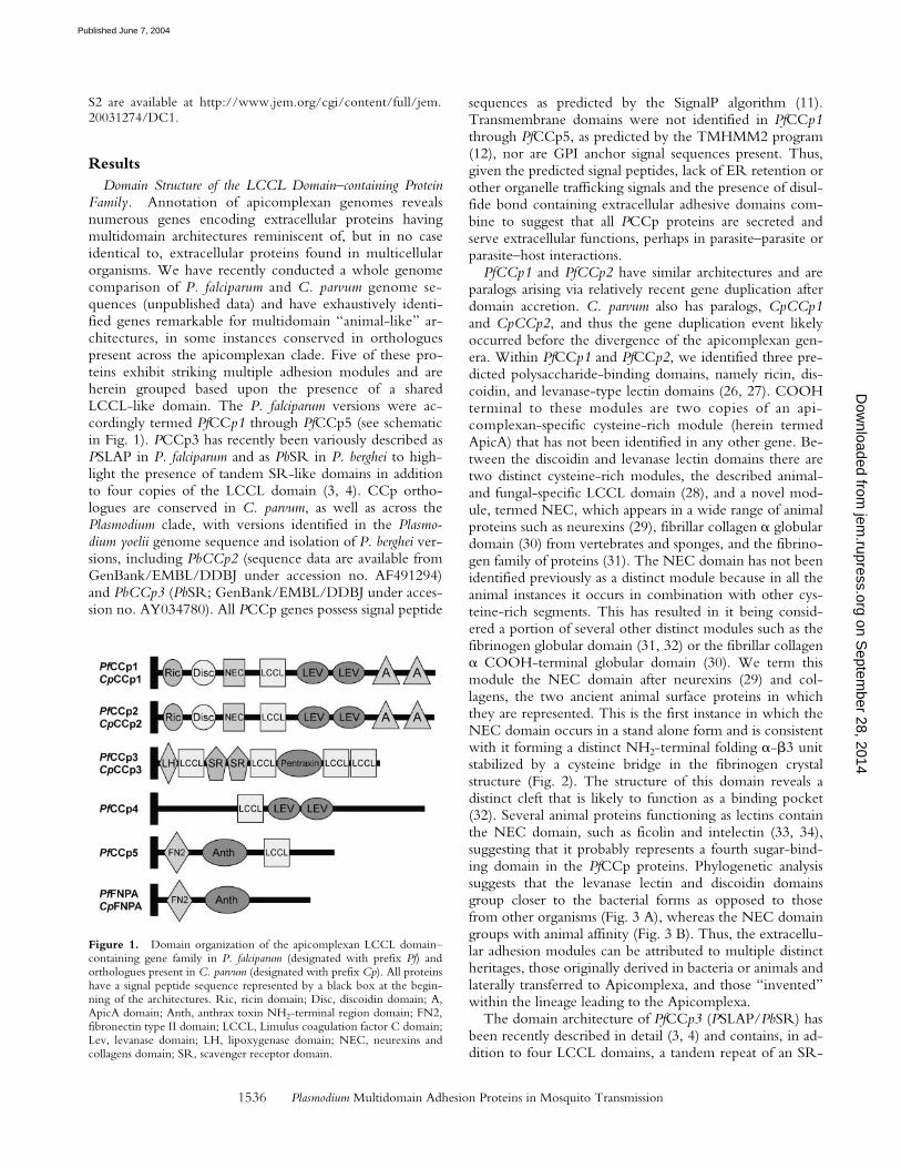
PfCCp1 and PfCCp2 have similar architectures and areparalogs arising via relatively recent gene duplication afterdomain accretion. C. parvum also has paralogs, CpCCp1and CpCCp2, and thus the gene duplication event likelyoccurred before the divergence of the apicomplexan gen-era. Within PfCCp1 and PfCCp2, we identified three pre-dicted polysaccharide-binding domains, namely ricin, dis-coidin, and levanase-type lectin domains (26, 27). COOHterminal to these modules are two copies of an api-complexan-specific cysteine-rich module (herein termedApicA) that has not been identified in any other gene. Be-tween the discoidin and levanase lectin domains there aretwo distinct cysteine-rich modules, the described animal-and fungal-specific LCCL domain (28), and a novel mod-ule, termed NEC, which appears in a wide range of animalproteins such as neurexins (29), fibrillar collagen � globulardomain (30) from vertebrates and sponges, and the fibrino-gen family of proteins (31). The NEC domain has not beenidentified previously as a distinct module because in all theanimal instances it occurs in combination with other cys-teine-rich segments. This has resulted in it being consid-ered a portion of several other distinct modules such as thefibrinogen globular domain (31, 32) or the fibrillar collagen� COOH-terminal globular domain (30). We term thismodule the NEC domain after neurexins (29) and col-lagens, the two ancient animal surface proteins in whichthey are represented. This is the first instance in which theNEC domain occurs in a stand alone form and is consistentwith it forming a distinct NH2-terminal folding �-�3 unitstabilized by a cysteine bridge in the fibrinogen crystalstructure (Fig. 2). The structure of this domain reveals adistinct cleft that is likely to function as a binding pocket(32). Several animal proteins functioning as lectins containthe NEC domain, such as ficolin and intelectin (33, 34),suggesting that it probably represents a fourth sugar-bind-ing domain in the PfCCp proteins. Phylogenetic analysissuggests that the levanase lectin and discoidin domainsgroup closer to the bacterial forms as opposed to thosefrom other organisms (Fig. 3 A), whereas the NEC domaingroups with animal affinity (Fig. 3 B). Thus, the extracellu-lar adhesion modules can be attributed to multiple distinctheritages, those originally derived in bacteria or animals andlaterally transferred to Apicomplexa, and those “invented”within the lineage leading to the Apicomplexa.
The domain architecture of PfCCp3 (PSLAP/PbSR) hasbeen recently described in detail (3, 4) and contains, in ad-dition to four LCCL domains, a tandem repeat of an SR-
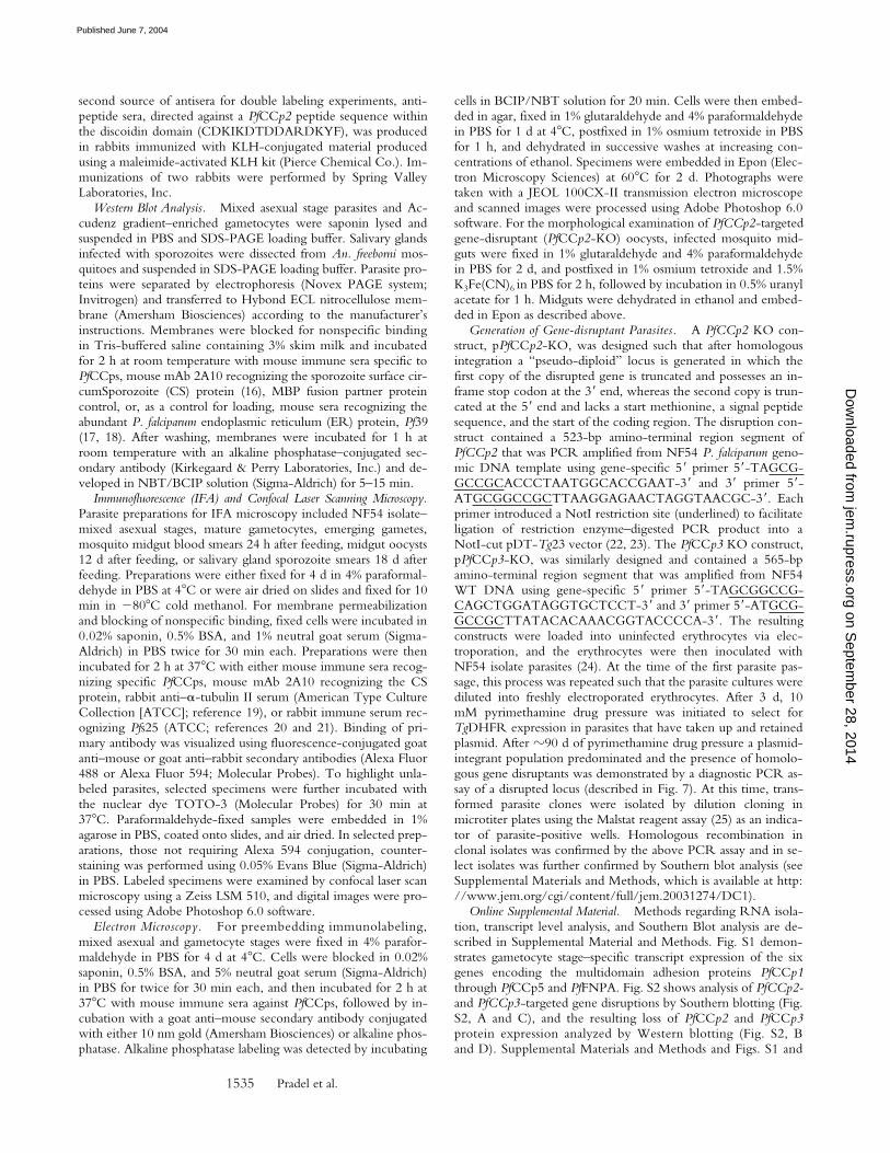
Figure 1. Domain organization of the apicomplexan LCCL domain–containing gene family in P. falciparum (designated with prefix Pf) andorthologues present in C. parvum (designated with prefix Cp). All proteinshave a signal peptide sequence represented by a black box at the begin-ning of the architectures. Ric, ricin domain; Disc, discoidin domain; A,ApicA domain; Anth, anthrax toxin NH2-terminal region domain; FN2,fibronectin type II domain; LCCL, Limulus coagulation factor C domain;Lev, levanase domain; LH, lipoxygenase domain; NEC, neurexins andcollagens domain; SR, scavenger receptor domain.
on Septem
ber 28, 2014jem
.rupress.orgD
ownloaded from
Published June 7, 2004
Pradel et al.1537
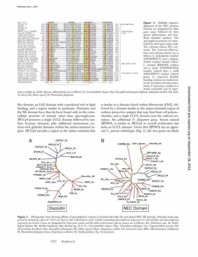
like domain, an LH2 domain with a predicted role in lipidbinding, and a region similar to pentraxin. Pentraxin andthe SR domain have thus far been found only in the extra-cellular proteins of animals other than apicomplexans.PfCCp4 possesses a single LCCL domain followed by tan-dem levanase domains, plus additional anonymous cys-teine-rich globular domains within the amino-terminal re-gion. PfCCp5 encodes a region at the amino terminus that
is similar to a domain found within fibronectin (FN2), fol-lowed by a domain similar to the amino-terminal region ofanthrax-protective antigen that may bind host cell polysac-charides, and a single LCCL domain near the carboxy ter-minus. An additional P. falciparum gene, herein namedPfFNPA, is similar to PfCCp5 in overall architecture butlacks an LCCL domain. Given that PfFNPA has an appar-ent C. parvum orthologue (Fig. 1), the two genes are likely
Figure 2. Multiple sequencealignment of the NEC domain.Proteins are designated by theirgene name followed by theirspecies abbreviations and Gen-Bank identifier numbers. Theapicomplexan proteins are repre-sented by their gene names only.The coloring reflects 85% con-sensus. The consensus abbrevia-tions and coloring scheme are asfollows: h, hydrophobic residues(LIYFMWACV) and l, aliphatic(LIAV) residues shaded yellow;c, charged (KERDH) residuesand p, polar (STEDRKHNQ)residues colored blue; s, small(SAGDNPVT) residues coloredgreen; C, conserved disulfidebonding cysteines are shaded red.In the secondary structure anno-tation, E represents a residue in astrand (extended) and H repre-
sents a residue in a helix. Species abbreviations are as follows: Ce, Caenorhabditis elegans; Dm, Drosophila melanogaster; Ephmu, Ephydatia muelleri; Hd, Hali-otis discus; Hs, Homo sapiens; Pf, Plasmodium falciparum.
Figure 3. Phylogenetic trees showing affinity of apicomplexan versions to bacterial discoidin (A) and animal NEC (B) domains. Principal nodes sup-ported by bootstrap values of �65% are shown with a filled green circle. Clades containing apicomplexan sequences are colored blue and apicomplexansequences are boxed. Genes are designated by their gene names and the abbreviated source species names are as follows: Art, Arthrobacter spp.; At, Arabi-dopsis thaliana; Bh, Bacillus halodurans; Bsp, Bacillus spp. D-2; Ce, Caenorrhabditis elegans; Clpe, Clostridium perfringens; Cp, Cryptosporidium parvum; Dd,Dictyostelium discoidium; Dm, Drosophila melanogaster; Hs, Homo sapiens; Hyro, Hypomyces rosellus; Lal, Lactococcus lactis; Mivi, Micromonospora viridifaciens;Pf, Plasmodium falciparum; Scoe, Streptomyces coelicolor; Xf, Xylella fastidiosa; Yp, Yersinia pestis.
on Septem
ber 28, 2014jem
.rupress.orgD
ownloaded from
Published June 7, 2004
Plasmodium Multidomain Adhesion Proteins in Mosquito Transmission1538
to have diverged in P. falciparum from a common ancestorthat was similar in architecture to PfFNPA, followed by ac-cretion of an LCCL domain in PfCCp5.
Expression of PfCCps during the Parasite Life Cycle.Transcript expression was studied by real time RT-PCRanalysis using RNA isolated from asexual and gameto-cyte intraerythrocytic in vitro–cultivated P. falciparum para-sites. Expression was gametocyte stage specific for PfCCp1through PfCCp4 and PfFNPA, in comparison with tran-script expression controls for asexual (AMA-1) and game-tocyte (Pfs48/45) stage–specific genes (Fig. S1, available
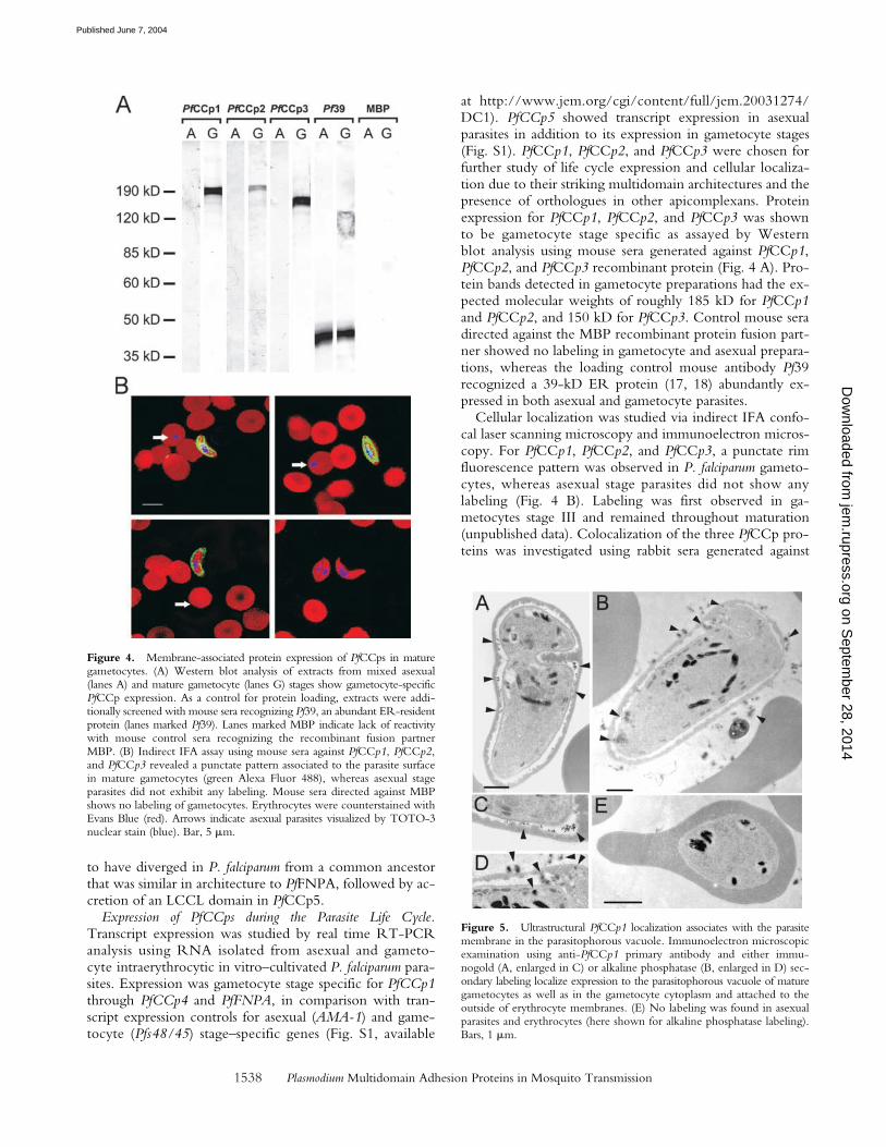
at http://www.jem.org/cgi/content/full/jem.20031274/DC1). PfCCp5 showed transcript expression in asexualparasites in addition to its expression in gametocyte stages(Fig. S1). PfCCp1, PfCCp2, and PfCCp3 were chosen forfurther study of life cycle expression and cellular localiza-tion due to their striking multidomain architectures and thepresence of orthologues in other apicomplexans. Proteinexpression for PfCCp1, PfCCp2, and PfCCp3 was shownto be gametocyte stage specific as assayed by Westernblot analysis using mouse sera generated against PfCCp1,PfCCp2, and PfCCp3 recombinant protein (Fig. 4 A). Pro-tein bands detected in gametocyte preparations had the ex-pected molecular weights of roughly 185 kD for PfCCp1and PfCCp2, and 150 kD for PfCCp3. Control mouse seradirected against the MBP recombinant protein fusion part-ner showed no labeling in gametocyte and asexual prepara-tions, whereas the loading control mouse antibody Pf39recognized a 39-kD ER protein (17, 18) abundantly ex-pressed in both asexual and gametocyte parasites.
Cellular localization was studied via indirect IFA confo-cal laser scanning microscopy and immunoelectron micros-copy. For PfCCp1, PfCCp2, and PfCCp3, a punctate rimfluorescence pattern was observed in P. falciparum gameto-cytes, whereas asexual stage parasites did not show anylabeling (Fig. 4 B). Labeling was first observed in ga-metocytes stage III and remained throughout maturation(unpublished data). Colocalization of the three PfCCp pro-teins was investigated using rabbit sera generated against
Figure 4. Membrane-associated protein expression of PfCCps in maturegametocytes. (A) Western blot analysis of extracts from mixed asexual(lanes A) and mature gametocyte (lanes G) stages show gametocyte-specificPfCCp expression. As a control for protein loading, extracts were addi-tionally screened with mouse sera recognizing Pf39, an abundant ER-residentprotein (lanes marked Pf39). Lanes marked MBP indicate lack of reactivitywith mouse control sera recognizing the recombinant fusion partnerMBP. (B) Indirect IFA assay using mouse sera against PfCCp1, PfCCp2,and PfCCp3 revealed a punctate pattern associated to the parasite surfacein mature gametocytes (green Alexa Fluor 488), whereas asexual stageparasites did not exhibit any labeling. Mouse sera directed against MBPshows no labeling of gametocytes. Erythrocytes were counterstained withEvans Blue (red). Arrows indicate asexual parasites visualized by TOTO-3nuclear stain (blue). Bar, 5 �m.
Figure 5. Ultrastructural PfCCp1 localization associates with the parasitemembrane in the parasitophorous vacuole. Immunoelectron microscopicexamination using anti-PfCCp1 primary antibody and either immu-nogold (A, enlarged in C) or alkaline phosphatase (B, enlarged in D) sec-ondary labeling localize expression to the parasitophorous vacuole of maturegametocytes as well as in the gametocyte cytoplasm and attached to theoutside of erythrocyte membranes. (E) No labeling was found in asexualparasites and erythrocytes (here shown for alkaline phosphatase labeling).Bars, 1 �m.
on Septem
ber 28, 2014jem
.rupress.orgD
ownloaded from
Published June 7, 2004
Pradel et al.1539
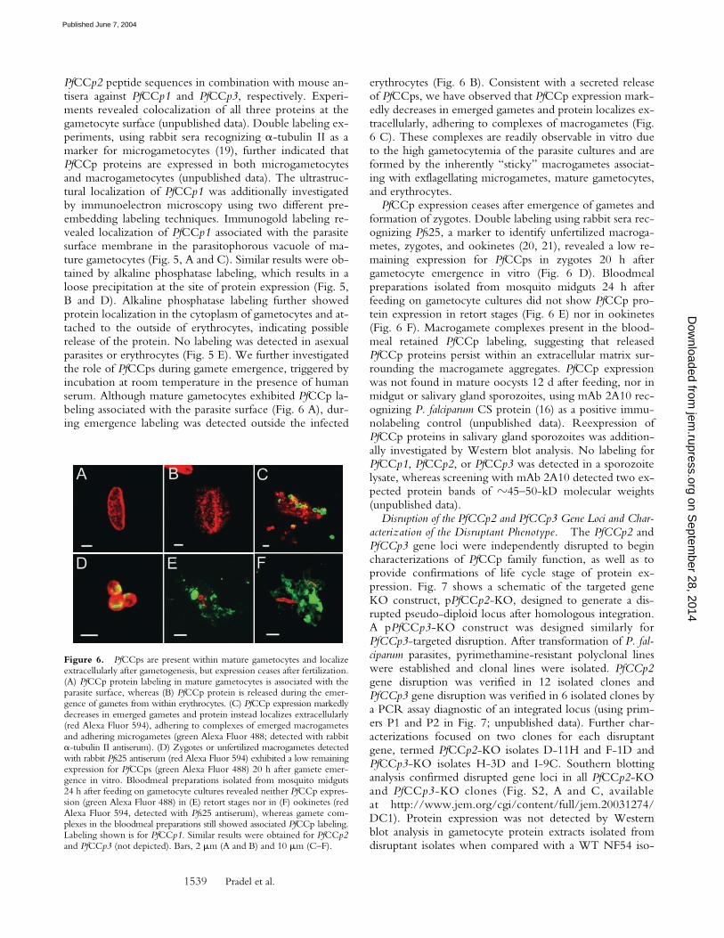
PfCCp2 peptide sequences in combination with mouse an-tisera against PfCCp1 and PfCCp3, respectively. Experi-ments revealed colocalization of all three proteins at thegametocyte surface (unpublished data). Double labeling ex-periments, using rabbit sera recognizing �-tubulin II as amarker for microgametocytes (19), further indicated thatPfCCp proteins are expressed in both microgametocytesand macrogametocytes (unpublished data). The ultrastruc-tural localization of PfCCp1 was additionally investigatedby immunoelectron microscopy using two different pre-embedding labeling techniques. Immunogold labeling re-vealed localization of PfCCp1 associated with the parasitesurface membrane in the parasitophorous vacuole of ma-ture gametocytes (Fig. 5, A and C). Similar results were ob-tained by alkaline phosphatase labeling, which results in aloose precipitation at the site of protein expression (Fig. 5,B and D). Alkaline phosphatase labeling further showedprotein localization in the cytoplasm of gametocytes and at-tached to the outside of erythrocytes, indicating possiblerelease of the protein. No labeling was detected in asexualparasites or erythrocytes (Fig. 5 E). We further investigatedthe role of PfCCps during gamete emergence, triggered byincubation at room temperature in the presence of humanserum. Although mature gametocytes exhibited PfCCp la-beling associated with the parasite surface (Fig. 6 A), dur-ing emergence labeling was detected outside the infected
erythrocytes (Fig. 6 B). Consistent with a secreted releaseof PfCCps, we have observed that PfCCp expression mark-edly decreases in emerged gametes and protein localizes ex-tracellularly, adhering to complexes of macrogametes (Fig.6 C). These complexes are readily observable in vitro dueto the high gametocytemia of the parasite cultures and areformed by the inherently “sticky” macrogametes associat-ing with exflagellating microgametes, mature gametocytes,and erythrocytes.
PfCCp expression ceases after emergence of gametes andformation of zygotes. Double labeling using rabbit sera rec-ognizing Pfs25, a marker to identify unfertilized macroga-metes, zygotes, and ookinetes (20, 21), revealed a low re-maining expression for PfCCps in zygotes 20 h aftergametocyte emergence in vitro (Fig. 6 D). Bloodmealpreparations isolated from mosquito midguts 24 h afterfeeding on gametocyte cultures did not show PfCCp pro-tein expression in retort stages (Fig. 6 E) nor in ookinetes(Fig. 6 F). Macrogamete complexes present in the blood-meal retained PfCCp labeling, suggesting that releasedPfCCp proteins persist within an extracellular matrix sur-rounding the macrogamete aggregates. PfCCp expressionwas not found in mature oocysts 12 d after feeding, nor inmidgut or salivary gland sporozoites, using mAb 2A10 rec-ognizing P. falciparum CS protein (16) as a positive immu-nolabeling control (unpublished data). Reexpression ofPfCCp proteins in salivary gland sporozoites was addition-ally investigated by Western blot analysis. No labeling forPfCCp1, PfCCp2, or PfCCp3 was detected in a sporozoitelysate, whereas screening with mAb 2A10 detected two ex-pected protein bands of �45–50-kD molecular weights(unpublished data).
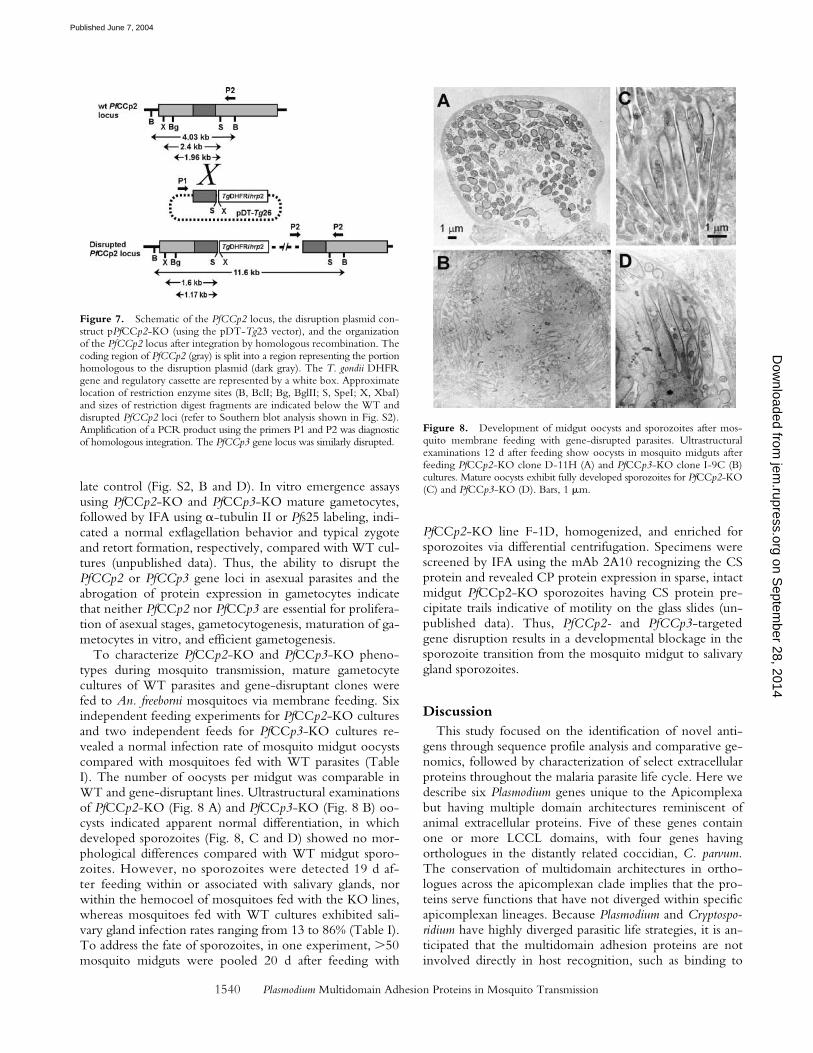
Disruption of the PfCCp2 and PfCCp3 Gene Loci and Char-acterization of the Disruptant Phenotype. The PfCCp2 andPfCCp3 gene loci were independently disrupted to begincharacterizations of PfCCp family function, as well as toprovide confirmations of life cycle stage of protein ex-pression. Fig. 7 shows a schematic of the targeted geneKO construct, pPfCCp2-KO, designed to generate a dis-rupted pseudo-diploid locus after homologous integration.A pPfCCp3-KO construct was designed similarly forPfCCp3-targeted disruption. After transformation of P. fal-ciparum parasites, pyrimethamine-resistant polyclonal lineswere established and clonal lines were isolated. PfCCp2gene disruption was verified in 12 isolated clones andPfCCp3 gene disruption was verified in 6 isolated clones bya PCR assay diagnostic of an integrated locus (using prim-ers P1 and P2 in Fig. 7; unpublished data). Further char-acterizations focused on two clones for each disruptantgene, termed PfCCp2-KO isolates D-11H and F-1D andPfCCp3-KO isolates H-3D and I-9C. Southern blottinganalysis confirmed disrupted gene loci in all PfCCp2-KOand PfCCp3-KO clones (Fig. S2, A and C, availableat http://www.jem.org/cgi/content/full/jem.20031274/DC1). Protein expression was not detected by Westernblot analysis in gametocyte protein extracts isolated fromdisruptant isolates when compared with a WT NF54 iso-
Figure 6. PfCCps are present within mature gametocytes and localizeextracellularly after gametogenesis, but expression ceases after fertilization.(A) PfCCp protein labeling in mature gametocytes is associated with theparasite surface, whereas (B) PfCCp protein is released during the emer-gence of gametes from within erythrocytes. (C) PfCCp expression markedlydecreases in emerged gametes and protein instead localizes extracellularly(red Alexa Fluor 594), adhering to complexes of emerged macrogametesand adhering microgametes (green Alexa Fluor 488; detected with rabbit�-tubulin II antiserum). (D) Zygotes or unfertilized macrogametes detectedwith rabbit Pfs25 antiserum (red Alexa Fluor 594) exhibited a low remainingexpression for PfCCps (green Alexa Fluor 488) 20 h after gamete emer-gence in vitro. Bloodmeal preparations isolated from mosquito midguts24 h after feeding on gametocyte cultures revealed neither PfCCp expres-sion (green Alexa Fluor 488) in (E) retort stages nor in (F) ookinetes (redAlexa Fluor 594, detected with Pfs25 antiserum), whereas gamete com-plexes in the bloodmeal preparations still showed associated PfCCp labeling.Labeling shown is for PfCCp1. Similar results were obtained for PfCCp2and PfCCp3 (not depicted). Bars, 2 �m (A and B) and 10 �m (C–F).
on Septem
ber 28, 2014jem
.rupress.orgD
ownloaded from
Published June 7, 2004
Plasmodium Multidomain Adhesion Proteins in Mosquito Transmission1540
late control (Fig. S2, B and D). In vitro emergence assaysusing PfCCp2-KO and PfCCp3-KO mature gametocytes,followed by IFA using �-tubulin II or Pfs25 labeling, indi-cated a normal exflagellation behavior and typical zygoteand retort formation, respectively, compared with WT cul-tures (unpublished data). Thus, the ability to disrupt thePfCCp2 or PfCCp3 gene loci in asexual parasites and theabrogation of protein expression in gametocytes indicatethat neither PfCCp2 nor PfCCp3 are essential for prolifera-tion of asexual stages, gametocytogenesis, maturation of ga-metocytes in vitro, and efficient gametogenesis.
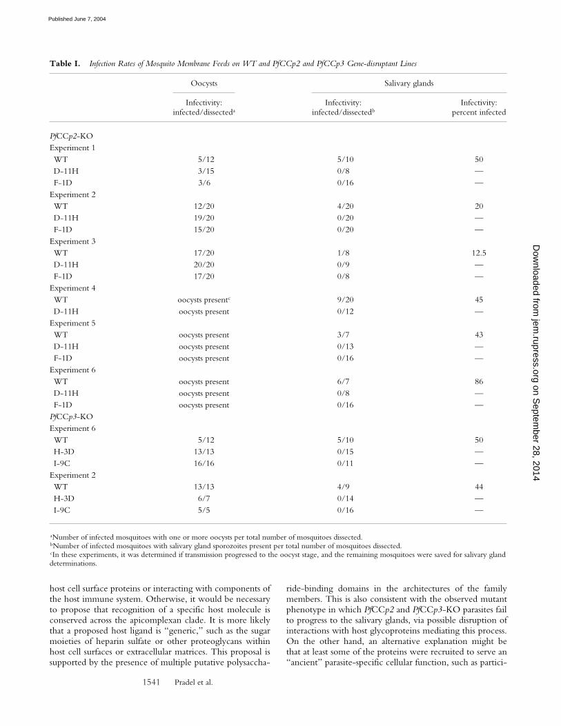
To characterize PfCCp2-KO and PfCCp3-KO pheno-types during mosquito transmission, mature gametocytecultures of WT parasites and gene-disruptant clones werefed to An. freeborni mosquitoes via membrane feeding. Sixindependent feeding experiments for PfCCp2-KO culturesand two independent feeds for PfCCp3-KO cultures re-vealed a normal infection rate of mosquito midgut oocystscompared with mosquitoes fed with WT parasites (TableI). The number of oocysts per midgut was comparable inWT and gene-disruptant lines. Ultrastructural examinationsof PfCCp2-KO (Fig. 8 A) and PfCCp3-KO (Fig. 8 B) oo-cysts indicated apparent normal differentiation, in whichdeveloped sporozoites (Fig. 8, C and D) showed no mor-phological differences compared with WT midgut sporo-zoites. However, no sporozoites were detected 19 d af-ter feeding within or associated with salivary glands, norwithin the hemocoel of mosquitoes fed with the KO lines,whereas mosquitoes fed with WT cultures exhibited sali-vary gland infection rates ranging from 13 to 86% (Table I).To address the fate of sporozoites, in one experiment, �50mosquito midguts were pooled 20 d after feeding with
PfCCp2-KO line F-1D, homogenized, and enriched forsporozoites via differential centrifugation. Specimens werescreened by IFA using the mAb 2A10 recognizing the CSprotein and revealed CP protein expression in sparse, intactmidgut PfCCp2-KO sporozoites having CS protein pre-cipitate trails indicative of motility on the glass slides (un-published data). Thus, PfCCp2- and PfCCp3-targetedgene disruption results in a developmental blockage in thesporozoite transition from the mosquito midgut to salivarygland sporozoites.
DiscussionThis study focused on the identification of novel anti-
gens through sequence profile analysis and comparative ge-nomics, followed by characterization of select extracellularproteins throughout the malaria parasite life cycle. Here wedescribe six Plasmodium genes unique to the Apicomplexabut having multiple domain architectures reminiscent ofanimal extracellular proteins. Five of these genes containone or more LCCL domains, with four genes havingorthologues in the distantly related coccidian, C. parvum.The conservation of multidomain architectures in ortho-logues across the apicomplexan clade implies that the pro-teins serve functions that have not diverged within specificapicomplexan lineages. Because Plasmodium and Cryptospo-ridium have highly diverged parasitic life strategies, it is an-ticipated that the multidomain adhesion proteins are notinvolved directly in host recognition, such as binding to
Figure 7. Schematic of the PfCCp2 locus, the disruption plasmid con-struct pPfCCp2-KO (using the pDT-Tg23 vector), and the organizationof the PfCCp2 locus after integration by homologous recombination. Thecoding region of PfCCp2 (gray) is split into a region representing the portionhomologous to the disruption plasmid (dark gray). The T. gondii DHFRgene and regulatory cassette are represented by a white box. Approximatelocation of restriction enzyme sites (B, BclI; Bg, BglII; S, SpeI; X, XbaI)and sizes of restriction digest fragments are indicated below the WT anddisrupted PfCCp2 loci (refer to Southern blot analysis shown in Fig. S2).Amplification of a PCR product using the primers P1 and P2 was diagnosticof homologous integration. The PfCCp3 gene locus was similarly disrupted.
Figure 8. Development of midgut oocysts and sporozoites after mos-quito membrane feeding with gene-disrupted parasites. Ultrastructuralexaminations 12 d after feeding show oocysts in mosquito midguts afterfeeding PfCCp2-KO clone D-11H (A) and PfCCp3-KO clone I-9C (B)cultures. Mature oocysts exhibit fully developed sporozoites for PfCCp2-KO(C) and PfCCp3-KO (D). Bars, 1 �m.
on Septem
ber 28, 2014jem
.rupress.orgD
ownloaded from
Published June 7, 2004
Pradel et al.1541
host cell surface proteins or interacting with components ofthe host immune system. Otherwise, it would be necessaryto propose that recognition of a specific host molecule isconserved across the apicomplexan clade. It is more likelythat a proposed host ligand is “generic,” such as the sugarmoieties of heparin sulfate or other proteoglycans withinhost cell surfaces or extracellular matrices. This proposal issupported by the presence of multiple putative polysaccha-
ride-binding domains in the architectures of the familymembers. This is also consistent with the observed mutantphenotype in which PfCCp2 and PfCCp3-KO parasites failto progress to the salivary glands, via possible disruption ofinteractions with host glycoproteins mediating this process.On the other hand, an alternative explanation might bethat at least some of the proteins were recruited to serve an“ancient” parasite-specific cellular function, such as partici-
Table I. Infection Rates of Mosquito Membrane Feeds on WT and PfCCp2 and PfCCp3 Gene-disruptant Lines
Oocysts Salivary glands
Infectivity:infected/dissecteda
Infectivity:infected/dissectedb
Infectivity:percent infected
PfCCp2-KOExperiment 1WT 5/12 5/10 50D-11H 3/15 0/8 —F-1D 3/6 0/16 —
Experiment 2WT 12/20 4/20 20D-11H 19/20 0/20 —F-1D 15/20 0/20 —
Experiment 3WT 17/20 1/8 12.5D-11H 20/20 0/9 —F-1D 17/20 0/8 —
Experiment 4WT oocysts presentc 9/20 45D-11H oocysts present 0/12 —
Experiment 5WT oocysts present 3/7 43D-11H oocysts present 0/13 —F-1D oocysts present 0/16 —
Experiment 6WT oocysts present 6/7 86D-11H oocysts present 0/8 —F-1D oocysts present 0/16 —
PfCCp3-KOExperiment 6WT 5/12 5/10 50H-3D 13/13 0/15 —I-9C 16/16 0/11 —
Experiment 2WT 13/13 4/9 44H-3D 6/7 0/14 —I-9C 5/5 0/16 —
aNumber of infected mosquitoes with one or more oocysts per total number of mosquitoes dissected.bNumber of infected mosquitoes with salivary gland sporozoites present per total number of mosquitoes dissected.cIn these experiments, it was determined if transmission progressed to the oocyst stage, and the remaining mosquitoes were saved for salivary glanddeterminations.
on Septem
ber 28, 2014jem
.rupress.orgD
ownloaded from
Published June 7, 2004
Plasmodium Multidomain Adhesion Proteins in Mosquito Transmission1542
pation in a stage-specific aspect of the parasitophorous vac-uole, formation of structures associated with the parasitepellicule, or serving in the formation of an extracellularmatrix surrounding one or more emerged parasite stages.Either of the two proposed models must take into accounta role for the multiple putative polysaccharide-binding do-mains within the LCCL domain proteins. It is likely thatthe expression stage specificity is conserved across generaand support for CCp function might ultimately be pro-vided by the study of life cycle stage of expression and lo-calization in additional apicomplexans, such as Cryptosporid-ium and Toxoplasma.
Transcript expression studies show abundant gameto-cyte-specific expression for five of the six multidomain ad-hesion proteins. Protein expression and immunolocaliza-tion studies for three selected proteins, PfCCp1, PfCCp2,and PfCCp3, indicate that the proteins colocalize andare released during gametogenesis and remain associatedwith gamete aggregates. Gametocyte-specific expression ofPfCCp proteins is supported by high throughput massspectrometry proteomic analysis of P. yoelii rodent malariasexual stage parasites (35), as well as a recent study showinggametocyte-specific transcript and protein expression ofPSLAP/PfCCp3 in P. falciparum (3). In the latter study, cel-lular localization revealed submembranous expression orsecretion to the parasitophorous vacuole and erythrocytecytoplasm of mature gametocytes. This localization patternis similar to that observed in our indirect IFA and immuno-electron microscopy studies.
To begin to address function of the LCCL domain fam-ily members, we have disrupted the PfCCp2 and PfCCp3gene loci and have shown that neither gene is essential forasexual proliferation or required for efficient gametocyto-genesis. Despite abundant gametocyte stage expression inWT parasites, gene-disruptant lines formed gametes effi-ciently and, after membrane feeds to anophelene mosqui-toes, formed midgut wall oocysts with efficiency similar toWT. Oocysts mature normally and intact sporozoites arereleased, but a block in transmission occurred during themature midgut oocyst to salivary gland sporozoite transi-tion. Hemocoel sporozoites were not detected, nor weresporozoites adhering to salivary glands. The fate of thegene-disruptant sporozoites is not yet known, with onepossibility being that the sporozoites remain and subse-quently degrade within the mosquito midgut. It is thus farnot possible to interpret the discrepancy between theprominent PfCCp transcript and protein expression ob-served in gametocyte stages and the manifestation of a phe-notype in PfCCp2 and PfCCp3 gene disruptants during latestages of midgut oocyst development. It is unlikely that thephenotype is due to an “epigenetic artifact” because itwould require proposing that a similar epigenetic pheno-type has occurred after disruption of either of the geneti-cally unlinked genes, PfCCp2 and PfCCp3. The oocyststage phenotype may either be linked in an as yet unknownfashion to protein function in gametocytes or might be dueto abrogation of undetected low level sporozoite stage pro-
tein expression. The latter hypothesis is supported by re-cent description of a CpCCP1/2 homologue expressed inCryptosporidium sporozoites (36, 37). CpCCP1/2 proteinalso localized to the parasitophorous vacuole in 72-h in-fected HCT-8 cells, thus drawing a parallel with Plasmo-dium gametocyte cellular localization in this study and pro-viding support that the LCCL domain family membersserve roles in multiple parasite stages.
Targeted gene disruption of PbSR (PbCCp3/PbSLAP) inthe rodent malaria model, P. berghei, also results in a blockin late stage oocyst maturation (4) and the phenotype is su-perficially similar to PfCCp2-KO and PfCCp3-KO. Incontrast to the PbSR study (4), we were unable to detectPfCCp3 protein expression in sporozoites by IFA andWestern blot assays using mouse sera directed against re-combinant protein corresponding to the tandem SR do-mains. Whereas Claudianos et al. (4) describe 40- and 60-kD proteins by Western blot and immunolocalization insporozoites using SR antipeptide sera, we detect a proteinwith the expected full-length molecular weight of 150 kDin P. falciparum gametocyte protein extracts, although wehave not addressed possible proteolytic maturation of theprotein during gamete emergence. The discrepancy in thetwo studies might be due to either sporozoite-specific pro-cessing resulting in a truncated protein contaminating pro-tease activity in the sporozoite preparations, or nonspecificreactivity of one or more antibody reagents. Similar to ourobservations, Delrieu et al. (3) show that PSLAP/PfCCp3is expressed in gametocytes and if gametocyte expressionholds for P. berghei, it would suggest that expression ofmultiple PfCCp proteins are essential for sporozoite devel-opment, whereas expression in gametocytes is marked butnot essential for in vitro development.
Transmission-blocking vaccines target sexual phase para-site antigens, thereby relying on human antibodies toinhibit parasite development within the mosquito host(38–40). The sexual stage expression and extracellular lo-calization of PfCCp1, PfCCp2, and PfCCp3 make them po-tential vaccine candidates, and targeted gene disruption ofPfCCp2 and PbSR/PfCCp3 further demonstrate that thesehighly conserved apicomplexan proteins are essential for P.falciparum development in the mosquito vector. PfCCptransmission-blocking assays are currently being pursued inour laboratory to investigate PfCCps as candidates for studyas subunits of a transmission-blocking vaccine.
We thank Heather Eisen, Darrow Deluca, and Amina Kurtovic fortechnical assistance. We also thank Dr. Elizabeth Nardin for kindlyproviding the mAb 2A10, and David Keister, Olga Muratova, andAndre Laughinghouse for mosquito and parasite material, mosquitorearing, and mosquito feeds. Drs. Kirk Deitsch and Carl Nathan arethanked for critically reviewing the manuscript.
This work was supported by the Niarchos Foundation. The De-partment of Microbiology and Immunology at Weill Medical Col-lege of Cornell University acknowledges the support of the Wil-liam Randolph Hearst Foundation.
Submitted: 28 July 2003Accepted: 12 April 2004
on Septem
ber 28, 2014jem
.rupress.orgD
ownloaded from
Published June 7, 2004

Pradel et al.1543
References1. Gardner, M.J., N. Hall, E. Fung, O. White, M. Berriman,
R.W. Hyman, J.M. Carlton, A. Pain, K.E. Nelson, S. Bow-man, et al. 2002. Genome sequence of the human malariaparasite Plasmodium falciparum. Nature. 419:498–511.
2. Carlton, J.M., S.V. Angiuoli, B.B. Suh, T.W. Kooij, M.Pertea, J.C. Silva, M.D. Ermolaeva, J.E. Allen, J.D. Selengut,H.L. Koo, et al. 2002. Genome sequence and comparativeanalysis of the model rodent malaria parasite Plasmodium yoeliiyoelii. Nature. 419:512–519.
3. Delrieu, I., C.C. Waller, M.M. Mota, M. Grainger, J. Lang-horne, and A.A. Holder. 2001. PSLAP, a protein with multi-ple adhesive motifs, is expressed in Plasmodium falciparum ga-metocytes. Mol. Biochem. Parasitol. 121:11–20.
4. Claudianos, C., J.T. Dessens, H.E. Trueman, M. Arai, J.Mendoza, G.A. Butcher, T. Crompton, and R.E. Sinden.2002. A malaria scavenger receptor-like protein essential forparasite development. Mol. Microbiol. 45:1473–1484.
5. Altschul, S.F., T.L. Madden, A.A. Schaffer, J. Zhang, Z.Zhang, W. Miller, and D.J. Lipman. 1997. Gapped BLASTand PSI-BLAST: a new generation of protein database searchprograms. Nucleic Acids Res. 25:3389–3402.
6. Notredame, C., D.G. Higgins, and J. Heringa. 2000. T-Cof-fee: a novel method for fast and accurate multiple sequencealignment. J. Mol. Biol. 302:205–217.
7. Rost, B. 1996. PHD: predicting one-dimensional proteinstructure by profile-based neural networks. Methods Enzymol.266:525–539.
8. Kumar, S., K. Tamura, I.B. Jakobsen, and M. Nei. 2001.MEGA2: molecular evolutionary genetics analysis software.Bioinformatics. 17:1244–1245.
9. Felsenstein, J. 1996. Inferring phylogenies from protein se-quences by parsimony, distance, and likelihood methods.Methods Enzymol. 266:418–427.
10. Hasegawa, M., H. Kishino, and N. Saitou. 1991. On themaximum likelihood method of molecular phylogenies. J.Mol. Evol. 32:443–445.
11. Nielson, H., J. Engelbrecht, S. Brunak, and G. von Heijne.2001. Identification of prokaryotic and eukaryotic signal pep-tides and prediction of their cleavage sites. Protein Eng. 10:1–6.
12. Krogh, A., B. Larsson, G. von Heijne, and E.L. Sonnham-mer. 2001. Predicting transmembrane protein topology witha hidden Markov model: application to complete genomes. J.Mol. Biol. 305:567–580.
13. Ifediba, T., and J.P. Vanderberg. 1981. Complete in vitro mat-uration of P. falciparum gametocytes. Nature. 294:364–366.
14. Templeton, T.J., D.B. Keister, O. Muratova, J.L. Procter,and D.C. Kaslow. 1998. Adherence of erythrocytes duringexflagellation of Plasmodium falciparum microgametes is de-pendent on erythrocyte surface sialic acid and glycophorins.J. Exp. Med. 187:1599–1609.
15. Bishop, A., and B.M. Gilchrist. 1946. Experiments upon thefeeding of Aedes aegypti through animal membranes with aview to applying this method to the chemotherapy of ma-laria. Parasitology. 37:85–100.
16. Nardin, E.H., V. Nussenzweig, R.S. Nussenzweig, W.E. Col-lins, K.T. Harinasuta, P. Tapchaisri, and Y. Chomcharn. 1982.Circumsporozoite proteins of human malaria parasites Plasmo-dium falciparum and Plasmodium vivax. J. Exp. Med. 156:20–30.
17. Rawling, D.J., and D.C. Kaslow. 1992. A novel 40-kDamembrane-associated EF-hand calcium binding protein inPlasmodium falciparum. J. Biol. Chem. 267:3976–3982.
18. Templeton, T.J., H. Fujioka, M. Aikawa, K.C. Parker, andD.C. Kaslow. 1997. Plasmodium falciparum Pfs40, renamedPf39, is localized to an intracellular membrane-bound com-partment and is not sexual stage-specific. Mol. Biochem. Para-sitol. 90:359–365.
19. Rawlings, D.J., H. Fujioka, M. Fried, D.B. Keister, M.Aikawa, and D.C. Kaslow. 1992. Alpha-tubulin II is a male-specific protein in Plasmodium falciparum. Mol. Biochem. Parasi-tol. 56:239–250.
20. Kaslow, D.C., I.A. Quakyi, C. Syin, M.G. Raum, D.B.Keister, J.E. Coligan, T.F. McCutchan, and L.H. Miller.1988. A vaccine candidate from the sexual stage of humanmalaria that contains EGF-like domains. Nature. 333:74–76.
21. Kaslow, D.C., I.C. Bathurst, T. Lensen, T. Ponnudurai, P.J.Barr, and D.B. Keister. 1994. Saccharomyces cerevisiae recombi-nant Pfs25 absorbed to alum elicits antibodies that block trans-mission of Plasmodium falciparum. Infect. Immun. 62:5576–5580.
22. Wu, Y., L.A. Kirkman, and T.E. Wellems. 1996. Transfor-mation of Plasmodium falciparum malaria parasites by homolo-gous integration of plasmids that confer resistance to pyr-imethamine. Proc. Natl. Acad. Sci. USA. 93:1130–1134.
23. Templeton, T.J., D.C. Kaslow, and D.A. Fidock. 2000. De-velopmental arrest of the human malaria parasite Plasmodiumfalciparum within the mosquito midgut via CTRP gene dis-ruption. Mol. Microbiol. 36:1–9.
24. Deitsch, K.W., C. Driskill, and T. Wellems. 2001. Transfor-mation of malaria parasites by the spontaneous uptake andexpression of DNA from human erythrocytes. Nucleic AcidsRes. 29:850–853.
25. Goodyer, I.D., and T.F. Taraschi. 1997. Plasmodium falcip-arum: a simple, rapid method for detecting parasite clones inmicrotiter plates. Exp. Parasitol. 86:158–160.
26. Baumgartner, S., K. Hofmann, R. Chiquet-Ehrismann, andP. Bucher. 1998. The discoidin domain family revisited: newmembers from prokaryotes and a homology-based fold pre-diction. Protein Sci. 7:1626–1631.
27. Ponting, C.P., and R.B. Russell. 2000. Identification of dis-tant homologues of fibroblast growth factors suggests a com-mon ancestor for all beta-trefoil proteins. J. Mol. Biol. 302:1041–1047.
28. Trexler, M., L. Banyai, and L. Patthy. 2000. The LCCLmodule. Eur. J. Biochem. 267:5751–5757.
29. Missler, M., R. Fernandez-Chacon, and T.C. Sudhof. 1998.The making of neurexins. J. Neurochem. 71:1339–1347.
30. Bork, P. 1992. The modular architecture of vertebrate col-lagens. FEBS Lett. 307:49–54.
31. Doolittle, R.F. 1992. A detailed consideration of a principaldomain of vertebrate fibrinogen and its relatives. Protein Sci.1:1563–1577.
32. Spraggon, G., S.J. Everse, and R.F. Doolittle. 1997. Crystalstructures of fragment D from human fibrinogen and itscrosslinked counterpart from fibrin. Nature. 389:455–462.
33. Lu, J., and Y. Le. 1998. Ficolins and the fibrinogen-like do-main. Immunobiology. 199:190–199.
34. Tsuji, S., J. Uehori, M. Matsumoto, Y. Suzuki, A. Matsuhisa,K. Toyoshima, and T. Seya. 2001. Human intelectin is anovel soluble lectin that recognizes galactofuranose in carbo-hydrate chains of bacterial cell wall. J. Biol. Chem. 276:23456–23463.
35. Lasonder, E., Y. Ishihama, J.S. Andersen, A.M. Vermunt, A.Pain, R.W. Sauerwein, W.M. Eling, N. Hall, A.P. Waters,
on Septem
ber 28, 2014jem
.rupress.orgD
ownloaded from
Published June 7, 2004
Plasmodium Multidomain Adhesion Proteins in Mosquito Transmission1544
H.G. Stunnenberg, et al. 2002. Analysis of the Plasmodiumfalciparum proteome by high-accuracy mass spectrometry. Na-ture. 419:537–542.
36. Tosini, F., S. Caccio, A. Tamburrini, G. La Rosa, and E.Pozio. 1999. Identification and characterization of three anti-genic proteins from Cryptosporidium parvum sporozoites usinga DNA library expressing poly-histidine tagged peptides. Int.J. Parasitol. 29:1925–1933.
37. Tosini, F., A. Agnoli, R. Mele, M.A.G. Moralez, and E.Pozio. 2004. A new modular protein of Cryptosporidium par-
vum, with ricin B and LCCL domains, expressed in the sporo-zoite invasive stage. Mol. Biochem. Parasitol. 134:137–147.
38. Carter, R., K.N. Mendis, L.H. Miller, L. Molineaux, and A.Saul. 2000. Malaria transmission-blocking vaccines–how cantheir development be supported? Nat. Med. 6:241–244.
39. Carter, R. 2001. Transmission blocking malaria vaccines.Vaccine. 19:2309–2314.
40. Kaslow, D.C. 2002. Transmission-blocking vaccines. Chem.Immunol. 80:287–307.
on Septem
ber 28, 2014jem
.rupress.orgD
ownloaded from
Published June 7, 2004
Copyright © 2022 FDOKUMEN