TAP (NXF1) Belongs to a Multigene Family of Putative RNA Export Factors with a Conserved Modular...
13
MOLECULAR AND CELLULAR BIOLOGY, 0270-7306/00/$04.0010 Dec. 2000, p. 8996–9008 Vol. 20, No. 23 Copyright © 2000, American Society for Microbiology. All Rights Reserved. TAP (NXF1) Belongs to a Multigene Family of Putative RNA Export Factors with a Conserved Modular Architecture ANDREA HEROLD, 1 MIKITA SUYAMA, 1 JOA ˜ O P. RODRIGUES, 2 ISABELLE C. BRAUN, 1 ULRIKE KUTAY, 3 MARIA CARMO-FONSECA, 2 PEER BORK, 1 AND ELISA IZAURRALDE 1 * European Molecular Biology Laboratory, D-69117 Heidelberg, Germany 1 ; Institute of Histology and Embryology, Faculty of Medicine, University of Lisbon, 1699 Lisbon Codex, Portugal 2 ; and Institute of Biochemistry, Swiss Federal Institute of Technology, CH-8092 Zu ¨rich, Switzerland 3 Received 12 July 2000/Returned for modification 15 August 2000/Accepted 6 September 2000 Vertebrate TAP (also called NXF1) and its yeast orthologue, Mex67p, have been implicated in the export of mRNAs from the nucleus. The TAP protein includes a noncanonical RNP-type RNA binding domain, four leu- cine-rich repeats, an NTF2-like domain that allows heterodimerization with p15 (also called NXT1), and a ubiq- uitin-associated domain that mediates the interaction with nucleoporins. Here we show that TAP belongs to an evolutionarily conserved family of proteins that has more than one member in higher eukaryotes. Not only the overall domain organization but also residues important for p15 and nucleoporin interaction are conserved in most family members. We characterize two of four human TAP homologues and show that one of them, NXF2, binds RNA, localizes to the nuclear envelope, and exhibits RNA export activity. NXF3, which does not bind RNA or localize to the nuclear rim, has no RNA export activity. Database searches revealed that although only one p15 (nxt) gene is present in the Drosophila melanogaster and Caenorhabditis elegans genomes, there is at least one additional p15 homologue (p15-2 [also called NXT2]) encoded by the human genome. Both human p15 homologues bind TAP, NXF2, and NXF3. Together, our results indicate that the TAP-p15 mRNA export path- way has diversified in higher eukaryotes compared to yeast, perhaps reflecting a greater substrate complexity. mRNAs are exported from the nucleus as large ribonucleo- protein complexes (mRNPs). To date, proteins directly impli- cated in this process include several nucleoporins and RNA binding proteins (hnRNPs), an RNA helicase of the DEAD- box family (Dbp5), and the nuclear pore complex (NPC)-as- sociated proteins Gle1p, TAP and Mex67p, and RAE1 (also called Gle2p) (reviewed in references 22, 28, and 32). Mex67p is essential for mRNA export in Saccharomyces cerevisiae, while RAE1 is essential for mRNA export in Schizosaccharomyces pombe (9, 27, 36). Their vertebrate homologues, TAP and RAE1, have also been implicated in the export of cellular mRNAs (6, 8, 12, 15, 20, 31). We identified TAP as the cellular factor which is recruited by the constitutive transport element (CTE) of simian type D retroviruses to promote nuclear export of their genomic RNAs (12). In Xenopus oocytes, titration of TAP with an excess of CTE RNA prevents cellular mRNAs from exiting the nucleus (12, 30, 33). This suggests a role for this protein in the export of cellular mRNA. Considerable progress has been made in defining TAP struc- tural and functional domains (see Fig. 1) and in identifying its binding partners. TAP partners include various nucleoporins (4, 17); p15 (also called NXT1), a protein related to nuclear transport factor 2 (NTF2) (7, 17); transportin, which mediates TAP nuclear import (4); and several mRNP-associated pro- teins, such as E1B-AP5, RAE1 (4), and members of the Yra1p/ REF family of proteins (37, 39). Binding of TAP to these mRNP-associated proteins is mediated by its N-terminal do- main (residues 1 to 372) (4, 39). This domain includes a non- canonical RNP-type RNA binding domain (RBD) and four leucine-rich repeats (LRRs) and exhibits general RNA binding affinity and specific binding to the CTE RNA (8, 12, 24). TAP heterodimerizes with p15 via its NTF2-like domain (residues 371 to 551) (4, 40). Nucleoporin binding by TAP in vitro and in vivo is mediated by a domain located at the very C-terminal end of the protein (residues 508 to 619) (4, 5). The similarities of this domain to the ubiquitin-associated (UBA) domain allowed the prediction that residues located in a con- served loop were implicated in TAP-nucleoporin interaction (40). In this study, we identify TAP homologues in Homo sapiens, Caenorhabditis elegans, and Drosophila melanogaster. The over- all domain organization of these proteins, including residues important for p15 and nucleoporin interaction, is conserved. Two of four putative human TAP homologues, NXF2 and NXF3, have been characterized in detail. These proteins show 57 and 44% sequence identity to TAP. Neither binds specifi- cally to the CTE RNA, indicating that they may exhibit differ- ent substrate specificities. Both proteins are localized within the nucleoplasm, but only NXF2 associates with the nuclear envelope and exhibits detectable RNA export activity. In ad- dition, a human homologue of p15 (p15-2 [also called NXT2]) was characterized. p15-2 binds to TAP, NXF2, and NXF3 with affinities similar to those of p15. Like p15-1, a fraction of p15-2 localizes to the nuclear envelope. Both human p15 homologues participate in RNA nuclear export. Together, our results indi- cate that the TAP-p15-mediated export pathway has diversi- fied in higher eukaryotes and in humans includes at least two p15 proteins and multiple TAP-like putative mRNA export factors. MATERIALS AND METHODS Homology searches and sequence analysis. Homologues of TAP and p15 were retrieved in expressed sequence tag (EST) databases, human genomic DNA, and the Drosophila and C. elegans genomes, as well as in various protein sequence databases, using the BLAST suite of programs (3). Multiple sequence alignments were constructed by using CLUSTAL W (42) and manually refined on the SEAVIEW alignment editor (11). To predict the genomic structure of TAP and * Corresponding author. Mailing address: EMBL, Meyerhofstrasse 1, D-69117 Heidelberg, Germany. Phone: 0049 6221 387 389. Fax: 0049 6221 387 518. E-mail: [email protected]. 8996
-
Upload
independent -
Category
Documents
-
view
0 -
download
0
Transcript of TAP (NXF1) Belongs to a Multigene Family of Putative RNA Export Factors with a Conserved Modular...

MOLECULAR AND CELLULAR BIOLOGY,0270-7306/00/$04.0010
Dec. 2000, p. 8996–9008 Vol. 20, No. 23
Copyright © 2000, American Society for Microbiology. All Rights Reserved.
TAP (NXF1) Belongs to a Multigene Family of Putative RNAExport Factors with a Conserved Modular Architecture
ANDREA HEROLD,1 MIKITA SUYAMA,1 JOAO P. RODRIGUES,2 ISABELLE C. BRAUN,1 ULRIKE KUTAY,3
MARIA CARMO-FONSECA,2 PEER BORK,1 AND ELISA IZAURRALDE1*
European Molecular Biology Laboratory, D-69117 Heidelberg, Germany1; Institute of Histology and Embryology,Faculty of Medicine, University of Lisbon, 1699 Lisbon Codex, Portugal2; and Institute of Biochemistry,
Swiss Federal Institute of Technology, CH-8092 Zurich, Switzerland3
Received 12 July 2000/Returned for modification 15 August 2000/Accepted 6 September 2000
Vertebrate TAP (also called NXF1) and its yeast orthologue, Mex67p, have been implicated in the export ofmRNAs from the nucleus. The TAP protein includes a noncanonical RNP-type RNA binding domain, four leu-cine-rich repeats, an NTF2-like domain that allows heterodimerization with p15 (also called NXT1), and a ubiq-uitin-associated domain that mediates the interaction with nucleoporins. Here we show that TAP belongs to anevolutionarily conserved family of proteins that has more than one member in higher eukaryotes. Not only theoverall domain organization but also residues important for p15 and nucleoporin interaction are conserved inmost family members. We characterize two of four human TAP homologues and show that one of them, NXF2,binds RNA, localizes to the nuclear envelope, and exhibits RNA export activity. NXF3, which does not bindRNA or localize to the nuclear rim, has no RNA export activity. Database searches revealed that although onlyone p15 (nxt) gene is present in the Drosophila melanogaster and Caenorhabditis elegans genomes, there is at leastone additional p15 homologue (p15-2 [also called NXT2]) encoded by the human genome. Both human p15homologues bind TAP, NXF2, and NXF3. Together, our results indicate that the TAP-p15 mRNA export path-way has diversified in higher eukaryotes compared to yeast, perhaps reflecting a greater substrate complexity.
mRNAs are exported from the nucleus as large ribonucleo-protein complexes (mRNPs). To date, proteins directly impli-cated in this process include several nucleoporins and RNAbinding proteins (hnRNPs), an RNA helicase of the DEAD-box family (Dbp5), and the nuclear pore complex (NPC)-as-sociated proteins Gle1p, TAP and Mex67p, and RAE1 (alsocalled Gle2p) (reviewed in references 22, 28, and 32). Mex67pis essential for mRNA export in Saccharomyces cerevisiae, whileRAE1 is essential for mRNA export in Schizosaccharomycespombe (9, 27, 36). Their vertebrate homologues, TAP andRAE1, have also been implicated in the export of cellularmRNAs (6, 8, 12, 15, 20, 31).
We identified TAP as the cellular factor which is recruitedby the constitutive transport element (CTE) of simian type Dretroviruses to promote nuclear export of their genomic RNAs(12). In Xenopus oocytes, titration of TAP with an excess ofCTE RNA prevents cellular mRNAs from exiting the nucleus(12, 30, 33). This suggests a role for this protein in the exportof cellular mRNA.
Considerable progress has been made in defining TAP struc-tural and functional domains (see Fig. 1) and in identifying itsbinding partners. TAP partners include various nucleoporins(4, 17); p15 (also called NXT1), a protein related to nucleartransport factor 2 (NTF2) (7, 17); transportin, which mediatesTAP nuclear import (4); and several mRNP-associated pro-teins, such as E1B-AP5, RAE1 (4), and members of the Yra1p/REF family of proteins (37, 39). Binding of TAP to thesemRNP-associated proteins is mediated by its N-terminal do-main (residues 1 to 372) (4, 39). This domain includes a non-canonical RNP-type RNA binding domain (RBD) and four
leucine-rich repeats (LRRs) and exhibits general RNA bindingaffinity and specific binding to the CTE RNA (8, 12, 24).
TAP heterodimerizes with p15 via its NTF2-like domain(residues 371 to 551) (4, 40). Nucleoporin binding by TAP invitro and in vivo is mediated by a domain located at the veryC-terminal end of the protein (residues 508 to 619) (4, 5). Thesimilarities of this domain to the ubiquitin-associated (UBA)domain allowed the prediction that residues located in a con-served loop were implicated in TAP-nucleoporin interaction (40).
In this study, we identify TAP homologues in Homo sapiens,Caenorhabditis elegans, and Drosophila melanogaster. The over-all domain organization of these proteins, including residuesimportant for p15 and nucleoporin interaction, is conserved.Two of four putative human TAP homologues, NXF2 andNXF3, have been characterized in detail. These proteins show57 and 44% sequence identity to TAP. Neither binds specifi-cally to the CTE RNA, indicating that they may exhibit differ-ent substrate specificities. Both proteins are localized withinthe nucleoplasm, but only NXF2 associates with the nuclearenvelope and exhibits detectable RNA export activity. In ad-dition, a human homologue of p15 (p15-2 [also called NXT2])was characterized. p15-2 binds to TAP, NXF2, and NXF3 withaffinities similar to those of p15. Like p15-1, a fraction of p15-2localizes to the nuclear envelope. Both human p15 homologuesparticipate in RNA nuclear export. Together, our results indi-cate that the TAP-p15-mediated export pathway has diversi-fied in higher eukaryotes and in humans includes at least twop15 proteins and multiple TAP-like putative mRNA exportfactors.
MATERIALS AND METHODS
Homology searches and sequence analysis. Homologues of TAP and p15 wereretrieved in expressed sequence tag (EST) databases, human genomic DNA, andthe Drosophila and C. elegans genomes, as well as in various protein sequencedatabases, using the BLAST suite of programs (3). Multiple sequence alignmentswere constructed by using CLUSTAL W (42) and manually refined on theSEAVIEW alignment editor (11). To predict the genomic structure of TAP and
* Corresponding author. Mailing address: EMBL, Meyerhofstrasse1, D-69117 Heidelberg, Germany. Phone: 0049 6221 387 389. Fax: 00496221 387 518. E-mail: [email protected].
8996

p15 homologues in various organisms, the Genewise program (http://www.sanger.ac.uk/Software/Wise2/) was employed. If cDNA and/or EST sequences wereavailable, the BLASTN program for genomic sequences was also used to deter-mine genomic structures.
Plasmids. Full-length human NXF2, NXF3, p15-2a, and p15-2b cDNAs wereamplified by PCR using the human testis Marathon-Ready cDNA library (Clon-tech) as a template and primers introducing the appropriate restriction sites. The39 primers were designed according to the predicted cDNAs and included stopcodons. The 59 primers were designed using sequence information obtained afterperforming 59 rapid amplification of cDNA ends reactions with the same humantestis cDNA library and oligonucleotides lying in the p15-2a, p15-2b, NXF2, andNXF3 coding region that is represented in the corresponding human genomicsequences or I.M.A.G.E. Consortium cDNA clones. The NXF2 sequence isrepresented in the IMAGp998P054498Q2 and IMAGp998C234110Q2 clones.NXF3 is represented in the IMAGp998M08574Q2 clone (see http://www.rzpd.defor further information). The p15-2a and p15-2b cDNAs are represented in thegenomic sequence AL031387. The complete NXF2 and NXF3 cDNA wascloned into pGEMT-easy (Promega) and sequenced. p15-2a cDNA was di-rectly cloned in pGEXCS (29) and sequenced. The cDNAs present in theseplasmids were used for all further subcloning steps.
To generate a plasmid for in vitro translation, NXF2 cDNA was excised frompGEM T-easy with BamHI and HindIII and inserted into the same restrictionsites present in pBSpALTER, a derivative of pALTER-Ex1 (Promega). NXF3was excised with the enzymes NcoI and BamHI and cloned into the NcoI/BamHIsites of pBSSK-HA, a derivative of the pBSSK(1) vector (Stratagene) with theb-globin 59 untranslated region inserted between the HindIII and EcoRI sites.For the coexpression assay (see Fig. 5F), p15-1 and p15-2a cDNAs were clonedin the NcoI/NotI or NcoI/BamHI sites of vector pET28c (Novagen), respectively.
Plasmids allowing the expression of glutathione S-transferase (GST) fusions offull-length NXF2 and NXF3 in Escherichia coli were generated by inserting thecoding sequences into the BamHI and NotI sites of pGEX4T-1 (NXF2) (Phar-macia) or the NcoI and BamHI sites present in pGEXCS (NXF3). The NXF2point mutants E598R, W599A, and N600A and the triple alanine substitution3xAla598 were generated by using an oligonucleotide-directed in vitro mutagen-esis system from Stratagene (Quick-change site-directed mutagenesis) in thecontext of pGEX4T-1-NXF2. The mutation 3xAla598 introduces a NotI site intothe coding sequence of NXF2. Plasmid pGEX4T-1-NXF2D598-626 was made bydigesting pGEX4T-1-NXF2 3xAla598 with PpuMI and NotI and inserting the re-leased fragment into pGEX4T-1-NXF2 cut with the same enzymes. Constructsencoding GST fusions of the NXF2 amino acids 1 to 377 and 102 to 203 (RBD)were generated by inserting the corresponding PCR products into pGEX4T-1 di-gested with BamHI and NotI. A plasmid encoding a GST fusion of NXF3 aminoacids 90 to 192 (RBD) was generated by inserting the corresponding PCRproduct into pGEXCS digested with NcoI-BamHI. All PCRs were performedwith the Expand high-fidelity PCR system (Roche). The integrity of the PCRproducts was confirmed by sequencing.
For expression of GFP fusions in mammalian cells, NXF2 and NXF3 cDNAswere excised from the pGEMT-easy clones with the restriction enzymesBamHI/HindIII (NXF2) or EcoRI/BamHI (NXF3) and inserted into the vectorpEGFP-C1 (Clontech) digested with BglII/HindIII or EcoRI/BamHI, respec-tively. pEGFP-N3 (Clontech) derivatives encoding Staphylococcus aureus proteinA (zz) fusions of p15-1 and p15-2a were constructed in two steps: first, p15-1 andp15-2a cDNAs were cloned into the vector pRN3zz using the NcoI/BamHIrestriction sites. Then, fragments encoding the zz tag fusions were released bydigesting the corresponding plasmids with HindIII/NotI and inserted intopEGFP-N3 cut with the same enzymes. Additional plasmids used in this studywere previously described (4, 8, 24, 39).
In vitro translation and expression of recombinant proteins. For generation of35S-labeled in vitro-translated proteins, the combined in vitro transcription-translation (TnT) kit from Promega was used following the instructions of themanufacturer. Translation was checked by sodium dodecyl sulfate-polyacrylam-ide gel electrophoresis (SDS-PAGE) and subsequent autoradiography. E. coliBL21(DE3) was used when proteins were expressed with the pGEX vectors,while E. coli M15[pREP4] was used for expressing proteins cloned into the pQEvectors. Recombinant proteins were purified as previously described (12).
GST pull-down and in vitro RNA binding assays. Gel retardation assays andGST pull-down assays were performed as previously described (4, 8, 39). For invitro synthesis of a 43-nucleotide RNA probe, pBluescribe was linearized withBamHI and transcribed with T3 RNA polymerase. The amounts of unlabeledcompetitor RNAs used per binding reaction are indicated in the figure legends.
Ran binding assays. The expression and purification of NTF2 and the expres-sion of importin fragments have been described previously (21). Wild-type Ranor zzRanQ69L was immobilized on immunoglobulin G (IgG)-Sepharose in thepresence of the Rna1 or RCC1 and energy-regenerating system, respectively. E.coli lysates (250 ml) supplemented with equal amounts (5 mM) of purified NTF2,p15-1, or p15-2a, and a lysate from E. coli expressing importin b (fragment1–452) were subjected to binding to 15 ml of IgG-Sepharose beads coated withRanGDP or RanQ69L-GTP. Binding was performed for 4 h at 4°C in a finalvolume of 1.5 ml of binding buffer (50 mM HEPES [pH 7.6], 200 mM NaCl, 2mM magnesium acetate). After being washed three times with binding buffer, thebound protein was eluted with 1.5 M MgCl2 and precipitated with isopropanol.
The starting material and the bound fractions were analyzed by SDS-PAGE andCoomassie blue staining.
Immunofluorescence. HeLa cells were transfected with FuGENE6 (Roche).Approximately 20 h after transfection, the cells were fixed with 3.7% formalde-hyde for 10 min and subsequently permeabilized with 0.5% Triton X-100 for 15min or were extracted first with 0.5% Triton X-100 for 1 min on ice and thenfixed in formaldehyde. Indirect immunofluorescence assays were performed aspreviously described (2). The zz fusion proteins were visualized with a rabbitpolyclonal anti-protein A antibody (Sigma) (dilution, 1:1,000 in phosphate-buff-ered saline supplemented with 5% fetal calf serum and 0.05% Tween 20) and asecondary Cy3-coupled anti-rabbit IgG antibody (diluted 1:4,000). Coverslipswere mounted in VectaShield medium (Vector Labs).
DNA transfection and CAT assays. Human 293 cells were transfected by thecalcium phosphate method. The cells were transfected at 50% confluency in6-cm-diameter dishes with a plasmid DNA mixture. This mixture consisted of 0.5mg of pDM138 (13, 14), 1 mg of pEGFP-C1 plasmid encoding green fluorescentprotein (GFP)-tagged versions of TAP or TAP homologues, and 1 mg of pEGFP-N3 derivatives encoding zz-tagged versions of p15-1 or p15-2a. The total amountof plasmid DNA transfected in each sample was held constant by adding theappropriate amount of the corresponding parental plasmids without an insert.The transfection efficiency was determined by including 0.5 mg of pCH110 plas-mid (Pharmacia) encoding b-galactosidase (b-Gal), as b-Gal expression fromthis vector is not affected by increasing levels of TAP expression. Quail QT6 cellswere transfected using Lipofectamine Plus (Life Technologies). The cells weretransfected at 50% confluency in six-well dishes with a plasmid DNA mixture.This mixture consisted of 0.5 mg of pCH110, 0.1 mg of pDM138 or pDM138-CTE, 0.5 mg of pEGFP-C1 plasmids encoding GFP-tagged versions of TAP orTAP homologues, 0.5 mg of pEGFP-N3zzp15-1, and 0.8 mg of pBSIISK. The cellswere harvested 40 h posttransfection, and chloramphenicol acetyltransferase(CAT) activity was measured as described previously (26). Protein expressionlevels were analyzed by Western blotting with anti-GFP antibodies kindly pro-vided by Patrick Keller.
Nucleotide sequence accession numbers. The NXF2, NXF3, p15-2a, andp15-2b sequence data have been submitted to the EMBL database under acces-sion numbers AJ277526, AJ277527, AJ277591, and AJ278323.
RESULTS
TAP belongs to a multigene family of evolutionarily con-served proteins. We performed an extensive search for TAPhomologues. Using BLAST searches (3), we identified two ho-mologues in the C. elegans genome, four in the Drosophilagenome, and, in addition to TAP itself, four putative homo-logues in the human genome, including the two previouslyidentified (5) (Fig. 1). In agreement with the Human GenomeNomenclature Committee, genes encoding TAP-like proteinsin higher eukaryotes were named nxf (for nuclear export fac-tor). To avoid confusion, we will call human NXF1 TAP.
In C. elegans, the two TAP-like genes were annotated only ashypothetical proteins (10). In Drosophila, three sequenceswere predicted genes (nxf1, nxf2, and nxf4) that show partialsimilarity to TAP, while the fourth sequence (nxf3) was de-tected in a portion of the genome for which no genes werepredicted (1). In the human genome, several ESTs and regionsin genomic DNA showed high similarity to TAP. These couldbe assembled into six nonidentical cDNA fragments and werefound to correspond to four distinct TAP homologues whenfull-length inserts of various EST clones were sequenced. TAPcDNA sequence and sequences from the fragments were usedto scan human genomic DNA to identify coding exons. Usingthe Genewise program, the genomic structure of the four hu-man candidate TAP homologues, in addition to those of TAPitself and of a pseudogene, were predicted (Fig. 1). TAPmapped on chromosome 11. An intronless region correspond-ing to nxf6 was located on chromosome 19. Due to its frag-mentary nature and the multiple frameshifts and stop codons(Fig. 1), nxf6 is likely to be a pseudogene. All four of the otherhomologous candidate genes were located on the X chromo-some (Fig. 1).
When the predicted gene structures are compared, all hu-man nxf genes show similar intron-exon patterns, with theexception of nxf6 (Fig. 1). In spite of the presence of frame-shifts and in-frame stop codons (Fig. 1), nxf5 and nxf4 have
VOL. 20, 2000 NXF MULTIGENE FAMILY OF TAP-LIKE PROTEINS 8997

8998 HEROLD ET AL. MOL. CELL. BIOL.

retained this genomic structure. cDNAs encoding various al-ternative splice forms of nxf5 have recently been isolated (Y.Lin, S. Frints, G. Froyen, and P. Marynen, submitted for pub-lication), and at least one EST (accession number, AI150002)that corresponds to nxf4 was identified, indicating that somesplice forms of these genes are expressed. Thus, the genesmight be subjected to alternative splicing, thereby avoiding theframeshifted exons. Alternative splicing seems to be an impor-tant mechanism in this gene family, as ESTs representingmultiple alternative spliced forms exist in the database. Fur-thermore, multiple splice forms were cloned by reverse tran-scription-PCR (data not shown; Lin et al., submitted; A. S.Zolotukhin and B. K. Felber, personal communication), sug-gesting that multiple protein variants may result from the ex-pression of human nxf genes.
Multiplication of nxf genes has occurred independently indifferent lineages. Phylogenetic analysis of the NXF proteinfamily indicates that separate gene duplication events haveoccurred in several eukaryotic lineages (Fig. 2). Thus, althoughhigher eukaryotes have several TAP homologues, they haveevolved independently, so there is no clear one-to-one rela-tionship of any isoforms.
Comparison of the deduced amino acid sequences of theTAP-like proteins in all species, including yeast, indicates thatthe overall domain organization of the protein family has beenevolutionarily conserved (40) (Fig. 1 and 3), although, due toalternative splicing, not all domains are always expressed. Atthe genomic level, the LRRs are present in all members of thefamily; however, ESTs representing H. sapiens NXF3 and thecDNA we have isolated lack exon 9 (Fig. 1) and, as a conse-quence, part of the LRRs (Fig. 3). With the exception ofD. melanogaster nxf4, the NTF2-like domain is also present inall nxf genes, including yeast mex67; however, in some forms ofH. sapiens nxf2, exon 18 is skipped, resulting in an internaldeletion within this domain and the introduction of a prema-ture stop codon downstream of the skipped exon (Fig. 1). TheUBA-like domain is absent in D. melanogaster nxf3 and nxf4and C. elegans nxf2 but is present in all human nxf genes.Nevertheless, the presence of premature stop codons in theH. sapiens NXF3, NXF4, and NXF5 genes (Fig. 3) results inproteins lacking part of this domain.
Characterization of the substrate binding domain of NXF2and NXF3. cDNAs encoding human NXF2 and NXF3 werecloned by reverse transcription-PCR and sequenced (Fig. 3).To investigate whether these proteins interact with E1B-AP5or the REF proteins and exhibit RNA binding activity, weperformed in vitro binding assays.
[35S]methionine-labeled NXF2 and NXF3 were synthesizedin vitro in rabbit reticulocyte lysates and assayed for binding toglutathione agarose beads coated with either GST-E1B-AP5,GST-REF1-II, or GST. Binding of TAP to the recombinantproteins was tested in parallel. Figure 4A shows that TAP,NXF2, and NXF3 could be selected on glutathione agarosebeads coated with E1B-AP5 (lane 3) but not on beads coated
with GST (lane 2). TAP and NXF2 bound to full-lengthREF1-II (fragment 1–163 [lane 6]) and to its C-terminal do-main (fragment 103–163 [lane 5]) but not to its RBD (fragment14–102 [lane 4]). In contrast, NXF3 did not interact withREF1-II. None of these interactions was affected by the pres-ence of RNase A, indicating that they were not RNA mediated(data not shown; see reference 39 for TAP).
RNA and CTE binding activities were assayed by electro-phoretic gel mobility retardation assays. To test general RNAbinding affinity, a 43-nucleotide-long 32P-labeled RNA probewas incubated with purified recombinant N-terminal domainof NFX2 (residues 1 to 377) fused to GST, and the resultingcomplexes were resolved in a native polyacrylamide gel andvisualized by autoradiography (Fig. 4B). As a control, bindingof GST-TAP (1 to 372) to the RNA probe was tested in par-allel. The N-terminal domains of TAP and NXF2 bound to theRNA probe (Fig. 4B, lanes 2 and 6). Formation of both pro-tein-RNA complexes was competed by the addition of increas-ing amounts of tRNA (Fig. 4B, lanes 3 to 5 and 7 to 9), sug-gesting that the proteins exhibit similar RNA binding affinities.The N-terminal domain of NXF3 could not be expressed inE. coli; however, full-length NXF3 coexpressed with p15-2a(see below) did not bind RNA (data not shown). The N-ter-minal domain of TAP contains a noncanonical RBD that ex-hibits general RNA binding activity (24). This domain is con-served in all vertebrate NXF proteins (24) (Fig. 1 and 3).Figure 4C shows that while the isolated RBDs of TAP andNXF2 bind RNA, the corresponding domain of NXF3 did notexhibit detectable RNA binding activity.
Binding to the CTE RNA probe was tested using in vitro-translated proteins. To assess the specificity of the interac-tion, the binding reactions were supplemented with increasingamounts of unlabeled CTE RNA or M36 RNA, a CTE deriv-ative which does not bind TAP (12). Under conditions in whichTAP bound specifically and with high affinity to the CTE RNA(Fig. 4D, lanes 3 to 6), neither NXF2 nor NXF3 interactedwith the CTE probe (Fig. 4D, lanes 7 and 8). Moreover, re-combinant NXF2 and NXF3, coexpressed in E. coli with p15-1or p15-2a, did not show specific CTE binding (data not shown).
Next, we tested the abilities of NXF2 and NXF3 to promoteCTE-dependent export of a precursor mRNA in quail cells. Inthis assay, TAP and TAP-like proteins were cotransfected withthe reporter plasmid pDM138 (14) and its derivative pDM138-CTE. These plasmids harbor a single intron containing theCAT coding sequence, which is excised when the RNA isspliced (Fig. 4E). Cells transfected with the pDM138 plasmidexpress only the spliced transcripts in the cytoplasm and thusyield only trace levels of CAT enzyme activity (14). The pres-ence of the CTE in the intron (pDM138-CTE) allows nuclearretention to be bypassed and export of the unspliced tran-scripts to be promoted (15). In quail cells, CTE-mediated ex-port of this precursor mRNA requires coexpression of humanTAP (15), which leads to an increase in CAT activity (Fig. 4F).This activation can be abolished by preventing TAP-nucleo-
FIG. 1. Intron-exon structure of the nxf genes. The domain organization of human TAP is indicated at the top. Hs, H. sapiens; Dm, D. melanogaster; Ce, C. elegans.When known, the chromosomal (chr.) locations are indicated below the gene names. Exons are colored according to the domains: purple, N-terminal portion foundonly in human homologues and C. elegans nxf1; yellow, RBD; green, LRR; red, NTF2-like domain; pink, linkers upstream and downstream of NTF2-like domain; cyan,UBA domain. 59 and 39 untranslated regions present in the ESTs or cDNA sequences are indicated by open boxes. Exons having no similarity to human TAP are gray.Introns are depicted as lines. The intron-exon structures are drawn to scale except for the long introns, which have breaks in the middle with the lengths indicated bynumbers. An alternative splicing pathway is shown by lines above the gene (nxf2). A skipped exon is shown in black and connected by dotted lines (nxf3). Somecharacteristics of the sequences are indicated above the exon by filled triangles: green, initiation codon; red, termination codon; black, in-frame stop codon; cyan,frameshift. In nxf2, a stop codon created by alternative splicing is indicated as a red open triangle. On the complementary strand of nxf6, there is a region showing weaksimilarity to other TAP gene sequences; this region is shown in shift vertically. Some introns for the human TAP and NXF5 genes are not shown because the genesare mapped on distinct fragments of the genome sequences and the lengths of the introns are not clear. On the right, a simplified phylogenetic tree (not to scale) isshown.
VOL. 20, 2000 NXF MULTIGENE FAMILY OF TAP-LIKE PROTEINS 8999

porin interaction (15) (Fig. 4F, TAPDNPC) or by deleting theRBD required for CTE binding (24). In contrast to TAP,neither NXF2 nor NXF3 could promote specific CTE-depen-dent export (Fig. 4F), although these proteins were expressedat comparable levels (data not shown).
Thus, NXF2 displays general affinity for RNA as reportedpreviously for TAP and Mex67p (12, 17, 35, 39) and interactswith E1B-AP5 and Ref1-II, whereas NXF3 only interacts withE1B-AP5 and does not exhibit detectable RNA binding activ-ity. Neither protein specifically interacts with the CTE RNA ormediates CTE-dependent export.
p15-2, a human p15 homologue that interacts with TAP andlocalizes to the nuclear rim. The NTF2-like domain of TAPheterodimerizes with p15 and is conserved in most members ofthe NXF family. Searches for p15 homologues revealed onenxt-like gene in Drosophila and C. elegans and two in availablehuman genomic sequences (Fig. 5A). The additional humannxt gene was named nxt2. The proteins encoded by these hu-man genes will be referred to as p15-1 and p15-2, respectively.nxt2 is located on the X chromosome, and its genomic struc-ture more closely resembles those of the homologues in otherspecies than does the intronless nxt1 gene on chromosome 20(Fig. 5A). Human EST sequences include two alternativesplice variants of nxt2 (p15-2a and p15-2b) which differ in their59 exons (Fig. 5A). The predicted p15-2 forms were confirmedby cloning and sequencing the corresponding cDNAs. Align-ment of the deduced amino acid sequence of p15-2a withknown homologues and phylogenetic analysis suggest that thetwo human nxt genes are the result of a recent duplication inthe nxt lineage (Fig. 5B and C).
The subcellular localization of p15-2a fused to two IgG-binding units of protein A from Staphylococcus aureus (zz tag)was analyzed in transfected HeLa cells. Figure 5D shows thatp15-2a was evenly distributed in the nucleoplasm and wasexcluded from the nucleolus. Furthermore, a fraction of theprotein was detected in the cytoplasm. To investigate whether
p15-2a associates with the nuclear envelope, transfected HeLacells were extracted with Triton X-100 prior to fixation (Fig.5D, 1Triton X-100). Under these conditions, most of the nu-cleoplasmic and cytoplasmic pools of the protein were solubi-lized. However, a fraction of p15-2a was resistant to detergentextraction and was clearly visualized in a rim at the nuclearperiphery. Similar results were obtained when p15-2a wasfused to GFP (data not shown). Thus, the subcellular localiza-tion of p15-2a is similar to that previously reported for p15-1(7, 17).
p15-1 interacts with TAP and is closely related to NTF2 (Fig.5C) (7, 17, 40). We therefore investigated whether p15-2acould interact with TAP or Ran. Lysates from E. coli supple-mented with equimolar amounts of recombinant purifiedp15-1, p15-2a, or NTF2 were incubated with IgG-Sepharosebeads coated with purified zzRanGDP, zzRanQ69L-GTP, orzzTAP. The RanQ69L mutant is GTPase deficient and re-mains in the GTP-bound form (19). In contrast to Black et al.(7), but in agreement with Katahira et al. (17), we could notobserve binding of p15-1 or p15-2a to Ran (Fig. 5E, lanes 7, 8,11, and 12). However, both proteins were selected on immo-bilized TAP (Fig. 5E, lanes 15 and 16), suggesting that theywere properly folded. In addition, we could not detect bindingof TAP-p15 heterodimers to RanGDP or to RanGTP (data notshown). Under the same conditions, NTF2 bound to RanGDP(Fig. 5E, lane 6), while the Ran binding domain of Importin b(fragment 1–452) bound RanQ69L-GTP (Fig. 5E, lane 13).Thus, both p15-1 and p15-2a directly interact with TAP but notwith Ran.
Next, we tested whether the p15 proteins could also interactwith NXF2 or NXF3. Untagged p15-1 and p15-2a were coex-pressed in E. coli along with TAP, NXF2, or NXF3 fused toGST. The bacterial lysates were incubated with glutathioneagarose beads, and after extensive washes, the bound proteinswere eluted with SDS-sample buffer. Both p15-1 and p15-2awere copurified with TAP, NXF2, and NXF3 but not with GST(Fig. 5F). Moreover, coexpression of the NXF2 and NXF3proteins with p15-1 or p15-2a significantly increased their sta-bility, as full-length NXF3 could not be expressed in E. coli inthe absence of p15 (data not shown).
NXF2, but not NXF3, binds to nucleoporins and localizes tothe nuclear rim. To test nucleoporin binding of NXF2 andNXF3, we immobilized bacterially expressed GST-CAN (frag-ment 1690–2090) or GST on gluthatione agarose beads. Thebeads were then incubated with in vitro-translated TAP, NXF2,or NXF3. Both TAP and NXF2 bound CAN, while no signif-icant binding was observed for NXF3 (Fig. 6A). In order todetermine whether NXF2 could interact with other FG-repeatcontaining nucleoporins, pull-down assays were performedwith recombinant NXF2 or TAP fused to GST and in vitro-translated CAN (fragment 1690–1894), Nup153 (fragment 895–1475), or full-length Nup98 and p62. The TAP mutants W594Aand D595R were used as negative controls (40). With theexception of Nup98, the nucleoporins tested bound to NXF2with efficiencies similar to those with TAP (Fig. 6B, lane 6versus lane 3).
Residues located at positions 593 to 595 in the TAP se-quence (NWD [Fig. 3]) have been implicated in nucleoporin
FIG. 2. Phylogenetic tree of NXF family sequences. The tree was drawn bythe neighbor-joining method (34). Abbreviations (other than those in Fig. 1):Mm, Mus musculus; Rn, Rattus norvegicus; Sc, S. cerevisiae; Sp, S. pombe.
FIG. 3. Multiple sequence alignment of NXF family sequences. First column, species names (Hs, H. sapiens; Dm, D. melanogaster; Ce, C. elegans); second column,protein names; third column, positions of the first aligned residues in each of the sequences. The positions conserved in 80% of the sequences are indicated in theconsensus line: a, aromatic (FHWY); c, charged (DEHKR); h, hydrophobic (ACFGHIKLMRTVWY); l, aliphatic (LIV); o, hydroxyl (ST); p, polar (CDEHKNQRST);s, small (ACDGNPSTV); t, turnlike (ACDEGHKNQRST); u, tiny (AGS). The assigned domains are indicated below the consensus line. Highly conserved residuesare indicated by colored boldface characters: orange, polar; light green, tiny; dark green, hydrophobic; blue, proline; light blue, hydroxyl; purple, cysteine. Exonboundaries are indicated by red marks.
9000 HEROLD ET AL. MOL. CELL. BIOL.

9001
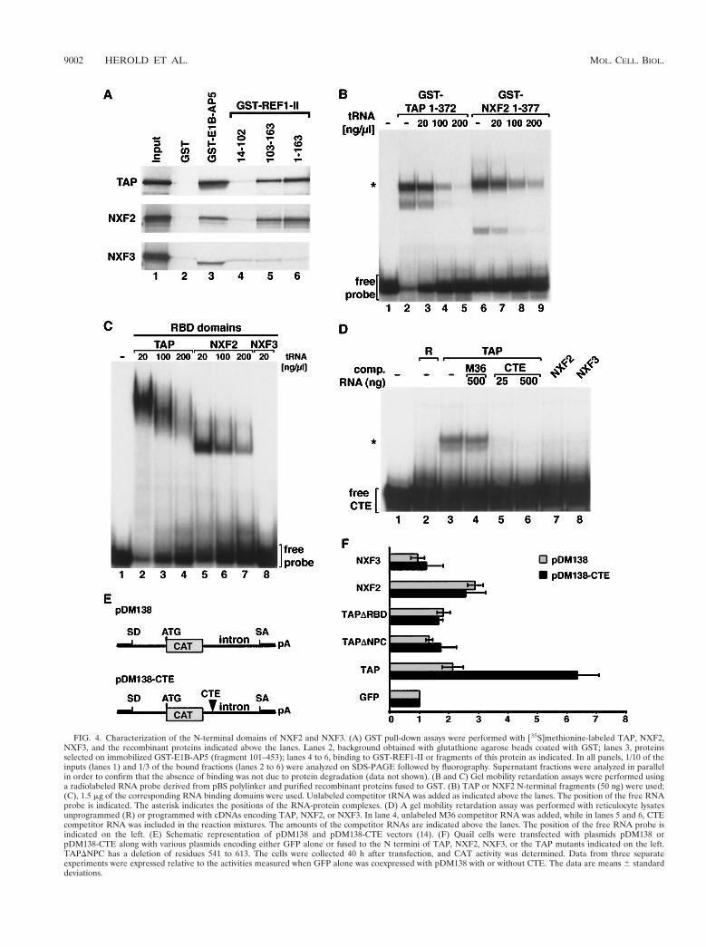
FIG. 4. Characterization of the N-terminal domains of NXF2 and NXF3. (A) GST pull-down assays were performed with [35S]methionine-labeled TAP, NXF2,NXF3, and the recombinant proteins indicated above the lanes. Lanes 2, background obtained with glutathione agarose beads coated with GST; lanes 3, proteinsselected on immobilized GST-E1B-AP5 (fragment 101–453); lanes 4 to 6, binding to GST-REF1-II or fragments of this protein as indicated. In all panels, 1/10 of theinputs (lanes 1) and 1/3 of the bound fractions (lanes 2 to 6) were analyzed on SDS-PAGE followed by fluorography. Supernatant fractions were analyzed in parallelin order to confirm that the absence of binding was not due to protein degradation (data not shown). (B and C) Gel mobility retardation assays were performed usinga radiolabeled RNA probe derived from pBS polylinker and purified recombinant proteins fused to GST. (B) TAP or NXF2 N-terminal fragments (50 ng) were used;(C), 1.5 mg of the corresponding RNA binding domains were used. Unlabeled competitor tRNA was added as indicated above the lanes. The position of the free RNAprobe is indicated. The asterisk indicates the positions of the RNA-protein complexes. (D) A gel mobility retardation assay was performed with reticulocyte lysatesunprogrammed (R) or programmed with cDNAs encoding TAP, NXF2, or NXF3. In lane 4, unlabeled M36 competitor RNA was added, while in lanes 5 and 6, CTEcompetitor RNA was included in the reaction mixtures. The amounts of the competitor RNAs are indicated above the lanes. The position of the free RNA probe isindicated on the left. (E) Schematic representation of pDM138 and pDM138-CTE vectors (14). (F) Quail cells were transfected with plasmids pDM138 orpDM138-CTE along with various plasmids encoding either GFP alone or fused to the N termini of TAP, NXF2, NXF3, or the TAP mutants indicated on the left.TAPDNPC has a deletion of residues 541 to 613. The cells were collected 40 h after transfection, and CAT activity was determined. Data from three separateexperiments were expressed relative to the activities measured when GFP alone was coexpressed with pDM138 with or without CTE. The data are means 6 standarddeviations.
9002 HEROLD ET AL. MOL. CELL. BIOL.

FIG. 5. p15-2a, a human p15-1 homologue, interacts with TAP and localizes to the nuclear rim. (A) Intron-exon structures of p15 family sequences. Protein codingregions and untranslated regions are colored red and white, respectively. The positions of initiation and termination codons are indicated by green and red triangles,respectively. For human p15-2a and -b, the alternative splicing pathway is shown by lines above the gene. The 59 exon of the p15-2a gene contains an open reading frameof five amino acids, while p15-2b gene 59 exon sequences contain an in-frame stop codon but no in-frame ATG. Therefore, translation of this mRNA may generatea truncated protein starting at methionine 29 in the p15-2a sequence. Alternatively, translation may start at the GTG codon (green open triangle), resulting in an openreading frame (gray), since this region does not show any similarity to the other p15 sequences. (B) Multiple sequence alignment of p15 family sequences. The symbolsare as in Fig. 3. (C) Phylogenetic tree of the NTF2 family sequences. The tree was drawn by the neighbor-joining method (34). Abbreviations (other than those in Fig.1 and 2): At, Arabidopsis thaliana; Nt, Nicotiana tabacum; G3BP, Ras-GAP SH3 domain binding protein; MKK3, MAP kinase kinase 3; NPK2, a tobacco protein kinase.(D) Subcellular localization of p15-2a. HeLa cells were transfected with a pEGFP-N3 plasmid derivative expressing a zz fusion of p15-2a. The fusion protein wasdetected throughout the nucleoplasm and cytoplasm and was excluded from the nucleolus (left). On the right, HeLa cells were extracted with (1) Triton X-100 priorto fixation. A punctate labeling pattern was visible at the nuclear periphery for the p15-2a protein. (E) Lysates from E. coli expressing the Ran binding domain ofImportin b (fragment 1–452) or supplemented with equimolar amounts of NTF2, p15-1, or p15-2a were incubated with IgG-Sepharose beads coated with purifiedzzRanGDP, zzRanQ69L-GTP, or zzTAP (fragment 61–619). After extensive washes, the bound proteins were eluted. One-hundredth of the inputs (lanes 1 to 4) and1/10 of the bound fractions (lanes 6 to 16) were analyzed by SDS-PAGE followed by Coomassie blue staining. (F) Lysates from E. coli expressing GST fusions of TAP,NXF2, or NXF3 together with untagged versions of p15-1 or p15-2a were incubated with glutathione agarose beads. After extensive washes, the bound proteins wereeluted with SDS sample buffer and analyzed by SDS-PAGE followed by Coomassie blue staining. Lanes 1 to 9, input lysates; lanes 11 to 19, bound fractions; lane 10,molecular mass markers (116, 97, 84, 66, 55, 45, 36, 29, 24, 20, and 14.2 kDa).
VOL. 20, 2000 NXF MULTIGENE FAMILY OF TAP-LIKE PROTEINS 9003

binding (40). We tested the effect of replacing the three loopresidues of NXF2 with alanines. This substitution (3xAla598)disrupts nucleoporin binding in vitro as efficiently as the deletionof the entire NXF2 C-terminal domain (D598–626) (Fig. 6C,
lanes 7 and 8). As is the case for TAP, a single substitution ofalanine for W599 was sufficient to disrupt nucleoporin binding(Fig. 6C, lane 5), while substitution of alanine for N600 or ofarginine for E598 had no significant effect (Fig. 6C, lanes 4 and 6).
FIG. 6. A fraction of NXF2 localizes to the nuclear rim. (A) GST pull-down assays were performed with [35S]methionine-labeled TAP, NXF2, or NXF3, and therecombinant proteins indicated above the lanes. One-tenth of the inputs (lanes 1, 4, and 7) and one-third of the bound fractions (lanes 2, 3, 5, 6, 8, and 9) were analyzedby SDS-PAGE followed by fluorography. (B) NXF2 interacts with multiple components of the NPC. GST pull-down assays were performed with the [35S]methionine-labeled nucleoporins indicated on the left of the panels and recombinant GST or TAP, TAP W594A, TAP D595R, or NXF2 fused to GST, as indicated above the lanes.One-tenth of the input (lane 1) and one-third of the bound fractions (lanes 2 to 6) were analyzed on SDS-PAGE followed by fluorography. (C) GST pull-down assayswere performed with the [35S]methionine-labeled nucleoporins indicated on the left of the panels and recombinant GST, GST-NXF2, or various NXF2 mutants asindicated above the lanes. Samples were analyzed as indicated for panel A. (D and E) HeLa cells were transfected with pEGFP-C1 plasmid derivatives expressing GFPfusions of TAP, NXF2, and NFX3 or various TAP or NXF2 mutants as indicated on the left. Approximately 20 h after transfection, the cells were fixed in formaldehyde,permeabilized with Triton X-100, and directly observed with a fluorescence microscope. For all proteins, the GFP signal was detected throughout the nucleoplasm(2Tx). Cytoplasmic staining was also detected for NXF3. On the right (1Tx), HeLa cells were extracted with Triton X-100 prior to fixation. A punctate labeling patternwas visible at the nuclear periphery for TAP and NXF2, while in cells transfected with NXF3, TAP D595R, or the NXF2 mutants, no GFP signal was detected at thenuclear rim.
9004 HEROLD ET AL. MOL. CELL. BIOL.

To analyze nucleoporin binding in vivo, the subcellular lo-calization of NXF2 and NXF3 fused to GFP was investigatedin transfected HeLa cells and compared to that of GFP-taggedTAP. GFP-tagged NXF2 and NXF3 were evenly detected inthe nucleoplasm and were excluded from the nucleolus. More-over, a fraction of NXF3 could be detected in the cytoplasm(Fig. 6D, 2Tx). To visualize a potential nuclear envelope as-sociation, transfected cells were extracted with Triton X-100prior to fixation (Fig. 6D, 1Tx). As previously observed forTAP, a fraction of NXF2 was resistant to detergent extractionand remained associated with the nuclear envelope whileNXF3 was not detected at the nuclear rim following Tritonextraction. Furthermore, NXF2 and TAP mutants that do notinteract with nucleoporins in vitro were not detected at thenuclear envelope upon Triton extraction (Fig. 6E).
NXF2, but not NXF3, can stimulate RNA export. We havedeveloped an assay that tests mRNA export stimulation ofTAP and its homologues. In this assay, TAP and TAP-like
proteins are cotransfected with the reporter plasmid pDM138(14) in human 293 cells. As mentioned above, transfection withthis plasmid yields only trace levels of CAT enzyme activity(13, 14). However, cotransfection with vectors expressing TAPand p15 nuclear retention to be bypassed and export of theunspliced transcripts to be promoted, increasing CAT activity(Fig. 7A). To test the effect of TAP-like proteins on cat geneexpression, human 293 cells were cotransfected with a constantamount of pDM138 reporter plasmid, plasmids encoding TAP,NXF2, or NXF3 together with plasmids encoding p15-1 orp15-2a (Fig. 7A and D). Protein expression levels were ana-lyzed by Western blotting (Fig. 7B and C). Overexpression ofthe TAP protein together with p15-1 increased cat expressionby 15- to 17-fold compared to the expression levels obtainedwhen only pDM138 vector was transfected, whereas coexpres-sion of TAP with p15-2a resulted in 13-fold activation of catexpression (Fig. 7A). Coexpression of NXF2 with p15-1 orp15-2a activated cat expression up to 10- or 6-fold, respectively.
FIG. 7. NXF2 exhibits general RNA nuclear export activity. (A to D) Human 293 cells were transfected with a mixture of plasmids encoding b-Gal, CAT, and eitherGFP alone (2) or fused to the N termini of TAP, NXF2, NXF3, and various TAP and NXF2 mutants as indicated on the left. pEGFP-N3 derivatives encoding zz-taggedversions of p15-1 and p15-2a were cotransfected as indicated. The cells were collected 40 h after transfection, and b-Gal and CAT activities were determined. Datafrom three separate experiments were expressed relative to the activities measured when GFP alone was coexpressed with pDM138. The data are means 6 standarddeviations. (B and C) Protein expression levels were analyzed by Western blotting with anti-GFP antibodies. (B) Arrowheads indicate the positions of NXF proteinsfused to GFP or of GFP itself, while the asterisks show the positions of p15 proteins.
VOL. 20, 2000 NXF MULTIGENE FAMILY OF TAP-LIKE PROTEINS 9005

This level of induction may reflect the lower expression levelsof NXF2 compared to TAP (Fig. 7B and C). In contrast, whencoexpressed with p15-1 or p15-2a, NXF3 had no effect on catactivity, even though its expression levels in the presence ofp15-1 were similar to that of TAP (Fig. 7A and B). Overex-pression of p15-1 or p15-2a, in the absence of exogenous TAPor in the presence of a deletion mutant of TAP (TAPD437–507) that no longer interacts with p15, had no effect on CATactivity (Fig. 7A and D). In contrast, overexpression of TAP orNXF2 in the absence of exogenous p15 resulted in a significantbut modest increase in CAT activity (Fig. 7A); however, thelevels of expression of TAP and NXF2 in the absence of p15were also reduced (Fig. 7B). Therefore, p15-1 and p15-2aappear to increase the stability of TAP-like proteins. In sum-mary, TAP and NXF2, when coexpressed with p15-1 or p15-2a,stimulate the export of the unspliced pre-mRNA, while NXF3did not stimulate CAT gene expression. The ability of TAP andNXF2 to export pDM138 pre-mRNA is likely to reflect thegenuine export activity of these proteins, as this activity couldbe abolished with various mutations in the TAP sequence,implying the need for a functional protein (Fig. 7D). In par-ticular, this activity requires the LRRs and the NTF2-like do-main. In contrast, the RBD and the NPC binding domain ofTAP were not strictly required (Fig. 7D) (see Discussion).Thus, the lack of RNA export activity of NXF3 may not be dueto the truncation of the UBA domain but to the deletion ofpart of the LRRs.
DISCUSSION
In this study, we present a combined evolutionary and func-tional characterization of the family of TAP-like proteins inhigher eukaryotes. Members of this protein family were calledNXF. We have further characterized two of four putative hu-man NXF proteins, namely, NXF2 and NXF3, and have shownthat NXF2 exhibits RNA export activity.
NXF proteins heterodimerize with p15. Previously, we haveshown that TAP heterodimerizes with p15-1 via its NTF2-likedomain (40). In this study, we show that the human genomeencodes at least two p15 homologues and that both interactwith TAP, NXF2, and NXF3 and participate in RNA nuclearexport. However, the precise role of p15 in TAP-mediatedexport is not well defined. p15 binding is not strictly requiredfor TAP-mediated export of CTE-bearing substrates (4, 8, 16)but appears to play a role in the general export activity of theNXF proteins (Fig. 7A and D). Whether these effects are dueto a direct role of p15 in export or to an increased stability ofNXF proteins when coexpressed with p15 is unclear.
The interaction between the NTF2-like domains of TAP andp15-1 is mediated by multiple rather than a few critical resi-dues (40). Indeed, mutation of residues located at the het-erodimer interface reduced but did not completely abolishheterodimerization in vitro (40). This provides an explanationfor the TAP, NXF2, and NXF3 interactions with p15-1 andp15-2a studied here, as these interactions occur even thoughnot all interface residues are conserved. The NTF2-like do-main also occurs in Mex67p, the S. pombe and S. cerevisiae,NXF homologue (40). In S. cerevisiae, this domain is impli-cated in interaction with a protein known as Mtr2p (35, 38).Mtr2p does not exhibit obvious sequence similarities with p15,but secondary-structure predictions suggest that it could be ap15 analogue (40).
Functional conservation of the UBA-like domain. Thenucleoporin binding domain of TAP includes a UBA domain(40). The known structure of UBA domains suggests a con-served loop (NWD at positions 593 to 595 in human TAP) is
involved in the interaction with nucleoporins. Replacement ofthese three residues by alanines was previously shown to blockthe ability of TAP to promote CTE-dependent export of aprecursor mRNA in quail cells (15). Moreover, single-amino-acid changes in this loop are sufficient to impair binding ofTAP and C. elegans NXF1 to nucleoporins in vitro and in vivo(40, 41) (Fig. 6). The mutational analysis of the UBA-likedomain of NXF2 presented here further supports the predic-tion that the conserved loop residues of the UBA-like domainhave a critical role in the interaction of NXF proteins withnucleoporins. Conversely, based on these observations, it waspossible to predict that NXF proteins having a truncated UBAdomain are unlikely to directly interact with nucleoporins. Thisprediction was confirmed for NXF3.
Surprisingly, in spite of its conservation, the UBA domain ofMex67p is not sufficient for nucleoporin binding, and Mex67placking the UBA domain can bind to nucleoporins in the pres-ence of Mtr2p (38). The possibility that vertebrate NXF pro-teins lacking the NPC binding domain can still interact withnucleoporins in the presence of p15 requires further investiga-tion; however, NXF3 did not associate with nucleoporins ei-ther in vivo or in vitro when coexpressed with p15 (data notshown).
TAP binds Nup98 in vitro, but this interaction is not re-quired for its localization at the nuclear rim, as in Nup982/2
cells, TAP remains at the nuclear envelope (43). The observa-tion that NXF2 localizes at the nuclear rim and can promoteexport of the pDM138 pre-mRNA in the absence of Nup98binding is consistent with the observation that Nup982/2 cellsdo not display an overt export inhibition phenotype (43). Thus,although TAP and NXF2 interact with multiple nucleoporinsin vitro, the nucleoporins that are responsible for their local-ization at the nuclear rim remain to be identified. Moreover,the mechanism by which NXF proteins regulate their bindingto nucleoporins is unknown.
Role of NXF proteins in RNA nuclear export. In yeast,Mex67p is involved in the export of bulk polyadenylated RNAs(36). Evidence for an essential role in mRNP export has beenrecently obtained for the C. elegans protein NXF1 (41). More-over, TAP is directly implicated in the export of simian type Dretroviral RNAs bearing the CTE (8, 12). Although we cannotexclude the possibility that some TAP homologues may have afunction other than export, the conservation of their structuralorganization and the observation that at least three membersof the NXF family are implicated in RNA export suggest thatthese proteins are likely to participate, directly or indirectly, inthe export of cellular mRNAs to the cytoplasm. The diversifi-cation of NXF proteins in higher eukaryotes compared to yeastmay reflect a greater substrate complexity or tissue-specificrequirements. Consistent with this possibility is the observationthat human NXF5 is expressed in brain (Lin et al., submitted).
NXF proteins may associate with cellular mRNPs directly,by binding to the mRNA, or indirectly, through interactionwith hnRNP-like proteins. Recently, several TAP partners thatcould facilitate its interaction with cellular mRNPs have beenidentified. These include E1B-AP5, RAE1, and members of anevolutionarily conserved family of hnRNP-like proteins, theYra1p/REF proteins (4, 37, 39). Aside from these, other RNAbinding proteins may also facilitate TAP binding to themRNPs, as Y14, an hnRNP-like protein that preferentiallyassociates with spliced mRNAs, has recently been shown to bepresent in a complex containing TAP (18). In vitro, Y14 inter-acts with TAP and NXF2 (data not shown); therefore, it ispossible that Y14, directly or indirectly, recruits NXF proteinsto mRNPs in a post-splicing-dependent manner (18). BesidesY14, several other proteins form splicing-dependent inter-
9006 HEROLD ET AL. MOL. CELL. BIOL.

actions with mRNA. These include the splicing coactivatorSRm160, the acute myeloid leukemia-associated protein DEK,and several unidentified proteins (23, 25). These proteins mayalso link NXFs with mRNAs. NXF proteins may then be moreefficiently loaded on mRNAs that have been produced by splic-ing. Therefore, it is possible that the pDM138 precursormRNA cannot efficiently compete with other mRNAs for bind-ing to NXFs, i.e., NXFs may be limiting for this substrate;hence, its export can be stimulated by overexpression of NXF-p15 heterodimers. Consistent with this is the observation thatoverexpression of TAP-p15 heterodimers does not stimulateexport of reporter mRNAs that are efficiently expressed (I. C.Braun and E. Izaurralde, unpublished data).
TAP-mediated export of pDM138 pre-mRNA requires theLRRs and the NTF2-like domain, while deletion of the RBDor of the NPC binding domain impaired but did not abolishexport (Fig. 7D). This is in sharp contrast to the situation ob-served when TAP-mediated export of pDM138-CTE is moni-tored in quail cells. In this case, both the RBD and the NPCbinding domain are absolutely required (Fig. 4F). The RBD ofTAP is required in cis to the LRRs for specific binding to theCTE RNA (24). Hence, this domain is essential for TAP-mediated export of CTE-containing substrates, whereas bind-ing of TAP to pDM138 pre-mRNA may be mediated by pro-tein-protein interactions. We have previously reported that therequirements for the NTF2-like and UBA domains of TAP aresubstrate dependent (4, 8). Indeed, in Xenopus oocytes, TAP-mediated export of CTE-bearing intron lariats is independentof these domains (8), while export of U6-CTE requires theUBA domain but not the NTF2-like domain (4). In quail cells,TAP-mediated export of pDM138-CTE pre-mRNA is abol-ished by mutations or deletions of the UBA domain (15) (Fig.4) but is only reduced by mutations preventing p15 binding(16). Finally, the C-terminal domain of Mex67p is importantbut not essential for mRNA export (38). Thus, the functional-ity of NXF protein domains may be influenced by the specificset of proteins associated with the RNP export substrate. Dis-covery of the precise roles of NXF protein domains and themechanism by which these proteins mediate directional trans-port of RNP substrates across the NPC remains an importantgoal for the future.
ACKNOWLEDGMENTS
The technical support of Michaela Rode is gratefully acknowledged.We thank Tom Hope and Matthias Dobbelstein for the kind gift ofplasmids pDM138 and pDM138-CTE. We thank Birthe Fahrenkrog,Maarten Fornerod, Iain W. Mattaj, and Christel Schmitt for criticalreading of the manuscript. We are grateful to Barbara Felber, GuyFroyen, and Peter Marynen for communicating their results prior topublication.
This study was supported by the German Ministry of Research andTechnology (BMBF), the Swiss National Science Foundation, the Eu-ropean Molecular Biology Organization (EMBO), and the HumanFrontier Science Program Organization (HFSPO).
A.H. and M.S. contributed equally to this work.
REFERENCES
1. Adams, M. D., S. E. Celniker, R. A. Holt, C. A. Evans, J. D. Gocayne, P. G.Amanatides, S. E. Scherer, P. W. Li, R. A. Hoskins, et al. 2000. The genomesequence of Drosophila melanogaster. Science 287:2185–2195.
2. Almeida, F., R. Saffrich, W. Ansorge, and M. Carmo-Fonseca. 1998. Micro-injection of anti-coilin antibodies affects the structure of coiled bodies. J. CellBiol. 142:899–912.
3. Altschul, S. F., T. L. Madden, A. A. Schaffer, J. Zhang, Z. Zhang, W. Miller,and D. J. Lipman. 1997. Gapped BLAST and PSI-BLAST: a new generationof protein database search programs. Nucleic Acids Res. 25:3389–3402.
4. Bachi, A., I. C. Braun, J. P. Rodrigues, N. Pante, K. Ribbeck, C. von Kobbe,U. Kutay, M. Wilm, D. Gorlich, M. Carmo-Fonseca, and E. Izaurralde. 2000.The C-terminal domain of TAP interacts with the nuclear pore complex and
promotes export of specific CTE-bearing RNA substrates. RNA 6:136–158.5. Bear, J., W. Tan, A. S. Zolotukhin, C. Tabernero, E. A. Hudson, and B. K.
Felber. 1999. Identification of novel import and export signals of humanTAP, the protein that binds to the CTE element of the type D retrovirusmRNAs. Mol. Cell. Biol. 19:6306–6317.
6. Bharathi, A., A. Ghosh, W. A. Whalen, J. H. Yoon, R. Pu, M. Dasso, and R.Dhar. 1997. The human RAE1 gene is a functional homologue of Schizo-saccharomyces pombe rae1 gene involved in nuclear export of Poly(A)1RNA. Gene 198:251–258.
7. Black, B. E., L. Levesque, J. M. Holaska, T. C. Wood, and B. Paschal. 1999.Identification of an NTF2-related factor that binds RanGTP and regulatesnuclear protein export. Mol. Cell. Biol. 19:8616–8624.
8. Braun, I. C., E. Rohrbach, C. Schmitt, and E. Izaurralde. 1999. TAP bindsto the constitutive transport element (CTE) through a novel RNA-bindingmotif that is sufficient to promote CTE-dependent RNA export from thenucleus. EMBO J. 18:1953–1965.
9. Brown, J. A., A. Bharathi, A. Ghosh, W. Whalen, E. Fitzgerald, and R. Dhar.1995. A mutation in the Schizosaccharomyces pombe rae1 gene causes de-fects in poly(A)1 RNA export and in the cytoskeleton. J. Biol. Chem. 270:7411–7419.
10. The C. elegans Sequencing Consortium. 1998. Genome sequence of thenematode C. elegans: a platform for investigating biology. Science 282:2012–2018.
11. Galtier, N., M. Gouy, and C. Gautier. 1996. SEAVIEW and PHYLO_WIN:two graphic tools for sequence alignment and molecular phylogeny. Comput.Appl. Biosci. 12:543–548.
12. Gruter, P., C. Tabernero, C. von Kobbe, C. Schmitt, C. Saavedra, A. Bachi,M. Wilm, B. K. Felber, and E. Izaurralde. 1998. TAP, the human homologueof Mex67p, mediates CTE-dependent RNA export from the nucleus. Mol.Cell 1:649–659.
13. Hope, T. J., X. Huang, D. McDonald, and T. G. Parslow. 1990. Steroid-receptor fusion of the human immunodeficiency virus type 1 Rev transacti-vator: mapping cryptic functions of the arginine-rich motif. Proc. Natl. Acad.Sci. USA 87:7787–7791.
14. Huang, X., T. J. Hope, B. L. Bond, D. McDonald, K. Grahl, and T. G.Parslow. 1991. Minimal Rev-response element for type 1 human immuno-deficiency virus. J. Virol. 65:2131–2134.
15. Kang, Y., and B. R. Cullen. 1999. The human TAP protein is a nuclearmRNA export factor that contains novel RNA-binding and nucleocytoplas-mic transport sequences. Genes Dev. 13:1126–1139.
16. Kang, Y., H. P. Bogerd, and B. R. Cullen. 2000. Analysis of cellular factorsthat mediate nuclear export of RNAs bearing the Mason-Pfizer monkey virusconstitutive transport element. J. Virol. 74:5863–5871.
17. Katahira, J., K. Straßer, A. Podtelejnikov, M. Mann, J. U. Jung, and E.Hurt. 1999. The Mex67p-mediated nuclear mRNA export pathway is con-served from yeast to human. EMBO J. 18:2593–2609.
18. Kataoka, N., J. Yong, V. N. Kim, F. Velazquez, R. A. Perkinson, F. Wang, andG. Dreyfuss. 2000. Pre-mRNA splicing imprints mRNA in the nucleus witha novel RNA-binding protein that persists in the cytoplasm. Mol. Cell 6:673–682.
19. Klebe, C., F. R. Bischoff, H. Ponstingl, and A. Wittinghofer. 1995. Interactionof the nuclear GTP-binding protein Ran with its regulatory proteins RCC1and RanGAP1. Biochemistry 34:639–647.
20. Kraemer, D., and G. Blobel. 1997. mRNA binding protein mrnp 41 localizesto both nucleus and cytoplasm. Proc. Natl. Acad. Sci. USA 94:9119–9124.
21. Kutay, U., E. Izaurralde, F. R. Bischoff, I. W. Mattaj, and D. Gorlich. 1997.Dominant-negative mutants of importin-b block multiple pathways of importand export through the nuclear pore complex. EMBO J. 16:1153–1163.
22. Mattaj, I. W., and L. Englmeier. 1998. Nucleocytoplasmic transport: thesoluble phase. Annu. Rev. Biochem. 67:256–306.
23. Le Hir, H., E. L. Maquat, and J. M. Moore. 2000. Pre-mRNA splicing altersmRNP composition: evidence for stable association of proteins at exon-exonjunctions. Genes Dev. 14:1098–1108.
24. Liker, E., E. Fernadez, E. Izaurralde, and E. Conti. The structure of themRNA nuclear export factor TAP reveals a cis arrangement of a non-canonical RNP domain and a leucine-rich-repeat domain. EMBO J., inpress.
25. McGarvey, T., E. Rosonina, S. McCracken, Q. Li, R. Arnaout, E. Mientjes,A. J. Nickerson, D. Awrey, J. Greenblatt, G. Grosveld, and B. J. Blencowe.2000. The acute myeloid leukemia-associated protein DEK forms a splicing-dependent interaction with exon-product complexes. J. Cell Biol. 150:309–320.
26. Morency, C. A., J. R. Neumann, and K. O. Russian. 1987. A novel rapid assayfor Chloramphenicol-acetyl transferase gene expression. BioTechniques 5:444–448.
27. Murphy, R., J. L. Watkins, and S. R. Wente. 1996. GLE2, a Saccharomycescerevisiae homologue of the Schizosaccharomyces pombe export factorRAE1, is required for nuclear pore complex structure and function. Mol.Biol. Cell 7:1921–1937.
28. Nakielny, S., and G. Dreyfuss. 1999. Transport of proteins and RNAs in andout of the nucleus. Cell 99:677–690.
29. Parks, T. D., K. K. Leuther, E. D. Howard, S. A. Johnston, and W. G.
VOL. 20, 2000 NXF MULTIGENE FAMILY OF TAP-LIKE PROTEINS 9007

Dougherty. 1994. Release of proteins and peptides from fusion proteins witha recombinant plant virus proteinase. Anal. Biochem. 216:413–417.
30. Pasquinelli, A. E., R. K. Ernst, E. Lund, C. Grimm, M. L. Zapp, D. Rekosh,M.-L. Hammarskjold, and J. E. Dahlberg. 1997. The constitutive transportelement (CTE) of Mason-Pfizer Monkey Virus (MPMV) accesses an RNAexport pathway utilized by cellular messenger RNAs. EMBO J. 16:7500–7510.
31. Pritchard, C. E. J., M. Fornerod, L. H. Kasper, and J. M. A. van Deursen.1999. RAE1 is a shuttling mRNA export factor that binds to a GLEBS-likeNUP98 motif at the nuclear pore complex through multiple domains. J. CellBiol. 145:237–253.
32. Ryan, K., and S. R. Wente. 2000. The nuclear pore complex: a proteinmachine bridging the nucleus and cytoplasm. Curr. Opin. Cell Biol. 12:361–371.
33. Saavedra, C. A., B. K. Felber, and E. Izaurralde. 1997. The simian retrovi-rus-1 constitutive transport element CTE, unlike HIV-1 RRE, utilises factorsrequired for cellular RNA export. Curr. Biol. 7:619–628.
34. Saitou, N., and M. Nei. 1987. The neighbor-joining method: a new methodfor reconstructing phylogenetic trees. Mol. Biol. Evol. 4:406–425.
35. Santos-Rosa, H., H. Moreno, G. Simos, A. Segref, B. Fahrenkrog, N. Pante,and E. Hurt. 1998. Nuclear mRNA export requires complex formation be-tween Mex67p and Mtr2p at the nuclear pores. Mol. Cell Biol. 18:6826–6838.
36. Segref, A., K. Sharma, V. Doye, A. Hellwig, J. Huber, R. Luhrmann, and E.Hurt. 1997. Mex67p, a novel factor for nuclear mRNA export, binds to bothpoly(A)1 RNA and nuclear pores. EMBO J. 16:3256–3271.
37. Straßer, K., and E. Hurt. 2000. Yra1p, a conserved nuclear RNA-bindingprotein, interacts directly with Mex67p and is required for mRNA export.EMBO J. 19:410–420.
38. Straßer, K., J. Baßler, and E. Hurt. 2000. Binding of the Mex67p/Mtr2pheterodimer to FXFG, GLFG, and FG repeat nucleoporins is essential fornuclear mRNA export. J. Cell Biol 150:695–706.
39. Stutz, F., A. Bachi, T. Doerks, I. C. Braun, B. Seraphin, M. Wilm, P. Bork,and E. Izaurralde. 2000. REF, an evolutionarily conserved family of hnRNP-like proteins, interacts with TAP/Mex67p and participates in mRNA nuclearexport. RNA 6:638–650.
40. Suyama, M., T. Doerks, I. C. Braun, M. Sattler, I. Izaurralde, and P. Bork.2000. Prediction of structural domains of TAP reveals details of its interac-tion with p15 and nucleoporins. EMBO Rep. 1:53–58.
41. Tan, W., A. S. Zolotukhin, J. Bear, D. J. Patenaude, and B. K. Felber. ThemRNA export in C. elegans is mediated by Ce-TAP1, an orthologue ofhuman TAP and S. cerevisiae Mex67p. RNA, in press.
42. Thompson, J. D., D. G. Higgins, and T. J. Gibson. 1994. CLUSTAL W:improving the sensitivity of progressive multiple sequence alignment throughsequence weighting, position-specific gap penalties and weight matrix choice.Nucleic Acids Res. 22:4673–4680.
43. Wu, X., L. H. Kasper, T. Ralitsa, R. T. Mantcheva, B. M. A. Fontoura, G. T.Mantchev, G. Blobel, and J. M. A. van Deursen. NUP98 is required forgastrulation, proper nuclear pore complex assembly and efficient proteinimport. J. Cell Biol., in press.
9008 HEROLD ET AL. MOL. CELL. BIOL.