African elephants Loxodonta africana amplify browse heterogeneity in African savanna

Published Ahead of Print 17 March 2008. 2008, 76(5):2008. DOI: 10.1128/IAI.01691-07. Infect. Immun.
BäumlerSantos, Satya Dandekar, Renée M. Tsolis and Andreas J.
L.D. George, Tatiane A. Paixão, Hortensia G. Rolán, Renato Ivan Godinez, Takeshi Haneda, Manuela Raffatellu, Michael Mucosa Serotype Typhimurium in the Intestinal
Salmonella entericaResponses Induced by T Cells Help To Amplify Inflammatory
http://iai.asm.org/content/76/5/2008Updated information and services can be found at:
These include:
REFERENCEShttp://iai.asm.org/content/76/5/2008#ref-list-1at:
This article cites 40 articles, 28 of which can be accessed free
CONTENT ALERTS more»articles cite this article),
Receive: RSS Feeds, eTOCs, free email alerts (when new
http://journals.asm.org/site/misc/reprints.xhtmlInformation about commercial reprint orders: http://journals.asm.org/site/subscriptions/To subscribe to to another ASM Journal go to:
on February 6, 2013 by U
C D
AV
IS S
HIE
LDS
LIBR
AR
Yhttp://iai.asm
.org/D
ownloaded from

INFECTION AND IMMUNITY, May 2008, p. 2008–2017 Vol. 76, No. 50019-9567/08/$08.00�0 doi:10.1128/IAI.01691-07Copyright © 2008, American Society for Microbiology. All Rights Reserved.
T Cells Help To Amplify Inflammatory Responses Induced bySalmonella enterica Serotype Typhimurium in the
Intestinal Mucosa�
Ivan Godinez, Takeshi Haneda, Manuela Raffatellu, Michael D. George, Tatiane A. Paixao,Hortensia G. Rolan, Renato L. Santos, Satya Dandekar,
Renee M. Tsolis, and Andreas J. Baumler*Department of Medical Microbiology and Immunology, School of Medicine, University of
California at Davis, One Shields Ave., Davis, California
Received 18 December 2007/Returned for modification 18 February 2008/Accepted 6 March 2008
Salmonella enterica serotype Typhimurium causes an acute inflammatory reaction in the ceca of streptomy-cin-pretreated mice. We determined global changes in gene expression elicited by serotype Typhimurium in thececal mucosa. The gene expression profile was dominated by T-cell-derived cytokines and genes whose expres-sion is known to be induced by these cytokines. Markedly increased mRNA levels of genes encoding gammainterferon (IFN-�), interleukin-22 (IL-22), and IL-17 were detected by quantitative real-time PCR. Further-more, the mRNA levels of genes whose expression is induced by IFN-�, IL-22, or IL-17, including genesencoding macrophage inflammatory protein 2 (MIP-2), inducible nitric oxide synthase (Nos2), lipocalin-2(Lcn2), MIP-1�, MIP-1�, and keratinocyte-derived cytokine (KC), were also markedly increased. To assess theimportance of T cells in orchestrating this proinflammatory gene expression profile, we depleted T cells byusing a monoclonal antibody prior to investigating cecal inflammation caused by serotype Typhimurium instreptomycin-pretreated mice. Depletion of CD3� T cells resulted in a dramatic reduction in gross pathology,a significantly reduced recruitment of neutrophils, and a marked reduction in mRNA levels of Ifn-�, Il-22, Il-17,Nos2, Lcn2, and Kc. Our results suggest that T cells play an important role in amplifying inflammatoryresponses induced by serotype Typhimurium in the cecal mucosa.
Salmonella enterica serotype Typhimurium elicits intestinalinflammation in the terminal ileum and colon of patients thatis characterized by a massive neutrophil influx (3, 17). Thegeneration of intestinal inflammatory responses during sero-type Typhimurium infection can be studied using streptomy-cin-pretreated mice (1). It has recently been proposed that theorchestration of serotype Typhimurium-induced intestinal in-flammation can be broken down into mechanisms involved inits induction and mechanisms involved in the amplification ofinflammatory responses in tissue (29).
Several factors for the initial induction of inflammatory re-sponses have been proposed using tissue culture models. Theseinclude cytokine and hepoxilin A3 production induced by theinvasion-associated type III secretion system (T3SS-1) of sero-type Typhimurium in intestinal epithelial cells (2, 9, 11, 16, 21),the activation of Toll-like receptors in epithelial cells and mac-rophages through pathogen-associated molecular patterns (12,22, 23, 34, 35, 39), and the stimulation of cytosolic nucleotide-binding and oligomerization domain-like receptors by a T3SS-1-dependent translocation of flagellin into the cytosol of mac-rophages (6, 18, 33).
Compared to factors involved in the initial induction ofinflammatory responses, relatively little is known about themechanisms involved in the subsequent amplification of in-
flammatory responses in the intestinal mucosa. It has beenproposed that mononuclear cells (macrophages and/or den-dritic cells) play an important role in initiating the amplifica-tion of responses against serotype Typhimurium in tissue bysecreting specific cytokines (29). For example, in other modelsof infection, mononuclear cells produce interleukin-23 (IL-23)and IL-18, two cytokines which stimulate T cells to produceIL-17 and gamma interferon (IFN-�), respectively (4, 6–8, 18,30, 37). These data suggest that the production of cytokines byT cells may be a central component of mechanisms that helpamplify inflammatory responses in tissue. However, the poten-tial contribution of T-cell-mediated amplification mechanismsto the development of the acute inflammatory reaction devel-oping in the intestinal mucosa during serotype Typhimuriuminfection has not yet been investigated. Here we determinedthe gene expression profile elicited by serotype Typhimuriumin the intestinal mucosa of streptomycin-pretreated mice andassessed the contribution of T cells to the orchestration ofintestinal inflammation.
MATERIALS AND METHODS
Bacterial strains and culture conditions. Serotype Typhimurium strain IR715is a fully virulent, nalidixic acid-resistant derivative of isolate ATCC 14028 andwas used in all experiments (32). Bacteria were cultured aerobically at 37°C inLuria-Bertani (LB) broth.
Purification of MAb. Hybridoma lines producing monoclonal antibodies(MAb) (rat immunoglobulin G [IgG]) against murine CD3 were purchased fromATCC (CRL-1975). Hybridoma lines were expanded and maintained in Iscove’smodified Dulbecco’s medium (Gibco catalog no. 12440) containing 10% fetalbovine serum and 5% antibiotic-antimycotic solution (Gibco catalog no. 15240-062). Cells were incubated at 37°C with 5% CO2. Every 2 to 3 days, media were
* Corresponding author. Mailing address: Department of MedicalMicrobiology and Immunology, School of Medicine, University of Cal-ifornia at Davis, One Shields Ave., Davis, CA 95616-8645. Phone:(530) 754-7225. Fax: (530) 754-7240. E-mail: [email protected].
� Published ahead of print on 17 March 2008.
2008
on February 6, 2013 by U
C D
AV
IS S
HIE
LDS
LIBR
AR
Yhttp://iai.asm
.org/D
ownloaded from

replaced and stored at �20°C until ready for MAb isolation. Medium superna-tant containing MAb was diluted 1:1 with 20 mM sodium phosphate in phos-phate-buffered saline (PBS), pH 7.0, and pumped through a protein G column(GE HiTrap catalog no. 17-0404-01) using a Bio-Rad peristaltic pump (BiologicLP) set at a rate of 1 ml/min. Columns were then washed using 10 ml of 20 mMsodium phosphate in PBS, pH 7.0, at a rate of 1 ml/min. Columns were elutedusing 5 ml of 0.1 M glycine-HCl, pH 2.7. The eluent was aliquoted in 1.5-mlEppendorf tubes containing 100 �l of 1 M Tris-HCl, pH 9.0, to adjust the pH.Sodium dodecyl sulfate-polyacrylamide gel electrophoresis was performed toqualitatively ascertain the purity of the MAb. Aliquots were then dialyzed againststerile PBS at 4°C for 48 h and subsequently filter sterilized by passage througha 0.2-�m syringe filter (Pall catalog product no. 4612). Protein concentrationswere determined using a Bio-Rad DC protein assay (catalog no. 500-0113through -0115) as described by the manufacturer.
Animal experiments. To study inflammation in the cecum, 8-week-old strep-tomycin-pretreated C57BL/6 mice were infected orally with Salmonella serotypesas described previously (1). In brief, groups of four mice were inoculated withstreptomycin (0.1 ml of a 200-mg/ml solution in PBS) intragastrically. Mice wereinoculated intragastrically 24 h later either with sterile LB broth or with bacteria(0.2 ml containing approximately 1 � 109 CFU/ml). At the indicated time pointsafter infection, mice were euthanized and samples of the cecum were collectedfor the isolation of mRNA and histopathological analysis. For bacteriologicanalysis, cecal contents, mesenteric lymph nodes, and the liver were homoge-nized and serial 10-fold dilutions were spread on agar plates containing theappropriate antibiotics.
For T-cell depletion experiments, mice were injected intraperitoneally oncedaily with 0.15 mg/animal of anti-CD3 MAb or an isotype control (purified ratIgG from Sigma) for 3 days. On the last day of antibody treatment, mice werepretreated with streptomycin as described above. Mice were inoculated intragas-trically 24 h later either with sterile LB broth or with bacteria (0.2 ml containingapproximately 1 � 109 CFU/animal). At 48 h after infection, mice were eutha-nized and samples of the cecum were collected for isolation of RNA and his-topathological analysis. The spleen from each animal was collected to obtainsplenocyte populations for analysis.
Gene expression profiling. For analysis of changes in gene expression afterserotype Typhimurium infection, tissue samples of murine cecum were collected,immediately snap-frozen in liquid nitrogen at the site of necropsy, and stored at�80°C until processing. RNA was then extracted from snap-frozen tissue with
Tri reagent (Molecular Research Center) according to the instructions of themanufacturer.
Gene expression profiles in cecal tissues were monitored utilizing a GeneChipmouse expression set 430-2.0 array, representing 45,000 transcripts (Affymetrix,Santa Clara, CA). For each treatment group, total cecal RNA from four micewas pooled and double-stranded cDNA was amplified and biotin labeled with aone-cycle target labeling kit (Affymetrix, Santa Clara, CA) according to themanufacturer’s instruction. Biotin-labeled cRNA was hybridized overnight (16 h)to mouse GeneChips and subsequently stained with a streptavidin-phycoerythrinconjugate and scanned with an Affymetrix 3000 scanner at an excitation wave-length of 488 nm. The fluorescence emitted at 570 nm was measured and used tocalculate and compare the expression levels of each gene under each experimen-tal condition. To minimize the occurrence of false positives in the array data, aminimum twofold difference in mRNA levels (P value of �0.05; 95% confidenceinterval) between control and experimental samples was used as the criterion foridentifying a “change” in gene expression. Statistical validation was determinedthrough the analysis of at least 11 independent 25-mer oligonucleotide probes foreach gene in each sample. Pathway analysis of microarray data was performedusing the Ingenuity Pathway Analysis web interface, the DAVID annotation tool,and EASE annotation software (http://david.abcc.ncifcrf.gov/). Statistically over-represented biological processes (pathways) were determined with better than95% confidence interval (P value of �0.05).
Real-time PCR. For quantitative analysis of mRNA levels, 1 �g of RNA fromeach sample was reverse transcribed in a 50-�l volume (TaqMan reverse tran-scription reagent; Applied Biosystems), and 4 �l of cDNA was used for eachreal-time reaction. Real-time PCR was performed using Sybr green (AppliedBiosystems) and a 7900HT fast real-time PCR system. The data were analyzedusing a comparative cycle threshold method (Applied Biosystems). Increases incytokine expression in infected mice were calculated relative to the average levelof the respective cytokine in four control animals from the corresponding timepoint after inoculation with sterile LB broth. A list of genes analyzed in this studywith the respective primers is provided in Table 1.
Flow cytometry. To prepare splenocytes, single-cell suspensions were prepared bypressing the textured base of a sterile 10-ml syringe on spleens in a circular motionin a 150-mm petri dish. The resulting suspension was passed through a cell strainer(70 �m pore size). Cells were then harvested at 200 � g for 10 min at 4°C. The cellpellet was resuspended in 10 ml ACK buffer (NH4Cl, 8.29 g/liter; KHCO3, 1 g/liter;Na2EDTA � 2H2O, 37.2 mg/liter) and incubated at room temperature for 10 min.
FIG. 1. Time course of cytokine responses in the ceca of strepto-mycin-pretreated mice during serotype Typhimurium infection. Foreach time point, changes in gene expression observed for serotypeTyphimurium-infected mice (n � 4) compared to that observed formock-infected mice (n � 4) were determined. The graph shows geo-metric means of increases (n-fold) in Mip-2, Kc, Il-6, Tnf-�, Il23p19,and Il-10 expression over time.
TABLE 1. Primers for real-time PCR
Gene Primer pair
Gapdha .......................5-TGTAGACCATGTAGTTGAGGTCA-35-AGGTCGGTGTGAACGGATTTG-3
Il-6 ..............................5-GAGGATACCACTCCCAACAGACC-35-AAGTGCATCATCGTTGTTCATACA-3
Il-12/Il-23p40..............5-GGAAGCACGGCAGCAGAATA-35-AACTTGAGGGAGAAGTAGGAATGG-3
Il-23p19 ......................5-TGTGCCTAGGAGTAGCAGTCCTGA-35-TTGGCGGATCCTTTGCAAGCAGAA-3
Il-17 ............................5-GCTCCAGAAGGCCCTCAGA-35-AGCTTTCCCTCCGCATTGA-3
Il-22 ............................5-GGCCAGCCTTGCAGATAACA-35-GCTGATGTGACAGGAGCTGA-3
Tnf-� .........................5-CATCTTCTCAAAATTCGAGTGACAA-35-TGGGAGTAGACAAGGTACAACCC-3
Mip-2 ..........................5-AGTGAACTGCGCTGTCAATGC-35-AGGCAAACTTTTTGACCGCC-3
Kc................................5-TGCACCCAAACCGAAGTCAT-35-TTGTCAGAAGCCAGCGTTCAC-3
Nos2............................5-TTGGGTCTTGTTCACTCCACGG-35-CCTCTTTCAGGTCACTTTGGTAGG-3
Lcn2............................5-ACATTTGTTCCAAGCTCCAGGGC-35-CATGGCGAACTGGTTGTAGTCCG-3
Mip-1� .......................5-CCATGACACTCTGCAACCAAGT-35-TCCGGCTGTAGGAGAAGCA-3
Mip-1 ........................5-TGCCCTCTCTCTCCTCTTGCT-35-CAGGAAGTGGGAGGGTCAGA-3
Ifn-� ............................5-TCAAGTGGCATAGATGTGGAAGAA-35-TGGCTCTGCAGGATTTTCATG-3
a Gapdh, gene encoding glyceraldehyde-3-phosphate dehydrogenase.
VOL. 76, 2008 T CELLS CONTRIBUTE TO S. ENTERICA INTESTINAL INFLAMMATION 2009
on February 6, 2013 by U
C D
AV
IS S
HIE
LDS
LIBR
AR
Yhttp://iai.asm
.org/D
ownloaded from

An additional 10 ml of ACK buffer was added and incubated at room temperaturefor five more minutes. Cells were centrifuged at 200 � g for 10 min at 4°C. Theresulting pellet was adjusted to a concentration of approximately 5 � 107 cells/ml inPBS containing 1% bovine serum albumin (BSA).
A volume of 50 �l of the splenocyte solution was treated with 0.25 �g ofanti-mouse CD16/32 (eBioscience clone 93) for 1 h at 4°C. Aliquots were sub-sequently stained for 1 h at 4°C with phycoerythrin-conjugated CD3 (BD clone17A2). The amount of each MAb used was determined in a titration experiment.
Cells were washed with 300 �l of 1% BSA in PBS and centrifuged at 200 � g.Cells were fixed in 4% Formalin overnight at 4°C. Cells were washed twice asdescribed above and resuspended in 300 �l of 1% BSA in PBS for flow cytom-etry. Cells were analyzed on a FACScan instrument, and results were analyzedusing FlowJo software (TreeStar, Inc., Ashland, OR).
Histopathology. Tissue samples were fixed in formalin, processed according tostandard procedures for paraffin embedding, sectioned at 5 �m, and stained withhematoxylin and eosin. A veterinary pathologist scored inflammatory changesusing a blind-sample analysis. Neutrophil counts were determined per high(�40)-magnification microscopy, and numbers were averaged from 10 micro-scopic fields per animal.
Statistical analysis. Microarray data were analyzed using model-based algo-rithms (dChip; http://biosun1.harvard.edu/complab/dchip) and t tests. Geneswith significantly (P � 0.05) altered expression were subjected to hierarchicalclustering and then to functional and statistical analyses of the genes in eachsubcluster. Changes in mRNA levels measured by real-time PCR underwentlogarithmic transformation, and percent values underwent angular transforma-tion prior to analysis by Student’s t test.
Microarray data accession numbers. Microarray data were deposited in theNational Center for Biotechnology Information (NCBI) Gene ExpressionOmnibus (GEO) database under the microarray data accession numberGSE10594.
RESULTS
Kinetics of cytokine production in the cecum during sero-type Typhimurium infection. We performed a pilot experimentto determine the kinetics of cytokine production in the cecalmucosa of mice. Six groups of four streptomycin-pretreatedmice were inoculated with sterile LB broth (mock infection) orwith serotype Typhimurium. One group of mock-infected miceand one group of serotype Typhimurium-infected mice were
FIG. 2. Serotype Typhimurium-induced increases and decreases inmRNA levels in the cecal mucosa. The graph shows the changes inexpression for genes whose transcript levels were significantlyincreased (n � 1,549) or decreased (n � 1,407) 48 h after serotypeTyphimurium infection compared to levels after mock infection.
FIG. 3. Gene expression profile of transcripts that were significantly decreased in the cecal mucosa during serotype Typhimurium infection. Biologicalanalysis of microarray data was performed using the Affymetrix NetAFFX web interface and the DAVID (http://david.abcc.ncifcrf.gov/) annotation tool.Statistically overrepresented (P � 0.05) biological processes within subclusters were identified using EASE (http://david.abcc.ncifcrf.gov/). Biologicalprocesses that were significantly overrepresented among decreased transcripts are indicated. LPS, lipopolysaccharide; RXR, retinoid X receptor.
2010 GODINEZ ET AL. INFECT. IMMUN.
on February 6, 2013 by U
C D
AV
IS S
HIE
LDS
LIBR
AR
Yhttp://iai.asm
.org/D
ownloaded from
euthanized at 24, 48, and 72 h after infection. RNA from thececum was extracted, and the expression levels of neutrophilchemoattractants (macrophage inflammatory protein 2 [MIP-2] and keratinocyte-derived cytokine [KC]), proinflammatorycytokines (tumor necrosis factor alpha [TNF-�], IL-6, and IL-23p19), and anti-inflammatory cytokines (IL-10) for each ani-mal were determined by quantifying the respective mRNAlevels using real-time PCR (Fig. 1). This analysis revealed thatproinflammatory cytokines were strongly induced in the cecalmucosa by 48 h after serotype Typhimurium infection, and thistime point was chosen for subsequent studies.
Characterization of serotype Typhimurium infection bygene expression profiling of the cecal mucosa. We reasonedthat if an amplification of inflammatory responses occurs dur-ing serotype Typhimurium infection, then genes whose expres-sion is induced by these amplification mechanisms would beexpected to dominate the list of genes whose transcript levelsare increased in infected intestinal tissue. Groups of four strep-tomycin-pretreated mice were inoculated with sterile LB broth(mock infection) or with serotype Typhimurium, and RNAfrom the cecum was extracted 48 h after infection. Comparedto levels for mock infection, levels of 1,549 transcripts wereelevated during serotype Typhimurium infection (�2-foldchange, P � 0.05), while levels of 1,407 transcripts were de-creased (Fig. 2).
A large majority of the genes whose transcript levels werereduced in the cecum during serotype Typhimurium infectionwere associated with the metabolic functions of the intestinalepithelium (Fig. 3). Multiple genes involved in metabolism oflipids, xenobiotics, carboxylic acids, carbohydrates, and aminoacids showed substantially decreased mRNA levels compared
to levels for healthy uninfected controls. Additionally, genesthat control retinoid X receptor-based signaling in response tolipopolysaccharide and/or IL-1 were found at reduced mRNAlevels.
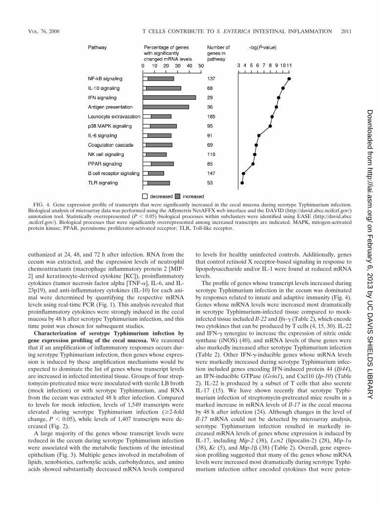
The profile of genes whose transcript levels increased duringserotype Typhimurium infection in the cecum was dominatedby responses related to innate and adaptive immunity (Fig. 4).Genes whose mRNA levels were increased most dramaticallyin serotype Typhimurium-infected tissue compared to mock-infected tissue included Il-22 and Ifn-� (Table 2), which encodetwo cytokines that can be produced by T cells (4, 15, 30). IL-22and IFN-� synergize to increase the expression of nitric oxidesynthase (iNOS) (40), and mRNA levels of these genes werealso markedly increased after serotype Typhimurium infection(Table 2). Other IFN-�-inducible genes whose mRNA levelswere markedly increased during serotype Typhimurium infec-tion included genes encoding IFN-induced protein 44 (Ifi44),an IFN-inducible GTPase (Gvin1), and Cxcl10 (Ip-10) (Table2). IL-22 is produced by a subset of T cells that also secreteIL-17 (15). We have shown recently that serotype Typhi-murium infection of streptomycin-pretreated mice results in amarked increase in mRNA levels of Il-17 in the cecal mucosaby 48 h after infection (24). Although changes in the level ofIl-17 mRNA could not be detected by microarray analysis,serotype Typhimurium infection resulted in markedly in-creased mRNA levels of genes whose expression is induced byIL-17, including Mip-2 (38), Lcn2 (lipocalin-2) (28), Mip-1�(38), Kc (5), and Mip-1 (38) (Table 2). Overall, gene expres-sion profiling suggested that many of the genes whose mRNAlevels were increased most dramatically during serotype Typhi-murium infection either encoded cytokines that were poten-
FIG. 4. Gene expression profile of transcripts that were significantly increased in the cecal mucosa during serotype Typhimurium infection.Biological analysis of microarray data was performed using the Affymetrix NetAFFX web interface and the DAVID (http://david.abcc.ncifcrf.gov/)annotation tool. Statistically overrepresented (P � 0.05) biological processes within subclusters were identified using EASE (http://david.abcc.ncifcrf.gov/). Biological processes that were significantly overrepresented among increased transcripts are indicated. MAPK, mitogen-activatedprotein kinase; PPAR, peroxisome proliferator-activated receptor; TLR, Toll-like receptor.
VOL. 76, 2008 T CELLS CONTRIBUTE TO S. ENTERICA INTESTINAL INFLAMMATION 2011
on February 6, 2013 by U
C D
AV
IS S
HIE
LDS
LIBR
AR
Yhttp://iai.asm
.org/D
ownloaded from

tially derived from T cells or encoded products whose expres-sion is regulated by these cytokines.
Real-time PCR analysis demonstrates a dramatic increasein mRNA levels for Il-22, Ifn-�, and Il-17 during serotypeTyphimurium infection. To confirm results from gene expres-sion profiling, mRNA levels of cytokines for selected geneswere monitored by quantitative real-time PCR at 48 h afterinfection by using the ceca from groups of four serotype Ty-phimurium-infected and four mock-infected mice. Quantita-tive measurements by real-time PCR showed that Il-22 (579-fold), Ifn-� (554-fold), and Il-17 (149-fold) were the cytokine
genes whose mRNA levels were elevated most dramaticallyafter serotype Typhimurium infection (Fig. 5). Furthermore,the mRNA levels of genes regulated by IL-22, IL-17, and/orIFN-� were also markedly increased, including Mip-2 (168-fold), Kc (84-fold), Lcn2 (58-fold), Mip-1� (17-fold), andMip-1 (23-fold). In summary, real-time PCR analysis sug-gested that marked increases in the mRNA levels of Il-22,Il-17, and Ifn-� were among the changes in the gene expressionprofile that exhibited the greatest magnitudes. Furthermore,our data confirmed the marked increase in mRNA levels ofgenes induced by IL-22, IL-17, and IFN-�, which had beensuggested by gene expression profiling (Table 2).
The detection of increased mRNA levels of genes associatedwith immunity and inflammation (Fig. 4 and 5) was accompa-nied by the development of pathological changes in the cecalmucosa (Fig. 6). At 48 h after inoculation, the ceca fromserotype Typhimurium-infected mice contained little contentsand had severe gross pathological changes, characterized byreduced size and thickening of the cecal wall (Fig. 6B). At 48 hafter infection, the histopathology was characterized by epithe-lial erosion, neutrophil infiltration in the mucosa, and markededema in the submucosa (Fig. 6C), which was consistent withprevious reports describing cecal inflammation in this model(1, 24).
T cells are major contributors to intestinal inflammationelicited by serotype Typhimurium. Gene expression profilingand real-time PCR analysis suggested that the host response toserotype Typhimurium infection is dominated by a dramaticincrease in the mRNA levels of Il-22, Il-17, and Ifn-� and ofgenes whose expression can be induced by the cytokines en-
FIG. 5. Cytokine expression elicited by serotype Typhimurium instreptomycin-pretreated mice 48 h after infection, as measured byquantitative real-time PCR. Changes in gene expression observed forserotype Typhimurium-infected mice (n � 4) compared to that ob-served for mock-infected mice (n � 4) were determined. Data areshown as geometric means of changes (n-fold) � standard errors,determined for RNA from individual mice.
TABLE 2. List of transcripts related to immunity and inflammationwhose levels were elevated more than 10-fold (P � 0.05) in
serotype Typhimurium-infected cecal tissue
Gene
Gene expressionprofiling of
mRNA from cecaof serotype
Typhimurium-infected micecompared to
those of mock-infected mice
Avg fold changedetermined byreal-time PCRconfirmation
Foldchange P value
Cxcl2 (Mip-2) 97.9 0.001 168Cxcl5 62.3 0.001 NDa
Il-1� 57.2 0.002 NDCccl3 (Mip-1a) 53.9 0.002 17.3Ifn-� 53.9 0.002 554Irg 1 40.3 0.001 ND
S100a8 (calgranulin A) 37.0 0.001 NDS100a9 (calgranulin B) 32.5 0.001 NDNos2 (iNOS) 30.6 0.008 56.9b
Il-22 27.9 0.002 579Il-6 26.7 0.002 44.0Lcn2 (lipocalin 2) 26.6 0.003 58.3
Fpr-rs2 25.5 0.003 NDIl-1 21.0 0.004 NDCxcl1 (Kc) 20.6 0.005 83.7Mmp3 20.5 0.003 NDCcl4 (Mip-1) 20.4 0.003 22.7Il-33 18.8 0.001 ND
Ccl2 17.4 0.003 NDIl-1rn (IL-1 receptor antagonist) 15.9 0.006 NDIl-18bp (IL-18 binding protein) 15.8 0.002 NDCcl7 15.6 0.007 NDSell (lymphocyte selectin) 15.2 0.005 NDMmp8 14.0 0.007 ND
Ltf (lactoferrin) 13.6 0.017 NDIfi44 (IFN-induced protein 44) 13.6 0.004 NDMmp10 13.4 0.007 NDCcr5 12.4 0.004 NDCxcl10 (Ip-10) 11.9 0.002 NDGvin1 (IFN-inducible GTPase) 11.6 0.006 ND
Cxcl11 11.1 0.006 NDCxcl9 10.7 0.005 NDItk (IL-2-inducible T-cell kinase) 10.7 0.037 NDTnf-� 10.2 0.011 85.8
a ND, not determined.b Determined for mock-depleted C57BL/6 mice.
2012 GODINEZ ET AL. INFECT. IMMUN.
on February 6, 2013 by U
C D
AV
IS S
HIE
LDS
LIBR
AR
Yhttp://iai.asm
.org/D
ownloaded from

coded by these three genes (Table 2; Fig. 5). Since T cells area potential source of IL-22, IL-17, and IFN-� (4, 15, 30), thesedata raised the possibility that genes whose mRNA levels wereincreased most dramatically during serotype Typhimurium in-fection may participate in a T-cell-mediated amplification ofresponses in tissue. The intestine is the largest reservoir of Tcells in the human body, and this cell type is present abun-dantly throughout the lamina propria, thus making this celltype well suited for amplifying inflammatory responses. Toassess the importance of T cells in orchestrating inflammation,two groups of four mice were treated with rat anti-mouse CD3MAb to deplete T cells. As a control, two groups of four micewere treated with nonspecific rat IgG control antibody (mockdepletion). All four groups of mice were streptomycin pre-treated. Four CD3-depleted and four mock-depleted micewere inoculated with sterile LB broth (mock infection), andthe remaining two groups were infected with serotype Typhi-murium. At 48 h after inoculation, organs were collected foranalysis.
Spleens from all animals were processed for flow cytometryto monitor T-cell depletion. Splenocytes from mock-depletedmice contained on average 36.2% CD3� T cells. This percent-age remained unchanged when mock-depleted mice were in-fected with serotype Typhimurium (35.5% CD3� T cells) (Fig.7). In contrast, CD3� T cells composed on average 0.12% ofthe splenocyte population in mice treated with the anti-mouseCD3 MAb. Similarly, splenocytes from CD3-depleted miceinfected with serotype Typhimurium contained on average0.12% of CD3� T cells. These data suggested that T cells hadbeen depleted effectively by treatment with rat anti-mouseCD3 MAb.
RNA was extracted from the cecum to investigate the de-velopment of proinflammatory changes in mRNA levels in
each group. Mock infection of CD3-depleted mice resulted inmRNA levels of proinflammatory cytokines similar to thosefound after mock infection of mock-depleted mice. However,compared to mock infection of mock-depleted mice, serotypeTyphimurium infection of mock-depleted mice resulted inmarkedly increased mRNA levels of genes encoding inflam-matory cytokines, including Il-22 (197-fold) (Fig. 8A), Ifn-�(228-fold) (Fig. 8B), Il-17 (92-fold) (Fig. 8C), Kc (60-fold) (Fig.8D), Nos2 (57-fold) (Fig. 8E), and Tnf-� (16-fold) (Fig. 8F). Incontrast, this marked induction of proinflammatory cytokineexpression was not observed to occur during serotype Typhi-murium infection of CD3-depleted mice (Fig. 8). CD3 deple-tion dramatically reduced the ability of serotype Typhimuriumto elicit increased mRNA levels of Il-22, Il-17, and Ifn-� and ofgenes regulated by the encoded cytokines. These data sug-gested that T cells were an important source of IL-22, IL-17,and IFN-� production in the intestinal mucosa 48 h after se-rotype Typhimurium infection.
To determine whether attenuated inflammatory responses inCD3-depleted mice were due to a large decrease in the bac-terial load, bacteria were recovered from cecal contents, themesenteric lymph node, and the liver. CD3 depletion resultedin increased bacterial loads in the liver (Fig. 9) (P � 0.05).More bacteria were also recovered from cecal contents andmesenteric lymph nodes of CD3-depleted mice than fromthose of mock-depleted mice, but these differences were notsignificant. In conclusion, CD3-depleted mice exhibited similar(i.e., in the cecum and mesenteric lymph node) or increased(i.e., in the liver) bacterial burdens, while expression of inflam-matory cytokines was markedly reduced. These data suggestedthat reduced cytokine responses in CD3-depleted mice werenot caused by a reduction in bacterial numbers.
We next investigated the biological consequences of the
FIG. 6. Gross pathological (A and B) and histopathological (C) appearance of the murine cecum. (A) Cecum of a streptomycin-pretreatedmouse 48 h after inoculation with sterile LB broth, with a normal appearance. (B) Cecum of a streptomycin-pretreated mouse 48 h after inoculationwith serotype Typhimurium. Note the reduced size of the cecum, which was devoid of contents and had a whitish discoloration and gelatinousappearance, indicating severe edema. (C) Murine cecum 48 h after infection with serotype Typhimurium, shown at low magnification (�10) toillustrate edema in the submucosa. The inset at the bottom right shows a section of the cecum from an animal 48 h after inoculation with sterileLB broth at the same magnification (�10).
VOL. 76, 2008 T CELLS CONTRIBUTE TO S. ENTERICA INTESTINAL INFLAMMATION 2013
on February 6, 2013 by U
C D
AV
IS S
HIE
LDS
LIBR
AR
Yhttp://iai.asm
.org/D
ownloaded from

blunted cytokine responses observed in T-cell-depleted miceby investigating the development of cecal inflammation. At48 h after inoculation, ceca from mock-depleted mice infectedwith serotype Typhimurium contained little contents and hadsevere gross pathological changes, characterized by reducedsize and thickening of the cecal wall. This appearance wastypical of the gross pathological changes elicited by serotypeTyphimurium at 48 h after infection in this model (Fig. 6B). Incontrast, ceca from serotype Typhimurium-infected CD3-de-pleted mice did not show gross pathological changes and ap-peared similar to ceca from mock-infected mice (Fig. 6A).Histopathological evaluation revealed epithelial erosion, neu-trophil infiltration in the mucosa, and edema in the submucosaof mock-depleted mice infected with serotype Typhimurium.These pathological changes were reduced drastically in sero-type Typhimurium-infected CD3-depleted mice (Fig. 10A).Compared to serotype Typhimurium-infected mock-depletedmice, serotype Typhimurium-infected CD3-depleted mice re-cruited markedly reduced numbers of neutrophils into to thececal mucosa (Fig. 10B), which correlated well with the re-duced levels detected in this group of mRNA levels for theneutrophil chemoattractant gene Kc (Fig. 8D) and the reducedtranscript levels for Il-17 (Fig. 7C), encoding a cytokine con-trolling expression of neutrophil chemoattractants (5, 38).
DISCUSSION
Serotype Typhimurium causes a localized gastroenteritis inhumans that is characterized by a massive neutrophil influx inthe terminal ileum and colon. Upon translocation through theintestinal epithelium, bacteria are found mainly within mono-nuclear cells (i.e., macrophages or dendritic cells) or neutro-phils in the lamina propria (25, 27). Some of the cytokinesreleased by mononuclear cells after their stimulation with bac-teria may help to amplify inflammatory responses in tissue(29). The gene expression profiles elicited in macrophages orepithelial cells infected with serotype Typhimurium have beendescribed previously (23, 26, 39). However, since only a verysmall fraction of cells in infected tissue contain bacteria, cyto-kine production by these cells may be limited in scope. Incontrast, responses that are amplified effectively in tissuewould be expected to be responsible for the most prominentchanges in gene expression observed in the cecal mucosa dur-ing serotype Typhimurium infection. Consistent with this idea,we found that mRNA levels upregulated most dramaticallyduring serotype Typhimurium infection of the cecal mucosaincluded genes encoding T-cell-derived cytokines (i.e., IL-22,IL-17, and IFN-�) and genes whose expression is known to beregulated by these cytokines. The predominance of these re-
FIG. 7. Fractions of T cells present in splenocyte populations of mice (n � 8) treated with anti-mouse CD3 MAb (CD3 depletion) or mice (n �8) treated with nonspecific IgG antibody (mock depletion). Four mock-depleted and four CD3-depleted mice were infected with serotypeTyphimurium, while the remaining animals were mock infected. Splenocytes were isolated 48 h after infection, and cells were analyzed by flowcytometry. The number in each box indicates the fraction of cells within the lymphocyte population that was scored CD3�. PE, phycoerythrin.
2014 GODINEZ ET AL. INFECT. IMMUN.
on February 6, 2013 by U
C D
AV
IS S
HIE
LDS
LIBR
AR
Yhttp://iai.asm
.org/D
ownloaded from

sponses in vivo was not apparent from previous studies of geneexpression profiles determined in tissue culture (23, 26, 39).Results from in vivo gene expression profiling were consistentwith a central role of T cells in amplifying responses in tissue.
Importantly, CD3� T-cell depletion resulted in a dramaticattenuation of intestinal inflammatory responses during sero-type Typhimurium infection. These data provided compellingsupport for the idea that T cells play an important role inamplifying local inflammatory responses during infection ofthe intestinal mucosa by serotype Typhimurium.
It is currently not clear by which mechanisms T cells in thececal mucosa are stimulated to produce IL-22, IL-17, andIFN-� during serotype Typhimurium infection. Previous stud-ies have implicated mononuclear cells as possible sources forcytokines that would stimulate the release of IL-22, IL-17, andIFN-� by T cells. For example, serotype Typhimurium flagellinis translocated by the T3SS-1 into the cytosol of macrophages(33), where it is sensed by Ipaf, a cytosolic nucleotide-bindingand oligomerization domain-like receptor (6, 18). Recognitionof flagellin by Ipaf leads to activation of caspase 1, which inturn cleaves precursors of IL-18 and IL-1 into their activeforms (10, 20). Examination of splenic tissue from mice showsthat IL-18 can stimulate antigen-experienced T cells to rapidlysecrete IFN-� during bacterial infection by an antigen-inde-pendent mechanism, thereby significantly amplifying early ef-fector responses in vivo (30). In a mouse model of Klebsiellapneumoniae lung infection, bacterial stimulation of Toll-likereceptors on dendritic cells results in IL-23 production, which
FIG. 8. Changes in gene expression elicited 48 h after serotype Typhimurium infection in the cecal mucosa of mock-depleted mice (filled bars) orCD3-depleted mice (open bars), as measured by quantitative real-time PCR. Data are expressed as changes of mRNA levels over mRNA levels detected inmock-infected, mock-depleted mice. Data represent geometric means � standard errors. Statistical significance of differences is indicated by P values.
FIG. 9. Recovery of serotype Typhimurium from the organs ofmock-depleted mice (filled bars) or CD3-depleted mice (open bars).Bars represent geometric means � standard errors of CFU/organ(cecal contents) or CFU/g tissue (mesenteric lymph node and liver).Statistical significance of differences is indicated by P values.
VOL. 76, 2008 T CELLS CONTRIBUTE TO S. ENTERICA INTESTINAL INFLAMMATION 2015
on February 6, 2013 by U
C D
AV
IS S
HIE
LDS
LIBR
AR
Yhttp://iai.asm
.org/D
ownloaded from

in turn triggers the rapid production of IL-17 by T cells. Thisparacrine IL-23-mediated mechanism also triggers the releaseof IL-17 from T cells in a mouse model of Mycobacterium bovisinfection (36). However, additional work is required to deter-mine whether these pathways also participate in the amplifi-cation of inflammatory responses in the intestinal mucosa dur-ing serotype Typhimurium infection.
The T-cell products IL-22, IL-17, and IFN-� likely play animportant role in the amplification of intestinal inflammatoryresponses. IL-17 is important for efficient neutrophil recruit-ment, as shown by its ability to induce the production of neu-trophil chemoattractants (CXC chemokines) in human bron-chial epithelial cells in vitro (13) and its ability to stimulate
neutrophil recruitment in rat airways in vivo upon intratrachealinstillation (14). Consistent with these findings, CD3� T-celldepletion blunted Il-17 expression during serotype Typhi-murium infection and resulted in a dramatic attenuation ofneutrophil recruitment in the cecal mucosa. Furthermore, IL-22, IL-17, and/or IFN-� can activate various effector mecha-nisms of the innate immune system, including expression ofiNOS (19, 40), lipocalin-2 (28), and human -defensin (13).The induction of these antimicrobial responses may contributeto a reduction in the numbers of competing microorganisms,which has been shown to be beneficial for serotype Typhi-murium during intestinal colonization (31). A beneficial effectof inflammation on the recovery of serotype Typhimuriumfrom cecal contents is observed at 5 days after streptomycintreatment, when normal composition of the intestinal micro-biota is restored. However, at the time point chosen for ourstudy (i.e., at 3 days after streptomycin treatment), normalcomposition of the intestinal microbiota had not yet been re-stored, which may explain why we did not observe that intes-tinal inflammation increased the numbers of serotype Typhi-murium bacteria in the cecal contents. Instead, we observedincreased translocation of serotype Typhimurium to the liverin CD3-depleted mice, which may be due to a defect in induc-ing neutrophil influx and antimicrobial responses in the intes-tinal mucosa of these animals. In conclusion, our data suggestthat one function of intestinal T cells is to amplify innateimmune responses by producing cytokines that regulate innateimmune functions.
ACKNOWLEDGMENTS
This investigation was conducted in a facility constructed with sup-port from Research Facilities Improvement Program grant numberC06 RR12088-01 from the National Center for Research Resources,National Institutes of Health. Work in A.J.B.’s laboratory was sup-ported by Public Health Service grants AI040124, AI044170, andAI065534. T.A.P. and R.L.S. are recipient of fellowships from CNPq(Conselho Nacional de Desenvolvimento Cientıfico e Tecnologico,Brasılia, Brazil). I.G. was supported by Public Health Service grantAI06055.
REFERENCES
1. Barthel, M., S. Hapfelmeier, L. Quintanilla-Martınez, M. Kremer, M.Rohde, M. Hogardt, K. Pfeffer, H. Russmann, and W.-D. Hardt. 2003. Pre-treatment of mice with streptomycin provides a Salmonella enterica serovarTyphimurium colitis model that allows analysis of both pathogen and host.Infect. Immun. 71:2839–2858.
2. Chen, L. M., S. Hobbie, and J. E. Galan. 1996. Requirement of cdc42 forsalmonella-induced cytoskeletal and nuclear responses. Science 274:2115–2118.
3. Day, D. W., B. K. Mandal, and B. C. Morson. 1978. The rectal biopsyappearances in Salmonella colitis. Histopathology 2:117–131.
4. D’Orazio, S. E., M. J. Troese, and M. N. Starnbach. 2006. Cytosolic local-ization of Listeria monocytogenes triggers an early IFN-gamma response byCD8� T cells that correlates with innate resistance to infection. J. Immunol.177:7146–7154.
5. Dubin, P. J., and J. K. Kolls. 2007. IL-23 mediates inflammatory responsesto mucoid Pseudomonas aeruginosa lung infection in mice. Am. J. Physiol.Lung Cell Mol. Physiol. 292:L519–L528.
6. Franchi, L., A. Amer, M. Body-Malapel, T. D. Kanneganti, N. Ozoren, R.Jagirdar, N. Inohara, P. Vandenabeele, J. Bertin, A. Coyle, E. P. Grant, andG. Nunez. 2006. Cytosolic flagellin requires Ipaf for activation of caspase-1and interleukin 1beta in salmonella-infected macrophages. Nat. Immunol.7:576–582.
7. Happel, K. I., P. J. Dubin, M. Zheng, N. Ghilardi, C. Lockhart, L. J.Quinton, A. R. Odden, J. E. Shellito, G. J. Bagby, S. Nelson, and J. K. Kolls.2005. Divergent roles of IL-23 and IL-12 in host defense against Klebsiellapneumoniae. J. Exp. Med. 202:761–769.
8. Happel, K. I., M. Zheng, E. Young, L. J. Quinton, E. Lockhart, A. J. Ramsay,J. E. Shellito, J. R. Schurr, G. J. Bagby, S. Nelson, and J. K. Kolls. 2003.
FIG. 10. Effect of CD3 depletion on the histopathology of themurine cecum 48 h after infection with serotype Typhimurium. (A) Allpanels are shown at the same magnification (�10) to illustrate themagnitude of edema in serotype Typhimurium-infected animals. Thetwo panels on the left show the normal appearance of the cecal mucosain mock-infected animals. The second panel from the right shows thececum of a mock-depleted animal 48 h after serotype Typhimuriuminfection. Note the marked thickening of the intestinal wall due toedema in the submucosa. The right panel shows the appearance of thececal mucosa in a CD3-depleted animal 48 h after serotype Typhi-murium infection. (B) Neutrophil recruitment into the cecal mucosa.The numbers of neutrophils per microscopic field were determined bya veterinary pathologist during a blind examination of slides from thececal mucosa. Data represent means � standard errors. Statisticalsignificance of differences is indicated by P values.
2016 GODINEZ ET AL. INFECT. IMMUN.
on February 6, 2013 by U
C D
AV
IS S
HIE
LDS
LIBR
AR
Yhttp://iai.asm
.org/D
ownloaded from

Cutting edge: roles of Toll-like receptor 4 and IL-23 in IL-17 expression inresponse to Klebsiella pneumoniae infection. J. Immunol. 170:4432–4436.
9. Hardt, W. D., L. M. Chen, K. E. Schuebel, X. R. Bustelo, and J. E. Galan.1998. S. typhimurium encodes an activator of Rho GTPases that inducesmembrane ruffling and nuclear responses in host cells. Cell 93:815–826.
10. Hersh, D., D. M. Monack, M. R. Smith, N. Ghori, S. Falkow, and A. Zych-linsky. 1999. The salmonella invasin SipB induces macrophage apoptosis bybinding to caspase-1. Proc. Natl. Acad. Sci. USA 96:2396–2401.
11. Hobbie, S., L. M. Chen, R. J. Davis, and J. E. Galan. 1997. Involvement ofmitogen-activated protein kinase pathways in the nuclear responses andcytokine production induced by Salmonella typhimurium in cultured intes-tinal epithelial cells. J. Immunol. 159:5550–5559.
12. Huang, F. C., A. Werne, Q. Li, E. E. Galyov, W. A. Walker, and B. J.Cherayil. 2004. Cooperative interactions between flagellin and SopE2 in theepithelial interleukin-8 response to Salmonella enterica serovar Typhi-murium infection. Infect. Immun. 72:5052–5062.
13. Kao, C. Y., Y. Chen, P. Thai, S. Wachi, F. Huang, C. Kim, R. W. Harper, andR. Wu. 2004. IL-17 markedly up-regulates beta-defensin-2 expression inhuman airway epithelium via JAK and NF-kappaB signaling pathways. J. Im-munol. 173:3482–3491.
14. Laan, M., Z. H. Cui, H. Hoshino, J. Lotvall, M. Sjostrand, D. C. Gruenert,B. E. Skoogh, and A. Linden. 1999. Neutrophil recruitment by human IL-17via C-X-C chemokine release in the airways. J. Immunol. 162:2347–2352.
15. Liang, S. C., X. Y. Tan, D. P. Luxenberg, R. Karim, K. Dunussi-Joannopoulos,M. Collins, and L. A. Fouser. 2006. Interleukin (IL)-22 and IL-17 are coex-pressed by Th17 cells and cooperatively enhance expression of antimicrobialpeptides. J. Exp. Med. 203:2271–2279.
16. McCormick, B. A., C. A. Parkos, S. P. Colgan, D. K. Carnes, and J. L.Madara. 1998. Apical secretion of a pathogen-elicited epithelial chemoat-tractant activity in response to surface colonization of intestinal epithelia bySalmonella typhimurium. J. Immunol. 160:455–466.
17. McGovern, V. J., and L. J. Slavutin. 1979. Pathology of salmonella colitis.Am. J. Surg. Pathol. 3:483–490.
18. Miao, E. A., C. M. Alpuche-Aranda, M. Dors, A. E. Clark, M. W. Bader, S. I.Miller, and A. Aderem. 2006. Cytoplasmic flagellin activates caspase-1 andsecretion of interleukin 1beta via Ipaf. Nat. Immunol. 7:569–575.
19. Miljkovic, D., and V. Trajkovic. 2004. Inducible nitric oxide synthase activa-tion by interleukin-17. Cytokine Growth Factor Rev. 15:21–32.
20. Monack, D. M., W. W. Navarre, and S. Falkow. 2001. Salmonella-inducedmacrophage death: the role of caspase-1 in death and inflammation. Mi-crobes Infect. 3:1201–1212.
21. Mrsny, R. J., A. T. Gewirtz, D. Siccardi, T. Savidge, B. P. Hurley, J. L.Madara, and B. A. McCormick. 2004. Identification of hepoxilin A3 ininflammatory events: a required role in neutrophil migration across intestinalepithelia. Proc. Natl. Acad. Sci. USA 101:7421–7426.
22. Nau, G. J., J. F. Richmond, A. Schlesinger, E. G. Jennings, E. S. Lander, andR. A. Young. 2002. Human macrophage activation programs induced bybacterial pathogens. Proc. Natl. Acad. Sci. USA 99:1503–1508.
23. Nau, G. J., A. Schlesinger, J. F. Richmond, and R. A. Young. 2003. Cumu-lative Toll-like receptor activation in human macrophages treated with wholebacteria. J. Immunol. 170:5203–5209.
24. Raffatellu, M., R. L. Santos, D. Chessa, R. P. Wilson, S. E. Winter, C. A.Rossetti, S. D. Lawhon, H. Chu, T. Lau, C. L. Bevins, L. G. Adams, and A. J.Baumler. 2007. The capsule encoding the viaB locus reduces interleukin-17expression and mucosal innate responses in the bovine intestinal mucosaduring infection with Salmonella enterica serotype Typhi. Infect. Immun.75:4342–4350.
25. Reis, B. P., S. Zhang, R. M. Tsolis, A. J. Baumler, L. G. Adams, and R. L.Santos. 2003. The attenuated sopB mutant of Salmonella enterica serovarTyphimurium has the same tissue distribution and host chemokine responseas the wild type in bovine Peyer’s patches. Vet. Microbiol. 97:269–277.
26. Rosenberger, C. M., M. G. Scott, M. R. Gold, R. E. Hancock, and B. B.Finlay. 2000. Salmonella typhimurium infection and lipopolysaccharide stim-ulation induce similar changes in macrophage gene expression. J. Immunol.164:5894–5904.
27. Santos, R. L., S. Zhang, R. M. Tsolis, A. J. Baumler, and L. G. Adams. 2002.Morphologic and molecular characterization of Salmonella typhimurium in-fection in neonatal calves. Vet. Pathol. 39:200–215.
28. Shen, F., M. J. Ruddy, P. Plamondon, and S. L. Gaffen. 2005. Cytokines linkosteoblasts and inflammation: microarray analysis of interleukin-17- andTNF-alpha-induced genes in bone cells. J. Leukoc. Biol. 77:388–399.
29. Srikanth, C. V., and B. J. Cherayil. 2007. Intestinal innate immunity and thepathogenesis of Salmonella enteritis. Immunol. Res. 37:61–78.
30. Srinivasan, A., R. M. Salazar-Gonzalez, M. Jarcho, M. M. Sandau, L.Lefrancois, and S. J. McSorley. 2007. Innate immune activation of CD4 Tcells in salmonella-infected mice is dependent on IL-18. J. Immunol. 178:6342–6349.
31. Stecher, B., R. Robbiani, A. W. Walker, A. M. Westendorf, M. Barthel, M.Kremer, S. Chaffron, A. J. Macpherson, J. Buer, J. Parkhill, G. Dougan, C.von Mering, and W. D. Hardt. 2007. Salmonella enterica serovar typhi-murium exploits inflammation to compete with the intestinal microbiota.PLoS Biol. 5:2177–2189.
32. Stojiljkovic, I., A. J. Baumler, and F. Heffron. 1995. Ethanolamine utilizationin Salmonella typhimurium: nucleotide sequence, protein expression, andmutational analysis of the cchA cchB eutE eutJ eutG eutH gene cluster. J.Bacteriol. 177:1357–1366.
33. Sun, Y. H., H. G. Rolan, and R. M. Tsolis. 2007. Injection of flagellin into thehost cell cytosol by Salmonella enterica serotype Typhimurium. J. Biol.Chem. 282:33897–33901.
34. Tallant, T., A. Deb, N. Kar, J. Lupica, M. J. de Veer, and J. A. DiDonato.2004. Flagellin acting via TLR5 is the major activator of key signalingpathways leading to NF-kappa B and proinflammatory gene program acti-vation in intestinal epithelial cells. BMC Microbiol. 4:33.
35. Tukel, C., M. Raffatellu, A. D. Humphries, R. P. Wilson, H. L. Andrews-Polymenis, T. Gull, J. F. Figueiredo, M. Wong, K. S. Michelsen, M. Akcelik,L. G. Adams, and A. J. Baumler. 2005. CsgA is a pathogen-associatedmolecular pattern of Salmonella enterica serotype Typhimurium that is rec-ognized by Toll-like receptor 2. Mol. Microbiol. 58:289–304.
36. Umemura, M., A. Yahagi, S. Hamada, M. D. Begum, H. Watanabe, K.Kawakami, T. Suda, K. Sudo, S. Nakae, Y. Iwakura, and G. Matsuzaki. 2007.IL-17-mediated regulation of innate and acquired immune response againstpulmonary Mycobacterium bovis bacille Calmette-Guerin infection. J. Im-munol. 178:3786–3796.
37. Ye, P., P. B. Garvey, P. Zhang, S. Nelson, G. Bagby, W. R. Summer, P.Schwarzenberger, J. E. Shellito, and J. K. Kolls. 2001. Interleukin-17 andlung host defense against Klebsiella pneumoniae infection. Am. J. Respir.Cell Mol. Biol. 25:335–340.
38. Ye, P., F. H. Rodriguez, S. Kanaly, K. L. Stocking, J. Schurr, P. Schwarzen-berger, P. Oliver, W. Huang, P. Zhang, J. Zhang, J. E. Shellito, G. J. Bagby,S. Nelson, K. Charrier, J. J. Peschon, and J. K. Kolls. 2001. Requirement ofinterleukin 17 receptor signaling for lung CXC chemokine and granulocytecolony-stimulating factor expression, neutrophil recruitment, and host de-fense. J. Exp. Med. 194:519–527.
39. Zeng, H., A. Q. Carlson, Y. Guo, Y. Yu, L. S. Collier-Hyams, J. L. Madara,A. T. Gewirtz, and A. S. Neish. 2003. Flagellin is the major proinflammatorydeterminant of enteropathogenic Salmonella. J. Immunol. 171:3668–3674.
40. Ziesche, E., M. Bachmann, H. Kleinert, J. Pfeilschifter, and H. Muhl. 2007.The interleukin-22/STAT3 pathway potentiates expression of inducible ni-tric-oxide synthase in human colon carcinoma cells. J. Biol. Chem. 282:16006–16015.
Editor: A. Camilli
VOL. 76, 2008 T CELLS CONTRIBUTE TO S. ENTERICA INTESTINAL INFLAMMATION 2017
on February 6, 2013 by U
C D
AV
IS S
HIE
LDS
LIBR
AR
Yhttp://iai.asm
.org/D
ownloaded from
Copyright © 2022 FDOKUMEN