Synthesis, Characterization, Physicochemical, and Photophysical Studies of Redox Switchable NIR Dye...
14
Int. J. Electrochem. Sci., 10 (2015) 6092 - 6105 International Journal of ELECTROCHEMICAL SCIENCE www.electrochemsci.org Synthesis, Characterization, Physicochemical and Electrochemical Studies of Novel Donor Acceptor Chromophores Abdullah M. Asiri 1,2 , Salman A. Khan 1,* , Hadi Mussa Basisi 1 1 Chemistry Department, Faculty of Science, King Abdulaziz University, P.O. Box 80203, Jeddah, Saudi Arabia 21589 2 Center of Excellence for Advanced Materials Research, King Abdulaziz University, P.O. Box 80203, Jeddah, Saudi Arabia 21589 * E-mail: [email protected] Received: 22 September 2014 / Accepted: 27 May 2015 / Published: 24 June 2015 Three donor acceptor chromophores were synthesized by Knoevenagel condensation. Structures of the chromophoes were conformed by the elemental analysis and EI-MS, FT-IR, 1 H-NMR, 13 C-NMR spectroscopy. Absorbance and fluorescence spectra of the chromophores were studied in different solvent provide that all the chromophores are good absorbent and emission. All chromophores give same behavior red shift in absorbance and emission spectra as polarity of the solvents increase. Photophysical properties including, oscillator strength, extinction coefficient, transition dipole moment and stokes shift were investigated in order to investigate the physicochemical behaviors of synthesized chromophores. The HOMO energy levels of compound 1, 2 and 3 respectively were calculated front the onset oxidation potential of cyclic voltammogram in acetonitrile. Keywords: Knoevenagel condensation, Stokes shift; Dipole moment, Cyclic voltammogram 1. INTRODUCTION Donor acceptor chromophores have drawn much attention in the recent decades due to their attractive potential in photonic device [1]. In the last two decades great attempt has been made in the field of synthetic chemistry in order to design the organic molecules used in material sciences [2] such as optical and photonic imaging [3], electrochemical sensing [4], langmuir films and photoinitiated polymerization with high non linear optical properties by enhancing the molecular polarizabilities of the constitute NLO energetic units [5-7]. The compound containing with donor - acceptor framework employable as monomeric active unit is based on the well known donor acceptor system constituted by a л –conjugated system with strong electron donor/withdrawing groups [8]. Use of substituents such as
-
Upload
independent -
Category
Documents
-
view
4 -
download
0
Transcript of Synthesis, Characterization, Physicochemical, and Photophysical Studies of Redox Switchable NIR Dye...

Int. J. Electrochem. Sci., 10 (2015) 6092 - 6105
International Journal of
ELECTROCHEMICAL SCIENCE
www.electrochemsci.org
Synthesis, Characterization, Physicochemical and
Electrochemical Studies of Novel Donor Acceptor
Chromophores
Abdullah M. Asiri1,2
, Salman A. Khan1,*
, Hadi Mussa Basisi1
1Chemistry Department, Faculty of Science, King Abdulaziz University, P.O. Box 80203, Jeddah,
Saudi Arabia 21589 2
Center of Excellence for Advanced Materials Research, King Abdulaziz University, P.O. Box 80203,
Jeddah, Saudi Arabia 21589 *E-mail: [email protected]
Received: 22 September 2014 / Accepted: 27 May 2015 / Published: 24 June 2015
Three donor acceptor chromophores were synthesized by Knoevenagel condensation. Structures of the
chromophoes were conformed by the elemental analysis and EI-MS, FT-IR, 1H-NMR,
13C-NMR
spectroscopy. Absorbance and fluorescence spectra of the chromophores were studied in different
solvent provide that all the chromophores are good absorbent and emission. All chromophores give
same behavior red shift in absorbance and emission spectra as polarity of the solvents increase.
Photophysical properties including, oscillator strength, extinction coefficient, transition dipole moment
and stokes shift were investigated in order to investigate the physicochemical behaviors of synthesized
chromophores. The HOMO energy levels of compound 1, 2 and 3 respectively were calculated front
the onset oxidation potential of cyclic voltammogram in acetonitrile.
Keywords: Knoevenagel condensation, Stokes shift; Dipole moment, Cyclic voltammogram
1. INTRODUCTION
Donor acceptor chromophores have drawn much attention in the recent decades due to their
attractive potential in photonic device [1]. In the last two decades great attempt has been made in the
field of synthetic chemistry in order to design the organic molecules used in material sciences [2] such
as optical and photonic imaging [3], electrochemical sensing [4], langmuir films and photoinitiated
polymerization with high non linear optical properties by enhancing the molecular polarizabilities of
the constitute NLO energetic units [5-7]. The compound containing with donor - acceptor framework
employable as monomeric active unit is based on the well known donor acceptor system constituted by
a л –conjugated system with strong electron donor/withdrawing groups [8]. Use of substituents such as
Int. J. Electrochem. Sci., Vol. 10, 2015
6093
NO2 and NR2 group can remarkably increase the push pull effect [9]. Photophysical properties such as,
dipole moment, oscillator strength, solvatochromic, piezochromic, florescent quantum yield and
photostability of the chromophore depends on the efficiency of the intramolecular charge transfer from
these two terminal groups (D-π- A), therefore on the nature and the length between the two terminal
with π-bond conjugation are also the most significant role to formative the behavior of compounds
[10,11]. Many reactions have been reported for the formation of donor-acceptor (D-π- A
chromophores. However, Knoevenagel condensation is one of the most imperative reactions for the
formation of donor-acceptor chromophores by the reaction of carbonyl compounds with active
methylene carbon in the presence of some Lewis acids or Lewis base followed by a nucleophilic
addition and dehydration reaction [12]. Different synthetic methods were reported for Knoevenagel
reaction, such as normal/refluxing in the solvent [13], ultrasonication, microwave radiation [14], solid-
phase reaction and photosensitization [15]. Due to numerous application of donor-acceptor
chromophores and continues work on the photophysical studies, in this paper we are reporting the
synthesis of novel donor acceptor chromophores and their photophysical, electrochemical
investigation.[13].
2. EXPERIMENTAL
2.1. Chemicals and reagents
All the solvents (A.R.) used in this work were of spectroscopic grade, appropriate aldehyde and
6-methoxy-1,2,3,4-tetrahydro-naphthalin-1-one were purchased from Acros Organic.
2.2. Apparatus
UV-Vis electronic absorption spectra were recorded using a 1 cm quartz cell on a Shimadzu
UV-1650 PC spectrophotometer. Steady state emission spectra were record using Shimadzu RF 5301
PC spectrofluorphotometer using a rectangular quartz cell of dimension 0.2 cm 3 cm. 1H-NMR and
13C-NMR spectra were recorded in CDCl3 on a Brucker DPX 600 at 600MHz and 150 MHz
spectrometer using tetramethyl silane (TMS) as internal standard. IR spectra were recorded on
Shimadzu FT-IR 8400S. Thomas Hoover capillary melting apparatus were used to determine the
melting points of the chromophores.
2.3. General method for the synthesis of donor acceptor chromophores (1-3)
A mixture of the 6-methoxy-1,2,3,4-tetrahydro-naphthalin-1-one (2 g, 0.011 mol) and
appropriate aldehyde (0.011 mol), in ethanol (99.9%) (25 mL) in the prescience of KOH with staring
at room temperature for 6-8h. Stirring was continued until all starting material had been consumed.
(TLC) solvent system chloroform : methane (2:8). After the completion of the reaction, the reaction
Int. J. Electrochem. Sci., Vol. 10, 2015
6094
mixture was pored in an ice cooled water and the precipitate thus obtained was filtered washed with
distilled water and recrystallized by chloroform and few drop of distilled ethanol.
2.3.1.(2E)-2-[(1-benzyl-1H-indol-3-yl)methyllidene]-6-methoxy-1,2,3,4-tetrahydronaphthalene-1-one
(1)
Yellow colure solid (Chloroform); Yield: 77.3%; M.p. 98 °C; EI-MS m/z (rel. int. %): 395 (72)
[M+1]+.
; IR (KBr) vmax cm-1
: 2939 (C-H), 1692 (C=O), 1560 (C=C), 1132 (C-N); 1H NMR (600MXz
CDCl3) δ: 8.18 (s, 1H, CH), 8.02-6.70 (m, 13H, CH Aromatic), 3.80 (s, 3H, OCH3), 2.96-2.91 (m, 2H,
CH2), 2.37 (s, 2H, CH2), 2.17-2.09 (m, 2H, CH2); 13
CNMR (CDCl3) δ: 191.83, 186.95, 163.95, 163.42,
145.56 (C- β), 140.40, 139.95, 137.93, 133.10, 130.70, 128.54, 127.40, 126.89, 123, 122.89 120.84,
119.35, 113.22, 112.25, 109.17, 108.72, 55.47, 37.97, 30.19, 29.35, 27.51, 23.40, 13.58, 13.85;; Anal.
calc.for C27H23NO2: C, 82.42, H, 5.89, N, 3.56. Found: C, 82.39, H, 5.83, N, 3.51.
2.3.2.(2E)-6-methoxy-2-[(1-methyl-1H-pyrrol-2-yl)methylidene]-1,2,3,4-tetrahydronaphthalene-1-one
(2)
Yellow colure solid (Chloroform); Yield: 76.3%; M.p. 148 °C; EI-MS m/z (rel. int. %): 269
(72) [M+1]+.
; IR (KBr) vmax cm-1
: 2948 (C-H), 1656 (C=O),1584 (C=C), 1132 (C-N); 1H NMR
(600MXz CDCl3) δ: 8.14 (s, 1H, CH), 8.02 (d, 1H, CHAr, J= 8.4 Hz), 8.09 (d, 1H, CHAr, J= 9.0
Hz),7.81 (s, 1H, CHAr), 6.81 (d, 1H, CHAr, J=7.8 Hz), 6.72 (dd, 1H, CHAr, J= 6.8 Hz), 6.58 (d, 1H,
CHAr, J= 7.8 Hz), 3.69 (s, 3H, OCH3), 2.96-2.91 (m, 2H, CH2), 2.62-2.59 (m, 2H, CH2); 13
CNMR
(CDCl3) δ: 197.25 (C=O), 186.12, 163.55, 146.97, 145.38 (C- β), 130.48, 125.97, 116.27, 113.76,
109.02, 55.44, 38.93, 36.42, 31.38, 28.63, 27.26; Anal. calc.for C17H17NO2: C, 76.38, H, 6.41, N, 5.24.
Found: C, 76.32, H, 6.38, N, 5.21.
2.3.3.(2E)-2-[(9-ethyl-9H-carbazol-3-yl)methyllidene]-6-methoxy-1,2,3,4-tetrahydronaphthalene-1-
one(3)
Yellow colure solid (Chloroform); Yield: 72.5%; M.p. 126 °C; EI-MS m/z (rel. int. %): 383
(78) [M+1]+.
; IR (KBr) vmax cm-1
: 2951 (C-H), 1682 (C=O), 1578 (C=C), 1123 (C-N); 1H NMR
(600MXz CDCl3) δ: 8.21 (s, 1H, CH), 8.16-6.70 (m, 10H, CHAromatic) 4.42-4.39 (q, CH2-CH3), 3.85
(s, 3H, OCH3), 2.96-2.91 (m, 2H, CH2), 2.62-2.60 (m, 2H, CH2), 1.49-1.45 (t, N-CH2-CH3); 13
CNMR
(CDCl3) δ: 195.25 (C=O), 186.95, 163.42, 145.55(C- β), 140.40, 139.95, 137.73, 133.10, 130.37,
128.37, 126.84 (C-Aromatic), 123.06, 120.56, 119.38, 113.20, 109.17, 108.72, 55.47, 38.93, 37.97,
30.19, 29.35, 27.51, 23.40, 13.88, 13.85; Anal. calc.for C26H23NO2: C, 81.86, H, 6.08, N, 3.67; Found:
C, 81.82, H, 6.02 N, 3.63.

Int. J. Electrochem. Sci., Vol. 10, 2015
6095
2.4. Cyclic voltammetry measurements
All prepared solutions were thoroughly degassed with oxygen free nitrogen, and a nitrogen
atmosphere was maintained above the solution throughout experimental studies.
Three electrode cell configuration linked to an EG and G model 283 Potentiostat were used to
measure the cyclic voltammetry of the chrompohores. The surface of platinum electrode was 7.85 x 10-
3 cm
2 as a working electrode, Counter electrode as a coiled platinum wire and refrence electrode as a
saturated Ag/AgCl. The potential was calculated with relative to the Ag/AgCl reference electrode at
25oC and 0.1 mol/L tetraethyl ammonium chloride (TEACl) as background electrolyte. The functioning
electrode was polished on a polisher Ecomet grinder. Cyclic voltammetric information was obtained at
scan rate ranging from 0.02 to 5 V/s in non aqueous media at (25 ± 2) oC. Cyclic voltammograms were
recorded after background calculation and iR recompense to reduce double-layer charging current and
solution resistance.
3. RESULT AND DISCUSSION
3.1. Chemistry
The synthesis of donor acceptor chromophores (1-3) are straight forward and the compounds
were isolated in good yield (Scheme 1). The chromophores were synthesized by the reaction of 6-
methoxy-1,2,3,4-tetrahydro-naphthalin-1-one and appropriate aldehyde in the presence of KOH [16].
The newly synthesized chromophores are stable in the in the solution as well as solid state. The
structure of all the chrompohores were conformed by the spectral data EI-MS, FT-IR, 1H-NMR,
13C-
NMR and purity of the chromophores further conformed by the elemental analysis. The select
characteristic IR band positions give important signal for the structure of the chromophores. The
chromophores showed intense bands at 1560-1584 cm-1
due to v (C=C) stretch, which is prove the
formation of donor accepter chromophores. Further proof for the formation of chromophores was
obtained from the 1H-NMR spectra, which provide indicative tools for the positional clarification of
the protons. Assignments of the signals are based on the intensity patterns and chemical shifts. The
aromatic protons of chromophores 1 -3 are shown as s,d, dd, m in the range ppm for the compounds. A
Singlet due to =C-H proton in the chromophores 1-3 were observed at δ 8.14-8.21 respectively. The
appearance of singlet, doublet, and multiplets at δ 6.58-8.16 was due to aromatic protons in
chromophores 1-3. The appearance of two multiplets at δ 2.96-2.91 and δ 2.62-2.09 was due to the
benzylic protons (C5-H and C6-H respectively) in chromophores 1-3. 13
C NMR (CDCl3) spectra of
chromophores were recorded in CDCl3 and spectral signals are in good agreement with the possible
structures. The carbonyl carbon of the chromophores usually appears at δ 191.83-197.25 for
compounds in 13
C NMR spectrum. The β- carbon atoms with respect to the carbonyl group give rise to
characteristic signals in between δ 145.56-145.38. Finally the structures of the chromophores were
conformed by molecular ion peak from the mass spectra. The mass spectrum of chromophores 1 -3
shows a molecular ion peak (M+.
) m/z 395, 269 and 383. All the chromophores give analogous
fragmentation pattern.

Int. J. Electrochem. Sci., Vol. 10, 2015
6096
O
OCH
3
O
OCH
3 N
N
CH3
O
OCH
3
N
O
OCH
3
CH2
Ar
N
O
H
CH2
Ar
N
CH3
O
H
KOH
N
O
H6-8h
Scheme 1. Schematic diagram showing the synthesis of compounds (1-3).
3.2. Spectral behavior of donor acceptor chromophores in different media (1-3)
Table 1. Physicochemical data of chromophore no. 1
Solvent
f
N
TE
ET (30)
Kcal
mol-1
ab(nm)
em(nm)
ε
M -1
cm-1
f
μ 12
Debye
(cm-1
)
DMSO 0.266 1.52 80.08 357 433 20400 0.40 30.16 4995
EtOH 0.305 1.53 80.53 355 425 37690 0.58 43.62 4640
CHCl3 0.217 1.57 81.68 350 416 25520 0.46 34.25 4533
Acetonitrile 0.274 1.62 83.35 343 421 18900 0.40 29.64 5402
Dioxan 0.148 1.63 83.59 342 416 21200 0.44 31.95 5201
n-Hexane 0.0014 1.67 84.83 337 402 25400 0.48 34.27 4798
Table 2. Physicochemical data of chromophore no. 2
Solvent
f
N
TE
ET (30)
Kcal
mol-1
ab(nm)
em(nm)
ε
M -1
cm-1
f
μ 12
Debye
(cm-1
)
DMSO 0.266 1.30 72.93 392 437 19190 0.20 16.74 2627
Int. J. Electrochem. Sci., Vol. 10, 2015
6097
EtOH 0.305 1.28 72.19 396 424 25060 0.16 14.09 1668
CHCl3 0.217 1.32 73.49 389 424 19400 0.16 13.57 2122
Acetonitrile 0.274 1.38 75.43 379 425 20350 0.23 18.46 2856
Dioxan 0.148 1.38 75.63 378 423 20080 0.22 18.09 2815
n-Hexane 0.0014 1.47 78.33 365 421 21770 0.31 24.51 3645
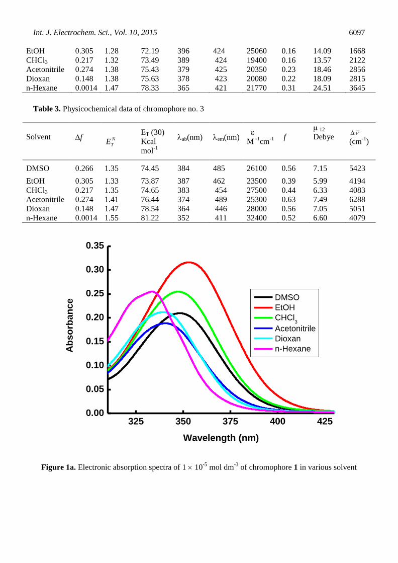
Table 3. Physicochemical data of chromophore no. 3
Solvent
f
N
TE
ET (30)
Kcal
mol-1
ab(nm)
em(nm)
ε
M -1
cm-1
f
μ 12
Debye
(cm-1
)
DMSO 0.266 1.35 74.45 384 485 26100 0.56 7.15 5423
EtOH 0.305 1.33 73.87 387 462 23500 0.39 5.99 4194
CHCl3 0.217 1.35 74.65 383 454 27500 0.44 6.33 4083
Acetonitrile 0.274 1.41 76.44 374 489 25300 0.63 7.49 6288
Dioxan 0.148 1.47 78.54 364 446 28000 0.56 7.05 5051
n-Hexane 0.0014 1.55 81.22 352 411 32400 0.52 6.60 4079
325 350 375 400 4250.00
0.05
0.10
0.15
0.20
0.25
0.30
0.35
Ab
so
rban
ce
Wavelength (nm)
DMSO
EtOH
CHCl3
Acetonitrile
Dioxan
n-Hexane
Figure 1a. Electronic absorption spectra of 1 10
-5 mol dm
-3 of chromophore 1 in various solvent

Int. J. Electrochem. Sci., Vol. 10, 2015
6098
400 450 500 550 6000
5
10
15
20
Em
issio
n I
nte
nsit
y (
arb
. u
nit
s)
Wavelength (nm)
DMSO
EtOH
CHCl3
Acetonitrile
Dioxane
n-Hexane
Figure 1b. Emission spectra of 1 10
-5 mol dm
-3 of chromophore 2 in various solvent
325 350 375 400 425 450 4750.00
0.05
0.10
0.15
0.20
0.25
0.30
Ab
so
rban
ce
Wavelength (nm)
DMSO
EtOH
CHCl3
Acetonitrile
Dioxan
n-Hexane
Figure 2a. Electronic absorption spectra of 1 10-5
mol dm-3
of chromophore 3 in various solvent

Int. J. Electrochem. Sci., Vol. 10, 2015
6099
400 425 450 475 500 525 550 5750
5
10
15
20
25
30
Em
issio
n I
nte
nsit
y (
arb
. u
nit
s)
Wavelength (nm)
DMSO
EtOH
CHCl3
Acetonitrile
Dioxan
n-Hexane
Figure 2b. Emission spectra of 1 10-5
mol dm-3
of chromophore 2 in various solvent
325 350 375 400 425 4500.00
0.05
0.10
0.15
0.20
0.25
0.30
0.35
Ab
so
rban
ce
Wavelength (nm)
DMSO
EtOH
CHCl3
Acetonitrile
Dioxan
n-Hexane
Figure 3a. Electronic absorption spectra of 1 10
-5 mol dm
-3 of chromophore 3 in various solvent
Int. J. Electrochem. Sci., Vol. 10, 2015
6100
350 400 450 500 550 600 6500
50
100
150
200
250
300
350
Em
issio
n I
nte
nsit
y (
arb
. u
nit
s)
Wavelength (nm)
DMSO
EtOH
CHCl3
Acetonitrile
Dioxan
n-Hexane
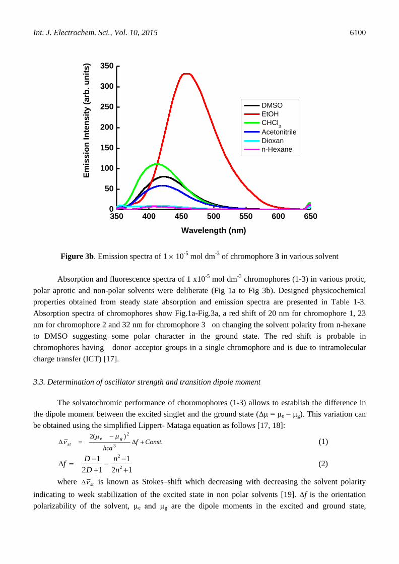
Figure 3b. Emission spectra of 1 10
-5 mol dm
-3 of chromophore 3 in various solvent
Absorption and fluorescence spectra of 1 x10⁻⁵
mol dm-3
chromophores (1-3) in various protic,
polar aprotic and non-polar solvents were deliberate (Fig 1a to Fig 3b). Designed physicochemical
properties obtained from steady state absorption and emission spectra are presented in Table 1-3.
Absorption spectra of chromophores show Fig.1a-Fig.3a, a red shift of 20 nm for chromophore 1, 23
nm for chromophore 2 and 32 nm for chromophore 3 on changing the solvent polarity from n-hexane
to DMSO suggesting some polar character in the ground state. The red shift is probable in
chromophores having donor–acceptor groups in a single chromophore and is due to intramolecular
charge transfer (ICT) [17].
3.3. Determination of oscillator strength and transition dipole moment
The solvatochromic performance of choromophores (1-3) allows to establish the difference in
the dipole moment between the excited singlet and the ground state (∆μ = μe – μg). This variation can
be obtained using the simplified Lippert- Mataga equation as follows [17, 18]:
.)(2
3
2
Constfhca
ge
st
(1)
12
1
12
12
2
n
n
D
Df (2)
where st is known as Stokes–shift which decreasing with decreasing the solvent polarity
indicating to week stabilization of the excited state in non polar solvents [19]. ∆f is the orientation
polarizability of the solvent, µe and µg are the dipole moments in the excited and ground state,
Int. J. Electrochem. Sci., Vol. 10, 2015
6101
respectively which measures both electron mobility and dipole moment of the solvent molecule. c is
the speed of light in vacuum, a is the Onsager cavity radius and h is Planck's constant, n and ε are the
refractive index and dielectric constant of the solvent for equation 2 respectively. The Onsager cavity
radius was chosen to be 4.2 Å because this value is comparable to the radius of a typical aromatic
fluorophore [20].
ss is the Stokes shifts of the chromophores (1-3) in different solvents were deliberate, as
shown in Table 1-3, using the following the equation [17]:
emabss (3)
where ss is the difference between λmax of the ab and me indicate the wavenumbers of
absorption and emission maxima (cm−1
) respectively.
The change in dipole moments ( ) between the excited singlet and ground state were
calculated from the slop of plot of Stokes shifts ( ss ) and orientation polarizability of the solvent
(f) as 1.54, -5.95 and 1.28 debye for chromophore 1, 2 and 3 respectively, positive value for
chromophore 1 & 3 indicating that the excited sate is more polor than the ground state and negative
value for chromophore 2 indicating that the ground sate is more polor than the excited state.
The change in transition dipole moments ( 12
) between the excited singlet and ground state
of chromophore 1-3 in various solvents were calculated as in Table 1-3, using the equation 4 [21].
max
7
2
121072.4 E
f
(4)
where Emax is the maximum energy of absorption in cm-1
and f is the oscillator strength.
The oscillator strength (f), can be calculated using the following equation:
df )(1032.4 9 (5)
where represents the numerical value of wavenumber (cm−1
) and ε is the extinction
coefficient (Lmol−1
cm−1
). Oscillator strength values of chromophores (1-3) in various solvents were
calculated from the equation no. 5 and reported in Table 1-3, [22].
3.4. Fluorescence polarity study of chromophores (1-3)
The emission spectrum of chromophores correlates with increasing polarity of the solvent (Fig.
1b- Fig. 3b). It is apparent that the emission maxima undergo a red shift of 31 nm for chromophore 1,
16 nm for chromophore 2 and 74 nm for chromophores 3 on increasing the polarity from n-Hexane to
DMSO, suggesting the emission state is more polar than the ground state [17].
ET (30) and N
TE is the empirical Dimroth polarity parameter of chromophores (1-3) was also
premeditated according to the following equation [23].

Int. J. Electrochem. Sci., Vol. 10, 2015
6102
4.32
7.30)(
solventEE TN
T (6)
max
Tλ
28591(solvent)E (7)
where maxλ corresponds to the peak wavelength (nm) in the red region of the intramolecular
charge transfer absorption of the bitain dye. All the choromophores have bathochromic when solvent
polarity increase from n-hexane to DMSO indicates that the polarity of chromophores and
photoinduced intramolecular charge transfer (ICT) occurs in the singlet excited state, therefore
increasing the excitation.
3.5. Electrochemical properties of chromophores 1-3
-1.0 -0.5 0.0 0.5 1.0
-0.0001
0.0000
0.0001
0.0002
0.0003
0.0004
0.0005
C
urr
en
t / 1
e-5 A
Potential / V
(1)
Figure 4. Cyclic voltammogram of chromophore 1 in acetonitrile at scan rate of 2V/s
-1.0 -0.5 0.0 0.5 1.0
-0.00012
-0.00006
0.00000
0.00006
0.00012
0.00018
0.00024
0.00030
Potential / V
Cu
rre
nt / 1
e-5 A
(2)
Figure 5. Cyclic voltammogram of chromophore 2 in acetonitrile at scan rate of 2V/s
Int. J. Electrochem. Sci., Vol. 10, 2015
6103
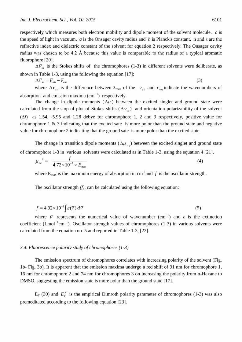
-1.0 -0.5 0.0 0.5 1.0
-0.0001
0.0000
0.0001
0.0002
0.0003
0.0004
Cu
rre
nt / 1
e-5 A
Potential / V
(3)
Figure 6. Cyclic voltammogram of chromophore 3 in acetonitrile at scan rate of 2V/s
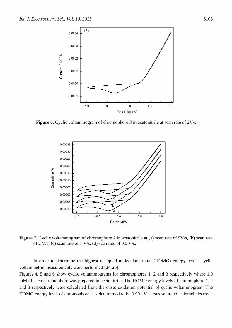
-1.0 -0.5 0.0 0.5 1.0
-0.00010
-0.00005
0.00000
0.00005
0.00010
0.00015
0.00020
0.00025
0.00030
0.00035
Cu
rre
nt/1
e-5A
Potential/V
a
b
c
d
Figure 7. Cyclic voltammogram of chromophore 2 in acetonitrile at (a) scan rate of 5V/s, (b) scan rate
of 2 V/s, (c) scan rate of 1 V/s, (d) scan rate of 0.5 V/s.
In order to determine the highest occupied molecular orbital (HOMO) energy levels, cyclic
voltammetric measurements were performed [24-26].
Figures 4, 5 and 6 show cyclic voltammograms for chromophores 1, 2 and 3 respectively where 1.0
mM of each chromophore was prepared in acetonitrile. The HOMO energy levels of chromophore 1, 2
and 3 respectively were calculated front the onset oxidation potential of cyclic voltammogram. The
HOMO energy level of chromophore 1 is determined to be 0.991 V versus saturated calomel electrode

Int. J. Electrochem. Sci., Vol. 10, 2015
6104
(SCE) (-5.76 eV versus vacuum), for chromophore 2 lies at 0.982 V versus SCE (-5.63 eV versus
vacuum) and for chromophore 3 lies at 0.962 V versus SCE (-5.57 eV versus vacuum). Based on the
offset absorption spectra and HOMO energy, lowest unoccupied molecular orbital (LUMO) energy
levels are determined to be -2.853 eV, -2.780 eV and -2.750 eV for ch 1, 2 and 3, respectively.
A comparative study of cyclic voltammograms for chromophore 2 in acetonitrile at different scan rates
is shown in fig. 7. Energetic data for chromophores 1, 2 and 3, respectively are listed in Table 4.
Table 4. Electrochemical properties of the chromophores 1-3.
Compounds E 1/2 E HUMO (eV) E LUMO (eV) ΔE spect (eV)
1 0.991 -5.7613 -2.862 2.78
2 0.982 -5.6312 -2.793 2.81
3 0.962 -5.5713 -2.734 2.85
From cyclic voltammogram, It Seems the electrochemical behavior of chromophores 1, 2 and 3
follow EC mechanism.
4. CONCLUSION
Three donor-acceptor chromophores were synthesized by the reaction appropriate aldehyde and
6-methoxy-1,2,3,4-tetrahydro-naphthalin-1-one by Knoevenagel condensation. Photophysical
properties including, oscillator strength, extenction coefficient, transition dipole moment and stokes
shift were investigated in order to investigate the physicochemical behaviors of synthesized
chromophores on the basis of the polarity of solvent. The absorption spectra of chromophores exhibit
an intramolecular charge transfer band; which showed a positive solavotochromism in different
solvents. The emission spectra of the chromophores also reveal the intramolecular charge transfer band
character. These findings confirm that there is a significant electron transfer between the donating
moiety and the accepting fragment through the π conjugated. The HOMO energy levels of
chromophores 1, 2 and 3 respectively were calculated front the onset oxidation potential of cyclic
voltammogram.
ACKNOWLEDGEMENTS
This Project was funded by the King Abdulaziz City for Science and Technology (KACST) through
National Science, Technology and Innovation Plan (NSTIP) under grant number 8-ENE198-3. The
authors, therefore, acknowledge with thanks KACST for support for Scientific Research. Also, the
authors are thankful to the Deanship of Scientific Research (DSR), King Abdulaziz University for their
technical support.
References
1. A. M. Asiri, S. A. Khan, Meter. Lett., 65 (2011) 1749.
Int. J. Electrochem. Sci., Vol. 10, 2015
6105
2. A. A. Asiri, S. A. Khan, S. I. Hallag, J. New Mater. Electrochem. Syst., 14 (2011) 251-258.
3. A. M. Asiri, S. A. Khan, M. S. Al-Amodi, K. A. Alamry, Bull. Kore. Chem. Soc., 33 (2012) 1900-
1906.
4. H. M. Marwani, A. M. Asiri, S. A Khan, J. Lumin., 136 (2013) 296-302.
5. A. M. Asiri, S. A Khan, S. A Al-daly, Spectrochim. Acta A. 95 (2012) 279-284.
6. X. Wang, F. Jin, W. Zhang, X. Tao, X. Duan, M. Jiang, Dyes & Pigments 88 (2011) 57-64.
7. C. Carlini, F. Ciardelli, D. Donati, F. Gurzoni, Polymer, 24 (1983) 599-606.
8. S. A. El-Daly, A. M. Asiri, K. A. Alamary, S. A. Khan, J. Lumin., 137 (2013) 6-14.
9. A M. Asiri, S. A Khan, H. M. Marwani, K. Sharma, J. Photochem. Photobiol., B 120 (2013) 82-
89.
10. A. M. Asiri, M. Mehmet, S. A. Khan, I. U. Khan, M. N. Arshad, Acta Cryst. (2009). E65o 1169.
11. H. M. Marwani, A. M. Asiri, S. A. Khan, Russ. J. Bioorg. Chem., 38 (2012) 5330538.
12. S. A. Khan, A. M. Asiri, S. H. Al-Thaqafy, H. M. Faidallah, S. A. El-Daly, Spectrochim. Acta A.
133 (2014) 141-148.
13. Y. Yang, H. Yao, F. Xi, E. Gao, J. Mol. Catal. A: Chem., 390 (2014) 198-205.
14. P. D. Torre, E. Osorio, J. H. Alzate-Morales, J. Caballero, J. Trilleras, L. Astudillo-Saavedra, I.
Brito, A. Cardenas, J. Quiroga, M. Gutierrez, Ultrason. Sonochem., 21( 2014) 1666-1674.
15. A. M. Asiri, H. M. Marwani, S. A. Khan, S. A. El-Daly, J Fluoresc., 23 (2013) 1271-1278.
16. A. M. Asiri, H. M. Marwani, S. A. Khan, Saudi Chem. Soc., 18 (2014) 392-397.
17. J. K. Dey, S. K. Dogra, Bull. Chem. Soc. Jpn. 64 (1991) 3142-3152.
18. W. E. Acree, D. C. Wilkins, S. A. Tucker, J. M. Griffin, J. R. Powell, J. Phys. Chem., 98 (1994)
2537-2544.
19. S. Kumar, V. C. Rao, R. C. Rastogi, Spectrochim. Acta A. 57 (2001) 41-47.
20. M. Ravi, A. Samanta, T. P. Radhakrishnan, J. Phys. Chem., 98 (1994) 9133-9136.
21. M. Ravi, T. Soujanya, A. Samanta, T. P. Radhakrishnan, J. Chem. Soc. Faraday Trans., 91
(1995) 2739- 2742.
22. N. J. Turro, Molecular photochemistry (frontiers in chemestry), 1st ed., W. A. Benjamin, Inc.,
Reading, MA, (1965) 286.
23. B. J. Coe, J. A. Harris, I. Asselberghs, K. Clays, G. Olbrechts, A. Persoons, J. T. Hupp, R. C.
Johnson, S. J. Coles, M. B. Hursthouse, K. Nakatani, Adv. Funct. Mater., 12 (2002) 110-116.
24. Q. Yu, S. Lu, M. Zhang, P. Wang, J. Phy. Chem. 113 (2009) 14559-14566.
25. R. A. Sheikh, I.A. Rahman, M. A. Malik, N. Luddin, S. M. Masudi, S. A. Al-Thabaiti, Int. J.
Electrochem. Sci., 8 (2013) 6972.
26. S. A. Khan, A. M. Asiri, K. A. Alamary, M. A. Malik, The Scientific World Journal, ID 592375
(2014) 1-9
© 2015 The Authors. Published by ESG (www.electrochemsci.org). This article is an open access
article distributed under the terms and conditions of the Creative Commons Attribution license
(http://creativecommons.org/licenses/by/4.0/).