Localization of potent photosensitizers in human tumor LOX by means of laser scanning microscopy
Upload
rr-researchCategory
view
0download
0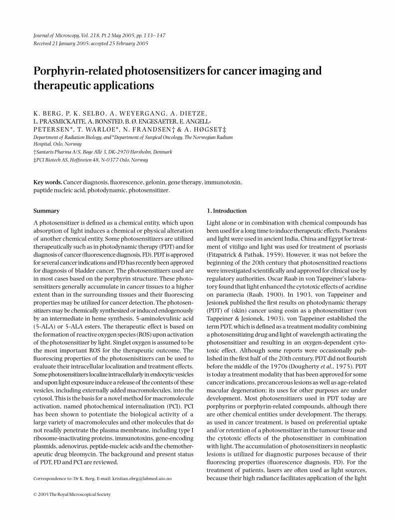
© 2005 The Royal Microscopical Society
Journal of Microscopy, Vol. 218, Pt 2 May 2005, pp. 133–147
Received 21 January 2005; accepted 25 February 2005
Blackwell Publishing, Ltd.
Porphyrin-related photosensitizers for cancer imaging and therapeutic applications
K . B E RG , P. K . S E L B O , A . W E Y E RG A N G , A . D I E T Z E , L. PRASMICKAITE, A. BONSTED, B. Ø. ENGESAETER, E. ANGELL-P E T E R S E N *, T. WA R L O E *, N. F R A N D S E N † & A . H Ø G S E T ‡
Department of Radiation Biology, and
*
Department of Surgical Oncology, The Norwegian Radium Hospital, Oslo, Norway
†
Santaris Pharma A/S, Bøge Allé 3, DK-2970 Hørsholm, Denmark
‡
PCI Biotech AS, Hoffsveien 48, N-0377 Oslo, Norway
Key words.
Cancer diagnosis, fluorescence, gelonin, gene therapy, immunotoxin, paptide nucleic acid, photodynamic, photosensitizer.
Received 21 January 2005;
accepted 25 February 2005
Summary
A photosensitizer is defined as a chemical entity, which uponabsorption of light induces a chemical or physical alterationof another chemical entity. Some photosensitizers are utilizedtherapeutically such as in photodynamic therapy (PDT) and fordiagnosis of cancer (fluorescence diagnosis, FD). PDT is approvedfor several cancer indications and FD has recently been approvedfor diagnosis of bladder cancer. The photosensitizers used arein most cases based on the porphyrin structure. These photo-sensitizers generally accumulate in cancer tissues to a higherextent than in the surrounding tissues and their fluorescingproperties may be utilized for cancer detection. The photosen-sitizers may be chemically synthesized or induced endogenouslyby an intermediate in heme synthesis, 5-aminolevulinic acid(5-ALA) or 5-ALA esters. The therapeutic effect is based onthe formation of reactive oxygen species (ROS) upon activationof the photosensitizer by light. Singlet oxygen is assumed to bethe most important ROS for the therapeutic outcome. Thefluorescing properties of the photosenisitizers can be used toevaluate their intracellular localization and treatment effects.Some photosensitizers localize intracellularly in endocytic vesiclesand upon light exposure induce a release of the contents of thesevesicles, including externally added macromolecules, into thecytosol. This is the basis for a novel method for macromoleculeactivation, named photochemical internalization (PCI). PCIhas been shown to potentiate the biological activity of alarge variety of macromolecules and other molecules that donot readily penetrate the plasma membrane, including type Iribosome-inactivating proteins, immunotoxins, gene-encodingplasmids, adenovirus, peptide-nucleic acids and the chemother-apeutic drug bleomycin. The background and present statusof PDT, FD and PCI are reviewed.
1. Introduction
Light alone or in combination with chemical compounds hasbeen used for a long time to induce therapeutic effects. Psoralensand light were used in ancient India, China and Egypt for treat-ment of vitiligo and light was used for treatment of psoriasis(Fitzpatrick & Pathak, 1959). However, it was not before thebeginning of the 20th century that photosensitized reactionswere investigated scientifically and approved for clinical use byregulatory authorities. Oscar Raab in von Tappeiner’s labora-tory found that light enhanced the cytotoxic effects of acridineon paramecia (Raab, 1900). In 1903, von Tappeiner andJesionek published the first results on photodynamic therapy(PDT) of (skin) cancer using eosin as a photosensitizer (vonTappeiner & Jesionek, 1903). von Tappeiner established theterm PDT, which is defined as a treatment modality combininga photosensitizing drug and light of wavelength activating thephotosensitizer and resulting in an oxygen-dependent cyto-toxic effect. Although some reports were occasionally pub-lished in the first half of the 20th century, PDT did not flourishbefore the middle of the 1970s (Dougherty
et al
., 1975). PDTis today a treatment modality that has been approved for somecancer indications, precancerous lesions as well as age-relatedmacular degeneration; its uses for other purposes are underdevelopment. Most photosensitizers used in PDT today areporphyrins or porphyrin-related compounds, although thereare other chemical entities under development. The therapy,as used in cancer treatment, is based on preferential uptakeand/or retention of a photosensitizer in the tumour tissue andthe cytotoxic effects of the photosensitizer in combinationwith light. The accumulation of photosensitizers in neoplasticlesions is utilized for diagnostic purposes because of theirfluorescing properties (fluorescence diagnosis, FD). For thetreatment of patients, lasers are often used as light sources,because their high radiance facilitates application of the light
Correspondence to: Dr K. Berg. E-mail: [email protected]

134
K . B E RG
E T A L .
© 2005 The Royal Microscopical Society,
Journal of Microscopy
,
218
, 133–147
through thin optical fibres (Brancaleon & Moseley, 2002).This is, however, not a prerequisite for the photodynamicresponse and non-laser sources are often used on skin lesions.
2. Photosensitization
2.1. Photochemistry
The photocytotoxic effects, which are utilized in PDT, areinitiated by the absorption of light by a photosensitizer. A moleculethat has absorbed a light quantum is excited to the first orhigher excited states (Fig. 1). The higher excited states aredissipated very rapidly (picoseconds) down to the first excitedsinglet state with a lifetime of the order of nanoseconds. This
excited state can be deactivated by release of heat (non-radiativedecay), emitted as fluorescence of a wavelength equal to or longerthan that of the excitation light (utilized for cancer diagnosis)or undergo intersystem crossing (ISC). Photochemistry mayoccur from the first excited singlet state, but usually with lowprobability owing to the short life-time of this state. Isomerizationreactions however, may in some cases be of importance. ISC isusually forbidden, but macrocyclic molecules with conjugateddouble bond systems (
π
-electron system) are exceptions andallow for ISC to long-lived (microseconds to milliseconds)triplet states. The triplet state may react in two ways, either bya type I mechanism involving electron or hydrogen atomtransfer from one molecule to the other or by a type II mecha-nism involving energy transfer to molecular oxygen (Fig. 2).
Fig. 1. Jablonski diagram showing the electronic statesfollowing absorption of light by a photosensitizer. Theblack arrow indicates the excitation of the photosensitizerto the excited singlet states (1P1). The other arrows indicatealternative pathways for dissipation of the energy inthe excited state. ISC, intersystem crossing; 3P1, thesensitizer in the triplet state. See text for details.
Fig. 2. Photochemical reaction pathways from thetriplet state of a photosensitizer. See text for details.

P O R P H Y R I N - R E L AT E D P H O TO S E N S I T I Z E R S F O R CA N C E R I M AG I N G
135
© 2005 The Royal Microscopical Society,
Journal of Microscopy
,
218
, 133–147
Both mechanisms may occur simultaneously and their relativeimportance depends on the surroundings and the nature ofthe substrate molecules. A significant amount of indirectevidence, i.e. by means of quenchers of
1
O
2
and degradationof photosensitizers, and some direct evidence exists for theimportance of
1
O
2
in the photoinduced processes of PDT(Moan
et al
., 1979; Sonoda
et al
., 1987; Niedre
et al
., 2002,2003). Singlet oxygen causes mainly membrane damage byoxidizing amino acids (tryptophan, cysteine, histidine,methionine and phenylalanine), unsaturated fatty acidsand cholesterol. Additionally, guanine may also be oxidized bysinglet oxygen.
2.2. Photosensitizers
A photosensitizer is defined as a chemical entity, which uponabsorption of light induces a chemical or physical alteration ofanother chemical entity. A good photosensitizer should absorbphotons efficiently (i.e. high extinction coefficient), have ahigh quantum yield of triplet formation and the triplet stateshould be long lived in order to have time to react with neigh-bouring target molecules. Most compounds that form tripletstates that are able to produce reactive oxygen species (ROS)have tricyclic, heterocyclic or porphyrin-like ring structureswith conjugated double bonds (
π
-electron system).
2.2.1. Chemically synthesized porphyrin-based photosensitizers.
Most photosensitizers and all approved photosensitizers (withthe exception of methylene blue) used in PDT are based on orrelated to the tetrapyrrole macrocycle (Dolmans
et al
., 2003).Porphyrins consist of four pyrrole subunits linked together by
four methine bridges (Fig. 3). This tetrapyrrole ring structureis named porphin and derivatives of porphins are namedporphyrins. Tetrapyrroles are naturally occurring pigments,which are used in many biological processes. These tetrapyrrolesare not able to induce any photochemical or photobiophysicalreactions in other compounds or are rapidly quenched in theirnormal surroundings, e.g. chlorophyll. In all these tetrapyrrolesa metal ion is coordinated in the middle of the compounds. Thepresence of a coordinated metal ion and its electronic propertiesare of importance for the photocytotoxic potential of porphyrins.By removal of the metal tetrapyrroles become efficient pho-tosensitizers as well as acquiring fluorescing properties, e.g. byremoval of iron from heme. Most efficient porphyrin-basedphotosensitizers therefore lack coordinated metal ions. Coor-dinated metal ions increase the probability for non-radiativedecay of the triplet state, paramagnetic metals such as Fe
3+
being much more efficient than diamagnetic metals such asAl
3+
and Mg
2+
. Several metallophotosensitizers have, how-ever, been developed for clinical purposes. Although in mostcases they have lower quantum yields for cell inactivation, theyhave other properties, e.g. improved solubility and stability,which makes them interesting as therapeutic substances. Themetals used include Zn, Pd, Sn, Ru, Pt, Lu, Gd and Al.
Porphyrins and porphyrin-related dyes used in PDT havesubstituents in the peripheral positions of the pyrrole rings, onthe four methine carbons (
meso
-positions) and/or coordinatedmetals. These derivatives are synthesized to influence water/ lipidsolubility, amphiphilicity, pKa and stability of the compounds(MacDonald & Dougherty, 2001). These parameters determinethe biodistribution of the compounds, i.e. the intracellularlocalization, tissue distribution and pharmacokinetics.
Fig. 3. Basic structure of some photosensitizers.

136
K . B E RG
E T A L .
© 2005 The Royal Microscopical Society,
Journal of Microscopy
,
218
, 133–147
2.2.2. Endogenously synthesized photosensitizers.
5-Aminole-vulinic acid (5-ALA) is a prodrug for application in PDT andFD that has attracted great attention the last 15 years (Peng
et al
., 1997a, b; Berg, 2001; Kormeili
et al
., 2004; Lopez
et al
.,2004). This treatment is based on heme synthesis. A mainregulatory step in the heme pathway is linked to 5-ALAsynthase (ALAS) activity, which induces 5-ALA formationfrom glycine and succinyl-CoA (Fig. 4). Heme can inhibit theenzyme directly as well as the transcription, translation andtransport of the protein into mitochondria. Thus, treatmentof cells with 5-ALA overrides the regulatory step of the hemepathway and induces a high activity through the pathway.Treatment with 5-ALA has been shown to induce accumula-tion of porphyrins, mainly protoporphyrin IX (PpIX), whichsensitize cells to photoinactivation. Almost all the intermediates
in the heme pathway are porphyrinogens, which are notphotochemically active, but protoporphyrinogen is convertedto the photosensitizer PpIX by means of protoporphyrinogenoxidase or spontaneously when accumulated in 5-ALA-treatedcells. Esterification of the carboxyl group in 5-ALA may improvethe therapeutic as well as diagnostic effect of 5-ALA by, forexample, increasing the rate of cellular uptake and penetrationrate in tissue (Kormeili
et al
., 2004). Ferrochelatase incorporatesFe
2+
into PpIX and converts it into the photochemically inactiveporphyrin heme.
3. Utilization of photosensitizers in diagnosis
The photosensitizers used in PDT accumulate preferentially inneoplastic lesions as compared with the surrounding normal
Fig. 4. The heme biosynthetic pathway. The heme synthesis as shown in the figure occurs partly in the mitochondria as indicated on the left side of thefigure and partly in the cytosol. Externally added 5-ALA is passing the plasma membrane through amino acid transporters and enters the heme synthesispathway leading to temporary accumulation of protoporphyrin IX.

P O R P H Y R I N - R E L AT E D P H O TO S E N S I T I Z E R S F O R CA N C E R I M AG I N G
137
© 2005 The Royal Microscopical Society,
Journal of Microscopy
,
218
, 133–147
tissue. The ratio of tumour tissue to normal surroundingtissue is usually about 2–3 : 1 for chemically synthesizedphotosensitizers (Fig. 5), but might in some cases (e.g. braintumours) be much higher (Zimmermann
et al
., 2001; Yang
et al
., 2003). The mechanisms involved in this preferentialdistribution are not fully understood, but several properties ofthe tumour tissue seem to be of importance. The high numberof LDL receptors, low interstitial pH, reduced lymphatic drain-age, and large interstitial space with high amounts of collagen
and leaky vasculature all tend to favour the accumulation ofphotosensitizers in tumours tissues (Kessel, 1986; Henderson& Bellnier, 1989; Peng
et al
., 1991). The selectivity may be fur-ther influenced and improved by incorporation of the photo-sensitizers into delivery vehicles such as liposomes (Bourre
et al
., 2003), antibodies (Duska
et al
., 1999; van Dongen
et al
.,2004) and chemically synthesized polymers (Shiah
et al
., 2000).The systemic use of chemically synthesized photosensitizers
for diagnostic purposes may be hampered by prolonged skinand eye photosensitivity and is still not approved for clinicalutilization. In contrast, endogenous porphyrins accumulatedafter treatment with 5-ALA and 5-ALA ester derivatives havebeen applied locally (e.g. skin and bladder) with no skin photo-sensitization and short half-life on the treatment area (< 24 h,Fig. 6) (Ericson
et al
., 2003; Schmidbauer
et al
., 2004). The spe-cificity is high [tumour-to-skin ratio approximately 10 : 1 basedon fluorescence (Fritsch
et al
., 1998; E. Angell-Petersen
et al.
,unpublished data)] for reasons that are not fully clear, butinvolves changes in the activities of the enzymes in the hemesynthetic pathway and increased penetration through sur-face layers covering tumours (e.g. reduced stratum corneum)(Stefanidou
et al
., 2000; Berg, 2001; Peng
et al
., 2001). The use of5-ALA hexyl ester for detection of bladder cancer (papillomaand carcinoma-in-situ) has recently been approved in Sweden(Table 1) and the method may be utilized for diagnosis of otherindications, such as in the gastrointestinal tractus (Endlicher
et al
., 2001; Mayinger
et al
., 2001; Claydon & Ackroyd, 2004).Systemic delivery of 5-ALA has been evaluated for fluorescence-guided resection of brain cancers with promising results(Stummer
et al
., 2003; Bogaards
et al
., 2004). Local adminis-tration of chemically synthesized photosensitizers, such as thenon-porphyrin-type hypericin, has also shown promising results(Joudi & Konety, 2004).
4. Utilization of photosensitizers in therapy
The first photosensitizer that was used and approved for PDT isa mixture of a large variety of porphyrins (Moan & Peng, 2003;
Fig. 5. Detection of photosensitizer distribution in a tumour-bearingmouse. Forty-eight hours after i.p. injection of the photosensitizerAlPcS2a (10 mg kg−1) the mouse was investigated via an IVIS™ ImagingSystem 100. High fluorescence intensity from the photosensitizer appearsred and low intensity appears blue according to the scale shown. Highintensity was detected in the tumour region (WiDr human colorectaladenocarcinoma, see arrow).
Table 1. Photosensitizers approved for use in PDT and fluorescence diagnosis.
Sensitizer Trade name IndicationsActivation wavelength (nm)
HpD/porfymer sodium Photofrin Cervical dysplasia and cancer, Barrett’s oesophageous; endobroncheal, oesophageal, gastric and papillary bladder cancer
630
m-THPC Foscan Head and neck cancer 652BPD-MA Verteporfin Age-related macula degeneration 689SnEt2 Purlytin Age-related macula degeneration 664AlPcS (approved in Russia) Photosense Head and neck cancer 6705-ALA Levulan Actinic keratosis 6355-ALA-methyl ester Metvix Actinic keratosis, basal cell carcinoma 6355-ALA hexyl ester Hexvix Diagnosis of bladder cancer ∼400

138
K . B E RG
E T A L .
© 2005 The Royal Microscopical Society,
Journal of Microscopy
,
218
, 133–147
Brown
et al
., 2004). It was named Hematoporphyrin Deriva-tive (HpD) and later a somewhat purified version was developedand named Photofrin. The structure of the most active com-pounds in this mixture has been much debated and it hasbeen suggested that dihematoporphyrin ether or ester is themost efficient photosensitizer in this mixture (Fig. 7). This isfar from an ideal photosensitizer for use in PDT because of itscomposition and the difficulty involved in synthesizing repro-ducible batches (although this has been reduced as a problemcommercially), far from optimal spectral properties, longhalf-life in patients, and skin and eye photosensitivity for 4–6 weeks after injection. It is, however, approved for use in manyindications; a large number of patients have been treated withthis compound and it is still the most broadly used photosensi-tizer in the clinic.
The absorption spectrum of a porphyrin as shown in Fig. 8comprises of a Soret band and four Q-bands. Naturally occur-ring porphyrins, such as heme and other cytochromes, andmelanin are the main chromophores in tissues. If photoactiva-tion of photosensitizers located deep into a tissue is required,wavelengths above 600 nm are therefore usually necessary.However, the extinction coefficient of porphyrins is low at thiswavelength range (Sharman
et al
., 1999). New photosensitizershave therefore been developed with higher extinction coeffi-cient in this wavelength range and with peak absorption athigher wavelengths (Figs 3 and 8). There is an upper limit,
however, to how far into the infrared region a photosensitizercan absorb light and still induce singlet oxygen. This upperlimit is set by the energy required to excite O
2
, which is 0.96 eV(22 kcal mol
−
1
). The upper wavelength limit is therefore 850–900 nm depending on the energetic level of the triplet state ofthe photosensitizer. The therapeutic window is therefore agreedto be 600–800 nm for
in vivo
treatment, limited by haemoglobinand melanin light absorption in the low-wavelength region.
The therapeutic effect of PDT may be due to direct cell killingand vascular damage, and there are also indications for theimportance of immune responses (Dolmans
et al
., 2003).The importance of these pathways for therapeutic effectsdepends on the photosensitizer and its formulation, the meansof administration and the time between administration of thephotosensitizer and exposure to light.
Shortly after intravenous injection the photosensitizers aremainly located in the vasculature (Fig. 9). PDT may inducevasoconstriction during or immediately after treatment, andthrombus formation has been observed a few hours aftertreatment. These effects will lead to vascular shutdown andtissue hypoxia and anoxia, causing delayed tumour growth.Targeting the vasculature is utilized in the treatment of age-related macular degeneration (AMD), in which the photosen-sitizer is injected systemically shortly before light exposure ofthe macula (van den Bergh, 2001). The reduced vision qualityobserved by AMD patients is due to new vessels formed in
Fig. 6. Fluorescence-based detection of cancer. Multifocal nodular basal cell carcinoma of the skin visualized by application of a cream containing 5-ALAmethyl ester for 3 h followed by inspection during exposure to blue light (A, upper figure) or white light (A, lower figure). The red fluorescence is due toPpIX formation in the cancer cells. Similarly, bladder cancer may be visualized after 1 h instillation with 5-ALA hexyl ester (B) followed by inspectionduring exposure to blue light (upper panel) or white light (lower panel). The figures show a carcinoma-in-situ (left figures) and papillary tumours (rightfigures). The pictures of the bladders were kindly provided by PhotoCure ASA and taken by Dr Dirk Zaak, Grosshadern Hospital, Münich.

P O R P H Y R I N - R E L AT E D P H O TO S E N S I T I Z E R S F O R CA N C E R I M AG I N G
139
© 2005 The Royal Microscopical Society,
Journal of Microscopy
,
218
, 133–147
the macular area. PDT performed with the photosensitizerlocated in the blood induces damage to the neovasculature,resulting in a slower development of the disease. Severalclinical protocols are now also initiated for treatment of cancers,
such as prostate cancer, where a similar photosensitizer–lighttime interval as for AMD is performed in an attempt to shutdown the tumour blood supply (Huang
et al
., 2004; Weersink
et al
., 2004).
Fig. 7. Photosensitizers presently approved for clinicalapplications. SnEt2, Tin etiopurpurin; mTHPC, tetra(meso-hydroxy) phenyl chlorin; BPD-MA, benzopor-phyrin derivative monoacid ring A; 5-ALA, 5-aminolevulinic acid; Photofrin is a mixture of severalcompounds as described in the text; PpIX, protopor-phyrin IX (other photoactive porphyrins may also beformed). See Table 1 for further details.
Fig. 8. Absorption spectra of photosensitizers. A fullspectrum is shown for a porphyrin-type photosensitizerand additionally the location of the main peaks forphotosensitizers developed for PDT are shown on thefigure (not in scale).

140
K . B E RG
E T A L .
© 2005 The Royal Microscopical Society,
Journal of Microscopy
,
218
, 133–147
The pharmacokinetics of photosensitizers usually fit acceptablyinto two- or three-compartment models (Derendorf & Hochhaus,1995), but with large sensitizer-dependent variation in bloodhalf-life. The tissue uptake and consequently skin sensitivityvary widely among the photosensitizers. After the redistribu-tion from the blood the photosensitizer may localize in theparenchyma cells and the stroma depending on the structuralproperties of the photosensitizer, and a mixture of vascularshutdown due to targeting the endothelial lining and directtumour cell damage induce tumour eradication. The photo-sensitizers approved for therapeutic purposes and their struct-ures are shown in Fig. 7 and Table 1.
The use of 5-ALA and 5-ALA esters for endogenous forma-tion of photosensitizers has attracted great attention owing tothe short photosensitivity period after application of 5-ALA/5-ALA esters. These compounds are applied locally, e.g. topicallyin a cream or in solution for bladder instillation, or systemically(applies only for 5-ALA thus far). Shortly after application(typically 1–3 h) sufficient amounts of the photosensitizers(PpIX and other photoactive porphyrins) have accumulatedin the neoplastic lesions to allow for diagnosis and therapy(Table 1). More detailed information of the clinical status ofPDT can be found in Brown
et al.
(2004).
Fig. 9. Vital microscopic analysis of tissue localization of the photosensitizeraluminium phthalocyanine disulphonate (AlPcS2a). A transparent dorsalchamber based on viable tissue growing in between two glass plates inathymic mice was used for visual inspection (see Brustad et al., 1997, forfurther details). AlPcS2a (10 mg kg−1) was injected i.p. 4 h (upper panels)or 24 h (lower panels) before visual inspection. The tissue was evaluatedby means of phase contrast white light (left panel) or fluorescence (rightpanel, λex = 590–650 nm, λem > 660 nm). The tissues were not exposedto therapeutic doses of light and the fluorescence reflects the localizationof the photosensitizer in tissue not exposed to light.
Fig. 10. Proposed localization of sulphonated photosensitizers in endocytic vesicles. The figure shows a schematic drawing of an endocytic vesicle anddrawings of the main location of a tetrasulphonated (tetra(4-sulphonatophenyl)porphine, TPPS4) and an amphiphilic disulphonated photosensitizer(tetraphenylporphine disulphonate (TPPS2a) and aluminium phthalocyanine disulphonate (AlPcS2a)) (not to scale).

P O R P H Y R I N - R E L AT E D P H O TO S E N S I T I Z E R S F O R CA N C E R I M AG I N G
141
© 2005 The Royal Microscopical Society,
Journal of Microscopy
,
218
, 133–147
5. Photochemical internalization: a novel technology for light-directed delivery of macromolecules
The utilization of macromolecules in therapy of cancer andother diseases is becoming increasingly relevant (Sullenger &Gilboa, 2002; Everts & Curiel, 2004; Selkirk, 2004; Wels
et al
.,2004). Recent advances in molecular biology and biotech-nology have made it possible to improve targeting and to designcytotoxic agents or DNA complexes for clinical application(Curiel, 1999; Wels
et al
., 2004). To achieve the expectedbiological effect of these macromolecules internalization tothe cell cytosol is in many cases crucial. Although new deliverysystems have improved the cellular uptake of macromolecules,tissue penetration, cellular uptake and efficient transfer of themolecules into the cytosol of the target cell are still fundamen-tal obstacles. At the intracellular level, the most fundamentalobstruction to cytoplasmic or nuclear targeting of therapeuticmolecules is the membrane barrier of the endocytic vesicles(Cho
et al
., 2003).
Photochemical internalization (PCI) is a novel technology,which may improve the therapeutic effect of macromoleculesand other molecules that accumulate in endocytic vesicles(Berg
et al
., 1999; Hogset
et al
., 2004). PCI utilizes photo-sensitizers with physicochemical properties that lead toaccumulation in endocytic vesicles (Berg
et al
., 1990). Uponexposure to light the contents of vesicles containing suchphotosensitizers are released into the cytosol in a functionallyactive form (Berg
et al
., 1991; Berg & Moan, 1994). Examplesof photosensitizers and tentative localization in endocyticvesicles are shown in Fig. 10. The most efficient photosensitizersfor use in PCI have an amphiphilic structure with a hydrophilicpart inhibiting penetration through cellular membranes, asillustrated in Figs 10 and 11. Upon exposure to light, ROS, ofwhich singlet oxygen is assumed to be dominant, are formed.Singlet oxygen has a short range of action (< 20 nm; Moan& Berg, 1991) and leads to release of the contents of the endo-cytic vesicles (Figs 11 and 12). Although the exact structureof the damaged vesicles has not yet been revealed, results
Fig. 11. In photochemical internalization (PCI), photosensitizer molecules and macromolecules accumulate in endocytic vesicles, as indicated on thefigure. The most efficient photosensitizers for application in PCI are mainly located in the membranes of the vesicles. Upon illumination of thephotosensitizer, reactive oxygen species, mainly singlet oxygen, are formed, constituents of the membranes are destroyed (as indicated on the figure) andpassage of macromolecules to the cytosol is allowed.

142
K . B E RG
E T A L .
© 2005 The Royal Microscopical Society,
Journal of Microscopy
,
218
, 133–147
with dextran and viral particles indicate that relatively largeparticles can escape the vesicles after PCI. The release ofmacromolecules from endocytic vesicles is exemplified inFig. 12 by release of peptide nucleic acids and the possibleclinical utilization exemplified with the activation of theprotein toxin saporin (Fig. 13). The various macromoleculesthat have been shown to increase their biological function byPCI are described in Table 2. PCI of therapeutic macromole-cules has been shown to increase the necrotic depth of thetreatment and to increase the fraction of complete remissionsin animal models (Selbo
et al
., 2001b; A. Dietze
et al
., unpub-lished data).
In cancer therapy as well in the treatment of many otherdiseases there is a need for improved specificity of the treatment.PCI relies on the use of photosensitizers, which accumulate
Fig. 12. PCI-induced endosomal release. (A) PCI-induced release of the photosensitizer AlPcS2a in vitro. Fluorescence photomicrographs of AlPcS2a inKLN205 lung cancer cells in culture. The cells were incubated with 5 µg mL−1 AlPcS2a for 18 h. After 4 h chase in drug-free medium the cells were exposedto 15 s microscopy light (mercury lamp), which photochemically induced a release of AlPcS2a from the endocytic vesicles, and pictures taken 30 s afterphotochemical activation. (B) PCI-induced release of the photosensitizer AlPcS2a in vivo. Fluorescence photomicrographs of AlPcS2a in granular tissue bymeans of vital microscopy (see Fig. 9). The photosensitizer AlPcS2a (10 mg kg−1) was injected i.p. 96 h before analysis. The granular intracellularlocalization of AlPcS2a is visualized by fluorescence microscopy before (middle) and after photochemical activation and relocalization (right) of AlPcS2a.(C) PCI-induced relocalization of a peptide nucleic acid (PNA). HEPG2 hepatoma cells were incubated for 18 h with 5 µg mL−1 AlPcS2a. After a further 4 hof incubation with a fluorescein-labelled PNA (5 µm) linked to N-acetylgalactosamine (GalNAc) for receptor-mediated cellular uptake the cells werephotographed before and 1 h after the exposure to 15 min of red light (1.5 mW cm−2). Green fluorescence represents PNA and red fluorescence AlPcS2a.
Fig. 13.
PCI of epidermal growth factor-targeted saporin. NuTu19 cellswere treated with TPPS
2a
and 5 p
m
of a complex of epidermal growthfactor (EGF) and saporin linked together by a non-covalent biotin-streptavidin binding. The cells were exposed to light and cell survivalmeasured by the MTT assay (Hogset
et al
., 2000).

P O R P H Y R I N - R E L AT E D P H O TO S E N S I T I Z E R S F O R CA N C E R I M AG I N G
143
© 2005 The Royal Microscopical Society,
Journal of Microscopy
,
218
, 133–147
preferentially in neoplastic lesions and induce a cytotoxic reac-tion only in the light-exposed areas. A further improved treat-ment specificity may, however, be established by cell-specifictargeting of the photosensitizer or the therapeutic macro-molecule (Figs 13 and 14). Several targeting methods are underdevelopment, such as the use of antibodies and derivativesthereof, receptor ligands, peptides, tissue-specific promoters andreplication. Some targeting principles have been evaluatedin combinations with PCI, such as immunotoxins (Selbo
et al
.,2000b), transferrin- and epidermal growth factor-conjugatedvectors (Prasmickaite
et al
., 2000; Kloeckner
et al
., 2004) andRGD sequences for integrin-targeting (Engesæter
et al., in press).These results indicate that PCI can be used in combinationwith other targeting principles to further improve therapeuticspecificity.
6. Concluding remarks
Chemically and endogenously synthesized photosensitizershave properties that may be utilized both experimentally andclinically. Use of the preferential accumulation of photosensi-tizers in neoplastic lesions is already approved for detection ofbladder cancer and is under development for detection ofother cancer diseases. Therapeutically, thousands of patientshave been treated over the last 25 years. In some specialities,such as dermatological oncology and ophthalmology, PDT iswidely used but in other specialities it still remains marginal.The reasons for this may be many. The use of photosensitizersfor cancer detection and treatment is a new approach thatrequires new and costly equipment. This may cause clinicaland hospital resistance. One bottleneck has been the need for
Table 2. Documented PCI-enhanced biological effect of macromolecules. The table presents all macromolecules where PCI has been shown to enhance its biological function.
Macromolecules Photosensitizer References
1. Proteinsgelonin TPPS4, TPPS2a, AlPCS2a,
Chlorine e6(Berg et al., 1999; Selbo et al., 2000a;Dietze et al., 2003)
saporin TPPS2a (Berg et al., 1999)horseradish peroxidase TPPS2a (Berg et al., 1999)
Targeted proteinsMOC-31-gelonin TPPS2a, AlPcS2a, ALA-PPIX (Selbo et al., 2001a, b)
2. Genes (reporters and therapeutic-relevant)Virus mediated:AdCMV-lacZ, AdCMV-eGFP,AdCMV-luc TPPS2a (Dietze et al., 2003; Bonsted et al., 2004)AdCMV-lacZ AlPcS2a (Hogset et al., 2002)AAV TPPS2a (Bonsted et al., in press)Non-virus mediated:Polylysine (with reporter gene eGFP) TPPS2a, TPPS4, AlPcS2a (Prasmickaite et al., 2000, 2001; Dietze et al., 2003)Polyethyleneimine (PEI) (with reporter genes eGFP, luciferase and therapeutic gene HSV-thymidine kinase)
TPPS2a, AlPcS2a (Prasmickaite et al., 2000, 2004)
Cationic lipids (with reporter eGFP) AlPcS2a (Hellum et al., 2003)Targeted gene delivery
Epidermal growth factor (EGFR)-targeted TPPS2a, AlPcS2a (Kloeckner et al., 2004)PEI (with reporter genes eGFP, luciferase and therapeutic gene HSV-thymidine kinaseGlucosylated PEI with p53 gene TPPS2a (Ndoye et al., 2004a, b)PEI with PTEN gene TPPS2a (Maurice-Duelli et al., 2004)AdTRAIL TPPS2a unpublished
OligonucleotidesPeptide nucleic acids (PNA) AlPcS2a, TPPS2a (Folini et al., 2003)
3. ChemotherapeuticalsBleomycin TPPS2a, AlPcS2a unpublished
In vivo experiments gelonin
WiDr, THX, TAX-1, HUH-7, CT26, U87MG AlPCS2a (Selbo et al., 2001b)bleomycin
WiDr, TAX-1, CT-26 AlPCS2a unpublished
eGFP, enhanced green fluorescence protein; AdCMV, adenovirus with reporter gene regulated by the CMV promoter; AAV, adenoassociated virus.

144 K . B E RG E T A L .
© 2005 The Royal Microscopical Society, Journal of Microscopy, 218, 133–147
expensive (including running costs) and unreliable lasers. Therecent development of high-power diode lasers with emissionat relevant wavelengths has, however, been a major achievementin that they are easy to use, reliable and less expensive. Further-more, the treatment requires optimization of several parameterssuch as drug and light doses, drug–dose interval, fluences rate,activating wavelength and light applicators. These parametersand the need for efficient penetration of light through the targettissues have resolved in suboptimal therapeutic effectiveness.Combination treatments such as PCI may, however, improvethe effectiveness of photosensitizer-based therapies.
One of the major assumed limitations of PDT (and PCI) is itslack of therapeutic effect on distant metastasis. The possibilityof PDT as an anti-tumour vaccine as indicated by BarbaraHenderson and colleagues is therefore highly interesting(Gollnick et al., 2002). Similar documentation has beenreported by others (Canti et al., 1994) and indicates that PDTcould induce a systemic immune effect, although this has sofar not been documented in patients who have received PDT.Furthermore, macromolecular therapies may by themselvesbe more efficient towards micrometastasis than towards largesolid tumours (Schmidt et al., 1999). One may therefore spec-ulate that a combination of targeted macromolecules andphotodynamic treatment (e.g. PCI) may improve both thelocal control as well as the metastatic spread. However, it stillremains to be seen whether such combination treatments canattenuate the spread of metastatic tumours.
The potential of using photosensitizers for cancer diagnosishas been known for a long time, but has not until recentlybeen approved for any indications. This is an exciting utiliza-tion of photosensitizers and the approved use for diagnosis ofother indications is to be expected in the future.
Experimentally, photosensitizers have been evaluated formany purposes. Cationic photosensitizers in combination withlight seem efficient against both Gram-positive and Gram-negativebacteria (Demidova & Hamblin, 2004; Maisch et al., 2004).Photosensitizers may also be linked to targeting moieties forselective inactivation of biomolecules, e.g. to nucleotide sequencesfor inhibition of single gene expression (Dobrikov et al., 1997;Cream et al., 2002), tissue-specific activation of photosensitizingbeacons (Chen et al., 2004), and antibodies, growth factorsand peptides towards cell-specific receptors (Allen et al., 1999;Duska et al., 1999; van Dongen et al., 2004).
References
Allen, C.M., Sharman, W.M., La Madeleine, C., Weber, J.M., Langlois, R.,Ouellet, R. & van Lier, J.E. (1999) Photodynamic therapy: tumor target-ing with adenoviral proteins. Photochem. Photobiol. 70, 512–523.
Berg, K. (2001) Basic principles of 5-aminolevulinic-acid based photody-namic therapy. Photodynamic Therapy and Fluorescence Diagnosis inDermatology (ed. by P. Calzavara-Pinton, B. Ortel and R. M. Szeimies),pp. 115–162. Elsevier, Amsterdam.
Berg, K., Madslien, K., Bommer, J.C., Oftebro, R., Winkelman, J.W.& Moan, J. (1991) Light induced relocalization of sulfonated
Fig. 14. Illustration of how PCI can improve specificityand efficacy of the macromolecular and photodynamictherapies.

P O R P H Y R I N - R E L AT E D P H O TO S E N S I T I Z E R S F O R CA N C E R I M AG I N G 145
© 2005 The Royal Microscopical Society, Journal of Microscopy, 218, 133–147
meso-tetraphenylporphines in NHIK 3025 cells and effects of dosefractionation. Photochem. Photobiol. 53, 203–210.
Berg, K. & Moan, J. (1994) Lysosomes as photochemical targets. Int. J.Cancer, 59, 814–822.
Berg, K., Selbo, P.K., Prasmickaite, L., Tjelle, T.E., Sandvig, K., Moan, J.,Gaudernack, G., Fodstad, O., Kjolsrud, S., Anholt, H., Rodal, G.H.,Rodal, S.K. & Hogset, A. (1999) Photochemical internalization: a noveltechnology for delivery of macromolecules into cytosol. Cancer Res. 59,1180–1183.
Berg, K., Western, A., Bommer, J.C. & Moan, J. (1990) Intracellular local-ization of sulfonated meso-tetraphenylporphines in a human carci-noma cell line. Photochem. Photobiol. 52, 481–487.
van den Bergh, H. (2001) Photodynamic therapy of age-related maculardegeneration: history and principles. Semin. Ophthalmol. 16, 181–200.
Bogaards, A., Varma, A., Collens, S.P., Lin, A., Giles, A., Yang, V.X., Bilbao, J.M.,Lilge, L.D., Muller, P.J. & Wilson, B.C. (2004) Increased brain tumorresection using fluorescence image guidance in a preclinical model.Lasers Surg. Med. 35, 181–190.
Bonsted, A., Engesaeter, B.O., Hogset, A., Maelandsmo, G.M., Prasmickaite, L.,Kaalhus, O. & Berg, K. (2004) Transgene expression is increased byphotochemically mediated transduction of polycation-complexedadenoviruses. Gene Ther. 11, 152–160.
Bonsted, A., Hogset, A., Hoover, F. & Berg, K. (2005) Photochemicalenhancement of glue delivery to glioblastoma cells is dependent on thevector applied. Anticancer Res. in press.
Bourre, L., Thibaut, S., Fimiani, M., Ferrand, Y., Simonneaux, G. &Patrice, T. (2003) In vivo photosensitizing efficiency of a diphenylchlo-rin sensitizer: interest of a DMPC liposome formulation. Pharmacol. Res.47, 253–261.
Brancaleon, L. & Moseley, H. (2002) Laser and non-laser light sources forphotodynamic therapy. Lasers Med. Sci. 17, 173–186.
Brown, S.B., Brown, E.A. & Walker, I. (2004) The present and future roleof photodynamic therapy in cancer treatment. Lancet Oncol. 5, 497–508.
Brustad, T., Voss, T., Selbo, P.K., Gartner, K., Holmøy, T., Nesland, J.M. &Berg, K. (1997) Vital Microscopy and its use to elucidate photodynamiceffects. Internet Photochem. Photobiol. 3, http://www.photobiology.com/v1/selbo/vitalmic.htm,.
Canti, G., Lattuada, D., Nicolin, A., Taroni, P., Valentini, G. & Cubeddu, R.(1994) Antitumor immunity induced by photodynamic therapy withaluminum disulfonated phthalocyanines and laser light. AnticancerDrugs, 5, 443–447.
Chen, J., Stefflova, K., Niedre, M.J., Wilson, B.C., Chance, B., Glickson, J.D.& Zheng, G. (2004) Protease-triggered photosensitizing beacon basedon singlet oxygen quenching and activation. J. Am. Chem. Soc. 126,11450–11451.
Cho, Y.W., Kim, J.D. & Park, K. (2003) Polycation gene delivery systems:escape from endosomes to cytosol. J. Pharm. Pharmacol. 55, 721–734.
Claydon, P.E. & Ackroyd, R. (2004) 5-Aminolaevulinic acid-induced pho-todynamic therapy and photodetection in Barrett’s esophagus. Dis.Esophagus, 17, 205–212.
Cream, C.W., Kavanagh, Y.T., O’Keeffe, C.M., Lawler, M.P., Stevenson, C.,Davies, R.J., Boyle, P.H. & Kelly, J.M. (2002) Targeting of photooxidativedamage on single-stranded DNA representing the bcr-abl chimericgene using oligonucleotide-conjugates containing [Ru(phen)3](2+)-like photosensitiser groups. Photochem. Photobiol. Sci. 1, 1024–1033.
Curiel, D.T. (1999) Strategies to adapt adenoviral vectors for targeteddelivery. Ann. NY Acad. Sci. 886, 158–171.
Demidova, T.N. & Hamblin, M.R. (2004) Photodynamic therapy targetedto pathogens. Int. J. Immunopathol. Pharmacol. 17, 245–254.
Derendorf, H. & Hochhaus, G. (1995) Handbook of Pharmacokinetics/Phar-macodynamic Correlation. CRC Press.
Dietze, A., Bonsted, A., Hogset, A. & Berg, K. (2003) Photochemical inter-nalization enhances the cytotoxic effect of the protein toxin gelonin andtransgene expression in sarcoma cells. Photochem. Photobiol. 78, 283–289.
Dobrikov, M.I., Gaidamakov, S.A., Gainutdinov, T.I., Koshkin, A.A. &Vlassov, V.V. (1997) Sensitized photomodification of single-strandedDNA by a binary system of oligonucleotide conjugates. Antisense NucleicAcid Drug Dev. 7, 309–317.
Dolmans, D.E., Fukumura, D. & Jain, R.K. (2003) Photodynamic therapyfor cancer. Nat. Rev. Cancer, 3, 380–387.
van Dongen, G.A., Visser, G.W. & Vrouenraets, M.B. (2004) Photosensitizer–antibody conjugates for detection and therapy of cancer. Adv. DrugDeliv. Rev. 56, 31–52.
Dougherty, T.J., Grindey, G.B., Fiel, R., Weishaupt, K.R. & Boyle, D.G.(1975) Photoradiation therapy. II. Cure of animal tumors with hemat-oporphyrin and light. J. Natl Cancer Inst. 55, 115–121.
Duska, L.R., Hamblin, M.R., Miller, J.L. & Hasan, T. (1999) Combinationphotoimmunotherapy and cisplatin: effects on human ovarian cancerex vivo. J. Natl Cancer Inst. 91, 1557–1563.
Endlicher, E., Rummele, P., Hausmann, F., Krieg, R., Knuchel, R.,Rath, H.C., Scholmerich, J. & Messmann, H. (2001) Protoporphyrin IXdistribution following local application of 5-aminolevulinic acid andits esterified derivatives in the tissue layers of the normal rat colon. Br. J.Cancer, 85, 1572–1576.
Engesaeter, B.Ø., Bonsted, A., Berg, K., et al. (2005) PCI enhanced ade-noviral transduction employs the known uptake mechanism of adeno-viral particles. Cancer Gene Therapy, in press.
Ericson, M.B., Sandberg, C., Gudmundson, F., Rosen, A., Larko, O. &Wennberg, A.M. (2003) Fluorescence contrast and threshold limit:implications for photodynamic diagnosis of basal cell carcinoma. J.Photochem. Photobiol. B, 69, 121–127.
Everts, M. & Curiel, D.T. (2004) Transductional targeting of adenoviralcancer gene therapy. Curr. Gene Ther. 4, 337–346.
Fitzpatrick, T.B. & Pathak, M.A. (1959) Historic aspects of methoxsalenand other furocoumarins. J. Invest. Dermatol. 23, 229–231.
Folini, M., Berg, K., Millo, E., Villa, R., Prasmickaite, L., Daidone, M.G.,Benatti, U. & Zaffaroni, N. (2003) Photochemical internalization of apeptide nucleic acid targeting the catalytic subunit of human telomer-ase. Cancer Res. 63, 3490–3494.
Fritsch, C., Homey, B., Stahl, W., Lehmann, P., Ruzicka, T. & Sies, H.(1998) Preferential relative porphyrin enrichment in solar keratosesupon topical application of delta-aminolevulinic acid methylester. Pho-tochem. Photobiol. 68, 218–221.
Gollnick, S.O., Vaughan, L. & Henderson, B.W. (2002) Generation of effec-tive antitumor vaccines using photodynamic therapy. Cancer Res. 62,1604–1608.
Hellum, M., Hogset, A., Engesaeter, B.O., Prasmickaite, L., Stokke, T.,Wheeler, C. & Berg, K. (2003) Photochemically enhanced gene deliverywith cationic lipid formulations. Photochem. Photobiol. Sci. 2, 407–411.
Henderson, B.W. & Bellnier, D.A. (1989) Tissue localization of photosensi-tizers and the mechanism of photodynamic tissue destruction. CibaFound. Symp. 146, 112–125; discussion 125–130.
Hogset, A., Ovstebo, E.B., Prasmickaite, L., Berg, K., Fodstad, O. &Maelandsmo, G.M. (2002) Light-induced adenovirus gene transfer, anefficient and specific gene delivery technology for cancer gene therapy.Cancer Gene Ther. 9, 365–371.
Hogset, A., Prasmickaite, L., Selbo, P.K., Hellum, M., Engesaeter, B.O.,

146 K . B E RG E T A L .
© 2005 The Royal Microscopical Society, Journal of Microscopy, 218, 133–147
Bonsted, A. & Berg, K. (2004) Photochemical internalisation in drugand gene delivery. Adv. Drug Deliv. Rev. 56, 95–115.
Hogset, A., Prasmickaite, L., Tjelle, T.E. & Berg, K. (2000) Photochemicaltransfection: a new technology for light-induced, site-directed genedelivery. Hum. Gene Ther. 11, 869–880.
Huang, Z., Chen, Q., Trncic, N., LaRue, S.M., Brun, P.H., Wilson, B.C.,Shapiro, H. & Hetzel, F.W. (2004) Effects of Pd-bacteriopheophorbide(TOOKAD)-mediated photodynamic therapy on canine prostatepretreated with ionizing radiation. Radiat. Res. 161, 723–731.
Joudi, F.N. & Konety, B.R. (2004) Fluorescence cystoscopy and bladdersurveillance. Curr. Opin. Urol. 14, 265–270.
Kessel, D. (1986) Porphyrin–lipoprotein association as a factor in porphyrinlocalization. Cancer Lett. 33, 183–188.
Kloeckner, J., Prasmickaite, L., Hogset, A., Berg, K. & Wagner, E. (2004)Photochemically enhanced gene delivery of EGF receptor-targetedDNA polyplexes. J. Drug Target, 12, 205–213.
Kormeili, T., Yamauchi, P.S. & Lowe, N.J. (2004) Topical photodynamictherapy in clinical dermatology. Br. J. Dermatol. 150, 1061–1069.
Lopez, R.F., Lange, N., Guy, R. & Bentley, M.V. (2004) Photodynamictherapy of skin cancer: controlled drug delivery of 5-ALA and its esters.Adv. Drug Deliv. Rev. 56, 77–94.
MacDonald, I.J. & Dougherty, T.J. (2001) Basic principles of photody-namic therapy. J. Porphyrins Phthalocyanines, 5, 105–129.
Maisch, T., Szeimies, R.M., Jori, G. & Abels, C. (2004) Antibacterialphotodynamic therapy in dermatology. Photochem. Photobiol. Sci. 3,907–917.
Maurice-Duelli, A., Ndoye, A., Bouali, S., Leroux, A. & Merlin, J.L. (2004)Enhanced cell growth inhibition following PTEN nonviral gene transferusing polyethylenimine and photochemical internalization in endome-trial cancer cells. Technol. Cancer Res. Treat. 3, 459–465.
Mayinger, B., Neidhardt, S., Reh, H., Martus, P. & Hahn, E.G. (2001)Fluorescence induced with 5-aminolevulinic acid for the endoscopicdetection and follow-up of esophageal lesions. Gastrointest. Endosc. 54,572–578.
Moan, J. & Berg, K. (1991) The photodegradation of porphyrins in cellscan be used to estimate the lifetime of singlet oxygen. Photochem. Photo-biol. 53, 549–553.
Moan, J. & Peng, Q. (2003) An outline of the hundred-year history of PDT.Anticancer Res. 23, 3591–3600.
Moan, J., Pettersen, E.O. & Christensen, T. (1979) The mechanism ofphotodynamic inactivation of human cells in vitro in the presence ofhaematoporphyrin. Br. J. Cancer, 39, 398–407.
Ndoye, A., Bouali, S., Dolivet, G., Leroux, A., Erbacher, P., Behr, J.P.,Berg, K., Guillemin, F. & Merlin, J.L. (2004a) Sustained gene transferand enhanced cell death following glucosylated-PEI-mediated p53gene transfer with photochemical internalisation in p53-mutated headand neck carcinoma cells. Int. J. Oncol. 25, 1575–1581.
Ndoye, A., Merlin, J.L., Leroux, A., Dolivet, G., Erbacher, P., Behr, J.P., Berg, K.& Guillemin, F. (2004b) Enhanced gene transfer and cell death follow-ing p53 gene transfer using photochemical internalisation of glucosylatedPEI-DNA complexes. J. Gene Med. 6, 884–894.
Niedre, M., Patterson, M.S. & Wilson, B.C. (2002) Direct near-infraredluminescence detection of singlet oxygen generated by photodynamictherapy in cells in vitro and tissues in vivo. Photochem. Photobiol. 75,382–391.
Niedre, M.J., Secord, A.J., Patterson, M.S. & Wilson, B.C. (2003) In vitrotests of the validity of singlet oxygen luminescence measurements as adose metric in photodynamic therapy. Cancer Res. 63, 7986–7994.
Peng, Q., Berg, K., Moan, J., Kongshaug, M. & Nesland, J.M. (1997a) 5-
Aminolevulinic acid-based photodynamic therapy: principles andexperimental research. Photochem. Photobiol. 65, 235–251.
Peng, Q., Moan, J. & Cheng, L.S. (1991) The effect of glucose administra-tion on the uptake of photofrin II in a human tumor xenograft. CancerLett. 58, 29–35.
Peng, Q., Soler, A.M., Warloe, T., Nesland, J.M. & Giercksky, K.E. (2001)Selective distribution of porphyrins in skin thick basal cell carcinomaafter topical application of methyl 5-aminolevulinate. J. Photochem.Photobiol. B, 62, 140–145.
Peng, Q., Warloe, T., Berg, K., Moan, J., Kongshaug, M., Giercksky, K.E.& Nesland, J.M. (1997b) 5-Aminolevulinic acid-based photodynamictherapy. Clinical research and future challenges. Cancer, 79, 2282–2308.
Prasmickaite, L., Hogset, A. & Berg, K. (2001) Evaluation of differentphotosensitizers for use in photochemical gene transfection. Photochem.Photobiol. 73, 388–395.
Prasmickaite, L., Hogset, A., Olsen, V.M., Kaalhus, O., Mikalsen, S.O. &Berg, K. (2004) Photochemically enhanced gene transfectionincreases the cytotoxicity of the herpes simplex virus thymidine kinasegene combined with ganciclovir. Cancer Gene Ther. 11, 514–523.
Prasmickaite, L., Hogset, A., Tjelle, T.E., Olsen, V.M. & Berg, K. (2000) Roleof endosomes in gene transfection mediated by photochemical inter-nalisation (PCI). J. Gene Med. 2, 477–488.
Raab, O. (1900) Ueber die wirkung fluorescierenden stoffe auf infusoriennach versuchen von O.Raab. Z. Biol. 39, 524–546.
Schmidbauer, J., Witjes, F., Schmeller, N., Donat, R., Susani, M. &Marberger, M. (2004) Improved detection of urothelial carcinoma insitu with hexaminolevulinate fluorescence cystoscopy. J. Urol. 171,135–138.
Schmidt, M., Maurer-Gebhard, M., Groner, B., Kohler, G., Brochmann-Santos, G. & Wels, W. (1999) Suppression of metastasis formation by arecombinant single chain antibody-toxin targeted to full-length andoncogenic variant EGF receptors. Oncogene, 18, 1711–1721.
Selbo, P.K., Kaalhus, O., Sivam, G. & Berg, K. (2001a) 5-Aminolevulinicacid-based photochemical internalization of the immunotoxin MOC31-gelonin generates synergistic cytotoxic effects in vitro. Photochem.Photobiol. 74, 303–310.
Selbo, P.K., Sandvig, K., Kirveliene, V. & Berg, K. (2000a) Release of gelo-nin from endosomes and lysosomes to cytosol by photochemical inter-nalization. Biochim. Biophys. Acta, 1475, 307–313.
Selbo, P.K., Sivam, G., Fodstad, O., Sandvig, K. & Berg, K. (2000b) Photo-chemical internalisation increases the cytotoxic effect of the immunotoxinMOC31-gelonin. Int. J. Cancer, 87, 853–859.
Selbo, P.K., Sivam, G., Fodstad, O., Sandvig, K. & Berg, K. (2001b) In vivodocumentation of photochemical internalization, a novel approach tosite specific cancer therapy. Int. J. Cancer, 92, 761–766.
Selkirk, S.M. (2004) Gene therapy in clinical medicine. Postgrad. Med. J.80, 560–570.
Sharman, W.M., Allen, C.M. & van Lier, J.E. (1999) Photodynamic thera-peutics: basic principles and clinical applications. Drug Discov. Today, 4,507–517.
Shiah, J.G., Sun, Y., Peterson, C.M., Straight, R.C. & Kopecek, J. (2000)Antitumor activity of N-(2-hydroxypropyl) methacrylamide copolymer-Mesochlorine e6 and adriamycin conjugates in combination treatments.Clin. Cancer Res. 6, 1008–1015.
Sonoda, M., Krishna, C.M. & Riesz, P. (1987) The role of singlet oxygen inthe photohemolysis of red blood cells sensitized by phthalocyaninesulfonates. Photochem. Photobiol. 46, 625–631.
Stefanidou, M., Tosca, A., Themelis, G., Vazgiouraki, E. & Balas, C. (2000)In vivo fluorescence kinetics and photodynamic therapy efficacy of

P O R P H Y R I N - R E L AT E D P H O TO S E N S I T I Z E R S F O R CA N C E R I M AG I N G 147
© 2005 The Royal Microscopical Society, Journal of Microscopy, 218, 133–147
delta-aminolevulinic acid-induced porphyrins in basal cell carcinomasand actinic keratoses; implications for optimization of photodynamictherapy. Eur. J. Dermatol. 10, 351–356.
Stummer, W., Reulen, H.J., Novotny, A., Stepp, H. & Tonn, J.C. (2003)Fluorescence-guided resections of malignant gliomas – an overview.Acta Neurochir. Suppl. 88, 9–12.
Sullenger, B.A. & Gilboa, E. (2002) Emerging clinical applications of RNA.Nature, 418, 252–258.
von Tappeiner, H., & Jesionek A. (1903) Terapeutiche Versuche mit fluo-rescierenden Stoffen. Munch. Med. Worchenschr. 47, 2042–2044.
Weersink, R.A., Forbes, J., Bisland, S., Trachtenberg, J., Elhilali, M.,Brun, P.H. & Wilson, B.C. (2005) Assessment of cutaneous photosensi-tivity of TOOKAD (WST09) in pre-clinical animal models and in patients.Photochem. Photobiol. 81, 106–113.
Wels, W., Biburger, M., Muller, T., Dalken, B., Giesubel, U., Tonn, T. &Uherek, C. (2004) Recombinant immunotoxins and retargeted killercells: employing engineered antibody fragments for tumor-specifictargeting of cytotoxic effectors. Cancer Immunol. Immunother. 53, 217–226.
Yang, V.X., Muller, P.J., Herman, P. & Wilson, B.C. (2003) A multispectralfluorescence imaging system: design and initial clinical tests in intra-operative Photofrin-photodynamic therapy of brain tumors. Lasers Surg.Med. 32, 224–232.
Zimmermann, A., Ritsch-Marte, M. & Kostron, H. (2001) mTHPC-mediatedphotodynamic diagnosis of malignant brain tumors. Photochem. Photo-biol. 74, 611–616.
Copyright © 2022 FDOKUMEN