Antibiotic mediated synthesis of gold nanoparticles with potent ...
Upload
independentCategory
view
7download
0
Cancer Letters, 58 (1991) 17-27
Elsevier Scientific Publishers Ireland Ltd.
17
Localization of potent photosensitizers in human tumor LOX by means of laser scanning microscopy
Q. Penga, J. Moan”, G. Farrantsb, H.E. Danielsenb and C. Rimingtona
‘Department of Biophysics and bLaboratory for Electron Microscopy and Image Analysis, The Norwegian Institute for
Cancer Research, hontebello, 0310 Oslo 3 (Norway)
(Received 26 February 1990) (Revision received 25 June 1990)
(Accepted 25 June 1990)
Summary
By means of laser scanning fluorescence microscopy the in&a&moral localization pat- terns of several photosensitizers in LOX tumors in nude mice were studied. Lipophilic dyes such as TPPS, (tetraphenylporphine monosulfonate), TPPSzO (tetraphenylporphine disulfonates with the sulfonate groups on adja- cent rings), AIPCSl (aluminium phthalocyan- ine monosulfonate) and A/PC& (aluminium
phthalocyanine disulfonates) localized mainly in tumor cells. The fluorescence intensity of these dyes increased from 4 h to 48 h postin-
jection and the fluorescence was still obser- vable 120 h postinjection. The more hydro- philic dyes such as TPPS3 (tetraphenylpor- phine trisulfonates), TPP& (tetraphenylpor- phine tetrasulfonates), and A/PC& (olum- inium phthczlocyanine tetrasulfonates) localized
mainly extracellularly in the tumorous stroma. The fluorescence intensity of these dyes de- creased from 4 h to 48 h postinjection. 120 h
postinjection no significant fluorescence of these dyes could be seen in the tumors. P-11 (Photofrin II), 3-THPP [ tetra(3-hydroxy-
phenyl)porphine] , TPP&, (tetraphenylpor-
Correspondence to: Q. Peng, Department of Biophysics, The
Norwegian Institute for Cancer Research, Montebello, 0310
Oslo 3, Norway.
phine drsulfonates with the sulfonate groups on opposite rings) and AIPCSs (aluminum
phthalocyanine trisulfonates) had a combined localization pattern, i.e. a strongly cytoplasmic membrane-localizing pattern and a weakly in- tracellular distribution pattern, although some fluorescence could be seen in the tumorous stroma. The data are discussed in relation to
what is known about the in oioo photosensitiz- ing efficiency of some of the dyes.
Keywords: photodynamic therapy; photo- sensitizer; localization; melanoma; nude mice; laser scan microscopy.
Introduction
Photodynamic therapy (PDT) of tumors is being evaluated in experimental research as well as in clinical trials [l-7]. It seems possible that for several cancers PDT can provide a favourable alternative to existing conventional treatment modalities [S- 111.
Photofrin II (P-II) is the sensitizer used in most clinical trials. Since this drug contains several porphyrin components with supposed- ly different tumorlocalizing abilities and dif- ferent photosensitizing efficiencies and since it has a low absorbance in the red region of the spectrum, new sensitizers are being sought
0304-3835/91/$03.50 0 1991 Elsevier Scientific Publishers Ireland Ltd.
Published and Printed in Ireland
18
[12--131. The new sensitizers include por- phyrins, phthalocyanines as well as tetraphenyl porphines with different lipid/wa- ter partition coefficients. The lipid/water parti- tion coefficient seems to play a major role for the properties of a dye relevant to PDT. It is a determinant of the aggregation properties of a dye, which, in turn, may be important for the accumulation and retention of the dye in the tumor [3]. It is a determinant of the quantum yield of photoinactivation of cells in vitro in the presence of the dye [13] and it seems to play an important role for the photosensitizing effi- ciency of the dye in vivo [14]. For dyes with similar yields of singlet oxygen, the photosen- sitizing efficiency may be related to different in- tratumoral localization patterns. Photofrin II acts both by sensitizing the tumor cells and by sensitizing components of the circulation system in the tumors [15- 181.
Tetraphenylporphine monosulfonate (TPP- S,) and tetraphenylporphine disulfonate with the sulfonate groups on adjacent rings (TPP- Sz,) were shown to localize in the tumor cells while tetraphenylporphine sulfonate with two oppositely localized sulfonate groups and tri- and tetra-sulfonates (TPP&, TPPSs and TPP&, respectively) were shown to localize in stroma and not in tumor cells after being in- jected in tumorbearing mice [19]. Different in- tratumoral localization patterns may explain why TPP!&, which is taken up in mouse tumors to a degree similar to that of P-II [20], is a significantly less efficient in vivo sensitizer than P-II [14].
In the present work we have applied a laser scan microscope to study the intratumoral localization patterns of several sensitizers rele- vant to PDT, namely Photofrin II, tetra- (3-hydroxyphenyl) porphine (3-THPP) , TPPSi, TPPSz,, TPPSz,, TPPSs, TPPS4 and aluminium phthalocyanines with one to four sulfonate groups (AIPCSi, AIPCSz, AlPCSs and AIPCS4).
Matedalsandlbtbods
Chemicals Stock solution of Photofrin II (2.5 mg/ml)
was obtained from Photomedica Inc. (Raritan,
NJ, U.S.A.) and stored at -20°C. P-II con- tained 6-10 % hematoporphyrin (HP), and monomers of hydroxyethylvinyldeuteropor- phyrin (HVD) and protoporphyrin (PP) as well as dimers and aggregates as described elsewhere [21-221. Tetra(3_hydroxyphenyl)- porphine (3-THPP) was obtained from Por- phyrin Products, Logan, UT, U.S.A. Solution of 3-THPP was made by dissolving it in a small amount of 0.05 M NaOH and diluting with physiological saline. Sulfonated meso-tetra- phenylporphines (TPPSi, TPPSz,, TPP!&,, - TPP& and TPP!$, respectively) were provid- ed by Porphyrin Products, Logan, UT. Each of these analogs was of high purity showing a single peak on HPLC [23]. Stock solutions of TPPS4 were made up in Dulbecco’s phosphate buffered saline (PBS) (GIBCO), of TPPSs in 0.05 M NaOH and of TPPSz,, TPPSz, and TPPSi in DMSO. Sulfonated alu- minium phthalocyanines (AIPCSi, AIPCSz, AIPCSs and AIPC&) were obtained from Por- phyrin Products, Logan, UT. The purity of these analogs was assessed by HPLC [24-251. The dye called AlPCSz in the pres- ent work is probably AIPCSa, while our AlPCSs contains some AlPCSz, 1241. With these limitations the phthalocyanines were found > 90% pure. A separation of AlPCSs and AlPCSz, is a complicated procedure and was not attempted by us. Stock solutions of AlPCSz, AIPCSs and AIPCS4 were prepared in PBS while AlPCSi was dissolved in a small amount of 40% ethanol in PBS (one-third of final volume) and diluted by PBS. All solutions of TPPS,s and AlPCS,s were sonicated for 5 min (.Elma Transsonic, Type T400, West Ger- many) before use to reduce the degree of ag- gregation.
All chemicals used were of the highest purity commercially available.
Animals Female Balb/c nu/nu athymic nude mice
were purchased from the Laboratory Breeding and Research Center, GL. Bomholtgaard, Ry, Denmark and kept under specific-pathogen- free conditions. At the start of the experiments the mice were 7-S weeks old weighing
19
18-22 g. They were housed three mice per cage with autoclaved filter covers in a room with subdued light at constant temperature (24-26°C) and humidity (30-50 W). Food and bedding were sterilized and tap water was given ad libitum in sterilized bottles.
Tumor model The human malignant melanoma tumor line
LOX was established in 1987 as an S.C. xenograft in Balb/c nude mice from an amelanotic axillary lymph-node metastasis of a 58-year-old Caucasian male patient suffering from malignant melanoma. The melanoma tis- sue was transplanted directly into athymic nude mice without previous adaptation to in vitro culture conditions. The LOX cells have shown a remarkable stability and similarity to the cells of the patient’s tumor with respect to morphology, karyotype and chemosensitivity
[261. The LOX tumor was propagated by serial
transplantation into Balb/c nude mice. Non- necrotic tumor material for inoculation was ob- tained by sterile dissection of large flank tumors from syngeneic nude mice. Macroscopically viable tumor tissue was gently minced with a pair of scissors and forced repeatedly through a 20-gauge sterile needle to make a tumor tissue suspension. 0.02 ml of the tumor sus- pension was injected subcutaneously into the right flank of the syngeneic nude mice. The rate of successful transplants was 100% in this present study. No spontaneous necrosis was observed in the tumors which grew to reach 6-8 mm transverse diameter as measured by a caliper every second day.
When the tumor reached the appropriate size (350 mm3) 10 mg/kg body weight of each dye was intraperitoneally administered. At 4,48 and 120 h (three mice per time period and per dye) after injection of the dyes the animals were killed by cervical dislocation and the tumor tissues were sampled. Tumor tissue samples from control mice injected with 0.2 ml PBS were also collected.
Laser scanning microscopy (LSM) The laser scan microscope (Zeiss, West Ger-
many) is based on a conventional microscope
(Axioplan, G42-210) with epifluorescence and transmitted light (or a phase contrast system). Excitation light and detection of fluorescence were performed as follows: For the AIPCS,s, a 5 mW helium-neon laser, emitting light at 633 nm was used for excitation. A long-pass filter (> 640 nm) was placed on the emission side. For the TPPS,s, 3-THPP and P-II, a 10 mW argon laser, emitting light at 514 nm for TPPS,s and 3-THPP and at 488 nm for P-II, was used for excitation. A long-pass filter (> 520 nm) was placed on the emission side. The intensity of the laser beam could be adjusted by neutral density filters.
Fluorescence micrographs were obtained by means of a high sensitivity photomultiplier tube (PMT). Contrast and brightness of the image could be controlled by changing the photomultiplier gain and by subtracting DC levels from the amplified signal. An LSM system software with high resolution TV flat- screen RGB monitor controlled the video printer and the SLR camera.
The principal advantage of using a laser scan microscope, apart from the spectral purity which can significantly reduce tissue auto- fluorescene compared to that seen with con- ventional UV lamp illumination, is that the ex- panded laser beam can generate highly collimated light which can be focused with high resolution to extremely small spots (the spot di- ameter can be smaller than 0.5 pm). Further- more, the laser beam can be scanned by a servo-controlled galvanometer scanner. After scanning, the scan-zoom can be decreased or increased by a factor of up to 8 which proves to be useful at very high magnifications for the visualization of fine structures without lack of contrast. Since the high sensitivity of LSM allows short exposure times the photobleaching of the photosensitizers was reduced to a minimum. Such photobleaching is a serious hindrance in conventional fluorescence microscopy of cells and tissues containing fluorescing photosensitizers such as those used in the present work.
Preparation of tissue sample Tumor tissue samples were immersed in li-
20
quid nitrogen immediately after excision from the animals. The frozen tissue blocks were then mounted in OCT medium (Tissue Tek II embedding compound, BDH). Sections of 8 pm thickness were cut using a Cryostat. The sections were mounted on clean glass slides and stored at - 80°C until examined. A series of sections from each sample block were cut, from which 2 sections were stained with haematoxylin and eosin (H and E) for light microscopic identification of tissue structures (Nikon, Optiphot, Japan), the rest of the sec- tions were examined by means of LSM. The frozen sections were coded before being evaluated semiquantitatively for fluorescence intensity which was evaluated as no
fluorescence (0); weak (1); moderate (2) and strong (3). Since the fluorescence excitation spectra as well as the emission spectra are of identical shape for the TPPS, family of dyes, such a semiquantitative comparison can be made. The same is also true for the AlPCS, family.
Results and Discussion
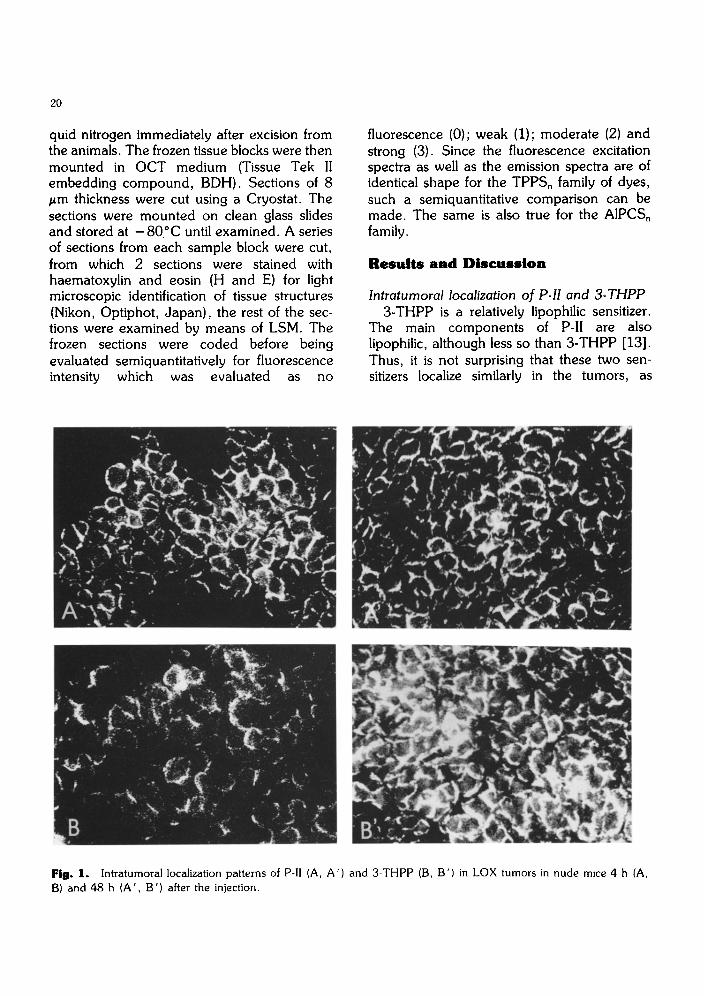
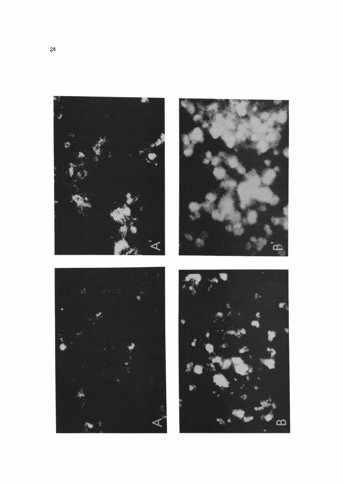
lntratumoral localization of P-11 and 3-THPP 3-THPP is a relatively lipophilic sensitizer.
The main components of P-II are also lipophilic, although less so than 3-THPP [13]. Thus, it is not surprising that these two sen- sitizers localize similarly in the tumors, as
Fig. 1. Intratumoral localization patterns of P-II (A, A’) and 3-THPP (B, B’) in LOX tumors in nude mice 4 h (A,
B) and 48 h (A’, B’) after the injection.
21
shown in Fig. 1. 3-THPP was definitely loca- lized on the membrane and some in the cytoplasm of tumor cells. The fluorescence in- tensity appeared to increase from 4 h to 48 h after the injection. P-11 was also mainly localiz- ed on the membrane and some in the cytoplasm of tumor cells, although some fluorescence in the stroma could be seen. This is in agreement with reports which indicate that PDT of tumors containing this sensitizer leads to a damage to the tumor cells as well as to the vascular system [15- 171. The present data in- dicate that the intratumoral localization pattern of P-II is similar at 4 h and 48 h after the injec- tion, although the fluorescence intensity seem- ed to be slightly higher at 48 h. The latter observation is in agreement with our quan- titative measurements of the fluorescence of the tumor extracts (data not shown).
Intratumoral localization of TPPS,s The intratumoral localization patterns of
TPPS1 and TPP&, were quite similar and strikingly different from those of TPPSz,, TPPS3 and TPPS4 (Fig. 2, Table I). TPPSl
Table 1. A semiquantitative comparison of fluor- escence intensity in stroma and parenchyma of the tumor tissue among different analogs of TPPS, at various inter- vals after i.p. injections of 10 mg/kg.
Dyes Time (h) after injection
4 48 120
TPPS, la/lb O/l O/l TPPS,, O-l/l O/2-3 O/l TPPS,, l-2/0 l/l o/o TPPS, 2/o l-Z/O o/o TPPS, 3/o 2/o o/o
‘Means fluorescence intensity in the stroma of the tumor tissue
bMeans fluorescence intensity in the parenchyma of the tumor tissue.
Grades were described as follows: 0: no fluorescence; 1: weak; 2: moderate; 3: strong.
and TPPS*, tended to localize mainly in- tracellularly, while TPP!& and TPPS4 localized mainly in the extracellular space. TPPSZ, was mainly distributed on the cytoplasmic mem- brane, and some also in the cytoplasm of tumor cells and tumorous stroma. There was a slight change in the localization patterns of TPPSl and TPPSZ, from 4 h to 48 h after the injection (a weak fluorescence was seen in the extracellular space at 4 h but not at 48 h, see Fig. 2 and Table I) while the localization pat- terns of TPP!&,, TPP!& and TPPS4 were similar at 4 h and 48 h (data shown only for 48 h in Fig. 2). These findings are in excellent agreement with the localization patterns in the pancreatic tumor Pant 02 in C57BL/6 mice reported by Kessel [ 191, although the data are not explicitly shown in their paper. The lipophilic dyes (TPPS1 and TPP!&,) were re- tained in the tumors for a much longer time (even at 120 h significant fluorescence inten- sities were seen, Table I) than were the more hydrophilic ones (TPP&,, TPPS3 and TPPSJ the fltiorescence of which could not be seen at 120 h postinjection, Table I).
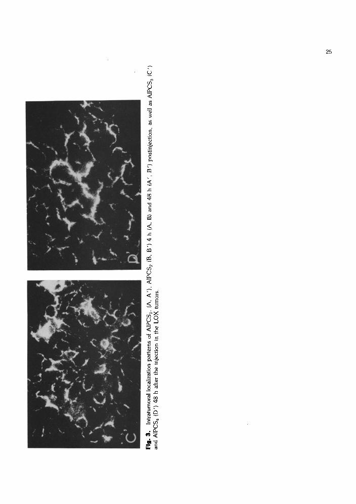
Intratumoral localization of AIPCS,s The localization patterns of this family of
dyes, as shown in Fig. 3 and semiquantitative- ly described by Table II, corresponded to the patterns of the TPPS, family: The hydrophob- ic members [AIPC!$ and AlPCS:! (i.e. AIPCS2J] localized mainly intracellularly, while the more hydrophilic member, AIPC& localized mainly extracellularly. AIPCSj had a membrane-localizing pattern and a weakly in- tracellular distribution pattern. Some fluor- escence was also found in the stroma. The fluorescence intensity of AIPCS, and AIPCS, increased from 4 h to 48 h after the injection while the fluorescence intensity of AIPCS3 and AIPCS4 decreased from 4 h to 48 h postinjection (Table 2). At 120 h postinjection there were still significant amounts of fluoresc- ing AlPC!$ and AlPCS2 in the tumor cells, while no fluorescence was seen in the samples from mice injected with AlPCS3 and AIPCS+ This is the first time that the localization pattern
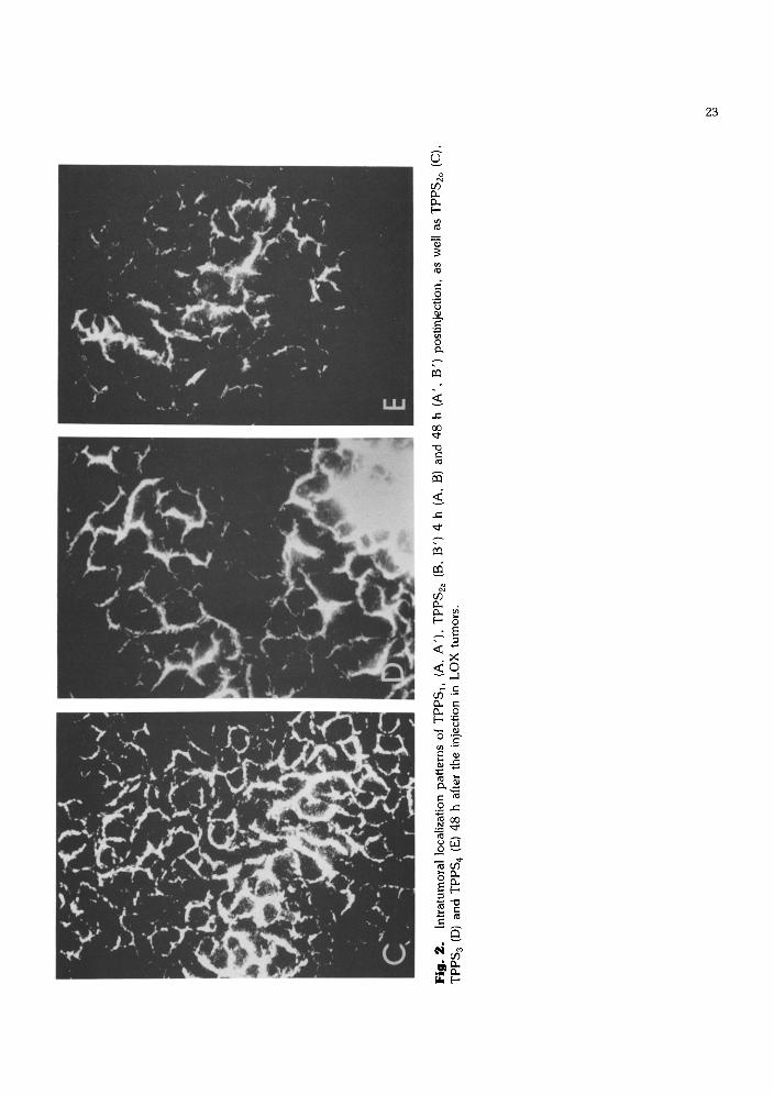
Fig.
2.
In
trat
umor
al l
ocal
izat
ion
patt
erns
of
TPPS
,, (A
, A
’),
TPPS
,, (B
, B’
) 4
h (A
, B)
and
48
h
(A’,
B’
) po
stin
ject
ion,
as
wel
l as
TPP
S,,
(C),
TPPS
, (D
) an
d TP
PS,
(E)
48
h af
ter
the
inje
ctio
n in
LO
X
tum
ors.
_-
--
--
--
--
_~-
--
--
--
__-
--
--
--
--
-_
. ._
- ..
I._
_
.._
. _
..Y
_
,__
_
-
--
--
__
I .r_
- _
1-
--
--
__
.-
-_
24
Fig
. 3.
ln
trat
umor
al
loca
lizat
ion
patte
rns
of A
IPC
S,,
(A,
A’)
, A
IPC
S,
(B,
B’)
4
h (A
, B
) an
d 48
h
(A’,
B
’)
post
inje
ctio
n.
as w
ell
as A
IPC
S,
(C’)
an
d A
IPC
S,
(D’)
48
h
afte
r th
e in
ject
ion
in t
he
LO
X
tum
ors.
_---
----
----
I---
----
----
----
--_,
_-
----
----
----
----
----
----
---
26
Table II. A semiquantitative comparison of fluor- escence intensity in stroma and parenchyma of the tumor tissue among different species of AIPCS, at various in- tervals after i.p. injections of 10 mg/kg.
Dyes Time (h) after injection
4 48 120 References
AIPCS, o-la/lb O/l O/l
AIPCS, O-l/Z o/3 O/l AIPCS, 2--3/O Z/O-l o/o AIPCS, 3/o 2/o o/o
a,bGrades were as described in Table I.
of the fluorescence of AIPC with different sulfonations in human tumor xenografts has been reported. In the case of AlPCS4 we have measured the kinetics of accumulation and retention of fluorescing material by quantitative methods. The results of these measurements are in agreement with the present observations and will be presented separately.
Conclusion
In the present tumor model lipophilic dyes such as TPP&, TPPSza, AlPC& and AlPCS2, localize intracellularly while the more hydrophilic dyes like TPP&, TPPS4 and AlPC& localize mainly in the tumorous stroma. The former dyes tend to be retained for a longer time in the tumors than the latter ones. P-II, 3-THPP, TPP!& and AlPCSs had a combined localization pattern, i.e. a strongly cytoplasmic membrane-localizing pattern and a weakly intracellular distribution pattern, although some fluorescence in the stroma could be seen.
The difference in intratumoral localization may explain why TPPS4and AlPC& seem to be less efficient PDT sensitizers than P-II in spite of the fact that they are taken up by tumors (at least at 24 h postinjection) to a similar degree as is P-II [14,20,27].
Acknowledgements
The present work was supported by The Association for International Cancer Research. We thank Mrs Ellen Hellesylt for expert techni- cal assistance and Professor 0. Fodstad for providing the human tumor LOX.
1
2
3
4
5
6
7
8
9
10
11
Ash, D. and Brown, S.B. (1989) Photodynamic therapy-
achievements and prospects. Br. J. Cancer, 60, 151. Gomer, C.J., Rucker, N., Ferrario, A. and Wong, S.
(1989) Review: Properties and applications of
photodynamic therapy. Radiat. Res., 120, l-18.
Moan, J. (1986) Porphyrin photosensitization and
phototherapy. Photochem. Photobiol., 43, 681-690.
Moan, J., Berg, K., Kvam, E.. Western, A., Malik, Z.,
Ruck. A. and Schneckenburger, H. (1989) Intracellular
localization of photosensitizers. In: Photosensitizing com-
pounds: their chemistry, biology and clinical use, pp.
95- 111. Editors: G. Bock and S. Harnett. Wiley,
Chichester (Ciba Foundation Symposium 146).
Henderson, B.W. and Bellnier, D.A. (1989) Tissue
localization of photosensitizers and mechanism of
photodynamic tissue destruction. In: Photosensitizing com-
pounds: their chemistry, biology and clinical use, pp.
112-130. Editors: G. Bock and S. Harnett. Wiley,
Chichester (Ciba Foundation Symposium 146).
Dougherty, T.J. (1989) Introduction. In: Photosensitizing
compounds: their chemistry, biology and clinical use, pp.
l-3. Editors: G. Bock and S. Harnett. Wiley, Chichester
(Ciba Foundation Symposium 146).
Kaye, A.H. (1989) Photoradiation therapy of brain
tumors. In: Photosensitizing compounds: their chemistry,
biology and clinical use, pp. 209-224. Editors: G. Bock
and S. Harnett. Wiley, Chichester (Ciba Foundation Sym-
posium 146).
Sandeman, D.R. (1986) Photodynamic therapy in the
management of malignant gliomas: a review. Laser Med.
Sci.. 1,163-174.
Spikes, J.D. and Jori. G. (1987) Photodynamic therapy of
tumors and other diseases using porphyrins. Laser Med.
sci., 2. 3-15. Kato, H., Kawate. N., Kinoshita, K.. Yamamoto, H.,
Furukawa, K. and Hayata, Y. (1989) Photodynamic
therapy of early-stage lung cancer. In: Photosensitizing
compounds: their chemistry, biology and clinical use, pp. 183-197. Editors: G. Bock and S. Harnett. Wiley.
Chichester (Ciba Foundation Symposium 146).
Jocham, D., Beer, M., Baumgartner. R., Staehler, G. and
Unsold, E. (1989) Long-term experience with integral photodynamic therapy of TIS bladder carcinoma. In:
Photosensitizing compounds: their chemistry, biology and
clinical use, pp. 198-208. Editors: G. Bock and S.
Harnett. Wiley, Chichester (Ciba Foundation Symposium 146)

27
12
13
14
15
16
17
18
19
20
van Lier, J.E. (1988) New sensitizers for photodynamic
therapy of cancer. In: Light in biology and medicine, pp.
133-141. Editors: R. Douglas, J. Moan and F. Dall’Ae-
qua. Plenum Press.
Moan, J., Peng. Q., Evensen. J.F., Berg, K., Western, A.
and Rimington. C. (1987) Photosensitizing efficiencies,
tumor-and cellular uptake of different photosensitizing
drugs relevant for photodynamic therapy of cancer.
Photochem. Photobiol.. 46, 713-721.
Evensen. J.F. and Moan, J. (1987) A test of different
photosensitizers for photodynamic treatment of cancer in a
murine tumor model. Photochem. Photobiol., 46, 859-865.
Henderson, B.W., Waldow, S.M., Mang. T.S., Potter,
W.R.. Malone, P.B. and Dougherty, T.J. (1985) Tumor
destruction and kinetics of tumor cell death in two ex-
perimental mouse tumors following photodynamic
therapy. Cancer Res., 45. 572-576. Franken. N.A.P., van Delft. J.L., Dubbelman. T.M.A.R.,
De Wolff-rouendaal, D.. Oosterhuis, J.A.. Star, W.M. and
Marijnissen, H.P.A. (1985) Hematoporphyrin-derivative
photoradiation treatment of experimental malignant
melanoma in the anterior chamber of the rabbit. Curr. Eye Res.. 4, 641-654
Star, W.M.. Marijnissen, H.P.A., Van der Berg-Blok.
A.E., Versteeg. J.A.C.. Franken, K.A.P. and Reinhold,
H.S. (1986) Destruction of rat mammary tumor and nor-
mal tissue microcirculation by hematoporphyrin-derivative
photoradiation observed in vivo in sandwich observation
chambers. Cancer Res., 46. 2532-2540.
Zhou, C.N. (1989) Mechanisms of tumor necrosis induced
by photodynamic therapy. J Photochem. Photobiol. B.
Biol.. 3, 299-318.
Kessel. D.. Thompson, P.. Saatio. K. and Nantwi, K.D.
(1987) Tumor localization and photosensitization by sulfonated derivatives of tetraphenylporphine. Photochem. Photobiol., 45, 787-790
Peng, Q.. Evensen. J-F.. Rimington. C. and Moan, J.
(1987) A comparison of different photosensitizing dyes
with respect to uptake by C3H-tumors and tissues of mice.
Cancer Lett.. 36, I- 10.
21 Moan, J. and Sommer, S. (1983) Uptake of the com-
ponents of hematoporphyrin derivative by cells and
tumors. Cancer Lett., 21. 167-174.
22 Sommer, S.. Rimington, C. and Moan, J. (1987) Por-
phyrin derivatives having physrcal and chemical
characteristics similar to those of the active components of
hematoporphyrin derivative and with very strong photosensitizing effects. Photochem. Photobiol. B: Biol.,
1. 241-246.
23 Kongshaug, M., Moan, J. and Brown, S.B. (1989) The
distribution of porphyrins with different tumorlocalizing
ability among human plasma proteins. Br. J. Cancer. 59,
184- 188.
24 Berg, K., Bommer, J.C. and Moan, J. (1989) Evaluation
of sulfonated aluminium phthalocyanines for use in
photochemotherapy. Cellular uptake studies. Cancer
Lett.. 44, 7-15
25 Peng, Q.. Nesland. J.M., Moan, J., Evensen. J.F.,
Kongshaug, M and Rimington, C. (1990) Localization of
fluorescent Photofrin II and aluminium phthalocyanine
tetrasulfonate in transplanted human malignant tumor
LOX and normal tissues of nude mice using high light-
sensitive video intensification microscopy. Int. J Cancer,
45, 972-979.
26 Fodstad. 0.. Aadal, S.. McMenamin, M , Nesland, J.M.
and Pihl, A. (1988) A new experimental metastasis model
in athymic nude mice, the human malignant melanoma
LOX. Int. J. Cancer, 41, 442-449
27 Brasseur, N.. Ali. H., Langlois. R and van Lier. J.E.
(1988) Biological activities of phthalocyanines-IX.
Photosensitization of V-79 Chinese hamster cells and
EMT-6 mouse mammary tumor by selectively sulfonated
zinc phthalocyanines. Photochem. Photobiol.. 47, 705-711.
Copyright © 2022 FDOKUMEN


![Poly[di(carboxylatophenoxy)phosphazene] is a potent adjuvant for intradermal immunization](https://static.fdokumen.com/doc/165x107/6335c6c4a1ced1126c0af097/polydicarboxylatophenoxyphosphazene-is-a-potent-adjuvant-for-intradermal-immunization.jpg)