
S Gupta JCan Res Ther targeted delivery of photosensitizers
11
314 Journal of Cancer Research and Therapeutics - July-September 2011 - Volume 7 - Issue 3 Seema Gupta 1-3 , Bilikere S. Dwarakanath 1 , N. K. Chaudhury 1 , Anil K. Mishra 4 , K. Muralidhar 5 , Viney Jain 1,6 1 Division of Radiation Biosciences, Institute of Nuclear Medicine and Allied Sciences, Brig S K Mazumdar Road, Delhi, India, 2 Department of Radiation Oncology, 3 Sylvester Comprehensive Cancer Center, University of Miami, Miami, FL 33136, USA, 4 Division of Cyclotron and Radiopharma- ceuticals, Institute of Nuclear Medicine and Allied Sciences, Brig S K Mazumdar Road, Delhi, India, 5 Department of Zoology, University of Delhi, Delhi, India, 6 Eco-Development Foundation, New Delhi, India For correspondence: Dr. Seema Gupta, Department of Radiation Oncology, Sylvester Comprehensive Cancer Center, University of Miami, 1550 Northwest 10 th Avenue, PAP Building, Room 117, Miami, FL 33136, USA. E-mail: sgupta3@med. miami.edu In vitro and in vivo targeted delivery of photosensitizers to the tumor cells for enhanced photodynamic effects ABSTRACT Background: Efficacy of photodynamic therapy can be enhanced by improving uptake, localization, and sub-cellular localization of sensitizers at the sensitive targets. Materials and Methods: Uptake, localization, and photodynamic effects of hematoporphyrin derivative (HpD, Photosan-3; PS-3) and disulfonated aluminum phthalocyanine (AlPcS 2 ) were studied either encapsulated in liposomes or conjugated to a monoclonal antibody to carcinoembryonic antigen (anti-CEA) in a brain glioma cell line, BMG-1. Results: Although the total uptake with encapsulated or conjugated sensitizers was less than the free sensitizers, photodynamic efficiency was higher due to the localization of the sensitizer at the sensitive targets. Biodistribution of intravenously administered technetium ( 99m Tc)-labeled PS-3 analyzed by gamma camera imaging showed maximum accumulation in the liver followed by tumor. Tumor/ muscle (T/N) ratio of free PS-3 was higher compared to encapsulated or conjugated PS-3 but the accumulation of PS-3 significantly reduced in brain and cutaneous tissue following modulated delivery. Pharmacokinetics suggested faster accumulation of encapsulated and conjugated PS-3 in the tumor. Conclusion: Localization of sensitizers at sensitive targets and reduced accumulation in normal tissues with liposome encapsulation and antibody conjugation suggest that these two delivery systems can potentially enhance the efficacy of photodynamic treatment. KEY WORDS: 99m Tc labeling, biodistribution, liposome encapsulation, localization, monoclonal antibody, photodynamic treatment, photosensitizer, sub-cellular localization, targeted delivery, uptake Original Article INTRODUCTION Efficacy of photodynamic treatment (PDT) depends on several factors such as uptake and localization of the photosensitizer, mode of light delivery, physiological status of the cells or the target tissue, and photophysical properties of the sensitizer like singlet and triplet quantum yields and life- times. Methods/techniques that help in increased accumulation and retention of the sensitizer in the target tissue could be one of the approaches to differentially enhance the damage to the tumor tissue as compared to the normal tissue. Also, the sub-cellular localization of the sensitizers at sites that are most sensitive to the photodynamic treatment is another important factor influencing the photodynamic efficacy, since singlet oxygen has a short half-life, and therefore can cause damage at or near the site of its production. Therefore, there is considerable amount of interest in investigations pertaining to the optimization of uptake, distribution, and localization of the sensitizers both in vitro and in vivo. Selective damage to the tumor can be achieved by increased accumulation and retention of the sensitizer in the tumor, [1-4] development of better sensitizers, [5,6] light sources and delivery systems, [7] and modulation of metabolism to selectively enhance the manifestation of PDT-induced damage in the tumor. [8-11] New photosensitizers that absorb significantly in the far red region with higher yields of singlet oxygen and reduced localization in skin have been developed. [12,13] Several delivery systems such as photosensitizer conjugates, dendrimers, micelles, liposomes, and nanoparticles, besides the application of photochemical internalization (PCI) technology for intracellular delivery of macromolecules have been recently described. [14-16] In the present study, localization, distribution, and targeting of the sensitizers, photosan-3 (PS-3) and disulfonated phthalocyanine (AlPcS 2 ) were modulated by encapsulating in liposomes and conjugating with monoclonal antibodies. Both liposome encapsulation and antibody conjugation resulted in localization of sensitizers at sensitive targets and reduced accumulation in the normal Access this article online Access this article online Website: Website: www.cancerjournal.net DOI: DOI: 10.4103/0973-1482.87035 PMID: PMID: *** Quick Response Code: Quick Response Code: [Downloaded free from http://www.cancerjournal.net on Friday, May 17, 2013, IP: 122.162.88.153] || Click here to download free Android application for this journal
-
Upload
writerightindia -
Category
Documents
-
view
2 -
download
0
Transcript of S Gupta JCan Res Ther targeted delivery of photosensitizers
314 Journal of Cancer Research and Therapeutics - July-September 2011 - Volume 7 - Issue 3
Seema Gupta1-3, Bilikere S. Dwarakanath1, N. K. Chaudhury1, Anil K. Mishra4, K. Muralidhar 5, Viney Jain1,6
1Division of Radiation Biosciences, Institute of Nuclear Medicine and Allied Sciences, Brig S K Mazumdar Road, Delhi, India, 2Department of Radiation Oncology, 3Sylvester Comprehensive Cancer Center, University of Miami, Miami, FL 33136, USA,4Division of Cyclotronand Radiopharma-ceuticals, Institute of Nuclear Medicine and Allied Sciences, Brig S K Mazumdar Road, Delhi, India, 5Department of Zoology, University of Delhi, Delhi, India, 6Eco-Development Foundation,New Delhi, India
For correspondence: Dr. Seema Gupta, Department of Radiation Oncology, Sylvester Comprehensive Cancer Center, University of Miami, 1550 Northwest 10th Avenue, PAP Building, Room 117, Miami, FL 33136, USA. E-mail: [email protected]
In vitro and in vivo targeted delivery of photosensitizers to the tumor cells for enhanced photodynamic effects
ABSTRACTBackground: Efficacy of photodynamic therapy can be enhanced by improving uptake, localization, and sub-cellular localization of sensitizers at the sensitive targets.
Materials and Methods: Uptake, localization, and photodynamic effects of hematoporphyrin derivative (HpD, Photosan-3; PS-3) and disulfonated aluminum phthalocyanine (AlPcS2) were studied either encapsulated in liposomes or conjugated to a monoclonal antibody to carcinoembryonic antigen (anti-CEA) in a brain glioma cell line, BMG-1.
Results: Although the total uptake with encapsulated or conjugated sensitizers was less than the free sensitizers, photodynamic efficiency was higher due to the localization of the sensitizer at the sensitive targets. Biodistribution of intravenously administered technetium (99mTc)-labeled PS-3 analyzed by gamma camera imaging showed maximum accumulation in the liver followed by tumor. Tumor/muscle (T/N) ratio of free PS-3 was higher compared to encapsulated or conjugated PS-3 but the accumulation of PS-3 significantly reduced in brain and cutaneous tissue following modulated delivery. Pharmacokinetics suggested faster accumulation of encapsulated and conjugated PS-3 in the tumor.
Conclusion: Localization of sensitizers at sensitive targets and reduced accumulation in normal tissues with liposome encapsulation and antibody conjugation suggest that these two delivery systems can potentially enhance the efficacy of photodynamic treatment.
KEY WORDS: 99mTc labeling, biodistribution, liposome encapsulation, localization, monoclonal antibody, photodynamic treatment, photosensitizer, sub-cellular localization, targeted delivery, uptake
Original Article
INTRODUCTION
Efficacy of photodynamic treatment (PDT) depends on several factors such as uptake and localization of the photosensitizer, mode of light delivery, physiological status of the cells or the target tissue, and photophysical properties of the sensitizer like singlet and triplet quantum yields and life-times. Methods/techniques that help in increased accumulation and retention of the sensitizer in the target tissue could be one of the approaches to differentially enhance the damage to the tumor tissue as compared to the normal tissue. Also, the sub-cellular localization of the sensitizers at sites that are most sensitive to the photodynamic treatment is another important factor influencing the photodynamic efficacy, since singlet oxygen has a short half-life, and therefore can cause damage at or near the site of its production. Therefore, there is considerable amount of interest in investigations pertaining to the optimization of uptake, distribution, and localization of the sensitizers both in vitro and in vivo.
Selective damage to the tumor can be achieved by increased accumulation and retention of the sensitizer in the tumor,[1-4] development of better sensitizers,[5,6] light sources and delivery systems,[7] and modulation of metabolism to selectively enhance the manifestation of PDT-induced damage in the tumor.[8-11] New photosensitizers that absorb significantly in the far red region with higher yields of singlet oxygen and reduced localization in skin have been developed.[12,13] Several delivery systems such as photosensitizer conjugates, dendrimers, micelles, liposomes, and nanoparticles, besides the application of photochemical internalization (PCI) technology for intracellular delivery of macromolecules have been recently described.[14-16]
In the present study, localization, distribution, and targeting of the sensitizers, photosan-3 (PS-3) and disulfonated phthalocyanine (AlPcS
2)
were modulated by encapsulating in liposomes and conjugating with monoclonal antibodies. Both liposome encapsulation and antibody conjugation resulted in localization of sensitizers at sensitive targets and reduced accumulation in the normal
Access this article onlineAccess this article onlineWebsite: Website: www.cancerjournal.netDOI: DOI: 10.4103/0973-1482.87035PMID: PMID: ***Quick Response Code:Quick Response Code:
[Downloaded free from http://www.cancerjournal.net on Friday, May 17, 2013, IP: 122.162.88.153] || Click here to download free Android application for this journal
Avinash K
Rectangle
315Journal of Cancer Research and Therapeutics - July-September 2011 - Volume 7 - Issue 3
Gupta, et al.: Targeted delivery of photosensitizers
tissues, suggesting that both these delivery systems have potential to selectively enhance local tumor control following photodynamic treatment.
MATERIALS AND METHODS
ChemicalsThe commercially available form of the photosensitizer, hematoporphyrin derivative, was obtained as PS-3 from SeeLab (Hamburg, Germany). Antibody to carcinoembryonic-antigen (CEA) was provided by CIM, Hawana, Cuba. This was conjugated to PS-3 using water-soluble activating agents, N-(3-dimethylaminopropyl)-N’-ethylcarbodiimide hydrochloride (EDC-C8H17N3·HCl) and N-Hydroxy sulfosuccinimide sodium salt (S-NHS-C4H4NNaO6S). Sephadex G-50 (Pharmacia) was used to separate conjugated PS-3 from free PS-3. Fluorescein Isothiocyanate (FITC)-conjugated secondary antibody (mouse anti-IgG) was from Sigma Chemical Co., USA, and stannous chloride (SnCl
2, used as a reducing agent) from Glaxo Laboratories.
Disulfonated aluminum phthalocyanine (AlPcS2) was prepared
and characterized in Institute of Nuclear Medical and Allied Science (INMAS), Delhi.[17] 99mTechnetium for radioactive labeling was generated at INMAS, Delhi.
DL-alpha-PC dipalmitoyl (C16:0) (1,2-dihexadecanyl-rac-glycero-3-phosphocholine, C
40H
80NO
8P) (DPPC, SIGMA), cholesterol
(5-cholesten-3beta-ol, C27
H46
O, SIGMA), dialysis tubing (cellulose membrane, SIGMA, Size- mol. wt. cut off 12000 or more), P-100 (Biogel), Osmium tetraoxide (Agar Scientific Ltd., UK) were used for liposome preparation and characterization.
All other chemicals were of analytical grade and obtained from Glaxo Laboratories (Qualigens) or E-Merck, India.
Tumor cell lineMurine Ehrlich ascites tumor (EAT, F15) cells were obtained from University of Frankfurt, Germany[18] and used for animal studies.
Human cerebral glioma cell line (BMG-1; DNA index = 0.95; wild-type p53), established from a mixed glioma[19] was used for in vitro studies.
Monolayer BMG-1 cells were grown in Dulbecco’s modified Eagle’s medium (DMEM) with 5% fetal calf serum (FCS), penicillin (100 units/ml), streptomycin (50 g/ml) and nystatin (2 g/ml). Stock cultures were sub-cultured every third day after harvesting the cells with 0.05% trypsin and seeding 8 × 10³ cells/cm2 in tissue culture flasks to maintain the cells in the expo nential phase. All experiments were carried out with exponentially growing cells.
Animal modelsSwiss albino, strain ‘A’ male mice inbred at INMAS were used as experimental animal model. EAT cells were grown in the
peritoneal cavity of three-month-old male ‘A’ strain mice and harvested on every seventh day after inoculation. Viable cells excluding 0.5% trypan blue were counted using Neubauer chamber and 25 million cells were injected intraperitoneally for propagation. For implantation of solid tumors, EAT cells (15 × 106) were injected subcutaneously in 0.1-0.3 ml volume in the right hind leg of the mice.
After tumor implantation, animals were observed for tumor growth. Tumor diameters were measured in three mutually perpendicular directions with the help of a caliper and volumes calculated using the formula 4/3π r3, where r is the mean radius of tumor. The tumor doubling time was estimated to be around 2 days. Mice were sacrificed when the tumor reached a volume of ~4500 mm3 to avoid tumor burden-related discomfort to the animal as per the United Kingdom Coordinating Committee on Cancer Research (UKCCCR) guidelines for the welfare of animals in experimental neoplasia.[20] Further, to examine the labeling and presence of free technetium in the liposome preparation, labeled liposomes were injected intravenously through dorsal ear veins in rabbits of New Zealand strain weighing 3-3.5 kg. Images were taken using planar gamma camera equipped with pinhole collimator as described.
All experiments were conducted according to the guidelines established by CPCEA, Indian National Science Academy (INSA), and European Society of animal handling after obtaining the permission from institute’s animal ethics committee.
Preparation and characterization of liposomesIncorporation of photosensitizer in small unilamellar vesicles (SUV) was carried out by co-dissolving dipalmitoyl phosphatidylcholine (DPPC) (20 mg), cholesterol (2 mg), and PS-3 (150 g) in 3 ml of methanol in a round bottom flask. Methanol was evaporated completely in a rotoevaporator (Model S B, Buchi, Switzerland) at 40°C. After evaporation a thin film was deposited on the inner surface of the flask, which was dried completely overnight in a lyophilizer (Herysun, India) and redissolved in 10 ml of phosphate buffered saline (PBS). The resultant suspension was sonicated (B. Braun, Labsonic U, USA) by keeping the sample immersed in ice for 20 min and the preparation was kept at room temperature for about 30 min.
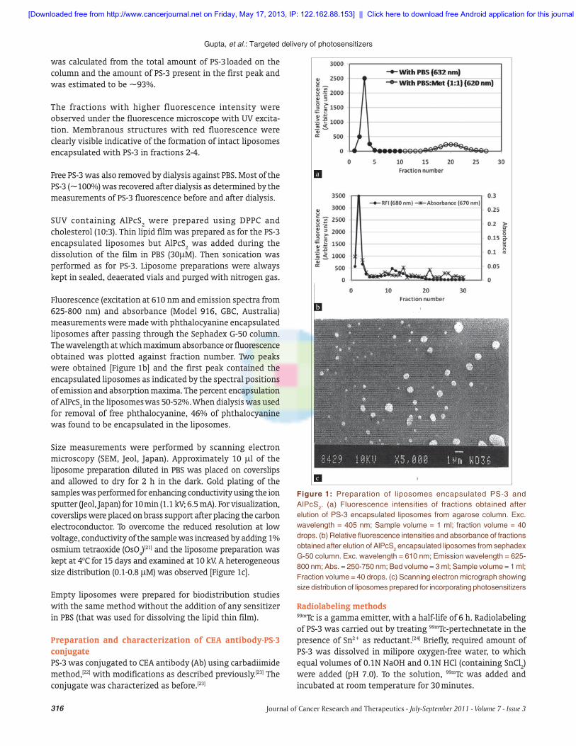
The liposomal suspension containing PS-3 was run through an agarose (SIGMA, type 1A) column. The preparation was initially eluted with PBS and about 10 fractions containing 40 drops each were collected. Then the eluent was changed to PBS: methanol (1:1) and 16 fractions were collected. Fractions were analyzed by PS-3 fluorescence (Model JY3C, Jobin Yvon, France) and the fluorescence intensity was plotted against fraction number. Two peaks were obtained [Figure 1a]. The spectral positions of emission maximum (632, 690 nm) of the fractions in the first peak indicated incorporation of PS-3 in the lipid environment of SUV. The free PS-3 was detected in later fractions as revealed by the spectral positions of fluorescence peaks at 620 and 683 nm. Finally, the efficiency of incorporation
[Downloaded free from http://www.cancerjournal.net on Friday, May 17, 2013, IP: 122.162.88.153] || Click here to download free Android application for this journal
316 Journal of Cancer Research and Therapeutics - July-September 2011 - Volume 7 - Issue 3
Gupta, et al.: Targeted delivery of photosensitizers
was calculated from the total amount of PS-3 loaded on the column and the amount of PS-3 present in the first peak and was estimated to be ~93%.
The fractions with higher fluorescence intensity were observed under the fluorescence microscope with UV excita-tion. Membranous structures with red fluorescence were clearly visible indicative of the formation of intact liposomes encapsulated with PS-3 in fractions 2-4.
Free PS-3 was also removed by dialysis against PBS. Most of the PS-3 (~100%) was recovered after dialysis as determined by the measurements of PS-3 fluorescence before and after dialysis.
SUV containing AlPcS2 were prepared using DPPC and
cholesterol (10:3). Thin lipid film was prepared as for the PS-3 encapsulated liposomes but AlPcS
2 was added during the
dissolution of the film in PBS (30M). Then sonication was performed as for PS-3. Liposome preparations were always kept in sealed, deaerated vials and purged with nitrogen gas.
Fluorescence (excitation at 610 nm and emission spectra from 625-800 nm) and absorbance (Model 916, GBC, Australia) measurements were made with phthalocyanine encapsulated liposomes after passing through the Sephadex G-50 column. The wavelength at which maximum absorbance or fluorescence obtained was plotted against fraction number. Two peaks were obtained [Figure 1b] and the first peak contained the encapsulated liposomes as indicated by the spectral positions of emission and absorption maxima. The percent encapsulation of AlPcS
2 in the liposomes
was 50-52%.
When dialysis was used
for removal of free phthalocyanine, 46% of phthalocyanine was found to be encapsulated in the liposomes.
Size measurements were performed by scanning electron microscopy (SEM, Jeol, Japan). Approximately 10 l of the liposome preparation diluted in PBS was placed on coverslips and allowed to dry for 2 h in the dark. Gold plating of the samples was performed for enhancing conductivity using the ion sputter (Jeol, Japan) for 10 min (1.1 kV; 6.5 mA). For visualization, coverslips were placed on brass support after placing the carbon electroconductor. To overcome the reduced resolution at low voltage, conductivity of the sample was increased by adding 1% osmium tetraoxide (OsO
4)[21] and the liposome preparation was
kept at 4oC for 15 days and examined at 10 kV. A heterogeneous size distribution (0.1-0.8 M) was observed [Figure 1c].
Empty liposomes were prepared for biodistribution studies with the same method without the addition of any sensitizer in PBS (that was used for dissolving the lipid thin film).
Preparation and characterization of CEA antibody-PS-3 conjugatePS-3 was conjugated to CEA antibody (Ab) using carbadiimide method,[22] with modifications as described previously.[23] The conjugate was characterized as before.[23]
Figure 1: Preparation of liposomes encapsulated PS-3 and AlPcS2. (a) Fluorescence intensities of fractions obtained after elution of PS-3 encapsulated liposomes from agarose column. Exc. wavelength = 405 nm; Sample volume = 1 ml; fraction volume = 40 drops. (b) Relative fl uorescence intensities and absorbance of fractions obtained after elution of AlPcS2 encapsulated liposomes from sephadex G-50 column. Exc. wavelength = 610 nm; Emission wavelength = 625-800 nm; Abs. = 250-750 nm; Bed volume = 3 ml; Sample volume = 1 ml; Fraction volume = 40 drops. (c) Scanning electron micrograph showing size distribution of liposomes prepared for incorporating photosensitizers
Radiolabeling methods99mTc is a gamma emitter, with a half-life of 6 h. Radiolabeling of PS-3 was carried out by treating 99mTc-pertechnetate in the presence of Sn2+ as reductant.[24] Briefly, required amount of PS-3 was dissolved in milipore oxygen-free water, to which equal volumes of 0.1N NaOH and 0.1N HCl (containing SnCl
2)
were added (pH 7.0). To the solution, 99mTc was added and incubated at room temperature for 30 minutes.
[Downloaded free from http://www.cancerjournal.net on Friday, May 17, 2013, IP: 122.162.88.153] || Click here to download free Android application for this journal
317Journal of Cancer Research and Therapeutics - July-September 2011 - Volume 7 - Issue 3
Gupta, et al.: Targeted delivery of photosensitizers
Quality Control was performed by spiking 2 l of labeled PS-3 on two strips of silica gel Thin layer chromatography (TLC) paper and were dipped in (i) acetone (100%) and (ii) solvent mixture (ethyl acetate:acetone:water:liquid ammonia, 3:7:3:0.3). Free 99mTc moves with acetone. Both labeled and free 99mTc move with solvent mixture and reach at the top, while reduced/hydrolyzed 99mTc remains at the bottom. After the run, top and bottom portions of the strip were counted separately with a gamma counter (Electronics Corporation of India Limited, ECIL). Percent free 99mTc and reduced/hydrolyzed 99mTc were calculated. Labeling efficiency was estimated to be more than 95%.
PS-3 was labeled with 99mTc using SnCl2 as reductant as
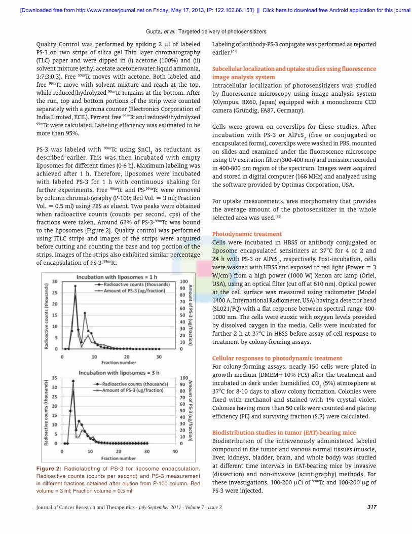
described earlier. This was then incubated with empty liposomes for different times (0-6 h). Maximum labeling was achieved after 1 h. Therefore, liposomes were incubated with labeled PS-3 for 1 h with continuous shaking for further experiments. Free 99mTc and PS-99mTc were removed by column chromatography (P-100; Bed Vol. = 3 ml; Fraction Vol. = 0.5 ml) using PBS as eluent. Two peaks were obtained when radioactive counts (counts per second, cps) of the fractions were taken. Around 62% of PS-3-99mTc was bound to the liposomes [Figure 2]. Quality control was performed using ITLC strips and images of the strips were acquired before cutting and counting the base and top portion of the strips. Images of the strips also exhibited similar percentage of encapsulation of PS-3-99mTc.
Labeling of antibody-PS-3 conjugate was performed as reported earlier.[23]
Subcellular localization and uptake studies using fluorescence image analysis systemIntracellular localization of photosensitizers was studied by fluorescence microscopy using image analysis system (Olympus, BX60, Japan) equipped with a monochrome CCD camera (Gründig, FA87, Germany).
Cells were grown on coverslips for these studies. After incubation with PS-3 or AlPcS
2 (free or conjugated or
encapsulated forms), coverslips were washed in PBS, mounted on slides and examined under the fluorescence microscope using UV excitation filter (300-400 nm) and emission recorded in 400-800 nm region of the spectrum. Images were acquired and stored in digital computer (166 MHz) and analyzed using the software provided by Optimas Corporation, USA.
For uptake measurements, area morphometry that provides the average amount of the photosensitizer in the whole selected area was used.[25]
Photodynamic treatmentCells were incubated in HBSS or antibody conjugated or liposome encapsulated sensitizers at 37°C for 4 or 2 and 24 h with PS-3 or AlPcS
2, respectively. Post-incubation, cells
were washed with HBSS and exposed to red light (Power = 3 W/cm2) from a high power (1000 W) Xenon arc lamp (Oriel, USA), using an optical filter (cut off at 610 nm). Optical power at the cell surface was measured using radiometer (Model 1400 A, International Radiometer, USA) having a detector head (SL021/ FQ) with a flat response between spectral range 400-1000 nm. The cells were euoxic with oxygen levels provided by dissolved oxygen in the media. Cells were incubated for further 2 h at 37°C in HBSS before assay of cell response to treatment by colony-forming assays.
Cellular responses to photodynamic treatmentFor colony-forming assays, nearly 150 cells were plated in growth medium (DMEM+10% FCS) after the treatment and incubated in dark under humidified CO
2 (5%) atmosphere at
37°C for 8-10 days to allow colony formation. Colonies were fixed with methanol and stained with 1% crystal violet. Colonies having more than 50 cells were counted and plating effi ciency (PE) and surviving fraction (S.F.) were calculated.
Biodistribution studies in tumor (EAT)-bearing miceBiodistribution of the intravenously administered labeled compound in the tumor and various normal tissues (muscle, liver, kidneys, bladder, brain, and whole body) was studied at different time intervals in EAT-bearing mice by invasive (dissection) and non-invasive (scintigraphy) methods. For these investigations, 100-200 Ci of 99mTc and 100-200 g of PS-3 were injected.
Figure 2: Radiolabeling of PS-3 for liposome encapsulation. Radioactive counts (counts per second) and PS-3 measurement in different fractions obtained after elution from P-100 column. Bed volume = 3 ml; Fraction volume = 0.5 ml
[Downloaded free from http://www.cancerjournal.net on Friday, May 17, 2013, IP: 122.162.88.153] || Click here to download free Android application for this journal
318 Journal of Cancer Research and Therapeutics - July-September 2011 - Volume 7 - Issue 3
Gupta, et al.: Targeted delivery of photosensitizers
For invasive method, tumor-bearing mice were injected via tail vein with 0.1-0.2 ml of labeled PS-3. The animals were anaesthetized with chloroform and sacrificed at different time intervals. Tumors were dissect ed free of adjacent normal tissue. Samples of muscles were taken from the thigh. Kidney and liver pieces were also removed, rinsed in water, and blotted dry on tissue paper. Each sample was weighed and radioactivity counted using gamma counter (ECIL, India). Tissue levels of activity were expressed as percent injected dose per gram of tissue.
To follow the distribution of activity by non-invasive method, scanning was started 1 h after the animals were injected with the labeled PS-3 into the tail vein. Animals were mounted on a thermocol platform and restrained by adhesive tape. Activity distribution data were collected at different time intervals using a gamma camera (Model-EGC 1400A, ECIL, India) equipped with a pinhole camera, coupled to a computer system (ECIL, India). The gamma camera consisted of a thallium activated sodium iodide crystal (16 inches diameter and ½ inch thickness) and 37 photomultiplier tubes (PMT) of 2 inches diameter. Similar counts (100 K) were collected for each animal and activities of various regions of interest (ROIs) were expressed as average counts per pixel of the ROI. Tumor to normal tissue ratios were calculated from the absolute counts.
RESULTS
In vitro studiesEffects of antibody conjugation and liposome encapsulation of photosensitizers were investigated by studying the uptake, sub-cellular localization, and photodynamic effects in a human glioma cell line (BMG-1).
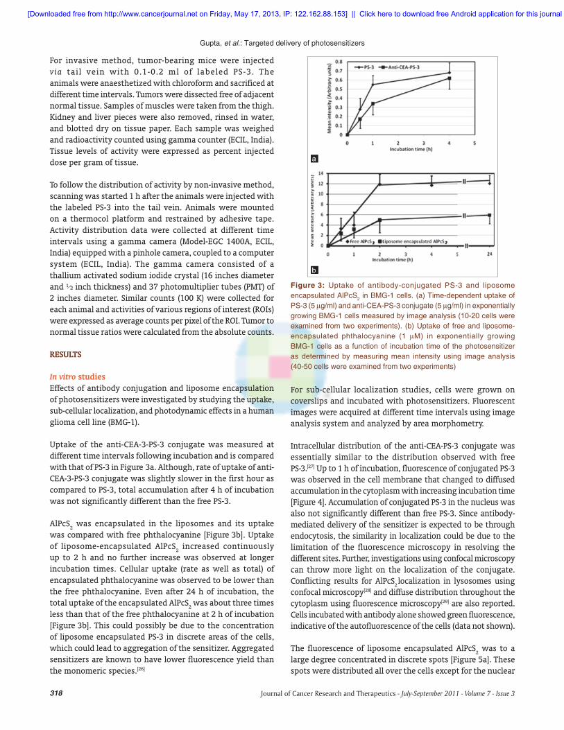
Uptake of the anti-CEA-3-PS-3 conjugate was measured at different time intervals following incubation and is compared with that of PS-3 in Figure 3a. Although, rate of uptake of anti-CEA-3-PS-3 conjugate was slightly slower in the first hour as compared to PS-3, total accumulation after 4 h of incubation was not significantly different than the free PS-3.
AlPcS2 was encapsulated in the liposomes and its uptake
was compared with free phthalocyanine [Figure 3b]. Uptake of liposome-encapsulated AlPcS
2 increased continuously
up to 2 h and no further increase was observed at longer incubation times. Cellular uptake (rate as well as total) of encapsulated phthalocyanine was observed to be lower than the free phthalocyanine. Even after 24 h of incubation, the total uptake of the encapsulated AlPcS
2 was about three times
less than that of the free phthalocyanine at 2 h of incubation [Figure 3b]. This could possibly be due to the concentration of liposome encapsulated PS-3 in discrete areas of the cells, which could lead to aggregation of the sensitizer. Aggregated sensitizers are known to have lower fluorescence yield than the monomeric species.[26]
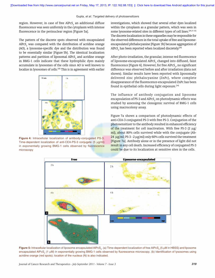
For sub-cellular localization studies, cells were grown on coverslips and incubated with photosensitizers. Fluorescent images were acquired at different time intervals using image analysis system and analyzed by area morphometry.
Intracellular distribution of the anti-CEA-PS-3 conjugate was essentially similar to the distribution observed with free PS- 3. [27] Up to 1 h of incubation, fluorescence of conjugated PS-3 was observed in the cell membrane that changed to diffused accumulation in the cytoplasm with increasing incubation time [Figure 4]. Accumulation of conjugated PS-3 in the nucleus was also not significantly different than free PS-3. Since antibody-mediated delivery of the sensitizer is expected to be through endocytosis, the similarity in localization could be due to the limitation of the fluorescence microscopy in resolving the different sites. Further, investigations using confocal microscopy can throw more light on the localization of the conjugate. Conflicting results for AlPcS
2 localization in lysosomes using
confocal microscopy[28] and diffuse distribution throughout the cytoplasm using fluorescence microscopy[29] are also reported. Cells incubated with antibody alone showed green fluorescence, indicative of the autofluorescence of the cells (data not shown).
The fluorescence of liposome encapsulated AlPcS2 was to a
large degree concentrated in discrete spots [Figure 5a]. These spots were distributed all over the cells except for the nuclear
Figure 3: Uptake of antibody-conjugated PS-3 and liposome encapsulated AlPcS2 in BMG-1 cells. (a) Time-dependent uptake of PS-3 (5 g/ml) and anti-CEA-PS-3 conjugate (5 g/ml) in exponentially growing BMG-1 cells measured by image analysis (10-20 cells were examined from two experiments). (b) Uptake of free and liposome-encapsulated phthalocyanine (1 M) in exponentially growing BMG-1 cells as a function of incubation time of the photosensitizer as determined by measuring mean intensity using image analysis (40-50 cells were examined from two experiments)
[Downloaded free from http://www.cancerjournal.net on Friday, May 17, 2013, IP: 122.162.88.153] || Click here to download free Android application for this journal
319Journal of Cancer Research and Therapeutics - July-September 2011 - Volume 7 - Issue 3
Gupta, et al.: Targeted delivery of photosensitizers
region. However, in case of free AlPcS2 an additional diffuse
fluorescence was seen uniformly in the cytoplasm with intense fluorescence in the perinuclear region [Figure 5a].
The pattern of the discrete spots observed with encapsulated AlPcS
2 was compared with the distribution of acridine orange
(AO), a lysosome-specific dye and the distribution was found to be essentially similar [Figure 5b]. The identical localization patterns and partition of liposomal AlPcS
2 and acridine orange
in BMG-1 cells indicate that these hydrophilic dyes mainly accumulate in lysosomes of the cells since AO is well known to localize in lysosomes of cells.[30] This is in agreement with earlier
investigations, which showed that several other dyes localized within the cytoplasm as a granular pattern, which was seen in some lysosome-related sites in different types of cell lines.[29,31-33] The discrete localization in these organelles may be responsible for the observed differences in the total uptake of free and liposome-encapsulated phthalocyanine [Figure 3b] because aggregation of AlPcS
2 has been reported when localized discretely.[29]
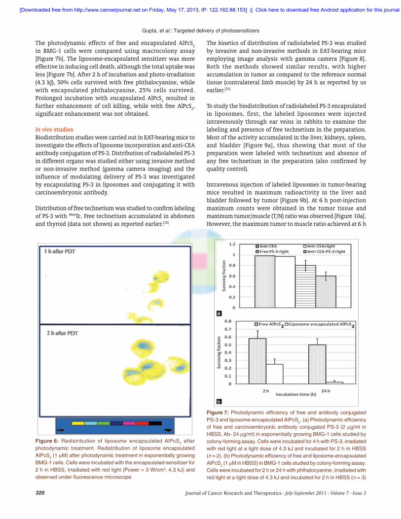
After photo-irradiation, the granular, intense red fluorescence of liposome-encapsulated AlPcS
2 changed into diffused, faint
fluorescence [Figure 6]. However, for free AlPcS2, no significant
difference was observed before and after irradiation (data not shown). Similar results have been reported with liposomally delivered zinc phthalocyanine (ZnPc), where complete disappearance of the fluorescence-encapsulated ZnPc has been found in epithelial cells during light exposure.[34]
The influence of antibody conjugation and liposome encapsulation of PS-3 and AlPcS
2 on photodynamic effects was
studied by assessing the clonogenic survival of BMG-1 cells using macrocolony assay.
Figure 7a shows a comparison of photodynamic effects of anti-CEA-3 conjugated PS-3 with free PS-3. Conjugation of the photosensitizer to the antibody resulted in enhanced efficiency of the treatment for cell inactivation. With free PS-3 (2 g/ml), about 80% cells survived while with the conjugate (Ab- 24 g/ ml; PS-3 - 2 g/ml) only 60% cells survived the treatment [Figure 7a]. Antibody alone or in the presence of light did not result in any cell death. Increased efficiency of conjugated PS-3 could be due to its localization at sensitive sites in the cells.
Figure 4: Intracellular localization of antibody-conjugated PS-3. Time-dependent localization of anti-CEA-PS-3 conjugate (5 g/ml) in exponentially growing BMG-1 cells observed by fluorescence microscopy
Figure 5: Intracellular localization of liposome encapsulated AlPcS2. (a) Time-dependent localization of free AlPcS2 (5 M in HBSS) and liposome encapsulated AlPcS2 (1 M) in exponentially growing BMG-1 cells observed by fl uorescence microscopy. (b) Identifi cation of lysosomes using acridine orange (red spots); location of the nucleus (N) is also indicated.
[Downloaded free from http://www.cancerjournal.net on Friday, May 17, 2013, IP: 122.162.88.153] || Click here to download free Android application for this journal
320 Journal of Cancer Research and Therapeutics - July-September 2011 - Volume 7 - Issue 3
Gupta, et al.: Targeted delivery of photosensitizers
The photodynamic effects of free and encapsulated AlPcS2
in BMG-1 cells were compared using macrocolony assay [Figure 7b]. The liposome-encapsulated sensitizer was more effective in inducing cell death, although the total uptake was less [Figure 7b]. After 2 h of incubation and photo-irradiation (4.3 kJ), 50% cells survived with free phthalocyanine, while with encapsulated phthalocyanine, 25% cells survived. Prolonged incubation with encapsulated AlPcS
2 resulted in
further enhancement of cell killing, while with free AlPcS2,
significant enhancement was not obtained.
In vivo studiesBiodistribution studies were carried out in EAT-bearing mice to investigate the effects of liposome incorporation and anti-CEA antibody conjugation of PS-3. Distribution of radiolabeled PS-3 in different organs was studied either using invasive method or non-invasive method (gamma camera imaging) and the influence of modulating delivery of PS-3 was investigated by encapsulating PS-3 in liposomes and conjugating it with carcinoembryonic antibody.
Distribution of free technetium was studied to confirm labeling of PS-3 with 99mTc. Free technetium accumulated in abdomen and thyroid (data not shown) as reported earlier.[35]
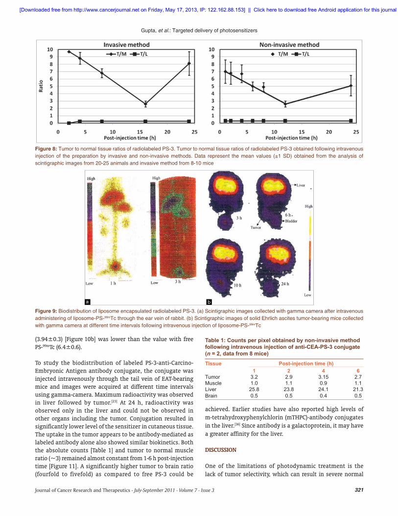
The kinetics of distribution of radiolabeled PS-3 was studied by invasive and non-invasive methods in EAT-bearing mice employing image analysis with gamma camera [Figure 8]. Both the methods showed similar results, with higher accumulation in tumor as compared to the reference normal tissue (contralateral limb muscle) by 24 h as reported by us earlier.[23]
To study the biodistribution of radiolabeled PS-3 encapsulated in liposomes, first, the labeled liposomes were injected intravenously through ear veins in rabbits to examine the labeling and presence of free technetium in the preparation. Most of the activity accumulated in the liver, kidneys, spleen, and bladder [Figure 9a], thus showing that most of the preparation were labeled with technetium and absence of any free technetium in the preparation (also confirmed by quality control).
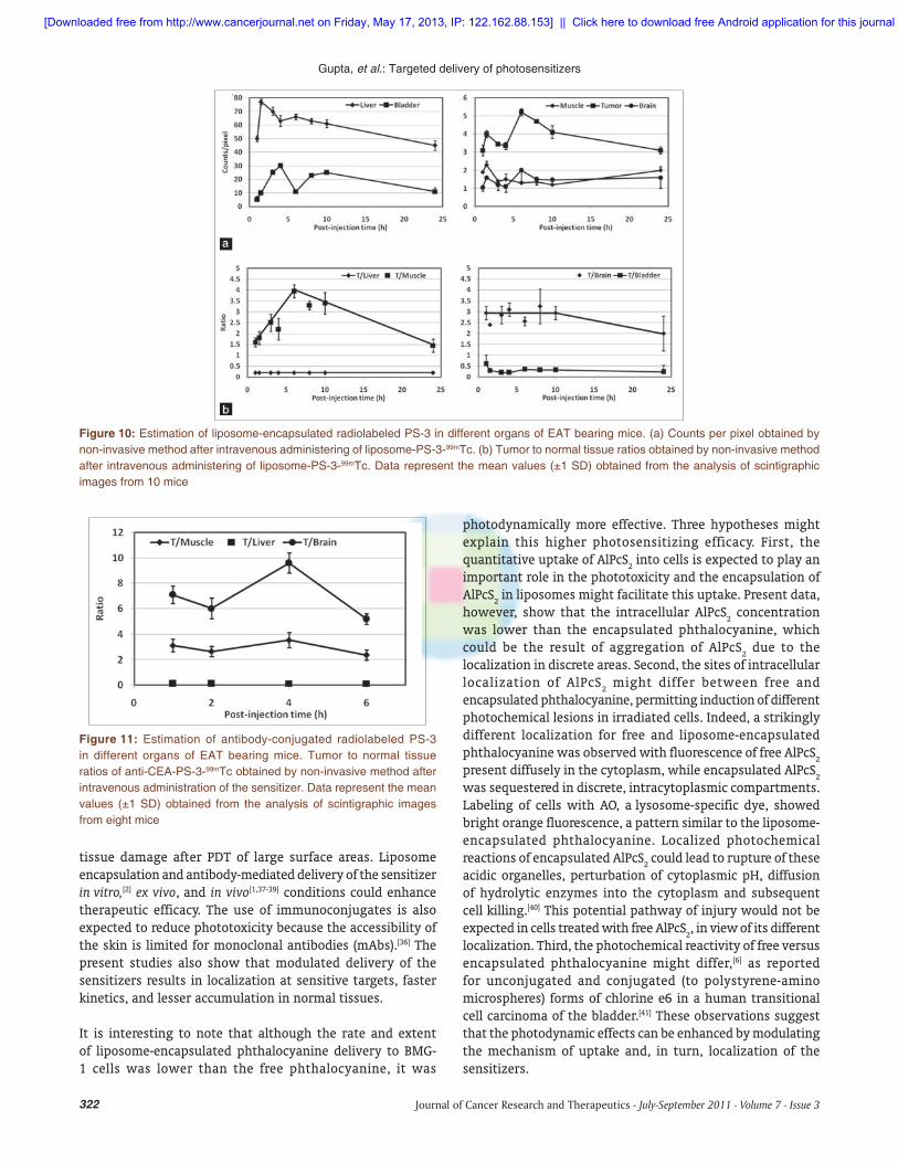
Intravenous injection of labeled liposomes in tumor-bearing mice resulted in maximum radioactivity in the liver and bladder followed by tumor [Figure 9b]. At 6 h post-injection maximum counts were obtained in the tumor tissue and maximum tumor/muscle (T/N) ratio was observed [Figure 10a]. However, the maximum tumor to muscle ratio achieved at 6 h
Figure 6: Redistribution of liposome encapsulated AlPcS2 after photodynamic treatment. Redistribution of liposome encapsulated AlPcS2 (1 M) after photodynamic treatment in exponentially growing BMG-1 cells. Cells were incubated with the encapsulated sensitizer for 2 h in HBSS, irradiated with red light (Power = 3 W/cm2; 4.3 kJ) and observed under fl uorescence microscope
Figure 7: Photodynamic effi ciency of free and antibody conjugated PS-3 and liposome-encapsulated AlPcS2. (a) Photodynamic effi ciency of free and carcinoembryonic antibody conjugated PS-3 (2 g/ml in HBSS, Ab- 24 g/ml) in exponentially growing BMG-1 cells studied by colony-forming assay. Cells were incubated for 4 h with PS-3, irradiated with red light at a light dose of 4.3 kJ and incubated for 2 h in HBSS (n = 2). (b) Photodynamic effi ciency of free and liposome-encapsulated AlPcS2 (1 M in HBSS) in BMG-1 cells studied by colony-forming assay. Cells were incubated for 2 h or 24 h with phthalocyanine, irradiated with red light at a light dose of 4.3 kJ and incubated for 2 h in HBSS (n = 3)
[Downloaded free from http://www.cancerjournal.net on Friday, May 17, 2013, IP: 122.162.88.153] || Click here to download free Android application for this journal
321Journal of Cancer Research and Therapeutics - July-September 2011 - Volume 7 - Issue 3
Gupta, et al.: Targeted delivery of photosensitizers
Table 1: Counts per pixel obtained by non-invasive method following intravenous injection of anti-CEA-PS-3 conjugate (n = 2, data from 8 mice)
Tissue Post-injection time (h)1 2 4 6
Tumor 3.2 2.9 3.15 2.7Muscle 1.0 1.1 0.9 1.1Liver 25.8 23.8 24.1 21.3Brain 0.5 0.5 0.4 0.5
Figure 8: Tumor to normal tissue ratios of radiolabeled PS-3. Tumor to normal tissue ratios of radiolabeled PS-3 obtained following intravenous injection of the preparation by invasive and non-invasive methods. Data represent the mean values (±1 SD) obtained from the analysis of scintigraphic images from 20-25 animals and invasive method from 8-10 mice
Figure 9: Biodistribution of liposome encapsulated radiolabeled PS-3. (a) Scintigraphic images collected with gamma camera after intravenous administering of liposome-PS-99mTc through the ear vein of rabbit. (b) Scintigraphic images of solid Ehrlich ascites tumor-bearing mice collected with gamma camera at different time intervals following intravenous injection of liposome-PS-99mTc
(3.94±0.3) [Figure 10b] was lower than the value with free PS-99mTc (6.4±0.6).
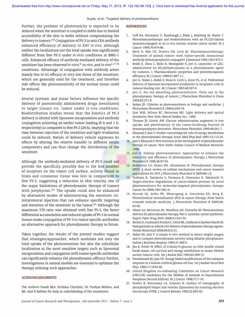
To study the biodistribution of labeled PS-3-anti-Carcino-Embryonic Antigen antibody conjugate, the conjugate was injected intravenously through the tail vein of EAT-bearing mice and images were acquired at different time intervals using gamma-camera. Maximum radioactivity was observed in liver followed by tumor.[23] At 24 h, radioactivity was observed only in the liver and could not be observed in other organs including the tumor. Conjugation resulted in significantly lower level of the sensitizer in cutaneous tissue. The uptake in the tumor appears to be antibody-mediated as labeled antibody alone also showed similar biokinetics. Both the absolute counts [Table 1] and tumor to normal muscle ratio (~3) remained almost constant from 1-6 h post-injection time [Figure 11]. A significantly higher tumor to brain ratio (fourfold to fivefold) as compared to free PS-3 could be
achieved. Earlier studies have also reported high levels of m-tetrahydroxyphenylchlorin (mTHPC)-antibody conjugates in the liver.[36] Since antibody is a galactoprotein, it may have a greater affinity for the liver.
DISCUSSION
One of the limitations of photodynamic treatment is the lack of tumor selectivity, which can result in severe normal
[Downloaded free from http://www.cancerjournal.net on Friday, May 17, 2013, IP: 122.162.88.153] || Click here to download free Android application for this journal
322 Journal of Cancer Research and Therapeutics - July-September 2011 - Volume 7 - Issue 3
Gupta, et al.: Targeted delivery of photosensitizers
tissue damage after PDT of large surface areas. Liposome encapsulation and antibody-mediated delivery of the sensitizer in vitro,[2] ex vivo, and in vivo[1,37-39] conditions could enhance therapeutic efficacy. The use of immunoconjugates is also expected to reduce phototoxicity because the accessibility of the skin is limited for monoclonal antibodies (mAbs).[36] The present studies also show that modulated delivery of the sensitizers results in localization at sensitive targets, faster kinetics, and lesser accumulation in normal tissues.
It is interesting to note that although the rate and extent of liposome-encapsulated phthalocyanine delivery to BMG-1 cells was lower than the free phthalocyanine, it was
photodynamically more effective. Three hypotheses might explain this higher photosensitizing efficacy. First, the quantitative uptake of AlPcS
2 into cells is expected to play an
important role in the phototoxicity and the encapsulation of AlPcS
2 in liposomes might facilitate this uptake. Present data,
however, show that the intracellular AlPcS2 concentration
was lower than the encapsulated phthalocyanine, which could be the result of aggregation of AlPcS
2 due to the
localization in discrete areas. Second, the sites of intracellular localization of AlPcS
2 might differ between free and
encapsulated phthalocyanine, permitting induction of different photochemical lesions in irradiated cells. Indeed, a strikingly different localization for free and liposome-encapsulated phthalocyanine was observed with fluorescence of free AlPcS
2
present diffusely in the cytoplasm, while encapsulated AlPcS2
was sequestered in discrete, intracytoplasmic compartments. Labeling of cells with AO, a lysosome-specific dye, showed bright orange fluorescence, a pattern similar to the liposome-encapsulated phthalocyanine. Localized photochemical reactions of encapsulated AlPcS
2 could lead to rupture of these
acidic organelles, perturbation of cytoplasmic pH, diffusion of hydrolytic enzymes into the cytoplasm and subsequent cell killing.[40] This potential pathway of injury would not be expected in cells treated with free AlPcS
2, in view of its different
localization. Third, the photochemical reactivity of free versus encapsulated phthalocyanine might differ,[6] as reported for unconjugated and conjugated (to polystyrene-amino microspheres) forms of chlorine e6 in a human transitional cell carcinoma of the bladder.[41] These observations suggest that the photodynamic effects can be enhanced by modulating the mechanism of uptake and, in turn, localization of the sensitizers.
Figure 10: Estimation of liposome-encapsulated radiolabeled PS-3 in different organs of EAT bearing mice. (a) Counts per pixel obtained by non-invasive method after intravenous administering of liposome-PS-3-99mTc. (b) Tumor to normal tissue ratios obtained by non-invasive method after intravenous administering of liposome-PS-3-99mTc. Data represent the mean values (±1 SD) obtained from the analysis of scintigraphic images from 10 mice
Figure 11: Estimation of antibody-conjugated radiolabeled PS-3 in different organs of EAT bearing mice. Tumor to normal tissue ratios of anti-CEA-PS-3-99mTc obtained by non-invasive method after intravenous administration of the sensitizer. Data represent the mean values (±1 SD) obtained from the analysis of scintigraphic images from eight mice
[Downloaded free from http://www.cancerjournal.net on Friday, May 17, 2013, IP: 122.162.88.153] || Click here to download free Android application for this journal
323Journal of Cancer Research and Therapeutics - July-September 2011 - Volume 7 - Issue 3
Gupta, et al.: Targeted delivery of photosensitizers
Further, the problem of phototoxicity is expected to be reduced when the sensitizer is coupled to mAbs due to limited accessibility of the skin to mAbs without compromising the delivery to tumors.[36] Conjugation of PS-3 to anti-CEA antibody enhanced efficiency of delivery to EAT in vivo, although neither the localization nor the total uptake was significantly different from free PS-3 under in vitro conditions in BMG-1 cells. Enhanced efficacy of antibody-mediated delivery of the sensitizer has been observed in vitro,[2] ex vivo, and in vivo[1,37- 39] conditions. Advantage of conjugation with antibody arises mainly due to its efficacy at very low doses of the sensitizer, which are generally used for the treatment, and therefore side effects like photosensitivity of the normal tissue could be reduced.
Several systemic and tissue factors influence the specific delivery of parenterally administered drugs (sensitizers) to target tissues viz. tumor under in vivo conditions. Biodistribution studies reveal that the kinetics of PS-3 delivery is altered with liposome encapsulation and antibody conjugation achieving an earlier tumor loading (6 h and 1 h, respectively) as compared to free PS-3 (24 h), implying that the time between injection of the sensitizer and light irradiation could be reduced. Nature of delivery vehicle expresses its effects by altering the relative transfer to different serum components and can thus change the distribution of the sensitizer.
Although the antibody-mediated delivery of PS-3 could not provide the specificity, possibly due to the less number of receptors on the tumor cell surface, activity found in brain and cutaneous tissue was less in comparison to free PS-3, suggesting a reduction in skin toxicity, one of the major limitations of photodynamic therapy of tumors with porphyrins.[42] The uptake could also be enhanced by alternative modes of administering the sensitizer viz. intratumoral injection that can enhance specific targeting and retention of the sensitizer in the tumor.[23] Although the maximum T/N ratio was obtained with free PS-3, the faster differential accumulation and reduced uptake of PS-3 in normal tissues make conjugation of PS-3 to tumor-specific antibodies an alternative approach for photodynamic therapy in future.
Taken together, the results of the present studies suggest that strategies/approaches, which modulate not only the total uptake of the photosensitizer but also the subcellular localization at the most sensitive targets such as liposomal encapsulation and conjugation with tumor-specific antibodies can significantly enhance the photodynamic efficacy. Further, investigations in animal models are essential to optimize the therapy utilizing such approaches.
ACKNOWLEDGMENTS
The authors thank Mrs. Krishna Chuttani, Dr. Pushpa Mishra, and Mr. Anil K Babbar for help in radiolabeling of the sensitizers.
REFERENCES
1. Goff BA, Hermanto U, Rumbaugh J, Blake J, Bamberg M, Hasan T. Photoimmunotherapy and biodistribution with an OC125-chlorin immunoconjugate in an in vivo murine ovarian cancer model. Br J Cancer 1994;70:474-80.
2. Mew D, Wat CK, Towers GH, Levy JG. Photoimmunotherapy: Treatment of animal tumors with tumor-specific monoclonal antibody-hematoporphyrin conjugates. J Immunol 1983;130:1473-7.
3. Reddi E, Zhou C, Biolo R, Menegaldo E, Jori G. Liposome- or LDL-administered Zn (II)-phthalocyanine as a photodynamic agent for tumours. I. Pharmacokinetic properties and phototherapeutic efficiency. Br J Cancer 1990;61:407-11.
4. Jori G, Tomio L, Reddi E, Rossi E, Corti L, Zorat PL, et al. Preferential delivery of liposome-incorporated porphyrins to neoplastic cells in tumour-bearing rats. Br J Cancer 1983;48:307-9.
5. Jori G. Far red absorbing photosensitizers: Their use in the photodynamic therapy of tumors. J Photochem Photobiol A Chem 1992;62:371-8.
6. Spikes JD. Chlorins as photosensitizers in biology and medicine. J Photochem Photobiol B 1990;6:259-74.
7. Star WM, Wilson BC, Patterson MS. Light delivery and optical dosimetry. New York: Marcel Dekker Inc.; 1992.
8. Thomas JP, Girotti AW. Glucose administration augments in vivo uptake and phototoxicity of the tumor-localizing fraction of hematoporphyrin derivative. Photochem Photobiol 1989;49:241-7.
9. Khanum F, Jain V. Studies concerning the role of energy metabolism in the photodynamic therapy. Assoc Med Phys Bull 1987;12:113-24.
10. Jain V, Khanum F. New horizons in the development of photodynamic therapy of cancer. New Delhi: Indian Council of Medical Research; 1990.
11. Jori G. Tumour photosensitizers: Approaches to enhance the selectivity and efficiency of photodynamic therapy. J Photochem Photobiol B 1996;36:87-93.
12. Robertson CA, Evans DH, Abrahamse H. Photodynamic therapy (PDT): A short review on cellular mechanisms and cancer research applications for PDT. J Photochem Photobiol B 2009;96:1-8.
13. Toshima K, Tanimoto S, Tsumura K, Umezawa K, Takahashi D. Target-selective degradation of cancer-related proteins by novel photosensitizers for molecular-targeted photodynamic therapy. Cancer Sci 2009;100:1581-4.
14. Norum OJ, Selbo PK, Weyergang A, Giercksky KE, Berg K. Photochemical internalization (PCI) in cancer therapy: From bench towards bedside medicine. J Photochem Photobiol B 2009;96:83-92.
15. Sibani SA, McCarron PA, Woolfson AD, Donnelly RF. Photosensitiser delivery for photodynamic therapy. Part 2: Systemic carrier platforms. Expert Opin Drug Deliv 2008;5:1241-54.
16. Bechet D, Couleaud P, Frochot C, Viriot ML, Guillemin F, Barberi-Heyob M. Nanoparticles as vehicles for delivery of photodynamic therapy agents. Trends Biotechnol 2008;26:612-21.
17. Yadav HS, Jain V. A simple in vitro method to detect singlet oxygen and to compare photodynamic activity using alkaline phosphatase. Indian J Biochem Biophys 1994;31:490-5.
18. Jha B, Pohlit W. Effect of 2-deoxy-D-glucose on DNA double strand break repair, cell survival and energy metabolism in euoxic Ehrlich ascites tumour cells. Int J Radiat Biol 1992;62:409-15.
19. Dwarkanath BS, Jain VK. Energy linked modifications of the radiation response in a human cerebral glioma cell line. Int J Radiat Oncol Biol Phys 1989;17:1033-40.
20. United Kingdom Co-ordinating Committee on Cancer Research (UKCCCR) Guidelines for the Welfare of Animals in Experimental Neoplasia (Second Edition). Br J Cancer 1998;77:1-10.
21. Fowler K, Bottomley LA, Schreier H. Surface of tomography of phospholipid bilayer and vesicles (liposomes) by scanning electron microscopy (STM). J Control Release 1992;22:283-92.
[Downloaded free from http://www.cancerjournal.net on Friday, May 17, 2013, IP: 122.162.88.153] || Click here to download free Android application for this journal
324 Journal of Cancer Research and Therapeutics - July-September 2011 - Volume 7 - Issue 3
Gupta, et al.: Targeted delivery of photosensitizers
22. Lewis MR, Raubitschek A, Shively JE. A facile, water-soluble method for modification of proteins with DOTA. Use of elevated temperature and optimized pH to achieve high specific activity and high chelate stability in radiolabeled immunoconjugates. Bioconjug Chem 1994;5:565-76.
23. Gupta S, Mishra AK, Muralidhar K, Jain V. Improved targeting of photosensitizers by intratumoral administration of immunoconjugates. Technol Cancer Res Treat 2004;3:295-301.
24. Babbar AK, Singh AK, Goel HC, Chauhan UP, Sharma RK. Evaluation of (99m)Tc-labeled photosan-3, a hematoporphyrin derivative, as a potential radiopharmaceutical for tumor scintigraphy. Nucl Med Biol 2000;27:587-92.
25. Miller GG, Brown K, Moore RB, Diwu ZJ, Liu J, Huang L, et al. Uptake kinetics and intracellular localization of hypocrellin photosensitizers for photodynamic therapy: A confocal microscopy study. Photochem Photobiol 1995;61:632-8.
26. Redmond RW, Land EJ, Truscott TG. Aggregation effects on the photophysical properties of porphyrins in relation to mechanisms involved in photodynamic therapy. Adv Exp Med Biol 1985;193:293-302.
27. Gupta S, Dwarakanath BS, Muralidhar K, Jain V. Cellular uptake, localization and photodynamic effects of haematoporphyrin derivative in human glioma and squamous carcinoma cell lines. J Photochem Photobiol B 2003;69:107-20.
28. Moan J, Berg K, Anholt H, Madslien K. Sulfonated aluminium phthalocyanines as sensitizers for photochemotherapy. Effects of small light doses on localization, dye fluorescence and photosensitivity in V79 cells. Int J Cancer 1994;58:865-70.
29. Peng Q, Farrants GW, Madslien K, Bommer JC, Moan J, Danielsen HE, et al. Subcellular localization, redistribution and photobleaching of sulfonated aluminum phthalocyanines in a human melanoma cell line. Int J Cancer 1991;49:290-5.
30. Allison AC, Young MR. Vital staining and fluorescence microscopy of lysosomes. North Holland, Amsterdam; 1969.
31. Slater TF, Riley PA. Photosensitization and lysosomal damage. Nature 1966;209:151-4.
32. Austen JD, McConnell S, Carrano CJ, Tsutsul M. Intracellular localization of meso-Tetra(p-sulfophenyl)porphine: A potential tumor localizing agent. Cancer Treat Rep 1978;62:511-8.
33. Santus R, Kohen C, Kohen E, Reyftmann JP, Morliere P, Dubertret L, et al. Permeation of lysosomal membranes in the course of
photosensitization with methylene blue and hematoporphyrin: Study by cellular microspectrofluorometry. Photochem Photobiol 1983;38:71-7.
34. Ruck A, Beck G, Bachor R, Akgun N, Gschwend MH, Steiner R. Dynamic fluorescence changes during photodynamic therapy in vivo and in vitro of hydrophilic A1(III) phthalocyanine tetrasulphonate and lipophilic Zn(II) phthalocyanine administered in liposomes. J Photochem Photobiol B 1996;36:127-33.
35. Rousseau J, Ali H, Lamoureux G, Lebel E, van Lier JE. Synthesis, tissue distribution and tumor uptake of 99mTc- and 67Ga-tetrasulfophthalocyanine. Int J Appl Radiat Isot 1985;36:709-16.
36. Vrouenraets MB, Visser GW, Stewart FA, Stigter M, Oppelaar H, Postmus PE, et al. Development of meta-tetrahydroxyphenylchlorin-monoclonal antibody conjugates for photoimmunotherapy. Cancer Res 1999;59:1505-13.
37. Goff BA, Bamberg M, Hasan T. Photoimmunotherapy of human ovarian carcinoma cells ex vivo. Cancer Res 1991;51:4762-7.
38. Goff BA, Bamberg M, Hasan T. Experimental photodynamic treatment of ovarian carcinoma cells with immunoconjugates. Antibody Immunoconj Radiopharm 1992;5:191-9.
39. Goff BA, Blake J, Bamberg MP, Hasan T. Treatment of ovarian cancer with photodynamic therapy and immunoconjugates in a murine ovarian cancer model. Br J Cancer 1996;74:1194-8.
40. Zdolsek JM, Olsson GM, Brunk UT. Photooxidative damage to lysosomes of cultured macrophages by acridine orange. Photochem Photobiol 1990;51:67-76.
41. Bachor R, Shea CR, Gillies R, Hasan T. Photosensitized destruction of human bladder carcinoma cells treated with chlorin e6-conjugated microspheres. Proc Natl Acad Sci U S A 1991;88:1580-4.
42. Zhou CN, Yang WZ, Ding ZX, Wang YX, Shen H, Fan XJ, et al. The biological effects of photodynamic therapy on normal skin in mice–II. An electron microscopic study. Adv Exp Med Biol 1985;193:111-5.
Dispatch and return notification by E-mail The journal now sends email notification to its members on dispatch of a print issue. The notification is sent to those members who have provided their email address to the association/journal office. The email alerts you about an outdated address and return of issue due to incomplete/incorrect address.
If you wish to receive such email notification, please send your email along with the membership number and full mailing address to the editorial office by email.
Cite this article as: Gupta S, Dwarakanath BS, Chaudhury NK, Mishra AK, Muralidhar K, Jain V. In vitro and in vivo targeted delivery of photosensitizers to the tumor cells for enhanced photodynamic effects. J Can Res Ther 2011;7:314-24.
Source of Support: S G was a recipient of fellowship from University Grants Commission and Council of Scientifi c and Industrial Research, Government of India, Confl ict of Interest: None declared.
[Downloaded free from http://www.cancerjournal.net on Friday, May 17, 2013, IP: 122.162.88.153] || Click here to download free Android application for this journal


![1,3,4-oxadiazol-2-yl]sulfanyl}acetamides as suitable ther](https://static.fdokumen.com/doc/165x107/6319f8931e5d335f8d0b61c0/134-oxadiazol-2-ylsulfanylacetamides-as-suitable-ther.jpg)