Impact of Photosensitizers Activation on Intracellular Trafficking and Viscosity
13
Impact of Photosensitizers Activation on Intracellular Trafficking and Viscosity Kelly Aubertin 1 , Stéphanie Bonneau 2 , Amanda K. A. Silva 1 , Jean-Claude Bacri 1 , François Gallet 1 , Claire Wilhelm 1* 1 Laboratoire Matière et Systèmes Complexes (MSC), CNRS and Université Paris Diderot, Paris, France, 2 Laboratoire Jean Perrin-CNRS, Université Pierre et Marie Curie, Paris 6, Paris, France Abstract The intracellular microenvironment is essential for the efficiency of photo-induced therapies, as short-lived reactive oxygen species generated must diffuse through their intracellular surrounding medium to reach their cellular target. Here, by combining measurements of local cytoplasmic dissipation and active trafficking, we found that photosensitizers activation induced small changes in surrounding viscosity but a massive decrease in diffusion. These effects are the signature of a return to thermodynamic equilibrium of the system after photo-activation and correlated with depolymerization of the microtubule network, as shown in a reconstituted system. These mechanical measurements were performed with two intracellular photosensitizing chlorins having similar quantum yield of singlet oxygen production but different intracellular localizations (cytoplasmic for mTHPC, endosomal for TPCS2a). These two agents demonstrated different intracellular impact. Citation: Aubertin K, Bonneau S, Silva AKA, Bacri J-C, Gallet F, et al. (2013) Impact of Photosensitizers Activation on Intracellular Trafficking and Viscosity. PLoS ONE 8(12): e84850. doi:10.1371/journal.pone.0084850 Editor: Nikolai Lebedev, US Naval Reseach Laboratory, United States of America Received August 9, 2013; Accepted November 19, 2013; Published December 27, 2013 Copyright: © 2013 Aubertin et al. This is an open-access article distributed under the terms of the Creative Commons Attribution License, which permits unrestricted use, distribution, and reproduction in any medium, provided the original author and source are credited. Funding: This work was supported by the by the European commission's FP7 (project Magnifyco NMP4-SL-2009-228622). The funders had no role in study design, data collection and analysis, decision to publish, or preparation of the manuscript. Competing interests: The authors have declared that no competing interests exist. * E-mail: [email protected] Introduction Photodynamic therapy uses photo-activated organic molecules (porphyrins, chlorins, phthalocyanins, etc.) with very special photo-physical properties. Owing to their triplet state, irradiation of these photosensitizers generates reactive species such as singlet oxygen [1]. The lifetime of these molecular species is very short [2,3] and their action is highly localized [4–6]. The ability of photosensitizers to target a specific cellular compartment explains their potential to modify and control cellular physiology. For example, photochemical internalization (PCI) using the photoactivation of photosensitizers that localize in endosomes, induces endosomal membrane changes, enabling release into the cytosol of an active substance stored in the endosomal compartment [7,8]. More marked changes occur if the photosensitizer is localized in the cytoplasm - particularly affecting mitochondria, endoplasmic reticulum or functional organelles - and potentially leading to cell death by necrosis or apoptosis [9,10]. This approach, called photodynamic therapy (PDT), is already used in the clinic for cancer treatment [11]. While the cellular effects of photosensitizers (cytotoxicity, membrane permeabilization, …) have been extensively explored [12,13], their impact on intracellular mechanics and trafficking are much less documented [14,15]. In particular, as the cytotoxic effect is based on the production of very short- lived reactive oxygen species, it is important to know the mechanical properties of the medium through which these species must diffuse in order to reach their target. A first approach to this issue was recently developed using a photosensitive molecule coupled to a molecular rotor whose orientation could be optically determined, allowing the diffusion properties of the molecule to be followed [15]. However, the relationship between diffusion and dissipation is not direct, especially in a living system [16–19]. For a Newtonian fluid at thermodynamic equilibrium, the diffusion coefficient is directly related to the viscosity, through the Stokes-Einstein relationship. This particular expression of the Fluctuation-Dissipation Theorem (FDT) can be generalized to any non-newtonain fluid, provided that it remains at thermodynamical equilibrium. However, in the intracellular medium, molecular motors regularly consume ATP, converting chemical energy into the mechanical work needed to sort and transport each cargo to its final destination [20–23]. The intracellular space is thus, by its nature, a system far from thermal equilibrium, thus challenging the validity of the fluctuation-dissipation theorem in this situation. The ratio of the PLOS ONE | www.plosone.org 1 December 2013 | Volume 8 | Issue 12 | e84850
-
Upload
independent -
Category
Documents
-
view
0 -
download
0
Transcript of Impact of Photosensitizers Activation on Intracellular Trafficking and Viscosity

Impact of Photosensitizers Activation on IntracellularTrafficking and ViscosityKelly Aubertin1 Steacutephanie Bonneau2 Amanda K A Silva1 Jean-Claude Bacri1 Franccedilois Gallet1 ClaireWilhelm1
1 Laboratoire Matiegravere et Systegravemes Complexes (MSC) CNRS and Universiteacute Paris Diderot Paris France 2 Laboratoire Jean Perrin-CNRS Universiteacute Pierre etMarie Curie Paris 6 Paris France
Abstract
The intracellular microenvironment is essential for the efficiency of photo-induced therapies as short-lived reactiveoxygen species generated must diffuse through their intracellular surrounding medium to reach their cellular targetHere by combining measurements of local cytoplasmic dissipation and active trafficking we found thatphotosensitizers activation induced small changes in surrounding viscosity but a massive decrease in diffusionThese effects are the signature of a return to thermodynamic equilibrium of the system after photo-activation andcorrelated with depolymerization of the microtubule network as shown in a reconstituted system These mechanicalmeasurements were performed with two intracellular photosensitizing chlorins having similar quantum yield of singletoxygen production but different intracellular localizations (cytoplasmic for mTHPC endosomal for TPCS2a) Thesetwo agents demonstrated different intracellular impact
Citation Aubertin K Bonneau S Silva AKA Bacri J-C Gallet F et al (2013) Impact of Photosensitizers Activation on Intracellular Trafficking andViscosity PLoS ONE 8(12) e84850 doi101371journalpone0084850
Editor Nikolai Lebedev US Naval Reseach Laboratory United States of America
Received August 9 2013 Accepted November 19 2013 Published December 27 2013
Copyright copy 2013 Aubertin et al This is an open-access article distributed under the terms of the Creative Commons Attribution License which permitsunrestricted use distribution and reproduction in any medium provided the original author and source are credited
Funding This work was supported by the by the European commissions FP7 (project Magnifyco NMP4-SL-2009-228622) The funders had no role instudy design data collection and analysis decision to publish or preparation of the manuscript
Competing interests The authors have declared that no competing interests exist
E-mail clairewilhelmuniv-paris-diderotfr
Introduction
Photodynamic therapy uses photo-activated organicmolecules (porphyrins chlorins phthalocyanins etc) with veryspecial photo-physical properties Owing to their triplet stateirradiation of these photosensitizers generates reactive speciessuch as singlet oxygen [1] The lifetime of these molecularspecies is very short [23] and their action is highly localized[4ndash6]
The ability of photosensitizers to target a specific cellularcompartment explains their potential to modify and controlcellular physiology For example photochemical internalization(PCI) using the photoactivation of photosensitizers that localizein endosomes induces endosomal membrane changesenabling release into the cytosol of an active substance storedin the endosomal compartment [78] More marked changesoccur if the photosensitizer is localized in the cytoplasm -particularly affecting mitochondria endoplasmic reticulum orfunctional organelles - and potentially leading to cell death bynecrosis or apoptosis [910] This approach calledphotodynamic therapy (PDT) is already used in the clinic forcancer treatment [11]
While the cellular effects of photosensitizers (cytotoxicitymembrane permeabilization hellip) have been extensively
explored [1213] their impact on intracellular mechanics andtrafficking are much less documented [1415] In particular asthe cytotoxic effect is based on the production of very short-lived reactive oxygen species it is important to know themechanical properties of the medium through which thesespecies must diffuse in order to reach their target A firstapproach to this issue was recently developed using aphotosensitive molecule coupled to a molecular rotor whoseorientation could be optically determined allowing the diffusionproperties of the molecule to be followed [15]
However the relationship between diffusion and dissipationis not direct especially in a living system [16ndash19] For aNewtonian fluid at thermodynamic equilibrium the diffusioncoefficient is directly related to the viscosity through theStokes-Einstein relationship This particular expression of theFluctuation-Dissipation Theorem (FDT) can be generalized toany non-newtonain fluid provided that it remains atthermodynamical equilibrium However in the intracellularmedium molecular motors regularly consume ATP convertingchemical energy into the mechanical work needed to sort andtransport each cargo to its final destination [20ndash23] Theintracellular space is thus by its nature a system far fromthermal equilibrium thus challenging the validity of thefluctuation-dissipation theorem in this situation The ratio of the
PLOS ONE | wwwplosoneorg 1 December 2013 | Volume 8 | Issue 12 | e84850
chemical energy input over thermal fluctuations energy canthen be measured as described for aging complex systems[24] as well as for the cell interior [17] This ratio can reach1000 implying diffusion coefficients 1000 times greater thanthose deduced from viscosity measurements using FDT
To access this ratio or more simply to measure dissipativeand diffusive properties independently one must be able atthe same time and in the same system to make a passivemeasurement (ldquofluctuationrdquo) as well as an active measurement(ldquodissipationrdquo) This is the approach we used here takingadvantage of magnetic micromanipulation techniquesdeveloped in recent years to measure local viscoelasticity [25]while at the same time following the transport modalities ofintracellular vesicles
Two types of chlorins m-THPC (hydrophobic) and TPCS2a(more hydrophilic) were chosen as photosensitizers as theirdifferent hydrophobicities affect their intracellular localization[26ndash28] Endosomes containing magnetic nanoparticles wereused as both probes of the trafficking and magnetic probes forthe local viscosity within irradiated cells
Combined measurement of rheological properties and activetransport according to the photosensitizer localization and theduration of the treatment and post-treatment steps shouldprovide information on the intracellular mechanisms of action ofphotodynamic stress a largely unexplored area
Results
Simultaneous internalization of photosensitizers andmagnetic nanoparticles by tumor cells
Incubation of cells with magnetic nanoparticles andphotosensitizers led to internalization of both components(Figure 1A) Each cell contained an average of 107
nanoparticles equivalent to 10 pg of iron The nanoparticlespenetrate through the endocytic pathway and cluster withinendosomes as previously demonstrated [29ndash31]
Then if a homogeneous magnetic field is applied eachendosome acquires a magnetic moment which aligns with itsneighbors under the effect of magnetic dipole forces Thiscreates small intracellular chains that can be manipulated viathe external magnetic field The two photosensitizers are alsointernalized at dose equivalent of 02 fmoles per cell onaverage However m-THPC spreads in the cytoplasmiccompartment while TPCS2a is found in the endosomalcompartments
After irradiation (deposited energy between 15 and 30 Jcm2) the cellsrsquo metabolic activity was not reduced during thefirst two hours (Figure 1B) In contrast a strong decrease incell viability was detected 24 hours after exposure
We thus obtained magnetic sensors inside cells containingphotosensitizers with different intracellular targets within arange of concentrations at which treatment is cytotoxic at longtimes
Probing intracellular trafficking with magneticendosomes Diffusion measurements
We first aimed at exploring the impact of photodynamicstress on intracellular trafficking Endosomes loaded with
magnetic nanoparticles are easily detectable by opticalmicroscopy It is possible to track their position (xy) in a time-resolved manner (image capture every 100 ms for 100 s) Infigure 2 left images show a few example of endosometrajectories (color lines) superimposed to cells before photo-activation After photo-activation one can see a dramaticdecrease in trafficking (strong shortening of all track lengths)
To quantify such an effect the mean square displacementltΔrsup2(t)gt was calculated for each trajectory (see Methodsequation 4)
Figure 3A shows the ltΔrsup2(t)gt for different treatmentconditions control cells and microtubule disrupted cells m-THPC treatment TPCS2a treatment both at various exposuretimes In all cases ltΔrsup2(t)gt obeys a power law with time
ltΔr sup2 t gt thinsp=2D1stα (1)
α characterizes the type of movement (confinedαlt1diffusiveα=1 directedαgt1 more details are given in theMethods section) D1sreflects the amplitude of motion at thecharacteristic time t=1s Figure 3B summarizes all mean D1s
values extracted from the mean square displacements datapresented in Figure 3A and includes all complementarycontrols D1sfell when the microtubules were depolymerizedand when the photosensitizers were irradiated the fall beingproportional to the dose of irradiation and demonstrating areduction of intracellular motions The value of the exponent α(extracted from the fitting of the curves shown in Figure 3A)provides information related to the mode of the observedmotions In untreated cells exponent αasymp13 is a signature of asuper-diffusive motion related to active transport of theendosomes along the microtubule network by its associatedmolecular motors [1632ndash34] Indeed the same cells treatedwith nocodazole a microtubule depolymerizing agent exhibitan average exponent of 08 indicating confined movementThe same effect was observed after irradiation increasing withthe amount of light energy deposited With m-THPC irradiationfor 1 s or 5 s reduced the exponent to 10 or 06 respectivelywhile with TPCS2a irradiation for 1s 5 s or 20 s reduced theexponent to 12 11 or 07 respectively
At this stage of our understanding the drop in endosomestrafficking after irradiation could be due either to an inhibition ofthe motor-mediated out-of-equilibrium endosomes motions orto a stiffening of the mechanical environment causing anincrease of the local viscosity as previously reported [15] Inthis latter case the viscosity η should rise by the same factoras the recorded drop in displacements (that is up to a hundred-fold) as further discussed in the Discussion section The nextstep therefore requires performing dissipative measurements ofthe endosomes surrounding viscosity to validate one or theother hypothesis
Probing the intracellular viscosity with magneticmanipulation
Such a measurement of the viscosity requires an externallyapplied stress on an internal probe The idea here is to exploitthe magnetic properties of the labeled endosomes to makethem align in chains and impose the chain rotation by an
Intracellular Mechanics Following Photoactivation
PLOS ONE | wwwplosoneorg 2 December 2013 | Volume 8 | Issue 12 | e84850
external magnetic rotating field the lag in time for theendosomal chains to align with the rotated field is ameasurement of viscosity The technical difficulty however is toproduce a rotating magnetic field strong enough to align
magnetic endosomes and deliver a sufficient magnetic torqueto permanently rotate these chains To do so we designed aminiaturized magnetic device (Figure 4) consisting of a set of 4home-made small coils magnetizing 4 soft iron engineered
Figure 1 Intracellular localization of the photosensitizers and the magnetic nanoparticles and metabolic activity (A) Co-internalization in PC3 tumor cells of photosensitizer molecules (mTHPC and TPCS2a) and magnetic nanoparticles mTHPClocalizes in the cytoplasm while TPCS2a is found in vesicular structures The magnetic nanoparticles are concentrated in lateendosomes and lysosomes and align in the direction of the applied magnetic field as shown by electron microscopy (inset) (B)Metabolic activity was not modified 2 hours after exposure regardless of the conditions The subsequent steps therefore involvedviable cells However a noteworthy cytotoxic effect was detected the following day with more than a 80 fall in cellular activity Theerror bars represent the standard error of the mean (SEM)doi 101371journalpone0084850g001
Intracellular Mechanics Following Photoactivation
PLOS ONE | wwwplosoneorg 3 December 2013 | Volume 8 | Issue 12 | e84850
pieces separated by only 06 mm All details and pictures arepresented in the Methods section ldquomagnetic devicerdquo Thanks tothe small spacing the magnetic field created in the center canbe tuned between 0 to 70 mT and its direction can be adjustedwithin the cell sample plane by tuning the current supplyingeach coil In particular if the two pairs of coils are supplied withan alternating current 90deg out of phase the produced magneticfield rotates in the cells plane Chains of endosomes thenundergo a magnetic torque (defined in the Methods) whichforces them to rotate along with the field (if the latter is strongenough) with an angular delay φ (between the direction of thechain and the magnetic field examples given in Figure 5A)This delay is due to the opposition to rotation of thesurrounding medium (creating a viscous restoring torqueproportional to the viscosityη details also given in theMethods) The measure of φ thus provides direct access to theviscosity η according to
η=μ0m2
32πκsin 2φ
F (2)
whereμ0=4π10minus7kg m Aminus2sminus2 m is the endosomes magneticmoment at the applied field Fthe frequency of the field and κ apreviously calibrated geometric factor (κ=67 149 and 247 forchains containing 2 3 and 4 endosomes respectively [3536]
To retrieve quantitative viscosity values we also needed anindependent measurement of the magnetic moment of eachendosomes To do so we extracted them from the cell andinjected them into a magnetophoresis chamber subjected to acalibrated magnetic gradient (280 Tm) Each endosomemigrated with an average speed of about 30 microms whichcorresponds to a magnetic moment for an applied field of50 mT (the one selected for the experiments)ofm=(18plusmn02)10minus16A m2
Before photoactivation the mean value of the viscosityretrieved was 025plusmn003 Pas The error corresponds to thestandard error of the mean (SEM) averaged over more than 25independent measurements One must note however that thestandard deviation is large (in order 45) due to the variabilityof the viscosity within the cytoplasm
Neither the presence of a photosensitizer without irradiationnor cell irradiation without a photosensitizer affected this valueAfter cell exposition to the photosensitizer and irradiation theintracellular viscosity slightly increased to values in order04 Pas (all values are shown in Figure 5B) and the increasewas significant considering a confidence level ofplt001 Bycontrast after nocodazole treatment the mean viscosity valueslightly decreases to 02 Pas but not significantly
Therefore while the diffusion coefficients D1s decreased afterexposure by a factor at least 20 and up to 100 with asignificance at a confidence level of plt0001 for all measuresthe viscosity values for irradiated cells only increased by afactor 2 or lower compared to the ones of untreated cells andwith less significance (plt001) This means that the irradiationstrongly impact the endosomes transport but not theirsurrounding viscosity bringing the system back to a situationclose to equilibrium These results could be explained by theinhibition of molecular motors mediated transport onmicrotubules and we therefore investigated further the impacton the microtubules of photoactivation with the investigatedphotosensitizers
Microtubules targets of the photodynamic effectMicrotubules are in perpetual remodeling [3738] and have
yet been identified as a target during photodynamic treatment[39ndash41] and at this stage it is logical to hypothesize that anaction of the two photosensitizers on the microtubules network
Figure 2 Monitoring of intracellular trafficking The dark compartments (endosomes filled with magnetic nanoparticles) werefollowed for 100 s and their trajectories (before and after exposure) were superimposed on the image of the cell obtained at theoutset of the monitoring phase Movements were clearly inhibited by the action of the photosensitizer (mTHPC and TPCS2a 5 sand 20 s of exposure respectively)doi 101371journalpone0084850g002
Intracellular Mechanics Following Photoactivation
PLOS ONE | wwwplosoneorg 4 December 2013 | Volume 8 | Issue 12 | e84850
may be responsible for the observed decrease in activetrafficking As the reactive species photo-induced via thephotosensitizers present a very short action range theirsubcellular localization may strongly impact their respectiveeffects For TPCS2a unlike for m-THPC the produced oxygenspecies must first cross the membrane of the endosome beforecoming close to the microtubule However their ability to reactwith these membranes is well-known - and used as basis ofPCI [4243] Consequently it is expected that the diffuse-labelled photosensitizer m-THPC reacts faster with themicrotubules To test this possibility we developed an in vitro
system of reconstituted microtubules and followed theirdynamics after irradiation Within the range of intracellularphotosensitizer concentrations (02 fmoles per cell) irradiationhad a significant depolymerizing effect (Figure 6) immediatelyafter 5 s of irradiation with a m-THPC concentration of 2 or 5microM the microtubule depolymerization took only a few secondsFor TPCS2a two kinds of experiments were performed withthe aim to mimic in a reconstructed system the effectsobserved within cells First results similar to those of m-THPCwere obtained with TPCS2a in solution within the sameconcentration range This highlights that both photosensitizers
Figure 3 Quantification of intracellular trafficking (A) The mean square displacement (ltΔrsup2(t)gt ) was calculated for allconditions according to equation (4) and averaged over 30 independent measurements For means of clarity only the mainconditions are represented unlabeled cells non exposed (control light blue) unlabeled cells treated with nocodazole (dark blue)cells treated with m-THPC and exposed for 1s (red) or 5s (dark red) and cells treated with TPCS2a and exposed for 5s (green) or20s (dark green) (B) Diffusion coefficient (D1s) defined in equation (1) It corresponds to the value of the mean square displacementat 1s (ltΔrsup2(1s)gt ) It is shown for all tested conditions the ones presented in part A as well as additional ones including unlabeledcells exposed for 20s (blue) cells treated with both photosensitizers (m-THPC light red and TPCS2a very light green) but nonexposed and cells treated with TPCS2a and exposed 1s (light green) The error bars represent the standard error of the mean(SEM) NS (Non Significant) corresponds to pgt005 corresponds to 001ltplt005 and corresponds to plt 0001doi 101371journalpone0084850g003
Intracellular Mechanics Following Photoactivation
PLOS ONE | wwwplosoneorg 5 December 2013 | Volume 8 | Issue 12 | e84850
which have similar singlet oxygen quantum yields act similarlyin solution as photo-depolymerization agent However withincells the subcellular localization of TPCS2a is strongly differentfrom that of m-THPC it remains sequestrated withinendosomes Then in a second experiment TPCS2a-labelledendosomes were prepared and used as photo-depolymerization agent in the same way as free TPCS2a (iewithin the same photosensitizer concentrations determined byfluorescence spectroscopy) The TPCS2a activity is thenstrongly slackened as compared to free TPCS2a corroboratingthe trapping role of the endosomal membrane for the photo-induced reactive species
Discussion
Here we examined the intracellular impacts of thephotodynamic effect Photosensitizers are used in variousbiomedical applications from induction of cell death for cancertherapy (PDT) to triggering the release of endocytisedmacromolecules (PCI) [944] We chose two ldquostandardrdquophotosensitizers mTHPC and TPCS2a for this study Thesetwo molecules which have very similar chemical formulas(chlorin rings) differ only by a terminal functional group Thissimple difference makes TPCS2a less hydrophobic thanmTHPC and strongly modifies the intracellular localization(cytoplasmic for mTHPC and endosomal for TPCS2a) It isinteresting to note that the therapeutic functions of the twophotosensitizers also differ m-THPC (FOSCAN) is used in theclinic providing effective PDT while TPCS2a is used within thecontext of drug delivery as PCI agent to induce endosomalmembrane rupture and the release of active molecules into thecytoplasm We used this well-controlled model to study theeffect of photodynamic stress on intracellular mechanics
Viscosity changes of the intracellular medium followingirradiation have only been studied previously in a single modelsystem using spectrally resolved fluorescence measurements
of a porphyrin-dimer-based molecular rotor [15] Such anapproach is only valid if the measurement is made atthermodynamic equilibrium This does not hold if the probe islocalized in endosomes which appears to be the case in thislatter model the rotor-photosensitizer appears as a small dot inthe cytoplasm suggesting that it is localized in vesicles(presumably endosomes) The observed diffusion of themolecule thus reflects both its diffusion within the vesicle andthe diffusiontransport of the vesicle itself
Here we chose to investigate the properties of theintracellular microenvironment after photosensitizers activationby using a dual approach on the one hand we measuredviscosity directly from the angular delay between a chain ofmagnetic spheres (endosomes) and a rotating magnetic fieldand on the other hand we followed the spontaneousmovement of the same objects and thus derived a diffusioncoefficient
For a Newtonian fluid at thermodynamic equilibrium thediffusion coefficient D is related to the viscosity η of thesurrounding medium according to the Stokes-Einsteinequation
D=kT 6πηa (3)
where a is the endosome probe diameter Note that Dcorresponds to our parameter D1sin the case of Browniandiffusion where exponent α is equal to 1 and equation (1)becomes the equation of diffusionltΔrsup2(t)gt =2Dt In our presentcase the intracellular medium surrounding the endosomes isnot a Newtonian fluid (because of the cytoskeleton structureand the presence of membrane compartments) and thecoefficient D1s only represents the amplitude of diffusion at thecharacteristic time t=1s Also strictly speaking the viscosity ofthe medium cannot be defined since the relationship betweenvelocity and drag force is not linear and η must be seen as aviscous coefficient at a characteristic time Nevertheless at
Figure 4 Magnetic device (Left) The magnetic set-up was adapted to an inverted microscope with a plan 63x oil immersionlens (Middle) It consists of two pairs of home-made coils magnetizing four engineered soft iron pieces (Right) The space betweenthe magnetic pieces is 06 mm creating a strong magnetic field in the center (up to 70 mT for the maximum applied current of 1A)doi 101371journalpone0084850g004
Intracellular Mechanics Following Photoactivation
PLOS ONE | wwwplosoneorg 6 December 2013 | Volume 8 | Issue 12 | e84850
thermal equilibrium a rough generalization of the Stokes-Einstein relation may be written as equation (3) Let us thenderive the viscosity from the diffusive measurements accordingto Stokes Einstein equation for the purpose of demonstrationbefore treatment one retrievesηfrom diffusion=0007 Pas and after
treatments for the different conditions ηfrom diffusion=007Pas(mTHPC 1s) ηfrom diffusion=02 Pas(mTHPC 5s) ηfrom
diffusion=003 Pas(TPCS2a 5s) ηfrom diffusion=05 Pas(TPCS2a20s) The effective viscosity deduced from the diffusivityincrease after irradiation However as the system is out
Figure 5 Measuring intracellular viscosity (A) Example of rotations of a chain of magnetic endosomes before and afterexposure for the mTHPC (top) or the TPCS2a (bottom) photosensitizers The phase lag φ between the direction of the magneticfield (dotted white line) and the direction of the selected chain (red arrow) are indicated in each case For these two examples theviscosity around the observed chain slightly increased after treatment from 011plusmn001thinspPasto018plusmn003 Pas Pas in the mTHPCcase and from 025plusmn005thinspPasto 055plusmn004thinspPasin the TPCS2a case (B) Mean viscosity (averaged over 25 independentmeasurements at least) is shown for each condition (for unlabeled control cells for unlabeled cells exposed for 20s for unlabeledcells treated with nocodazole (10microM 30min) for cells treated with m-THPC (without exposition or exposed for 1s and 5s) for cellstreated with TPCS2a (without exposition or exposed for 1s 5s and 20s) Controls and cells treated with photosensitizers but notexposed all display the same viscosity mean value in order 025 Pas After nocodazole treatment the mean viscosity value slightlydecreases to 02 Pas but not significantly while after exposure of the photosensitizers it increases to 04 Pas at maximum Thisincrease is significant considering a confidence level of plt001 The error bars represent the standard error of the mean (SEM) NS(Non Significant) corresponds to p gt 005 and corresponds to plt001doi 101371journalpone0084850g005
Intracellular Mechanics Following Photoactivation
PLOS ONE | wwwplosoneorg 7 December 2013 | Volume 8 | Issue 12 | e84850
Figure 6 Monitoring microtubule depolymerization (A) Schematic representation of the cover glass chamber used in the invitro microtubule assay featuring an inner space filled by capillarity (B) Network of reconstituted microtubules immersed in a bathcontaining a photosensitizer With m-THPC or TPCS2a at a concentration of 2 or 5 microM in solution complete depolymerizationoccurred very rapidly within less than 10 seconds We can see first the destabilization (1s) of the microtubule structure and secondthe depolymerization When TPCS2a-labelled endosomes are used as photodynamic agent at the same concentration thedepolymerization is strongly delayeddoi 101371journalpone0084850g006
Intracellular Mechanics Following Photoactivation
PLOS ONE | wwwplosoneorg 8 December 2013 | Volume 8 | Issue 12 | e84850
equilibrium it does not reflect a real increase in the viscosity itcorresponds instead to a decrease of the intracellulartrafficking The value for untreated cells is far removed from theone obtained with the direct dissipation measurementsη=025Pas) while after exposure the derived values get closer themeasured ones (η=04 Pas) Equation (3) is therefore stronglyviolated in untreated cells indicating a deviation fromequilibrium linked to ATP-dependent phenomena such asdirected trafficking mediated by molecular motors Afterirradiation the intracellular environment gets closer toequilibrium (in particular for the longest exposure withTPCS2a) This provides indirect evidence that photosensitizersactivation inhibits intracellular trafficking bringing the systemcloser to thermodynamic equilibrium where diffusion iscontrolled primarily by thermal energy Finally it must be notedthat this study demonstrates that deriving a viscosity fromdiffusive measurements is not valid and that any conclusionconcerning an increase in viscosity after photosensitizersactivation measured with such passive approach [15] must beconsidered with care It should also be noted that themeasured viscosity here increased only slightly by a factorless than 2 but significantly (due to the important number ofmeasures performed always more than 25)
It is interesting to compare the magnitude of the effectsbetween the two photosensitizers for the same dose ofirradiation The two photosensitizers have the same quantumyield of singlet oxygen so that any differences between themcan only result from differences in their modes of interactionwith the cell Thus 5s of irradiation of cells loaded withTPCS2a had little effect on peri-endosomal viscosity orendosome diffusion while the same irradiation of cells loadedwith m-THPC strongly modified both parameters These resultswere expected given the cytoplasmic localization of m-THPCas its immersion in the microtubule network makes it easier forshort-lived singlet oxygen molecules to reach their target Incontrast the singlet oxygen produced by TPCS2a beinglocated within endosomes must diffuse through the endosomemembrane prior to hit a cytoplasmic target such asmicrotubules
To demonstrate further this hypothesis and to give directevidence that the photosensitizer excitation impacts themicrotubules network we directly tested the irradiation on areconstituted network of microtubules immersed in a bathcontaining the two photosensitizers in solution atconcentrations equivalent to those measured inside the cellThe effect was immediate in the whole concentration rangeirradiation entirely depolymerized the microtubules within a fewseconds At the lower concentration range depolymerizationoccurred more slowly and was sometimes only partial but themicrotubule structure was nevertherless strongly modified Thisobservation showing that microtubules are targets ofphotodynamic therapy supports the results of some previousstudies Indeed microtubule network has been identified as aprime target for anticancer photodynamic therapy [11] so thatmicrotubule inhibitors have been tested as enhancers forphotodynamic therapy [45] Interestingly these studiescorresponding to the early stages of the process of PCIinvention lead the authors to consider lysosomes and
microtubules as main targets In fact the localization of thephotosensitizer dictates which of these entities the preferredtarget is
Anyway the inhibition of the microtubules polymerizationwithin NHIK 3025 cells during photodynamic treatments hasbeen studied with various photosensitizers in particular withTPPSn [3940] Actually cytoskeleton damages notably onmicrotubules network during PDT are partly involved in theinitiation of the cell death [4146] More recently the binding ofporphyrin derivative photosensitizers to tubulin dimers wasdescribed [47] together with the photo-induced modification ofthe physical properties of the MTs network by reactive speciesgenerated directly by the light interaction with the fluorophorelabeling the tubulin [48]
Finally we can support that regarding to their subcellularlocalization the photodynamic effect of m-THPC and TPCS2acan be deciphered by two different scenarios MTs are primarytargets for the cytosolic photosensitizer m-THPC whereas animportant quenching of this effect results from the entrapping ofthe TPCS2a within the endosomal membrane whichconstitutes a competitive target As a consequence largerdoses of photosensitizer or light are necessary to overcomethis barrier for endosomal TPCS2a to approach the potency ofmTHPCs ability to disrupt microtubules and cause drasticdecreases of active trafficking
Conclusion
Here the dual effects of photosensitizers activation on theviscous and diffusive intracellular properties are evidenced forthe first time
The dominant effect concerns the inhibition of the endosomaltrafficking which is found up to 100-fold lower after irradiationBy contrast the viscosity did not increase enough to explainsuch an effect demonstrating that the ligand (or binding site)for the photosensitizer is the major determinant of its ability todisrupt intracellular transport through the depolymerization ofmicrotubules
Also interesting is the combining of active and passivemeasurements which allow us to compare the viscosityextrapolated from the passive diffusive measurements(assuming that the system is at thermal equilibrium) and themeasured one they differ by two orders of magnitude in controlcells which are clearly out-of-equilibrium systems by contrastthey come closer to each other after irradiation thermalequilibrium is approached through the partial inhibition of theactive motions mediated by molecular motors
Finally at the same concentration cells treated with m-THPC are more impacted that the ones treated with TPCS2ain keeping with their intracellular location and their use for celltreatments
Materials and Methods
PhotosensitizersTPCS2a (Benzenesulfonic acid 44-(78-dihydro-1520-
diphenyl-21H23H-porphine-510-diyl)bis-) was kindly providedby PCI Biotech AS Oslo Norway as a powder with a purity of
Intracellular Mechanics Following Photoactivation
PLOS ONE | wwwplosoneorg 9 December 2013 | Volume 8 | Issue 12 | e84850
more than 98 m-THPC (5101520-tetra(3-hydroxyphenyl)chlorin) was purchased from Frontier Scientific(Logan UT USA) Photosensitizer stock solutions wereprepared in ethanol All the solutions were handled in the dark
Cell labelingThe cells are Human Prostatic Cancer Cell lines (PC-3
ATCCreg CRL-1435trade) and were cultured in T75 flasks at 37degCin 5 CO2 in Dulbeccorsquos modified Eaglersquos medium (DMEM)completed with 10 fetal bovine serum and 1 penicillin andstreptomycin antibiotics
Tumor cell were brought to confluence in glass-bottomed 35-mm Petri dishes for high magnification immersion microscopy(63X objective) The cells were then incubated with RoswellPark Memorial Institute medium (RPMI) containing citratecoated magnetic nanoparticles (maghemite negativelycharged 8 nm in diameter iron concentration 2mM)[3149] andthe photosensitizers (m-THPC or TPCS2a) at a concentrationof 5microM After incubation for 2 hours the cells were rinsed threetimes and placed in complete DMEM The next day they wereirradiated and observed under a thermalized invertedmicroscope (at magnification 63x)
To assess the influence of the microtubules on theintracellular trafficking and viscosity cells were treated with amicrotubule-disrupting drug namely nocodazole (SigmaAldrich 30 min 10 mM)
IrradiationThe cells were exposed to light under the microscope with a
wavelength of 470 nm corresponding to one of the twoexcitation peaks of the photosensitizers used As thephotosensitizers then emit at 650 nm a fluorescence image isrecorded systematically in order to observe the intracellularlocalization and intensity of the photosensitizers Irradiationtimes of 1s 5s andor 20s were used corresponding todeposited energies of 15 Jcm2 75 Jcm2 and 30 Jcm2 Thefluorescent pictures were taken with a camera Cool Snap(CoolSnap HQ) The effect of irradiation on cellular metabolismwas studied by using the Alamar blue test for metabolic activity
Monitoring of intracellular traffickingWe videomonitored the cells at a acquisition rate of 10fps
during 100s with a high speed camera Phantom (PhantomV91 Vision Research) to observe the spontaneous motion ofthe endosomes Their trajectories were extracted using ImageJ software (Analyse Particles module) and the coordinates(x(t)y(t)) retreived The mean square displacement ltΔrsup2(t)gt ofeach trajectory was then computed with Excel softwareaccording to
ltΔr sup2 t gt = x t+ t minusx t 2+ y t+ t minusy t 2t= Δx2 t + Δy2 t (4)
where brackets denotes averagingIn most situations ltΔrsup2(t)gt follows a power law with time
ltΔrsup2(t)gt =2D1stα defined in the Results as equation (1)Generally speaking the value of the exponent α characterizesthe diffusive behavior in the cell α=1corresponds to a purediffusive behavior in a newtonian fluid at thermal equilibrium
the motion is sub-diffusive (confined) for an exponentαlt1 andsuper-diffusive (directed) for an exponent 1ltαlt2(α=2corresponding to ballistic motion) [50]
Extraction of endosomes and determination of themagnetic moment by magnetophoresis
The day after incubation cells were detached with trypsinand centrifuged at 4degC for 5min (250 g) The cells wereresuspended in HB (250mM sucrose 3mM imidazole) plus1mM DTT and 11000 PIC (protease inhibitor cocktail) andcentrifuged twice After the last centrifugation the cells wereresuspended in 1 mL of buffer and disrupted by extruding 10times through a 23G needle a treatment that leaves nucleiintact Then this cell lysate were centrifuged 6 min at 700 g(4degC) to remove the nuclei The post-nuclear supernatant werethen placed against a magnet for 1h and the magneticendosomes were collected in BRB-taxol-DTT (1X-10microM-5mM)
To measure the magnetization of the endosomes a thin rodof nickel (50microm) was trapped in a chamber made of two coverglasses creating a 150mT magnetic field and 280 mTmmmagnetic gradient (characterized with magnetic beads ofknown magnetization) in the window of observation 50micromapart from the rod A small volume (10microl) of the solutioncontaining the magnetic endosomes was inserted in thechamber and the rod was magnetized by an external magnetThe velocity of the endosomes was measured and themagnetization of the endosomes deduced by equilibrating themagnetic force with the viscous drag (see 51 for more details)The magnetic moment of endosomes at saturation was foundequal to msat=(33plusmn05)10minus15A m2(equivalent to (26plusmn4)103
nanoparticles per endosome) This saturation is reached forapplied magnetic field over 200 mT At 50 mT for instance (thefield selected for the experiments here) the nanoparticles aremagnetized at only 55 of their saturation value (see forinstance [REF] for a magnetisation curve) and the magneticmoment per endosome equalsm=055msat=(18plusmn02)10minus15A m2
Magnetic endosomes chainingIn the presence of a magnetic field the magnetic endosomes
should align in the direction of the field to form small chainswithin the cytoplasm Indeed the interaction energy between
two endosomesEdipoleminusdipole= minus2μom2
a3 where a is the
endosome diameter (06 microm) is in order103kBT once formedinside the cell the chains are stable against thermalfluctuations In a 50 mT magnetic field chains from two to fourendosomes were observed within the cells
Magnetic deviceThe magnetic device to apply a rotational magnetic field was
specially designed in the laboratory and is composed of fourcoils which cores are made of soft iron Pictures of the devicesare shown in the Figure 4 The coils are connected by pairsand supply by an alternative current The space between eachtip is about 600 microns yielding a strong magnetic field (50 mTfor the one selected in this study corresponding to a 065 Acurrent possibly reaching 70 mT for a 1A current) in the plane
Intracellular Mechanics Following Photoactivation
PLOS ONE | wwwplosoneorg 10 December 2013 | Volume 8 | Issue 12 | e84850
of the cells The magnetic field B rotates if the two pairs of coilsare supplied with sinusoidal currents displaying the samefrequency but 90deg out of phase the generated magnetic fieldsin the xminusyplane Bxand By are sinusoidal with the samefrequency but with a phase lag of 90deg meaning that Bx=0when By is maximum and reciprocally The resulting magneticfield thus display always the same modulus but its directionrotates in the xminusy at the frequency of the currents applied Alight-emitting diode (LED) is mounted at the top of the device toilluminate the sample This same diode is connected to theinput signal to trigger its turning of at the exact instant whenBx=0 (and thus calibrate the magnetic field angle for all thecaptured frames) Finally the whole set-up was adapted to amicromanipulator mounted on a Leica DMIRB microscope(thermostated at 37degC by cubeampbox Life Imaging Services)and was systematically sterilized prior to use
Magnetic rotation of the chains principles andviscosity measurement
When the magnetic field rotates the applied magnetic torquedepends on the angle φ between the chain and the field
Γmagn=Γosin 2φ
2 Γo is calculated by summing the torques
exerted between pairs of endosomes in the chain each due tothe magnetic dipole interaction with its neighbors
Γo=3μom2
4πN2
d3 where N is the number of endosomes in the
chain When the field is continuously rotating at a constantfrequencyF=dθdt this magnetic moment equilibrates with the
viscous torqueΓviscoel= κηVdθdt where η is the viscosity of the
medium surrounding the chain at the frequency of field rotation(02 Hz) V the volume of the chain and κ a previouslycalibrated geometric factor (see 3536 for more details) Themeasurement of the angular delay φ thus provides direct
access to the viscous coefficientη η=μom2
32πκsin 2φ
F (defined as
equation 2) Such a measurement of the local viscosity wasvalidated using magnetic beads (MyOnem=10minus14A m2) andperforming the measure in glycerol (999 purity η=035Pasat 37degC) The magnetic local measurement performedretrieved a valueηmeas=0359plusmn005 Pas in excellent agreementwith the calibrated value
In vitro microtubules polymerizationThe tubulin was provided from Cytoskeleton (Cytoskelton
Inc Denver USA) It was stored at the concentration of10mgml at -80degC in BRB 1X buffer containing glycerol (50)and guanosine triphosphate (GTP) (1mM) Microtubules werepolymerized from a 14 mixture of rhodamine-labeled tubulinand unlabeled tubulin in BRB 1X buffer containing GTP (1mM)MgCl2 (25mM) and DMSO (1mM) This mixture was incubatedat 37degC for 30min in order to allow for microtubule
polymerization The microtubules were then diluted at least 30fold with a mixture of BRB 1X Taxol (10microM) and dithiothreitol(DTT) (5mM) for stabilization The microtubules were stored atroom temperature and used within two weeks (adapted from[52])
In vitro Microtubule assayChambers used for the in vitro microtubule assay featured
two overlaid silanized cover glasses (2650 and 2222mm)The inner space of chambers was created by positioning thinstrips of parafilm between the cover glasses (Figure 6A) Thechambers were sealed by heating at 100degC The inner spacebetween the cover glasses was less than 05mmcorresponding to a volume of 10-15 microl Different solutions wereinjected and penetrated into the inner space by capillarity
First we incubated the chamber with Anti-α-tubulin (5 min)followed by F127 (Pluronic) (10-15 min) to passivate thesurface and then microtubules (15 min) including a washingstep with BRB 1X buffer between each incubation Aftermicrotubule incubation the chamber was washed with BRB 1Xbuffer containing Taxol (10microM) and DTT (5mM) The last stepconsisted in injecting the last solution and the photosensitizer(free in solution or inside endosomes) at differentconcentrations Concerning the free photosensitizer conditiontriton X-100 was added to the solution at 03 finalconcentration in order to prevent photosensitizer aggregationFor the endosome solution the photosensitizer (TPCS2a)concentration was determined by fluorescence spectroscopy(SLM Aminco Bowan Series 2 spectrometer) at the excitationwavelength of 410 nm
StatisticsData are presented as mean values plusmn standard error of the
mean (SEM) Numbers of independent measurements were ngt 30 for passive measurements n gt 25 for activemeasurements t-test with Welchrsquos correction was performed todetermine a significant difference between the test and controlgroups using Prism 30 version of GraphPad software (USA) Aminimum of 99 confidence level was considered significant indicatesplt0001 indicatesplt001 indicatesplt005 NS(Non Significant) indicatespgt005
Acknowledgements
The authors thank Ceacutecile Leduc for help with the microtubulesin vitro test Damien Robert for help in the magnetic devicedevelopment and Julien Tailleur for fruitful discussions
Author Contributions
Conceived and designed the experiments KA SB AS JCB FGCW Performed the experiments KA SB AS CW Analyzed thedata KA SB FG CW Contributed reagentsmaterialsanalysistools KA SB CW Wrote the manuscript KA FG CW
Intracellular Mechanics Following Photoactivation
PLOS ONE | wwwplosoneorg 11 December 2013 | Volume 8 | Issue 12 | e84850
References
1 DeRosa MC Crutchley RJ (2002) Photosensitized singlet oxygen andits applications Coordination Chemistry Reviews 233ndash234 351-371
2 Kuimova MK Yahioglu G Ogilby PR (2009) Singlet oxygen in a cellSpatially dependent lifetimes and quenching rate constants J AmChem Soc 131 332-340 doi101021ja807484b PubMed 19128181
3 Ogilby PR (2010) Singlet oxygen There is indeed something newunder the sun Chem Soc Rev 39 3181-3209 doi101039b926014pPubMed 20571680
4 Breitenbach T Kuimova MK Gbur P Hatz S Schack NB et al (2009)Photosensitized production of singlet oxygen Spatially-resolved opticalstudies in single cells Photochem Photobiol Sci 8 442-452 doi101039b809049a PubMed 19337656
5 Hatz S Poulsen L Ogilby PR (2008) Time-resolved singlet oxygenphosphorescence measurements from photosensitized experiments insingle cells Effects of oxygen diffusion and oxygen concentrationPhotochem Photobiol 84 1284-1290 doi101111j1751-1097200800359x PubMed 18435700
6 Heuvingh J Bonneau S (2009) Asymmetric oxidation of giant vesiclestriggers curvature-associated shape transition and permeabilizationBiophys J 97 2904-2912 doi101016jbpj200908056 PubMed19948119
7 Berg K Selbo PK Prasmickaite L Tjelle TE Sandvig K et al (1999)Photochemical internalization A novel technology for delivery ofmacromolecules into cytosol Cancer Res 59 1180-1183 PubMed10096543
8 Selbo PK Sivam G Fodstad Y Sandvig K Berg K (2001) In vivodocumentation of photochemical internalization a novel approach tosite specific cancer therapy International Journal of Cancer 92761-766 doi1010021097-0215(20010601)925 PubMed 11340584
9 Bonneau S Vever-Bizet C (2008) Tetrapyrrole photosensitisersdeterminants of subcellular localisation and mechanisms ofphotodynamic processes in therapeutic approaches Expert Opinion onTherapeutic Patents 18 1011-1025 doi101517135437761891011
10 Petrovajova D Jancura D Miskovsky P Chorvat D Jr Chorvatova A etal (2013) Monitoring of singlet oxygen luminescence and mitochondrialautofluorescence after illumination of hypericinmitochondria complexA time-resolved study Laser Physics Letters 10
11 Dougherty TJ Gomer CJ Henderson BW Jori G Kessel D et al(1998) Photodynamic therapy J Natl Cancer Inst 90 889-905 doi101093jnci9012889 PubMed 9637138
12 Castano AP Demidova TN Hamblin MR (2005) Mechanisms inphotodynamic therapy Part three - Photosensitizer pharmacokineticsbiodistribution tumor localization and modes of tumor destructionPhotodiagnosis and Photodynamic Therapy 2 91-106 doi101016S1572-1000(05)00060-8
13 Castano AP Demidova TN Hamblin MR (2005) Mechanisms inphotodynamic therapy Part two - Cellular signaling cell metabolismand modes of cell death Photodiagnosis and Photodynamic Therapy 21-23 doi101016S1572-1000(05)00030-X
14 Vikdal M Generalov R Berg K The photosensitizer disulfonatedaluminum phthalocyanine reduces uptake and alters trafficking of fluidphase endocytosed drugs in vascular endothelial cellsmdashImpact onefficacy of photochemical internalization Biochemical Pharmacology
15 Kuimova MK Botchway SW Parker AW Balaz M Collins HA et al(2009) Imaging intracellular viscosity of a single cell duringphotoinduced cell death Nat Chem 1 69-73 doi101038nchem120PubMed 21378803
16 Brangwynne CP Koenderink GH MacKintosh FC Weitz DA (2008)Cytoplasmic diffusion Molecular motors mix it up J Cell Biol 183583-587 doi101083jcb200806149 PubMed 19001127
17 Wilhelm C (2008) Out-of-equilibrium microrheology inside living cellsPhys Rev Lett 101 028101 PubMed 18764230
18 MacKintosh FC Schmidt CF (2010) Active cellular materials Curr OpinCell Biol 22 29-35 doi101016jceb201001002 PubMed20089390
19 Bursac P Fabry B Trepat X Lenormand G Butler JP et al (2007)Cytoskeleton dynamics Fluctuations within the network BiochemBiophys Res Commun 355 324-330 doi101016jbbrc200701191PubMed 17303084
20 Stuhrmann B Soares E Silva M Depken M MacKintosh FCKoenderink GH (2012) Nonequilibrium fluctuations of a remodeling invitro cytoskeleton Phys Rev E Stat Nonlin Soft Matter Phys 86020901 PubMed 23005716
21 Brangwynne CP Koenderink GH MacKintosh FC Weitz DA (2009)Intracellular transport by active diffusion Trends Cell Biol 19 423-427doi101016jtcb200904004 PubMed 19699642
22 Goldstein D Elhanan T Aronovitch M Weihs D (2013) Origin of activetransport in breast-cancer cells Soft Matter 9 7167-7173 doi101039c3sm50172h
23 Bruno L Salierno M Wetzler DE Despoacutesito MA Levi V (2011)Mechanical properties of organelles driven by microtubule-dependentmolecular motors in living cells PLOS ONE 6 e18332 PubMed21483765
24 Abou B Gallet F (2004) Probing a nonequilibrium Einstein relation in anaging colloidal glass Physical Review Letters 93160603-160601-160603-160604
25 Robert D Aubertin K Bacri JC Wilhelm C (2012) Magneticnanomanipulations inside living cells compared with passive tracking ofnanoprobes to get consensus for intracellular mechanics Phys Rev EStat Nonlin Soft Matter Phys 85 011905 PubMed 22400589
26 Mojzisova H Bonneau S Brault D (2007) Structural and physico-chemical determinants of the interactions of macrocyclicphotosensitizers with cells Eur Biophys J 36 943-953 doi101007s00249-007-0204-9 PubMed 17628795
27 Wang JTW Berg K Hoslashgset A Bown SG MacRobert AJ (2013)Photophysical and photobiological properties of a sulfonated chlorinphotosensitiser TPCS2a for photochemical internalisation (PCI)Photochemical and Photobiological Sciences 12 519-526 doi101039c2pp25328c PubMed 23232550
28 Yow CMN Chen JY Mak NK Cheung NH Leung AWN (2000) Cellularuptake subcellular localization and photodamaging effect ofTemoporfin (mTHPC) in nasopharyngeal carcinoma cells Comparisonwith hematoporphyrin derivative Cancer Lett 157 123-131 doi101016S0304-3835(00)00453-5 PubMed 10936672
29 Salas-Cortes L Ye F Tenza D Wilhelm C Theos A et al (2005)Myosin Ib modulates the morphology and the protein transport withinmulti-vesicular sorting endosomes J Cell Sci 118 4823-4832 doi101242jcs02607 PubMed 16219689
30 Verderio C Cagnoli C Bergami M Francolini M Schenk U et al(2012) TI-VAMPVAMP7 is the SNARE of secretory lysosomescontributing to ATP secretion from astrocytes Biology of the cell under the auspices of the European Cell Biology Organization 104213-228
31 Riviegravere C Wilhelm C Cousin F Dupuis V Gazeau F et al (2007)Internal structure of magnetic endosomes European Physical JournalE Soft Matter 22 1-10 doi101140epjee2007-00014-1 PubMed17334684
32 Vale RD (2003) The molecular motor toolbox for intracellular transportCell 112 467-480 doi101016S0092-8674(03)00111-9 PubMed12600311
33 Arcizet D Meier B Sackmann E Raumldler JO Heinrich D (2008)Temporal analysis of active and passive transport in living cells PhysRev Lett 101 248103 PubMed 19113674
34 Robert D Nguyen TH Gallet F Wilhelm C (2010) In vivo determinationof fluctuating forces during endosome trafficking using a combination ofactive and passive microrheology PLOS ONE 5 e10046 PubMed20386607
35 Marion S Wilhelm C Voigt H Bacri JC Guilleacuten N (2004)Overexpression of myosin IB in living Entamoeba histolytica enhancescytoplasm viscosity and reduces phagocytosis J Cell Sci 1173271-3279 doi101242jcs01178 PubMed 15226399
36 Wilhelm C Browaeys J Ponton A Bacri JC (2003) Rotational magneticparticles microrheology the Maxwellian case Phys Rev E Stat NonlinSoft Matter Phys 67 011504 doi101103PhysRevE67011504PubMed 12636503
37 Dogterom M Leibler S (1993) Physical aspects of the growth andregulation of microtubule structures Phys Rev Lett 70 1347-1350 doi101103PhysRevLett701347 PubMed 10054353
38 Mitchison T Kirschner M (1984) Dynamic instability of microtubulegrowth Nature 312 237-242 doi101038312237a0 PubMed6504138
39 Berg K (1992) The unpolymerized form of tubulin is the target formicrotubule inhibition by photoactivated tetra(4-sulfonatophenyl)porphine Biochim Biophys Acta 1135 147-153 doi1010160167-4889(92)90130-4 PubMed 1616935
40 Berg K Moan J Bommer JC Winkelman JW (1990) Cellular inhibitionof microtubule assembly by photoactivated sulphonated meso-tetraphenylporphines Int J Radiat Biol 58 475-487 doi10108009553009014551821 PubMed 1975609
41 Juarranz A Espada J Stockert JC Villanueva A Polo S et al (2001)Photodamage induced by Zinc(II)-phthalocyanine to microtubulesactin alpha-actinin and keratin of HeLa cells Photochem Photobiol 73
Intracellular Mechanics Following Photoactivation
PLOS ONE | wwwplosoneorg 12 December 2013 | Volume 8 | Issue 12 | e84850
283-289 Available online at doi1015620031-8655(2001)0730283PIBZIP20CO2 PubMed 11281025
42 Berg K Moan J (1997) Lysosomes and microtubules as targets forphotochemotherapy of cancer Photochem Photobiol 65 403-409 doi101111j1751-10971997tb08578x PubMed 9077120
43 Berg K Nordstrand S Selbo PK Tran DTT Angell-Petersen E et al(2011) Disulfonated tetraphenyl chlorin (TPCS 2a) a novelphotosensitizer developed for clinical utilization of photochemicalinternalization Photochemical and Photobiological Sciences 101637-1651 doi101039c1pp05128h PubMed 21773635
44 Berg K Selbo PK Weyergang A Dietze A Prasmickaite L et al (2005)Porphyrin-related photosensitizers for cancer imaging and therapeuticapplications J Microsc 218 133-147 doi101111j1365-2818200501471x PubMed 15857375
45 Ma LW Berg K Danielsen HE Kaalhus O Iani V et al (1996)Enhanced antitumour effect of photodynamic therapy by microtubuleinhibitors Cancer Lett 109 129-139 doi101016S0304-3835(96)04437-0 PubMed 9020912
46 Liu T Wu LY Berkman CE (2010) Prostate-specific membraneantigen-targeted photodynamic therapy induces rapid cytoskeletal
disruption Cancer Lett 296 106-112 doi101016jcanlet201004003PubMed 20452720
47 Tian F Johnson EM Zamarripa M Sansone S Brancaleon L (2007)Binding of porphyrins to tubulin heterodimers Biomacromolecules 83767-3778 doi101021bm700687x PubMed 18020394
48 Guo H Xu C Liu C Qu E Yuan M et al (2006) Mechanism anddynamics of breakage of fluorescent microtubules Biophys J 902093-2098 doi101529biophysj105071209 PubMed 16387782
49 Fayol D Luciani N Lartigue L Gazeau F Wilhelm C (2013) Managingmagnetic nanoparticle aggregation and cellular uptake a preconditionfor efficient stem-cell differentiation and MRI tracking Adv HealthcMater 2 313-325 doi101002adhm201200294 PubMed 23184893
50 Bruno L Levi V Brunstein M Despoacutesito MA (2009) Transition tosuperdiffusive behavior in intracellular actin-based transport mediatedby molecular motors Phys Rev E Stat Nonlin Soft Matter Phys 80011912 PubMed 19658734
51 Andriola Silva AK Di Corato R Gazeau F Pellegrino T Wilhelm C(2012) Magnetophoresis at the nanoscale Tracking the magnetictargeting efficiency of nanovectors Nanomedicine (Lond) 71713-1727 doi102217nnm1240 PubMed 22709344
52 Herold C Leduc C Stock R Diez S Schwille P (2012) Long-rangetransport of giant vesicles along microtubule networks Chemphyschem13 1001-1006 doi101002cphc201100669 PubMed 22213552
Intracellular Mechanics Following Photoactivation
PLOS ONE | wwwplosoneorg 13 December 2013 | Volume 8 | Issue 12 | e84850
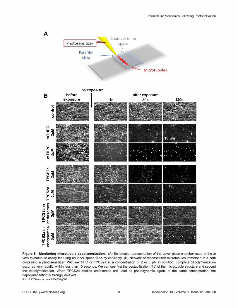