ÿþS o n y E r i c s s o n M o b i l e C o m m u n i c a t i o n s ...
Upload
moscowstateCategory
view
0download
0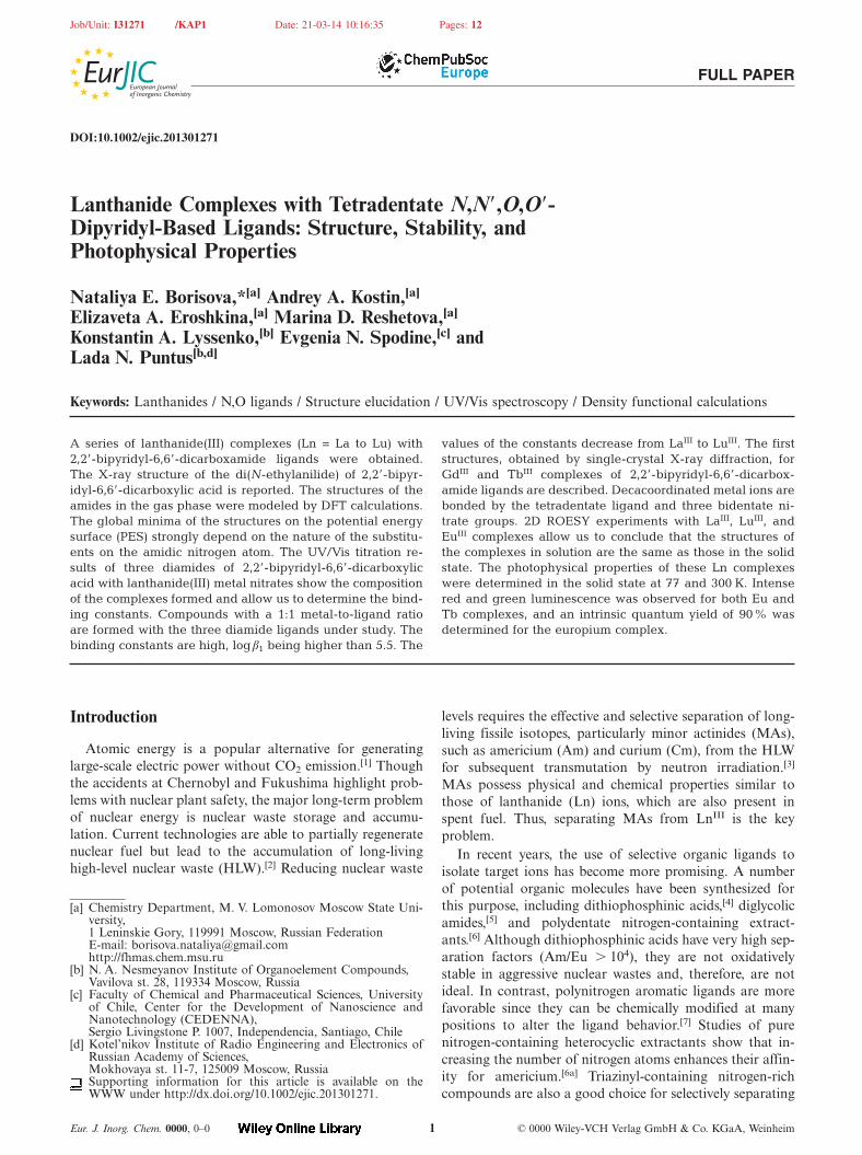
Job/Unit: I31271 /KAP1 Date: 21-03-14 10:16:35 Pages: 12
FULL PAPER
DOI:10.1002/ejic.201301271
Lanthanide Complexes with Tetradentate N,N�,O,O�-Dipyridyl-Based Ligands: Structure, Stability, andPhotophysical Properties
Nataliya E. Borisova,*[a] Andrey A. Kostin,[a]
Elizaveta A. Eroshkina,[a] Marina D. Reshetova,[a]
Konstantin A. Lyssenko,[b] Evgenia N. Spodine,[c] andLada N. Puntus[b,d]
Keywords: Lanthanides / N,O ligands / Structure elucidation / UV/Vis spectroscopy / Density functional calculations
A series of lanthanide(III) complexes (Ln = La to Lu) with2,2�-bipyridyl-6,6�-dicarboxamide ligands were obtained.The X-ray structure of the di(N-ethylanilide) of 2,2�-bipyr-idyl-6,6�-dicarboxylic acid is reported. The structures of theamides in the gas phase were modeled by DFT calculations.The global minima of the structures on the potential energysurface (PES) strongly depend on the nature of the substitu-ents on the amidic nitrogen atom. The UV/Vis titration re-sults of three diamides of 2,2�-bipyridyl-6,6�-dicarboxylicacid with lanthanide(III) metal nitrates show the compositionof the complexes formed and allow us to determine the bind-ing constants. Compounds with a 1:1 metal-to-ligand ratioare formed with the three diamide ligands under study. Thebinding constants are high, logβ1 being higher than 5.5. The
Introduction
Atomic energy is a popular alternative for generatinglarge-scale electric power without CO2 emission.[1] Thoughthe accidents at Chernobyl and Fukushima highlight prob-lems with nuclear plant safety, the major long-term problemof nuclear energy is nuclear waste storage and accumu-lation. Current technologies are able to partially regeneratenuclear fuel but lead to the accumulation of long-livinghigh-level nuclear waste (HLW).[2] Reducing nuclear waste
[a] Chemistry Department, M. V. Lomonosov Moscow State Uni-versity,1 Leninskie Gory, 119991 Moscow, Russian FederationE-mail: [email protected]://fhmas.chem.msu.ru
[b] N. A. Nesmeyanov Institute of Organoelement Compounds,Vavilova st. 28, 119334 Moscow, Russia
[c] Faculty of Chemical and Pharmaceutical Sciences, Universityof Chile, Center for the Development of Nanoscience andNanotechnology (CEDENNA),Sergio Livingstone P. 1007, Independencia, Santiago, Chile
[d] Kotel’nikov Institute of Radio Engineering and Electronics ofRussian Academy of Sciences,Mokhovaya st. 11-7, 125009 Moscow, RussiaSupporting information for this article is available on theWWW under http://dx.doi.org/10.1002/ejic.201301271.
Eur. J. Inorg. Chem. 0000, 0–0 © 0000 Wiley-VCH Verlag GmbH & Co. KGaA, Weinheim1
values of the constants decrease from LaIII to LuIII. The firststructures, obtained by single-crystal X-ray diffraction, forGdIII and TbIII complexes of 2,2�-bipyridyl-6,6�-dicarbox-amide ligands are described. Decacoordinated metal ions arebonded by the tetradentate ligand and three bidentate ni-trate groups. 2D ROESY experiments with LaIII, LuIII, andEuIII complexes allow us to conclude that the structures ofthe complexes in solution are the same as those in the solidstate. The photophysical properties of these Ln complexeswere determined in the solid state at 77 and 300 K. Intensered and green luminescence was observed for both Eu andTb complexes, and an intrinsic quantum yield of 90% wasdetermined for the europium complex.
levels requires the effective and selective separation of long-living fissile isotopes, particularly minor actinides (MAs),such as americium (Am) and curium (Cm), from the HLWfor subsequent transmutation by neutron irradiation.[3]
MAs possess physical and chemical properties similar tothose of lanthanide (Ln) ions, which are also present inspent fuel. Thus, separating MAs from LnIII is the keyproblem.
In recent years, the use of selective organic ligands toisolate target ions has become more promising. A numberof potential organic molecules have been synthesized forthis purpose, including dithiophosphinic acids,[4] diglycolicamides,[5] and polydentate nitrogen-containing extract-ants.[6] Although dithiophosphinic acids have very high sep-aration factors (Am/Eu � 104), they are not oxidativelystable in aggressive nuclear wastes and, therefore, are notideal. In contrast, polynitrogen aromatic ligands are morefavorable since they can be chemically modified at manypositions to alter the ligand behavior.[7] Studies of purenitrogen-containing heterocyclic extractants show that in-creasing the number of nitrogen atoms enhances their affin-ity for americium.[6a] Triazinyl-containing nitrogen-richcompounds are also a good choice for selectively separating
Job/Unit: I31271 /KAP1 Date: 21-03-14 10:16:35 Pages: 12
www.eurjic.org FULL PAPER
MAs and can function in solvents like octanol-1. However,extractants that can be used in fluorinated solvents (e.g.,trifluoromethylphenyl sulfone or m-trifluoromethyl nitro-benzene) have not yet been developed.
We designed a heteropolydentate ligand that possessestwo soft nitrogen-containing heterocycles and two carbox-amide groups for binding actinides, which are slightlyharder than lanthanides. We expected to achieve high selec-tivity for Am, and preliminary results showed that the syn-thesized ligands [diamides of 2,2�-bipyrydil-6,6�-dicarbox-ylic acid (DPDAs)] were highly selective for AmIII in com-parison with EuIII.[8] Moreover, ligand affinity to lanth-anides decreased linearly with increasing atomic number ofthe lanthanide. The extracting ability of DPDAs heavily de-pends on the structure of their side amidic chain. The mosteffective extraction occurs when the amide contains botharomatic and aliphatic substituents as its amidic groups,and no extraction was observed when a long aliphatic di-amide chain was used. We performed quantum chemicalcalculations on the structures of several DPDAs to deter-mine how the side amidic group affected metal binding. Inaddition, we synthesized and analyzed the structure of theLn–DPDA complexes that form in the organic phase. Sta-bility constants for Ln ions (lanthanum to lutetium) weremeasured in the presence of several DPDA ligands to deter-mine the effect of ionic radii on the stability of the DPDAcomplexes.
From another point of view, the luminescence of lanthan-ide ions is in the focus of intense research efforts becauseof the wide technical and analytical applications. The litera-ture in this field is growing fast, and a number of reviewshave been published during the last ten years to accumulateand analyze information in this popular area.[9] The mostimportant applications of the photophysical properties oflanthanide ions are lighting (luminophores[10] and electrolu-minophores[11]) and bioanalytics (biochemical sensors[12]
and medical imaging[9a,9c,13]). Moreover, photophysicalproperties are very important for the estimation of the fur-ther possible applications of new luminescent complexes. Toshed light on the possible optical applications of lanthan-ide–DPDA complexes, we also investigated their photo-physical properties in this work.
Results and Discussion
Structures of the 2,2�-Dipyridyl-6,6�-dicarboxamide Ligands1H NMR spectroscopic data show that all three DPDA
molecules (LPh, LBu2, and LOct2) possess highly symmetricalCs geometry (Figures S1–S3). The two pyridine rings areequivalent, and the amide side arms have identical chemicalshifts. These data indicate the existence of an inversion cen-ter in the ligands. Studying the restricted intramolecular ro-tations of the amide groups in [D3]acetonitrile indicatedmoderate energy barriers (16.02 kcal/mol for LBu2
[8a] and16.74 kcal/mol for LOct2). For ligand LPh, only one con-former with cis-orientation of the ethyl substituent was ob-served.[8a] This strongly supported the observation of the
Eur. J. Inorg. Chem. 0000, 0–0 © 0000 Wiley-VCH Verlag GmbH & Co. KGaA, Weinheim2
rotating-frame Overhauser effect (ROESY)[14] for ligandLPh. The corresponding cross peak was found between thebroad singlet of the amidic phenyl ring and the 3�-positionof the pyridine ring (Figure S4).
The solid-state structure was determined for the LPh li-gand (Figure 1, Tables S1 and S2). LPh possesses a centerof symmetry at the C14–C14a bond of the bipyridyl moiety.The phenyl groups are trans in relation to the amide oxygen.The phenyl rings are located in close proximity to the pyr-idine rings of the bipyridyl unit, and the torsion angle C10–C9–N2–C1 is –14.5°. Due to steric hindrance, the dihedralangle of the bulky phenyl and pyridyl groups is 64°. Theamide moieties significantly deviate from the bipyridylplane; moreover, the carboxamide group is distorted fromthe planes of both the pyridine and the phenyl rings andhas corresponding dihedral angles of 42° and 64°, respec-tively. Thus, there is no evidence for electronic conjugationbetween the two aromatic phenyl and pyridyl rings and thecarboxamide group. Consequently, the energy of the rota-tional barrier of the carboxamide group is likely lower as aresult of the CO–N bond. The crystal packing of LPh isdescribed in Figure S5.
Figure 1. Molecular structure of LPh, showing the atom numberingscheme. Displacement ellipsoids are drawn at the 50% probabilitylevel, and H atoms are shown as small spheres of arbitrary radius.
The calculated geometry of one potential energy surface(PES, see below) minimum for LPh was in good agreementwith its solid-state structure. Therefore, LPh has a similarstructure in both the solid state (X-ray crystallography data)and in solution (NMR spectroscopic data).
DFT Calculations for the Diamide Ligands
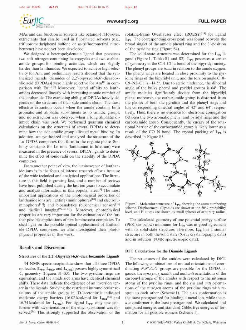
The structures of the amides were calculated by DFT.The following combinations of mutual orientations of coor-dinating N,N�,O,O�-groups are possible for the DPDA li-gands: the syn,syn, syn,anti, and anti,anti orientations of thecarbonyl groups of the amides with respect to the nitrogenatoms of the pyridine rings, and the syn and anti orienta-tions of the nitrogen atoms of the pyridine rings with re-spect to each other (Scheme 1). The s-s-s conformation isthe most preorganized for binding a metal ion, while the a-a-a conformer is the least preorganized. We calculated andcompared energies and standard Gibbs free energies of for-mation for all possible isomers (Scheme 1).
Job/Unit: I31271 /KAP1 Date: 21-03-14 10:16:35 Pages: 12
www.eurjic.org FULL PAPER
Scheme 1. Possible conformations of the DPDA ligands with their relative Gibbs free energies (in kcal/mol).
Minima on the PES for all six possible isomers were onlyobserved for LPh, which contains the phenyl substituent inthe amide group. Two minima, corresponding to the a-s-a and a-a-a conformers, were observed for the tetraalkyl-substituted LOct2 and LBu2. The global PES minimum ofLOct2 and LBu2 corresponds to the least preorganized con-former (a-a-a), and no minima correspond to the most pre-organized conformation (s-s-s). The partially preorganizedconformer (s-a-s) corresponds to the global PES minimumof LPh. The s-s-s conformer is most prone to metal ionbinding and its minimum energy is 3.64 kcal/mol higherthan that of the s-a-s conformer. The a-s-a conformer hasthe highest PES minimum with 7 kcal/mol.
The geometry of the LPh a-a-a conformer was calculatedfor the gas phase and was found to be close to that of themolecule’s X-ray structure (Table S2). For the tetraalkyl-substituted amides, the second PES minimum was calcu-lated to be 3.21 and 5.49 kcal/mol higher than that of themain a-a-a conformer for LBu2 and LOct2, respectively. Ageometrical analysis of the molecules corresponding to PESminima showed that, regardless of the amide substituents,pyridine rings are unfolded at 178–179° for anti conformers(Table S3). In contrast, the syn conformers have torsionangles of 30° between the two rings, which agrees more withthe gauche conformation. Similarly, we obtained averageangles of 135�6° between the carbonyl groups and pyr-idine rings for anti conformers, regardless of the amide sub-stituent. The average angle for the syn conformers, whichwas only found for LPh, is 63 �6°, which also agrees with agauche conformation.
As expected, the amide substituents substantially affectthe ligand geometry. For all tetraalkyl-substituted amides(LBu2 and LOct2), the carboxamide groups and α-carbonatoms of the substituents lie in one plane in all conforma-tions, and the corresponding torsion angles never exceed 2°.For the anilide (LPh), the aromatic rings form a 60° anglewith the carboxamide plane, and the orientation depends
Eur. J. Inorg. Chem. 0000, 0–0 © 0000 Wiley-VCH Verlag GmbH & Co. KGaA, Weinheim3
on those of the carbonyl group and pyridine ring. All synconformations of the carbonyl group have trans-phenyl sub-stituents with respect to the pseudo double amide C–Nbond. In contrast, the anti conformations of the carbonylgroup have cis-phenyl groups. This was clearly reflected inthe torsion angles between the C=O bond of the carbonylgroup and the N–C bond of the phenyl substituent(Table S3). The syn and anti torsion angles are 168�4° and3 �4°, respectively; the torsion angle reaches 26.33° in onlyone case (a-s-a). Neither the amide substituent nor the con-formational behavior affects the heteroatom charges(Table S3).
The amide with an aromatic substituent (LPh) is morepreorganized for binding metal ions than its tetraalkyl-sub-stituted analogs (LOct2 and LBu2). The energy differencesbetween the conformers are not high and do not exceed5.5 kcal/mol for LOct2 and LBu2 and 7.5 kcal/mol for LPh.However, such low energy values cannot explain the con-siderable difference in extraction ability we observed. Thus,we also studied the rotational barriers of the pyridine ringsand amide groups. The rotation of the pyridine rings aboutthe C–C bond is similar for all three amides (Figure S6).The maximum rotational barriers of the pyridine rings are9.25, 6.15, and 6.67 kcal/mol for LPh, LBu2, and LOct2,respectively. In contrast, the rotation of the carboxamidegroup about the C–C bond is heavily dependent on theamide side chain and is characterized by significant rota-tional barriers (Figure S7). The barrier is much higher forthe tetraalkyl-substituted amides (21.05 kcal/mol for LOct2
and 17.34 kcal/mol for LBu2) than that for the anilide amide(14.11 kcal/mol for LPh). This distribution of rotational bar-rier energy is similar to the changes in extraction abilityfor this series of amides. LPh has much higher distributioncoefficients compared to LBu2, while LOct2 does not extractAm or Eu. The a-a-a conformer, which is the least preorga-nized for metal ion binding, corresponds to the global mini-mum for tetraalkyl-substituted amides and has to overcome
Job/Unit: I31271 /KAP1 Date: 21-03-14 10:16:35 Pages: 12
www.eurjic.org FULL PAPER
a considerable energy barrier (the rotation of the carbox-amide group) to bind metal ions. The anilide LPh has aglobal minimum corresponding to the partially preorga-nized s-a-s conformer, and only a slight energy barrier (theunfolding of the pyridine rings) needs to be overcome formetal ion binding.
This reasoning is supported by knowing the energy re-quired for binding-site reorganization for each ligand.Higher ligand affinity is closely related with ligand confor-mity and complementarity.[15] The latter is considered as theenergy required for structural reorganization of the ligandduring the complexation. We calculated the total energy ofbinding-site reorganization by using the difference betweenthe global PES minimum and the energy of the bound li-gand (see Experimental Section). All the ligands had non-zero energies for binding-site reorganization for binding aEu ion. The lowest reorganization energy required was ob-served for LPh (17.40 kcal/mol), and more reorganizationenergy was required for LBu2 and LOct2 (21.48 and23.45 kcal/mol, respectively). Thus, LPh is more preorga-nized for binding EuIII and has lower energy barriers toovercome, resulting in a better extraction ability comparedto LBu2 and LOct2. Moreover, this further explains whyLOct2, which is the least preorganized, cannot extract metalions.
LnIII Complexes with LPh
We studied complexes of LPh, the most promising ex-tractant, with LnIII to understand the differences betweenAmIII and EuIII extraction by the DPDA ligands. Thesecomplexes are directly synthesized by reacting hydratednitrates of LnIII with LPh in acetonitrile heated to refluxand have the same LnLPh(NO3)3 composition for alllanthanide ions (LaIII to LuIII; Scheme 2).
Complex yields decreased from lanthanum (70 %) to sa-marium and europium (61–65%) and returned to 70% fromgadolinium to ytterbium and lutetium. The complexes werecharacterized by standard spectroscopic techniques; the in-frared spectra of the synthesized complexes were almostidentical, and the spectra for all complexes significantly dif-fered from those of the free ligand. The band near1610 cm–1 corresponding to the first amide band is verysensitive for metal binding. The spectra of the observedamide bands decreased from 1639 cm–1 for the free ligandto 1610 cm–1 for the corresponding complexes, which con-firms the complexation of metal ions with the amidic oxy-gen atom. N=O bands for NO3 anions were observed near
Scheme 2. Synthesis of LnLPh(NO3)3.
Eur. J. Inorg. Chem. 0000, 0–0 © 0000 Wiley-VCH Verlag GmbH & Co. KGaA, Weinheim4
1456 cm–1 for all of complexes. Less intense bands, whichcorrespond to symmetrical and asymmetrical deformationof the O–N–O vibrations in the NO3 anions, were also ob-served. Mass spectra obtained by MALDI-TOF showedonly one ion (LnLPh[NO3]2+) in the laser desorption-ioniza-tion mode. Secondary peaks originating from the exchangereaction between nitrate and cinnamic acid anions were ob-served in the MALDI mode in the presence of a cinnamicacid matrix. All mass spectra showed peaks derived frommononuclear complexes of LnLPh(NO3)3.
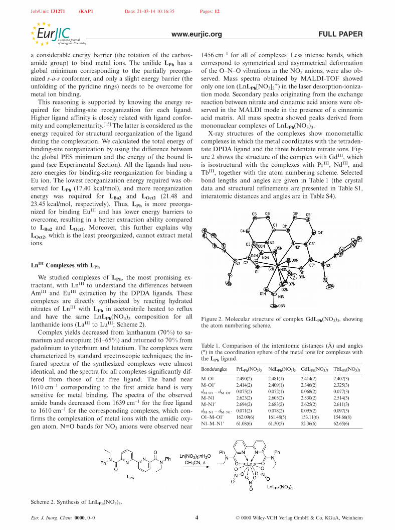
X-ray structures of the complexes show monometalliccomplexes in which the metal coordinates with the tetraden-tate DPDA ligand and the three bidentate nitrate ions. Fig-ure 2 shows the structure of the complex with GdIII, whichis isostructural with the complexes with PrIII, NdIII, andTbIII, together with the atom numbering scheme. Selectedbond lengths and angles are given in Table 1 (the crystaldata and structural refinements are presented in Table S1,interatomic distances and angles are in Table S4).
Figure 2. Molecular structure of complex GdLPh(NO3)3, showingthe atom numbering scheme.
Table 1. Comparison of the interatomic distances (Å) and angles(°) in the coordination sphere of the metal ions for complexes withthe LPh ligand.
Bonds/angles PrLPh(NO3)3 NdLPh(NO3)3 GdLPh(NO3)3 TbLPh(NO3)3
M–O1 2.490(2) 2.481(1) 2.414(2) 2.402(3)M–O1� 2.414(2) 2.409(1) 2.346(2) 2.325(3)dM–O1 – dM–O1� 0.075(2) 0.072(1) 0.068(2) 0.077(3)M–N1 2.623(2) 2.605(2) 2.530(2) 2.514(3)M–N1� 2.694(2) 2.683(2) 2.625(2) 2.611(3)dM–N1 – dM–N1� 0.071(2) 0.078(2) 0.095(2) 0.097(3)O1–M–O1� 162.09(6) 161.48(5) 153.11(6) 154.66(8)N1–M–N1� 61.08(6) 61.30(5) 52.36(6) 62.65(6)

Job/Unit: I31271 /KAP1 Date: 21-03-14 10:16:35 Pages: 12
www.eurjic.org FULL PAPER
Table 2. Chemical shifts (ppm) of the protons of the LPh ligand and the corresponding LnIII complexes.
CH3 CH2 CH5�–Py CHPh CH4�–Py CH3�–Py
LPh t 1.18 q 3.93 m 7.03–7.32 t 7.74 d 7.57LaLPh(NO3)3 t 1.26 q 4.09 d 6.99 m 7.31–7.53 t 7.80 d 8.19EuLPh(NO3)3 b room temp. –0.27 br.s. 2.34 br.d. 3.42 br.s. 7.39 b room temp. 5.29 br.d. 3.64LuLPh(NO3)3 t 1.29 q 4.10 d 6.85 m 7.38–7.45 7.48–7.58 t 7.90 d 8.30
The coordination sphere of the metal ion can be de-scribed as a distorted two-capped square antiprism in whichN2, O8N, O1�, O5N and O1, O4N, O2N, O7N define twodistorted square faces of the antiprism; the faces are cappedwith N2� and O1N, respectively. The two tetragonal facesare nearly parallel (1.2–1.7°) and are separated by approxi-mately 2.1 Å. The Ln atom is located at the middle of thedistance between the faces. The N2, O8N, O1�, O5N face iscapped with the N2� pyridine atom, which is placed underthe center of the face.
The second face is capped with the oxygen atom O1Nfrom the nitrate ion – notable is that the second oxygenatom, O2N, of this nitrate ion lies in the square face, andthis shifts the O2N cap from the center to the square as aresult of the short interatomic distance in the nitrate. Theangle between the Ln–O2N bond and the perpendiculardistance to the face plane is about 15°. Such coordinationof the metal atom leads to the inequivalence of the twopyridine nitrogen atoms of the bipyridine unit and the twooxygen atoms of the carboxamide groups because of sterichindrance. This steric hindrance between NO3 and carbox-amide groups causes the significant difference in torsionangles: O1–C7–C3–N2 25.4–28.9° and O1�–C7�–C3�–N2�7.5–7.2. As a consequence, the M–O1� bond in all membersof this isostructural series is systematically shorter than M–O1 while the M–N2� bond is longer than M–N2. The differ-ence in M–N and M–O bonds for these formally equivalentparts of the ligand is almost independent from the natureof the metal atom and therefore also from the lanthanidecontraction. The independence of the above differencesfrom the nature of the metal center is probably a result ofvariations in the torsion angle N2–C1–C1�–N2�, which areequal to 5.0 and 4.6° for Nd and Pr complexes and increaseup to 12.8 and 11.0° for Gd and Tb. Thus, the strength ofthe metal-to-ligand bonding is mostly governed by thesteric repulsion between the carboxamide groups, and thenitrate ligand and is less affected by lanthanide radii.
NMR spectra were recorded for the diamagnetic LaIII
and LuIII and for the EuIII complexes (Table 2, Figures S8and S9). The diamagnetic spectra have similar peak pat-terns and differ in only chemical shifts. The 3�- and 4�-pyr-idine proton peaks of the ligand are shielded as a result ofmetal ion binding. The deshielded cone of the neighboringphenyl ring causes an unexpected shift up for the 5�-pyr-idine ring protons. Complexes with metals of a higheratomic number shield more the pyridine protons.
The ROESY[14] spectrum was recorded for the diamag-netic complexes: cross peaks occur between the 5�-pyridinering proton and the o-protons of the phenyl ring (Fig-ures S10–S12). This confirms the conclusion that these pro-
Eur. J. Inorg. Chem. 0000, 0–0 © 0000 Wiley-VCH Verlag GmbH & Co. KGaA, Weinheim5
tons are deshielded in the complexes compared to those inthe free ligand. Together, these data show that the com-plexes maintain the same conformation in solution as theyhave in crystal form.
Formation Constants of LnDPDA(NO3)3 Complexes inSolution
NMR spectroscopic data and the chemical synthesesprovided the initial evidence of complex stability in aceto-nitrile solution. UV/Vis spectrophotometric titration wasused to quantitatively assess the ability of the ligand to bindLn ions in acetonitrile solution (see Supporting Infor-mation). The absorbance spectrum of the ligands in aceto-nitrile is characterized by two broad bands in the UV range(near 293 and 245 nm). A new band near 310 nm with goodisosbestic behavior was observed as increasing amounts ofmetal salt were added. On the basis of our X-ray and NMRspectroscopic data, we expected complexes to form at a 1:1stoichiometric ratio in solution. The 1:1 ratio was observedfor all the metals, and the titration curves increasedsmoothly with a marked endpoint at a M/L value of 1:1.
The binding constants β were calculated according toEquations (1), (2), (3), and (4):
(1)
(2)
(3)
(4)
where Abs is absorbance at the chosen wavelength, εML isthe molar absorbance coefficient of the ML(NO3)3 complexat the chosen wavelength, and cL and cM are total ligandand metal salt concentrations, respectively. Logβ values forthe LnIII complexes are shown in Table 3 and Figure S13.All the DPDA ligands have high affinities for LnIII ionswith log β values greater than 5.5. These values are lowerthan the logβ1 values measured for the related 2,6-pyridine-dicarboxylic acid (PDAs: PyiPr2 and PyEt2) diamides(Table 3). The LPh and LOct2 constants do not vary as muchbetween lanthanide ions, and LOct2 has the highest con-stants. The binding constants of both LPh and LOct2 tendto decrease from lanthanum to lutetium, but the decreaseis not constant. LBu2 has similar binding constants for all
Job/Unit: I31271 /KAP1 Date: 21-03-14 10:16:35 Pages: 12
www.eurjic.org FULL PAPER
lanthanide ions. Generally, the binding constant is lowerfor cerium(III) than lanthanum(III) and praseodymium(III)(Table 3) for all three ligands, and the binding constants forthe tetraalkyl-substituted ligands have the smallest value forcerium(III). If the anomalous behavior of cerium is ex-cluded, we can conclude that the DPDA ligands bind metalions with larger ionic radii better.
Table 3. Logβ1 values (with accuracies in parentheses) for the bind-ing of the PDA and DPDA ligands with lanthanide(III) ions inacetonitrile.
M LPh LBu2 LOct2 PyiPr2[16] PyEt2
[17] PyBn2[16]
La 6.2(2) 5.5(3) 6.7(2) 7.8(5) 7.4(3) 5.4(5)Ce 5.6(1) 5.3(1) 5.9(2) 7.4(4) 7.6(3)Pr 5.9(1) 5.1(3) 6.5(4) 8.0(6) 7.6(3)Nd 5.9(1) 5.7(2) 6.6(2) 7.7(5) 7.5(3)Sm 5.7(1) 5.3(3) 6.2(1) 8.5(6) 7.3(3)Eu 5.6(1) 6.1(3) 6.2(1) 8.3(6) 8.3(3) 4.9(5)Gd 5.7(1) 5.2(3) 6.2(1) 7.9(7) 7.9(3)Tb 5.7(1) 5.5(2) 6.1(1) 7.6(6) 8.2(3)Dy 5.7(1) 5.1(2) 6.3(1) 7.7(5) 7.5(3)Ho 5.7(2) 5.4(3) 6.3(1) 8.3(6) 7.3(4)Er 5.4(1) 6.3(2) 6.3(2) 8.3(5) 7.7(4)Tm 5.9(2) 5.4(3) 6.5(1) 7.9(6) 8.5(3)Yb 5.6(2) 5.9(3) 6.4(1) 7.7(4) 8.5(3)Lu 5.5(2) 5.7(4) 6.4(1) 7.6(4) 8.1(3) 5.3(5)
In contrast to the DPDA ligands, PDA ligands(Scheme 3) complex with hydrated triflates of LnIII at 1:1,1:2, and 1:3 ratios in acetonitrile (Table 3).[16,17] In general,β1 values of the PDA ligands are higher than those of thebipyridyl analogs, except for that of PyBn2, which is veryclose to that of LOct2. Logβ1 values of PyEt2 and PyiPr2 arecomparable and experience an electrostatic trend (i.e., aslight increase in binding constants with decreasing metalion size), as expected, which results from the contraction ofionic radii along the lanthanide series.[16]
Scheme 3. Structure of the PDA ligands, whose complexation withLnIII was studied.
Photophysical Properties of LnDPDA(NO3)3 Complexes inthe Solid State
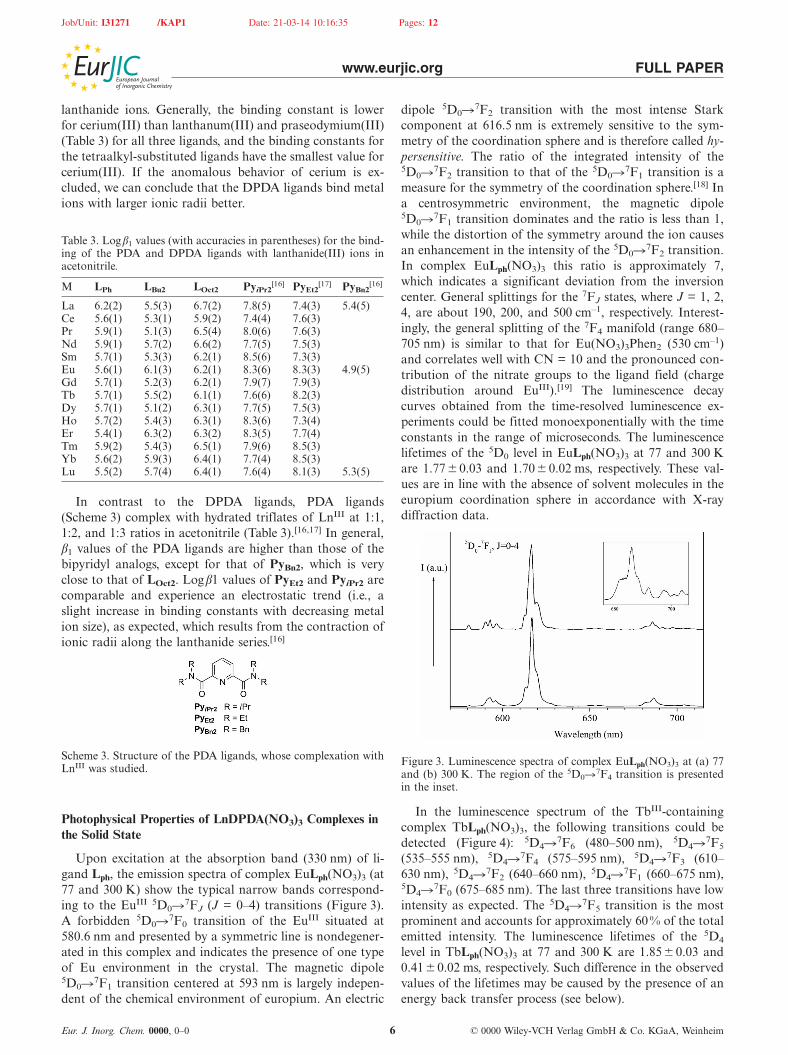
Upon excitation at the absorption band (330 nm) of li-gand Lph, the emission spectra of complex EuLph(NO3)3 (at77 and 300 K) show the typical narrow bands correspond-ing to the EuIII 5D0�7FJ (J = 0–4) transitions (Figure 3).A forbidden 5D0�7F0 transition of the EuIII situated at580.6 nm and presented by a symmetric line is nondegener-ated in this complex and indicates the presence of one typeof Eu environment in the crystal. The magnetic dipole5D0�7F1 transition centered at 593 nm is largely indepen-dent of the chemical environment of europium. An electric
Eur. J. Inorg. Chem. 0000, 0–0 © 0000 Wiley-VCH Verlag GmbH & Co. KGaA, Weinheim6
dipole 5D0�7F2 transition with the most intense Starkcomponent at 616.5 nm is extremely sensitive to the sym-metry of the coordination sphere and is therefore called hy-persensitive. The ratio of the integrated intensity of the5D0�7F2 transition to that of the 5D0�7F1 transition is ameasure for the symmetry of the coordination sphere.[18] Ina centrosymmetric environment, the magnetic dipole5D0�7F1 transition dominates and the ratio is less than 1,while the distortion of the symmetry around the ion causesan enhancement in the intensity of the 5D0�7F2 transition.In complex EuLph(NO3)3 this ratio is approximately 7,which indicates a significant deviation from the inversioncenter. General splittings for the 7FJ states, where J = 1, 2,4, are about 190, 200, and 500 cm–1, respectively. Interest-ingly, the general splitting of the 7F4 manifold (range 680–705 nm) is similar to that for Eu(NO3)3Phen2 (530 cm–1)and correlates well with CN = 10 and the pronounced con-tribution of the nitrate groups to the ligand field (chargedistribution around EuIII).[19] The luminescence decaycurves obtained from the time-resolved luminescence ex-periments could be fitted monoexponentially with the timeconstants in the range of microseconds. The luminescencelifetimes of the 5D0 level in EuLph(NO3)3 at 77 and 300 Kare 1.77 �0.03 and 1.70� 0.02 ms, respectively. These val-ues are in line with the absence of solvent molecules in theeuropium coordination sphere in accordance with X-raydiffraction data.
Figure 3. Luminescence spectra of complex EuLph(NO3)3 at (a) 77and (b) 300 K. The region of the 5D0�7F4 transition is presentedin the inset.
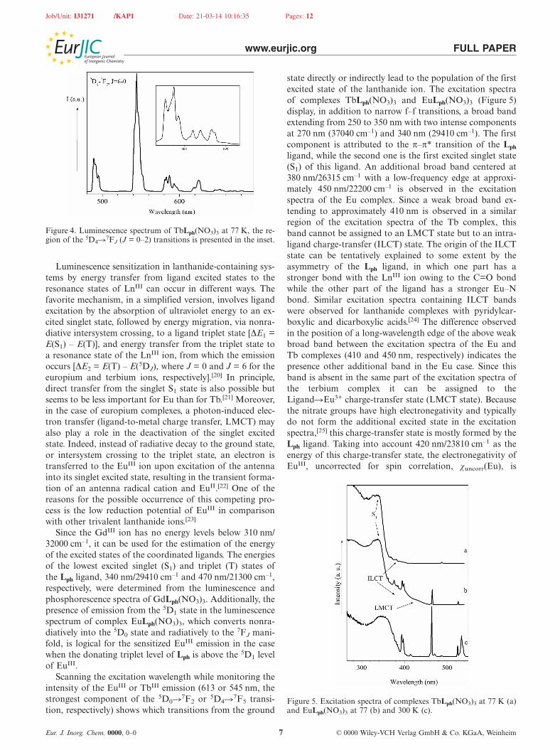
In the luminescence spectrum of the TbIII-containingcomplex TbLph(NO3)3, the following transitions could bedetected (Figure 4): 5D4�7F6 (480–500 nm), 5D4�7F5
(535–555 nm), 5D4�7F4 (575–595 nm), 5D4�7F3 (610–630 nm), 5D4�7F2 (640–660 nm), 5D4�7F1 (660–675 nm),5D4�7F0 (675–685 nm). The last three transitions have lowintensity as expected. The 5D4�7F5 transition is the mostprominent and accounts for approximately 60 % of the totalemitted intensity. The luminescence lifetimes of the 5D4
level in TbLph(NO3)3 at 77 and 300 K are 1.85� 0.03 and0.41�0.02 ms, respectively. Such difference in the observedvalues of the lifetimes may be caused by the presence of anenergy back transfer process (see below).
Job/Unit: I31271 /KAP1 Date: 21-03-14 10:16:35 Pages: 12
www.eurjic.org FULL PAPER
Figure 4. Luminescence spectrum of TbLph(NO3)3 at 77 K, the re-gion of the 5D4�7FJ (J = 0–2) transitions is presented in the inset.
Luminescence sensitization in lanthanide-containing sys-tems by energy transfer from ligand excited states to theresonance states of LnIII can occur in different ways. Thefavorite mechanism, in a simplified version, involves ligandexcitation by the absorption of ultraviolet energy to an ex-cited singlet state, followed by energy migration, via nonra-diative intersystem crossing, to a ligand triplet state [ΔE1 =E(S1) – E(T)], and energy transfer from the triplet state toa resonance state of the LnIII ion, from which the emissionoccurs [ΔE2 = E(T) – E(5DJ), where J = 0 and J = 6 for theeuropium and terbium ions, respectively].[20] In principle,direct transfer from the singlet S1 state is also possible butseems to be less important for Eu than for Tb.[21] Moreover,in the case of europium complexes, a photon-induced elec-tron transfer (ligand-to-metal charge transfer, LMCT) mayalso play a role in the deactivation of the singlet excitedstate. Indeed, instead of radiative decay to the ground state,or intersystem crossing to the triplet state, an electron istransferred to the EuIII ion upon excitation of the antennainto its singlet excited state, resulting in the transient forma-tion of an antenna radical cation and EuII.[22] One of thereasons for the possible occurrence of this competing pro-cess is the low reduction potential of EuIII in comparisonwith other trivalent lanthanide ions.[23]
Since the GdIII ion has no energy levels below 310 nm/32000 cm–1, it can be used for the estimation of the energyof the excited states of the coordinated ligands. The energiesof the lowest excited singlet (S1) and triplet (T) states ofthe Lph ligand, 340 nm/29410 cm–1 and 470 nm/21300 cm–1,respectively, were determined from the luminescence andphosphorescence spectra of GdLph(NO3)3. Additionally, thepresence of emission from the 5D1 state in the luminescencespectrum of complex EuLph(NO3)3, which converts nonra-diatively into the 5D0 state and radiatively to the 7FJ mani-fold, is logical for the sensitized EuIII emission in the casewhen the donating triplet level of Lph is above the 5D1 levelof EuIII.
Scanning the excitation wavelength while monitoring theintensity of the EuIII or TbIII emission (613 or 545 nm, thestrongest component of the 5D0�7F2 or 5D4�7F5 transi-tion, respectively) shows which transitions from the ground
Eur. J. Inorg. Chem. 0000, 0–0 © 0000 Wiley-VCH Verlag GmbH & Co. KGaA, Weinheim7
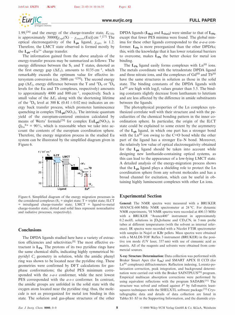
state directly or indirectly lead to the population of the firstexcited state of the lanthanide ion. The excitation spectraof complexes TbLph(NO3)3 and EuLph(NO3)3 (Figure 5)display, in addition to narrow f–f transitions, a broad bandextending from 250 to 350 nm with two intense componentsat 270 nm (37040 cm–1) and 340 nm (29410 cm–1). The firstcomponent is attributed to the π–π* transition of the Lph
ligand, while the second one is the first excited singlet state(S1) of this ligand. An additional broad band centered at380 nm/26315 cm–1 with a low-frequency edge at approxi-mately 450 nm/22200 cm–1 is observed in the excitationspectra of the Eu complex. Since a weak broad band ex-tending to approximately 410 nm is observed in a similarregion of the excitation spectra of the Tb complex, thisband cannot be assigned to an LMCT state but to an intra-ligand charge-transfer (ILCT) state. The origin of the ILCTstate can be tentatively explained to some extent by theasymmetry of the Lph ligand, in which one part has astronger bond with the LnIII ion owing to the C=O bondwhile the other part of the ligand has a stronger Eu–Nbond. Similar excitation spectra containing ILCT bandswere observed for lanthanide complexes with pyridylcar-boxylic and dicarboxylic acids.[24] The difference observedin the position of a long-wavelength edge of the above weakbroad band between the excitation spectra of the Eu andTb complexes (410 and 450 nm, respectively) indicates thepresence other additional band in the Eu case. Since thisband is absent in the same part of the excitation spectra ofthe terbium complex it can be assigned to theLigand�Eu3+ charge-transfer state (LMCT state). Becausethe nitrate groups have high electronegativity and typicallydo not form the additional excited state in the excitationspectra,[25] this charge-transfer state is mostly formed by theLph ligand. Taking into account 420 nm/23810 cm–1 as theenergy of this charge-transfer state, the electronegativity ofEuIII, uncorrected for spin correlation, χuncorr(Eu), is
Figure 5. Excitation spectra of complexes TbLph(NO3)3 at 77 K (a)and EuLph(NO3)3 at 77 (b) and 300 K (c).
Job/Unit: I31271 /KAP1 Date: 21-03-14 10:16:35 Pages: 12
www.eurjic.org FULL PAPER
1.99,[26] and the energy of the charge-transfer state, ECTS,is approximately 30000[χopt(X) – χuncorr(Eu)] cm–1.[27] Theoptical electronegativity of the Lph ligand, χopt, is 1.2.Therefore, the LMCT state observed is formed mostly bythe Lph�Eu3+ charge transfer.
The information gained from the above analysis of theenergy-transfer process may be summarized as follows: Theenergy difference between the S1 and T states, denoted asthe first energy gap (ΔE1), amounts to 8135 cm–1, whichremarkably exceeds the optimum value for effective in-tersystem conversion (ca. 5000 cm–1[28]). The second energygap (ΔE2, energy difference between the T and 5D0 or 5D4
levels for the Eu and Tb complexes, respectively) amountsto approximately 4000 and 800 cm–1, respectively. Such asmall value of the ΔE2 along with the shortening lifetimeof the 5D4 level at 300 K (0.41� 0.02 ms) indicates an en-ergy back transfer process, which promotes luminescencequenching in complex TbLph(NO3)3. The intrinsic quantumyield of the europium-centered emission calculated bymeans of Werts’ formula[29] for complex EuLph(NO3)3 isQEu
Eu = 90 %, which is reasonable when we take into ac-count the contents of the europium coordination sphere.Therefore, the energy migration process in the studied Eusystem can be illustrated by the simplified diagram given inFigure 6.
Figure 6. Simplified diagram of the energy migration processes inthe considered complexes (S1 = singlet state; T = triplet state; ILCT= intraligand charge-transfer state; LMCT = ligand-to-metalcharge-transfer state; dotted and solid lines represent nonradiativeand radiative processes, respectively).
Conclusions
The DPDA ligands studied here have a variety of extrac-tion efficiencies and selectivities.[8] The most effective ex-tractant is LPh. The protons of its two pyridine rings havethe same chemical shifts, indicating highly symmetrical bi-pyridyl Cs geometry in solution, while the amidic phenylring was shown to be located near the pyridine ring. Thesegeometries were confirmed by DFT calculations for gas-phase conformations; the global PES minimum corre-sponded with the s-a-s conformer, while the next lowestPES corresponded with the a-s-s conformer. In contrast,the amidic groups are unfolded in the solid state with theoxygen atom located near the pyridine ring; thus, the mole-cule is not as preorganized for metal ion binding in thisstate. The solution and gas-phase structures of the other
Eur. J. Inorg. Chem. 0000, 0–0 © 0000 Wiley-VCH Verlag GmbH & Co. KGaA, Weinheim8
DPDA ligands (LBu2 and LOct2) were similar to that of LPh,except that fewer PES minima were found. The global min-ima for these other ligands corresponded to the a-a-a con-former. LPh is more preorganized than the other DPDAs;this, with the knowledge that it has lower rotational barriersto overcome, makes LPh the better choice for metal ionbinding.
The LPh ligand easily forms complexes with LnIII ions.The metals coordinate with the tetradentate DPDA ligandand three nitrate ions, and the complexes of GdIII and TbIII
have the same structures in solution as those in the solidstate. The binding constants of the DPDA ligands withLnIII are high with logβ1 values greater than 5.5. The bind-ing constants slightly decrease from lanthanum to lutetiumand are less affected by the difference in amide substituentsbetween the ligands.
The photophysical properties of the Ln complexes syn-thesized correlate well with their structure and with the pe-culiarities of the chemical bonding pattern in the inner co-ordination sphere. In particular, the origin of the ILCTstate could be explained to some extent by the asymmetryof the Lph ligand, in which one part has a stronger bondwith the LnIII ion owing to the C=O bond while the otherpart of the ligand has a stronger Eu–N bond. Moreover,the relatively low value of optical electronegativity obtainedfor the Lph ligand should be taken into account whiledesigning new lanthanide-containing optical systems, asthis can lead to the appearance of a low-lying LMCT state.A detailed analysis of the energy-migration process showsthat the Lph ligand plays a shielding role to protect the Lncoordination sphere from any solvent molecules and has abroad channel for excitation, which can be useful in ob-taining highly luminescent complexes with other Ln ions.
Experimental SectionGeneral: The NMR spectra were measured with a BRUKERAVANCE-600 MHz NMR spectrometer at 24 °C. For dynamicNMR experiments, 1H NMR spectra were recorded at 400.13 MHzwith a BRUKER “Avance400” instrument in approximately0.2 mol/L solutions in [D8]toluene and CD3CN, in 5 mm probetubes at different temperatures (with the solvent as internal refer-ence). IR spectra were recorded with a Nicolet FTIR spectrometerwith samples in Nujol or KBr pellets. Mass spectra were obtainedwith a MALDI-TOF Reflex 3 instrument (BRUKER) in the posi-tive ion mode (UV laser, 337 nm) with use of cinnamic acid asmatrix. All of the reagents and solvents were obtained from com-mercial sources.
X-ray Structure Determination: Data collection was performed withBruker Smart Apex (for LPh) and SMART APEX II CCD (forLnIII complexes) diffractometers. Reflection indexing, Lorentz-po-larization correction, peak integration, and background determi-nation were carried out with the Bruker SAINTPLUS[30] program.Empirical multiscan absorption corrections were performed byusing equivalent reflections with the program SADABS.[31] Thestructure was solved and refined against F2 by full-matrix least-squares techniques with the SHELXTL software package.[32] Crys-tallographic data and details of data collection are listed inTables S1–S3 in the Supporting Information, and the diamide crys-

Job/Unit: I31271 /KAP1 Date: 21-03-14 10:16:35 Pages: 12
www.eurjic.org FULL PAPER
tal packing is shown in Figure S1. CCDC-930542 [for LPh], -977838[for TbLPh(NO3)3], -961723 [for GdLPh(NO3)3], -977836 [forNdLPh(NO3)3], and -977837 [for PrLPh(NO3)3] contain the supple-mentary crystallographic data for this paper. These data can beobtained free of charge from The Cambridge CrystallographicData Centre via www.ccdc.cam.ac.uk/data_request/cif.
Computation Details: All calculations were carried out with thePRIRODA quantum chemistry program.[33] The gradient-correctedexchange-correlation Perdew, Burke, and Ernzerhof (PBE) func-tional[34] was used for the calculations. The efficient resolution ofidentity (RI) and parallel implementation of evaluating both Cou-lomb and exchange-correlation integrals with optimized fittingGaussian basis sets in the PRIRODA code permits the perform-ance of calculations of the molecular systems with a large numberof basis functions.[33] A large integration grid (which comprisesabout 800000 points over calculated molecules) with a 5� 10–9 ac-curacy parameter of the adaptively generated grid was used. Thisparameter is responsible for the precision of the exchange-corre-lation energy per atom. The 10–7 threshold on the orbital gradientsat the energy calculations tag and the 10–5 threshold on the molecu-lar gradient at the geometry optimization procedure were em-ployed. In all calculations, the spin-restricted formalism was cho-sen. For all atoms except hydrogen, Stevens–Basch–Krauss (SBK)effective core pseudopotentials were used.[35] The valence shellswere described by basis sets with the following contraction schemes[311/1] on H; [311/311/11] on C, N, and O. This basis set is denotedas SBK-TZ. The energy of the ligand bound by the EuIII ion wascalculated as proposed in ref.[15b] For the calculation of Eu com-plexes, we used the scalar-relativistic theory[36] and the relativisticfull-electron basis sets L1[37] ([2,1]/[6,2] for H; [3,2,1]/[10,7,3] for C,N, and O; [9,8,6,3,1]/[30,29,20,14,6] for Eu). All geometries for thereacting species and transition states were completely optimizedwithout any symmetry constraints. Systematic vibrational analysiswas performed to confirm whether an optimized geometry corre-sponds to a transition state with only one imaginary frequency orto a local minimum without an imaginary frequency. Zero-point-vibrational-energy (ZPVE) corrections were taken into account incalculating the energetics of the reaction pathways. The rigid rotorand harmonic oscillator models were used for evaluation of thetemperature (at 298 K) and entropy corrections for subsequent cal-culation of the Gibbs free energies of the processes under dis-cussion. The same method and program have been successfullyused earlier for the study of the mechanism of the catalytic oxi-dation of benzyl alcohol by the copper complex of the polymer2,2�-bipyridyl-containing ligand.[38]
Photophysical Study: Luminescence measurements (spectra andlifetimes) were recorded with a Fluorolog FL 3–22 spectrometerfrom Horiba–Jobin–Yvon–Spex at 293 and 77 K. Phosphorescencelifetimes (τ) were measured with samples put into quartz capillaries;they are averages of at least three independent measurements. Thedecays were monitored at the maxima of the emission spectra. Thesingle or bi-exponential decays were analyzed with Origin® 7.0.
General Method for the Preparation of the LPh Complexes of Lan-thanides: A mixture of lanthanide trinitrate hydrate (0.52 mmol)with LPh (0.52 mmol) in dry acetonitrile (80 mL) was heated toreflux until complete dissolution of the starting ligand occurred.After cooling of the reaction mixture, large crystals of the complexwere collected and dried. The mother liquor was collected and re-duced in volume twice under vacuum, a second crop of crystallinepowder being collected by filtration.
LaLPh(NO3)3: Yield 70%. C28H26LaN7O11 (775.46): calcd. C 43.37,H 3.38, N 12.64; found C 43.37, H 3.43, N 12.42. IR (KBr): ν̃ =
Eur. J. Inorg. Chem. 0000, 0–0 © 0000 Wiley-VCH Verlag GmbH & Co. KGaA, Weinheim9
1610.27, 1571.70, 1494.56, 1463.71, 1428.99, 1294.00 cm–1.MALDI-TOF MS: m/z (%) = 713 (100) [LaLPh(NO3)2]+. 1H NMR(400 MHz, [D3]acetonitrile): δ = 1.26 (t, J = 7.15 Hz, 3 H) 2.27 (s,1 H) 4.09 (d, J = 7.21 Hz, 2 H) 6.99 (d, J = 7.95 Hz, 1 H) 7.31–7.53 (m, 5 H) 7.80 (t, J = 8.01 Hz, 1 H) 8.19 (d, J = 7.82 Hz, 1 H)ppm. 13C NMR (101 MHz, [D3]acetonitrile): δ = 11.91, 48.94,125.77, 128.42, 128.92, 130.23, 131.22, 140.92, 141.88, 151.57,154.65, 163.45 ppm.
CeLPh(NO3)3: Yield 72%. C28H26CeN7O11·H2O: calcd. C 42.32, H3.55, N 12.34; found C 42.85, H 3.24, N 12.41. MALDI-TOF MS:m/z (%) = 732 (100) [CeLPh(NO3)2H2O]+.
PrLPh(NO3)3: Yield 75%. C28H26N7O11Pr (777.46): calcd. C 43.26,H 3.37, N 12.61; found C 43.19, H 3.08, N 12.57. IR (KBr): ν̃= 1608.34, 1583.27, 1571.70, 1494.56, 1463.71, 1428.99, 1419.35,1297.86, 1282.43, 1263.15 cm–1. MALDI-TOF MS: m/z (%) = 715(100) [PrLPh(NO3)2]+.
NdLPh(NO3)3: Yield 61%. C28H26N7NdO11 (780.79): calcd. C43.07, H 3.36, N 12.56; found C 43.06, H 3.37, N 12.58. IR (KBr):ν̃ = 1610.27, 1571.70, 1494.56, 1463.71, 1428.99, 1299.79,1265.07 cm–1. MALDI-TOF MS: m/z (%) = 718 (100)[NdLPh(NO3)2]+.
SmLPh(NO3)3: Yield 61%. C28H26N7O11Sm (786.95): calcd. C42.74, H 3.33, N 12.46; found C 42.81, H 3.22, N 12.35. IR (KBr):ν̃ = 1610.27, 1583.27, 1571.70, 1494.56, 1465.63, 1428.99, 1301.71,1290.14 cm–1. MALDI-TOF MS: m/z (%) = 726 (100)[SmLPh(NO3)2]+.
EuLPh(NO3)3: Yield 65%. C28H26EuN7O11 (788.51): calcd. C 42.65,H 3.32, N 12.43; found C 42.81, 42.61; H 3.30, 3.34; N 12.27,12.31. IR (KBr): ν̃ = 1610.30, 1583.30, 1573.65, 1496.51, 1465.66,1311.38, 1294.02, 761.76, 700.04, 674.97 cm–1. MALDI-TOF MS:m/z (%) = 727 (100) [EuLPh(NO3)2]+, 812 (13) [EuLPh-(NO3)(PhCHCHCO2H)]+, 449 (20) [LPh – H]+. 1H NMR(400 MHz, [D3]acetonitrile): δ = –0.27 (br. t, 3 H) 2.34 (br. s, 2 H)3.42 (br. d, J = 7.34 Hz, 1 H) 3.64 (br. d, J = 7.83 Hz, 1 H) 5.29(br. t, J = 7.58 Hz, 1 H) 7.39 (br. s, 5 H) ppm.
GdLPh(NO3)3: Yield: 70%. C28H26N7O11Gd·2CH3CN (875.90):calcd. C 43.88, H 3.68, N 14.39; found C 43.81, H 3.61, N 14.19.MALDI-TOF MS: m/z (%) = 732 (100) [GdLPh(NO3)2]+.
TbLPh(NO3)3: Yield: 70%. MALDI-TOF MS: m/z (%) = 733 (30)[TbLPh(NO3)2]+, 819 (100) [TbLPh(NO3)(PhCHCHCO2)]+, 904(100) [TbLPh(PhCHCHCO2)2]+, 449 (15) [LPh – H]+.
DyLPh(NO3)3: Yield: 76%. C28H26DyN7O11 (799.05): calcd. C42.09, H 3.28, N 12.27; found C 42.25, H 3.35, N 12.19. IR (KBr):ν̃ = 1610.27, 1583.27, 1573.63, 1496.49, 1463.71, 1430.92, 1303.64,1282.43 cm–1. MALDI-TOF MS: m/z (%) = 738 (100)[DyLPh(NO3)2]+.
HoLPh(NO3)3: Yield: 70%. C28H26HoN7O11 (801.48): calcd. C41.96, H 3.27, N 12.23; found C 41.78, H 3.33, N 12.00. IR (KBr):ν̃ = 1610.27, 1583.27, 1573.63, 1498.42, 1463.71, 1452.14, 1303.63,1282.43 cm–1. MALDI-TOF MS: m/z (%) = 739 (100)[HoLPh(NO3)2]+.
ErLPh(NO3)3: Yield: 62%. C28H26ErN7O11 (803.81): calcd. C 41.84,H 3.26, N 12.20; found C 41.82, H 3.27, N 12.01. IR (KBr): ν̃= 1610.27, 1583.27, 1571.70, 1496.49, 1463.71, 1452.14, 1305.57,1282.43 cm–1. MALDI-TOF MS: m/z (%) = 741 (100) [ErLPh-(NO3)2]+.
TmLPh(NO3)3: Yield: 70%. C28H26N7O11Tm (805.49): calcd. C41.75, H 3.25, N 12.17; found C 41.82, H 3.27, N 12.24. IR (KBr):ν̃ = 1612.28, 1583.35, 1573.71, 1496.56, 1465.71, 1452.21, 1430.99,
Job/Unit: I31271 /KAP1 Date: 21-03-14 10:16:35 Pages: 12
www.eurjic.org FULL PAPER
1384.71, 1288.28 cm–1. MALDI-TOF MS: m/z (%) = 743 (100)[TmLPh(NO3)2]+.
YbLPh(NO3)3: Yield: 87 %. C28H26N7O11Yb (809.59): calcd. C41.54, H 3.24, N 12.11; found C 41.51, H 3.41, N 11.86. IR (KBr):ν̃ = 1610.27, 1583.27, 1573.63, 1498.42, 1463.71, 1452.14, 1430.92,1304.93, 1286.29 cm–1. MALDI-TOF MS: m/z (%) = 748 (100)[YbLPh(NO3)2]+.
LuLPh(NO3)3: Yield: 78%. C28H26LuN7O11 (811.52): calcd. C41.44, H 3.23, N 12.08; found C 41.67, H 3.31, N 12.15. IR (KBr):ν̃ = 1612.28, 1583.35, 1571.78, 1510.06, 1494.64, 1463.78, 1452.21,1307.56, 1297.92, 1290.21 cm–1. MALDI-TOF MS: m/z (%) = 749(100) [LuLPh(NO3)2]+. 1H NMR (400 MHz, [D3]acetonitrile): δ =1.29 (t, J = 7.09 Hz, 3 H) 4.10 (q, J = 7.34 Hz, 2 H) 6.85 (d, J =7.95 Hz, 1 H) 7.38–7.45 (m, 2 H) 7.48–7.58 (m, 3 H) 7.90 (t, J =8.25 Hz, 1 H) 8.30 (d, J = 7.82 Hz, 1 H) ppm. 13C NMR (151 MHz,[D3]acetonitrile): δ = 11.81, 50.23, 126.14, 128.84, 130.98, 131.66,141.46, 144.89, 149.80, 156.57, 173.78 ppm.
Supporting Information (see footnote on the first page of this arti-cle): NMR spectra (1D and 2D), atomic coordinates of minimaon the PES (DFT), crystallographic data, atomic coordinates andmolecule geometries from X-ray diffraction experiments, UV/Vistitrations.
Acknowledgments
This research is supported by the International Science and Tech-nology Center (ISTC) (project N3364), Russian Foundation for Ba-sic Research (RFBR) (projects 10-03-01163, 09-03-00603, 09-03-12065, 10-03-00898, and 13-03-01041), and the Ministry of Educa-tion and Science of the Russian Federation (program14.513.11.0090). The Joint Supercomputer Center of the RussianAcademy of Science (JSCC, Moscow) is acknowledged for the com-puting facilities. E. N. S. thanks the Basal Project FB 0807 for fin-ancial support. The NMR spectroscopic measurements were car-ried out in the Laboratory of Magnetic Tomography and Spec-troscopy, Faculty of Fundamental Medicine, of Moscow State Uni-versity.
[1] R. G. Watts, Engineering Response to Global Climate Change:Planning a Research and Development Agenda, 2nd ed., Tay-lor & Francis Group, 2013.
[2] J. M. McKibben, Radiochim. Acta 1984, 36, 3.[3] a) H. A. Abderrahim, D. De Bruyn, G. Van den Eynde, S.
Michiels, WIREs Energy Environ. 2013, 60–69; b) C. Zicari, C.Andenna, G. Felicioni, J. Solid Waste Technol. Management2013, 39, 75–86.
[4] a) C. Hill, C. Madic, P. Baron, M. Ozawa, Y. Tanaka, J. AlloysCompd. 1998, 271–273, 159–162; b) G. Ionova, S. Ionov, C.Rabbe, C. Hill, C. Madic, R. Guillaumont, J. C. Krupa, SolventExtr. Ion Exch. 2001, 19, 391–414; c) A. Bhattacharyya, P. K.Mohapatra, V. K. Manchanda, Radiochim. Acta 2010, 98, 141–147; d) B. Coupez, C. Boehme, G. Wipff, J. Phys. Chem. B2003, 107, 9484–9490; e) D. R. Peterman, L. R. Martin, J. R.Klaehn, M. K. Harrup, M. R. Greenhalgh, T. A. Luther, J. Ra-dioanal. Nucl. Chem. 2009, 282, 527–531.
[5] a) S. A. Ansari, P. N. Pathak, V. K. Manchanda, M. Husain,A. K. Prasad, V. S. Parmar, Solvent Extr. Ion Exch. 2005, 23,463–479; b) S. Tachimori, Y. Sasaki, S.-i. Suzuki, Solvent Extr.Ion Exch. 2002, 20, 687–699; c) G. Modolo, H. Asp, C. Schrei-nemachers, H. Vijgen, Solvent Extr. Ion Exch. 2007, 25, 703–721; d) G. Modolo, H. Asp, H. Vijgen, R. Malmbeck, D.
Eur. J. Inorg. Chem. 0000, 0–0 © 0000 Wiley-VCH Verlag GmbH & Co. KGaA, Weinheim10
Magnusson, C. Sorel, Solvent Extr. Ion Exch. 2007, 25, 62–76.[6] a) H. H. Dam, D. N. Reinhoudt, W. Verboom, Chem. Soc. Rev.
2007, 36, 367–377; b) M. Nilsson, S. Andersson, F. Drouet, C.Ekberg, M. Foreman, M. Hudson, J. O. Liljenzin, D. Magnus-son, G. Skarnemark, Solvent Extr. Ion Exch. 2006, 24, 299–318; c) M. Nilsson, S. Andersson, C. Ekberg, M. R. S. Fore-man, M. J. Hudson, G. Skarnemark, Radiochim. Acta 2006, 94,103–106; d) M. Nilsson, C. Ekberg, M. Foreman, M. Hudson,J. O. Liljenzin, G. Modolo, G. Skarnemark, Solvent Extr. IonExch. 2006, 24, 823–843; e) M. J. Hudson, C. E. Boucher, D.Braekers, J. F. Desreux, M. G. B. Drew, M. R. S. J. Foreman,L. M. Harwood, C. Hill, C. Madic, F. Marken, T. G. A.Youngsa, New J. Chem. 2006, 30, 1171–1183; f) M. J. Hudson,L. M. Harwood, D. M. Laventine, F. W. Lewis, Inorg. Chem.2013, 52, 3414–3428; g) D. M. Whittaker, T. L. Griffiths, M.Helliwell, A. N. Swinburne, L. S. Natrajan, F. W. Lewis, L. M.Harwood, S. A. Parry, C. A. Sharrad, Inorg. Chem. 2013, 52,3429–3444.
[7] F. W. Lewis, L. M. Harwood, M. J. Hudson, P. Distler, J. John,K. Stamberg, A. Núñez, H. Galán, A. G. Espartero, Eur. J.Org. Chem. 2012, 1509–1519.
[8] a) M. Alyapyshev, V. Babain, N. Borisova, I. Eliseev, D. Kir-sanov, A. Kostin, A. Legin, M. Reshetova, Z. Smirnova, Poly-hedron 2010, 29, 1998–2005; b) M. Y. Alyapyshev, V. A. Babain,N. E. Borisova, R. N. Kiseleva, D. V. Safronov, M. D. Re-shetova, Mendeleev Commun. 2008, 18, 336–337.
[9] a) J. Yuan, G. Wang, TrAC Trends Anal. Chem. 2006, 25, 490–500; b) J.-C. G. Bünzli, C. Piguet, Chem. Soc. Rev. 2005, 34,1048–1077; c) J.-C. G. Bünzli, Chem. Rev. 2010, 110, 2729–2755; d) J. C. G. Bünzli, in Spectroscopic Properties of RareEarths in Optical Materials, vol. 83 (Eds.: R. Hull, J. Parisi,R. M. Osgood, Jr., H. Warlimont, G. Liu, B. Jacquier),Springer, Berlin, Heidelberg, 2005, pp. 462–499.
[10] S. Shionoya, W. M. Yen, Phosphor Handbook, CRC Press Inc.,Boca Raton, FL, 33431, USA, 1999.
[11] J. Kido, Y. Okamoto, Chem. Rev. 2002, 102, 2357.[12] a) K. Matsumoto, J. G. Yuan in Metal Ions in Biological Sys-
tems, Vol. 40 (Eds.: A. Sigel, H. Sigel), Marcel Dekker Inc.,New York, 2003; b) J. Karvinen, V. Laitala, M.-L. Mäkinen,O. Mulari, J. Tamminen, J. Hermonen, P. Hurskainen, I. Hem-milä, Anal. Chem. 2004, 76, 1429–1436; c) J. Vuojola, M.Syrjänpää, U. Lamminmäki, T. Soukka, Anal. Chem. 2012, 85,1367–1373; d) S. Mizukami, T. Yamamoto, A. Yoshimura, S.Watanabe, K. Kikuchi, Angew. Chem. Int. Ed. 2011, 50, 8750–8752; Angew. Chem. 2011, 123, 8909; e) J. Hovinen, P. M. Guy,Bioconjugate Chem. 2009, 20, 404–421; f) R. Badugu, J. R.Lakowicz, C. D. Geddes, Bioorg. Med. Chem. 2005, 13, 113–119; g) S. Mizukami, K. Tonai, M. Kaneko, K. Kikuchi, J. Am.Chem. Soc. 2008, 130, 14376–14377.
[13] a) E. Soini in Rapid Methods and Automation in Microbiologyand Immunology (Ed.: K. O. Habermehl), Springer, Berlin,1985, pp. 414–421; b) K. Hanaoka, K. Kikuchi, S. Kobayashi,T. Nagano, J. Am. Chem. Soc. 2007, 129, 13502–13509; c) B. K.McMahon, T. Gunnlaugsson, J. Am. Chem. Soc. 2012, 134,10725–10728; d) L. Albizu, M. Cottet, M. Kralikova, S. Stoev,R. Seyer, I. Brabet, T. Roux, H. Bazin, E. Bourrier, L. Lam-arque, C. Breton, M.-L. Rives, A. Newman, J. Javitch, E. Trin-quet, M. Manning, J.-P. Pin, B. Mouillac, T. Durroux, Nat.Chem. Biol. 2010, 6, 587–594.
[14] A. Bax, D. G. Davis, J. Magn. Reson. (1969–1992) 1985, 63,207–213.
[15] a) B. P. Hay, J. R. Rustad, J. Am. Chem. Soc. 1994, 116, 6316–6326; b) B. P. Hay, D. Zhang, J. R. Rustad, Inorg. Chem. 1996,35, 2650–2658; c) C. de Sahb, L. A. Watson, J. Nadas, B. P.Hay, Inorg. Chem. 2013, 52, 10632–10642.
[16] T. Le Borgne, J.-M. Bénech, S. Floquet, G. Bernardinelli, C.Aliprandini, P. Bettens, C. Piguet, Dalton Trans. 2003, 3856–3868.
[17] F. Renaud, C. Piguet, G. Bernardinelli, J.-C. G. Bünzli, G.Hopfgartner, Chem. Eur. J. 1997, 3, 1646–1659.

Job/Unit: I31271 /KAP1 Date: 21-03-14 10:16:35 Pages: 12
www.eurjic.org FULL PAPER
[18] a) A. F. Kirby, D. Foster, F. S. Richardson, Chem. Phys. Lett.1983, 95, 907; b) A. F. Kirby, F. S. Richardson, J. Phys. Chem.1983, 87, 2544.
[19] L. N. Puntus, V. F. Zolin, Russ. J. Coord. Chem. 2003, 29, 574–581.
[20] N. Sabbatini, M. Guardigli, J.-M. Lehn, Coord. Chem. Rev.1993, 123, 201.
[21] M. Kleinerman, J. Chem. Phys. 1969, 51, 2370.[22] S. I. Klink, L. Grave, D. N. Reinhoudt, F. C. J. M. van Veggel,
M. H. V. Werts, F. A. J. Geurts, J. W. Hofstraat, J. Phys. Chem.A 2000, 104, 5457.
[23] A. J. Bard, R. Parsons, J. Jordan, Standard Potentials in Aque-ous Solution, Marcel Dekker Inc., New York, 1985.
[24] a) L. N. Puntus, V. F. Zolin, V. A. Kudryashova, V. A. Tsaryuk,J. Legendziewicz, P. Gawryszewska, R. Szostak, Phys. SolidState 2002, 44, 1440–1444; b) V. F. Zolin, L. N. Puntus, V. I.Tsaryuk, V. A. Kudryashova, J. Legendziewicz, P. Gawryszew-ska, R. Szostak, J. Alloys Compd. 2004, 380, 279–284.
[25] V. Tsaryuk, V. Zolin, L. Puntus, V. Savchenko, J. Legendziew-icz, J. Sokolnicki, R. Szostak, J. Alloys Compd. 2000, 300–301,184–192.
[26] R. Demirbilek, J. Heber, S. I. Nikitin, Proc. SPIE 2002, 4766,47–50.
[27] C. K. Jörgensen, Mol. Phys. 1962, 5, 271.
Eur. J. Inorg. Chem. 0000, 0–0 © 0000 Wiley-VCH Verlag GmbH & Co. KGaA, Weinheim11
[28] M. Latva, H. Takalo, V.-M. Mukkala, C. Matachescu, J. C.Rodríguez-Ubis, J. Kankare, J. Lumin. 1997, 75, 149–169.
[29] M. H. V. Werts, R. T. F. Jukes, J. W. Verhoeven, Phys. Chem.Chem. Phys. 2002, 4, 1542–1548.
[30] Saint, version 8.27A, Bruker AXS Inc., Madison, Wisconsin,USA, 1997–2012.
[31] R. H. Blessing, Acta Crystallogr. 1995, A51, 33–38.[32] G. M. Sheldrick, Acta Crystallogr. 2008, A64, 112–122.[33] a) D. Laikov, Y. Ustynyuk, Russ. Chem. Bull. Int. Ed. 2005, 54,
820–826; b) D. N. Laikov, Chem. Phys. Lett. 1997, 281, 151–156.
[34] a) J. P. Perdew, K. Burke, M. Ernzerhof, Phys. Rev. Lett. 1996,77, 3865; b) J. P. Perdew, K. Burke, M. Ernzerhof, Phys. Rev.Lett. 1997, 78, 1396.
[35] a) W. J. Stevens, H. Basch, M. Krauss, J. Chem. Phys. 1984, 81,6026–6033; b) W. J. Stevens, M. Krauss, H. Basch, P. G. Jasien,Can. J. Chem. 1992, 70, 612–630.
[36] R. G. Dyall, J. Chem. Phys. 1994, 100, 2118–2127.[37] D. N. Laikov, Chem. Phys. Lett. 2005, 416, 116–120.[38] P. M. Polestshuk, T. V. Magdesieva, Inorg. Chem. 2010, 49,
3370–3386.Received: September 29, 2013
Published Online: �
Job/Unit: I31271 /KAP1 Date: 21-03-14 10:16:35 Pages: 12
www.eurjic.org FULL PAPER
Lanthanide Complexes
N. E. Borisova,* A. A. Kostin,E. A. Eroshkina, M. D. Reshetova,K. A. Lyssenko, E. N. Spodine,L. N. Puntus .................................... 1–12
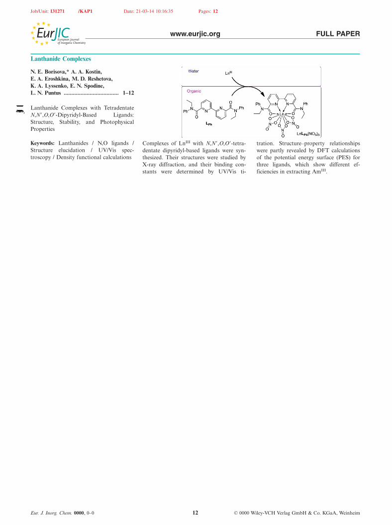
Lanthanide Complexes with TetradentateN,N�,O,O�-Dipyridyl-Based Ligands:Structure, Stability, and PhotophysicalProperties
Keywords: Lanthanides / N,O ligands / Complexes of LnIII with N,N�,O,O�-tetra- tration. Structure–property relationshipsStructure elucidation / UV/Vis spec- dentate dipyridyl-based ligands were syn- were partly revealed by DFT calculationstroscopy / Density functional calculations thesized. Their structures were studied by of the potential energy surface (PES) for
X-ray diffraction, and their binding con- three ligands, which show different ef-stants were determined by UV/Vis ti- ficiencies in extracting AmIII.
Eur. J. Inorg. Chem. 0000, 0–0 © 0000 Wiley-VCH Verlag GmbH & Co. KGaA, Weinheim12
Copyright © 2022 FDOKUMEN