Photophysical evaluation of substituted zinc phthalocyanines ...
259
• • •
-
Upload
khangminh22 -
Category
Documents
-
view
1 -
download
0
Transcript of Photophysical evaluation of substituted zinc phthalocyanines ...

Durham E-Theses
Photophysical evaluation of substituted zinc
phthalocyanines as sensitisers for photodynamic therapy
Stanley, Claire Frances
How to cite:
Stanley, Claire Frances (1997) Photophysical evaluation of substituted zinc phthalocyanines as sensitisers
for photodynamic therapy, Durham theses, Durham University. Available at Durham E-Theses Online:http://etheses.dur.ac.uk/4681/
Use policy
The full-text may be used and/or reproduced, and given to third parties in any format or medium, without prior permission orcharge, for personal research or study, educational, or not-for-pro�t purposes provided that:
• a full bibliographic reference is made to the original source
• a link is made to the metadata record in Durham E-Theses
• the full-text is not changed in any way
The full-text must not be sold in any format or medium without the formal permission of the copyright holders.
Please consult the full Durham E-Theses policy for further details.

Academic Support O�ce, Durham University, University O�ce, Old Elvet, Durham DH1 3HPe-mail: [email protected] Tel: +44 0191 334 6107
http://etheses.dur.ac.uk
2
PHOTOPHYSICAL EVALUATION OF SUBSTITUTED ZINC PHTHALOCYANINES AS SENSITISERS FOR PHOTODYNAMIC
THERAPY
Claire Frances Stanley
Department of Chemistry, University of Durham, Durham.
Submitted in partial fulfilment of the requirements for the degree of Doctor of Philosophy, University of Durham.
September 1997. The copyright of this thesis rests with the author. No quotation from it should be published without the written consent of the author and information derived from it should be acknowledged.
3 APR 1998

DECLARATION
The work described in this thesis was carried out in the Chemistry Department of the University of Durham between September 1994 and August 1997. This thesis is the work of the author except where acknowledged by reference, and has not been submitted for any other degree.
STATEMENT OF COPYRIGHT
The copyright of this thesis rests with the author. No quotation from it should be published without her prior written consent and information derived from it should be acknowledged.

ABSTRACT
Zinc phdialocyanines (ZnPc) are currently being investigated in relation to their use as sensitisers for Photodynamic Therapy (PDT). In particular, the photophysical properties of these dyes are of interest since their ability to generate the cytotoxic species, singlet oxygen (^02), is believed to be central to their role in causing tumour necrosis. In this study, a detailed investigation of the photophysical properties of substituted zinc phthalocyanines under various conditions is described.
Two novel |3-tetra substituted zinc phthalocyanines have been synthesised, ZnPc(CMe(C02Me)2)4 and ZnPc(CHMeC02H)4. The nature of peripheral substituents has littie effect on triplet state or singlet oxygen production by ZnPc, however, ZnPc(CHMeC02H)4 displays a remarkable sensitivity to the ionic strength of nonaqueous solutions. Ion concentrations below lO*^ mol dm'^ induce dimerisation whilst concentrations greater than this promote monomerisation. This behaviour is attributed to ion pairing effects. Photophysical properties of substituted zinc phthalocyanines in heterogenous media and on solid substrates are also described.
The temperature and pH of solvent media greatly influence the photophysical propenies of phthalocyanines. Octadecyl zinc phthalocyanine (CIO) aggregates upon cooling to 77 K in ether-pentane-alcohol (5:5:2) solution. Additional structure in the absorption spectrum is observed, accompanied by the appearance of a fluorescence emission band at 760 nm. Aluminium phthalocyanine chloride in methanol dimerises upon addition of 2.5 x 10'^ mol dm"3 fluoride ions. Dimer species are characterised by a blue shifted peak in the absorption spectrum and are non-fluorescent. These results are ascribed to different aggregate geometries and discussed in terms of exciton theory. Low pH induces stepwise protonation of the azomethine bridges of the phthalocyanine ring, Pc -1- nH"*" V PcHn""'', where n = 0 to 4. Protonation results in significant changes in absorption, fluorescence and triplet state properties of the phthalocyanine. A dramatic decrease in singlet oxygen generation by the phthalocyanine (Oa (n = 0) = 0.54, (n = 1) = 0.075)
is reported, and occurs under surprisingly mild conditions (pK^ of ZnPcS2 in 1% Triton X-100/H2O = 4.4).
The propensity of ZnPc's to bind to serum protein and to participate in electron transfer reactions with potential electron donors is discussed.
ACKNOWLEDGEMENTS
My most sincere thanks are due to so many people that I barely know where to begin. Firstly, for trusting me with all that expensive lab equipment, smiling so graciously when 'accidents happened' and generally being supportive and encouraging, I wish to express my gratitude to Dr. A. Beeby without whom this thesis would never have happened. Thanks also to the other Beebettes - Catherine and Allison (and Ian) for answering (and asking) those 'stupid questions', drinks when times were bad, drinks when times were good and generally ensuring my time in Lab CG 7 was an enjoyable one.
I am very grateful to Prof. D. Phillips, Dr. G. Rumbles, Dr. B. Crystall and their groups for use of the Time Correlated Single Photon Counting Apparatus at Imperial College, London and their helj) concerning measurements. I would also like to acknowledge Prof. M . Cook (University of East Anglia), Dr. N. McKeown (Manchester University) and Prof. M . R. Bryce (University of Durham) for supply of octadecyl zinc phthalocyanine, hexadecyl metal free phthalocyanine and thiafulvalene substituted phthalocyanines respectively. Dr. R. J. Leatherbarrow, Imperial College, should also be recognised for supplying the curve fitting program, Grafit. I am indebted to Dr. A. W. Parker (Rutherford Appleton Laboratory) and Dr. M. Crampton (University of Durham) for helpful theoretical discussions and Dr. S. Faulkner (University of Durham) for proof reading of this thesis and help with NMR measurements. Thank you to EPSRC for providing funding to allow this project to take place.
A special thank you is due to housemates, Pete, Catherine and Allison (as above) for looking after me so well over the last couple of years and in particular the last few months. I will always be grateful for the numerous cups of tea and cooked dinners that were delivered to my desk to make sure I didn't starve. Dave, Jonathan, Allison (again!) and the Duke of Edinburgh gang - weekends away with you provided me with a complete escape from chemistry - worrying about the cold / heat / rain / midges / bUsters etc. became much more important - thank you for keeping me sane.
Finally, I would like to thank everyone who had to Usten to me moan about writing this thesis for the best part of five months, especially Mum, Dad, Nicola, Louise and the IRC mob - thanks folks - it's all over!

CONTENTS
Declaration 1 Abstract 2 Acknowledgements 3 Contents 4 List of Figures 8
List of Tables 15 Common Abbreviations 17 Structures of Substituted Zinc Phthalocyanines 19
Chapter One Introduction
1.1 Background 20 1.2 Principle of PDT 20
1.3 Electronic Structure of Phthalocyanines 26 1.4 Transport and Delivery Systems 30 1.5 Retention and Localisation 31 1.6 Aim 34 1.7 References 35
Chapter Two Experimental Techniques
2.1 Photophysical Measurements 42
2.1.1 UVA^isible Absorption Spectroscopy 42 2.2.2 Ruorescence Spectroscopy 42
2.1.2.1 Spectra 42 2.1.2.2 Ruorescence Quantum Yields 44 2.1.2.3 Fluorescence Anisotropy 45
2.1.3 Time Correlated Single Photon Counting 46 2.1.3.1 Introduction to Measuring Fluorescence Lifetimes 46 2.1.3.2 Fxperimental Arrangement 47
2.1.4 Nanosecond Laser Flash Photolysis 48 2.1.4.1 Introduction to Rash Photolysis 48 2.1.4.2 Experimental Setup 50
2.1.4.3 Triplet Lifetime Measurements 51
2.1.4.4 Triplet Extinction Coefficients 53
2.1.4.5 Transient Absorption Spectra 53
2.1.4.6 Triplet Quantum Yields 54 2.1.5 Singlet Oxygen Phosphorescence Detection 55
2.1.5.1 Introduction to Singlet Oxygen 55 2.1.5.2 Experimental Methods 56
2.1.6 Low Temperature Measurements 58 2.1.7 Diffuse Reflectance Spectroscopy 59
2.1.7.1 Theory 59 2.1.7.2 Experimental 64
2.2 Synthesis 66
2.2.1 Synthesis of Substituted Zinc Phtiialocyanines 66 2.2.2 Metallation of H2(BuO)8Pc and H2(Ci6H34)8Pc 68
2.3 References 69
Chapter Three Properties of Novel Phthalocyanine Sensitisers
3.1 Introduction 72 3.2 Materials and Metiiods 73
3.2.1 Materials 73 3.2.2 Metiiods 73
3.3 Results and Discussion 75 3.3.1 Synthesis 75
3.3.2 Properties in Homogeneous Solution 76 3.3.2.2 Results 76 3.3.2.3 Discussion 86
3.3.3 Properties in Heterogeneous Solution 96 3.3.3.1 Micellar Systems 96 3.3.3.2 Photophysical Properties 97
3.3.4 Properties in tiie Solid State 100 3.4 Conclusion 104 3.5 References 105
Chapter Four Dimerisation of Phthalocyanines
4.1 Introduction I l l
4.2 Theoretical Background 112
4.2.1 Exciton Theory 112
4.2.2 Fluorescence Anisotropy 116
4.3 Materials and Methods 118

4.4 Results and Discussion 119 4.4.1 Ruorescence Anisotropy of Phthalocyanines 119
4.4.1.1 Preparative Studies 119 4.4.1.2 Anisotropy Measurements 123 4.4.1.3 Conclusion 130
4.4.2 Dimerisation of Phthalocyanines 131 4.4.2.1 Aluminium Phthalocyanine Chloride (AlPcCl) 131 4.4.2.2 1,4,8,11,15,18,22,25-Octadecyl Zinc Phtiialocyanine (CIO) 139 4.4.2.3 Conclusions 154
4.5 References 155
Chapter Five Protonation of Phthalocyanines
5.1 Introduction 160 5.2 Materials and Methods 161 5.3 Results and Discussion 161
5.3.1 tBu4ZnPc in Acidified AlcohoUc Solution 161 5.3.1.1 Results 161 5.3.1.2 Discussion 167
5.3.2 ZnPcS2 in 1% Triton X-100 solution 173 5.3.3 Solid State Measurements 177
5.4 Biological Relevance 180 5.5 Conclusion 182 5.6 References 182
Chapter 6 Ligand Binding and Electron Transfer Properties
6.1 Introduction 186 6.1.1 Ligand Binding 186 6.1.2 Electron Transfer Properties 187
6.1.2.1 Theory 187 6.1.2.2 Electron Transfer Reactions of Phthalocyanines 190
6.1.3 Aim 191 6.2 Materials and Methods 192
6.2.1 Materials 192 6.2.2 Methods 194
6.3 Results and Discussion 195
6.3.1 Ligand Binding 195

6.3.LI Cyanide Ions and Tetrahydrofuran 195 6.3.1.2 Bovine Serum Albumin 198
6.3.2 Electron Transfer Reactions 206 6.3.2.1 Simple Organic Quenchers 206 6.3.2.2 Biological Molecules 219
6.4 Conclusion 229
6.5 References 231
Summary 238
Appendix A A Novel Switch for Singlet Oxygen Measurements
A . l Introduction 239
A.2 Materials and Methods 240 A.2.1 Materials 240 A.2.2 Experimental 240 A.2.3 The Switch 241
A.3 Results and Discussion 242 A.3.1 Electrical properties 242 A.3.2 Performance 243
A.4 Conclusions 248 A.5 References 248
Appendix B 249 Appendix C 252
Publications 253 Conferences 254 Seminars 255

LIST OF FIGURES
Chapter 1 Title of Figure Page
Figure 1.1 The principle of PDT 21
Figure 1.2 Jablonski diagram showing absorption and deactivation processes of sensitsers 22
Figure 1.3 Destructive pathways of PDT 23 Figure 1.4 Formation of singlet oxygen 24 Figure 1.5 UVA^'isible absorption spectrum of ZnPc in 1%
pyridine / toluene solution 27 Figure 1.6 Structure of tetra ^butyl metal free phtiialocyanine 28 Figure 1.7 Energy level transitions of Q and Soret bands 28 Figure 1.8 Electron density of the HOMO, HOMO-1 and LUMO
ofZnPc 29 Figure 1.9 Factors affecting retention of sensitisers in a tumour 31 Figure 1.10 Schematic representation and glossary of a tumour
and cell 32
Chapter! Title of Figure Page
Figure 2.1 Experimental arrangement for recording emission
spectra from phthalocyanine species emitting in the near-IR 43
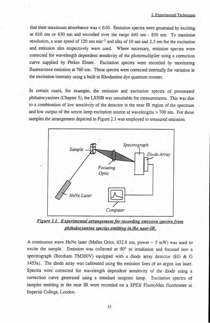
Figure 2.2 Experimental arrangement for measurement of
fluorescence anisotropy 46 Figure 2.3 Time correlated single photon counting setup 48 Figure 2.4 Principle of flash photolysis 49 Figure 2.5 Typical transient decay 49 Figure 2.6 Three dimensional flash photolysis measurements 50 Figure 2.7 Experimental arrangement of flash photolysis
apparatus 51 Figure 2.8 Experimental arrangement for singlet oxygen
measurements 57
Figure 2.9 Typical IO2 decay (Sensitiser: ZnPc(CHMeC02H)4, Solvent: EtOH) 57
Figure 2.10 Specular and diffuse reflection 60 Figure 2.11 Parameters for Kubelka-Monk theory 61

Figure 2.12 Diffuse reflectance flash photolysis: Experimental arrangement 66
Figure 2.13 Synthetic scheme for production of acid and ester substituted zinc phthalocyanine 67
Chapters Title of Figure Page
Figure 3.1 Absorbance spectra of ZnPc(CMe(C02Me)2)4. a) CHCI3, b) MeOH, c) Calculated dimer spectrum 77
Figure 3.2 A. Aggregation of ZnPc(CHMeC02H)4 in EtOH induced by potassium acetate. B. Disaggregation at high ionic strength 78
Figure 3.3 UVA^is spectra of ZnPc(CHMeC02H)4 in PBS. a. pH = 7.4 b. pH = 11 c. pH = 2 d. pH = 7.4 (0.02 mmoldm-3CTAB) 79
Figure 3.4 Solvent effect on (UVms.) of tBu4ZnPc 80
Figure 3.5 Absorption and emission spectra of
ZnPc(CMe(C02Me)2)4 in CHCI3 81 Figure 3.6 Transient absorbance of ZnPc(CMe(C02Me)2)4 in
CHCI3 vs laser power 82 Figure 3.7 Effect of temperature (a. RT, b. 77 K) on the UVA^is
spectra of A. ZnPcS2 in EtOH (inset: magnification of Q band peak), B. tBu4ZnPc in EPA, C. ZnPc(CHMeC02H)4 in EPA, D.
ZnPc(CMe(C02Me)2)4 in EPA, E. CIO in EPA 84 Figure 3.8 Excitation and emission spectra at 77 K. A. ZnPcS2
in EtOH, B. ZnPc(CMe(C02Me)2)4 in EPA 85 Figure 3.9 a and (3 positions of zinc phthalocyanine 87
Figure 3.10 Relationship between solvent refractive index and Xj^^ (UVms.) of tBu4ZnPc 93
Figure 3.11 S tructure of Triton X-100 96 Figure 3.12 Simple diagram of a normal micelle 97 Figure 3.13 Beer-Lambert plots of tBu4ZnPc and
ZnPc(CHMeC02H)4 in 1 % TX/PBS 98
Figure 3.14 Absorption specQ-a of CIO in 1% TXR/PBS and 1%
TX/PBS 99 Figure 3.15 Remission functions of phthalocyanines on silica. A.
tBu4ZnPc. B. ZnPc(CHMeC02H)4 101

Figure 3.16 A. Excitation and emission spectra of ^Bu4ZnPc on cellulose. B. Corrected for artefacts 101
Figure 3.17 a. Typical diffuse reflectance transient decay, b. Residuals of monoexponential fit 103
Figure 3.18 Diffuse reflectance transient absorption spectrum of tBu4ZnPc on cellulose 103
Chapter 4 Title of Figure Page
Figure 4.1 Energy level splitting on exciton coupling of a 'slipped' dimer 113
Figure 4.2 Exciton coupling of an 'obhque' dimer 114 Figure 4.3 Exciton coupling of an 'out-of-plane' dimer 115
Figure 4.4 Representation of - Angle between absorption and emission transition dipoles 116
Figure 4.5 Temperature dependence of UYA^isible absorption spectrum of tBu4H2Pc in EPA 119
Figure 4.6 Temperature dependence of fluorescence excitation and emission spectra of 'BU4H2PC in EPA 120
Figure 4.7 Temperature dependence of emission from tBu4H2Pc in EPA at 665 nm 121
Figure 4.8 Excitation spectrum and anisotropy of ^Bu4H2Pc in EPA at 77 K. Inset: r ^ of tBu4H2Pc and tBu4ZnPc by excitation into the Soret band 123
Figure 4.9 a) Emission spectrum of tBu4H2Pc, b) Anisotropy in EPA at 77 K 124
Figure 4.10 Dependence of x^^ of tBu4H2Pc in 1% TX/H2O on emission and excitation wavelength 124
Figure 4.11 Anisotropy of 'Bu4ZnPc as a function of excitation wavelength 125
Figure 4.12 Temperature dependence of r^v of tBu4ZnPc in EPA 126 Figure 4.13 A. Aggregation of 2 x lO'^ mol dm- AlPcCl + F"
B. Disaggregation of 2 x 10'^ mol dm-3 AlPcCl + F" 131 Figure 4.14 Rate of formation of (AlPc)2F-. Inset: Fit of first
order rate constant 133 Figure 4.15 a) Absorption, b) Excitation and c) Emission spectra
of (AlPc)2F-inMeOH 134
10

Figure 4.16 Transient absorption spectra of AlPcCI and (AIPc)2F" in MeOH 135
Figure 4.17 Equihbria involved in dimerisation of AlPcCI in MeOH upon addition of F" 136
Figure 4.18 Geometry of (AIPc)2F' dimer and AIPCF2" monomer 138 Figure 4.19 Absorption spectra of CIO at 293 K and 77 K and
emission spectra of CIO at 293 K and 77 K in EPA 140 Figure 4.2 Concentration dependence of absorption spectra of
CIO in EPA at 77 K 141 Figure 4.21 Effect of phthalocyanine concentration on the
emission spectra of CIO in EPA at 77 K 141 Figure 4.22 Temperature dependence of the excitation spectra of
CIO in EPA. A. = 725 nm, B. = "785 nm 142 Figure 4.23 Anisotropy of emission from CIO in EPA at 77 K. a)
^em = 725 nm b) ^gm = 785 nm 143 Figure 4.24 A. Transient decay of CIO in EPA at 200 K, .p^be =
700 nm. B. Effect of temperature on rate of triplet decay 145
Figure 4.25 Transient absorption spectra of CIO in EPA. A. 77 K, B. 200 K 146
Figure 4.26 Emission intensity vs. absorbance due to monomeric and dimeric CIO in EPA at 77 K 148
Figure 4.27 Energy level splitting of CIO in EPA at 77 K 150 Figure 4.28 Equilibria upon excitation of CIO in EPA at 77 K 153
Chapters Title of Figure Page
Figure 5.1 Absorption and fluorescence excitation spectra of PcHnn+ in EtOH (n = 0 to 4) 162
Figure 5.2 Ruorescence emission spectra of PcHn"+ in EtOH (n = 0 t o 4 ) 164
Figure 5.3 Anisotropy of tBu4ZnPc and PcH+ in EPA at 77 K 164 Figure 5.4 Time resolved fluorescence decays of PcHn"+ in
EtOH(n = 0 to4) 165
Figure 5.5 Triplet transient decays of tBu4ZnPc and 'Bu4ZnPcH+ 167
Figure 5.6 Effect of protonation on conjugation of the ring 168 Figure 5.7 Structure of tBu4ZnPcH+ 169
11

Figure 5.8 Dependence of kjP and kj^, on AE (S^ <- Sq) of
PcHn"+ (n = 0 to 4) 170
Figure 5.9 Triplet state equilibria of PcH+ 171
Figiu-e 5.10 UVA^isible absorption spectra of Fresh' and 'Old'
(days to weeks) solutions of ZnPcS2 in 1% Triton X-
IOO/H2O solution 173
Figure 5.11 Excitation and emission spectra of 'New' species
formed in an old solution of ZnPcS2 in 1 % TX/H2O 174
Figure 5.12 Absorbance spectra of ZnPcS2 in buffers ranging from
pH 7 to 1 175
Figure 5.13 Emission spectra of ZnPcS2 in buffers ranging from
pH 7 to 1. Inset: Ratio of emission from
unprotonated and protonated phthalocyanine species
(679/707 nm) 176
Figure 5.14 Effect of pH on the intensity ratio 679 / 707 nm for
ZnPcS2 in buffer 176
Figure 5.15 Reflectance spectra of ZnPcS2 and tBu4ZnPc on
cellulose. Inset: ZnPcS2 on cotton 178
Figure 5.16 Excitation and emission spectra of tBu4ZnPc and
ZnPcS2 on cellulose 178
Figure 5.17 Structure of cellulose 179
Chapter 6 Title of Figure Page
Figure 6.1 Potential energy profile of electron transfer 188
Figure 6.2 Processes involved in fluorescence quenching by
electron transfer 189
Figure 6.3 Structures of Pc(TTF)8 and Pc(TTF)4 193
Figure 6.4 UVMsible absorption spectra of ZnPcS2 in MeOH +
CN- ions 196
Figure 6.5 Hildebrand-Benesi plots of ZnPcS2 + CN" in MeOH 195
Figure 6.6 Disaggregation of ZnPcS2 in PBS upon addition of
BSA 199
Figure 6.7 Triplet transient decay of tBu4ZnPc in 1% TX/PBS +
4 X 10⁻⁴ mol dm-3 BSA. Irradiation by A. 4 laser
shots B. 30 laser shots C. 44 laser shots 201
Figure 6.8 Increase in absorbance at 322 nm for tBu4ZnPc + 5 x
10" mol dm"3 BSA upon irradiation by red light.
12

(Effect of irradiation on a phthalocyanine only solution is also shown) 203
Figure 6.9 CIO triplet state quenching by O2 in 1 % TX/PBS 204 Figure 6.10 Structure of simple organic quenchers 206 Figure 6.11 Stem-Volmer plot showing quenching of ZnPc
emission by DABCO in 1% pyr/tol 207 Figure 6.12 Formation of DABCO...ZnPcDABCO ion pair upon
irradiation of ZnPc in 1% pyr/tol in the presence of >
0.01 mol dm-3 DABCO 208
Figure 6.13 Stem-Volmer plot of ZnPc singlet state quenching by dimethylaniline in 1% pyr/tol 209
Figure 6.14 Stem-Volmer plot of ZnPc fluorescence quenching by
TMPD in 1% pyr/tol. Fit of static quenching model 210 Figure 6.15 Application of finite sink approximation to TMPD
quenching data 212 Figure 6.16 ^ZnPc* triplet state quenching by TMPD in 1%
pyiTtol 213 Figure 6.17 Stem-Volmer plots for quenching of ^Bu4ZnPc and
CIO in 1% TX/H2O by TTF in 10% TX/H20 215 Figure 6.18 Structures of amino acids 219 Figure 6.19 Increase in rate of decay of ^KTI* states of 'Bu4ZnPc
in MeOH upon addition of ^BOC methionine 220 Figure 6.20 Formation and decay of ^Pc.Met* complex 221 Figure 6.21 Molecular orbital description of exciplex formation 222 Figure 6.22 Stmctures of ascorbic acid (AA) and ascorbic acid
palmitate (AAP) 224
Figure 6.23 Quenching of the triplet state of ^Bu4ZnPc by ascorbic
acid and ascorbic acid palmitate in MeOH 225 Figure 6.24 Transient study of tBu4ZnPc in MeOH + 0.01 mol
dm"3 ascorbic acid palmitate. Deactivation of triplet
species and formation and decay of Pc- radicals 225 Figure 6.25 Transient absorption spectrum of tBu4ZnPc-- radical
in MeOH. a) At = 300 ^is, b) At = 1 ms 226
Figure 6.26 Equilibria of ascorbic acid (palmitate) and redox
couples at pH = 7 227
Figure 6.27 Comparison of tBu4ZnPc triplet state quenching by a)
ascorbic acid palmitate and b) ascorbic acid in 1%
TX/H2O solution 229
13

Appendix A Title of Figure Page
Figiu-eA.l Experimental arrangement 241
Figure A.2 Circuit diagram of switch 241 Figure A.3 Physical properties of the switch 242 Figure A.4 Decay trace of switching transient as the switch closes
following a 400 ns gate 243 Figure A.5 a) Huorescence recorded using 0.5 V/div. Scale, b)
Same reading on 1 mV/div. scale showing saturation of amplifier, c) Trace obtained using switch (1 mfWdiv.) 244
Figure A.6 Overlap of IO2 decays sensitised by Rose Bengal with and without the switch 245
Figure A.7 IO2 decay sensitised by phtiialocyanine without (a) and with (b) the switch 246
Figure A.8 A) Signal produced by a mixture of Styryl 9 and Rose Bengal with (a) and without (b) switch. B) Singlet oxygen decay viewed on correct scale 247
14

LIST OF TABLES
Chapters Title of Table Page
Table 3.1 Dimerisation constants of ZnPc(CMe(C02Me)2)4 76 Table 3.2 Effect of peripheral substitution on absoiption A jj x of
ZnPc 80 Table 3.3 A Effect of peripheral substitution on emission
characteristics of ZnPc 81
Table 3.3B Effect of peripheral substitution on fluorescence emission yield of ZnPc 82
Table 3.4 Triplet state properties of substituted ZnPc's 83 Table 3.5 Absorption and emission characteristics of
ZnPc(BuO)8 in various organic solvents at 293 K and
77 K 86 Table 3.6 Collation of substituent constant, a* and tiie
dimerisation constant, K of substituted ZnPc's 88 Table 3.7 Photophysical properties of substituted ZnPc's in
heterogeneous systems 99
Chapter 4 Title of Table Page
Table 4.1 Decrease in anisotropy of ^Bu4ZnPc in EPA with
increasing temperature 127 Table 4.2 Estimated rotational correlation times of tBu4ZnPc
and tBu4H2Pc in EPA and 1 % TX/H2O 130 Table 4.3 Absorption maxima of monomer and dimer species of
AlPcCI in MeOH 132 Table 4.4 EquiUbrium constants, rates and extinction
coefficients of AIPcCl dimerisation in MeOH upon addition of F- 134
Table 4.5 Ruorescence lifetime data of CIO in EPA at 293 K and 77 K 143
15

Chapters Title of Table Page
Table 5.1 Absoiption maxima of PcHnn+ (n = 0 to 4) in EtOH 162
Table 5.2 Absorbance and fluorescence data for PcHn""*" (n = 0
to 4) in EtOH 163
Table 5.3 Fluorescence lifetimes of PcHn"+ (n = 0 to 4) in EtOH.... 165
Table 5.4 Triplet state data for PcHnn+(n = 0 to 4) in EtOH 166
Table 5.5 Rate constants of radiative and non-radiative
deactivation pathways of PcHn"+ (n = 0 to 4) in EtOH.... 169
Table 5.6 Comparison of photophysical properties of
monoprotonated species of 'Bu4ZnPc, ZnPcS2 and
AIPCS2 in 1 % Triton X- IOO/H2O solution 177
Chapter 6 Title of Table Page
Table 6.1 Effect of THE on the photophysical properties of
ZnPc(CMe(C02Me)2)4 197 Table 6.2 Effect of bovine serum albumin on the photophysical
properties of substituted zinc phthalocyanines in 1%
TX/PBS 199
Table 6.3 BSA increases the fluorescence anisotropy, r ^ of
metallophthalocyanines 200
Table 6.4 Rate constants of triplet state quenching by oxygen in
the presence of 5 x IQ- mol dm-3 BSA 204
Table 6.5 Summary of measured electron transfer rates 216
Table 6.6 Calculated quenching rates of ^ZnPc* by simple
organic electron donors in 1% pyridine / toluene 217
Table 6.8 Calculated quenching rates of ^Bu4ZnPc* by amino
acids in MeOH 223
Table 6.9 Calculated quenching rates of ^Bu4ZnPc* by ascorbic
acid in MeOH 227
16

COMMON ABBREVIATIONS
Abbreviation Description
Phthalocyanines
AlPcCl
AIPCS2
(BuO)gZnPc
C5 CIO ZnC16 HpD P Pc tBu4ZnPc
ZnPc(CHMeC02H)4
ZnPc(CMe(C02Me)2)4
ZnPcS2
Aluminium phthalocyanine chloride Disulfonated aluminium phthalocyanine 1,4,8,11,15,18,22,25 octa-butoxy zinc phthalocyanine 1,4,8,11,15,18,22,25 octa-pentyl zinc phthalocyanine 1,4,8,11,15,18,22,25 octa-decyl zinc phthalocyanine 2,3,9,10,16,17,23,24 octa-hexadecyl zinc phthalocyanine Haematoporphyrin derivative Porphyrin Phthalocyanine p-tetra-tertbutyl zinc phthalocyanine p-tetra-l-carboxyethyl zinc phthalocyanine p-tetra-l,l-di(methoxycarbonyl)ethyl zinc phthalocyanine Disulfonated zinc phthalocyanine
Solvents
1% pyr/tol
1% TX/PBS 1%TX/H20 CTAB DMF
DMSO EPA Et20 EtOH IP MeOH MeTHF PBS T H E
TX TXR
1% Pyridine in toluene (v/v)
1% Triton X-100 in phosphate buffered saline (w/v) 1% Triton X-100 in distilled water (w/v) Hexadecyl trimethylammonium bromide
N, N-Dimethylformamide Dimethylsulfoxide Ether-isopentane-alcohol (5:5:2) Diethylether Ethanol Isopentane Methanol 2-Methyl tetrahydrofuran Phosphate buffered saline Tetrahydrofuran Triton X-100 Triton X-100 reduced
17

others
AA AAP bipy BSA DABCO DMA DMB DiPE EWHM HOMO Idl LUMO PDT TMPD
Symbols
^em
Op Tp
^Probe O t
(^Ag)02
(3Sg-)02
isc av
Ascorbic acid Acsorbic acid palmitate 4,4'-bipyridine Bovine serum albumin 1,4 diazabicyclo [2,2,2] octane N, N dimethylamline 1,4 dimethoxy benzene
Trans 1,2-dipyridyl ethylene Full Width at Half Maximum Highest Occupied Molecular Orbital Low density lipoprotein Lowest Unoccupied Molecular Orbital Photodynamic Therapy l,4-N,N,N',N'-tetramethylphenylenediamine
Extinction coefficient, dm^ mol' cm ^ Intrinsic fluorescence lifetime, ns Singlet oxygen lifetime, |is
Yield of singlet oxygen formation
Emission wavelength, nm Excitation wavelength, nm Huorescence quantum yield Measured fluorescence lifetime, ns Yield of internal conversion Wavelength of maximum absorbance, nm Probe wavelength in flash photolysis, nm Triplet quantum yield Triplet state lifetime, \is Singlet state molecular oxygen Singlet state molecular oxygen Ground state molecular oxygen
Rate of fluorescence, s"l Rate of internal conversion, s' Rate of intersystem crossing, s"
Measured steady state fluorescence anisotropy
18
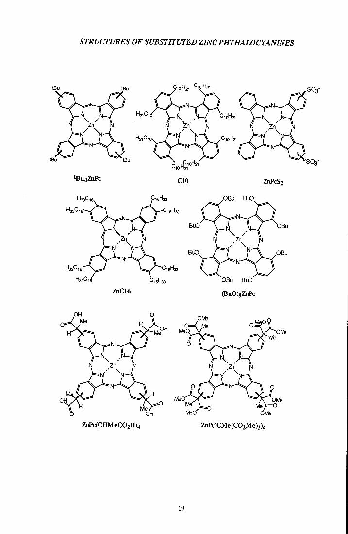
STRUCTURES OF SUBSTITUTED ZINC PHTHALOCYANINES
H21C1
tBu4ZhPc
10H21 CipH2i
CIO ZhPcS2
16' 133 33'-'16
16' '33
BuO'
OBu BuO
OBu
p B u
ZnC16
OBu B l O
(BuO)8ZnPc
Me°^ MeQ
OH Me'
ZhPc(CHMeC02H)4
PIVfe
OMe ZnPc(CMe(C02Me)2)4
19
I.l Background
Photodynamic Therapy, or PDT, is an alternative treatment for cancer which is currently undergoing clinical trials. The principle on which it is based has been known for almost one hundred years. Around that time, Meyer-Betz self administered haematopoiphyrin to determine its biological effectf l and Von Tapiener's laboratoryl J in Munich reported the destruction of skin tumours by the use of topical eosin and light. Progress was slow in the early years, the only major breakthrough coming in 1948 when Figge and co-workers' ] noticed that porphyrins and metalloporphyrins have a tendency to accumulate in neoplastic, traumatised and embryonic tissue. The build-up of porphyrins can be recognised by their characteristic red fluorescence upon irradiation by light. This principle was seized by Figgef'*J in 1955 and again by Lipson'^l in 1961, to show that porphyrins could be used to aid in the detection of malignant growths. Lipson and his group were also responsible for the development of HpDt l, one of the first compounds to be clinically tested as a photosensitiser for PDT. HpD is a haematoporphyrin derivative which was shown to exhibit greater localisation than the original porphyrin. Throughout the 1970's the destruction of various types of tumour such as bladder!' !, leukaemicl l, breastt J, lungt'°l and colon cancerst l, using HpD as a photosensitiser, was reported and it was suggested that singlet oxygen may be an important intermediate in the cytotoxic processf 2],[i3] Studies on HpD, including its chemical composition!'"! and mechanisms for its transport and distribution throughout the body continued throughout the 1980's and complications associated with using HpD were realised' l In 1985 Ben-Hurt' ! proposed phthalocyanines as a second generation class of sensitisers in an attempt to overcome these problems. Studies of both the biological and photophysical properties of this class of drugs have proved promising and research continues to increase understanding of the mechanism of cell destruction and sensitiser locaUsation.
1.2 Principle of PPn^-ium
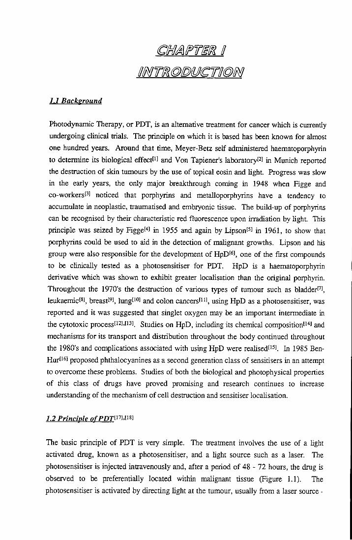
The basic principle of PDT is very simple. The treatment involves the use of a light activated drug, known as a photosensitiser, and a light source such as a laser. The photosensitiser is injected intravenously and, after a period of 48 - 72 hours, the drug is observed to be preferentially located within malignant tissue (Figure 1.1). The photosensitiser is activated by directing light at the tumour, usually from a laser source -
1. Introduction
red light is used to allow maximum tissue penetration. It is important to realise that the laser is not being used as an alternative for a scalpel but as a source of monochromatic, collimated light which can be directed down an optic fibre to allow access to internal tumours whilst still retaining intensity. The excited sensitiser is believed to interact with surrounding oxygen to produce cytotoxic species such as singlet oxygen and superoxide, leading to cell destruction.
Sensitiser Injection
24-72hrs later: Red light irradiation
Tumour cell death
Fisure 1.1. The principle of PDT.
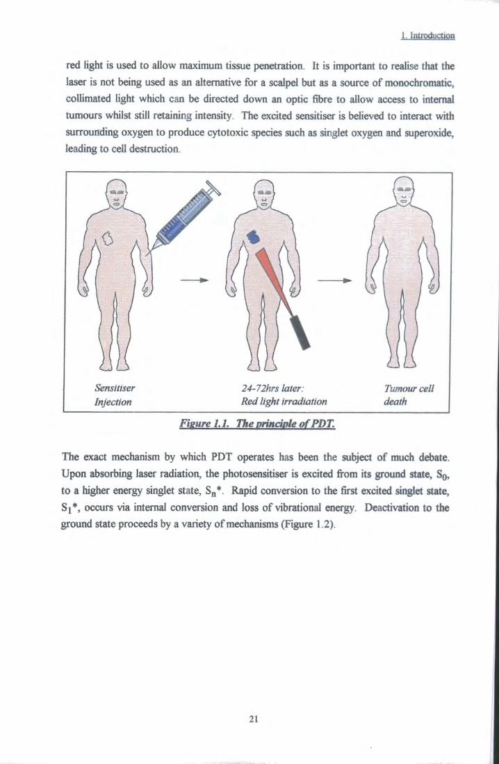
The exact mechanism by which PDT operates has been the subject of much debate. Upon absorbing laser radiation, the photosensitiser is excited fi-om its ground state, SQ, to a higher energy singlet state, S,,*. Rapid conversion to the first excited singlet state, Si*, occurs via internal conversion and loss of vibrational energy. Deactivation to the ground state proceeds by a variety of mechanisms (Figure 1.2).
21
1. Introduction
Absorption
Fluorescence
Triplet-Triplet Absorption
Energy Transfer
Phosphorescence
Phosphorescence (1269 nm)
IC = Internal Conversion
ISC = Intersystem Crossing
VR = Vibrational Relaxation
Figure 1.2. Jablonski diagram showing absorption and deactivation processes of sensitisers.
In the absence of quenchers, these include radiative emission (fluorescence), internal conversion (when the states involved are of the same spin multiplicity) and intersystem crossing. Intersystem crossing involves a ttansition between states of different multiplicity, Sj ^ Tj or Tj —> SQ, and is a prerequisite for population of the triplet state (Tj). This process is forbidden by the zero order approximationti l; however, the influence of spin-orbit perturbations render the pathway viable. It is the properties of the T j state that are of importance in the mechanism of PDT. In the absence of oxygen and other quenching species, triplet species decay via phosphorescence emission or by spin forbidden intersystem crossing to the ground state, SQ. However, in a biological environment, there are generally believed to be two types of reaction which may occur, Type I and Type II (Figure 1.3). The Type I mechanism involves interaction of the sensitiser triplet state with solvent molecules or biological substrates to produce radicals or radical ions by hydrogen atom or electron transfer. These radicals react with oxygen to produce the cytotoxic superoxide radical anion. Alternatively, the triplet state can interact directly with ground state oxygen in an energy transfer reaction to produce singlet oxygen, ( Ag) O2, 02.
22

1. Introduction
Dye
hv
Radicals and ^ubstrate 3 * ° 2 ^ , radical ions K ^0„
Type I Substrate
Oxidation Products
Typen
Oxidation Products
Fieure 1.3. Destructive pathways of PDT.
The formation of IO2 has been discussed in detail in a review by Keamsl oi. Production is believed to occur via an energy transfer mechanism from excited singlet (Equation 1.1) or triplet (Equation 1.2) sensitiser states to ground state molecular oxygen.
^Sens*+^02 >^Sens*+^02 (1.1)
^Sens*+^02-^hSens..02) > 02(^^g)+ ^Sensg (a)
\Sens..02 ) > 02( ^2 / ) + ^Sensg (b)
^(Sens..02 ) > 02(^^g~)+ ^SensQ (c)
^(Sens..02) (d)
(1.2)
where k j represents the diffusion controlled rate constant. In practice, singlet state quenching is rarely observed due to the short lifetime of these states with respect to the diffusion rate of molecules tiirough solution, i.e. the number of ^Sens* and 02 collisions is small. It can be seen from Equations 1.2a to 1.2d above, that singlet oxygen species result from only one out of nine possible colUsion complexes. This leads to a theoretical triplet state quenching constant, kQ2, of 1/9 k^. Experimentallyf U, values of an order of magnitude lower than were obtained, lending credence to this theory. Molecular orbital schemes for the formation of Ag and Sg+ are depicted in Figure 1.4.
23

1. Introduction
. - I v •> V
hens*
->
Sens.
Sens,
A V
Fieure 1.4. Formation of singlet oxygen.
lSg+ and lAg excited states of O2 occur at 13121 cm" and 7882 cm-l above the ground state respectively. The ratio of each state formed is dependent on the triplet state energy, E j , of the sensitiser. For E j values of ~ 160 kJ mol- (~ 13350 cm- ) or less,
Ag is the major product.
Both superoxide radical species and singlet oxygen cause oxidative destruction of tissue and hence may perform a vital role in the mechanism of PDT. However, it is generally believed that IO2 is the major intermediate responsible for photodynamic action although confirmation is prevented by lack of data regarding in vivo detection. Detection is difficult due to rapid quenching of IO2 in an in vivo environment, indeed, it has been shown that singlet oxygen phosphorescence generated by sensitisers incorporated into cells[22] is of greatiy reduced intensity and shorter lifetime with respect to that in a homogeneous environment. A summary of the competing processes is given below:
So + h v ^ S i * Si* ^ So + hv Si* So + heat S i * - ^ T i * T i * ->So + hv
T i * ^ So + heat T i * -i- Subs -> Sens- + Subs-T i * + 02(3lg-) M + 02(lAg)
Absorption I Ruorescence Internal Conversion Intersystem crossing Phosphorescence Intersystem crossing kisc[Ti] Electron transfer ki[Ti][Subs] Energy Transfer kii[Ti][02]
a kf{Si]
kic[Si]
kisc[Si]
kp[Ti]
24

1 • Introduction
The properties of the drug administered are extremely important and, whilst it is easy to specify those which make an ideal sensitiser, in real terms it is much more difficult due to the many biological factors which may have an influence. To avoid destruction of healdiy tissue, a sensitiser should be retained solely by neoplastic tissuef23). It should absorb in the red region of the spectrum with littie or no absorption in the ultraviolet region in order to minimise sensitisation to simlight after intravenous administration of high concentrations of dye and allow optimum penetration of tissue. It should have a defined chemical composition and, perhaps most important, are the photophysics of the sensitiser and its abihty to generate high concentrations of cytotoxic intermediates such as singlet oxygen. There have been large amounts of work carried out in order to develop photosensitisers which fulfil the criteria listed above.
Porphyrins, specifically a water soluble haematoporphyrin derivative and its purified form, marketed as Photofirin 11 , were the filrst compounds recognised to have potential as PDT sensitiserst J. These were shown to be clinically effective in many cases using both animal and human subjectsf24].[25] however, a number of problems were encountered. Dealing with a complex mixture of compounds made it difficult to replicate the exact formula. Storage times and synthetic routes both affected the composition and the extent of aggregation. Variationst J't ' J such as these lead to reduced biological efficiency in certain environments and different distributions between malignant and healthy tissue. The strong absorption band of porphyrins in the ultraviolet region of the spectrum also introduced a serious side effect to PDT treatmentl' l. Retention of significant amounts of porphyrin in the skin led to the patient becoming sensitised to sunlight in the period following treatment, thus risking the development of skin cancer. This was compounded by the low extinction coefficient of poiphyrins at 630 nm which made it necessary to inject high concentrations in order to obtain a satisfactory response.
As a result of these problems, a series of second generation sensitisers has been proposed. These include chlorins sj, purpurinst ], phenylporphyrinst "] and phthalocyaninest 'l It is the phthalocyanines (Pc) that are of interest in this study and detailed reviews of these and other sensitisers can be found elsewheret32],[33],[34] Phthalocyanines are derivatives of the porphyrin family in which the methine bridges have been replaced by nitrogen and the pyrrole moieties are extended by conjugation vwth extra benzene rings (Section 1.3). These modifications lead to a remarkably different absorption spectrum with a sharp peak at ~ 670 nm and no absorption between 400 nm and 600 nm. The large extinction coefficients of Pc's in the red region of the spectrum and their transparency at shorter wavelengths give them an immediate advantage over
25
1. Introduction
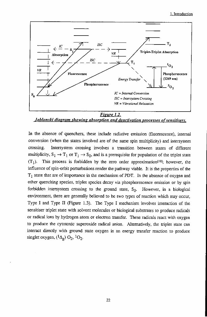
porphyrins as much lower doses are required and the risk of photosensitisation is substantially reduced. Coordination of certain metals to the Pc Mgand has been shown' l to create a more efficient PDT agent. In general, diamagnetic metals, e.g. Al, Ga and Zn, are the most suitable^s] (xj of ZnPc' ] = 270 |is) due to modifications of the photophysical properties which result in long triplet lifetimes and high quantum yields and therefore a greater ability to generate IO2. Paramagnetic metals such as Cu, Ni, Fe, Cr and Pd increase the rate of intersystem crossing (Ti SQ) and substantially reduce the lifetime of the triplet state (Xj of CuPct J = 0.04 |is). Sulphonated aluminium phthalocyanines have been the subject of many studiesP9].[40].t4i] nd shown to have great promise as future sensitisers. Incorporation of sulphonate groups onto the phthalocyanine ring renders the molecule soluble in aqueous media, useful for intravenous administration, however the efficacy has been shown to depend on the degree on sulphonationt'' ]. This leads to complications, as separation of the various isomers produced during AlPcSn synthesis is a formidable task. Alternative methods of controlling the hydrophilicity and lipophilicity of sensitiser macrocycles through the use of axial ligands on chemically pure phthalocyanines such as silicon phthalocyanine[''3l and ruthenium naphthalocyaninet'*'" have also shown promise.
1.3 Electronic Structure of Phthalocvanines
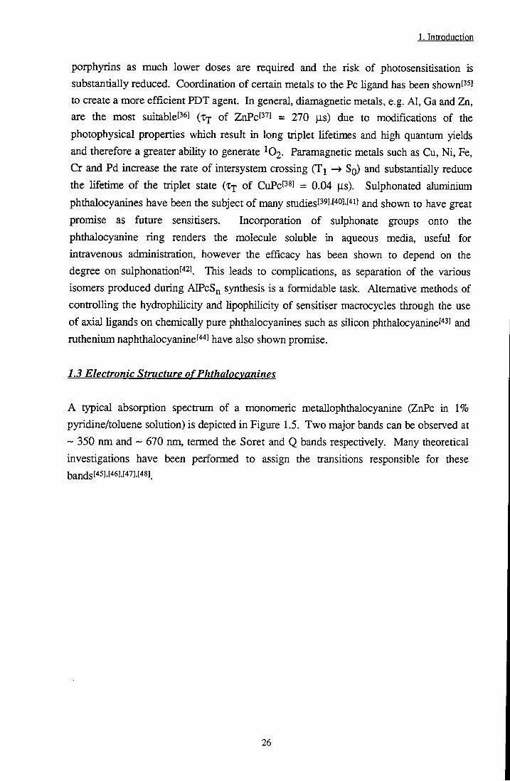
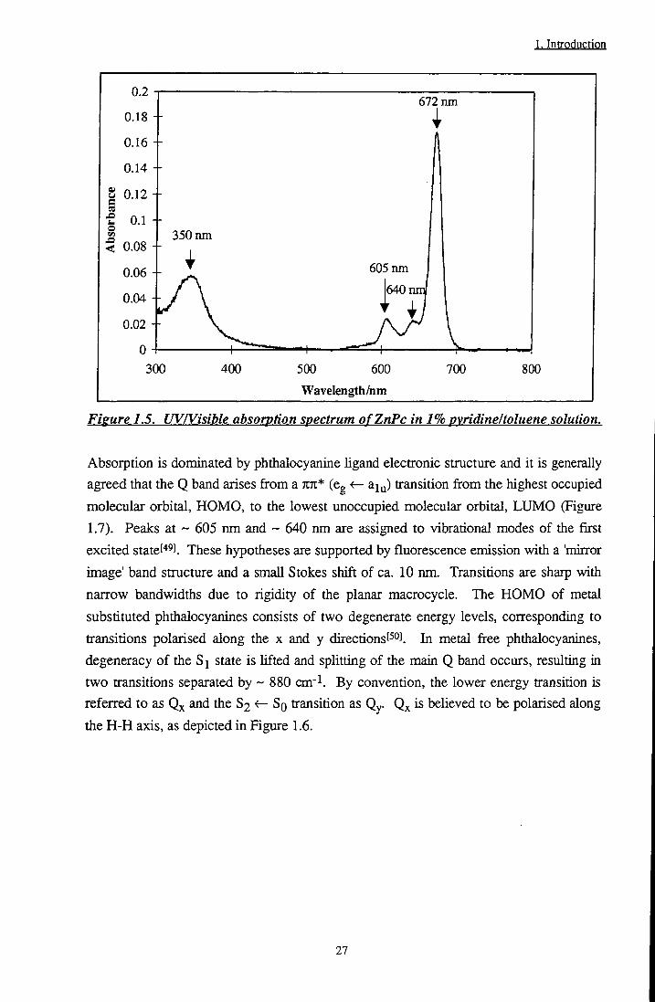
A typical absorption spectrum of a monomeric metallophthalocyanine (ZnPc in 1% pyridine/toluene solution) is depicted in Figure 1.5. Two major bands can be observed at ~ 350 nm and ~ 670 nm, termed the Soret and Q bands respectively. Many theoretical investigations have been performed to assign the transitions responsible for these bandsf'*5].[46].[47].[48]_
26
1. Introduction
0.2
0.18
0.16
0.14
I 0.12
< 0.08
0.1 +
0.06
0.02
0 4 300
672 nm 1
350 nm
^ 605 nm
jy/' \ j640nm
1 ^ !• I 1 . I T ^ - ^ ^ I v . . . ,
400 500 600
Wavelength/nm
700 800
Figure 1.5. UV/Visible absorption spectrum ofZnPc in 1% pyridine I toluene solution.
Absorption is dominated by phthalocyanine hgand electronic structure and it is generally
agreed that the Q band arises from a TIK* (eg <r- aiy) transition from the highest occupied
molecular orbital, HOMO, to the lowest unoccupied molecular orbital, LUMO (Figure
1.7). Peaks at ~ 605 nm and ~ 640 nm are assigned to vibrational modes of the first
excited statet"* !. These hypotheses are supported by fluorescence emission with a 'mirtor
image' band structure and a small Stokes shift of ca. 10 nm. Transitions are sharp with
narrow bandwidths due to rigidity of the planar macrocycle. The HOMO of metal
substituted phthalocyanines consists of two degenerate energy levels, corresponding to
transitions polarised along the x and y directions[^°J. In metal free phthalocyanines,
degeneracy of the Si state is lifted and splitting of the main Q band occurs, resulting in
two transitions separated by ~ 880 cm- . By convention, the lower energy transition is
referred to as and the S2 <- So transition as Qy. Qx is believed to be polarised along
the H-H axis, as depicted in Figure 1.6.
27

1. Introduction
Figure 1.6. Structure oftetra ^butvl metal free phthalocvanine.
Transitions responsible for Soret band absorption are more difficult to identify. Gouterman suggested the main absorption was a TCTt* transition of Cg < - a2u symmetry, i.e. HOMO-1 to LUMO (Figure 1.7), which was broadened by n7i* transitions of comparable energy.
Figure 1.7. Energy level transitions ofQ and Soret bands.
This has since been confirmed through absoiption and x-ray photoelectron spectroscopy
experimental studies performed by Nyokongf i] and Khalibt52]. Rosat oi performed
28

1. Introduction
density fijnctional calculations on the electronic states of several metallophthalocyanines and concluded that the Soret band consists of a mixture of several KTZ* and nn* transitions. Moriey et a/.t"] have calculated the position and magnitude of electron density in the HOMO, HOMO-1 and LUMO of zinc phthalocyanine (Figure 1.8). Q band absorption involves large changes in the electron distribution of the excited state with respect to the ground state. Density shifts from inner ring carbon atoms to nitrogen atoms. By contrast, Soret band absorption (HOMO-1 to LUMO) results in minimal changes in the electronic distribution.
N — Z n — N
HOMO HOMO-1
LUMO
Fiffure 1.8. Electron density of the HOMO. HOMO-1 andL UMO ofZnPc.
29
1. Introduction
1.4 Transport and Delivery Systems
One method of introducing photosensitisers in vivo is to use dehvery systemst ''] such as liposomes, oil emulsions, proteins or antibodies. In this way, hydrophobic sensitisers such as zinc phthalocyanine may be administered intravenously. There have been many studies investigating the suitability of potential vehicles and their effect on the transport mechanism and localisation of sensitisers. Schieweckl s] and Jorit l have shown that ZnPc may be incorporated into liposomes of various types in its monomeric form, retaining its phototoxic properties. For germanium phthalocyanines, Cremophor E L has been demonstrated to be most suitable since aggregation was observed in liposomal solutionis ). Liposomes and oil emulsions such as Cremophor E L are by far the most common methods chosen for solubUisation of hydrophobic dyes, however, deUvery via in vitro binding to low density lipoprotein' ! Gdl) or monoclonal antibodies' ! has also been demonstrated. After injection into the bloodstream, sensitiser molecules are believed to interact with human serum proteins[*°!. Human serum protein has many different components including albumin, low density Upoprotein (Idl), high density lipoprotein (hdl) and very low density lipoprotein (vldl). The affinity of many different sensitisers for individual components depends on their hydrophilic / lipophilic character. For example, it has been shown that the more hydrophilic dyes such as AlPcSn'^i! bind to albumin, whilst those of lipophUic character, e.g. ZnPc, will interact with low density lipoproteins' !-' !-' '*!. Protoporphyrin shows evidence of binding to Idl' !, chlorin E6 demonstrates binding to albumin and hdl but not Idl' ! and haematoporphyrin associates with albumin' ''!, thus, it is clear that binding is highly sensitive to the nature of the chosen sensitiser. The mechanism by which Uposomes are incorporated into Idl has been investigated by Rensen et alS^^^ who have shown that neither aggregation nor fusion of particles are involved. However, the exact mechanism has yet to be elucidated. Distribution to proteins is also affected by the nature of the delivery system. Work by Kessel'69] has demonstrated that Cremophor E L promotes binding of tin etiopurpurin to Idl whilst Polo'''°! and Jori''' ! have found that the nature of liposomal formulations also has an effect on the transport mode, e.g., l,2-dipalmitoyl-sn-glycero-3-phosphocholine (DPPC) and l-palmitoyl-2-oleoyl-sn-glycero-3-phosphochoUne (POPC) liposomes encourage release to lipoproteins whilst l,2-dimiristoyl-sn-glycero-3-phosphochohne (DMPC) allows delivery to both albumin and lipoprotein fractions.
30

l.Introdqction
1.5 Retention and Localisation
An understanding of the factors which influence and control the localisation and retention of photosensitisers within neoplastic tissue is an important area for development of photodynamic therapy. There has been a large body of work carried out in this area and many theories have been proposed. However, in vivo processes are complex and further study is necessary. Hypodieses concerning retention and localisation may be grouped loosely into two categories, one regarding the physical/biological properties of neoplastic tissue and the other concerning sensitiser characteristics such as its hydrophilicity (Figure 1.9).
BiodistnbuUon
Tumour Properties
Low pH
Sensitiser Properties
Idl Receptors
High Vascular
Permeabilityj
High Membrane
Potential
Hydrophilicity/
Hydrophobicity
Inefficient Lymphatic Drainage
1 Affinity
for Protein
pKa Aggregation State
Figure 1.9. Factors affecting retention of sensitisers in a tumour.
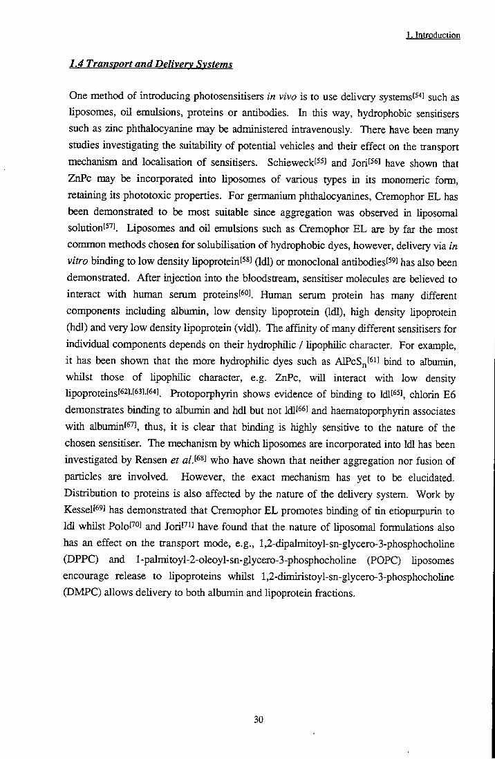
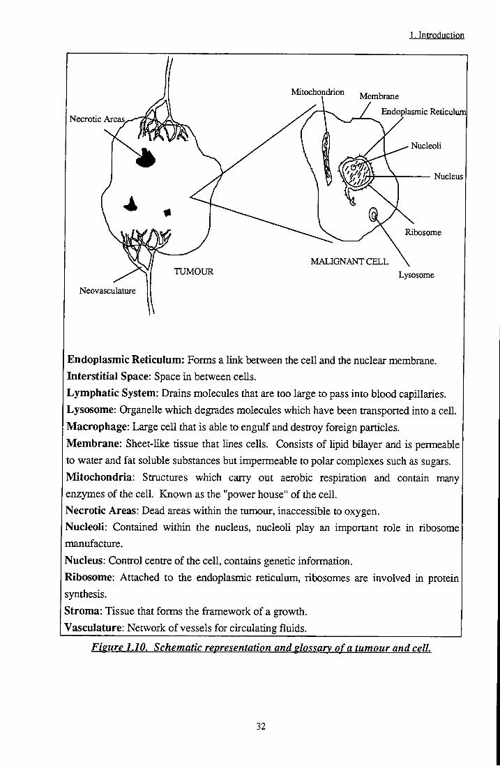
A tumour may be divided into five main areasf' J: Vasculature, which provides blood supply and nutrients to the growth; Necrotic or dead areas, prevalent in inner regions of the tumour where blood supply is poor; Immune cells, in particular macrophage cells capable of engulfing and destroying foreign particles; Stromal areas, which are composed largely of proteins such as coUagen and elastin and form the tissues that make up the framework of a growth and, of course, malignant tumour cells. A detailed description of the composition of a cell may be found in any standard biology pubhcation, such as 'Molecular Biology of the Cell' by Alberts et a/.f'^l. For the purposes of this text, however, a simple model may be considered (Figure 1.10).
31
1. Introduction
Mitochondrion
Necrotic Areas.
Membrane
/ Endoplasmic Reticulum]
Nucleoli
MALIGNANT C E L L TUMOUR
Nucleus
Ribosome
Lysosome
Neovasculature
Endoplasmic Reticulum: Forais a link between the cell and the nuclear membrane. Interstitial Space: Space in between cells. Lymphatic System: Drains molecules that are too large to pass into blood capillaries. Lysosome: Organelle which degrades molecules which have been transported into a cell. Macrophage: Large cell that is able to engulf and destroy foreign particles. Membrane: Sheet-like tissue that lines cells. Consists of hpid bilayer and is permeable to water and fat soluble substances but impermeable to polar complexes such as sugars. Mitochondria: Structures which carry out aerobic respiration and contain many enzymes of the cell. Known as the "power house" of the cell. Necrotic Areas: Dead areas within the tumour, inaccessible to oxygen. Nucleoli: Contained within the nucleus, nucleoli play an important role in ribosome manufacture.
Nucleus: Control centre of the cell, contains genetic information.
Ribosome: Attached to the endoplasmic reticulum, ribosomes are involved in protein synthesis. Stroma: Tissue that forms the framework of a growth. "Vasculature: Network of vessels for circulating fluids
Figure 1.10. Schematic representation and glossary of a tumour and cell.
32

1. Introduction
The vascular system of tumour growths has been shown to have a greater permeability to plasma proteins than that of healthy tissue!' ''!. This, in combination with a poor lymphatic drainage systemt'' ] leads to an accumulation of hydrophilic sensitisers in interstitial regionst' ^J. Intratumoral pH is low with respect to healthy tissue''' ] due to production of lactic acid and carbon dioxide by anaerobic glycolysis!''*'. Bech and Pettier have both considered the importance of pH. Becht'' ] studied the effect of pH on the production of protoporphyrin IX from 5-aminolevulinic acid (ALA) whilst Pottiert^oj j as shown that the biodistribution of haematoporphyrin is affected by pH. Tumours require a greater amount of cholesterol than healthy cells and, therefore, possess a higher concentration of low density lipoprotein (Idl) receptors'^'J possibly encouraging retention of hydrophobic sensitisers. Finally, the membrane potential of malignant cells is higher than that of healthy cells. This promotes transport of hydrophobic sensitisers through the cell membranel^ ', in particular cationic dye species, which have been observed to accumulate in mitochondiiat^^] of the ceU. Species which hypeipolarise the cell membrane, such as nigercin, have been shown to increase the efficiency of photodynamic actionfS''''f85)^ probably due to an increased uptake of the dye. For cell bound sensitisers, photodynamic action acts to destroy neoplastic growth directly. It is already clear that the site of localisation of sensitiser molecules varies according to the hydrophihcity. In general, hydrophilic sensitisers localise in the vascular stromal*^] since negative peripheral substituen^ prevent transport across cell membranes. Cell death is achieved as a result of damage to the blood vessels which supply the malignant growtht^'^l
The aggregation state, ionic distribution and pK^ of sensitisers are also important.
BeUnier and Lint^^l suggest that poiphyrin molecules are trapped in intracellular sites due
to aggregate formation that prevents exit through the ceU membrane. Work carried out
by Wood and co-workersJ has demonstrated the importance of ionic charge of
photosensitisers. In their study, ceUular uptake of cationic ZnPc sensitisers was far
greater than that of anionic or neutral species. The site of retention also differed.
Cationic and anionic species initially resided in lysosomes whilst neutral molecules
located in the plasma membrane. After irradiation, charged species redistributed to
nucleoli of the ceU. In addition, Johnson et alJ^^^ have shown that only positively
charged rhodamine dyes are capable of crossing the negatively charged cell membrane to
reside in the mitochondria of the cell. It follows that pK^ values of sensitisers will have
extreme importance in controlling localisation properties. Protonation will alter the ionic
state of the molecule and hence the propensity to cross the cell membrane.
Localisation is also affected by the delivery mechanism employed. Kessel has shownf^^]
that although tin etiopuipurin is efficiently delivered to cells when injected in an ethanolic
33
1. Introduction
solution, cytotoxicity was enhanced when a vehicle such as Cremophor EL was utilised. This implies different mechanisms of retention. Reddif '*' observed a two fold increase in tumour:normal tissue retention ratio in MS2 fibrosarcoma when Idl was used as delivery vehicle as opposed to liposomes. The kinetics and accumulation rate of a silicon naphthalocyanine sensitiser in urothelial ceU carcinoma were also observed to depend on the mode of deliveryt^il. It is clear that generalisations regarding transport mechanisms, localisation phenomena and cytotoxic pathways should be regarded with caution. Indeed, Peavy and co-workersf^^i h^ye shown that the biodistribution of AlPcS varied with tumour type, e.g. distribution throughout tumour tissue was observed in fibrosarcoma, however, in squamous cell carcinoma, sensitiser species were limited to the vascular stroma. It is important to improve understanding in this area of PDT since a knowledge of localisation and retention mechanisms would allow sensitisers to be tailored to provide the optimum conditions for effective treatment of cancer.
1.6 Aim
The aim of this study was to investigate the photophysical properties of substituted zinc
phthalocyanines under a range of conditions. The effect of ring modifications such as
peripheral substitution by acid, ester or alkyl groups has been considered. In addition,
the influence of environmental factors such as medium, temperature and pH has been
determined. The propensity of zinc phthalocyanine to interact with biological substrates
such as bovine serum albumin or DNA via binding or electron transfer reactions was also
of interest. In this way it was hoped to develop an understanding of the photoproperties
of zinc phthalocyanine in relation to its use as a sensitiser for photodynamic therapy.
A brief summary of chapters two to six is given below:
Chapter 2: Describes the photophysical techniques used throughout this work
including background theory necessary for a basic understanding of the
principles involved. Synthesis of two novel, substituted zinc
phthalocyanines, ZnPc(CMe(C02Me)2)4 and ZnPc(CHMeC02H)4 is also
reported.
Chapters: Discusses the solution state and photophysical properties of
ZnPc(CMe(C02Me)2)4 and ZnPc(CHMeC02H)4. The effect of
peripheral ligand substitution and the nature of the surrounding medium
(homogeneous, heterogeneous or solid state) on the photophysical
properties of substituted ZnPc's are also considered.
34

1. Introduction
Chapter 4: Dimerisation of phthalocyanines is investigated. The photophysical properties of a novel, fluorescent dimer / aggregate of CIO are reponed and compared with those of a face-to-face dimer of aluminium phthalocyanine. The properties of both species are discussed in terms of exciton theory. Chapter 4 also describes the steady state fluorescence anisotropy of phthalocyanines and how it may be used to study the nature of excited states.
Chapter 5: A complete photophysical evaluation of mono to tetra protonated species of tBu4ZnPc in ethanol is given. The tendency of ZnPcS2 to protonate in micellar media and on solid state substrates is also described.
Chapter 6: Discusses the propensity of substituted zinc phthalocyanines to bind to biological substrates such as protein and DNA. Their ability to participate in electron transfer reactions is also studied.
1.7 References
[1] Meyer-Betz F., Investigations on the biological (photodynamic) action of
haematoporphyrin and other derivatives of blood and bile pigments., Deut. Arch. Klin. Med., Ill, 476-503,1913.
[2] Von Tapiener H. and Jesionek A., Therapeutische Versuche mit fluoreszierenden
Stoffen, Muenchener Medizinische Wochenschrift, 50,2042-2051, 1903. [3] Figge F.J., Weiland G.S. and Manganiello L.O.J., Cancer detection and therapy.
Affinity of neoplastic, embryonic and traumatised tissue for poiphyrins and metalloporphyrins., Proc. of the Soc. Exp. Biol, and Med., 68, 640-641, 1948.
[4] Rasmussen D.S., Ward G.S. and Figge F.H.J., Fluorescence of human lymphatic and cancer tissues following high doses of intravenous hematoporphyrin.. Surgical Forum, 619-623, 1955.
[5] Lipson R.L., Baldes E.J. and Olsen A.M. , Hematoporphyrin derivative: a new aid for endoscopic detection of malignant disease., / . Thoracic and Cardiovascular Surgery, 42, 623-629, 1961.
[6] Lipson R.L., Baldes E.J. and Olsen A.M., The use of a derivative of
hematoporphyrin in tumour detection.,/. Nor. Cancer Inst., 26, 1-11, 1961. [7] Kelly J.F., Snell M.E. and Berenbaum M.C., Photodynamic destruction of human
bladder carcinoma., Br. J. Cancer, 31,237-244,1975.
35

1. Introduction
[8] Kessel D., Effects of photoactivated porphyrins at the cell surfaces of leukaemia L1210 ceUs., Biochem., 16, 3443-3449, 1977.
[9] Dougherty T.J., Lawrence G., Kaufman J.E., Boyle D., Weishaupt K.R. and Goldfarb A., Photoradiation in the treatment of reciurent breast cancer with hematoporphyrin derivative therapy., / . Nat. Cancer Inst., 62, 231 -237, 1979.
[10] Cortese A.A. and Kinsey J.H., Endoscopic management of lung cancer with
hematoporphyrin derivative phototherapy.. Mayo Clinic Proc, 57, 543-547, 1982. [11] Dougherty T.J., Lawrence G., Kaufman J.E., Weishaupt K.R., Goldfarb A. and
Mittleman A., Photoradiation therapy for the treatment of malignant tumours., Cancer Res., 38, 2628-2635, 1978.
[12] Weishaupt K.R., Gromer C.J. and Dougherty T.J., Identification of singlet oxygen as the cytotoxic agent in photo-inactivation of a murine tumor.. Cancer Res., 36, 2326,1976.
[13] Parker J.G., The importance of singlet delta oxygen in cancer photoradiation
therapy., John Hopkins APL Technical Digest, 5, 48-50, 1984.
[14] Andreoni A., Cubeddu R., De Silvestri S., Laporta P., Jori G. and Reddi E.,
Hematopoiphyrin derivative: Experimental evidence for aggregated species., Chem.
Phys.Lett.,SS, 33-36, 1982.
[15] Dougherty T.J., Studies on the structure of porphyrins contained in Photofrin E.,
Photochem. Photobiol, 45, 879-889, 1987. [16] Ben-Hur E. and Rosenthal I . , Photochemical generation of superoxide radical and
the cytotoxicity of phthalocyanines.. Int. J. Rad. Biol, 47,145-147, 1985. [17] Bensasson R.V., Land E.J. and Truscott T.G., Excited States and Free Radicals
in Biology and Medicine, p322, Oxford University Press, New York, 1993. [18] Ochsner M. , Photophysical and photobiological processes in the photodynamic
therapy of mmours., / . Photochem. Photobiol., 39, 1, 1997. [19] Turro N.J., Modern Molecular Photochemistry, University Science Books,
California, pl64,1991. [20] Keams D.R., Physical and chemical properties of singlet molecular oxygen.,
Chem. Rev.,11, 395, 1973.
[21] Gijzeman O.L.J., Kaulman F. and Porter G., Oxygen quenching of aromatic
triplet states in solution., / . Chem. Soc. Faraday Trans. II, 69, 708,1973.
[22] Oelckers S., Sczepan M., Hanke T. and Roder B., Time resolved detection of
singlet oxygen luminescence in red cell ghost suspensions., / . Photochem. Photobiol.
B.Biol., 39, 219, 1997.
[23] MacRobert A.J. and Phillips D., Photodynamic Therapy, Chemistry and Industry, 17, 1992.
36
1. Introduction
[24] Dougherty T.J., Grindey G.B., Fiel R., Weishaupt K.R. and Bryle D.G., Photoradiation therapy I I . Cure of animal tumors with hematoporphyrin and light., J. Nat. Cancer Inst., 55, 115-121,1975.
[25] Forbes I.J., Cowled P.A., Leong A.S.Y., Ward A.D., Black R.B., Blake R.B. and Jacka F.J., Phototherapy of human tumours using haematoporphyrin derivative., Med. J. Australia, 2,489-493,1980.
[26] Kessel D., Hematoporphyrin and HpD - Photophysics, photochemistry and Y)h.oioiher2i-py.,Photochem.Photobiol., 39,851-859, 1984.
[27] Moan J., Porphyrin photosensitisation and phototherapy., Photochem. Photobiol, 43,681-690, 1986.
[28] Truscott T.G., The photochemistry of haematoporphyrin and some related species., / . Chem. Soc. Faraday Trans. 2, 82, 2177-2181, 1986.
[29] Boriand C.F., McGarvey D.J., Morgan A.R. and Truscott T.G., Laser flash photolysis of purpurins, more potential photosensitisers of interest in photodynamic therapy., / . Photochem. Photobiol. B:Biol., 2,427-434, 1988.
[30] Bonnett R. and Berenbaum M. , Ciba Foundation Symposium, 14 Wiley Interscience Publ., Chichester, pp40-59,1989.
[31] Bonnett R., Photosensitisers of the porphyrin and phthalocyanine series for PDT., Chem. Soc. Rev., 19-33, 1995.
[32] Phillips D. and MacRobert A., Spectroscopic studies of photosensitisers and selectivity in photodynamic therapy.. Highlights Modern Biochemistry, 2, 1517-1525, 1989.
[33] Jori G., Far red absorbing photosensitisers - their use in the photodynamic therapy of tumours., / . Photochem. Photobiol. A:Chem., 371-378, 1992.
[34] Rosenthal I . , Phthalocyanines as photodynamic sensitisers., Photochem. Photobiol., 53, 859-870, 1991.
[35] Ford W.E., Rihter B.D., Kenney M.E. and Rodgers M.A.J., Photoproperties of alkoxy substituted phthalocyanines with deep red optical absorbance., Photochem. Photobiol., 50, 277-282, 1989.
[36] Chan W.S., Marshal J.F., Svensen R., Phillips, and Hart I.R., Photosensitising activity of phthalocyanine dyes screened against tissue culture cells., Photochem. Photobiol., 45,757-761, 1987.
[37] Murov S.L., Carmichael I . and Hug G.L., Handbook of Photochemistry, 2nd Edn., p40. Marcel Dekker Inc., New York, 1993.
[38] McVie J., Sinclair R.S. and Truscott T.G., Triplet states of copper and metal free phthalocyanine., / . Chem. Soc. Faraday Trans II, 74, 1870, 1978.
[39] Ben-Hur E. and Rosenthal I . , Photosensitisation of Chinese hamster cells by
water soluble phthalocyanines., Photochem. Photobiol., 43, 615-619,1986.
37

L Introduction
[40] Ambroz M. , MacRobert A.J., Morgan J., Rumbles G., Foley M.S.C. and Philips D., Time resolved fluorescence spectroscopy and intracellular imaging of aluminium phthalocyanine., / . Photochem. Photobiol. B.Biol., 27,105-117,1994.
[41] Marshall J.F., Chan W.S. and Hart I.R., Effect of photodynamic therapy on anti-tumour immune defences: Comparison of the photosensitisers hematoporphyrin derivative and chloroaluminium sulphonated phthalocyanine., Photochem. Photobiol., 49, 627-632, 1989.
[42] Moan J., Berg K., Bommer J. and Westem A., Action spectrum of phthalocyanines with respect to photosensitisation of cells., Photochem. Photobiol., 56, 171-175, 1992.
[43] He J., Larkin H.E., L i Y.S., Rihter B.D., Zaidi S.I.A., Rodgers M.A.J., Mukhtar H., Kenney M.E. and Oleinick N.L., The synthesis, photophysical and photobiological properties and in vitro structure activity relationships of a set of silicon phthalocyanine PDT photosensitisers., F/zorcc/zem. Photobiol., 65, 581, 1991.
[44] VoUano J.F., Bossard G.E., Martellucci S.A., Darkes M.C., Abrams M.J. and Brooks R.C., The synthesis and in vitro photodynamic activity of a series of novel ruthenium 2, 3 naphthalocyanines., / . Photochem. Photobiol. B.Biol., 37, 230, 1997.
[45] Schaffer A.M., Gouterman M. and Davidson E.R., Porphyrins X X V I I . Extended Huckel calculations on metal phthalocyanines and tetraazaporphins., Theoret. Chim. Acta, 30, 9,1973.
[46] Gouterman M. , Spectra of Porphyrins, / . Mol. Spec, 6, 138,1961. [47] Orti E. and Bredas J.L., Electronic structure of metal free phthalocyanine: A
valence effective Hamiltonian theoretical study., / . Chem. Phys., 89,1009,1988. [48] Orti E., Bredas J.L. and Clarisse C, Electronic structure of phthalocyanines.
Theoretical investigation of the optical properties of phthalocyanine monomers, dimers and crystals., / . Chem. Phys., 92,1228, 1990.
[49] Sevchenko A.N., Solov'ev K.N., Gradyushko A.T. and Shkirman S.F., Narrowband electronic spectra of metal derivatives of tetrabenzoporphine and phthalocyanine., Soviet Physics-Doklady, 11, 587, 1967.
[50] Rosa A. and Baerends E.J., Metal-macrocycle interaction in phthalocyanines. Density functional calculations of ground and excited states., Inorg. Chem., 33, 584, 1994.
[51] Nyokong T., Gasyna Z. and Stilknan M., Analysis of the absorption and magnetic circular dichroism spectra of zinc phthalocyanine and the K cation radical species [ZT\PC(-1)]-+., Inorg. Chem., 26, 1087, 1987.
[52] Khalib N., Boudjema B., Maitrot M. , Chermette H. and Porte L., Electronic structure of zinc phthalocyanine.. Can. J. Chem., 66,2313, 1988.
38
1. Introduction
[53] Morley J.O. and Charlton M.H., Theoretical investigation of the structiu-e and spectra of zinc phthalocyanines., / . Phys. Chem., 99, 1928, 1995.
[54] Reddi E., Role of delivery vehicles for photosensitisers in the photodynamic therapy of tumours., / . Photochem. Photobiol. B:Biol., 37,189, 1997.
[55] Schieweck K., Caparo H.G., Isele U., Van Hoogevest P., Ochsner M., Maurer T. and Bait E., CGP 55847 liposome delivered Zn(II) phthalocyanine as a phototherapeutic agent for tumours., S.P.I.E., 2078, 107,1994.
[56] Valduga G., Reddi E., Jori G., Cubeddu R., Taroni P. and Valentini G., Steady state and time resolved spectroscopic studies on Zn(n) phthalocyanine in liposomes., / . Photochem. Photobiol. B.Biol., 16, 331, 1992.
[57] Soncin M. , Polo L. , Reddi E., Jori G., Kenney M.G., Chang G. and Rodgers M.A.J., Effect of axial ligation and delivery system on the tumour localising and photosensitising properties of Ge(IV) octabutoxyphthalocyaiunes., Br. J. Cancer, 71, 727-732, 1995.
[58] Reddi E., Zhou C , Biolo R., Menegaldo E. and Jori G., Liposome or Idl administered Zn(n) phthalocyanine as a photodynamic agent for tumours. I Pharmacokinetic properties and phototherapeutic efficiency., Br. J. Cancer, 61, 407, 1990.
[59] Mew D., Wat C.K., Towers G.H.N, and Levy J.G., Photoimmunotherapy: Treatment of animal tumours with tumour specific monoclonal antibody -haematoporphyrin conjugate.,/./mmwrto/., 130,1473, 1983.
[60] Jori G., In vivo transport and pharmacokinetic behaviour of tumor photosensitisers., Ciba Foundation Symposium, Wiley Chich., 146,78-94, 1989.
[61] Reddi E., Transport modalities of photodynamic agents for tumors., S.P.I.E., 2078,246,1994.
[62] Polo L. , Reddi E., Garbo G.M., Morgan A.R. and Jori G., The distribution of the tumour photosensitisers Zn(II) phthalocyanine and Sn(rV) etiopurpurin among rabbit plasma proteins.. Cancer Lett., 66,217, 1992.
[63] Love W.G., Havenaar E.G., Lowe P.J. and Taylor P.W., Uptake of Zn(n) phthalocyanine by HepG2 cells expressing the low density lipoprotein receptor: studies with the liposomal formulation CGP55847., S.P.I.E., 2078, 381,1994.
[64] Versluis A.J., Rensen P.C.N., Kuipers M.E., Love W.G. and Taylor P.W., Interaction between Zn(II) phthalocyanine containing liposomes and human low density lipoprotein., / . Photochem. Photobiol. B.Biol., 23,141-148,1994.
[65] Reyftmann J.P., Morliere P., Goldstein S., Santus R., Dubertriet L. and Lagrange D., Interaction of human serum low density lipoproteins with porphyrins: A spectroscopic and photochemical study., Photochem. Photobiol., 40, 721, 1984.
39
1. Introduction
[66] Kessel D., Whitcomb K.L. and Schulz V., Lipoprotein-mediated distribution of N-aspartyl chlorin-E6 in the mouse., Photochem. Photobiol., 56, 51,1992.
[67] Grossweiner L . I and Goyad G.C., Binding of hematoporphyrin derivative to HSA., Photochem. Photobiol., 40,1,1984.
[68] Rensen P.C.N., Love W.G. and Taylor P.W., In vitro interaction of zinc(II)-phthalocyanine-containing Hposomes and plasma lipoproteins., / . Photochem. Photobiol. B.Biol., 26, 29,1994.
[69] Kessel D., Morgan A. and Garbo G.M., Sites and efficacy of photodamage by tin etiopurpurin in vitro using different delivery systems., Photochem Photobiol., 54, 193, 1991.
[70] Polo L. , Bianco G., Reddi E. and Jori G., The effect of liposomal formulations on the interaction of Zn(II) ph±alocyanine with isolated low and high density lipoproteins.. Int. J. Biochem. Cell Biol., 27, 1249, 1995.
[71] Jori G., Tumour photosensitisers: approaches to enhance the selectivity and
efficiency of photodynamic therapy., / . Photochem. Photobiol. B:Biol., 36, 87,1996. [72] Kessel D., PDT of Neoplastic Disease, CRC Press, Boston, 1990. [73] Alberts B., Bray D., Lewis J., Raff M., Roberts K. and Watson J.D., Molecular
Biology of the Cell, 2nd Edn., Garland Publishing, London, 1989. [74] Ackerman N.B. and Hechmer P.A., Failure of histamine type mediators to
enhance vascular permeability in experimental Mver metastases., Surg. Gynecol. Obstet., 146, 884,1978.
[75] Feldman G.B., Knapp R.C., Order S.E., and Hellman S., The role of lymphatic
obstruction in the formation of ascites in a murine ovarian carcinoma., Cancer Res., 32,1663, 1972.
[76] Roberts W.G. and Hasan T., Role of neovasculature and vascular permeability on the tumour retention of photodynamic agents., Cancer Res., 52, 924-930, 1992.
[77] Waddel W.J. and Bates R.G., Intracellular pH., Physiol. Rev., 49, 285,1969. [78] Albers C , Vanden Kerkhoff W., Vaupel P. and Muller KHeser W., Non-
bicarbonate buffering of ascites tumor cells in the rat as titrated by strong acids., Res. Physiol., 45, 273, 1981.
[79] Bech O., Berg K. and Moan J., The pH dependency of protoporphyria IX formulation in cells incubated with 5-aminolevulinic acid., Cancer Lett., 113, 25, 1997.
[80] Pottier B. and Kennedy J.C., New trends in photobiology. The possible role of ionic species in selective biodistribution of photochemotherapeutic agents toward neoplastic tissue., / . Photochem. Photobiol. B:Biol., 8, 1, 1990.
40
1. Introduction
[81] Milanesi C, Zhou C , Biolo R. and Jori G., Zn(n) phthalocyanine as a photodynamic agent for tumours. Studies on the mechanism of photosensitised tumour necrosis., Br. J. Cancer, 61, 846-850, 1990.
[82] Margaron P., Gregoire M.J., Scasnar V., Ali H. , Van Lier V.E., Structure-photodynamic activity relationships of a series of 4-substituted zinc phthalocyanines., Photochem. Photobiol., 63, 217, 1996.
[83] Johnson L.V., Summerhayes I.C. and Chen L.B., Decreased uptake and retention of Rhodamine 123 by mitochondria in feline sarcoma virus-transformed mink cells.. Cell, 28, 7, 1982.
[84] Lin C.W. and Shulok J.R., Enhancement of Nile Blue derivative induced photocytotoxicity by nigericin and low cytoplasmic pH., Photochem. Photobiol., 60, 143, 1994.
[85] Vames M.E., Bayne M.T. and Bright G.R., Reduction of the intracellular pH is not the mechanism for the synergistic interaction between photodynamic therapy and Nigericin., Photochem. Photobiol., 64, 853,1996.
[86] Kessel D., Thompson P., Saatio I.C. and Nuntun K.D., Tumour localisation and photosensitisation by sulphonated derivatives of tetraphenylporphine., Photochem. Photobiol., 45, 787,1987.
[87] Selman S.H., Kreimerbimbaum M. , Chaudhuri K., Garbo G.M., Seaman D.A., Keck R.W., Ben-Hur E. and Rosenthal I . , Photodynamic treatment of transplantable bladder tumors in rodents after pretreatment with chloroaluminium tetrasulfophtiialocyanine.,/. Urol, 136, 141,1986.
[88] Bellnier D.A. and Lin C.W., Photodynamic destruction of cultured human bladder tumor cells by haematoporphyrin derivative: effect of porphyrin molecular aggregation., Photobiochem. Photobiophys., 6, 357,1983.
[89] Wood S.R., Hokoyd J.A. and Brown S.B., The subcellular localisation of Zn(n) phthalocyanines and their redistribution on exposure to light., Photochem. Photobiol., 65, 397, 1997.
[90] Johnson L.V., Summerhays I.C. and Chen L.B., Decreased retention of Rhodamine 123 by mitochondria in feline sarcoma virus transformed mink cells., Cell, 28,7, 1982.
[91] Zuk M.M. , Rihter B.D., Kenney .E., Rodgers M.A.J. and Kreimer-Bimbaum M., Effect of delivery system on the pharmacokinetics and tissue distribution of bis(Di-isoutyl octadecylsiloxy)silicon 2,3-naphthalocyanine (isoBOCINC), a photosensitiser for tumor therapy., Photochem. Photobiol., 63, 132-140,1996.
[92] Peavy G.M., Krasieva T.B., Tromberg B.J., Eusantos E.D., Bems M.W., Variation in the distribution of a phthalocyanine photosensitiser in naturally occurring tumors of animals., / . Photochem. Photobiol. BiBiol, 27, 271-277, 1995.
41
In this work, photophysical techniques were employed to investigate the properties of
phthalocyanines under various conditions. A brief description of experimental methods
and relevant theory follows. Where further detail is required, the reader is referred to
comprehensive texts referenced throughout. The synthesis of P
-tetra-l,l-di(methoxycarbonyl)ethyl zinc phthalocyanine, ZnPc(CMe(C02Me)2)4, and (3
-tetra-l-carboxy ethyl zinc phthalocyanine, ZnPc(CHMeC02H)4 is described.
Modification of metal free phthalocyanines to produce the zinc metallated derivatives is
also included.
2.1 Photophysical Measurements
2.1.1 UV/Visible Absorption Spectroscopy
UVA^isible absorption spectra were carried out with an ATI Unicam UV2-100
spectrometer. Spectra were performed using quartz cuvettes, pathlength = 10 mm, over
an appropriate wavelength range (usually 300 nm to 800 nm). A baseline was obtained
using a suitable solvent prior to use. Molar extinction coefficients (e, dm^ mol"^ cm'^)
were determined using the Beer-Lambert law.
A = ecl = logio^ (2.1)
where A is the absorbance at the wavelength of interest, c is the concentration of
solution (mol dm-^), 1 is the pathlength of the cuvette (cm), IQ is the intensity of incident
radiation and I is the intensity of radiation after passing through the sample.
2.2.2 Fluorescence Spectroscopy 2.1.2.1 Spectra
Fluorescence emission and excitation spectra were recorded using a Perkin Ekner LS50B
luininescence spectrometer fitted with a red sensitive, Hamamatsu R928 photomultiplier
tube (PMT). The optical bench was controlled by FL-Winlab 3.0 running on an Elonex
P90 computer. In order to minimise inner filter effectst^l, samples were prepared such
2. Experimental Techniques
that their maximum absorbance was < 0.05. Emission spectra were generated by exciting at 610 nm or 630 nm and recorded over the range 640 nm - 850 nm. To maximise resolution, a scan speed of 120 nm min-1 and slits of 10 nm and 2.5 nm for the excitation and emission slits respectively were used. Where necessary, emission spectra were corrected for wavelength dependent sensitivity of the photomultiplier using a correction curve supplied by Perkin Elmer. Excitation spectra were recorded by monitoring fluorescence emission at 760 nm. These spectra were corrected intemally for variation in the excitation intensity using a built in Rhodamine dye quantum counter.
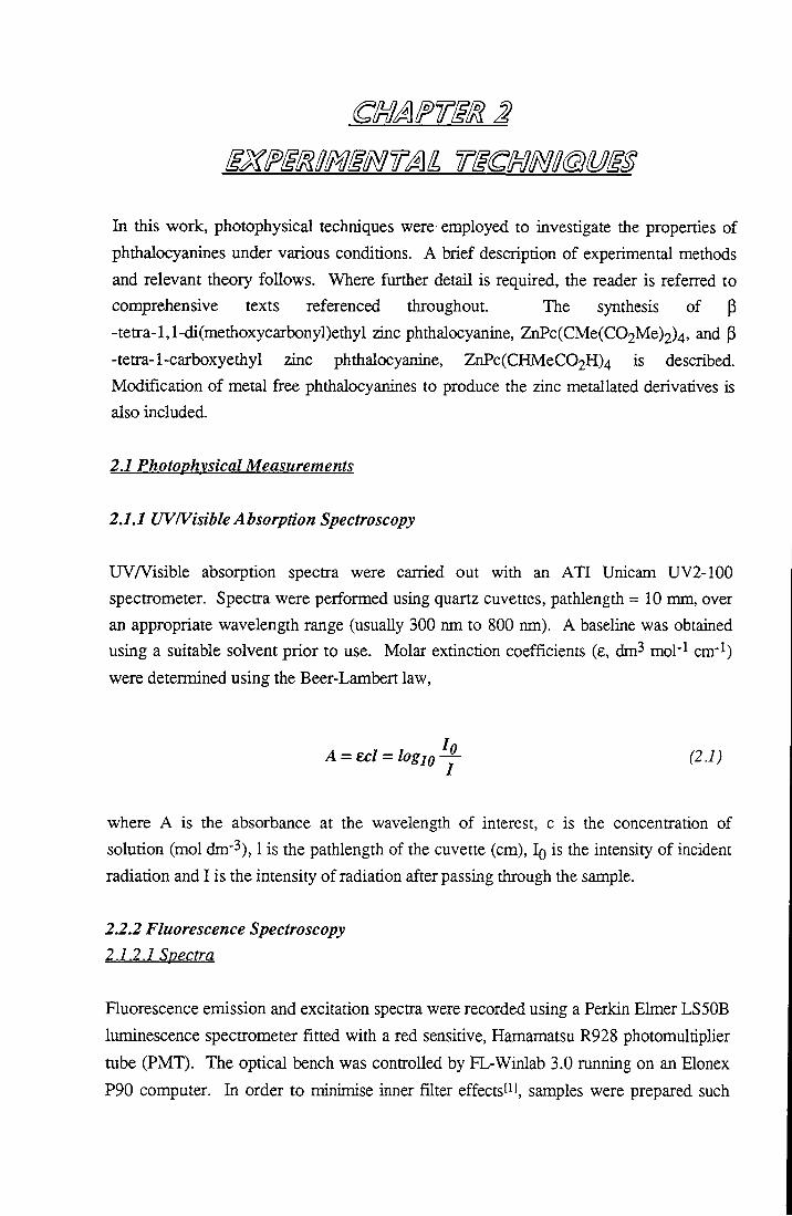
In certain cases, for example, the emission and excitation spectra of protonated phthalocyanines (Chapter 5), the LS50B was unsuitable for measurements. This was due to a combination of low sensitivity of the detector in the near IR region of the spectmm and low output of the xenon lamp excitation source at wavelengths > 700 nm. For these samples the airangement depicted in Figure 2.1 was employed to measured emission.
Sample Spectrograph
Focusing Optic
Diode Array
HeNe Laser
Computer
Fieure 2.1. Experimental arrangement for recording emission spectra from phthalocvanine species emitting in the near-IR.
A continuous wave HeNe laser (Melles Griot, 632.8 nm, power ~ 5 mW) was used to excite the sample. Emission was collected at 90° to irradiation and focused into a spectrograph (Bentiiam TM300V) equipped witii a diode airay detector (EG & G 1453a). The diode array was calibrated using the emission lines of an argon ion laser. Spectra were corrected for wavelengtii dependent sensitivity of the diode using a correction curve generated using a standard tungsten lamp. Excitation spectra of samples emitting in tiie near IR were recorded on a SPEX FluoroMax fluorimeter at Imperial College, London.
43

2. Experimental Techniques
2.7,2.2 Fluorescence Quantum Yields
The fluorescence quantum yield is defined as the number of photons emitted as fluorescence as a ratio of the number of photons absorbed. This can be expressed in terms of competing decay processes by assuming steady state conditions, i.e. the rate of formation of singlet state equals the rate of decay.
Process Rate SQ + hv^Si la S i ^ S o + /2V kf[Si] Sj -^So + heat ^i^l^i] S i ^ T i k i J S i ]
Using the steady state approximation:
la^kf [SiJ+kic [Si]+ki,,[Si] (22)
^ <I>F=T . (2.5) Kf + kif. + kigf.
Fluorescence quantum yields determined in this work were obtained using the
comparative method of Williams et a/. PI in which the quantum yield is expressed as
Ax Istd n^td
where Ox and Ogj^ are the quantum yields of the unknown and standard respectively. A is the absorbance of the solution at the excitation wavelengtii, I is the integrated area of fluorescence emission and n is the refractive index of the solvent. Stock solutions of the unknown and standards were prepared with absorbances below 0.05 to minimise excitation and emission inner filter effects. Measurements were performed using several dilutions of each sample to ensure that concentration had no effect on the fluorescence yield. Chlorophyll a in ether (Opt^l = 0.32) and zinc phthalocyanine in 1 % pyridine / toluene (Opt''] = 0.3) were used as standards (maximum concentration = 3 x 10"" mol dm"3). Fluorescence emission was cortected for the wavelength dependent sensitivity of the detector and then integrated to obtain the area of emission, I . Manipulations of data points were carried out using Microsoft Excel.
44

2. Experimental Techniques
2.1.2.3 Fluorescence Anisotropv
Fluorescence anisotropy is a valuable analytical technique which may be used to obtain information regarding the size, shape and rotational motion of a fluorophore, tiie hfetime and nature of its emissive state and the temperature/viscosity of the surrounding solvent. The technique is based on the principle that when a fluorophore emits, the total emission intensity is composed of the sum of components polarised in the x, y and z directions (Ix, ly and Iz). When the exciting radiation is vertically polarised and oriented perpendicular to the direction of detection, I^ = ly, so the total intensity may be expressed by, I = l2+2Iy or I = Ivv+2IvH' where the subscripts refer to the direction of polarisation of the incident and emitted radiation respectively (V, vertical and H, horizontal). Anisotropy is defined as the difference in vertically and horizontally polarised emission relative to the total emission intensity, I W - I V H / I and can be expressed by Equation 2.5.
Iw +2GlvH
where G = I H V / ^ H H is a correction factor for the polarisation dependence of the apparatus.
Fluorescence anisotropy measurements were carried out using a Perkin Elmer LS50B luminescence spectrometer, with built in vertical and horizontal polarising filters to orientate incident and emitted radiation (Figure 2.2). Four scans were performed to obtain r; lw(^)' I V H ( ^ ) ' I H V ( ^ ) and I H H ( ^ ) - Data obtained from each scan was substituted into Equation 2.5 and analysed using Microsoft Excel to calculate r^y, tiie steady state fluorescence anisotropy.
45
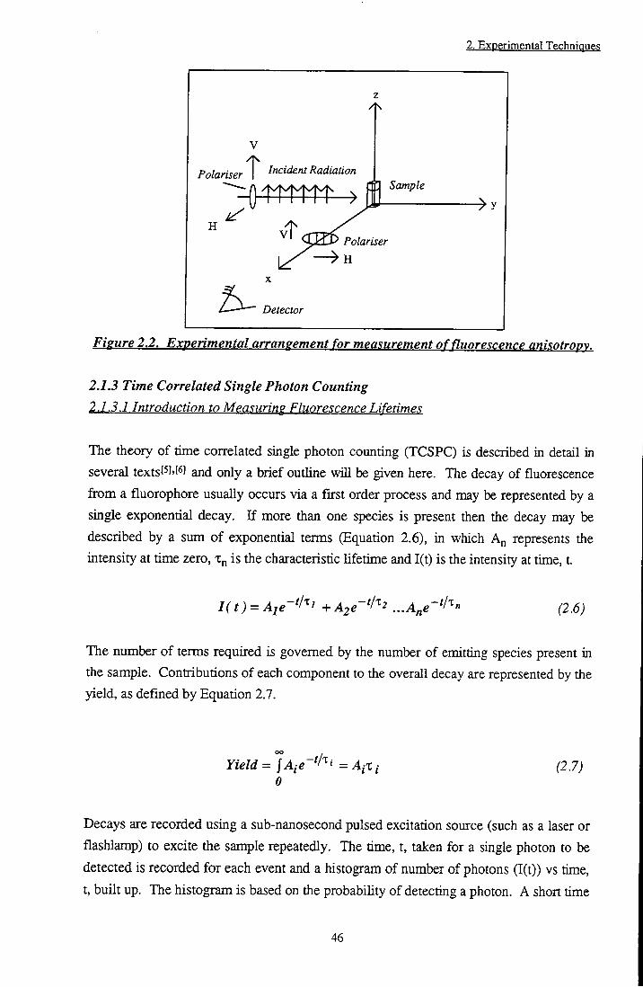
2. Experimental Techniques
V
Polariser Incident Radiation
Polariser
11 Sample
>
A - Detector
Figure 2.2. Experimental arrangement for measurement of fluorescence anisotropv.
2.1.3 Time Correlated Single Photon Counting 2.1.3.1 Introduction to Mea<;uring Fluorescence Lifetimes
The theory of time cortelated single photon counting (TCSPC) is described in detail in several textsl^l''^' and only a brief outline will be given here. The decay of fluorescence from a fluorophore usually occurs via a first order process and may be represented by a single exponential decay. I f more than one species is present then the decay may be described by a sum of exponential terms (Equation 2.6), in which A^ represents the intensity at time zero, is the characteristic lifetime and I(t) is the intensity at time, t.
I(t) = Aie-^l^i +A2e-^I''2 ...A^e'^l^" (2.6)
The number of terms required is governed by the number of emitting species present in the sample. Contributions of each component to the overall decay are represented by the yield, as defined by Equation 2.7.
Yield = °]Aie~^l'^i = A j X j 0
(2.7)
Decays are recorded using a sub-nanosecond pulsed excitation source (such as a laser or
flashlamp) to excite the sample repeatedly. The time, t, taken for a single photon to be
detected is recorded for each event and a histogram of number of photons (I(t)) vs time,
t, built up. The histogram is based on the probability of detecting a photon. A short time
46

2. Experimental Techniques
after excitation, the probability is greater than at large time intervals when the fluorescence has decayed. The time, t, is measured using a time to amplitude converter which may be started by the excitation pulse and stopped on detection of a photon, or vice versa. The latter case is only feasible when a highly regular excitation source such as a mode locked laser is employed. The measured decay consists of a convolution of the time response of the excitation source, the electronics of the apparatus and the true fluorescence decay. On analysis, the true fluorescence decay may be determined using a process termed 'iterative re-convolution'. The instrument response function is re-convolved with an exponential function with estimate values for A and x. These are varied until the best fit - as judged by non-linear least squares analysis - is obtained. The quality of a calculated fit is judged by statistical parameters such as the reduced y^, the Durbin-Watson parameter, random residuals and the correlation function.
2.1.3.2 Experimental Arrangement
Fluorescence lifetime measurements were carried out using the time correlated single photon counting equipment at Imperial College, London (Figure 2.3). The apparatus is described in detail elsewheret' ]. In brief, the experiment used a frequency doubled, mode locked Nd:YAG laser (Coherent, Antares 76S) to pump a cavity dumped dye laser (Coherent 701 dye laser & 7220 cavity dumper). DCM was used as the gain medium. This provided an excitation wavelength of 638 nm with a pulse duration of < 10 ps. Fluorescence was detected at 90° to the excitation beam using a microchannel plate photomultiplier (PHOTEK 413 LJ), discriminator (Ortec 584) and time to amplitude converter (Ortec 547). A double monochromator (Instruments SA 1602) was employed to select the desired wavelength of fluorescence emission. An intensity vs. time distribution was built up using a multichannel analyser (Canberra Series 35) which consisted of 512 channels with a chosen time interval of 36 ps per channel. Decays were recorded to 20000 counts in the peak channel. Samples were prepared such that their maximum absorbance did not exceed 0.05. The instrument response function was recorded by replacing the fluorophore with a scattering medium and monitoring the resulting signal at the wavelength of excitation. Decays were transferred to a computer and analysed using a non-linear least squares fitting program (Glife, Imperial College). The fitting function was judged to be acceptable when 0.8 < i} > 1.4, residual plots appeared random and the Durbin-Watson parameter had a value between 1.7 and 2.2.
47
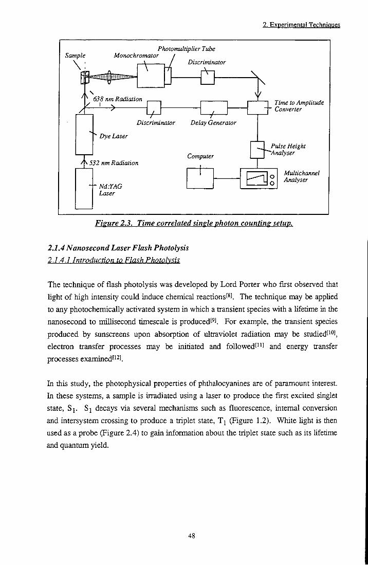
2. Experimental Techniqgeg
Sample
\ : Monochromator
Photomultiplier Tube
A
motor I Discriminator
\ 638 nm Radiation
7^ •7^
Time to Amplitude ~t~ Converter
Discriminator Delay Generator
Dye Laser
nm Radiation Computer
Pulse Height "Analyser
Nd:YAG Laser
Multichannel Analyser
Figure 2.3. Time correlated single photon counting setup.
2.1.4 Nanosecond Laser Flash Photolysis 2.1.4.1 Introduction to Flash Photolysis
The technique of flash photolysis was developed by Lord Porter who first observed that light of high intensity could induce chemical reactionsl^l The technique may be applied to any photochemically activated system in which a transient species with a lifetime in the nanosecond to millisecond timescale is produced^^ . For example, the transient species produced by sunscreens upon absorption of ultraviolet radiation may be studied' ^J, electron transfer processes may be initiated and foUowedt^'l and energy transfer processes examinedt^^l
In this study, the photophysical properties of phthalocyanines are of paramount interest. In these systems, a sample is irradiated using a laser to produce the first excited singlet state, $1- Si decays via several mechanisms such as fluorescence, internal conversion and intersystem crossing to produce a triplet state, T^ (Figure 1.2). White light is then used as a probe (Figure 2.4) to gain information about the triplet state such as its lifetime and quantum yield.
48
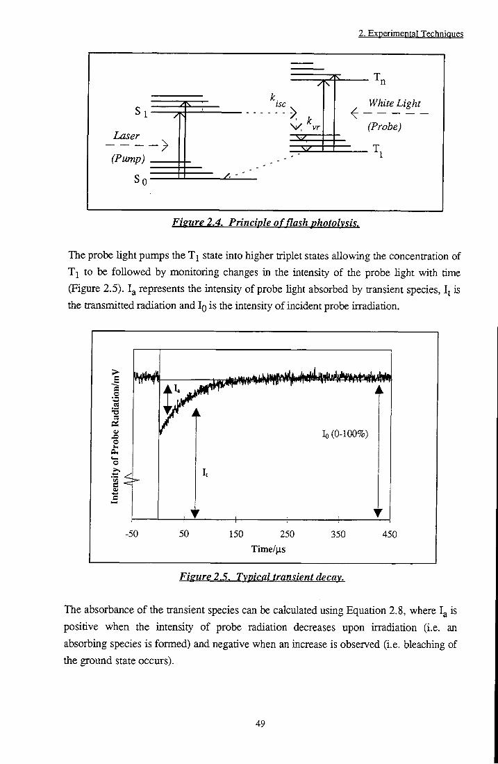
2. Experimental Techniques
White Light
Probe)
(Pump)
Fisure 2.4. Principle of flash photolysis.
The probe light pumps the state into higher triplet states allowing the concentration of Tl to be followed by monitoring changes in the intensity of the probe light with time (Figure 2.5). represents the intensity of probe light absorbed by transient species, I j is the transmitted radiation and IQ is the intensity of incident probe irradiation.
lo (0-100%)
Tinie/|xs
Figure 2.5. Typical transient decay.
The absorbance of the transient species can be calculated using Equation 2.8, where is
positive when the intensity of probe radiation decreases upon irradiation (i.e. an
absorbing species is formed) and negative when an increase is observed (i.e. bleaching of
the ground state occurs).
49
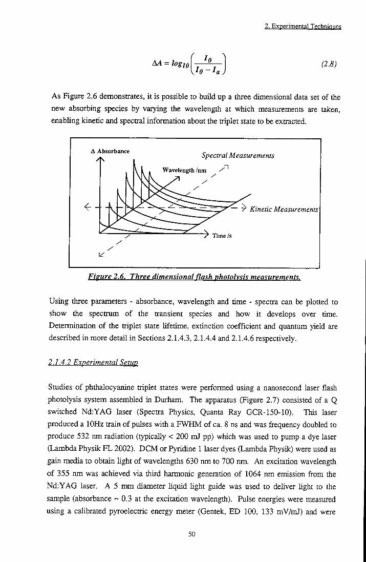
2. Experimental Techniques
AA = logio In-I.
(2.8)
As Figure 2.6 demonstrates, it is possible to build up a tiiree dimensional data set of tiie new absorbing species by varying the wavelength at which measurements are taken, enabling kinetic and spectral information about the triplet state to be extracted.
A Absorbance Spectral Measurements n
Wavelength /nm
^ ~ ^ ~ 7" Kinetic Measurements
Time /s
Figure 2.6. Three dimensional flash photolysis measurements.
Using three parameters - absorbance, wavelength and time - spectra can be plotted to show the spectrum of the transient species and how it develops over time. Determination of the triplet state lifetime, extinction coefficient and quantum yield are described in more detail in Sections 2.1.4.3, 2.1.4.4 and 2.1.4.6 respectively.
2.1.4.2 Experimental Setup
Studies of phthalocyanine triplet states were performed using a nanosecond laser flash photolysis system assembled in Durham. The apparatus (Figure 2.7) consisted of a Q switched Nd:YAG laser (Spectra Physics, Quanta Ray GCR-150-10). This laser produced a lOHz train of pulses with a FWHM of ca. 8 ns and was frequency doubled to produce 532 nm radiation (typically < 200 mJ pp) which was used to pump a dye laser (Lambda Physik FL 2002). DCM or Pyridine 1 laser dyes (Lambda Physik) were used as gain media to obtain light of wavelengths 630 nm to 700 nm. An excitation wavelength of 355 nm was achieved via third harmonic generation of 1064 nm emission from the Nd:YAG laser. A 5 mm diameter hquid light guide was used to dehver light to the sample (absorbance -0 .3 at the excitation wavelength). Pulse energies were measured using a calibrated pyroelectric energy meter (Gentek, ED 100, 133 mV/mJ) and were
50

2. Experimental Technianas
typically < 0.5 mJ. A tungsten-halogen lamp (100 W) was used to probe the transient species produced upon laser excitation. The probe beam was focused into the sample (irradiated volume 0.8 cm x 0.8 cm x 0.3 cm) using quartz lenses (100 mm focal length) and subsequently into a monochromator (Bentham TM300). The monochromator had a resolution of 2 nm per millimetre of slit width and was computer controlled.
Liquid Light Guide
Dye Laser
Tungsten Lamp
Power Meter
A Monochromator
PMT
Sample Focusing Optics
- V -
Computer Nd:YAG Laser
Digital Storage Oscilloscope
Fieure 2.7. Experimental arraneement of flash photolysis apparatus.
The intensity of light detected at a given wavelength was measured by a photomultiplier tube (Hamamatsu R928) with a 250 Q termination resistor. Transient decays were averaged over at least 32 laser pulses and displayed as voltage readings by a digital storage oscilloscope, DSO (Tektronix TDS 320). The oscilloscope was triggered extemally by a signal firom a photodiode as the laser was fired. Decays were transferred to a computer where data were converted into absorbance units using Equation 2.8 and analysed using Microsoft Excel and GrafiL
2.1.4.3 Triplet Lifetime Measurements
The rate of decay of triplet state species in solution is given by Equation 2.9, in which kp represents the rate of phosphorescence enaission, kg, is the intrinsic rate of non-radiative decay of triplet species, kyj-LTj] and kpJPc] are triplet-triplet annihilation and self
51
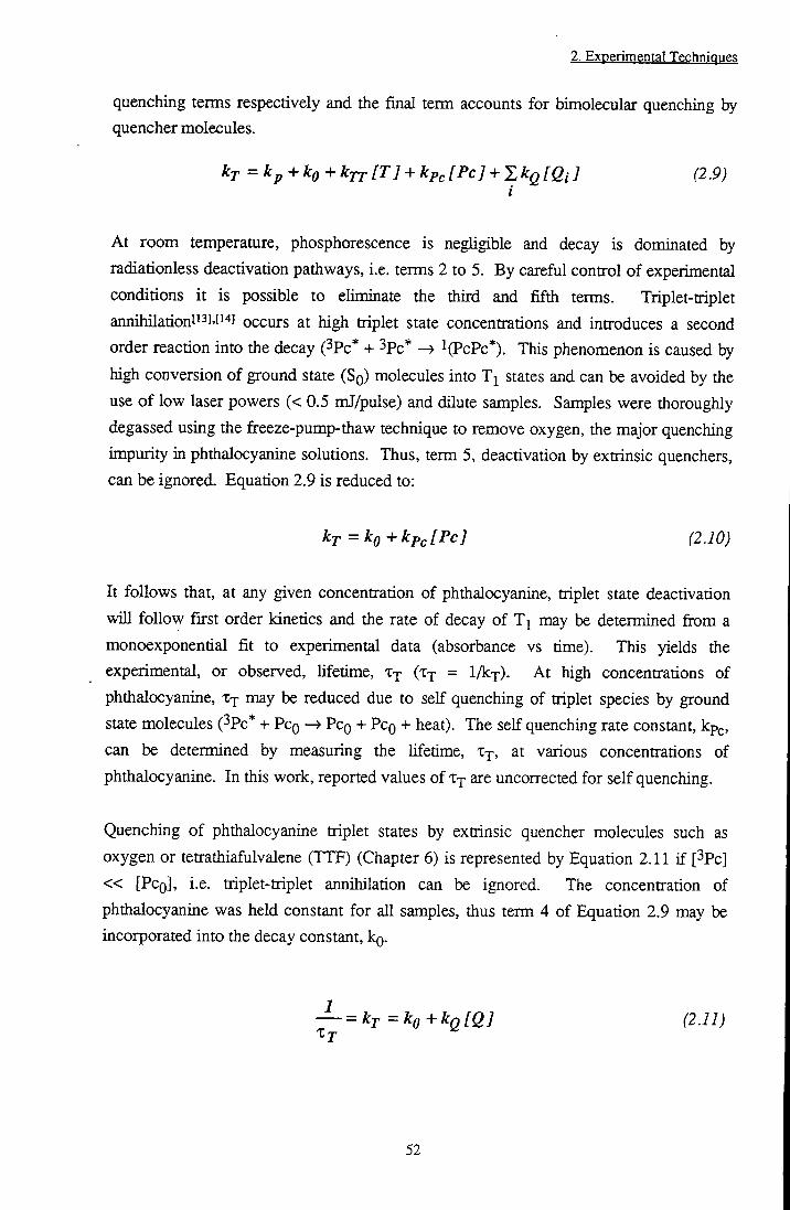
2. Experimental Techniques
quenching terms respectively and the final term accounts for bimolecular quenching by quencher molecules.
kr =kp+ko+kjr[T] + kp, [Pc] + S A:g [Qi ] (2.9)
At room temperature, phosphorescence is negligible and decay is dominated by radiationless deactivation pathways, i.e. terms 2 to 5. By careful control of experimental conditions it is possible to eliminate the third and fifth terms. Triplet-triplet aniu2iilationti3],[i4] occurs at high triplet state concentrations and introduces a second order reaction into tiie decay (3pc* + 3pc* l(PcPc*). This phenomenon is caused by high conversion of ground state (SQ) molecules into T j states and can be avoided by the use of low laser powers (< 0.5 mJ/pulse) and dilute samples. Samples were thoroughly degassed using the fireeze-pump-thaw technique to remove oxygen, the major quenching impurity in phthalocyanine solutions. Thus, term 5, deactivation by extrinsic quenchers, can be ignored. Equation 2.9 is reduced to:
kT=ko+kp,[Pc] (2.10)
It follows that, at any given concentration of phthalocyanine, triplet state deactivation will follow first order kinetics and the rate of decay of T^ may be determined from a monoexponential fit to experimental data (absorbance vs time). This yields the experimental, or observed, lifetime, X j (Xj = l / k j ) . At high concentrations of phthalocyanine, X j may be reduced due to self quenching of triplet species by ground state molecules (^Pc* + PCQ —» PCQ + PCQ -t- heat). The self quenching rate constant, kp ,, can be determined by measuring the lifetime, Xj, at various concentrations of phthalocyanine. In this work, reported values of X j are uncorrected for self quenching.
Quenching of phthalocyanine triplet states by extrinsic quencher molecules such as oxygen or tetrathiafulvalene (TTF) (Chapter 6) is represented by Equation 2.11 if [^Pc] « [PCQ], i.e. triplet-triplet annihilation can be ignored. The concentration of phthalocyanine was held constant for all samples, thus term 4 of Equation 2.9 may be incorporated into the decay constant, kg.
— =kT=ko+kQ[Q] (2.11) X j
52

2. Experimental Techniques
The quenching rate constant, kq, was determined by monitoring Xj as a function of quencher concentration, [Q]. kg can therefore be calculated from the gradient of a plot of l / X j vs [Q], whilst the intercept gives the triplet lifetime in the absence of quencher molecules.
2.1.4.4 Triplet Extinction Coefficients
At any given wavelength, the observed transient decay is a mixture of two components;
triplet state absorption and ground state bleaching. Jf it is assumed that the
concentration of SQ species bleached upon excitation is equal to the concentration of T j
species formed, then the observed change in absorbance may be given by Equation 2.12,
AA(X) = (ET()^)-es('^))[Ti]l (2.12)
where e j * and are the triplet and ground state extinction coefficients respectively, [T]
is the triplet state concentration and 1 is the path length (in cm). In this work, triplet state
extinction coefficients were determined using the singlet depletion methodti^l. It was
assumed that at the wavelength of maximum ground state absorption (i.e. 674 nm for
^Bu4ZnPc in MeOH), Ej was zero and Egfi J was 1.8 x 10^ mol"^ dm^ cm^ . AA may be
described by Equation 2.13 and [T^] calculated.
^ ^ ^ = - ^ s [ T i ] l (2.13)
For the majority of jjhthalocyanines the triplet state absorption band lies in a spectral
region where ground state absorption is neghgible (400 nm - 600 nm) and hence e j may
be calculated from Equation 2.12.
2.1.4.5 Transient Absorption Spectra
Transient absorption spectra were recorded by measuring AA at 5 nm wavelength
intervals over the range 400 nm to 800 nm. Decays were analysed using a suitable
exponential decay function (either mono- or bi- exponential fits were sufficient). AA at
tQ, the moment of excitation, and at a time delay. At, after excitation was noted. In this
way, a transient spectrum, AA vs. X, could be generated and its temporal characteristics
determined. Transient spectra were corrected for ground state absorption by adding a
fraction of the UVA^'isible absorption spectrum of the sample to the measured 'triplet
minus singlet' spectmm.
53
2. Experimental Techniques
2.1.4.6 Triplet Quantum Yields
Triplet quantum yields were determined using the comparative method' ' '. Solutions of the unknown and standard were prepared such that they were optically matched at the excitation wavelength. The quantum yield is defined in Equation 2.14.
_ No. of triplet state molecules formed 2 ^ No.of photons absorbed
The unknown and standard solutions have the same optical density, i.e. the number of
photons absorbed will be equal, therefore.
No.of Photons absorbed {]1X_ = LLIM. (2.15) •* V •'•St
where represents the quantum yield of triplet state formation, [T], is the
concentration of triplet formed, and x and std refer to the unknown and standard
respectively. Rearranging,
' IT]std
Using the Beer-Lambert law (Equation 2.1), the final equation, where e is the extinction
coefficient of the excited state and A is the transient absorbance, is obtained (Equation
2.17).
^ T . - ^ T ^ , ^ ^ (2.17)
In this study, laser irradiation was carried out at 355 nm, 638 nm or 700 nm using pulse
energies of < 0.5 mJ. Benzophenone in acetonitrile (kj^^ = 525 nm, = 1, EJ = 6250)[3], disulfonated aluminium phthalocyanine in methanol (Xjmax = ^ 0 nm, O-j- =
0.22, Ej = 36000)[i«] and zinc phthalocyanine in 1 % pyr/tol (^jmax = 490 nm, =
54

2. Experimental Techniques
0.58, E j = 36000)f'^l were used as standards. For each sample the power was varied and the change in absorbance of the sample determined by probing at ^xmax- - x -^std were obtained from the gradient of a graph of laser power vs AA (calculated using Equation 2.8) and substituted into Equation 2.17 to find O j . For samples where large overlap between transient and ground state absorption occurred, e.g. tri and tetra protonated phthalocyanines, <E>x was measured by monitoring ground state bleaching in preference to triplet state formation. Equation 2.17 was appHed, however e j was replaced with the singlet extinction coefficient, e^.
2.1.5 Singlet Oxygen Phosphorescence Detection 2.1.5.1 Introduction to Singlet Oxygen
Singlet oxygen, (lAg)02, (^02) is generated from phthalocyanine molecules almost exclusively through triplet state quenching by ground state oxygen, (^Lg-)02, via an encounter complexPO]. lAg is assumed to be the only reactive state in solution. This state is far more stable than ^Z^-^ and in gaseous media may have a lifetime of minutes due to the spin forbidden nature of the transition (^Ag ^Sg-)^!]. Equation 2.18 depicts the expression for the quantum yield of singlet oxygen formation,
4 . , = * , W £ i i _ f2.18,
where <I)j is the quantum yield of triplet state formation, ^AQ^ is the rate of quenching by
O 2 to 02(^Ag), kjo-, is tiie rate of quenching by O2 to give 02(^2g-) and k^j. & kj.
represent non-radiative and radiative deactivation pathways of T j respectively. In most
systems quenching by oxygen is very rapid and (kA02+k£02) » ^nr r> hence.
Equation 2.18 may be simphfied to yield Equation 2.19.
O A =<E>- 2 k&02 +^102 J
(2.19)
The rate terms in brackets represent the fraction of triplet state molecules quenched by ground state oxygen to yield singlet oxygen and is generally represented by S .
Therefore,
55
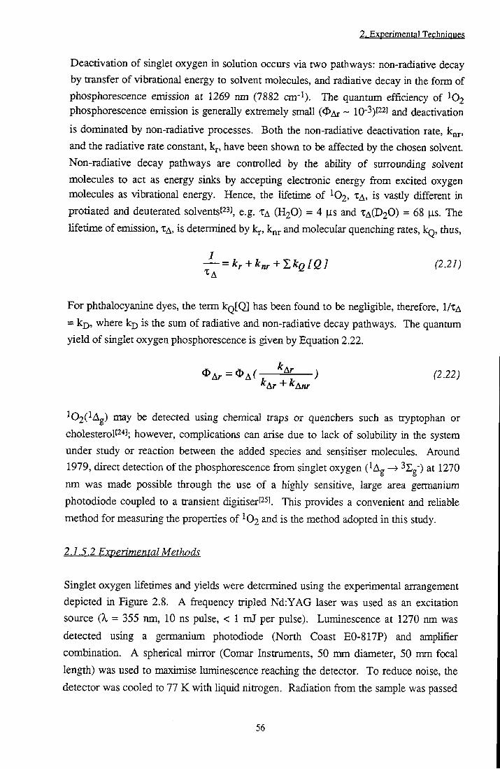
2. Experimental Techniques
Deactivation of singlet oxygen in solution occurs via two pathways: non-radiative decay by transfer of vibrational energy to solvent molecules, and radiative decay in the form of phosphorescence emission at 1269 nm (7882 cm'^). The quantum efficiency of phosphorescence emission is generally extremely small (<I>Ar ~ lO"^)^^] and deactivation
is dominated by non-radiative processes. Both the non-radiative deactivation rate, knr, and the radiative rate constant, k , have been shown to be affected by the chosen solvent. Non-radiative decay pathways are controlled by the ability of suirounding solvent molecules to act as energy sinks by accepting electronic energy from excited oxygen molecules as vibrational energy. Hence, the lifetime of ^02 , TA> is vastiy different in protiated and deuterated solventst^s], e.g. TA ( H 2 O ) = 4 |j,s and TA(D20) = 68 [is. The lifetime of emission, XA. is determined by kj., k^j. and molecular quenching rates, kg, thus,
— = kr+k^+Y.kQ[Q] (2.21)
For phthalocyanine dyes, the term kQ[Ql has been found to be negligible, therefore, I /TA
= ki), where kp is the sum of radiative and non-radiative decay pathways. The quantum
yield of singlet oxygen phosphorescence is given by Equation 2.22.
^ A r = ^ A ( - r ^ > ^2.22j ^Ar + *^Anr
^02(^Ag) may be detected using chemical traps or quenchers such as tryptophan or
cholesterolP'*]; however, complications can arise due to lack of solubihty in the system
under study or reaction between the added species and sensitiser molecules. Around
1979, direct detection of the phosphorescence from singlet oxygen (^Ag —> ^^g") at 1270
nm was made possible through the use of a highly sensitive, large area germanium
photodiode coupled to a transient digitiserPSJ. This provides a convenient and reliable
method for measuring the properties of ^ 6 2 and is the method adopted in this study.
2.1.5.2 Experimental Methods
Singlet oxygen lifetimes and yields were determined using the experimental arrangement
depicted in Figure 2.8. A frequency tripled Nd:YAG laser was used as an excitation
source (k = 355 nm, 10 ns pulse, < 1 mJ per pulse). Luminescence at 1270 nm was
detected using a germanium photodiode (North Coast E0-817P) and ampUfier
combination. A spherical mirtor (Comar Instruments, 50 mm diameter, 50 mm focal
length) was used to maximise luminescence reaching the detector. To reduce noise, the
detector was cooled to 77 K with hquid nitrogen. Radiation from the sample was passed
56
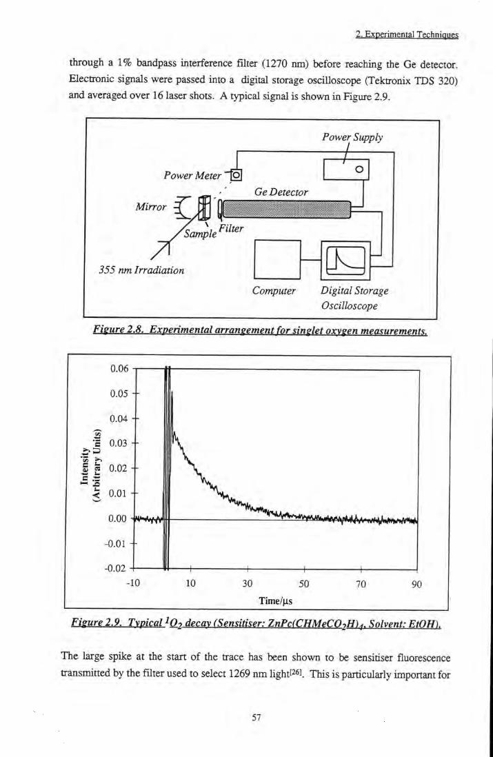
2. Experimental Techniques
through a 1% bandpass interference filter (1270 nm) before reaching the Ge detector. Electronic signals were passed into a digital storage oscilloscope (Tektronix TDS 320) and averaged over 16 laser shots. A typical signal is shown in Figiire 2.9.
Power Meter 4 Power Supply
Mirror | Ge Detector
355 nm Irradiation
Computer Digital Storage Oscilloscope
Fieure 2.8. Experimental arrangement for singlet oxv^en measurements.
0.06
0.05 +
e 0.03 +
S « 0.02 +
< 0.01 +
-0.01 +
-10 10 30 50
Time/fis 70 90
Fieure 2,9. Typical iQ-, decay (Sensitiser: ZnPcfCHMeCO.H)^. Solvent: EtOHl
The large spike at the start of the trace has been shown to be sensitiser fluorescence transmitted by the filter used to select 1269 nm lightpsi. This is particularly important for
57

2. Experimental Techniques
red emitting dyes such as phthalocyanines and, under certain experimental conditions, may cause saturation of the DSO amplifier (See Appendix A and Reference 26). An electronic switch has been developed to eliminate these effects^'^l This problem was not so acute when a Tektronix TDS 320 oscilloscope was employed, therefore, in this work, use of the switch was not required. Data was transferted to a computer and manipulated using a curve fitting package (Grafit). The lifetime of emission could be found direcdy by fitting a single exponential decay to the data (rate of decay (k) equals 1/XA)- Quanmm yields were determined by comparison with reference compounds. Samples of the unknown and standard were prepared such that the absorbance at the excitation wavelength was sunilar (~ 0.3). In this work. Rose Bengal in EtOH (OA = 0.86), MeOH or 1% Triton X-lOO/phosphate buffer saline (OA = 0.75) and tetraphenylporphyrin in 1% pyridine/toluene solution (<I>A = 0.8) or chloroform (<I>A = 0.55) were used as standardsP^l The intensity of the emission was measured as a function of laser power for the unknown and the standard, and the gradients, I , compared using Equation 2.23.
Std ^A = ^ A - . — ^ ^2.25j
A is the absorbance of the solution at the excitation wavelength and std and x refer to the
standard and unknown respectively. It is important that the unknown and reference
compound are used in the same solvent due to the large dependence of kn^ and k^ solvent
properties.
2.1.6 Low Temperature Measurements
Low temperature measurements were performed in ether-pentane-alcohol 5:5:2 (EPA)
solution or 100 % ethanol, cooled to 77 K using a liquid nitrogen cooled cryostat
(Oxford Instruments, D N 1704). Intermediate temperatures were obtained using a
temperature controller (Oxford Instruments, Intelligent Instrument Controller, ITC 4).
Samples were allowed to equihbrate for at least 30 minutes at each temperature.
UVA^'isible, fluorescence and flash photolysis spectrometers were adapted to
accommodate the cryostat.
58

2. Experimental Technique?
2.1.7 Diffuse Reflectance Spectroscopy^^^^-^^°^ 2.1.7.1 Theory 2.1.7.1.1 Reflection
Diffuse reflectance spectroscopy has been used since the early 1920's as a means of measuring the optical properties of heterogeneous and solid samples. Due to the inherent opacity of these substances, transmission measurements are impractical, thus techniques have been developed to obtain information via detection of radiation scattered from the surface of the sample. Reflection from matt surfaces has been shown to consist of two components, specular or regular reflection, and diffuse reflection. Specular reflection is weU known and occurs from highly polished or mirrored surfaces. Its behaviour is governed by Snell's Law (Equation 2.24), which defines the direction of reflected and transmitted beams of radiation, and Fresnel's equations t^'l from which Equations 2.25 and 2.26 are obtained. These determine the relative intensities of each polarised component of reflected hght.
Snell's Law: «n91 = s i n G a n d /ij s/nG ,• = n2 / (2.24)
where n^ and n2 are the refractive index values of each medium, Gj is die angle of
incidence, Gj is the angle of the transmitted beam and Gj. is the angle of the reflected
radiation.
RU = n^ cosQi-[n^ -sin^Qj]^^^
cosQi + [n^-sin^QiJ^^^ (2.25)
cosQj-ln^ -sin^Qj]^^^ n2
cosQi + [n^ -sin^^il 112 (2.26)
where Rjj is the intensity of reflected parallel polarised hght, R i is the intensity of
reflected perpendicularly polarised light and n is the relative index of refraction (n2/ni).
It should be noted that the proportion of specular reflection depends on both the angle of incidence, Gp and on the polarisation of the incident radiation. Reflected radiation retains the polarisation characteristics of the incident beam and is detected at a defined angle, Gj-, equal to Gj (Figure 2.10). For natural hght, the total reflected radiation is given by Rspec = (Rjj + Ri)/2 which, in the case of normal incidence, yields
59

2. Experimental Techniques
^Spec -n-1 n + 1 (2.27)
If the reflecting surface absorbs at the wavelength of irradiation, attenuation occurs according to Lamberts law (Equation 2.28).
(228)
where a, the absorption coefficient, is related to the absorption index of the mediiun, K by:
a = 4%K
(229)
Equation 2.27 now bccomesf l:
R _(n-l)^+(nK)^ spec ~ •} J
(n + i r + { ' n K r (2.30)
which predicts that as the absorbance increases, xht yield of specular reflection will also increase.
Specular Reflection
Diffuse Reflection
Refractive Index «/
Figure 2.10. Specular and diffuse reflection.
60

2. Experimental Techniques
Diffuse reflection occurs as a result of multiple scattering and absorption of an incident beam by individual sample particles. In contrast to specular reflection, diffuse reflection is unpolarised, isotropicaUy distributed at all angles of observation, and independent of die angle of incidence or polarisation of the incident Ught (Figure 10). It has been observed to be affected by the particle size and absorbing power of a reflecting sample. Several theories have been proposed to account for tiiese phenomena, however, die most commonly adopted is that proposed by Kubelka and Munk based on differential equationsP9].
Irradiated Surface(x = 0)
Dark Surface (x = d)
Figure 2.11. Parameters for Kubelka-Munk theory.
I f one considers light of intensity, IQ, falling on a sample of randomly distributed absorbers (Figure 2.11), the flux intensity will decrease as the radiation passes a distance, x, through the medium (Equation 2.31). By comparison, the intensity of the reflected hght flux, J, wil l increase as further scattering occurs (Equation 2.32).
dI(x) = -I(x)(K + S )dx + J(x)Sdx (2.31)
dJ(x) = J(x)(K + S)dx - I(x)Sdx (2.32)
K and S are the absorption and scattering coefficients respectively of the medium with
units cm"l. For an ideal diffuser, K = a = 2ec, where e is the Naperian extinction
coefficient (mol dm-3 cm'^) and c is the concentration (mol dm'^). The factor 2 accounts
for the increased average distance travelled by hght due to scattering. Equations 2.31
and 2.32 consider only the case where irradiation is perpendicular to the surface,
61

2. Experimental Techniques
however, the error introduced by this approximation is small when compared with the overall assumption of ideal diffuse reflection. In addition, these equations hold strictiy only for samples irradiated by diffuse tight. In practise, many experimental arrangements use a directed light source and the theory remains vaUd for an incident angle of 60°. Using Equation 2.33 for the diffuse reflectance, R, and the integral solutions of Equations 2.31 and 2.32 for a layer of thickness d, it can be shown (References 29 and 30) that the diffuse reflectance is given by Equation 2.34.
R=^ (2.33) ^0
sinHbSa) ^ K + S ,2-1x112 R = where a = and b =(a'^-l)^'^ a sinh( bSa) + b cosh( bS^ ) S
(2.34)
In the special case of infinite layer thickness or 'optically thick' samples, d ^ =«, and die
diffuse reflectance is given by the more useful Equation 2.35.
ni?) = f = ^^^f^ (2.35)
For a sample whose scattering coefficient is independent of wavelength, F(R) is termed the remission function and represents the absorption spectrum detennined by transmission spectroscopy. In practise, many differences may occur due to the assumptions made by Kubelka-Munk theory. The over-riding assumption of this theory is that of ideal diffuse reflection. Samples are supposed to consist of randomly distributed absorbers with a particle size far smaller than the thickness of the layer and to have infinite lateral dimension in order that edge effects may be neglected. Contributions due to specular reflection are ignored and though this is acceptable (but not strictiy true) for weakly absorbing samples, as the absorbance (K and K) increases, contributions due to specular reflection increase (Equation 2.30) whilst those due to diffuse reflection decrease (Equation 2.34). Therefore, for strongly absorbing samples it becomes unrealistic to discount specular reflection. Remission functions measured under diese conditions will underestimate the absorbance of a sample. Indeed, a paradoxical situation may occur where concentrated solutions will appear to absorb less than their dilute partners. This is a consequence of differences in die mean deptii, d, travelled by radiation through the medium. Light incident on a sample of weakly absorbing molecules wiU penetrate deeply and, hence, may be absorbed more efficientiy than in a
62

2. Experimental Techniques
concentrated sample where the penetration depth is small and large quantities of specular reflection occur. In addition, derivation of Equation 2.35 assumes that K and S are independent of x. This is true for a homogeneous sample of absorbers, however, when the concentration or absorbance of molecules changes with penetration depth a different analysis is required. This is discussed below for the case of transient formation upon laser excitation.
2.1.7.1.2 Diffuse Reflectance Flash Photolysis
Diffuse reflectance flash photolysis has been pioneered by Frank Wilkinson and his group at Loughborough[32],[33] The group recognised that the concentration of transient species produced by flash photolysis will decrease as incident radiation is absorbed on traveUing through the sample. Therefore, the assumption of Kubelka-Munk theory that K is independent of x is no longer vahd. It can be shown that the concentration of species produced by laser excitation falls off exponentially with penetration depthP^l.
Modifications to Kubelka-Munk theory are therefore required. Lin and Kant^^l have solved Equations 2.31 and 2.32 for tiie case where K varies exponentially with x at an analysing wavelength, a (Equation 2.36).
r 1 + TU+ L ' +... 7
6 h(h+l)2!
(2.36)
where R^g is the background reflectance at the analysing wavelength, y =(bgRs ) ^,
( y + l - R " ) 2K^T(0) 0 = , u = where K ^ is the absorption coefficient of the
be b,S
[K ^ +2K S 1^^^ transient species at the analysing wavelength, and b^ = — where K^ is
s the absoiption coefficient of the ground state at the excitation wavelength.
In diffuse reflectance flash photolysis experiments, the measured quantity is given by
Equation 2.37.
63

2. Experimental Techniques
— -1-RT (2.37) RB
where and R^{t) represent the reflectance before and after excitation respectively and R \ is the relative transient reflectance. Kessler et al. have shown^^ l by substitution of typical values of S (5 - 500 cm-l), R^g (0.7 - 0.95) and K ^ j (0.1 - 10 cm-l) into Equation 2.36 that for 1-R^ < 0.1, 1-R\ is linearly dependent on [T]. Assuming a unimolecular reaction, decay lifetimes may be determined by fitting a monoexponential curve to 1-R^ vs time. In bimolecular reactions, for example, triplet-triplet annihilation, no simple kinetic analysis is possible.
For the purposes of this smdy it was assumed that an exponential decrease of triplet species occurred within the sample, i.e. 1 - R \ a [T]. I f the sample concentration is low, or the laser power is high, complete sample conversion may occur at die surface. For this situation, the plug model must be considered. Under these conditions, Equation 2.35 is valid and hence, l-R-j- is proportional to [T] 1/2. xhis wiU not be discussed in detail here and the reader is referred to Reference 34 for more information.
2.7.7.2 Experimental
2.1.72.1 Sample Preparation
Substrates (cellulose, filter paper or siUca) were dried for 24 h in an oven at 100 °C prior to use. Bovine serum albumin was used as received. Solutions of phthalocyanine in ether, ethanol or methanol were mixed thoroughly with a known weight of substrate. Solvent was removed under vacuum with intermittent stirring of the sample until completely dry. A phthalocyanine/substrate weighting of ~ 50 |J.g g'^ for remission function and transient absorption measurements and ~ 6 )ig g" for fluorescence measurements was used.
2.1.7.2.2 Remission Function Measurements
The remission function, F(R), was determined using an adapted Perkin Ekner LS50B
spectrometer. The optimum arrangement permitted die sample to be irradiated at 60° as
required by Kubelka-Munk theory and enabled maximum collection of diffuse reflection
whilst minimising contributions due to specular reflection. Reflectance spectra were
recorded by performing an average of ten synchronous scans over the wavelength range
300 nm to 750 nm. Spectra were performed for substrate (Rgubstrate) sample
64

2. Experimental Techniques
(Rsample) executed using a neutral density filter in the emission beam and 5 nm slit widths.
F(R) was calculated using Equation 2.35 where,
j,^^Isarnple_ ^^^^^
^substrate
Calculation of the remission function using clean substrate as a reference compensates for any effects that the substrate may have on contributions to the signal resulting from specular reflection.
2.1.7.2.3 Fluorescence Measurements
Fluorescence measurements were carried out using the sample arrangement employed for remission function measurements. Emission scans were performed using sUt widths of 10 nm and 5 nm for excitation and emission monochromators respectively, a scan speed of 120 nm min"l and an excitation wavelength of 610 nm. Excitation spectra were obtained by monitoring emission at 760 nm (Slits: 5 nm and 10 nm (excitation and emission respectively). Scan speed: 60 nm min-^).
2.1.7.2.4 Flash Photolysis
Diffuse reflectance flash photolysis was carried out using the experimental airangement depicted in Figure 2.12. 355 nm radiation from a frequency tripled Nd:YAG laser (Spectra Physics, Quanta Ray GCR-150-10, 10 ns pulse, < 1 mJ per pulse) was used to excite the sample at 60° to the normal. In this way, focusing of specular reflection into the collection optics was avoided. A xenon lamp (Bentham 605) and filter combination (420 nm cutoff filter (Schott glass-GG420, Comar Instruments, UK) and V02+(aq) chemical filter) was used as a probe beam. Diffuse reflection from the sample was focused into a monochromator (Bentham TTVI300) and detected using a photomultiplier tube, PMT (Hamamatsu R928). Decays were recorded using a digital storage oscilloscope (Tektronix TDS 320) and transferred to a computer for analysis. Decays were fitted to exponential kinetics using, Grafit, a curve fitting package.
65

2. Experimental Technique.';
Sample
Laser Irradiation (355 nm)
Collection Optics
Monochromator
Photomultiplier Tube
Focusing Optics
Filter
Xenon Lamp
Digital Storage Oscilloscope
Computer
Fieure 2.12. Diffuse reflectance flash photolysis: Experimental arrangement.
2,2 Synthesis
22.1 Synthesis of Substituted Zinc Phthalocyanines
Figure 2.13 shows the synthetic scheme followed in the preparation of ZnPc(CMe(C02Me)2)4 and ZnPc(CHMeC02H)4. Compounds 2,3 and 4 were prepared following the literature method of Roze et a/.^^l.
i) Dimethyl 3,4 dicyanophenylmalonate (2)t"]. Nitrophthalonitrile, 1 (Acros, 7.53 g),
K2CO3 (Fison, purity 99%, 3.76 g), tetrabutylammonium bromide (Kodak, 1.04 g) and
dimethylmalonate (Aldrich, purity 97%, 10.06 ml) were placed in a round bottom flask
with a magnetic flea. Dimethylformamide (DMF - Aldrich, HPLC Grade, 80 ml) was
added with stirring and die flask purged with nitrogen. The solution was stirred at 50 "C
for 24 hr. Further aliquots of K2CO3 (5 g) were added at six and twelve hour intervals.
The resulting mixture was then poured onto ice, and the precipitate obtained separated
and dried. Purification was achieved by chromatography using a silica column with
dichloromethane (CH2a2 - BDH, Analar Grade) as eluent and drying under high
vacuum - yield = 69 %.
IR(Vmax /cm- l ) : 1730 and 1749 (C=0), 2239 (CN), 2956 (CH).
66

2. Experimental Techniques
5H(250 M H Z ; CDCI3): 3.79 (s, 6H, CO2CH3), 4.74 (s, I H , CH), 7.81-7.91 (m, 3H, ArH)
O g N ' - ^ ^ ^ C N CH2(COOMe)2 \ A ^ C N
Mel K O H ^
MeOH
CN
Me CN
4-nitro phthatomrile RMM=173
1
Dimethyl 3,4 dicyano phenylmatonate RMM = 258
Dimetltyl 3,4 dicyanophenyl 1,1 ethanedicarboxylate
RMM = 272
Zh Acetate
MeO
MeOH/EtOH
OMe
NaOH
MeO •0 Me'
Tecra-l-carboxyethyl zinc phthalocyanine
ZnPc(CHMeC02H)4
RMM = 861.9
5
Tetra-l,l-di(methoxycarbonyl)ethyl zinc phthalocyanine
ZhPc(CMe(C02Me)2)4 RMM=1154
Figure 2.13. Synthetic scheme for production of acid and ester substituted zinc
phthalocyanine.
ii) Dimethyl 3,4 dicyanophenyl 1,1 ethane dicarboxylate (3)[37]. Compound 2 (2.15
g), KOH (0.47 g) and Mel (Aldrich, 10 ml) were refluxed in methanol (50 ml) at 50 °C
for three hours. Additional aliquots of KOH (0.24 g) and Mel (10 ml) were added and
the solution refluxed for a further hour. Following this, the condenser was removed and
heating was continued for one hour to remove excess Mel. MeOH was removed in
vacuo and crude product separated by solvent extraction from water into CH2Q2 (3 x 50
67

2. Experimental Techniques
ml), to yield an oil which was purified by column chromatography on sihca using CH2Q2 as eluent. 3 was obtained as an oily solid after evaporation. Yield = 49 %. IR(Vmax/cm-l): 1727 (C=0), 2237 (CN), 2954 and 2997 (CH).
5H(250 MHz; CDCI3): 1.90 (s, 3H, CH3), 3.80 (s, 6H, CO2CH3), 7.76-7.87 (m, 3H, ArH)
iii) Tetra-l,l-di(methoxycarbonyl)ethyl zinc phthalocyanine (4)[37). Compound 3
(250 mg) was crushed with zinc acetate (Aldrich, purity 99.99 %, 250 mg) and heated
under an inert atmosphere to 200 °C for six hours. Product was purified by
chromatography on silica using 3 % MeOH:CH2Q2 as eluent and dried in a desiccator
under vacuum. Yield = 30 %.
IR(v„,ax /cm-l): 1721 (C=0)
Elemental Analysis: Found %: C, 58.03; H, 4.14; N , 9.41; Zn, 4.19. Calculated %: C,
58.3; H, 4.2; N, 9.7; Zn, 5.66.
\JY/Yis (CHCI3, \ n a x / n m (log e)): 347 (4.78), 611 (4.37), 677 (5.16)
iv) Tetra-1-carboxyethyI zinc phthalocyanine (5). Compound 4 was converted into
the tetra acid substituted phthalocyanine (5) using the method of Kahl and lit^s] 4 ^as
dissolved in MeOH (20 ml), sodium hydroxide (NaOH) in ethanol (20 ml, ~ 0.15 mol
dm"3) was added and the reactants heated under reflux for six hours. The solution was
concentrated under vacuum, dissolved in water and filtered to remove insoluble particles.
Acidification using hydrochloric acid (RCl, 0.1 mol dm'^), yielded a precipitate which
was separated by centrifugation and washed with distilled water (3 x 20 ml). The
product was dried under vacuum to yield a blue solid. Yield = 79 %.
IR (Vmax/cm-1): 2900 (OH), 1700 (C=0)
5H(250 M H Z ; D M S O ) 1.90 (d, 3H, CH3), 4.45-4.49 (m, IH , CH), 8.18-8.22 (m, IH,
ArH), 9.24-9.27 (m, 2H, ArH).
Elemental Analysis: Found %: C, 55.94; H, 3.68; N, 11.35; Zn, 5.63. Calculated %: C,
61; H , 3.72; N, 12.93; Zn, 7.55.
U V m s (CHCI3, >4nax /nm Oog e)): 349 (4.65), 610 (4.38), 673 (5.15)
2.2.2 Metallation ofH2{BuO)^Pc andH2{CuU^4)^Fc
Zinc derivatives of H2(BuO)8Pc and H2(Ci6H34)8Pc were prepared by refluxing with
excess zinc acetate in 1% pyridine/toluene solution for six hours I^^^. Aqueous HCl was
added to remove unreacted zinc acetate. The aqueous phase was removed and the
organic layer washed with distilled water several times. The organic fraction was
retained, dried with MgS04 and filtered. Solvent was removed under reduced pressure,
68
2. Experimental Techniques
the product passed through a silica column using dichloromethane as eluent and dried under vacuum.
2.3 References
[1] Dhami S., De Mello A.J., Rumbles G., Bishop S.M., Phillips D. and Beeby A., Phthalocyanine fluorescence at high concentration: Dimers or reabsorption effect?, Photochem. Photobiol, 61, 341-346,1995.
[2] Williams A.T.R., Winfield S.A. and Miller J.N., Relative fluorescence quantum yields using a computer controlled luminescence spectrometer.. Analyst, 108, 1067-1071, 1983.
[3] Murov S.L., Carmichael I . and Hug G.L., Handbook of Photochemistry, Marcel Dekker, Inc., 2nd Edn., New York, 1993.
[4] Vincett P.S., Voight E.M., Rieckhoff K.E., Phosphorescence and fluorescence of phthalocyanines., / . Chem. Phys., 55, 4131, 1971.
[5] O'Connor D.V. and Phillips D., Time Correlated Single Photon Counting, Academic Press, London, 1984.
[6] Birch D.J.S. and Imhof R.E. in Topics in Fluorescence Spectroscopy, Vol. 1: Techniques, Ed. Lakowicz J.R., Plenum Press, New York, ppl-95, 1991.
[7] Janot J.M., Beeby A., Bayley P.M. and PhiUips D., Preparative, analytical and fluorescence spectroscopic studies of sulphonated aluminum phthalocyanine sensitisers., Biophys. Chem., 41, 272, 1991.
[8] Poner G. and Norrish R.G.W., Chemical reactions produced by very high light intensities.. Nature, 154, 658, 1949.
[9] Patterson L . and Porter G., Lasers in photochemical kinetics., Chem. Br., 6, 246, 1970.
[10] Andrae I . , Bringhen A., Bohm F., Gonzenbach H., Hill T., Mukoy L and Truscott T.G., A UVA filter (4-ten-butyl-4'-methoxydibenzoylmediane): photoprotection reflects photophysical properties., / . Photochem. Photobiol. B.Biol., 37, 147, 1997.
[11] De la Tosa F.F., Laser flash photolysis studies of electron transfer in Complex I I I from yeast mitochondria., / . Photochem. Photobiol. B.Biol., 38, 184, 1997.
[12] Boch R., Mohtat N. , Lear Y., Amason J.T., Durst T. and Scalane J.C., Study of photoinduced energy and electron transfer in a-terthienyl-bovine serum albumin conjugates: A laser flash photolysis study., Photochem. Photobiol., 64, 92,1996.
[13] Frink M.E. and Ferraudi G.J., Excimer formation in triplet-triplet annihilation reactions of the lowest lying triplet excited state in aluminum(in), silicon(rV) and metal-free phthalocyanines - medium and magnetic field effects on the rate of reaction., Chem. Phys. Lett., 124, 576-578,1986.
69

2. Experimental Techniques
[14] Ohno T., Kato S., Yamada A. and Tanno T., Electron transfer reactions of the photoexcited triplet state of chloroaluminium phthalocyanine with aromatic amines, benzoquinones and coordination compounds of iron(n) and iron(III)., J. Phys. Chem., 87, 775-781,1983.
[15] Carmichael I . and Hug G.L., Triplet-triplet absorption spectra of organic molecules in condensed phases., / . Phys. Chem. Ref. Data, 15, 1, 1986.
[16] Simpson M.S.C., Beeby A., Bishop S.M., MacRobert A.J., Parker A.W. and Phillips D., Time resolved laser spectroscopy in biochemistry I I I . , S.PJ.E., 1640, 520, 1992.
[17] Amand B. and Bensasson R., Determination of triplet quantum yields by laser flash absorption spectroscopy., Chem. Phys. Lett., 34, 44, 1975.
[18] Foley M.S.C., The photophysics of sulphonated aluminium phthalocyanines as sensitisers for photodynamic therapy., PhD Thesis, Imperial College, London, 1994.
[19] Dhami S., Photophysics of phtiialocyanines in microheterogeneous systems., p58, PhD Thesis, Imperial College, London, 1996.
[20] Turro N.J., Modern Molecular Photochemistry, p583. University Science Press, CaUfomia, 1991.
[21] Badger R.M., Wright A.C. and Whitiock R.F., Absolute intensities of die discrete and continuous absorption bands of oxygen gas at 1.26 and 1.065|i and the radiative Ufetime of the ^Ag state of oxygen., / . Phys. Chem., 43, 4345, 1965.
[22] Krasnovsky A.A., Quantum yield of photosensitised luminescence and radiative lifetime of singlet (^Ag) molecular oxygen in solution., Chem. Phys. Lett., 81, 443, 1981.
[23] Losev A.P., Nichiporovich I.N., Byteva I .M., Drozdov N.N. and Aljghgami I.E., The perturbing effect of solvents on the luminescence rate constant of singlet molecular oxygen., Chem. Phys. Lett., 181,45,1991.
[24] Bensasson R.V., Land E.J. and Truscott T.G., Excited States and Free Radicals in Biology and Medicine., p i 18, Oxford University Press, New York, 1993.
[25] Byteva I .M. and Gurinovich G.P., Sensitised luminescence of oxygen in solutions., / . Lumin., 21, 17, 1979.
[26] Oldham T.C., Spectroscopic investigations of photodynamic mechanisms in cell
cultures., p85, PhD Thesis, Imperial College, 1996. [27] Beeby A., Parker A.W. and Stanley C.F., Elimination of fluorescence
contributions to singlet oxygen measurements using a novel electronic switch., J. Photochem. Photobiol. B.Biol., 37, 267,1997.
[28] WiLdnson F., Helman W.P., Ross A.B., Quantum yields for the photosensitised
formation of the lowest electronically excited singlet state of molecular oxygen in solution., / . Phys. Chem. Ref Data, 22, 113,1993.
70
2. Experimental Tgphnique?
[29] Wendlandt W.W.M. and Hecht H.G., Reflectance Spectroscopy, John Wiley and
Sons, New York, 1966. [30] Wilkinson F. and KeUy G., Diffuse Reflectance Hash Photolysis in Scaiano J.C.,
Handbook of Organic Photochemistry, CRC Press, Florida, 1, p293, 1989. [31] Harrick N.J., Total internal reflection and its application to surface studies., Ann.
N.Y. Acad. Sci., 101,928,1963. [32] Kessler R.W. and Wilkinson F., Diffuse reflectance triplet-triplet absorption
spectroscopy of aromatic hydrocarbons chemisorbed on 7-alumina., / . Chem. Soc.
Faraday Trans. 1, 77, 309, 1981. [33] Wilkinson F., Leicester P.A., Ferreira L.F.V. and Freire V.M.M.R.,
Photochemistry on surfaces: triplet-triplet energy transfer on microcrystalline cellulose
studied by diffuse reflectance transient absorption and emission spectroscopy.,
Photochem. Photobiol., 54, 599,1991.
[34] Oelkrug D., Honnen W., Wilkinson F. and Willsher C.J., Modelling of transient
production and decay following laser excitation of opaque materials., / . Chem. Soc, Faraday Trans. 2., 83,2081,1987.
[35] Lin T. and Kan H.K.A., Calculation of reflectance of a Ught diffuser with non
uniform absorption., / . Opt. Soc. Am., 38, 448, 1948.
[36] Kessler R.W., Krabichler G., Uhl S., Oelkrug D., Hagen W.P., Hyslop J. and
Wilkinson F., Transient decay following pulse excitation of diffuse scattering
samples., Opt. Acta, 30, 1099, 1983.
[37] Roze M.P., Berzin'sh E.L. and Neiland O.Y., Synthesis of 3,4
Dicyanophenyhnalonic esters and their use in the production of soluble
phthalocyanines., Z. Org. Khim., 28, 827, 1992.
[38] Kahl S.B. and L i J., Synthesis and characterisation of a boronated metallocyanine
for boron neutron capture theory., Inorg. Chem., 35, 3878, 1996.
[39] Cook M.J., Chambrier L, CrackneU S.J., Mayes D.A. and Russel D.A., Octaalkyl
zinc phtiialocyanines: Potential photosensitisers for use in the photodynamic therapy
of cancer., Photochem. Photobiol., 62, 542,1995.
71
MMiimm
3.1 Introduction
Zinc phthalocyanine (ZnPc) has proved to be an efficient sensitiser for photodynamic
therapyf'l. Use of zinc as the central metal ion results in high quantum yields of triplet
statePl and singlet oxygen formation. It has been shown to be effective at destroying
neoplastic cells in micet^l and exhibits a high uptake by tumoursf'*!. One of the
drawbacks of unsubstituted ZnPc, however, is very low solubihty in both aqueous and
organic solvents. This precludes direct injection of therapeutic quantities into the
bloodstream and hinders both in vitro and in vivo studies.
One method used to improve the solubihty of phthalocyanines is to incorporate
substituents onto the peripheral benzene ringsf^l. Pendant groups such as alkyl chainsf^l,
esterst''], ethersf^l, amidest ^ and alkoxyfi°) substituents have been shown to render
phtiialocyanines soluble in many organic solvents whilst hydrophilic sulfonic acid
substituentst^i] allow aqueous solutions to be prepared. In addition to improving
solubihty, the nature of peripheral substitution affects the cytotoxicity and selectivity of
the sensitiser. Ben-Hurf'^^ and Allenti^l found that anti-viral activity of ZnPc was
inversely proportional to the number of sulfonate substituents whilst a study by
Zimmermannt '*] revealed that cationic side chains such as trimethylammonium (Me3N"'")
groups induce selective photosensitisation of mitochondria. Rosenthal et a/.f^^l have
shown that sulfonate groups have no effect on the photophysical properties of ZnPc.
Similarly, although the absorption spectrum of 1,4,8,11,15,18,22,25-octabutoxy zinc
phthalocyanine was red shifted with respect to the parent moleculef^^l, the yield of triplet
state formation was essentially unaffected. Interestingly, although
1,4,8,11,15,18,22,25-octadecyl zinc phthalocyanine exhibits a smaller shift in the Sj <-
SQ transition than the octabutoxy substituted phthalocyanine, Ci(^2\' chains induce
significant changes in the triplet yield relative to unsubstituted zinc phthalocyaninef ' J.
Clearly, control over substiments of the phthalocyanine ring is an important means of
influencing both the photophysical and physiological properties of phthalocyanines m
vitro and in vivo.

3. Properties of Novel Phthalocvanine Sensitisers.
As an alternative to modification of the phthalocyanine ring, hydrophobic sensitisers may be incorporated into delivery vehicles such as Uposomesfi l These allow systemic injection to the bloodstream where association with proteins is believed to occurt l. The proteins act as carriers to deliver sensitisers to lipophilic sites in tumour tissue, where they accumulate. It is therefore important to determine the effect of micellar and soUd state media on the photophysics of any potential sensitisers.
The photophysical properties of a sensitiser are the core of photodynamic therapy and determine its ability to generate cytotoxic species (See Section 1.2). The measurement of photophysical parameters such as <I>p, and <I>A is therefore extremely important
when evaluating the potential efficacy of a dye. One of the aims of this study was to synthesise a novel, water soluble phthalocyanine and to investigate its solution state properties in homogeneous and heterogeneous media. The influence of peripheral substitution has also been investigated.
3.2 Materials and Methods 3.2.1 Materials
The synthesis and purification of P-tetra-l,l-di(methoxycarbonyl)ethyl zinc phthalocyanine (ZnPc(CMe(C02Me)2)4) and (3-tetra-l-carboxyethyl zinc phthalocyanine (ZnPc(CHMeC02H)4) was described in Chapter 2. Tetra tertbutyl zinc phthalocyanine (tBu4ZnPc) and disulfonated zinc phthalocyanine (ZnPcS2) were prepared by Dr. A. Beeby and used as received^o. 1,4,8,11,15,18,22,25-octadecyl zinc phthalocyanine (CIO) was a gift from Prof. M. J. Cook, University of East AnghaPH. Solvents were Analar grade or better and were used without further purification. Phosphate buffer saline (PBS) was purchased from Sigma and prepared using distilled water. Triton X -100, Triton X - 1 0 0 reduced and hexadecyl trimethylammonium bromide (CTAB -CH3(CH2)i5N(CH3)3Br) were purchased from Aldrich and used as received. Ether-pentane-alcohol (5 :5 :2) solution was prepared from diethyl ether, isopentane (IP) and ethanol.
32.2 Methods
Dimerisation constants were calculated by measuring absorbance at the wavelength of maximum monomer absorption ( ^ a x ) ^ range of phthalocyanine concentrations. Two analytical methods were employed, that of Tai et a/. [22] and that suggested by Fernandez et a/.[23]. Tai assumed a one step equilibrium between monomeric and dimeric phthalocyanine and also made the important assumption that the extinction coefficient of
73

3. Properties of Novel Phthalocvanine Sensitisers.
dimer, « the extinction coefficient of the monomer, at the monitored wavelength. This yielded Equation 3.1,
log[Ct(l-—)] = log( nK) + nlog[Ct(—)] (3.1)
where Cj is the total phthalocyanine concentration, e is the observed extinction
coefficient. Eg is the monomer extinction coefficient (assumed to be 180000 dm^ mol-l
cm-l at XjnsyP-*^), n is the average aggregation number and K is the dimerisation
constant. Plots of log [Ct(l-e/£s)] vs log [Ct(£/es)] yielded linear fits from which n may
be determined from the gradient and K estimated from the intercept. Equation 3.2 gives
the formula employed by Femandez[23].
A = j ^ I < 1 . 8 K C , ) " ^ - l , ^ ' - ^ l C , J ' ^ ' ' ' ^ ' / " - ' ] (32,
Q, K and have the same meaning as above and is the extinction coefficient of
dimeric species. The dimerisation constant was determined by fitting this equation to
experimental data using a curve fitting package (Grafit). C and were treated as
constants while and K were allowed to vary until the best fit was achieved.
Stock micellar solutions of tBu4ZnPc and ZnPc(CMe(C02Me)2)4 were prepared by
sonicating phthalocyanine in Triton X-100 (TX) or Triton X-100 reduced (TXR)
followed by dilution to 1% w/v surfactant solution (1.5 x 10"3 mol dm"^) using
phosphate buffered saline (PBS). Micellar solutions of CIO were prepared by
evaporation of toluene from a toluene/1% TX/PBS emulsion. CIO was dissolved in
toluene and thoroughly mixed with the surfactant solution. Toluene was removed by
stirring and blowing argon over the solution to assist evaporation. Water soluble
phthalocyanines (ZnPcS2 and ZnPc(CHMeC02H)4) were added directiy to 1% TX/PBS
solution and sonicated to achieve solubilisation. All surfactant solutions were prepared
immediately prior to use.
UV/Visible absorption spectra, fluorescence measurements, triplet state data and singlet
oxygen quantum yields were determined using the apparatus and sample conditions
described in Chapter 2 (pp 42-71).
74
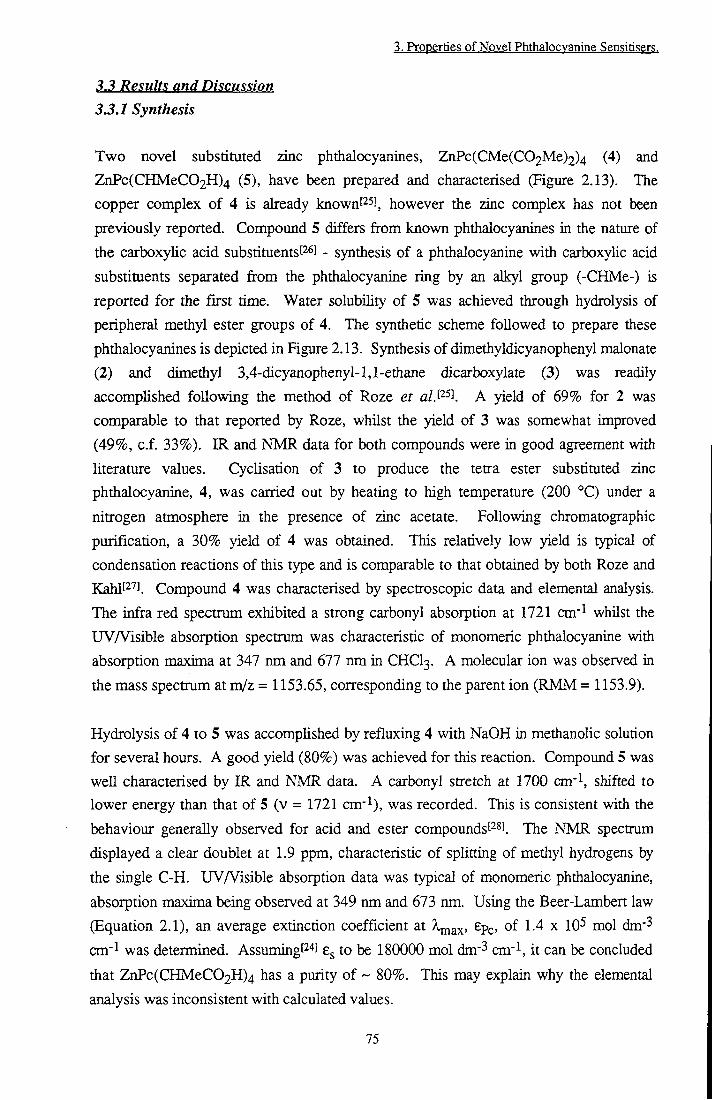
3. Properties of Novel Phthalocvanine Sensitisers.
3.3 Results and Discussion 3.3.1 Synthesis
Two novel substituted zinc phthalocyanines, ZnPc(CMe(C02Me)2)4 (4) and
ZnPc(CHMeC02H)4 (5), have been prepared and characterised (Figure 2.13). The
copper complex of 4 is already known[25], however the zinc complex has not been
previously reported. Compound 5 differs from known phthalocyanines in the nature of
the carboxyhc acid substituentsf^s] - synthesis of a phthalocyanine with carboxyHc acid
substituents separated from the phthalocyanine ring by an alkyl group (-CHMe-) is
reported for the first time. Water solubihty of 5 was achieved through hydrolysis of
peripheral methyl ester groups of 4. The synthetic scheme followed to prepare these
phthalocyanines is depicted in Figure 2.13. Synthesis of dimethyldicyanophenyl malonate
(2) and dimethyl 3,4-dicyanopheny 1-1,1-ethane dicarboxylate (3) was readily
accomphshed following the method of Roze et a/.I^^] ^ yield of 69% for 2 was
comparable to that reported by Roze, whilst the yield of 3 was somewhat improved
(49%, c.f. 33%). IR and NMR data for both compounds were in good agreement with
literature values. Cyclisation of 3 to produce the tetra ester substituted zinc
phthalocyanine, 4, was carried out by heating to high temperature (200 °C) under a
nitrogen atmosphere in the presence of zinc acetate. Following chromatographic
purification, a 30% yield of 4 was obtained. This relatively low yield is typical of
condensation reactions of this type and is comparable to that obtained by both Roze and
Kahl[27]. Compound 4 was characterised by spectroscopic data and elemental analysis.
The infra red spectrum exhibited a strong carbonyl absorption at 1721 cm'^ whilst the
UV/Visible absorption spectrum was characteristic of monomeric phthalocyanine with
absorption maxima at 347 nm and 677 nm in CHCI3. A molecular ion was observed in
the mass spectram at m/z = 1153.65, corresponding to tiie parent ion (RMM = 1153.9).
Hydrolysis of 4 to 5 was accomphshed by refluxing 4 with NaOH in methanohc solution
for several hours. A good yield (80%) was achieved for this reaction. Compound 5 was
well characterised by IR and NMR data. A carbonyl stretch at 1700 cm-^ shifted to
lower energy than that of 5 (v = 1721 cm-l), was recorded. This is consistent with the
behaviour generally observed for acid and ester compounds[2^]. The NMR spectmm
displayed a clear doublet at 1.9 ppm, characteristic of splitting of methyl hydrogens by
the single C-H. UVA^isible absorption data was typical of monomeric phthalocyanine,
absorption maxima being observed at 349 nm and 673 nm. Using the Beer-Lambert law
(Equation 2.1), an average extinction coefficient at ^ a x ' ^Pc ^•'^ mol dm'^
cm"^ was determined. Assuming[2'*] to be 180000 mol dm'^ cm-l, it can be concluded
that ZnPc(CHMeC02H)4 has a purity of ~ 80%. This may explain why the elemental
analysis was inconsistent with calculated values.
75

3. Properties of Novel Phthalocvanine Sensitisers.
Synthesis of 4 and 5 was successfully repeated on a larger scale (X3). Product yields were similar. Phthalocyanines 4 and 5 are expected to exist as a mixture of isomers with a statistical mixture of arrangements of the peripheral substituents. Perhaps not surprisingly, it was not possible to separate these by thin layer or column chromatography.
3.32 Properties in Homogeneous Solution 3.322 Results
3322.1 Solution State Properties
ZnPc(CMe(C02Me)2)4 and ZnPc(CHMeC02H)4 displayed very different solubilities.
Ester groups rendered the phthalocyanine soluble in non polar organic solvents such as
chloroform and toluene as well as methanol, THE and ethanol. In chloroform,
ZnPc(CMe(C02Me)2)4 was remarkably monomeric in character (Figure 3.1a), adhning
to the Beer-Lambert law (Equation 2.1) up to concentrations of 2 x ICH tool dm' .
Table 3.1 gives the dimerisation constants, K, calculated in various solvents using
Equations 3.1 and 3.2. Clearly a large discrepancy exists between each method of
analysis, however, the trend was consistent
MeOH
Toluene
CH2a2 CHClct
51 ±d 108 ±d 0.49 ± d
.c
22500 ± 2000
34000 ±3000
3100 ± 5 0 0 .c
a) Calculated using Equation 3.1, b) Calculated using Fernandez method c) Monomaric in concentration
range studied, d) Errors incurred using this equation were large: values quoted may vary by as much as
an order of magnitude.
Tahle 3,1. Dimemation constants 0fZnPc(CMe(CO2M£l2Lf
CHCI3 and CH2Q2 were the most suitable solvents. ZnPc(CMe(C02Me)2)4 was
monomeric in solutions of CHCI3 up to the detection limit of the spectrometer (1 x 10"
mol dm-3), whilst in CH2a2, a K value of 0.49^ (3100 ') was obtained. In methanol and
toluene, a greater tendency to aggregate was observed, dimer species being
characterised[29l by the appearance of an absorption peak at 633 nm (Figure 3.1b).
Figiure 3.1c shows the calculated absorption spectrum of pure dimer.
76

3. Properties of Novel Phthalocvanine Sensitisers.
0.6 +
0.4 +
Wavelength/nm
Figure 3.1. Absorption spectra of ZnPcfCMefCOoMe)^)^. 1 mm pathlength. a) CHCls (9.6 X10⁻⁵ mol dm-3), b) MeOH (2 x IQ-^ mol dm-3). c) Calculated dimer
spectrum.
The acid functionahty has hydrophilic properties, tiius ZnPc(CHMeC02H)4 was soluble
in polar solvents such as THE, methanol and ethanol but insoluble in dichloromethane
and toluene. Dimerisation in ethanol was found to be extremely sensitive to the ionic
strengdi of the solution. Addition of NaCl, zinc acetate, Ca(C104)2, HCl or NaOH (0 <
[ions] < 10-^ mol dm-3) was found to induce aggregation of ZnPc(CHMeC02H)4 in
EtOH (Figure 3.2A), whilst high ion concentrations (>10-3 mol dm'^) caused
monomerisation (Figure 3.2B). For samples with high ionic strength, the proportion of
dimer species was found to increase as any particular solution was diluted due to a
reduction in the ionic strength of the solution. This contradicts die general behaviour of
phthalocyanines which predicts a decreasing proportion of dimer as concentration
decreases [30'. Under the same conditions, the spectra obtained from ZnPcS2 were
consistent and predominantiy monomeric in character.
77

3. Properties of Novel Phthalocvanine Sensitisers.
0.55
0.5
S 0.25 X) ^ 0.2
0.1
0.05
0
- A ^
Increasing Ion Concentration
r
- B ^
- — = 2 = 2 ^ \ U
Increasing Ion Concentration
550 600 650 700
Wavelength/nm
750 800
Figure 3.2. A. Aggregation ofZnPc(CHMeC02H)^ in EtOH induced bv potassium
acetate, a. ~0 mol dm'^ b. IQ-^ mol dm'^.
B. Disaeereeation at high ionic strength a. IQ-^ mol dm'^ b. 5 x IQ-^ mol dm-3 c.6x
10-^ mol dm-3 d. 10'^ mol dm'^.
Low concentrations of tetrabutylammonium salts were found to promote aggregation but
at high concentrations monomerisation did not occur. In addition to dimer species at
633 nm, concentrated solutions of ZnPc(CHMeC02H)4 in THE showed an absorption
band at 704 nm, red shifted with respect to the main absorption peak. This disappeared
upon dilution of the phthalocyanine and by analogy to results described in Chapter 5 (pp
160-184) was ascribed to protonation of the phthalocyanine ring by H+ ions released
from the carboxyhc acid functionahties.
Both phthalocyanines were insoluble in distilled water. However, ZnPc(CHMeC02H)4
was soluble in phosphate buffered saline (PBS), pH = 7.4, in an aggregated form.
Clearly, since the ionic strength of PBS is high (140 mmol dm'^ Na/KCl), ions do not
promote monomerisation in an analogous manner to the situation in organic solvents.
However, disaggregation could be induced by addition of 0.02 mmol dm-^ hexadecyl
trimetiiylammonium bromide (CTAB). This concentration is well below the critical
micelle concentration[3i] (0.9 mmol dm-3) of this surfactant. Alkaline conditions had no
effect on the state of aggregation of ZnPc(CHMeC02H)4 whilst PBS acidified with HCl
to pH = 2 caused further aggregation (Figure 3.3).
78

3. Properties of Novel Phthalocvanine Sensitisers.
y 0.15
Wavelength/nm
Fieure 3.3. UV/Vis spectra ofZnPc(CHMeC02H)^ in PBS, a. PH = 7.4 b. PH = 11 C. PH = 2 d vH = 7.4 (0.02 mmol dm-3 CTAB).
3.3.2.2.2 Photophysical Properties
The energy of the Sj <- SQ transition of ^Bu4ZnPc was found to vary with solvent. Figure 3.4 shows the position of the absorption maximum (A^ax) ^U4ZnPc in a variety of solvents as a function of dielectric constant, e, dipole moment, |J., donor number, DN and refractive index, n^. No correlation was observed between the polarity or dipole moment of the solvent and A ax- ^ tentative link between the donor number of the solvent and \aax ^ impHed, while a reasonably clear linear dependency on the refractive index was observed.
79

3. Propfaties a( Novel Phthalncvanine. 5Sftnsirisftrs
A 680t
678 --
g 676 --
674
672--
670 --
668 0 10 20 30 40
Didcctric Craistant
^ 680 -
678 h
g 676-
674 -e B
672-•
•
670- •
668 10 20 30 Dmor Nunri)er
40
B 680 -
678 -- •
676 --S
^ 674 --
J 672 --
670 -•
668 1 2 3 DipcAe Moment/Debye
D 680t
678
676-H
1 674+ .
672 +
670 +
1.3 1.35 1.4 1.45 1.5 155 Refracdve Index
Figure 14, SQlvent m<^t Qn "K^ (UV/Vis,) Qf'Bu^ZnPf,
1 PhtftslOeyaAlne $otveiil
1 ZnPc l%pyr/tol 672 -
1 tBu4ZnPc l%pyr/tol 678 9 CIO l%pyr/tol 702 33
ZnPc MeOH 664a -tBu4ZnPc MeOH 673.5 2.5
ZnPc(CH]V[eC02H)4 MeOH 674 3
ZnPc(CMe(C02Me)2)4 MeOH 671 10
ZnPcSo MeOH 665.5 4.5
a This wavelength was calculated using Equation 3.3 and in toluene. Table 32. Effect of peripheral substitution on absorption of ZnPc.
80

3. Properties of Novel Phthalocvanine Sensitisers.
Peripheral substitution of ZnPc caused the wavelength of maximum absorption to be shifted to lower energy. The extent of the shift (AX) varies according to the nature of the substituent (Table 3.2). Tables 3.3A and 3.3B show the radiative emission properties of the excited singlet state of ZnPc and the effect of peripheral substitution. All the phthalocyanines studied have an excitation spectrum which exactly overlays the absorption spectrum, indicating the presence of just one absorbing species. The emission spectra have a Stokes shift of 8 nm ± 1 nm which was constant for all phthalocyanines except CIO. Good mirror symmetry of absorption and emission was obtained (Figure 3.5).
Wavelength/nm
Figure 3.5. a) Absorption and b) Emission Ck^y = 630 nm) suectrq of
ZnPcfCMe(C02Me)^)fin CHCh. Spectra have bfen nom^lM <U
Fc Solvent Stokes Shift
fam fnm /nm
ZnPc 1% pyr/tol 680 672 8
tBu4ZnPc EtOH 682 674 8
ZnPc(CHMeC02H)4a MeOH 683 674 9
ZnPc(CMe(C02Me)2)4 CHCI3 683 676 7
ZnPcS2 EtOH 674.5 667.5
CIO l%pyr/tol 717 702 15 1 a. 0.01 mol potassium acetate used to prepare monomeric species.
Table 3.3 A. Effect of perinheral substitution on emission characteristics of ZnPc.
81

3. Properties of Novel Phthalocvanine Sensitisers.
The quantum yield of radiative emission, Op, was essentially unaffected by peripheral substitution by tertiary butyl, ester or sulfonate groups, however carboxylic acid and decyl substituents caused a reduction in the fluorescence yield.
I»t
ZnPc
tBu4ZnPc
ZnPc(CHMeC02H)4
ZnPc(CHMeC02H)4
ZnPc(CMe(C02Me)2)4
ZnPcS2
CIO
l%pyr/tol
EtOH
MeOH
PBS
THF
EtOH
l%pyr/tol
0.3P2]
0.26 ±0.03 0.17 ± 0.02a
0.06 ± 0.005''
0.22 ±0.02
0.35 ±0.02
0.082 ±0.015
3.5 3.6
a. 0.01 mol dm- potassium acetate used to prepare monomeric species, b. Measured in PBS using
CTAB to monomerise.
Effect of peripheral substitution on fluorescence emission yield of ZnPc.
ZnPc(CHMeC02H)4 and ZnPc(CMe(C02Me)2)4 showed typical triplet state
characteristics. Measured traces could be described by monoexponential decays and a
good linear response of transient height to laser power was achieved at low laser powers
(Figure 3.6).
0.0045
0.004 +
0.0035 +
0.003 +
AA 0.0025 +
0.002 +
0.0015 +
0.001 +
0.0005 +
2 3 4 5 6 Pulse Energy /Arbitrary Units
Fieure 3.6. Transient absorbance ofZnPc(CMe(C02Me)2)^ in CHCl^ vs laser pulse
energy, (X^x = nm, Xprobe = ^90 nml
82
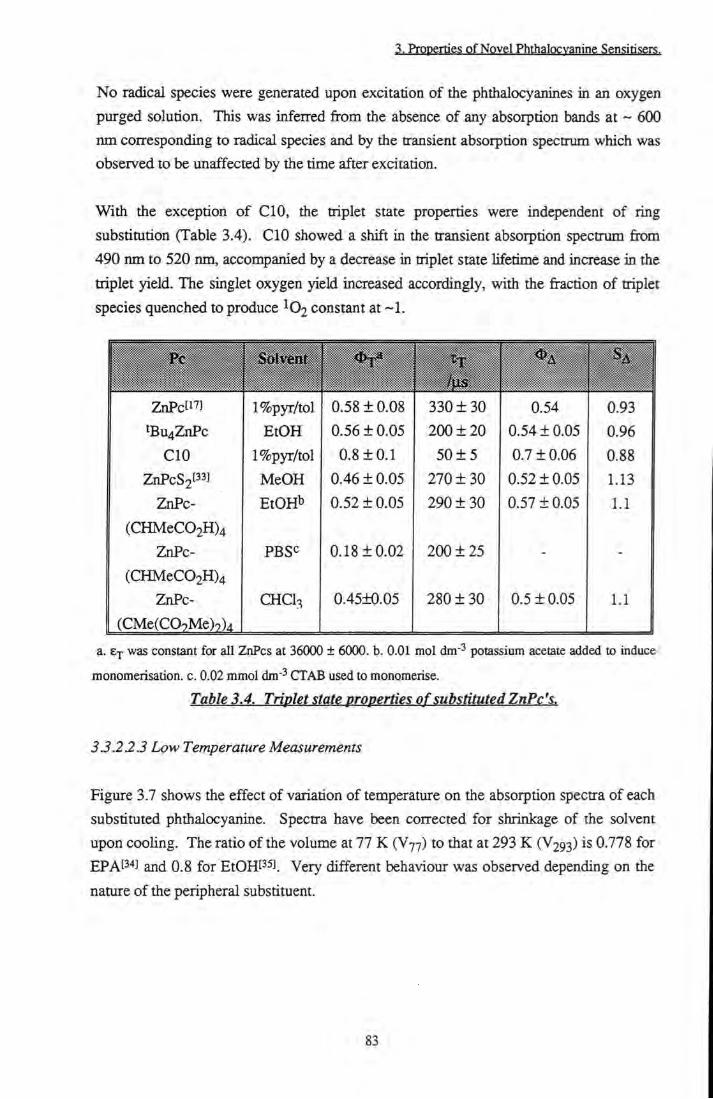
3. Properties of Novel Phthalocvanine Sensitisers.
No radical species were generated upon excitation of the phthalocyanines in an oxygen purged solution. This was infeired from the absence of any absorption bands at ~ 600 nm corresponding to radical species and by the transient absorption spectrum which was observed to be unaffected by the time after excitation.
With the exception of CIO, the triplet state properties were independent of ring
substitution (Table 3.4). CIO showed a shift in the transient absorption spectrum from
490 nm to 520 nm, accompanied by a decrease in triplet state lifetime and increase in the
triplet yield. The singlet oxygen yield increased accordingly, with the fraction of triplet
species quenched to produce ^©2 constant at ~1.
Pc Solvent fus
ZnPcti'J l%pyr/tol 0.58 ± 0.08 330 ± 30 0.54 0.93 tBu4ZnPc EtOH 0.56 ± 0.05 200 + 20 0.54 ±0.05 0.96
CIO l%pyr/tol 0.8 ±0 .1 50 ± 5 0.7 ±0.06 0.88 ZnPcS2t"l MeOH 0.46 ±0.05 270 ± 3 0 0.52 ±0.05 1.13
ZnPc- EtOHl' 0.52 ±0.05 290 ± 3 0 0.57 ±0.05 1.1
(CHMeC02H)4
ZnPc- PBSc 0.18 ±0.02 200 ± 2 5 - -
(CHMeC02H)4
ZnPc- CHCI3 0.45±0.05 280 ± 30 0.5 ±0.05 1.1
(CMe(C09Me)9)4
a. &p was constant for all ZnPcs at 36000 ± 6000. b. 0.01 mol dm~ potassium acetate added to induce
monomerisation. c. 0.02 mmol dm" CTAB used to monomerise.
Table 3.4. Triplet state properties of substituted ZnPc's.
3.3.22.3 Low Temperature Measurements
Figure 3.7 shows the effect of variation of temperature on the absorption spectra of each
substituted phthalocyamne. Spectra have been corrected for shrinkage of the solvent
upon cooling. The ratio of the volume at 77 K (V77) to tiiat at 293 K (V293) is 0.778 for
EPAP''] and 0.8 for EtOHt^si. Very different behaviour was observed depending on the
nature of the peripheral substituent.
83
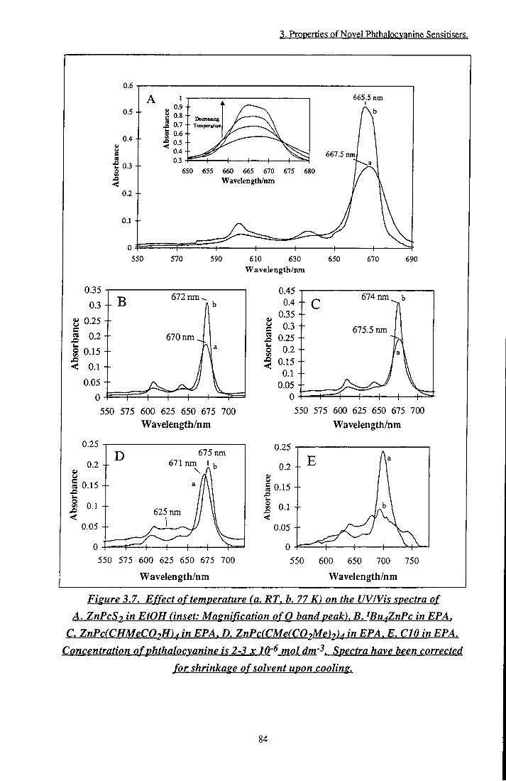
3. Properties of Novel Phthalocvanine Sensitisers.
0.6
0.5
0.4
0.3
J3 < 0.2 +
0.1 +
1 0.9 0.8 0.7 0.6 0.5 0.4 0.3
Dcocasiiig • TemperaEure
^^^l
665.5 nm I vb
550 570
667.5 nm
650 655 660 665 670 675 680 Wavelength/nm
610 630 Wavelength/mn
690
0.35 0.3
u e cs I <
0
T> 672 nm ^ - D b
670nm^ : J — 1 — 1 1 1
I 1 - f
0.45
0) c St u o < 0.1
0
- c 674 nm ^ b
675.5 nm
a \
1 —
550 575 600 625 650 675 700 Wavelength/nm
550 575 600 625 650 675 700 Wavelength/nm
0.25 675 nm
671 nm I
0.15 +
625 nm 0.05 +
0.25
0.2 --
s a 0.15 --x> o 0.1 --<
0.05 --
550 575 600 625 650 675 700
Wavelength/nm
550 600 650 700 750
Wavelength/nm
Figure 3.7. Effect of temperature (a. RT. b. 77 K) on the UV/Vis spectra of A. ZnPcSz in EtOH (inset: Magnification ofQ band peak), B. tBu^ZnPc in EPA,
C. ZnPc(CHMeC02H)^ in EPA. D. ZnPc(CMe(C02Me)2)^ in EPA. E. CIO in EPA. Concentration of phthalocyanine is 2-3 x 10'^ mol dm-^. Spectra have been corrected
for shrinkage of solvent upon cooling.
84
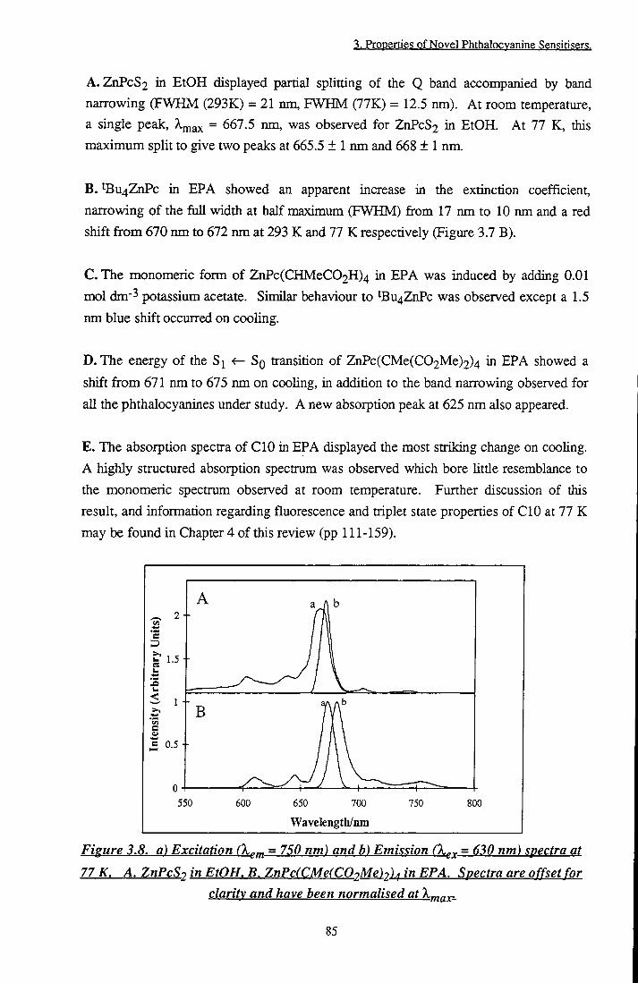
3. Properties of Novel Phthalocvanine Sensitisers.
A. ZnPcS2 in EtOH displayed partial splitting of the Q band accompanied by band narrowing (FWHM (293K) = 21 nm, FWHM (77K) = 12.5 nm). At room temperature, a single peak, = 667.5 nm, was observed for ZnPcS2 in EtOH. At 77 K, this maximum split to give two peaks at 665.5 ± 1 nm and 668 ± 1 nm.
B. tBu4ZnPc in EPA showed an apparent increase in the extinction coefficient, narrowing of the full width at half maximum (FWHM) from 17 nm to 10 nm and a red shift from 670 nm to 672 nm at 293 K and 77 K respectively (Figure 3.7 B).
C. The monomeric form of ZnPc(CHMeC02H)4 in EPA was induced by adding 0.01 mol dm-3 potassium acetate. Similar behaviour to ^Bu4ZnPc was observed except a 1.5 nm blue shift occurred on cooling.
D. The energy of the S <- Sq transition of ZnPc(CMe(C02Me)2)4 in EPA showed a
shift from 671 nm to 675 nm on cooling, in addition to the band narrowing observed for
all the phthalocyanines under study. A new absorption peak at 625 nm also appeared.
E . The absorption spectra of CIO in EPA displayed the most striking change on cooling. A highly structured absorption spectrum was observed which bore little resemblance to the monomeric spectrum observed at room temperature. Further discussion of this result, and information regarding fluorescence and triplet state propenies of CIO at 77 K may be found in Chapter 4 of this review (pp 111-159).
650 700
Wavelength/nm 750 800
Figure 3.8. a) Excitation Ckcm ~ Emission (X^^ = 630 nm) spectra at
77 K. A. ZnPcS2 in EtOH. B. ZnPc(CMe(C02Me)2)^ in EPA. Spectra are offset for clarity and have been normalised at
85
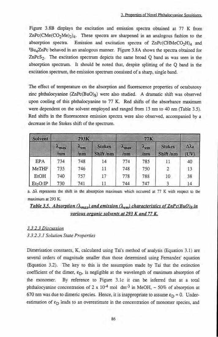
3. Properties of Novel Phthalocvanine Sensiti.sers.
Figure 3.8B displays the excitation and emission spectra obtained at 77 K from ZnPc(CMe(C02Me)2)4. These spectra are sharpened in an analogous fashion to the absorption spectra. Emission and excitation spectra of ZnPc(CHMeC02H)4 and ^Bu4ZnPc behaved in an analogous manner. Figure 3.8A shows the spectra obtained for ZnPcS2. The excitation spectrum depicts the same broad Q band as was seen in the absorption spectrum. It should be noted that, despite splitting of the Q band in the excitation spectrum, the emission spectrum consisted of a sharp, single band.
The effect of temperature on the absorption and fluorescence properties of octabutoxy
zinc phthalocyanine (ZnPc(BuO)g) were also studied. A dramatic shift was observed
upon cooling of this phthalocyanine to 77 K. Red shifts of the absorbance maximum
were dependent on the solvent employed and ranged from 13 nm to 40 nm (Table 3.5).
Red shifts in the fluorescence emission spectra were also observed, accompanied by a
decrease in the Stokes shift of the spectrum.
EPA
MeTHF
EtOH
EtoOrIP
734
735
740
730
748 746 757 741
14
11
17
11
774
748
778
744
785 750 788 747
11
2
10
3
40 13 38 14
a. AX represents the shift in the absorption maximum which occurred at 77 K with respect to the
maximum at 293 K.
Table 3 J. Absorption (X^g^f) and emission (X^tn) characteristics ofZnPc(BuO)g in
various organic solvents at293 K and 77 K.
3.3.2.3.1 Solution State Properties
Dimerisation constants, K, calculated using Tai's method of analysis (Equation 3.1) are
several orders of magnitude smaller than those determined using Fernandez' equation
(Equation 3.2). The key to this is the assumption made by Tai that the extinction
coefficient of the dimer, ep, is negligible at the wavelength of maximum absorption of
the monomer. By reference to Figure 3.1c it can be inferred that at a total
phthalocyanine concentration of 2 x 10" mol dm ^ in MeOH, ~ 50% of absorption at
670 nm was due to dimeric species. Hence, it is inappropriate to assume = 0. Under
estimation of Ej) leads to an overestimate in the concentration of monomer species, and
86
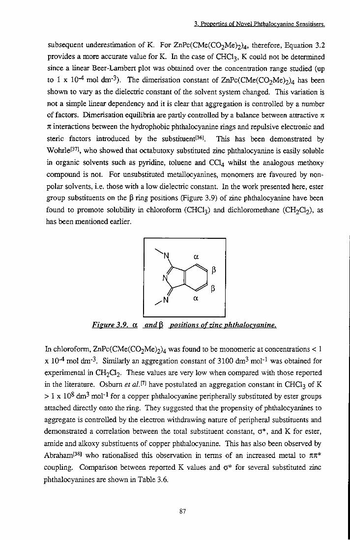
3. Properties of Novel Phthalocvanine Sensitisers.
subsequent underestimation of K. For ZnPc(CMe(C02Me)2)4, therefore. Equation 3.2 provides a more accurate value for K. In the case of CHCI3, K could not be determined since a linear Beer-Lambert plot was obtained over the concentration range studied (up to 1 X 10- mol dm-3). The dimerisation constant of ZnPc(CMe(C02Me)2)4 has been shown to vary as the dielectric constant of the solvent system changed. This variation is not a simple linear dependency and it is clear that aggregation is controlled by a number of factors. Dimerisation equilibria are partly controlled by a balance between attractive K n interactions between the hydrophobic phthalocyanine rings and repulsive electronic and steric factors introduced by the substituentt^^l. This has been demonstrated by WohrleP'''!, who showed that octabutoxy substituted zinc phthalocyanine is easily soluble in organic solvents such as pyridine, toluene and CCI4 whilst the analogous methoxy compound is not. For unsubstituted metallocyanines, monomers are favoured by non-polar solvents, i.e. those with a low dielectric constant. In the work presented here, ester group substituents on the P ring positions (Figtire 3.9) of zinc phthalocyanine have been found to promote solubility in chloroform (CHCI3) and dichloromethane (CH2CI2), as has been mentioned earlier.
\ a
h V a
Figure 3.9. a and P positions o f zinc phthalocvanine.
In chloroform, ZnPc(CMe(C02Me)2)4 was found to be monomeric at concentrations < 1 X 10"^ mol dm"3. Similarly an aggregation constant of 3100 dm^ mol'^ was obtained for
experimental in CH2Q2- These values are very low when compared with those reported
in the literature. Osbum et alS'^^ have postulated an aggregation constant in CHCI3 of K
> 1 X 10^ dm^ mol"^ for a copper phthalocyanine peripherally substituted by ester groups
attached directiy onto the ring. They suggested that the propensity of phthalocyanines to
aggregate is controlled by the electron withdrawing nature of peripheral substituents and
demonstrated a conrelation between the total substituent constant, a*, and K for ester,
amide and alkoxy substituents of copper phthalocyanine. This has also been observed by
Abraham[38] who rationalised this observation in terms of an increased metal to KK* coupling. Comparison between reported K values and a* for several substituted zinc
phthalocyanines are shown in Table 3.6.
87

3. Properties of Novel Phthalocvanine Sensitisers.
ssBSBSgw ,m • .-m... I.. .. • • •.' '.••SBSwggg;;wag
Substituent ' K Solvent Ref.
O-CO2BZ 0.34 -0.45 -0.11 1300 CHCI3 [7] -CioH2i -0.05 -0.14 -0.19 Monomeric to C6H12 [21]
1.5 X 10⁻⁴ mol dm-3 -C-(CH3)3
0 -0.07 -0.13 -0.2 2.5 X 10⁴ Toluene [23]
0.24 -0.23 0.01 2.04 X 10⁴ Toluene [23]
0 a
C(CH3)2CH2NH2l' -0.07 -0.13 -0.2 4x 10⁵ THE [23] a 0.47 -0.23 0.24 6x 10³ DMSO [40] Br 0.44 -0.19 0.25 4.7 X 10⁴ DMSO [40] I 0.39 -0.16 0.23 2.4 X 10⁴ DMSO [40]
a. Estimated as NHCOCH3, b. Estimated as €(0113)3. Table 3.6. Collation of substituent constant, a* and the dimerisation constant. K of
SUpstitiitcdZnPc'^r
These results show that many other factors besides a*, such as steric hindrance, specific interactions between substituents, and the presence of axial ligands to the metal must be taken into consideration. Amine groups have been shown to increase the aggregation tendencies of ZnPc in THE relative to those of butyl substituted ZnPc in toluene due to intermolecular hydrogen bonding between peripheral substituentst si. THE has the propensity to act as a ligand to the metal ion of metallophthalocyanines, inhibiting aggregation. Therefore, two competing effects must be considered: viz. enhanced aggregation due to hydrogen bonding interactions between substituents, and inhibition of aggregation due to the presence of an axial ligand which introduces steric hindrance to the approach of two molecules, a* for these substituents is not expected to be significantiy different. Halide substitutiont'*°l inhibited aggregation in the order Q > I > Br, though their effective substituent constants are approximately equal. Some other influence must, therefore, be responsible for measured differences in K. It is suggested that the low propensity of ZnPc(CMe(C02Me)2)4 to aggregate is a consequence of the extremely bulky nature of the diester substituents, introducing steric hindrance to the approach of two molecules in a similar fashion to the action of axial ligation. In addition, electronic effects which may promote aggregation due to the electron withdrawing natm-e of ester groups (a* = 0.5^^0 are reduced since the ester groups are not attached directly to the phthalocyanine but are removed by an alkyl linking unit (-CHMe-). In toluene and methanol, K is greater. This illustrates two different properties. Toluene is a non-polar solvent and ester groups are less soluble in this type of solvent than in CHCI3
88

3. Properties pf Novel Phthalocyanine Sensirisers.
or CH2Q2- Thus, unfavourable substituent/solvent interactions promote aggregation in a comparable manner to dimerisation promoted by hydrophobic alkyl chain substituentsf'*'! in aqueous solution. In methanol, unfavourable phthalocyanine ring/solvent interactions become the dominant factor so attractive 7i7t Van-der-Waals interactions outweigh favourable ester/solvent interactions and steric factors preventing association.
In the case of, ZnPc(CHMeC02H)4, the hydrophilic nature of these substituents favours
interaction with polar solvents, hence, this phthalocyanine is soluble in organic media
with high dielectric constants such as MeOH. Hydrogen bonding between carboxyl
groups and solvent molecules is possible, thus aiding solvation. An extreme sensitivity to
the ionic strength of the solution was observed. Many reports have been published
commenting on increased aggregation of ionic porphyrinsf'^^J/phthalocyaninest'i^ in
aqueous ionic media, however, little has been mentioned regarding the effect of ions in
organic systems. Indeed, Pastemack et a/.t' J have shown that addition of acetone to a
0.1 mol dm-3 ionic strength aqueous solution of nickel tetracarboxyphenylporphyrin
counteracts the electrolyte effect and shifts the dimerisation equilibrium towards the
monomeric form. Interestingly, the analogous zinc porphyrin was observed to remain
monomeric in 0.1 mol dm"^ salt solution. This was ascribed to a combination of two
effects; the presence of an axial ligand to the metal and non-planarity of the poiphyrin
ring. Dimerisation is enhanced due to ion pairing between the electrolyte and porphyrin
substituents.
A similar effect is likely to be responsible for aggregation of ZnPc(CHMeC02H)4 in
etiianol by Na+, K+, Mg2+, Zn2+ or tBu4N+ ions, irrespective of tiie nature of tiie ions.
The concentrations of electrolyte concerned (-10"^ mol dm'^) are considerably less than
those reported['*'']'t'*5] in aqueous solution (0.01 mol dm-3 or greater). In fact, with the
exception of tetraalkylammonium salts, monomeric ZnPc(CHMeC02H)4 species were
observed at ionic strengths of this magnitude ia ethanol. This behaviour illustrates the
complexities of aggregation phenomena. In aqueous solution, substituent groups, in this
case carboxylate ions, are hydrated. That is, they are surrounded by a solvation shell of
ordered water molecules. This has two conflicting effects. Firstiy, hydration reduces the
effective negative charge of carboxylate ions, therefore, ionic repulsion effects will be
lessened[''^l Secondly, acid groups are protected from ions in the bulk media. Of
course, electrolyte ions will also be highly solvated, e.g. the free energy of hydration!'*' ],
of Na+ ions is 375 kJ mol"! and that of Ca2+ is 1584 kJ mol-^. Hence large
concentrations of ions are necessary for ion pairing to occur. In non-aqueous solution,
ion solvation energies are lowert"* ] with respect to water, for example, the free energy
89
3. Properties of Novel Phthalocvanine Sen.sitisers.
change, AG^., on transferring a ^Bu4N+ ion from water to ethanoll"*'! is -1-I8 kJ mol'^ As a consequence of tiieir reduced solvation, ions also have a greater activity in nonaqueous solution - it follows that association with other species will occur far noore readily in these systems. Pairing of two entities is an entropically favourable process despite a reduction in the number of free species in solutionl oj. This is due to disruption of the solvation shells about both species concerned. The water/solvent molecules forming the solvation shell become more disordered as molecules are released into the bulk solution resulting in an overall increase in the entropy of the system (AS). The same effect is achieved by aggregation, in which two faces of monomaric dye molecules, previously in contact widi solvent molecules in the solvation shell, are lost Thus, aggregation of phthalocyanine molecules paired with metal ions is partially driven by energetically favourable entropy terms. An additional driving force for aggregation is the decrease in the diffusion coefficients of dyes which results fijom an increase in the electrolyte concentrationt il.
Disaggregation at concentrations of 0.01 mol dm-3 may also be explained using entropic
considerations. It has previouslyt j been observed that tetramethylammonium salts
induce the formation of Acridine Orange dimers in aqueous solution up to an ionic
strength of 0.3 mol dm-3 due to screening of positive charges on the dye by counter
anions. At greater concentrations, the aggregation constant started to decrease. This
was ascribed to 'structure breaking' characteristics of the salt. It was thought that
methylammonium ions acted to break apart the solvation shell of the dye. In this way
favourable entropy terms associated with dimerisation have been removed, AG is less
negative and aggregation becomes less energetically favourable. It is believed that
tetrabutylammonium salts act as structure makers in water[53].[54]. This has been
rationalised in terms of tiie influence of non-polar solutes on cooperative hydrogen bond
formation [5 1. These solutes serve to protect developing clusters from charge
fluctuations which interrupt bonding. Hence solvent order increases.
It is possible to use this argument for the results obtained here. At low concentrations of
electrolyte, aggregation was observed due to association of the carboxylic substituents of
the phthalocyanine and metal ions. However, at high concentrations (~ 0.01 mol dm^ ),
ordering of the ethanol solvent shell around phthalocyanine molecules is disrupted due to
tiie structure breaking properties of Na+, K+ and CIO4- ions, the driving force for
aggregation is removed, and ZnPc(CHMeC02H)4 becomes monomeric once more. By
contrast, ^U4N+a- and ^Bu4N+C104- salts increase ordering, thus disaggregation was
not observed. In the latter case, ordering by cations and disruption by anions act
competitively. It would appear that the ordering effect of the butylammonium ions is
90

3. Properties of Novel Phthalocyanine Sensitisers.
greater. It should be noted that the effect of ions on the ordering of non-aqueous solvents is generally less than that of water. Two dimensional hydrogen bonding, such as may occur in alcoholic solution, is less influenced by solute than the three dimensional H-bonding that exists in watert^s]. This is because non-aqueous solvents are iaherentiy less ordered systems than water (entropy of water = 69.9 J K"! mol'^ entropy of ethanol = 160JK-imoH)557].
The properties of ZnPc(CHMeC02H)4 in aqueous solution are very different. The
observed insolubility of the carboxylate in distilled water may be explained by reference
to the acid dissociation constants. Due to the separation of the carboxylate groups from
the phthalocyanine skeleton, phenylacetic acid' *] (pKg of 4.3 at 293 K in water) may be
used as a model for the pK^ of the phthalocyanine carboxylate groups. Accordingly, the
acid groups of ZnPc(CHMeC02H)4, would be expected to remain largely undissociated
in water (pH ~ 4.5-5) and consequentiy have little effect on the overall solubihty of the
phthalocyanine - c.f. ZnPc(CMe(C02Me)2)4. ZnPcS2 is soluble in H2O due to the
greater acidity of the sulfonic acid substituents in comparison to the carboxylate groups
above (pK^ = 2.5)[59]. PBS renders both ZnPc(CHMeC02H)4 and ZnPcS2 soluble. This
buffer has a pH of 7.4, a pH at which both sulfonates and carboxylates are fuUy
dissociated. This explanation is corroborated by the effects observed on altering the pH
of PBS. Alkaline conditions encouraged dissociation of the acidic groups, thus the state
of aggregation was unaffected. At low pH (2) further aggregation occiuxed due to
protonation of carboxylic acid side chains. Phosphate buffer saline (PBS) contains 0.1
mol dm-3 NaCl ions. In aqueous solution, ZnPc(CHMeC02H)4 is dimeric for the
reasons discussed above, i.e. intermolecular ionic repulsions between negatively charged
substituents are screened by hydration shells, favourable entropic considerations and
solvophobic forces[^°l Any structure breaking capacity of Na+ or CI" ions is too weak to
have any effect. CTAB, however, effectively induces monomerisation. Ben-Hur et
<2/.[6i] have used 1% hexadecyl trimethylammonium bromide (CTAB) solution to
monomerise sulfonated metallocyanines however, at this concentration (-0.03 mol
dm-3), monomerisation is most likely to be caused by incorporation into micelles. The
concentrations used in this study (0.02 mmol dm'^) were well below the cmc of CTAB
(0.9 mmol dm-^), therefore, disaggregation is not due to incorporation into micelles.
Hence, this effect may be explained in terms of ion pairing. In contrast to the effect of
ion pairing discussed above, where pairing in ethanol induced dimerisation due to linking
of substituent carboxyl groups by metal cations, here dimerisation is prevented due to
steric inhibitions introduced by pairing of bulky hexadecyl trimethylammonium groups to
the ionic side chains of the phthalocyanine.
91

3. Properties of Novel Phthalocvanine Sensitisers.
3.3.2.3.2 Photophysteal Properties
The effect of solvent on is a complex and involved issue. Foley!''''! reported a blue
shift in the absorption spectrum of AIPCS2 with increasing dielectric constant whilst
Harazonot*2] ^1. observed a red shift in the S^ <- SQ transition of titanium (IV) oxide
phthalocyaninate (TiOPc) as solvent polarity and dipole moment increased. It is
generally accepted that the wavelength of %%* transitions increases with solvent
polarityf^^J due to a greater stabilisation of the excited state with respect to the ground
state. The results obtained here (Figtire 3.4) show a random distribution of absorption
maxima with dielectric constant and solvent dipole moment. A possible connection
between the electron donating propensity (DN) of the solvent and X^a^ is suggested
whilst a reasonable linear correlation with refractive index was observed. These plots are
illustrative of the complexities encountered when trying to apply a simple trend.
In the case of phthalocyanines, it is not possible to consider purely one type of
intermolecular interaction. Electrostatic forces between solute and solvent, hydrogen
bonding capacity and the potential of the solvent to act as a ligand must all be
considered. Harazono attempted to separate these contributions with dubious
success [ ^1. Refractive index is a measure of the polarisability of a solvent. Limantara et alS^'*^ have demonstrated that the dependency of the and Qy transitions of
bacteriochlorophyll a on the refractive index of polar and non-polar solvents foUows
Equation 3.3. A red shift of absorption maxima was observed with increasing refractive
index.
2 _ j V =-d.R(nj)) + e where R(nD) = - ^ (3.3)
no +2
Application of Equation 3.3 where n^, is the refractive index of the solvent to the data
obtained for ^Bu4ZnPc yielded a plot (Figure 3.10) with a linear fit of gradient, -1900
cm-l (coefficient d) and intercept, 15300 cm'l (coefficient e). This is in good agreement
with Limantara's values of -2333 cm-l and 17680 cm-l (d and e respectively) for
bacteriochlorophyll a in polar solvents.
92

3. Properties of Novel Phthalocyanine Sensitisers.
14950
/cm'
14900 4-
14850
14800 4-
14750 4-
14700
y = -1900x+ 15300
-+- + 0.19 0.21 0.23 0.25 0.27 0.29 0.31
R(nD)
Figure 3.10. Relationship between solvent refractive index and X^^^^ (UV/Vis.) of ^Bu^ZnPc.
The effect of substituents on the position of the Q band absorption may be rationalised in
terms of their propensity to donate electrons to the % system of the phthalocyanine ring.
It has been shown experimentally that electron donating substituents on the
phthalocyanine rings cause red shifts in the absorption spectrum of ZnPct . The
magnimde of the shift was reported to depend on the site of peripheral substitution in
addition to the strength of the donor. This was corroborated by Ford et a/.t ^ in 1989
who demonstrated that the absorption spectrum of a substituted octabutoxyzinc
phthalocyanine was shifted -70 nm to longer wavelengths with respect to ZnPc in tiie
same solvent. In contrast, octabutoxy substimtion in the |3 positions had litde effect on
the position of ax ^ ' ^ - ^ he compounds investigated in this study, the octadecyl
substituents of CIO were observed to have the largest effect, inducing a red shift of 33
run with respect to the parent phthalocyanine. This is in agreement with a previous
studyt ' J of the basic photophysics of CIO. Alkyl chains are known to act as electron
donors when attached to a 7t electron systemt^^l A theoretical explanation for these
effects has recentiy been published by Morley et al\^''\ The Q band of phthalocyanine
molecules is a TCTC* transition or HOMO to LUMO transition. Substituent donors can
stabilise the LUMO by donating electrons to the nitrogen atoms of the ring, a and P
substituents both stabilise in this way, however, a substituents are more effective, thus a
greater decrease in the transition energy is observed. This explains the smaller shift of
^U4ZnPc with respect to CIO since the butyl groups were located in the (3 positions.
Electron withdrawing groups may also be expected to induce a small red shift.
93

3. Properties pf Npvgl Phthglocygnine Sensitisers,
Peripheral substitution had Uttie effect on the propensity of ZnPc's to produce singlet oxygen. This corroborates results by Fernandez et a/.P^l who found tiiat substiuition by nitrogen containing substiments neither enhanced nor depleted the yield of triplet state and singlet oxygen formation by ZnPc. Similarly, Ben-Hurt^^] has reported that sulfonate group substitution of aluminium phtiialocyanine had no effect on its photophysical properties. In this smdy, the fluorescence, triplet and singlet oxygen quantum yields remained approximately constant, with the notable exceptions of ZnPc(CHMeC02H)4 in 0.02 mmol dm-3 CTAB/PBS and CIO. In these cases, a dramatic decrease in <I>p from 0.25 to 0.06 and 0.082 respectively was observed. For ZnPc(CHMeC02H)4 in 0.02 mmol dm"3 CTAB/PBS the most reasonable explanation for tiiis and the low value of Op (0.18) is tiie presence of B r , a heavy atom which wiU act to quench die excited state. A comparable value of <t>p was measured by Valdugat^^l for ZnPc in CTAB miceUes. CIO displayed a concurrent increase in (0.8) with respect to unsubstituted ZnPc, suggesting that kjs^ has increasedf ' ].
3.3.2.3.3 Low Temperature Measurements
Sharpening of absorption and fluorescence spectra on lowering the temperature may be
attributedf^^! to a decrease in the thermal motion of surrounding solvent molecules,
leading to a reduced inhomogeneous broadening of the transition. Shifts in
more difficult to explain. Cupane et alP^^ observed a red shift upon cooHng a solution
of Ni(II) octaethyl porphyrin to 100 K. This was ascribed to an equilibrium between
planar and non-planar macrocyclic structures, shifting towards the lower energy non-
planar conformation at low temperatures. Phthalocyanines are remarkably planar in
structure, non-planarity only having been observed in special casest""], therefore, this
scenario is viewed as rather unlikely for this class of compounds. The hypothesis of
Hoshino et a/.f''^] ^o ascribe a shift of dimeric chlorophyll a from 695 nm to 706 nm on
cooling from 160 K to 77 K, to a change in configuration of the dimer is more
reasonable. It is feasible that rearrangement of solvent molecules around the
phthalocyanine and/or peripheral substituent configuration changes at low temperature
may be responsible for the shifts observed here. This has been demonstrated by the
effect of temperature on (BuO)8ZnPc {vide infra). Large red shifts in the absorption
spectrum upon cooling indicate that the nature of the solvent and potential interactions
with peripheral substituents of the phthalocyanine ring are very important. Shifts in EPA
and EtOH of ~ 40 nm suggest that stabilisation of the LUMO occurred. The most hkely
cause of this is formation of hydrogen bonds between solvent molecules and oxygen
atoms of the substituent. This is corroborated by behaviour in aprotic solvents, MeTHF
and Et20:IP, in which hydrogen bonding is impossible and far smaller shifts were
94

3. Properties of Novel Phthalocvanine Sensitisers.
obtained (AA, = 13 nm and 14 nm respectively). Not surprisingly, low temperatures resulted in a smaller Stokes shift of emission. This stems from reduced rotational deactivation pathways at 77 K due to the rigidity of the solvent glass which inhibits molecular motion. More interesting is the solvent dependency of tiie magnitude of the change. A significantiy larger decrease was observed in aprotic solvents (11 nm 2 nm) than in protic solvents (14 nm 11 nm) whilst the absolute value was larger for protic solvents, i.e. those capable of hydrogen bonding. Hydrogen bonding to the chromophore increases the number of vibrational deactivation modes available and hence facilitates energy loss to surrounding molecules. Protic solvents will therefore cause a larger decrease in the energy of emission with respect to absorption. At low temperatmres, the number of hydrogen bonds increased whilst vibrational motion is reduced. These conflicting effects mean that tiie Stokes shift is not reduced as significantiy in protic solvents as in aprotic solvents where the only process to be considered is that of reduced vibrational deactivation pathways.
Sheppard et al.^^^^ suggested that increased polarity of solvent molecules at low
temperature was responsible for a red shift in the absorption spectrum of non-ionised
merocyanine. It is generally accepted that Tin* transitions are red shifted with increased
solvent polarity, suggesting that this may be a feasible explanation; however, it has
already been shown that tiie Sj <- SQ transition energy of zinc phthalocyanines does not
exhibit straight forward behaviour with solvent polarity. The effect of temperature on
the refractive index of ethanolf'^l may be described by Equation 3.4.
^ = -3.7x10^ K ' (3.4) dl
where is the refractive index and T is the absolute temperature. dnjydT is negative
for most solvent media and demonstrates that as the temperamre of a solution is
lowered, the refractive index increases. Hence, by reference to Figures 3.4 and 3.10
which show a shift in X^^^^^ with TIQ, a bathochromic shift of X^^^soi ^ predicted. The
red shifts of ^Bu4ZnPc and ZnPc(CMe(C02Me)2)4 may be explained by this theorem.
ZnPc(CHMeC02H)4 and ZnPcS2 show an opposite shift suggesting a different
interaction which causes destabilisation by solvent molecules at low temperature.
The appearance of an absorption peak at 625 nm in the low temperature spectrum of
ZnPc(CMe(C02Me)2)4 (Figure 3.7D) may be ascribed to dimeric phtiialocyanine
species. Lack of additional emission at 77 K and the absence of this peak in an excitation
95
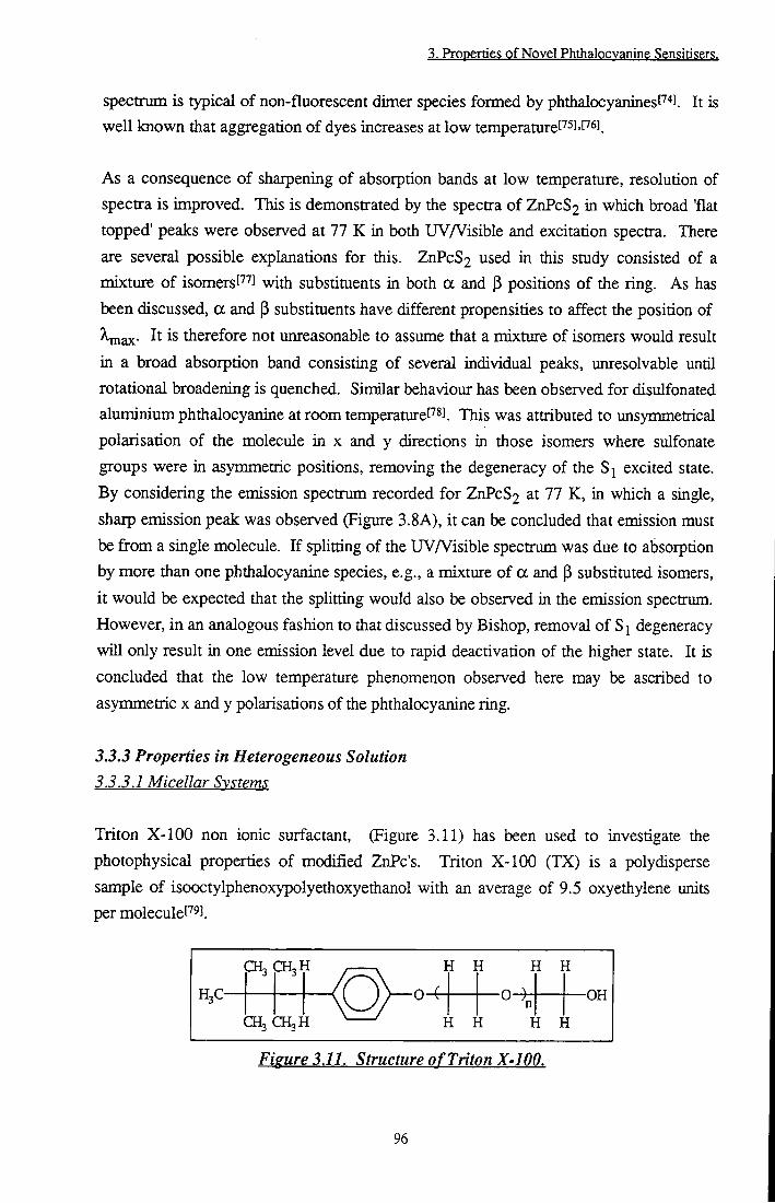
3. Properties of Novel Phthalocvanine Sensitisers.
spectrum is typical of non-fluorescent dimer species formed by phthalocyaninesf'''*!. It is well known that aggregation of dyes increases at low temperatureI''5J'C'' l.
As a consequence of sharpening of absorption bands at low temperature, resolution of spectra is improved. This is demonstrated by the spectra of ZnPcS2 in which broad 'flat topped' peaks were observed at 77 K in both UVA^isible and excitation spectra. There are several possible explanations for this. ZnPcS2 used in this study consisted of a mixture of isomersf' l with substituents in both a and (3 positions of the ring. As has been discussed, a and P substituents have different propensities to affect the position of \nQx. It is therefore not unreasonable to assume that a mixture of isomers would result in a broad absorption band consisting of several individual peaks, unresolvable until rotational broadening is quenched. Similar behaviour has been observed for disulfonated aluminium phthalocyanine at room temperaturef"'*!. This was attributed to unsymmetrical polarisation of the molecule in x and y directions in those isomers where sulfonate groups were in asymmetric positions, removing the degeneracy of the excited state. By considering the emission spectrum recorded for ZnPcS2 at 77 K, in which a single, sharp emission peak was observed (Figure 3.8A), it can be concluded that emission must be from a single molecule. If splitting of the UVA^isible spectrum was due to absorption by more than one phthalocyanine species, e.g., a mixture of a and p substituted isomers, it would be expected that the splitting would also be observed in the emission spectrum. However, in an analogous fashion to that discussed by Bishop, removal of degeneracy will only result in one emission level due to rapid deactivation of the higher state. It is concluded that the low temperature phenomenon observed here may be ascribed to asynraietric x and y polarisations of the phthalocyanine ring.
3.3.3 Properties in Heterogeneous Solution 3.3.3,1 Micellar Systems
Triton X-100 non ionic surfactant, (Figure 3.11) has been used to investigate the photophysical properties of modified ZnPc's. Triton X-100 (TX) is a polydisperse sample of isooctylphenoxypolyethoxyethanol with an average of 9.5 oxyethylene units per moleculef' J.
CH, CH,H
H3C-
CH3 CH3H
I (
i ] i ]
H H
-OH
H H
Fieure 3.11. Structure of Triton X-100.
96
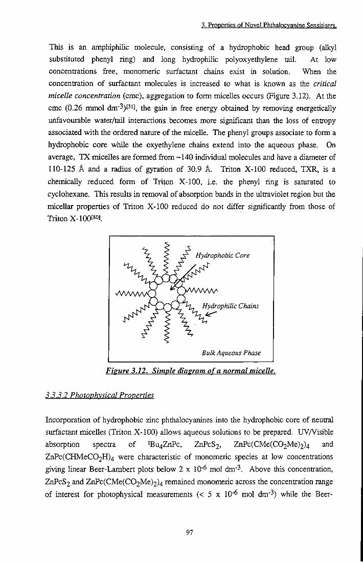
3. Properties of Novel Phthalocvanine Sensitisers.
This is an amphiphilic molecule, consisting of a hydrophobic head group (alkyl substituted phenyl ring) and long hydrophilic polyoxyethylene tail. At low concentrations free, monomeric surfactant chains exist in solution. When the concentration of surfactant molecules is increased to what is known as the critical micelle concentration (cmc), aggregation to form micelles occurs (Figure 3.12). At the cmc (0.26 mmol dm"3)f3i], the gain in free energy obtained by removing energetically unfavourable water/tail interactions becomes more significant than the loss of entropy associated with the ordered nature of the micelle. The phenyl groups associate to form a hydrophobic core while the oxyethylene chains extend into the aqueous phase. On average, TX micelles are formed from -140 individual molecules and have a diameter of 110-125 A and a radius of gyration of 30.9 A. Triton X-100 reduced, TXR, is a chemically reduced form of Triton X-100, i.e. the phenyl ring is saturated to cyclohexane. This results in removal of absorption bands in the ultraviolet region but the micellar properties of Triton X-100 reduced do not differ significantly from those of Triton X-100[80].
Hydrophobic Core
Hydrophilic Chains
Bulk Aqueous Phase
Figure 3.12. Simple diagram of a normal micelle.
3.3.3.2 Photophysical Properties
Incorporation of hydrophobic zinc phthalocyanines into the hydrophobic core of neutral surfactant micelles (Triton X-100) allows aqueous solutions to be prepared. UVA isible absorption spectra of ^Bu4ZnPc, ZnPcS2, ZnPc(CMe(C02Me)2)4 and ZnPc(CHMeC02H)4 were characteristic of monomeric species at low concentrations giving linear Beer-Lambert plots below 2 x 10" mol dm-3. Above this concentration, ZnPcS2 and ZnPc(CMe(C02Me)2)4 remained monomeric across the concentration range of interest for photophysical measurements (< 5 x 10⁻⁶ mol dm-3) while the Beer-
97
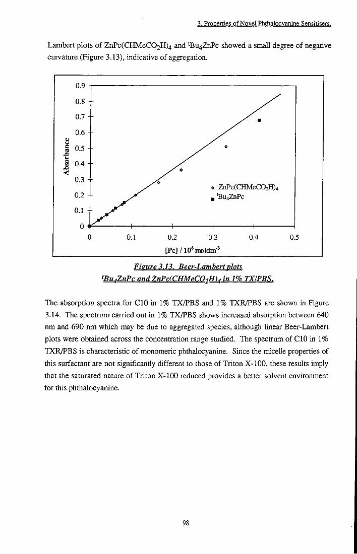
3. Properties of Novel Phthalocvanine Sensirisers.
Lambert plots of ZnPc(CHMeC02H)4 and tBu4ZnPc showed a small degree of negative curvature (Figure 3.13), indicative of aggregation.
o ZnPc(CHMeC02H)4 'Bu4ZiiPc
0.1 0.2 0.3
[Pc]/10*moldm-^
0.4 0.5
Figure 3.13. Beer-Lambert plots tBu^ZnPc and ZnPc(CHMeC02H)^ in 1% TX/PBS.
The absorption spectra for CIO in 1% TX/PBS and 1% TXR/PBS are shown in Figure 3.14. The spectrum carried out in 1% TX/PBS shows increased absorption between 640 nm and 690 nm which may be due to aggregated species, although linear Beer-Lambert plots were obtained across the concentration range studied. The spectrum of CIO in 1% TXR/PBS is characteristic of monomeric phthalocyanine. Since the micelle properties of this surfactant are not significantly different to those of Triton X-lOO, these results imply that the saturated nature of Triton X-100 reduced provides a better solvent environment for this phthalocyanine.
98
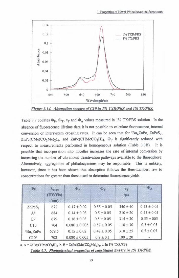
3. ftooerties of Novel Hitlialocvamne Sensitisers.
0.14 r
0.12
0.1 -
3 0.08 -
0.06 -<
0.04 -
0.02
1%TXR/FBS
Fieure 3.14. Absorption spectra of CIO in 1% TXR/PBS andl% TX/PBS.
Table 3.7 collates Op, and O^ values meaaired in 1% TX/PBS solution. In the
absence of fluorescence lifetime data it is not possible to calculate fluorescence, internal conva-sion or intersystem crossing rates. It can be seen that for •Bu4ZnPc, ZnPcS2, ZnPc(CMe(C02Me)2)4, and ZnPc(CHMeC02H)4, % is significantly reduced with respect to measurements performed in homogeneous solution (Table 3.3B). It is possible that incorporation into micdiles increases the rate of internal convosion by increasing the number of vibrational deactivation pathways available to the fluorophore. Alternatively, aggregation of phthalocyanines may be responsible. This is unlikely, however, since it has been shown that absorption follows the Beer-Lambert law to concentrations far greater than those used to determine fluorescence yields.
Pc (uv/vis)
/nm)
T t / s
ZnPcS2 672 0.17 ±0.02 0.55 ±0.05 340 ±40 0.53 ±0.05 Aa 684 0.14 ±0.03 0.5 ±0.05 210 ±20 0.55 ±0.05
Eb 679 0.16 ±0.03 0.5 ±0.05 315±30 0.55 ±005
CIO 704 0.080 ±0.005 0.57 ±0.05 110±30 0.5 ±0.05
tBu4ZnPc 678.5 0.15 ±0.02 0.48 ±0.05 310 ±25 0.5 ±0.05 702 0.080 ± 0.005 0.8 ± 0.1 100 ± 20 -
a. A = ZnPc(CHMeC02H)4, b. E = ZnPt(CMe(C02Me)j)4, c. In 1% TXR/PBS
Table 3.7. Photophvsical properties of substituted ZnPc's in 1% TX/PBS.
99
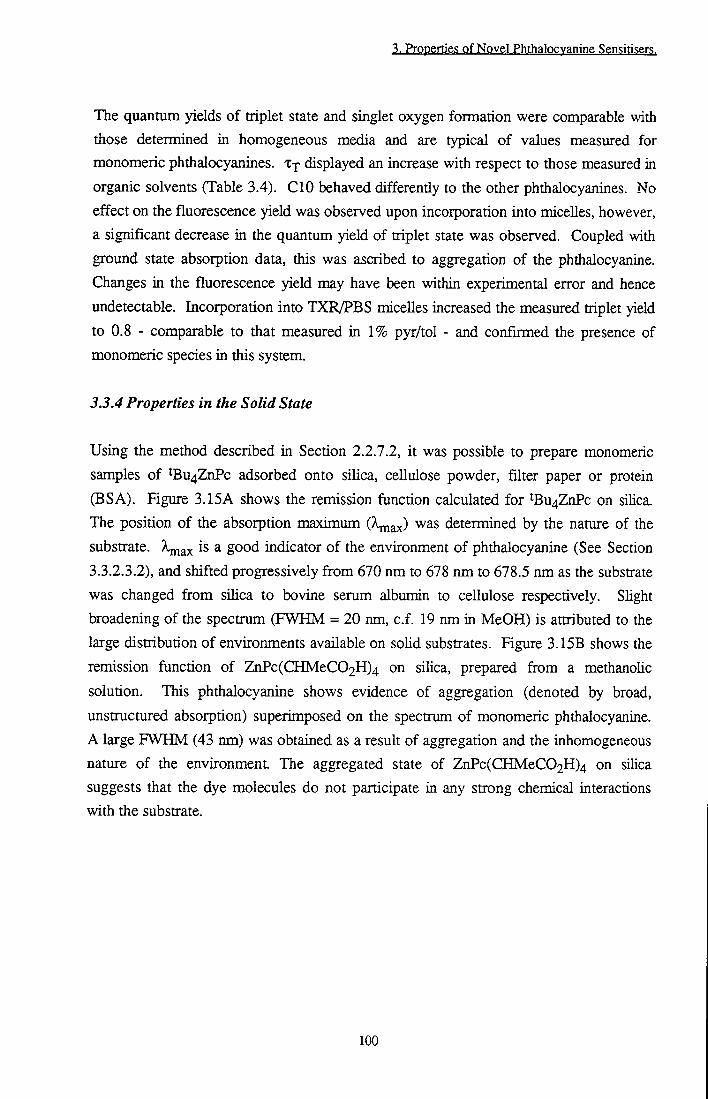
3. Properties of Novel Phthalocvanine Sensitisers.
The quantum yields of triplet state and singlet oxygen formation were comparable with those determined in homogeneous media and are typical of values measured for monomeric phthalocyanines. Xj displayed an increase with respect to those measured in organic solvents (Table 3.4). CIO behaved differentiy to the other phthalocyanines. No effect on the fluorescence yield was observed upon incorporation into micelles, however, a significant decrease in the quantum yield of triplet state was observed. Coupled with ground state absorption data, this was ascribed to aggregation of the phthalocyanine. Changes in the fluorescence yield may have been within experimental error and hence undetectable. Incorporation into TXR/PBS micelles increased the measured triplet yield to 0.8 - comparable to that measured in 1% pyr/tol - and confirmed the presence of monomeric species in this system.
3.3.4 Properties in the Solid State
Using the method described in Section 2.2.7.2, it was possible to prepare monomeric samples of ^Bu4ZnPc adsorbed onto silica, cellulose powder, filter paper or protein (BSA). Figure 3.15A shows the remission function calculated for tBu4ZnPc on sUica. The position of the absorption maximum (kjnax) was determined by the nature of the substrate. X ^ x is a good indicator of the environment of phthalocyanine (See Section 3.3.2.3.2), and shifted progressively from 670 nm to 678 nm to 678.5 nm as the substrate was changed from silica to bovine serum albumin to ceUulose respectively. Shght broadening of the spectrum (FWHM = 20 nm, c.f. 19 nm in MeOH) is attributed to the large distribution of environments available on solid substrates. Figure 3.15B shows the remission function of ZnPc(CHMeC02H)4 on siHca, prepared from a methanolic solution. This phthalocyanine shows evidence of aggregation (denoted by broad, unstructured absorption) superimposed on the spectrum of monomeric phthalocyanine. A large FWUM (43 nm) was obtained as a result of aggregation and the inhomogeneous nature of the environment. The aggregated state of ZnPc(CHMeC02H)4 on silica suggests that the dye molecules do not participate in any strong chemical interactions with the substrate.
100

3. Properties of Novel Phthalocvanine Sensitisers.
0.25
0.2 +
0.15 4-
F(R)o.i -
0.05 4-
04
A 670 nm
1
' B 680 nm
...1 ^ - i f " ^ J ..... 1
300 400 500 600 Wavelength/nm
700
Figure 3.15. Remission functions of phthalocyanines on silica. A. tBu^ZnPc. B. ZnPc(CHMeC02Hh. Spectra have been offset for clarity.
A 672nm686nm
a / \ f \ b 638 nm
786 nm 820 nm
610 nm
550 600 650 700 750 800 850 Wavelength/nm
Figure 3.16. A. Excitation (a) and emission (b) spectra of^Bu^ZnPc on cellulose (Xcm - ^cx ~ Corrected for artefacts. Spectra have been offset
for clarity.
101

3. Propertie? pf Npvel Phthalpcyaning $en$iti?er?.
The emission and excitation spectra obtained from a 'dilute' (~6 jig g" ) sample of ^Bu4ZnPc on cellulose are shown in Figure 3.16. Unexpected peaks were observed at 638 nm, 786 nm and 820 nm in the emission spectrum and 610 nm in the excitation spectrum. The intensity of these peaks relative to phthalocyanine emission was reduced by increasing the concentration of the sample. It was concluded, therefore, that these peaks arose due to an artefact rather than the behaviour of phthalocyanine
The source of these 'emission' bands was investigated. It was found that the positions of the peaks were independent of the substrate used, ruling out fluorescence or absorption artefacts of cellulose. When an empty sample holder was used (with quartz plate) these peaks were still observed, but when the same holder was used without a quartz plate, i.e. when teflon was the only scattering surface, a flat baseline was obtained. It was concluded that 'emission' originated from either the quartz window or an inherent impurity within it. Variation of the excitation wavelength or monitored emission wavelength caused peaks at 610 nm (P ) and 638 nm (P2) to shift (Figure 3.16A). The position of emission peaks at 786 nm and 820 nm remained constant. Clearly, spurious peaks are due to two different phenomena. It is probable that static peaks were caused by fluorescence emission from impurities in the quartz. The same difficulty was encountered whilst making singlet oxygen measurements (Appendix A). The energy difference, AEj, between ^ax(Pl) monitored emission wavelength, Pign,, was constant at 2700 ± 200 cm'l. AE2, the energy difference between \nax(^2)
wavelength of excitation, X^^, was 2850 ± 200 cm'l. AEj = AE2. It is tentatively suggested that this phenomenon may be due to a previously unreported Raman band of quartz or of an impurity therein. Spectra were corrected by scanning a 'blank', i.e. substrate only, as a background signal with subsequent subtraction from sample spectra. In this way, excitation and emission spectra resembhng solution state, monomeric phthalocyanine were obtained (Figure 3.16B).
Transient absorption decays obtained by diffuse reflectance flash photolysis fitted well to monoexponential decay curves OFigure 3.17), yielding lifetime values of 90 |is + 15 |j.s on silica and 130 |is ± 20 p.s on cellulose. These lifetimes are slightiy reduced compared with the lifetime observed in degassed homogeneous solution (170 |is). The decay of ^Bu4ZnPc on a cellulose substrate had a longer lifetime than that of phthalocyarune adsorbed on silica. There have been many reports of extended triplet state lifetimes of molecules adsorbed on cellulosef J'f ] Cellulose protects samples from oxygen quenching due to the rigidity of the cellulose matrix and poor mobility of oxygen molecules through dry celluloset ^ . This suggests that complete deoxygenation of the sample was not achieved. The transient absorption spectrum was centred at 490 nm,
102

3. Properties of Novel Phthalocvanine Sensitisers.
consistent with triplet species in solution and was independent of delay time (Figure 3.18).
0.01 +
e! 0.00
Time/|xs
Figure 3.17. a. Typical diffuse reflectance transient decay, 'kpj-ptc ~ = 355 nm. b. Residuals of monoexponential fit (offset for clarity).
0.025
0.02 +
I 0.015
< I 0.01 c 93
0.005 +
390 440 490 540 Wavelength/nm
590
Figure 3.18. Diffuse reflectance transient absorption spectrum of^Bu^ZnPc on cellulose, a. At = 5 \LS, b. At = 50 \is, c. At = 200 \is.
103

3. Properties of Novel Phthalocvanine Sensitisers.
Hence, it is possible to incorporate substituted zinc phthalocyanines onto solid substrates whilst retaining photophysical activity.
3.4 Conclusion
Two novel, substituted zinc phthalocyanines, ZnPc(CMe(C02Me)2)4 and ZnPc(CMe(C02H)4, have been synthesised. The photophysical properties, <I>p, <I)j, X j and OA , of ZnPc(CMe(C02H)4 and ZnPc(CMe(C02Me)2)4, are typical of substituted
zinc phthalocyanines making these ideal candidates as sensitisers for photodynamic therapy. ZnPc(CMe(C02Me)2)4 displays remarkably monomeric character in CHCI3 due to the bulky nature of its peripheral substituents, whilst ZnPc(CMe(C02H)4 was soluble in aqueous solution at physiological pH. Such solubility properties are important for intravenous administration in therapeutic applications, however in EtOH solution aggregation of this phdialocyanine displayed an unusual dependence on ionic strength. Aggregation was found to be promoted by ion concentrations between zero and 5 x 10" mol dm-3, while disaggregation occurred at concentrations > 10' mol dm^ . It is likely that ion pairing between acid substituents and cations results in aggregation at low ionic strengths. At higher ionic strengths, 'structure breaking' of solvation shells suixounding phthalocyanine removed energetically favourable entropic factors of aggregation, leading to regeneration of monomeric phthalocyanine species. It has also been shown that disaggregation in PBS can be induced by 0.02 mmol dm"3 CTAB due to steric inhibitions introduced by ion pairing. Considerable ion effects such as these may have important consequences in vivo where ion transport across cell membranes is a vital biological function' '*]. Certain sensitisers are known to locate in the cell membranel l In the case of ZnPc(CMe(C02H)4, its aggregation state, and consequentiy its efficacy as a sensitiser, may be controlled by the ionic strength of the surrounding medium.
The effect of peripheral substitution of ZnPc by various groups (X) has also been considered. The position of the absorption maximum, ^ g ^ ' found to be dependent on the position of substitution in the ring (a or |3) and the refractive index of the solvent. The photophysical properties of ZnPcX^ were unaffected by the identity of X, differences being observed only in the case of X = C10H21. By contrast, effects of low temperature (77 K) on the absorption and fluorescence spectra of ZnPcXj, were found to be highly dependent on the nature of substituents.
It has been shown that, despite their hydrophobicity, these phtiialocyanines may be incorporated into aqueous micellar systems or adsorbed onto solid substrates without
104

3. Properties of Novel Phthalocvanine Sensitisers.
loss of photoactivity. This has important consequences with respect to incorporation of phthalocyanines into lipid bUayers or onto tumour tissue in vivo where a change in physical (e.g. aggregation) or photophysical (e.g. <I>A, properties would render the
dye less efficient.
3.5 References
[1] Reddi E., Castro G.L., Biolo R. and Jori G., Phaimacokinetic studies with Zn(n) phthalocyanine in tumour bearing mice., Br. J. Cancer, 56, 597-600,1987.
[2] Chan W.S., Marshal J.F., Svensen R., Phillips, and Hart I.R., Photosensitising ability of phthalocyanine dyes screened against tissue cell cultures., Photochem. PhotobioL, 45, 757-761, 1987.
[3] Zhou C, Shunji C, Jinsheng D., Junlin L., Jori G. and Milanesi C, Apoptosis of mouse MS-2, fibrosarcoma cells induced by photodynamic therapy with Zn(n) phthalocyanine., / . Photochem. Photobiol. B.Biol., 33, 219-223,1996.
[4] Van Leengoed H.L.L.M., Cuomo V., Versteeg A.A.C., Van der Veen N., Jori G. and Star W.M., In vivo fluorescence and photodynamic therapy with Zn(II) phthalocyanine., Br. J. Cancer, 69, 840-845, 1994.
[5] Derkacheva V.M. and Luk'yanets E.A., Phthalocyanines and related compounds XVin. Phenoxy- and (phenylthio)-substituted phthalocyanines., / . Gen. Chem. USSR, 50, 1874, 1980.
[6] Mikhalenko S.A., Barkanova S.V., Lebelev D.L. and Luk'yanets E.A., Phthalocyanines and related compounds. IX Synthesis and electronic absorption spectra of tetra-4-t-butylphthalocyanine., / . Gen. Chem. USSR, 41, 2770, 1971.
[7] Osbum E.J., Chau L.K., Chen S.Y., Collins N., O'Brien D.F. and Armstrong N.R., Novel amphiphilic phthalocyanines: Formation of Langmuir-Blodgett and cast thin fihns., Langmuir, 12,4784, 1996.
[8] Koray A.R., Ahsen V. and Bekaroglu O., Preparation of a novel, soluble copper phthalocyanine with crown ether moieties., / . Chem. Sac. Chem. Comm., 932,1986.
[9] Mikhalenko S.A., Derkacheva S.V. and Luk'yanets E.A., Phthalocyanines and related compounds. Tetra and octa aminosubstituted phthalocyanine., / . Gen. Chem. (USSR), 51, 1405, 1981.
[10] Rihter B.D., Kenney M.E., Ford W.E. and Rodgers M.A.J., Synthesis and photoproperties of diamagnetic octabutoxyphthalocyanine with deep red optical absorbance., J.Am. Chem. Soc, 112, 8064, 1990.
[11] Martin P., Gouteraian M., Pepich B.V., Renzoni G.E. and Schindeze D.C., Effects of ligands, solvent and variable sulphonation on dimer formation of aluminium and zinc phthalocyaninesulphonate., Inorg. Chem., 30, 3305-3309, 1991.
105
3. Properties of Novel Phthalocvanine Sensitisers.
[12] Ben-Hur E., Hoeben R.C., Ormondt H.V., Dubbehnan T.M.A.R. and Steveninck J. v . , Photodynamic inactivation of retroviruses by phthalocyanines - the effects of sulfonation, metal ligand and fluoride., / . Photochem. Photobiol. B.Biol., 13, 145-152, 1992.
[13] Allen CM., Weber J.M. and Van Lier J.E., Sulfophthalocyanines for photodynamic inactivation of viruses in blood products - Effect of structural modifications., Photochem Photobiol., 62, 184-189,1995.
[14] Dummin H., Cemay Th., Zimmermann H.W., Selective photosensitisation of mitochondria in HeLa cells by cationic Zn(II) phthalocyanines. with lipophilic side chains., / . Photochem. Photobiol. B.Biol., 37, 219-229,1997.
[15] Rosenthal I . , Ben-Hur E., Greenberg S., Concepcion-Lam A., Drew D.M. and Leznoff C.C., The effect of substituents on phthalocyanine photocytotoxicity., Photochem. Photobiol., 46, 959,1987.
[16] Ford W.E., Rihter B.D., Kenney M.E. and Rodgers M.A.J., Photoproperties of alkoxy substituted phthalocyanines with deep red optical absorbance., Photochem. Photobiol., 50, 277-282,1989.
[17] Dhami S., Photophysics of phthalocyanines in microheterogeneous systems., p56, PhD Thesis, Imperial College, London, 1996.
[18] Valduga G., Reddi E. and Jori G., Steady state and time resolved spectroscopic studies on zinc(n) phthalocyanine in liposomes., / . Photochem. Photobiol. B.Biol., 16, 331-340, 1992.
[19] Polo L., Reddi E., Garbo G.M., Morgan A.R. and Jori G., The distribution of tiie tumour photosensitiser Zn(n) Phthalocyanine and Sn(rV) etiopurpurin among rabbit plasma proteins.. Cancer Lett., 66, 217-223,1992.
[20] Dr. A. Beeby, Unpublished Results. [21] Cook M.J., Chambrier I . , Cracknell S.J., Mayes D.A. and Russell D.A. Octa
aUcyl zinc phthalocyanines: Potential photosensitisers for use in the photodynamic therapy of cancer., Photochem. Photobiol., 62, 542-545, 1995.
[22] Tai S. and Hayashi N., Strong aggregation properties of novel phthalocyanines., /. Chem. Soc. Perkin Trans., 2, 1275, 1991.
[23] Fernandez A., Awruch J. and Dicelio L.E. Photophysical and aggregation studies of tBu4ZnPc's., Photochem. Photobiol., 63, 7S4-792, 1996.
[24] Simpson M.S.C., Beeby A., Bishop S.M., MacRobert A.J., Parker A.W. and Phillips D., Time resolved laser spectroscopy in biochemistry III . , S.PJ.E., 1640, 520, 1992.
[25] Roze M.P., Berzin'sn E.L. and Neiland O.Y., Synthesis of 3, 4 dicyanophenyl malonic esters and their use in the production of soluble phthalocyanines., Zh. Org. Khim., 28, 827, 1992.
106

3. Properties of Novel Phthalocvanine Sensitisers.
[26] Negri R.M., Zalts A., San Roman E.A., Aramendia P.F. and Braslavsky S.E., Carboxylated zinc phthalocyanines: Influence of dimerisation on the spectroscopic properties. An absorption, emission and thermal lensing study., Photochem. Photobiol., 53,311,1991.
[27] Kahl S.B. and Li J., Synthesis and characterisation of a boronated metallophthalocyanine for boron neutron capture therapy., Inorg. Chem., 35, 3878, 1996.
[28] Poucher C.J., The Aldrich Library of Infrared Spectra., Ed. m, Aldrich Chemical Company, Wisconsin, 1981.
[29] Kobayashi N. and Lever A.B.P. Cation or solvent induced supermolecular phthalocyanine formation: Crown ether substituted phthalocyanines., / . Am. Chem. Soc, 109,7433-7441,1987.
[30] Dhami S., DeMeUo A.J., Rumbles G., Bishop G., Bishop S.M., PhiUips D. and Beeby A., Phthalocyanine fluorescence at high concentration: dimers or reabsorption effect?, Photochem. Photobiol., 61, 341,1995.
[31] Kalyansundarum K., Photochemistry in Microheterogeneous Systems, p26, Academic Press, London, 1987.
[32] Vincett P.S., Voight E.M., Rieckhoff K.E., Phosphorescence and fluorescence of phthalocyanines., / . Chem. Phys., 55,4131, 1971.
[33] Phillips D., The photochemistry of sensitisers for photodynamic therapy.. Pure Appl. Chem., 67, 117,1995.
[34] Murov L.S., Carmichael I . and Hug G.L., Handbook of Photochem., p296. Marcel Dekker inc., 2nd Ed., 1993.
[35] It was assumed that V77/V293 for EtOH was equivalent to that reported in Reference 34 forEtOH:MeOH (4:1) solution.
[36] Abraham R.J., Burbridge P.A., Jackson A.H. and MacDonald D.B., The proton magnetic resonance spectra of porphyrins. Part IV. Coproporphyrin tetramethyl esters., / . Chem. Soc. B., 620,1966.
[37] Wohrle D. and Schmidt V., Octabutoxyphthalocyanine, a new electron donor., / . Chem. Soc. Dalton Trans., 549, 1988.
[38] Abraham R.J., Eivazi F., Pearson H. and Smith K.M., TtJt aggregation in metalloporphyrins: Causative factors., / . Chem. Soc. Chem. Comm., 699, 1976.
[39] All values taken fi-om Reference 34 Page 346 except that of O-CO2BZ. This value is contained in Reference 7.
[40] Zhang X.F., Influence of halogenation and aggregation on photosensitising properties of zinc phthalocyanine., / . Chem. Soc. Faraday Trans., 89, 3347,1993.
[41] Wyn-Jones E. and Gormally J., Aggregation Processes in Solution, Studies in Physical and Theoretical Chemistry, 26, 241, Elsevier, New York 1983.
107

3. Properties of Novel Phthalocvanine Sensitisers.
[42] Fukushima Y., Salt effect on the interaction of 22, 24 diprotonated 5,10,15,20 tetrakis (4-sulphonatophenyl) porphyrin with a P sheet structure of a zwitterionic polypeptide.. Bull. Chem. Soc. Jpn., 69, 1719-1726,1996.
[43] Pastemack R.F., Francesconi L., Raff D. and Spiro E., Aggregation of Ni(n), Cu(n) and Zn(II) derivatives of water soluble porphyrins., Inorg. Chem., 12, 2607, 1973.
[44] Hofstra U., Koehorst R.B.M., Schaafsma T.J., Excited state properties of water soluble porphyrin dimers., Chem. Phys. Lett., 130, 555,1986.
[45] Munro O.Q. and Marques H.M., Heme-peptide models for hemoproteins 2) N Acetylmicroperoxidase-8. Study of tiie K-K dimers formed at high ionic strengtii using a modified version of molecular exciton theory., Inorg. Chem., 35, 3768, 1996.
[46] Sheppard S.E. and Geddes A.L., Effect of solvents on the absorption spectra of dyes. IV. Water as solvent: A common pattem., / . Am. Chem. Soc, 66,1995,1944.
[47] Cox B.G., Modern Phase Liquid Kinetics, p46, Oxford Science Publications, New York, 1994.
[48] Parker A.J., The effects of solvation on the properties of anions in dipolar aprotic solvents., Quarterly Reviews, London: The Chemical Society, XVI, 163,1962.
[49] Isaacs N.S., Physical Organic Chemistry, John Wiley and Sons, New York, pl78, 1987.
[50] Nancollas G.H., Interactions in Electrolyte Solutions, Elsevier, London, pi33, 1966.
[51] Wyn-Jones E. and Gormally J., Aggregation Processes in Solution, Studies in Physical and Theoretical Chemistry, 26, 250, Elsevier, New York 1983.
[52] Robinson B.H., Loffler A. and Schwarz G., Thermodynamic behaviour of Acridine Orange in solution., / . Chem. Soc. Faraday Trans., 56,1973.
[53] Coetzee J.F. and Ritchie CD., Solute-Solvent Interactions, 1, p385, Marcel-Dekker Inc, New York, 1969.
[54] Marchon J.C., I.U.P.A.C. Non Aqueous Electrochemistry, p360, Butterworths, London, 1970.
[55] Frank H.S. and Wen W.Y., Ion solvent interaction. Structural aspects of ion solvent interaction in aqueous solutions: A suggested picture of water structure.. Disc. Fara. Trans., 24, 133,1957.
[56] Popovych 0. and Tomkins R.P.T., Non Aqueous Solution Chemistry, pl8, John Wiley and Sons, USA, 1981.
[57] Isaacs N.S., Physical Organic Chemistry, John Wiley and Sons, New York, pl72, 1987.
108
3. Properties of Novel Phthalocvanine Sensitisers.
[58] Serjeant E.P. and Dempsey B., lonisation Constants of Organic Acids in Aqueous Solution, I.U.P.A.C. Chemical Data Series, p23, Perganon Press, Oxford, 1979.
[59] Kortum G., Vogel W. and Andrussow K., Dissociation constants of organic acids in aqueous solution.. Pure Appl. Chem., 1, 466,1961.
[60] Margalit R. and Rotenberg M., Thermodynamics of porphyrin dimerisation in aqueous solution., Biochem. J., 219, 445,1984.
[61] Ben-Hur E., Dubbehnan T.M.A.R. and Steveninck J.V., The effect of fluoride on binding and photodynamic action of phthalocyanines with proteins., Photochem. Photobiol., 54, 703-707,1991.
[62] Harazono T. and Takagishi I . , Solvent effects on electronic states of phthalocyanines., 5m//. Chem. Soc. Jpn., 66, 1016, 1993.
[63] Turro N.J., Modern Molecular Photochemistry, pl07. University Science Press, California, 1991.
[64] Limantara L., Sakamoto S., Koyama Y. and Nagae H., Effects of non-polar and polar solvents on the and Qy energies of bacteriochlorophyll a and bacteriopheophytin a, Photochem. Photobiol., 65, 330, 1997.
[65] Kobayashi N. and Lever A.B.P., Cation or solvent induced supermolecular phthalocyanine formation. Crown ether substituted phthalocyanines., / . Am. Chem. Soc, 109,7433, 1987.
[66] Morrison R.T. and Boyd R.N., Organic Chemistry, pl65, 3rd Ed., New York University, Allyn and Bacon Inc., 1973.
[67] Morley J.O. and Charlton M.H., Theoretical investigation of the structure and spectra of zinc phthalocyanines., / . Phys. Chem., 99, 1928, 1995.
[68] Valduga G., Reddi E. and Jori G., Spectroscopic studies on Zn(II)Pc in homogeneous and microheterogeneous systems., / . Inorg. Biochem., 29, 59-65, 1987. [69] Sheppard S.E. and Brigham H.R., Some effects of solvents on the absorption
spectra of dyes. I l l Temperature and organic solutions of cyanine dyes., / . Am. Chem. Soc, 66, 380, 1944.
[70] Cupane A., Leone M., Cordone L., GUch H., Dreybrodt W., Unger E. and Schweiter-Stenner R., Conformational properties of Nickel(n) octaethylporphyrin in solution. 2. A low temperature optical absorption spectroscopy study., / . Phys. Chem., 100, 14192,1996.
[71] Friedel M.K., Hoskins B.F., Martin R.L. and Mason S.A., A new metal (11) phthalocyanine structure : X-Ray and Mossbauer studies of the tricUnic tin (H) phthalocyanine., Chem. Comm., 400,1970.
[72] Hoshino M., Lcehara K., Imamura M. and Hama Y., Temperature dependence of absorbance spectra of chlorophyll a in hydrocarbons over the temperature range
109

3. Properties of Novel Phthalocvanine Sensitisers.
300K-77K. Enthalpy and entropy changes in the formation of dimeric chlorophyll a.. Bull. Chem. Soc. Jpn., 56, 208, 1983.
[73] Scaiana J.C., Handbook of Organic Photochemistry, 1, p329, CRC Press, Florida, 1989.
[74] Foley M.S.C., The photophysics of sulphonated aluminium phthalocyanines as sensitisers for Photodynamic Therapy (PDT)., PhD Thesis, Imperial College, London, 1994.
[75] Ravikant M., Reddy D. and Chandrashekar T.K., Dimerisation effects on spectroscopic properties of water soluble porphyrins in aqueous and micellar media., J. Chem. Soc. Dalton Trans., 2103,1991.
[76] Wyn-Jones E. and Goraially J., Aggregation Processes in Solution, Studies in Physical and Theoretical Chemistry, 26,244, Elsevier, New York 1983.
[77] Ambroz A., Beeby A., MacRobert A.J., Simpson M.S.C., Svenson R.K. and Phillips D., P*reparative, analytical and fluorescence spectroscopic studies of sulphonated aluminium phthalocyanine photosensitisers., / . Photochem. Photobiol. B.Biol., 9, 87, 1991.
[78] Bishop S., Preparation and properties of phthalocyanine sensitisers for Photodynamic Therapy., p97, PhD Thesis, Imperial CoUege, London, 1993.
[79] Mittal K.L., Surfactants in Solution., p 182, 7, Plenum Press, New York, 1989. [80] Personal Communication. Crooks E., Bristol University. [81] Wilkinson F. and Ferreira L.F.V., Diffuse reflectance laser flash photolysis
studies of energy transfer at interfaces., / . Lumin., 40, 704,1988. [82] Levin P.P., Ferreira L.F.V. and Costa S.M.B., Diffuse reflectance laser photolysis
studies of geminate recombination kinetics of triplet radical pairs adsorbed on microcrystalline cellulose., Chem. Phys. Lett., 173, 277,1990.
[83] Murtagh J. and Thomas J.K., Effect of humidity and temperature on photoinduced reactions in cellulose., Chem. Phys. Lett., 148, 445, 1988.
[84] Smitii CA. and Wood E.J., Cell Biology, pl07. Chapman and Hall, London, 1992.
[85] Kessel D., Morgan A. and Garbo S.M., Sites and efficacy of photodamage by tin etiopuipurin in vitro using different delivery systems., Photochem. Photobiol., 54, 193, 1991.
110

miMimm rm^ ©IF PMimM@€mmms
4.1 Introduction
It has been demonstrated in Chapter 3 that dimerisation and aggregation of zinc phthalocyanines occurs readily and is highly dependent on the nature of peripheral ring substituents and the surrounding medium. In biological media, accumulation of phtiialocyanine molecules may lead to high local concentrations (lO^^ mol dm-3 to 10" mol dm"3)[ l't21 and therefore promote aggregation. Accordingly, it is extremely important to determine the efficacies of monomer and dimer components. In this section, the in vitro photophysical properties of phthalocyanine dimers are investigated. Luminescence from dimers has been a controversial topic for many years. It is generally accepted that it is the monomeric form of the dye which is responsible for photodynamic actiont ] and many reports have been pubUshed regarding the lack of fluorescence from aggregated species. It has been shownl' l'I J that the excited states of the well characterised face-to-face stacking dimer of \i-oxo aluminium (III) phthalocyanine are rapidly deactivated by non-radiative routes and Kobayashi and Levert l have demonstrated Stem-Volmer quenching of fluorescence from crown ether substituted phthalocyanines as dimerisation was induced by the addition of K+ ions. However, there have also been reports of luminescent phthalocyanine dimers. Yoon et a/.t' ] claimed to have observed emission from aggregated sulfonated aluminiiun phthalocyanine chloride at high concentrations and Kanezaki and co-workersf^l detected dimer transitions in the fluorescence excitation spectrum of ZnPc at 77 K. The work of Yoon has been questioned by Dhami et a/. who suggest that reabsorption was responsible for some of the effects observed. The behaviour of porphyrin dyes is markedly different. It is accepted that aggregates such as |i-oxo dimers of scandium octaethylporphyrin ((TPPSc)20)fi°l, aluminium tetraphenylpoiphyrin ((TPPAl)20)[ii^ and niobium tetraphenylporphyrin ((TPPNb)20)[i2] - which form face-to-face dimers with non-parallel monomer sub-units - exhibit intense fluorescence from the lowest excited singlet state. This difference in behaviour can be explained using exciton theoryti^l which describes the energy levels of an electronically excited dimer molecule based on its ground state geometry.
Aggregation of dye molecules results in the splitting of excited states. The aim of the work described in this chapter was to study the singlet and triplet state properties of

4. Dimerisation of Phthalocvanines.
phthalocyanine dimer species. In addition, the use of steady state fluorescence anisotropy as a means of studying excited state energy levels has been investigated. ^Bu4H2Pc was chosen as a model compound for anisotropy measurements due to the inherent non-degeneracy o f its first excited singlet state and its properties compared to those o f the metalled compound, tBu4ZnPc. Results obtained from this study were then applied to the study o f phthalocyanine dimer species produced by octaaUcyl metallophthalocyanines.
4.2 Theoretical Background
4.2.1 Exciton Theory
Effects of aggregation on the energy levels of phthalocyanine dye molecules may be
explained using exciton theory['2],[i3],(i4],[i5] which was developed by Kasha in the
1950's. The theory describes splitting of excited states which may occur in molecular
aggregates with strong electronic transitions in the monomer sub-units and negligible
electron overlap between those units. I f negligible overlap is assumed, molecular units
preserve their individuality and wave-functions for the aggregate species may be
determined in terms of the wave functions for the electronic states of monomer
components. The theory is based on a point-dipole approximation, i.e. i t is assumed that
transition dipole moments may be considered to be localised at the centre of the
molecule. The magnitude of splitting is dependent on the geometry of the constituent
monomers. Specifically, the angle between monomer transition moments and the line
connecting the monomer centres, 9 and the distance separating the sub-unit centres, r,
must be considered (Figure 4.1). Three main arrangements w i l l be discussed in this text:
A . Dimers consisting of monomer sub-units with coplanar, parallel transition moments,
B . Dimers with oblique transition moments,
C. Dimers with out-of-plane transition dipole moments.
Figure 4.1 illustrates the effect of 9 on the observed splitting of a dimer consisting of two
monomers with coplanar, parallel transition moments.
112
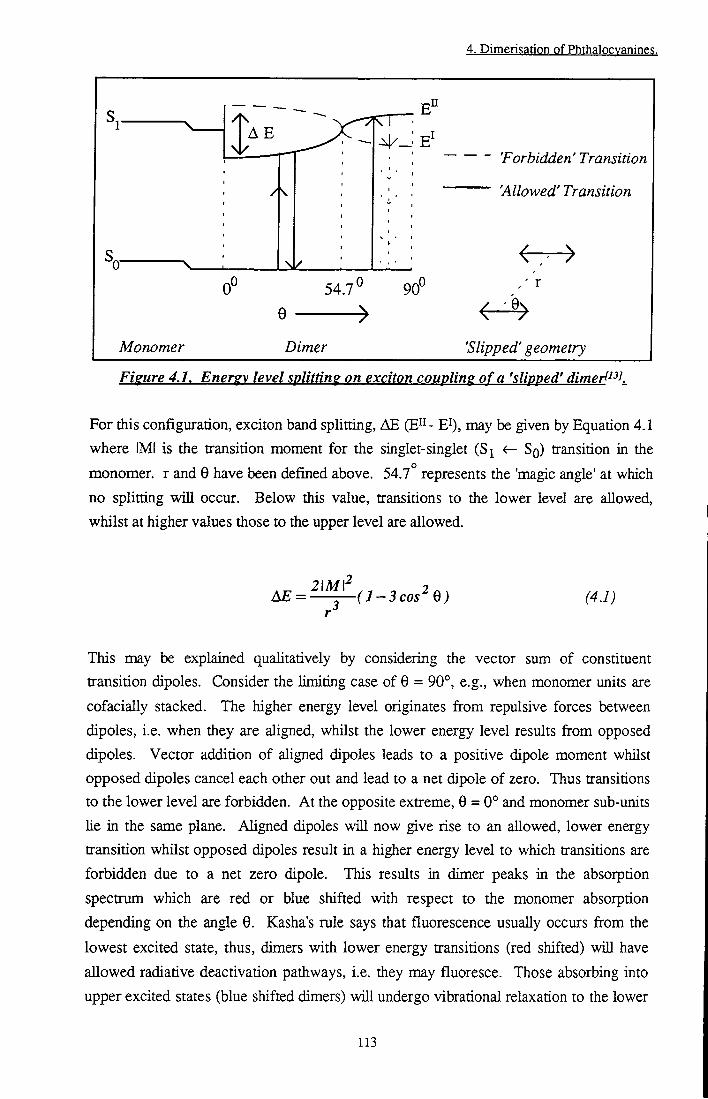
4. Dimerisation of Phthalocvanines.
\ 1 / 'Forbidden' Transition
'Allowed' Transition
' r
0
Monomer Dimer 'Slipped' geometry
Figure 4.1. Energy level splitting on exciton coupling of a 'slipped' dimert^^K
For this configuration, exciton band splitting, AE ( E " - E^, may be given by Equation 4.1
where IMI is die transition moment for the singlet-singlet (S^ <r- SQ) transition in the
monomer, r and 0 have been defined above. 54.7° represents the 'magic angle' at which
no splitting w i l l occur. Below this value, transitions to the lower level are allowed,
whilst at higher values those to the upper level are allowed.
A E = ^ ( i - 3 c o . ^ 0 ) (4.1)
This may be explained qualitatively by considering the vector sum of constituent
transition dipoles. Consider the limiting case of 0 = 90°, e.g., when monomer units are
cofacially stacked. The higher energy level originates from repulsive forces between
dipoles, i.e. when they are aligned, whilst the lower energy level results from opposed
dipoles. Vector addition of aligned dipoles leads to a positive dipole moment whilst
opposed dipoles cancel each other out and lead to a net dipole of zero. Thus transitions
to the lower level are forbidden. At the opposite extreme, 0 = 0° and monomer sub-units
he in the same plane. Aligned dipoles wi l l now give rise to an allowed, lower energy
transition whilst opposed dipoles result in a higher energy level to which transitions are
forbidden due to a net zero dipole. This results in dimer peaks in the absorption
spectrum which are red or blue shifted with respect to the monomer absorption
depending on the angle 0. Kasha's rule says that fluorescence usually occurs from the
lowest excited state, thus, dimers with lower energy transitions (red shifted) wi l l have
allowed radiative deactivation pathways, i.e. they may fluoresce. Those absorbing into
upper excited states (blue shifted dimers) wi l l undergo vibrational relaxation to the lower
113
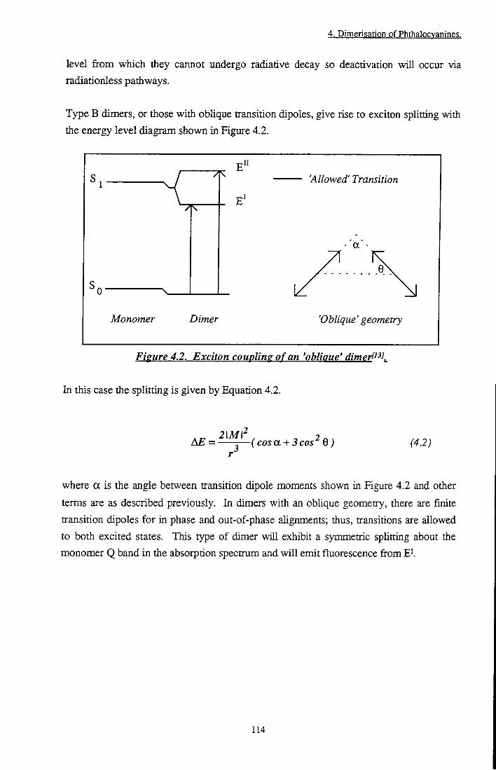
4. Dimerisation of Phthalocvanines.
level f rom which they cannot undergo radiative decay so deactivation wi l l occur via radiationless pathways.
Type B dimers, or those with oblique transition dipoles, give rise to exciton splitting with
the energy level diagram shown in Figure 4.2.
71 'Allowed' Transition
Monomer Dimer 'Oblique' geometry
Figure 4.2. Exciton coupling o f an 'oblique' dimerl^^K
In this case the splitting is given by Equation 4.2.
&E = ^^^^(cosa + 3cos^ Q) (42)
where a is the angle between transition dipole moments shown in Figure 4.2 and other
terms are as described previously. In dimers with an oblique geometry, there are finite
uransition dipoles for in phase and out-of-phase alignments; thus, transitions are allowed
to both excited states. This type of dimer wi l l exhibit a symmetric spUtting about the
monomer Q band in the absorption spectrum and w i l l emit fluorescence fi-om E^
114
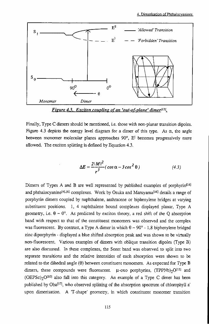
4. DimerisatiQn pf Phthalpcyanine?.
'Allowed' Transition
— 'Forbidden'Transition
90^
Monomer Dimer
Figure 4.3. Exciton coupling of an 'out-of-plane' dimerl^^K
Finally, Type C dimers should be mentioned, i.e. those with non-planar transition dipoles.
Figure 4.3 depicts the energy level diagram for a dimer of this type. As a, the angle
between monomer molecular planes approaches 90°, E^ becomes progressively more
allowed. The exciton splitting is defined by Equation 4.3.
2\Mf , , AE = — — ( c o s a-3 cos 0 ) (4.3)
Dimers of Types A and B are well represented by pubhshed examples of porphyrint^^J
and phthalocyaninef''J't^J complexes. Work by Osuka and Maruyamaf^^l details a range of
porphyrin dimers coupled by naphthalene, anthracene or biphenylene bridges at varying
substituent positions. 1, 4 naphthalene bound complexes displayed planar. Type A
geometry, i.e. 0 ~ 0°. As predicted by exciton theory, a red shift of the Q absorption
band with respect to that of the constituent monomers was observed and the complex
was fluorescent. By contrast, a Type A dimer in which 0 ~ 90° - 1,8 biphenylene bridged
zinc diporphyrin - displayed a blue shifted absorption peak and was shown to be virtually
non-fluorescent. Various examples of dimers with oblique transition dipoles (Type B)
are also discussed. In these complexes, the Soret band was observed to split into two
separate transitions and the relative intensities of each absorption were shown to be
related to the dihedral angle (0) between constituent monomers. As expected for Type B
dimers, these compounds were fluorescent. |j.-oxo porphyrins, (TPPNb)20['2] and
(0EPSc)20t i° ] also fall into this category. An example of a Type C dimer has been
published by Obati"^], observed splitting of the absorption spectrum of chlorophyll a'
upon dimerisation. A 'T-shape' geometry, in which constituent monomer transition
115
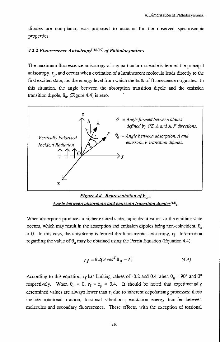
4. Dimerisation of Phthalocvanines.
dipoles are non-planar, was proposed to account for the observed spectroscopic properties.
42.2 Fluorescence Anisotropy^^^^'^^'^^ of Phthalocyanines
The maximum fluorescence anisotropy of any particular molecule is termed the principal
anisotropy, Tp, and occurs when excitation of a luminescent molecule leads directly to the
fiirst excited state, i.e. the energy level from which the bulk o f fluorescence originates. In
this situation, the angle between the absorption transition dipole and the emission
transition dipole, 9^, (Figure 4.4) is zero.
Vertically Polarised
Incident Radiation A\ A\ A\
O T ~l ~1 /
5 = Angle formed between planes defined by OZ, A and A, F directions.
^ = Angle between absorption, A and emission, F transition dipoles.
Figure 4.4. Representation ofQ^ -
Angle between absorption and emission transition dipolest^^K
When absorption produces a higher excited state, rapid deactivation to the emitting state
occurs, which may result in the absorption and emission dipoles being non-coincident, 9a
> 0. In this case, the anisotropy is termed the fundamental anisotropy, rf. Information
regarding the value of 9^ may be obtained using the Peixin Equation (Equation 4.4).
rf = 02(3cos'^ea-1) (4.4)
According to this equation, r f has limiting values of -0.2 and 0.4 when 9^ = 90° and 0°
respectively. When 9^ = 0, r f = rp = 0.4. I t should be noted that experimentally
determined values are always lower than rf due to inherent depolarising processes: these
include rotational motion, torsional vibrations, excitation energy transfer between
molecules and secondary fluorescence. These effects, with the exception of torsional
116

4. Dimerisation of Phthalocvanines.
vibrations, may be rninirnised by optimising experimental conditions. Secondary fluorescence may be eliminated by controlling the intensity of excitation radiation, energy transfer by using low concentrations of fluorophore and rotational motions via highly viscous or rigid solutions. The maximum anisotropy which may be obtained experimentally, having considered these effects is the limiting anisotropy, TQ. However, when molecules such as benzene and deuterobenzene are consideredt^^l, TQ (0.091 and 0.081 resp.) is vastiy different from the theoretical value of rp (0.4). In order to account for this difference, which is too large to be explained by the depolarising mechanisms mentioned previously, spatial oscUlators must be used instead of the linear oscillators assumed so far. In brief, the transition moment is considered to be composed of three dipoles, each oriented along one of the Cartesian coordinate axes, x, y and z. The axes are arranged such that the maximum dipole moment Ues along one axis and the minimum along another, thereby determining the direction of the third axis. Using spatial oscillators in this way, r f may be defined by Equation 4.5.
r/=fir,G - J OS) where F j represents the absorption intensities along each of the principal axes and
G j = EFjCos^Og where 0^ is the angle between the absorption and emission dipoles. For
anisotropic oscillators, = 1, r2 = r 3 = 0 and = cos- O , which leads to the original
Perrin Equation (Equation 4.4). For a symmetric, planar molecule such as benzene, F^ =
r2 = G j = G2 = 0.5 and F3 = G3 = 0, yielding a principal anisotropy, rp, of 0.1, thus
explaining the low values of rQ observed. This situation is also appUcable to molecules
with symmetry such as ZnPc. For an isotropic absorption and emission process no
anisotropic fluorescence would be expected.
Thus far, rotational motions of fluorophores have been neglected and, while this
assumption is valid in rigid or highly viscous media, rotations due to Brownian motion
must also be considered in the solution state. Under these conditions, the anisotropy is
time dependent and has been shown to decay exponentially following Equation 4.6
r ( t ) = roe-^^' (4.6)
where r(t) is the time dependent anisotropy, rQ is the limiting anisotropy and D is the time
dependent diffusion coefficient o f rotational motion. I t is possible to measure r(t) using
time correlated single photon counting techniques. However, r(t) has not been
117
4. DimerisatiQn pf Phthalocyanine?.
investigated in this study. Steady state conditions were employed. In this case, the time averaged value, r^y, of Equation 4.6 is applicable. This is expressed by the steady state Perrin Equation (Equation 4.7)
^ = 1 + ^ (4.7)
where T r is the rotational relaxation time, Tp is the fluorescence lifetime and rg is ± e
limiting anisotropy.
4.3 Materials and Methods
Aluminium phthalocyanine chloride was purchased from Kodak and used as received.
1,4,8,11,15,18,22,25- octadecyl zinc phthalocyanine (CIO) was a gift from Prof. M . J.
Cook, University of East Anglia, 2,3,9,10,16,17,23,24-octa-hexadecyl phthalocyanine
(H2C16) was a gif t from Dr. N . McKeown, University of Manchester and
1,4,8,11,15,18,22,25-octabutoxy phthalocyanine ((BuO)8H2Pc) was purchased from
Aldrich. H2C16 and (BuO)8H2Pc were converted into the zinc metallated derivatives
(ZnC16 and (BuO)8ZnPc respectively) via the procedure described in Section 2.2.2.
Methanol (MeOH), diethyl ether (Et20), ethanol (EtOH) and isopentane (IP) were
Analar grade or better and were used as received. 2-methyl tetrahydrofuran (MeTHF)
was purchased from Aldrich and distilled before use. Triton X-100 (TX-lOO),
teti-abutylammonium fluoride (^Bu4NF) - 98% purity, 1, 4 Diazabicyclo [2,2,2] octane
(DABCO) - 99% purity, trans 1,2-dipyridyl ethylene (DiPE) - 97% purity and
4,4'-bipyridine (bipy) - 98% purity were purchased from Sigma-Aldrich. AH were used
as received.
Anisotropy measurements were performed using the method described in Section
2.1.2.3. Rotational motion of fluorophores was minirtiised by cooling to 77 K in ether-
pentane-alcohol (5:5:2) (EPA) solution or by encapsulating molecules in Triton X-lOO
micelles. Dimer studies were carried out using the photophysical procedures and
apparatus described in Section 2.1. Samples of aluminium phthalocyanine chloride and
tetrabutylammonium fluoride (tBu4NF) were allowed to equilibrate in the dark for twelve
hours prior to measurement.
Photophysical techniques are described in Chapter 2 (pp 42-71).
118
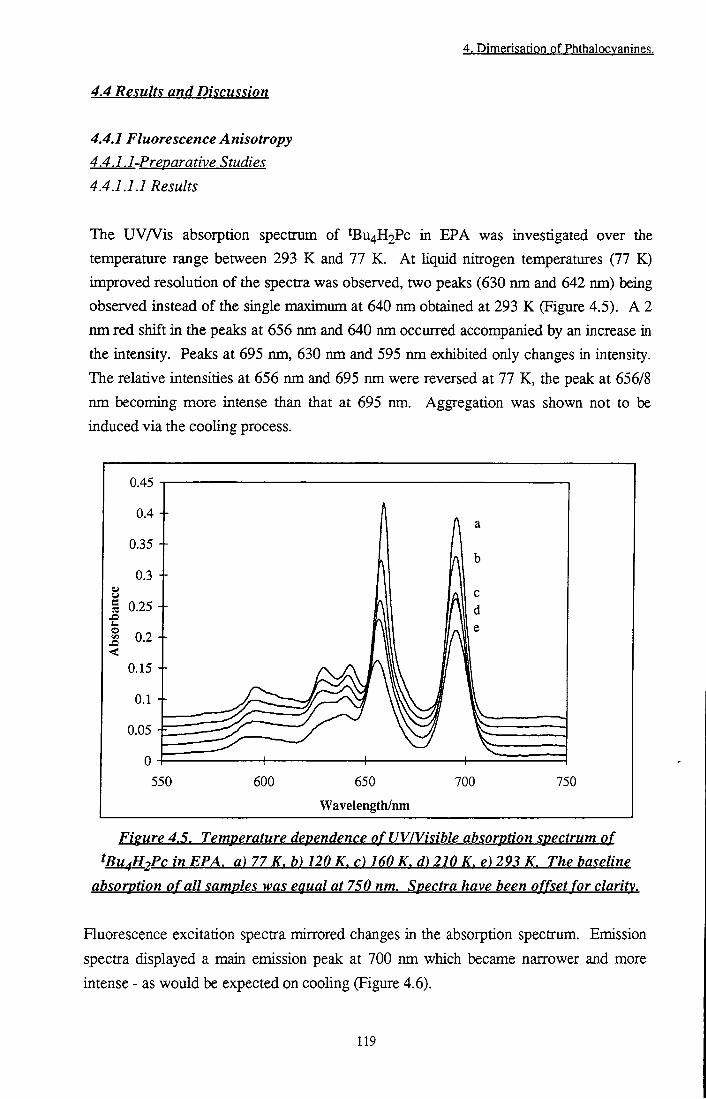
4. Dimerisation of Phthalocvanines.
4.4 Results and Discussion
4.4.1 Fluorescence Anisotropy
4.4.1.1-Preparative Studies
4.4.1.1.1 Results
The UVA^is absorption spectrum of ^Bu4H2Pc in EPA was investigated over the
temperature range between 293 K and 77 K. A t liquid nitrogen temperatures (77 K)
improved resolution of the spectra was observed, two peaks (630 nm and 642 nm) being
observed instead of the single maximum at 640 nm obtained at 293 K (Figure 4.5). A 2
nm red shift in the peaks at 656 nm and 640 nm occurred accompanied by an increase ia
the intensity. Peaks at 695 nm, 630 nm and 595 nm exhibited only changes in intensity.
The relative intensities at 656 nm and 695 nm were reversed at 77 K, the peak at 656/8
nm becoming more intense than that at 695 nm. Aggregation was shown not to be
induced via the cooling process.
0.45
0.4 +
S 0.25 +
« 0.2 +
0.05 -F
550 600 650
Wavelength/nm
700 750
Figure 4.5. Temperature dependence ofUV/Visible absorption spectrum of
tBu^H2Pc in EPA, a) 77 K. b) 120 K. c) 160 K. d) 210 K. e) 293 K. The baseline
absorption of all samples was equal at 750 nm. Spectra have been offset for clarity.
Fluorescence excitation spectra mirrored changes in the absorption spectrum. Emission
spectra displayed a main emission peak at 700 nm which became narrower and more
intense - as would be expected on cooling (Figure 4.6).
119

4. Dimerisation of PhthaJoo anines.
60
50 +
.•a 40
ja '-<
30 -
tens
i
20 -
s
10 665nni
550 600 650 700
Wavetength/nm
750 800
Fisure 4.6. Temperature dependence of fluorescence excitation CK^ff, = 760 nm]
a) 77 K. b) 293 K and emission spectra fk^^ = 630 nm). c) 77 K. <0 293 K. oi
^Bu^^PcinEPA
A n unexpected peak at 665 nm was observed in the emission spectrum at room
temperature which decreased as the sample was cooled to 77 K. The intensity o f this
emission relative to the main band at 700 nm was investigated using the Boltzmann
relation (Equation 4.8).
In ^700
^665 kT (4.8)
Figure 4.7 shows a linear dependence o f ln(l7oo/l665) vs 1/T and yielded a AE value of
1.7 X 10⁻²⁰ J or 10.7 kJ mol"' which is comparable to the result (9 kJ mol"') o f a crude
calculation o f AE using Equation 4.9.
AE = he
^665
he
•700 (4.9)
tBu4ZnPc in EPA at 77 K did not show any features beyond a sharpening o f the
absorption and emission peaks present at 293 K (Section 3.3.2.2.3).
120
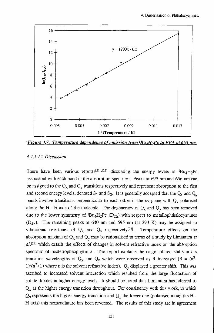
4. Dimerisation of Phthalocvanines.
16
14
12
10 +
8
6
4
y=1200x-0 .5
+ 0.003 0.005 0.007 0.009
1 / (Temperature / K)
0.011 0.013
Figure 4.7. Temperature dependence of emission from ^Bu^HjPc in EPA at 665 nm.
4.4.1.1.2 Discussion
There have been various reportst^^^'f^^J discussing the energy levels of 'BU4H2PC
associated with each band in the absorption spectrum. Peaks at 695 nm and 656 nm can
be assigned to the and Qy transitions respectively and represent absorption to the first
and second energy levels, denoted and $2. I t is generally accepted that the and Qy
bands involve transitions perpendicular to each other in the xy plane with polarised
along the H - H axis o f the molecule. The degeneracy of and Qy has been removed
due to the lower symmetry of IBU4H2PC (D2h) with respect to metallophthalocyanines
(D4h). The remaining peaks at 640 nm and 595 nm (at 293 K ) may be assigned to
vibrational overtones of Q^ and Qy respectivelyPSJ. Temperature effects on the
absorption maxima of Qx and Qy may be rationahsed in terms of a study by Limantara et
a/.l "*] which details the effects of changes in solvent refractive index on the absorption
spectrum of bacteriopheophytin a. The report explains the origin of red shifts in the
transition wavelengths of Q^ and Qy which were observed as R increased (R = (n^-
l ) / ( n 2 + l ) where n is the solvent refractive index). Qy displayed a greater shift. This was
ascribed to increased solvent interaction which resulted from the large fluctuation of
solute dipoles in higher energy levels. I t should be noted that Limantara has referred to
Qx as the higher energy transition throughout. For consistency with this work, in which
Qy represents the higher energy transition and the lower one (polarised along the H -
H axis) this nomenclature has been reversed. The results of this study are in agreement
121

4. Dimerisation of Phthalocvanines.
with Limantara's work. Qy demonstrated a red shift from 656 nm to 658 nm as the refractive index of the solvent increased due to cooling, whilst Q^ remained stationary. The additional peak at 642 nm which became clear at temperatures < 180 K has been observed before but not conclusively assignedPSJ. The most likely cause of this transition is absorption to a vibrational mode of Q^ or Qy. Leznoff and Leverl^^l suggest that a small amount of absorption may occur along the 'z' axis. Anisotropy data obtained in this study is in agreement with either of these explanations since absorption into a higher energy level than the emitting state is implied (Section 4.4.1.2).
As expected, the majority of fluorescence occurred from the lowest excited singlet state. Si, at a wavelength of 700 nm. Calculation of the energy difference between this emission band and the peak observed at 665 nm yielded an energy separation of 10.7 kJ mol"l which is consistent with published dataP2] for the energy difference (10.6 kJ mol'^) between the first and second unoccupied molecular orbitals, 6b3g and 6b2g. It is concluded that emission observed at 665 nm was due to fluorescence from the second excited state, 82- S2 emission is very uncommon since relaxation to the lowest excited state takes place very rapidly (ps), i.e. before emission has occurred. Soret band emission has previously been reported at 430 nm by Kobayashit^l, however, the authors themselves question its authenticity and suggest the observed emission band may be due to impurities in the sample. In the case of tBu4H2Pc, S and 82 excited states are very close to each other in energy, thus, thermal repopulation of 82 occurs. Consider a Boltzmann distribution of molecules in S and 82 excited states (Equation 4.10)
!^ = ^ kT nj
AE
e (410)
where n2 and n^ are the populations of 82 and Sj respectively, AE is the energy gap between states, k is the Boltzmann constant and T is the absolute temperature. AE has been shown to be ~ 10.7 kJ mol'^. Hence, when T is 293 K, ~ 1.5 % of excited state molecules will populate the 82 level, giving rise to detectable emission upon radiative decay. At 77 K the figure becomes neghgible (< 10'^ % ) . The excitation spectra resembled the absorption spectra and mimicked any changes that were observed with temperature. It was, therefore, concluded that the anisotropy of ^U4H2Pc and ^Bu4ZnPc could be studied without interference from aggregation effects.
122
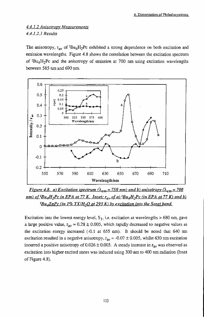
4. Dimerisation of Phthalocvanines.
4,4,1,2 Anisotropy Measurements 4.4.12.1 Results
The anisotropy, r^^ of ^Bu4H2Pc exhibited a strong dependence on both excitation and
emission wavelengths. Figure 4.8 shows the coirelation between the excitation spectrum
of tBu4H2Pc and the anisotropy o f emission at 700 nm using excitation wavelengths
between 585 nm and 690 nm.
0.05 +
5 0 . 3 300 325 350 375 400 Wavelength/nm
-0.2 550 570 590 610 630 650
Wavelength/nm
670 690 710
Figure 4.8. a) Excitation spectrum (X^^ - 750 nm) andb) anisotropy (X^^ = 700
nm) oftBu^H2Pc in EPA at 77 K. Inset: r^,, of a) tBu^HjPc (in EPA at 77 K) and b)
^Bu^ZnPc (in 1% TXIH2O at 293 K) b\ excitation into the Soret band.
Excitation into the lowest energy level, S j , i.e. excitation at wavelengths > 680 nm, gave
a large positive value, rgy = 0.28 ± 0.005, which rapidly decreased to negative values as
the excitation energy increased (-0.1 at 655 nm). It should be noted that 640 nm
excitation resulted in a negative anisotropy, r^v = -0.07 ± 0.005, whilst 630 nm excitation
incurred a positive anisotropy of 0.026 ± 0.005. A steady increase in r^^ was observed as
excitation into higher excited states was induced using 300 nm to 400 nm radiation (Inset
of Figure 4.8).
123

4. Dimerisation of Phtlialocvanines.
0.3
0.25 +
0,15 +
0.05 +
680 700 720 740 760
Wavelength/nm
780 800
Fisure 4.9. a) Emission spectrum of^Bu fH^Pc fk..^ = 630 nm),
b) AnisotroDV (X^^ = 610 itwi) in EPA at 77 K.
7 .
• 0.35O.4
•0.3O.35
• 0.25 .3
• 0.2-0.25
• 0.150.2
• 0.1-0.15
• O.OSO.l
• 0O.Q5
• -0.05O
• -0,1-0.05
T I I r o o o o O 00 C\ \o ^ ^ ^
o o q o — <s r~ Enisgkm Wavela^lMni
Fisure 4.10. Dependence of^BU fHiPc in 1% TX/H-,0 on emission and
excitation wavelength.
124
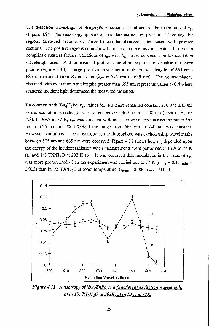
4. Dimerisation of Phthalocvanines.
The detection wavelength of tBu4H2Pc emission also influenced the magnitude of r^y (Figure 4.9). The anisotropy appears to modulate across the spectrum. Three negative regions (arrowed sections of Trace b) can be observed, interspersed with positive sections. The positive regions coincide with minima in the emission spectra. In order to complicate matters further, variations o f r^y with X^^ were dependent on the excitation wavelength used. A 3-dimensional plot was therefore required to visualise the entire picture (Figure 4.10). Large positive anisotropy at emission wavelengths of 665 nm -685 nm resulted from S2 emission (X^^ = 595 nm to 635 nm). The yellow plateau obtained with excitation wavelengths greater than 655 nm represents values > 0.4 where scattered incident light dominated the measured radiation.
By contrast with tBu4H2Pc, r^y values for ^ U 4 Z n P c remained constant at 0.075 ± 0.005
as the excitation wavelength was varied between 300 nm and 400 nm (Inset of Figure
4.8). In EPA at 77 K, r^y was constant with emission wavelength across the range 665
nm to 695 nm, in 1% TX/H2O the range from 665 nm to 740 nm was constant.
However, variations in the anisotropy as the fluorophore was excited using wavelengths
between 605 nm and 665 nm were observed. Figure 4.11 shows how r^y depended upon
the energy o f the incident radiation when measurements were performed in E P A at 77 K
(a) and 1% TX/H2O at 293 K (b). I t was observed that modulation in tiie value of r^y
was more pronounced when the experiment was carried out at 77 K (rj^^^ = 0.1, rmin =
0.003) tiian in 1% TX/H2O at room temperature, (r^ax = 0-086, Vj^in = 0.063).
0.14
0.12 +
0.08 4-
0.06 +
0.04 +
0.02 +
600 610 620 630 640 650
Excitation Wavelength/nm
660 670
Figure 4.11. Anisotropy of^Bu^ZnPc as a function of excitation wavelength.
a) in 1% TX/HoO at293K. h) in EPA at 77K.
125
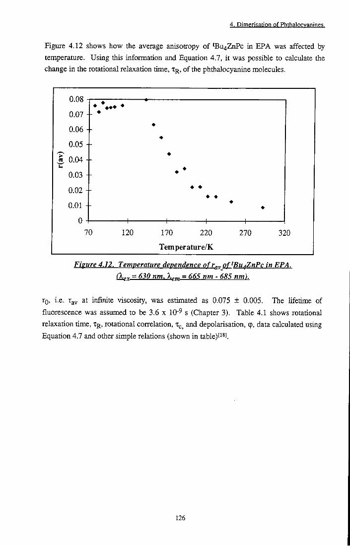
4. Dimerisation of Phthalocvanines.
Figure 4.12 shows how the average anisotropy of ^ U 4 Z n P c in EPA was affected by temperature. Using this information and Equation 4.7, i t was possible to calculate the change in the rotational relaxation time, T R , of the phthalocyanine molecules.
0.08
0.07
0.06 - -
0.05 - -
1 0.04
0.03 - -
0.02 - -
0.01
0
• • • •
• • • •
+ + 70 120 170 220
Tern per ature/K
270 320
Figure 4.12. Temperature dependence of r^y of^Bu^ZnPc in EPA.
(X(;^ = 630 nm. X^^^ = 665 nm - 5^5 nm).
XQ, i.e. r y at infinite viscosity, was estimated as 0.075 ± 0.005. The lifetime of
fluorescence was assumed to be 3.6 x lO'^ s (Chapter 3). Table 4.1 shows rotational
relaxation time, % , rotational correlation, x , and depolarisation, (p, data calculated using
Equation 4.7 and other simple relations (shown in table)f S3.
126

4. Dimerigatipn pf PhthalQgyanings,
/ns /ns 142 0.08 - -152 0.063 59 19.7 0.051
162 0.055 29 9.8 0.1
172 0.044 15.3 5.1 0.2
182 0.032 8.2 2.7 0.37
202 0.022 4.6 1.5 0.66
212 0.023 4.6 1.5 0.66
222 0.016 3 1 1
232 0.016 3 1 1
252 0.013 2.2 0.7 1.35
292 0.009 1.5 0.5 2 1 Table 4.1. Decrease in anisotropv oftBu^ZnPc in EPA with increasing temperature.
4.4.1.2.2 Discussion
^U4H2Pc belongs to die D2h point group, i.e. the lowest excited state is non-degenerate
(Section 4.4.1.2). ^Bu4H2Pc would, therefore, be expected to act as an anisotropic
oscillator. Excitation into the Qx band leads to emission from the same energy level,
= 0, and theory predicts a limiting anisotropy, rf, of 0.4. The absorption dipole of the Qy
transition (to the S2 excited state) lies perpendicular to that of the Qx transition. Since
emission occurs mainly from S^, the angle between the absorption and emission dipoles
is 90°. Therefore, excitation into the Qy band leads to an rf of -0.2. Experimentally
obtained rg values, of 0.3 and -0.1 at excitation wavelengths of 690 nm and 655 nm
respectively (X-gm = 700 nm), are in good agreement with rf and it is probable that
torsional vibrations are responsible for the small difference between rf and TQ (since the
medium is rigid, TQ may be approximated by r^y). Vibrations of the luminescent molecule
reduce the polarisation of emission and resuU in a reduced anisotropy. Between these
wavelengths, a gradual transition from 0.3 to -0.1 was obtained as mixing of excited
states occurred. Such a dependency on the frequency of exciting light was first observed
for porphyrins, analogous dye molecules, in 1957 by John Weigl ' l and is described in
more detail in a recent report by Maiti et a/.t^s]. A t 630 nm, 9^ increased to zero as
irradiation populated a vibrational level of S . Excitation at 640 nm resulted in a
negative anisotropy. This suggests that the absorbing state had a different dipole
moment to the emittimg state, i.e. absorption to a higher energy level than Qx occurred.
This may be explained by excitation into a vibrational energy level of Qy. In addition, the
suggestion of Leznoff and LeverPS] that a small amount of absorption occurs along the z-
127

4. Dimerisation of Phthalocyanines.
axis appears reasonable. Considering an angle of 90° between and Q^, theory predicts a maximum anisotropy of -0.2 (Equation 4.4). Experimentally, a value of -0.07 was calculated, however, this value is Hkely to be raised by state mixing of this transition with neighbouring absorption (630 nm) into a vibrational level of S .
Differences in anisotropy due to emission wavelength are more difficult to explain and have caused much controversy over the years. Turoverov^?] has demonstrated that the polarisation spectra of protein emission varies with emission wavelength. The magnitude of variation was affected by the wavelength of excitation. This behaviour is direcdy analogous to that observed in this study for ^Bu4H2Pc (Figure 4.9). Turoverov concluded that the observed dependence resulted from transitions to different vibrational states. On the other hand, GurinovichP°] ascribed an emission wavelength dependent polarisation of magnesium phthalocyanine to the presence of several different electronic transitions. The authors expressed the opinion that changes were too large to be accounted for by vibrational motion. In this case, when excitation wavelengths below 680 nm are considered, positive anisotropy at emission wavelengths from 665 nm to 685 nm may be ascribed to emission from S2. Since excitation at these wavelengths produces the emissive state directly (G^ = 0°), a large positive anisotropy is not surprising. When X em > 685 nm, the emitting state is S , thus, 6^ = 90°, and r^^ becomes negative. Reasons for further variation (i.e. those depicted in Figure 4.9) are less obvious. ^Bu4ZnPc shows no such dependence on emission wavelength. The observed dependence of r^y on the excitation wavelength of 'Bu4ZnPc is similarly difficult to account for. r^y was determined to have a mean value of 0.071 in both EPA at 77 K and TX-lOO at 293 K. This is consistent with a limiting anisotropy of 0.1, expected for a symmetric molecule in which = r2 = 0.5. Metallocyanines possess D^^ symmetry in which and Qy transitions are degenerate, thus the above condition applies. Bauer et alP^^ have reported a tendency for the anisotropy to decrease as the energy of excitation increases. They suggest that vibrational excess energy increases the torsional vibrations of the molecule and this, accompanied by a possible mixing of states, causes lower values of r^v An increase in the local temperature of the emitting molecule has also been proposed as a possible explanationf32]. Rapid relaxation of vibrational excess energy (VEE) in conjunction with slow dissipation of this energy causes an increase in the temperature of the fluorophore, thereby, increasing vibrations and lowering the anisotropy. Alternatively the energy may be dissipated into the surrounding solventf ] thus affecting the rotational depolarisation properties of the luminescent molecule. This issue is still under discussion and concrete conclusions as to the origin of the wavelength dependency have yet to be made. It is unlikely that any of these proposals can solely account for the X^^ dependency observed in this study since r^^ does not decrease
128

4. Dimerisation of Phthalocvanines.
continuously with increasing excitation energy. Two maxima were observed (625 nm and 650 nm) accompanied by a minimum at 635 nm in the wavelength range studied. This trend was observed in both EPA at 77 K and 1% TX/H2O at 293 K (although the effect is much less pronounced in Triton-X). It is possible that an averaging process occurred in 1% TX/H2O solution due to greater vibrational/rotational motion under these conditions.
Changes in the anisotropy as the temperature increased reflect increasing rotations and vibrations which depolarise radiative emission. This is exemplified by an increase in the probability of depolaiisation by Brownian motion, 9, that was observed on increasing the temperature fi-om 152 K to 292 K (1.76 ns'^) and die corresponding decrease in die rotational correlation time, x^, from 20 ns to 0.5 ns. It is possible to predict in various solvents using Equation 4.11fi^l.
x . = ^ (4.11) kT
where V = Volume of rotation of fluorophore, r] = viscosity of solvent, k = Boltzmann constant and T = temperature in Kelvin. The volume of rotation was estimated by assuming that rotation about the x or y axis represented the volume of a cylinder, defined using crystallographic dimensions of analogous phthalocyaninest^'']. tBu4ZnPc was estimated to be a square with 16 A by 16 A dimensionsl^s], i.e. r = 8 x lO'^^ m and h = 16 x 10'^^ m. The volume of rotation was therefore calculated to be 3.2 x 10" ^ m^ using the standard equation, V = Tcr h. For tBu4H2Pc, V = 1.3 x lO'^'^ m^. The viscosity of EPA at room temperature was approximated as the average of the three component solvents, i.e. 0.325 x lO'^ Pa s. x^ in Triton X was calculated using Ti = 140 x lO'^ Pa sf 5 and ri = 18 X 10" Pa sf ' l. These figures are only approximations. It should be noted that in micellar solution rotational dynamics within the micelle as well as rotation of the micelle itself (T^ = 72 ns for TX-lOO^s]) and translational diffusion of die fluorophore across the surface of the micelle must be consideredt^^]. Table 4.2 lists calculated for tBu4H2Pc and ^Bu4ZnPc in both EPA and TX.
129

4. Dimerisation of Phthalocvanines.
tBu4ZnPc EPA 293 0.26 tBu4H2Pc EPA 293 0.1 tBu4ZnPc 1% TX/H2O 293 111(a), 14(b)
tBu4H2PC 1% TX/H2O 293 47(a), 6(1')
tBu4ZnPc EPA 77 3.6 X 10¹³(C)
a) Ti = 140 X 10-3 Pa s, b) = 18 x lO'^ Pa s, c) TI = 1.2 x IQiO Pa s^SJ.
Table 4.2. Estimated rotational correlation times oftBu^ZnPc and tBu^H2Pc in EPA
andl%TXIH20.
Given the number of approximations which have been made to calculate Xc in this way, 0.26 ns is in very reasonable agreement with the value determined experimentally in EPA of 0.54 ns. It is evident tiiat in EPA at 293 K, rotational depolarisation factors reduce r^v to close to zero. However, in TX, a correlation time of > 6 ns is sufficiently large for polarised emission to be detected.
4.4.13Cnnr.lusion
It has been shown that phthalocyanines exhibit fluorescence anisotropy which may be partially explained using standard theory. The anisotropy of fluorescence ft-om tBu4H2Pc displayed an emission and excitation wavelength dependence which may be ascribed to different electronicMbrational transitions. A new and previously unreported fluctuation in the fluorescence anisotropy from ^Bu4ZnPc was observed as the excitation wavelength varied. This phenomenon was found to be dependent upon the physical conditions of the sample, e.g. the solvent and/or temperature. In order to use anisotropy as a technique to study the environment of a phthalocyanine molecule in solution, careful control of experimental conditions is required. The solvent and temperature as well as the excitation and emission wavelengths must be consistent i f a comparison between experiments is to be made.
130

4. Dimerisation of Phthalocyanines.
4.4.2 Dimerisation of Phthalocyanines 4.4.2.1 Aluminium Phthalocyanine Chloride (AlPcCl) 4.4.2.1.1 Results
Addition of small amounts of fluoride (F') ions to a methanolic solution of AlPcCI induced changes in the absorption spectrum which are characteristic of phthalocyanine dimer formationt^l Specifically, formation of a peak at ~ 631 nm was observed, in addition to a shift in Xmax of monomer absorption. X^ax underwent a continuous blue shift fi-om 673 nm to 662 nm upon increasing [F"] (Table 4.3). The yield and absorption maximum of dimeric species was dependent on the concentration of fluoride ions (Figure 4.13).
Increasing [F-]
Increasing [F-J
550 575 600 625 650
Wavelength/nm
675 700
Figure 4.13. A. Aggregation of 2 x lO'^ mol dm'^ AlPcCl + F-a)Omol dm-^,b)2.5 x 10'^ mol dm'^, c) 5x 10-^ mol d) 1 x 10⁻⁶ mol dm'^, e) 5 x 10⁻⁶ mol dm-3, f ) l x 10'^ mol dm-^ g) 2.5 X 10⁻⁵ mol dm-^. B. Disaggregation of2x lO'^ mol dm'^ AlPcCI + F: h) 5 x 10'^ mol dm-^, i) 7.5 x 10⁻⁵ fnol dm-3, j) 1 X 10⁻⁴ mol dm-^, k) 5 x 10⁻⁴ mol dm-^, I) 1 x 10-^ mol dm-^, m) 5 x 10-^ mol dm-^.
A andB have been offset for clarity.
131

4. Dimerisation of Phthalocvanines.
• • I B i i i l f ^^^^^^^^^^^^^^^^^^^^^^^^^ {Dialer) mm
0 673 NA 2.5 X 10⁻⁷ 672.5 638 5 X 10⁻⁷ 671 636 1 X 10⁻⁶ 671 635 5 X 10⁻⁶ 669 633 1 X 10⁻⁵ 668 631
2.5 X 10⁻⁵ 666 631 I x 10-4 663 631 1 X 10⁻³ 662 631
Table 4.3. Absorption maxima of monomer and dimer species of AlPcCI in MeOH.
At low concentrations of F- (5 x 10⁻⁷ ^-3 IQ 2.5 x 10⁻⁵ mol dm-3) aggregation was promoted, whilst at high concentrations (5 x 10⁻⁵ mol dm-3 to 0.01 mol dm-3) disaggregation occurred. Effects of dimerisation on the Soret band were more subtle - a 15 ± 3 nm blue shift of X^Q^ ' ^^S observed but littie change in intensity occmred. This is consistent with reported spectral data of crown ether substituted phthalocyanine dimerst^l. Aggregation constants, K, were determined by defining an equilibrium constant, KQ^, as depicted in Equation 4.12.
^obs -[ Dimer ]
[Monomer]" (4.12)
The extinction coefficient of dimer, was assumed to be zero at the maximum wavelength of monomer absorption, thus [Monomer] was determined using the Beer-Lambert law. Knowing values for [Monomer], [Pc]jQ^ai and assuming conservation of mass, i.e. [PcJxotal = [Monomer] + 2[Dimer], allowed the equilibrium concentration of dimeric species to be calculated. Values were corroborated using the method of TaiP^l, described in Section 3.2.2. KQ^^ values for various [F"] are collated in Table 4.4 and confirm the shift in equilibrium from monomer to dimer and back to monomer as the concentration of fluoride ions increased. Calculated equilibrium constants were consistent for both methods of analysis used. The rate of aggregation, koi^, was determined by monitoring absorbance at of the dimer as a function of time. A large variation was recorded. At fluoride concentrations less than one equivalent (< 10-6 mol dm-3), equilibrium was achieved rapidly (kg^g = 0.0075 s" ), whilst at high fluoride concentrations aggregation could be observed over the course of one hour.
132

4. Dimerisation of Phthalocvanines.
0.35
0.3
0.25 +
i 0.2
I 0.15 +
0.1 +
0.05 +
0.14
0.12
O.I
0.08
0.06 Time
1000 2000 3000
Time/s 631 nm
550 600 650 Wavelength/nm
700
Figure 4.14. Rate of formation of(AlPc)2F- (2 x IQ-^ mol dm'^ AlPcCl + 2.5 x lO'^ mol dm-^ F- in MeOH). a) 'Zero' seconds, o) 2700 seconds (180 s intervals between
traces). Dimer is characterised by growth of absorption at 631 nm. Inset: Fit of first order rate constant.
Figure 4.14 shows formation of dimer, (AlPc)2F% monitored by measuring the absorption spectrum of a solution containing phthalocyanine (2 x lO'^ mol dm-^) and fluoride (2.5 x 10"^ mol dm-3) at 3 minute intervals. The recording of the first scan was performed as quickly as possible after mixing and was taken as t=0. The inset shows a fit of first order kinetics to data obtained, ko s and Zjy, measured for a range of fluoride concentrations are also collated in Table 4.4. ep was calculated using the Beer-Lambert law (Equation 2.1) having corrected X^ax °^ ^ e dimer for monomer absorption. K^^,
and were not affected by the concentration of phthalocyanine over the range 4 x 10-' mol dm-3 to 4 x 10⁻⁶ moi dm-^.
133
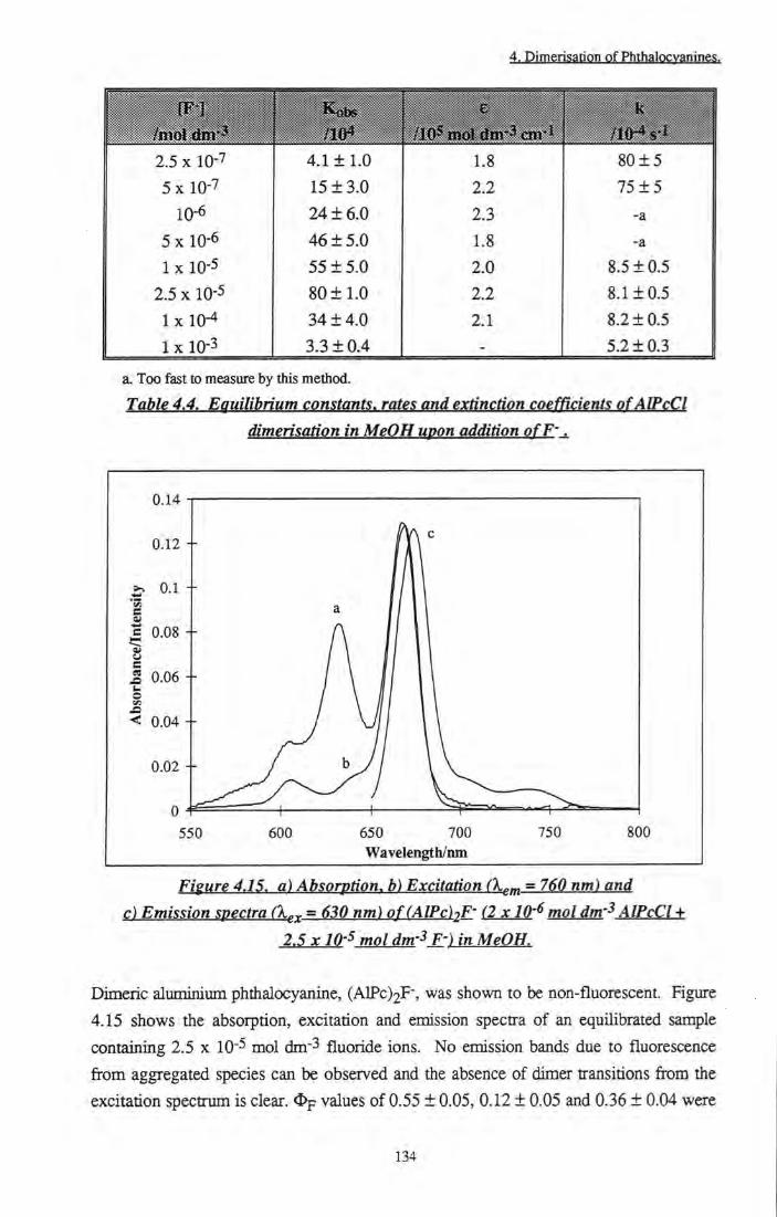
4. Dimerisation of Phthalocvanines.
llllllllll ^ ^ k
no* /105 mol dtn-3 cm-l
2.5 X 10⁻⁷ 4.111.0 1.8 80 ± 5
5 X 10⁻⁷ 15 ± 3.0 2.2 75 ± 5 10-6 24 ± 6 . 0 2.3 -a
5 X 10⁻⁶ 46 ± 5.0 1.8 -a 1 X 10⁻⁵ 55 ± 5.0 2.0 8.5 ±0 .5
2.5 X 10⁻⁵ 80 ± 1 . 0 2.2 8.1 ±0 .5 I x 10-4 34 ± 4 . 0 2.1 8.2 ±0 .5
1 X 10⁻³ 3.3 ± 0.4 - 5.2 ±0 .3
a. Too fast to measure by this method. Table 4.4. Equilibrium constants, rates and extinction coefficients ofAIPcCl
dimerisation in MeOH upon addition ofP-.
0.02 +
Wavelength/nm
Figure 4.15. a) Absorption, b) Excitation (X^m = 760 nm) and c) Emission spectra (X^^ = 630 nm) of(AlPc)2F- (2x10-^ mol dm'^ AlPcCl +
2.5 X10⁻⁵ mol dm'^ F') in MeOH.
Dimeric aluminium phthalocyanine, (AlPc)2F-, was shown to be non-fluorescent. Figure
4.15 shows the absorption, excitation and emission spectra of an equilibrated sample
containing 2.5 x lO'^ mol dm-3 fluoride ions. No emission bands due to fluorescence
from aggregated species can be observed and the absence of dimer transitions from the
excitation spectrum is clear. % values of 0.55 ± 0.05, 0.12 ± 0.05 and 0.36 ± 0.04 were
134

4. Dimerisation of Phthalocvanines.
determined for samples of AlPcCI (k^^ = 630 nm), AlPcCI -t- 2.5 x 10⁻⁵ mol dm-3 F" (k^^ = 620 nm) and AlPcCI -1-5x10⁻³ mol dm-3 F" (X^^^ = 630 nm) respectively.
Dimer species were also inactive with regard to the production of triplet state molecules. Figures 4.16 a and b show the transient absorption spectra obtained for samples of 2 x 10⁻⁶ mol dm-3 phthalocyanine with zero (Sample A) and 2.5 x 10⁻⁵ mol dm-3 (Sample B) fluoride respectively. A shift of monomer ground state bleaching was observed from 670 ± 2.5 nm to 665 ± 2.5 nm, consistent with the shift induced in the ground state absorption spectrum. Bleaching of dimer absorption (631 nm) did not occur.
-0.001 +
-0.003 +
S -0.004 +
0.005 +
-0.007 +
-0.008
•AlPca •AIPcF
400 450 500 550 600
Wavelength/nm
650
Figure 4.16. Transient absorption spectra of AlPcCI and (AlPc)2F- (AlPcCI + 2.5 x 10⁻⁵ mol dm-3 F-) in MeOH (X^^ = 638 nm).
This was confirmed by measurements. Sample B had a triplet yield of 0.08 ± 0.005 compared with a yield of 0.14 ± 0.03 from the parent phthalocyanine (Sample A) and 0.36 from Sample C (2 x 10⁻⁶ mol dm-3 = [AlPcCI] + 5 x lO'^ mol dm-3 = [F-]). Triplet lifetimes of 180 ± 15 [is, 285 ± 20 [is and 400 ± 30 |J.s were measured for samples A, B and C respectively.
Attempts to repeat this work using zinc phthalocyanine and various bidentate bridging
ligands were unsuccessful. Dimerisation induced by DABCO, bipy, diPE was
investigated using concentrations of each ligand ranging from 1 x 10- mol dm-^ to 0.1
mol dm-3 both at room temperature and 77 K. Despite, literature precedent for binding
135
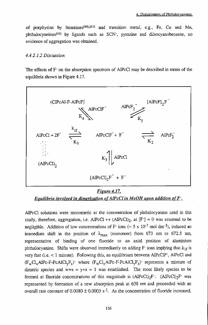
4. Dimerisation of Phthalocvanines.
of porphyrins by bisamines('*0J'[''il and transition metal, e.g., Fe, Co and Mn, phthalocyanines["2] by ligands such as SCN", pyrazine and diisocyanobenzene, no evidence of aggregation was obtained.
4.4.2.1.2 Discussion
The effects of F" on the absorption spectrum of AIPcCl may be described in terms of die
equilibria shown in Figure 4.17.
(ClPcAl-F-AlPcF) \ X AJPcClF' ^^^^2
[AlPcFl^F'
^ I f AlPcCI + 2F" AlPcClF"+ F '
K i AIPCF2"
A
(AlPcCI), AlPcCI
[A1PcC1]2F' + F '
Figure 4.17. Equilibria involved in dimerisation of AlPcCI in MeOH upon addition ofF:
AlPcCl solutions were monomeric at the concentration of phthalocyanine used in this study, therefore, aggregation, i.e. AlPcCl <-» (AIPcCl)2, at [F"] = 0 was assumed to be negligible. Addition of low concentrations of F" ions (~ 5 x lO'^ mol dm-3), induced an immediate shift in the position of A^ax (monomer) from 673 nm to 672.5 nm, representative of binding of one fluoride to an axial position of aluminium phthalocyanine. Shifts were observed immediately on adding F- ions implying that k^f is very fast (i.e. < 1 minute). Following this, an equilibrium between AlPcClF", AlPcCl and (F^QxAlPc-F-PcAlClyF^)- where (F^ClxAlPc-F-PcAlClyFz)- represents a mixture of dimeric species and w+x = y-i-z = 1 was established. The most likely species to be formed at fluoride concentrations of this magnitude is (AlPcCl)2F-. (AlPcCl)2F- was represented by formation of a new absorption peak at 638 nm and proceeded with an overall rate constant of 0.0080 ± 0.0005 s' . As the concentration of fluoride increased,
136

4. Dimerisation of Phthalocvanines.
the absorption maxima of monomeric and dimeric species shifted progressively towards lower wavelengths, reaching limiting values of 662 nm and 631 nm respectively. Monomer absorption at this wavelength may be ascribed to difluorinated species, AIPCF2-. Hypsochromic movement of dimeric phthalocyanine absorption suggests formation of more than one dimer species and indicates a shift of contributing absorption from (AlPcCl)2F- to tiiat of more highly fluorinated dimers, (ClPcAl-F-AlPcF)- and (AlPcF)2F-. A rate constant of 0.00080 ± 0.00005 s' was determined over the concentration range 10-5 mol dm-3 < [F-] < 10" mol dm-3. This is comparable to tiiat observed by Martin and co-workersf^J for the aggregation of tetrasulfonated aluminium phthalocyanine hydroxide (AIPCS4OH) in 0.2 mol dm-3 NaOH. Dimerisation was ascribed to the formation of a binuclear |i-oxo dimer, (AlS4Pc)20 and was observed to occur over four hours.
Experimentally determined equilibrium constants (Table 4.4) may be represented by
Equation 4.13 (See Appendix B).
r, _ K3K1 [F-]+ K^Kj' [ F - f + KsK2Ki^ [F']^ ^obs n (4-l:i)
(l + Ki[F-] + KiK2[F-]^ )^
Limiting cases of this equation may be considered. At low [F-], second and third
numerator terms will tend to zero and, if 1 » K^DP-], denominator terms will reduce to
1. Hence, KQ^ can be represented by Equation 4.14.
K„h,=K3Ki[F-] (4.14)
Thus, dimer formation is dominated by K3. At higher [F-], K4Ki[F-]2 and K^K2Ki^\F-]^ terms will become progressively more important and dimer formation shifts to
production of (ClPcAl-F-AlPcF)- and finally, (AlPcF)2F-. At very high fluoride
concentrations, i.e. i f Ki[F-] » 1 then Equation 4.13 reduces to
J. _K3+K4Kj[F-] + KsK2Kj[F-]' ^ObS y (4-i5)
Ki[F-](l + K2[F-])^
Thus, as the concentration of fluoride increases, KQ^JJ decreases as inactive, AIPCF2-
monomeric species are formed (Figure 4.13B). Rate determinations indicate that
(AlPcCl)2F- formation is very rapid (0.0075 s-^), whilst tiiat of (AlPcF)2F- is a factor of
137

4. Dimerisation of Phthalocvanines.
ten slower (0.0008 s'^). It was not possible to measure the formation of (CIPcAl-F-AlPcF)- direcdy, however, formation of dimer species with intermediate absorption wavelengths (e.g. 634 nm), the most likely position for this dimer, occurred very rapidly and kQ]^ may be estimated to be > 0.01 s' . All rate data fitted first order kinetics. This is surprising since dimerisation would be expected to be a second order process. However, kQjjj is a complex function of forward and reverse reactions of all equilibria present in solutiont'*^] and may appear as a 'pseudo' first order reaction.
By reference to exciton theory (Section 4.2.1), fluoride linked phthalocyanines may be
classified as Type A dimers in which 6 = 90° (Figure 4.18a), i.e. face-to-face dimers widi
cofacial, parallel transition dipole moments. In phthalocyanine molecules, transition
moments are thought to lie in the plane of the molecule along the N-M-N axes,
designated the x and y directions t ^ .
C1(F)
C1(F) a) Dimer b) Difluorinated Monomer
Figure 4.18. Geometry of (AlPc)2F- dimer (a) and AlPcFo' monomer (b).
This type of dimer has allowed transitions to the upper energy level but those to the lower level are forbidden. Thus dimer absorption appeared as a single peak, blue shifted with respect to the monomer. Exciton splitting, AE, of Type A dimers is represented by equation 4.1. In this case, AE was 1710 cm'^. The transition length, Mj., was determined using Equation 4.16^ ] and converted into the transition moment, M using Equation 4.17.
2 ^ _ e ^ AX
2513G X (4.16)
where G is die degeneracy of the state, AX is the half bandwidth, X is the wavelength of
maximum absorption and e^ax is the extinction coefficient.
138

4. Dimerisation of Phthalocvanines.
4Tzhceo ( 4 j y )
where e is the charge of an electron, h is Plank's constant, EQ is the permittivity of a vacuum and c is the velocity of light. Mj.^ was calculated to be 0.983 and M^ = 1.14 X 10⁻²³ n;i2 Thus, using Equation 4.1, assuming 0 is 90°, r was found to be 4.05 A. This is in excellent agreement with Martin's valuef^I of 4.27 A for (AlPcCl)20 and an interplanar distance of 4.11 A calculated by Kobayashi and Levert^l for crown ether substituted copper phthalocyanine. A crystal structure study by Wynnef''J showed the phthalocyanine rings of (AlPcCl)20 to be eclipsed, and separated by a distance of 4.27 A. These ring-ring distances are all slighdy larger than those reported for (AlPcF-)^ polymer (3.6 Ay**h Thus, by comparison with literature precedent, the assignment of the 631 nm absorption to (AlPcF)2F, a cofacial dimer with a ring separation of 4.05 A and a 6 value of 90° would seem reasonable.
Reduced fluorescence and triplet state formation of solutions containing dimeric species may be for one of two reasons: dimer inactivity or the effect of axial ligand replacement. It has already been demonstrated that replacement of sulfonated aluminium phthalocyanine axial hydroxide ligands by fluoride induced a small decrease in <I)p and a conciurent increase in Ojt''^]. This is corroborated by values determined using AlPcCI in this study. However, the measured fluorescence yield of (F^ClxAlPc-F-PcAlClyF^)" (0.12) is smaller than can be accounted for by fluoride binding (Op of AIPCF2- = 0.36). In addition, a decrease in was observed ( O j = 0.08) which is in contrast to an increase produced by fluoride binding {^j = 0.36). It is, therefore, concluded that reduced photophysical activity is due to the formation of inactive dimer species rather than replacement of axial chloride ligands by fluoride. Lack of fluorescence and triplet state production by this type of dimer is consistent with the predictions of exciton theory and also with observations made by Martini^] and Kobayashit J for other cofacial phthalocyanine dimers. This geometry is prevalent in phthalocyanine dimer formation['*6]-['*]'[6J, hence, phthalocyanine aggregates are generally accepted to be non-fluorescent with reduced phototoxicity.
^.^•2.2 l,4,dJlJ5J8,2225-Octadecyl Zinc Phthalocyanine (CIO) 4.42.2.1 Results
Cooling of a solution of CIO in EPA to 77 K resulted in a significant change in the
structure of the UVA^isible absorption spectrum (Figure 4.19). Peaks at 641 nm, 680
139

4. Dimerisation of Phthalocvanines.
nm, 693 nm, 705 nm and 741 nm were apparent compared with a single absorption peak at 698 nm observed at 293 K, characteristicf'*'''] of monomeric CIO. The change was a gradual one and thermal cycling experiments showed the temperature dependence to be reversible. The concentration of phdialocyanine was found to affect the changes observed at low temperature. Higher phthalocyanine concentrations resulted in a greater increase in structure on cooling (Figure 4.20). The nature of the peripheral substituents and their interaction with solvent molecules was found to be critical. The effect of temperature on the absorption spectrum of a-substituted ZnPc(X)8 in EPA decreased as die length of die alkyl chain substituent (X) decreased, i.e. C10H21 > C 5 H 1 1 > CH3. The same trend was observed in MeTHF where P-C16H33 substituents induced strucmreless aggregation of ZnPc at 77 K whilst C10H21 substituted ZnPc remained monomeric. a-butoxy ligands didn't cause any additional structure in the low temperature spectrum in EPA despite being isoelectronic with the a-C^Hn alkyl substituents.
0.25
0.2
f 0.15
1 c es •e 0.1
< 0.05 +
550 600 650 700 750
Wavelength/nm
800 850
Figure 4.19. Absorption spectra of CIO in EPA at a) 293 K (1.3 x 10-^ mol dm'^) and b) 77 K(1.7x 10⁻⁶ mol dm'^) and emission spectra of CIO in EPA at c) 293 K and
d) 77 K. (X^^ = 630 nm).
Fluorescence emission and excitation spectra exhibited similar behaviour to the
absorption spectra. Lxjwering of the sample temperature from 293 K to 77 K caused
emission to change from a single peak at 718 nm to two emission bands centred at 718
nm and 760 nm (Figure 4.19d). The relative yield of emission at 765 nm was observed
to increase as the concentration of CIO increased (Figure 4.21).
140
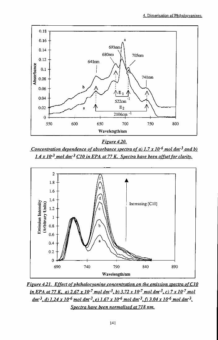
4. Dimerisation of Phthalocvanines.
0.18 J
0.16 --
0.14 --
0.12 --
e n
0.1 --
o 0.08 --
0.06 --
0.04 --
0.02
550
693mn/
680nm \ \ jQSnm
641nm
741mn
2106cm
600 650 700
Wavelength/nm
750 800
Figure 4.20. Concentration dependence of absorbance spectra of a) 1.7 x IQ-^ mol dm'^ and b)
1.4 X10-^ mol dm'^ CIO in EPA at 77 K. Spectra have been offset for clarity.
690
Increasing [CIO]
0.6 -H
740 790
Wavelength/nm
840 890
Figure 4.21. Effect of phthalocyanine concentration on the emission spectra of CIO
in EPA at 77 K. a) 2.67 x IQ-'^ mol dm-^. b) 3.72 x IQ-'^ mol dm-^. c)7x lO''^ mol
dm-3. d) 1.24x10-^ mol dm'^. e) 1.67 x lO'^ mol dm'l f) 3.04 x 10'^ mol dm'l
Spectra have been normalised at 718 nm.
141

4. Dimerisation of Phthalocvanines.
Excitation spectra obtained by monitoring emission wavelengths (XQ^) of 725 nm and
785 nm were very different, indicating the presence of at least two distinct species
(Figure 4.22).
2.5
2
e Si s
1.5
1 +
0.5
550 575 600 625 650
Wavelength/nm
675 700
2.5
C5
<
2 +
1.5 +
0.5 +
B
550 600 650 700
Wavelength/nm
750
Figure 4.22. Temperature dependence of the excitation spectra of CIO in EPA.
A. Xcm = 725 nm. B. X,^ = 785 nm. a) 293 K. b) 180 K. c) 170 K. d) 160 K. e) 150 K.
f ) 140 K. g) 77 K. Spectra have been offset for clarity.
142

4. Dimerisarion of Phthalocvanines.
The fluorescence anisotropy of each emission peak was deteimined using a range of excitation wavelengths (600 nm - 740 nm). The anisotropy of the first peak was determined by averaging r^y values obtained over the emission wavelengths 715 nm -725 nm and that of the second by averaging over the range 755 nm - 765 nm. Figure 4.23 shows large differences between values of r^y calculated for each peak.
0.45
0.4 +
0.35 +
0.3 +
0.25 +
»• 0.15 +
0.05 +
-0.05 +
590 610 630 650 670 690 710 730 750
Excitation Wavelengtli/iiiii
Fieure 4.23. Anisotropv of emission from CIO in EPA at 77 K.
The fluorescence lifetimes of emitting species were measured using time correlated single photon counting apparatus. Multiexponent decays were observed. Table 4.5 collates lifetimes measured by monitoring emission at 710 nm and 765 nm.
hm
Yield) VlSd|?C
«2
hm
Yield) VlSd|?C Yi^d> ^ ^ ^ ^ ^ ^
hm
Yield) VlSd|?C Yi^d> ^ ^ ^ ^ ^ ^
293 710 2.15 0.56 - 1.084 1.96 1 (78.73, 93.47) (21.27,6.53)
293 765 2.24 0.426 - 1.146 1.98 (114.06, 102.41) (-14.06, -2.41)
77 710 3.81 0.16 0.945 1.293 2.04
(36.1,88.4) (53.9, 5.55) (9.79, 6.05)
77 765 2.62 0.117 - 1.32 1.89 (116, 100.63) (-16.46, -0.63)
TabU4.5. Fluorescence lifetime data of CIO in EPA at 293 K and 77 K. X^^ = 638 nm.
143
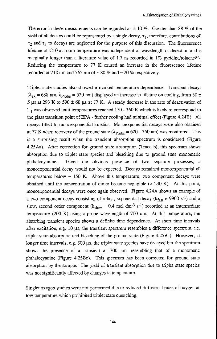
4. Dimerigation pf Phthglpcyaningg.
The error in these measurements can be regarded as ± 10 %. Greater than 88 % of the yield of all decays could be represented by a single decay, t j , therefore, contributions of X2 and X3 to decays are neglected for the puipose of this discussion. The fluorescence lifetime of CIO at room temperature was independent of wavelength of detection and is marginally longer than a literature value of 1.7 ns recorded in 1% pyridine/toluenef''*l. Reducing the temperature to 77 K caused an increase in the fluorescence lifetime recorded at 710 nm and 765 nm of ~ 80 % and ~ 20 % respectively.
Triplet state studies also showed a marked temperature dependence. Transient decays (^ex = 638 nm, Xp^^ = 520 nm) displayed an increase in lifetime on cooling, from 50 ± 5 |is at 293 K to 590 ± 60 |is at 77 K. A steady decrease in the rate of deactivation of T j was observed until temperatures reached 150 - 160 K which is likely to correspond to the glass transition point of EPA - further cooling had minimal effect (Figure 4.24B). All decays fitted to monoexponential kinetics. Monoexponential decays were also obtained at 77 K when recovery of the ground state (?iprobe = ^20 - 750 nm) was monitored. This is a surprising result when the transient absorption spectrum is considered (Figure 4.25 Aa). After correction for ground state absorption (Trace b), this spectrum shows absorption due to triplet state species and bleaching due to ground state monomeric phthalocyanine. Given the obvious presence of two separate processes, a monoexponential decay would not be expected. Decays remained monoexponential all temperatures below ~ 150 K. Above this temperature, two component decays were obtained until the concentration of dimer became neghgible (> 230 K). At this point, monoexponential decays were once again observed. Figure 4.24A shows an example of a two component decay consisting of a fast, exponential decay (kfa^j = 9900 s" ) and a slow, second order component (kgio^ = 0.4 mol dm^^ s' ) recorded at an intermediate temperature (200 K) using a probe wavelength of 700 nm. At this temperature, the absorbing ttansient species shows a definite time dependence. At short time intervals after excitation, e.g. 10 |is, the transient spectrum resembles a difference spectrum, i.e. triplet state absorption and bleaching of the ground state (Figure 4.25Ba). However, at longer time intervals, e.g. 300 |is, the triplet state species have decayed but the spectrum shows the presence of a transient at 700 nm, resembhng that of a monomeric phthalocyanine (Figure 4.25Bc). This spectrum has been corrected for ground state absorption by the sample. The yield of transient absorption due to triplet state species was not significantly affected by changes in temperature.
Singlet oxygen studies were not performed due to reduced diffusional rates of oxygen at
low temperature which prohibited triplet state quenching.
144
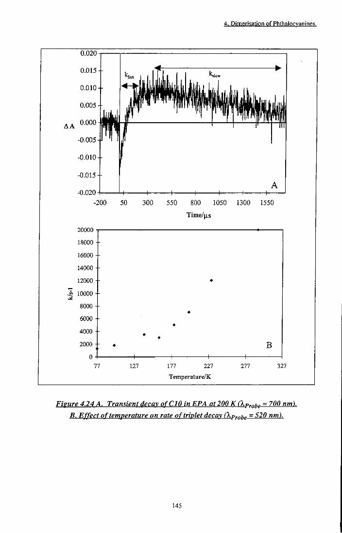
4. Dimerisarion of Phthalocvanines.
0.020
AA
-0.015 +
-0.020
20000
18000 --
16000 --
14000 --
12000
^ 10000
8000 - -
6000 --
4000 - -
2000 --
0
0.015 +
0.010 +
0.005 +
0.010 +
A +
-200 50 300 550 800 1050 1300
Time/us
1550
B
4-77 127
—I
177 227
Temperature/K
277 327
Figure 4.24 A. Transient decay of CIO in EPA at 200 K Ckprptc = 700 nm). B. Effect of temperature on rate of triplet decay Ckprpfjc = 520 nm).
145
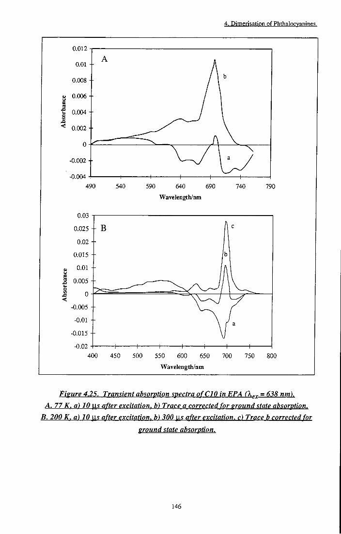
4. Dimerisation of Phthalocvanines.
0.012
0.01 --
0.008 --
g 0.006 +
I 0.004 -1-
^ 0.002-1-
-0.004 4
0.002 +
490 540 590 640 690
Wavelength/nm
740 790
0.03
a
<
-0.02
0.025 + B
0.005 +
-0.015
-t-400 450 500 550 600 650
Wavelength/nm
700 750 800
Figure 4.25. Transient absorption spectra of CIO in EPA (X^^ = 638 nm). A. 77 K, a) 10 \i.s after excitation, b) Trace a corrected for ground state absorption.
B. 200 K, a) 10 \is after excitation, b) 300 [is after excitation, c) Trace b corrected for
ground state absorption.
146

4, Dimerigation Qf Phth lQcyanings,
4.4.2.22 Discussion
The results observed when CIO was cooled to 77 K may be ascribed to dimerisation. Aggregation at low temperatures is not uncommonf''9].[50] and it is well estabhshed that low temperatures promote dimer formationt^'l due to thermodynamic considerations. The extent of aggregation was observed to increase as the concentration of phthalocyanine increased, consistent with the usual behaviour of molecules of this typel^'. Emission at 725 nm is assigned to residual monomer fluorescence. The excitation spectrum of this emission shows a splitting of the Q band at low temperatures, analogous to that observed at 77 K for ZnPcS2 in EtOH (Section 3.3.2.2.3) but more pronounced. A single emission peak results from both transitions, suggesting that degeneracy of the Sj excited state has been removed. Fluorescence anisotropy measurements (Figure 4.23) indicate this may be feasible since a negative value of r^^ was obtained at 690 nm (-0.05), i.e. the angle between the emission transition dipole and absorption transition dipole is > 54.7°. At excitation wavelengths below 660 nm, anisotropy was approximately constant at 0.075, consistent with the average value deterntined for monomeric ^U4ZnPc
(Section 4.4.1.2). A similar trend was observed by Gouterman[i°l for the fluorescence polarisation of |i.-oxo scandium porphyrin dimers. In these molecules, the polarisation was constant at 0.1 except in a region of excited state splitting where negative polarisation was observed (Pmin = -O.lf^^l). The reason for removal of degeneracy is not clear. Due to the inherent symmetry of this phthalocyanine it would be expected that X and y polarisation axes would be identical, as indeed they appear to be at room temperature. It would seem that as the temperature is lowered, interactions between the CIO chains render the molecule asymmetric. Explanation of this phenomenon becomes more difficult when the absorption spectrum of a more dilute sample of CIO (1.4 x 10" mol dm-3) is considered (Figure 4.20a). At this concentration, a significant fraction of monomeric species remain at 77 K and it is clear that splitting of the monomer absorption band did not occur.
Emission at 718 nm was accompanied by the appearance of a new band at 760 nm (Figure 4.19d). The excitation spectrum obtained by monitoring emission at 785 nm (Figure 4.22B(g)) is almost identical to the absorption spectrum and propounds that aggregated phthalocyanine species are responsible for this emission. It can be shown (Figure 4.26) that emission at 718 nm is linearly related to absorption by monomer species, A^, whilst, that at 760 nm is proportional to dimer absorbance, A^j. and A^j were calculated using Equations 4.18 and 4.19. Equation 4.18 was used to calculate the spectrum of pure dimer species.
147

4. Dimerisation of Phthalocvanines.
^DTotalC^) = AoncC^)-Const. A^uiX) (4.18)
where A^JQ^I is the absorbance of pure dimer species, A^onc the absorbance of a
concentrated CIO sample, is the absorbance of a dilute CIO sample and Const, is a
normalising factor. By subtracting a fraction of A^JQ^ from CIO samples of interest
(Equation 4.19), absorbance due to residual monomer, and hence dimer, species was
determined.
A('k)-( yAj)Total('^)) = ^MTotal(^) where
y^DTotal('^) = ^d('^) ^MTotal('^) = ^mC^)
(4.19)
6000
5000 +
S _4000 g S S O I 3000
E I eg ^ I §,2000 <
1000
•
J y=627000x-59 •
i (Monomer / Huorescence:
i Left Hand Scale) • • y y
y = 225400X + 1500 (Dimer Fluorescence: Right Hand Scale)
1 i L 1 1 1 1
18000
16000
14000
12000
10000
8000
6000
4000
2000
0
0)
i t t i o O
0.01 0.02 0.03 0.04
Absorbance
0.05 0.06 0.07
Figure 4.26.
Emission intensity vs absorbance due to monomeric and dimeric CIO in EPA at 77 K.
Such a linear correlation confirms that emission at 760 nm results from dimeric
phthalocyanine. From the gradient of each fit it may be calculated that at 77 K -
0.35<I>pM where Op^ and ^^YM. ^ e corrected fluorescence quantum yields of dimer
and monomer respectively. This result is very unusual and the first convincing evidence
to support the existence of luminescence from phthalocyanine dimers bound solely by
148

4. Dimerisation of Phthalocvanines.
Van der Waals forces. Fluorescence lifetime measurements support the theory of dimer emission at 760 nm. An increase of only 20% was observed on cooling compared with an increase of 80% in the lifetime of emission at 710 nm. This may be attributed to availabihty of radiationless deactivation pathways in dimeric species. As T is lowered, vibrational and rotational motions are reduced and with them ki^, thus an increase in tp was observed. In dimeric phthalocyanine, additional radiationless pathways such as disaggregation/recombination become available, counteracting to some extent the effects of temperature. Hence, a much smaller increase in radiative lifetime was observed in the emission of dimeric species (760 nm).
Dimer absorption was represented by two symmetrically displaced pairs of transitions, Ej (640 nm & 741 nm) and E2 (680 nm & 705 nm) (Figure 4.19). At a first glance, tiiese pairs of transitions may be ascribed to the absorption bands of CIO dimers in which exciton splitting of the excited state has occurred and transitions are allowed to both energy levels created. This criterion is met by the geometrical arrangements B or C considered in Section 4.2.1, ditner species with oblique, coplanar transition dipoles or 'clamshell' type geometry, or those with out-of-plane dipoles, i.e. cofacial, staggered phthalocyanine dimers. 'Clamshell' phthalocyanines have been synthesised by Leznoff and co-workers by linking phthalocyanine rings through units such as oxygent^^], hydrocarbon chainst ^] QJ- alkoxy unitsC '''. The extent of coupling between monomer subunits was dependent on the nature of the bridge and existed as a dynamic equihbrium between various geometries. At room temperature, the absorption spectra of these complexes represented a summation of all conformations co-existing in solution and displayed distinct similarities to the monomer absorption spectrum. The fluorescence emission spectrum was also representative of monomer emission, indicating an open 'clamshell' arrangement with littie interaction between rings. At 77 K, the absorption spectra displayed a greater degree of coupling, denoted by more intense absorption between 600 nm and 650 nm. A new emission peak at 750 nm accompanying these transitions was ascribed to emission from an exciton coupled excited state. This behaviour is analogous to that observed CIO. Emission at 750 nm is of comparable energy to the emission observed from CIO dimers (760 nm). It would, therefore, seem reasonable to assign E^ and E2 to the electronic transitions of CIO dimers with clamshell arrangements of differing r and/or 0. This is quantified by Equation 4.2 which demonstrates that small values of r and 6 give rise to a greater exciton splitting than when separation between molecules is large or the tilt angle is small. It was not possible to calculate the separation distance, r, since a and 9 are unknown quantities.
149

4. Dimerisation of Phthalocyanines.
Further consideration of the low temperature emission spectra in conjunction with the absorption spectrum casts doubt upon this theory. Only one peak due to dimer emission was obtained. I f two different dimer configurations were responsible for transitions observed in the absorption spectrum, as discussed above, it is reasonable to expect two emission peaks to be detected. In addition, differences between the excitation spectrum (Figure 4.22B, g) and absorption spectrum (Figure 4.19b) would be expected. Contributions of monomer fluorescence to excitation monitored at 785 nm may be considered to be negligible due to the vast excess of dimer fluorescence detected at this wavelength. It is suggested that a situation similar to that depicted in Figure 4.27 must be responsible for the observed behaviour. Exciton coupling of monomer excited states occurred to give rise to five allowed transitions in the excited state dimer. A shift in the centre of gravity, D, arises due to electrostatic interaction between monomer units. Fast vibrational relaxation from each of these levels to the lowest excited state E' took place, from where emission was observed. Fluorescence anisotropy measurements confirm an energy level arrangement of this type. By analogy to r^^ measurements for 'BU4H2PC (Section 4.4.1.2), a large positive anisotropy at 740 nm (0.38) may be explained by excitation into the emitting energy level of a molecule with a non-degenerate excited state, in this case E^ This value is very close to rf = 0.4, the theoretical maximum anisotropy which occurs when O = 0°. Troughs at 640 nm and 680 nm correspond to excitation into E^^ and E^ respectively, r^y values of -0.007 and -0.054 at these wavelengths correlate to angles of 55.5°and 60.4° (Equation 4.4) between E^^ & E' and E^ & E respectively. These values represent minimum limits for 63 since it is probable that mixing of states artificially raises the measiu-ed value. This is demonstrated by transitions to E"^ and E ^ in which an inaeasingly positive anisotropy was observed as the excitation wavelength approached that required for population of E^.
Monomer Dmer
Fieure 4.27. Energy level splitting o f CIO in EPA at 77 K.
150

4. Dimerisation of Phthalocvanines.
The reasons behind splitting of the monomer excited state into five energy levels are complex and cannot be explained by simple exciton theory. Similar observations have been made by other groups regarding the chemically bonded n-oxo dimers of silicon phthalocyanine. Ferencz et a/.t ^ report the presence of five absorption peaks in the spectnun of the |i-oxo dimer of tetra(methoxy) tetraoctyloxy substituted silicon phthalocyanine in non aromatic solvents such as dichloromethane and THF. Peaks are similarly distributed with respect to monomer absorption to those observed in this study for CIO aggregation. The oscillator strengtii of each of tiiese transitions was affected by the nature of tiie solvent and Schryver suggests tiiat solvent/dye interactions influence excitonic and vibrational coupling between the ring but states that a conclusive physical explanation is not possible based on current knowledge. Oddos-MarceK^^^ and coworkers extended this work and suggest that transitions from the HOMO of one monomer to the LUMO of another (ion pair or charge transfer configurations) may be responsible for some of tiie ground state transitions observed and the changes invoked as a function of environment. These are made possible by strong coupling between monomers. Aggregation of dimers was also suggested as a conceivable explanation, however, a satisfactory interpretation of these effects is still awaited. It is also possible that inequivalent coupling between x and y dipole moments may be responsible. This has been observed for a planar, binuclear zinc phthalocyanine, studied by Kobayashi and co-workerst^'^l In planar phthalocyanine dimers, dipoles oriented along the x axis lie along the same line, i.e. 9 = 0°, whilst dipoles oriented along the y axis are perpendicular to r, i.e. 0 = 90°. This gives rise to two pairs of energy levels. Transitions will be allowed to the lower energy level of the first pair (x dipole couphng) and the higher energy level of the second pair (c.f. rules for Type A dimers), leading to the observation of a doublet ia the UVA^isible absorption spectrum. Non-equivalent splitting of x and y dipoles in which transitions to both energy levels were allowed may explain E^ E", E ^ and E^ energy levels of CIO at 77 K. E™ would then have to be ascribed to residual monomer absorption.
A decrease in the non-radiative decay processes of the triplet species of CIO at 77 K is
not unexpected. Similar phenomena have been observed for the decay of triplet excited
states of pyrazinet^^l and propynal^^^l at low temperature. Data collected for the decay of
pyrazine excited states was shown to display Arrhenius type behaviour, i.e. an activation
controlled process was responsible for the decrease in triplet state lifetime observed at
high temperature. It was suggested that this was due to vibronic mixing of higher
excited triplet states above a threshold energy. Data measured for ^ClO* deactivation
did not follow Arrhenius behaviour, i.e. inhibition of more than one process was
151

4. Dimerisation of Phthalocvanines.
responsible for prolonged lifetimes at 77 K. Specifically, a decrease in vibrational deactivation through long hydrocarbon side chains due to changes in Boltzmann population of higher vibrational levels and a reduction in quenching by ground state molecules, C I O (T^) + C I O (SQ) C I O (SQ) + C I O (SQ), due to an increase in viscosity of the solution. Below the glass transition temperature of EPA (~ 150 K - 160 K), movement of molecules is severely restricted; further reduction of the temperature having littie effect on the triplet state lifetime. More surprising is the apparent independence of triplet yield on temperature since temperature is a direct measure of tiie aggregation state of C I O . It is generally accepted tiiat aggregated phthalocyanines do not produce triplet state species due to the short lived nature of the S states. C I O dimers have unique excited state properties which may be explained in terms of exciton theory. Kasha[^°l'[i3] commented on enhanced triplet state formation in dimers v^ith reduced fluorescence yields. <E>D has been calculated to be - 35 % of <I>p, thus, it is feasible that some degree of enhancement may be observed in this case. In addition, transient measurements of substituted |i-oxo SiPc dimer by Ferenczt^^l, showed the position and yield of triplet state species to be very similar to tiiat produced by the monomer. Russegger et a/.f J have shown that in the absence of compHcating factors such as aggregation, <E>jsc is unaffected by temperature.
At a temperature of 200 K, probing at wavelengtiis where ground state absorption
occurred, a decay with two clear components was observed (Figure 4.24A). The short
lived component of this decay (kf^st = 9900 s" ) has a comparable decay rate to triplet
state deactivation (k = 8300 s" at 200 K) and may be ascribed to recovery of ground
state equilibrium after bleaching due to triplet formation. The decay fits to
monoexponential kinetics despite contributions from two processes, i.e. 3Pc*-> Pc and
3(Pc)2*->(Pc)2- From this it can be infeixed that a dynamic equihbrium between 3pc*
and 3(Pc)2*, i.e. ^Pc* -t- Pc v = ^ • Pc2*, exists within the lifetime of the triplet state.
Thus, kfajt is an average of the rate of deactivation of both species. The key to the
identity of the slow component (kgio^ = 0.4 dm^ mol'^ s'^) hes in the transient absorption
spectrum. A distinct similarity to monomeric C I O was observed implying that
monomeric species are being produced on excitation ((Pc)2 — ^ 2Pc). Figure 4.28
summarises the processes occurring upon excitation.
152

4. Dimerisation of Phthalocyanines.
Excited State Equilibrium
^ 3, -J *
Pc +Pc
^Pc*
Pc-f-Pc
•"fast
(Pc), / i s '
^Pc),
I hv
^ (f<=>2 Ground State Equilibrium
slow
Pc + Pc
Monomer Formation
Figure 4.28. Equilibria upon excitation of CIO in EPA at77 K.
At 200 K, monomers formed upon irradiation may diffuse through the solution to form separated species, k^io^ obeyed second order kinetics indicating a bimolecular process and may be ascribed to monomer-monomer recombination. Ferraudi and Srisankart H have reported a similar reaction when dimeric cobalt and copper phthalocyanines were irradiated with ultraviolet light. Transient species were observed as a result of redox chemistry which produced two radical phthalocyanine species. Recombination to the ground state proceeded via a second order process. At 77 K, monoexponential decays were obtained despite evidence from the transient absorption spectrum of the presence of monomeric phthalocyanine. This may be because at 77 K, triplet decay and monomer-monomer recombination occur at comparable rates; separation of each process being prevented by experimental limitations (signal to noise ratio). Alternatively, it is possible that in a rigid medium, diffusional separation of monomers is impossible so irradiation produced dimeric species with enlarged intemuclear separation (such that excitonic coupling did not occur), resulting in an absorption spectrum representative of constituent monomer sub-units. Since separation into two distinct species has not occurred, relaxation would be expected to occur via a first order process. This theory is corroborated by behaviour observed at 160 K and 180 K in which complex decays were obtained. These temperatures are just above the glass transition temperature of EPA, thus, it is possible that both processes contributed to a cenain extent.
Attempts to induce aggregation of this type using different peripheral substituents were
unsuccessful. Monahan[62],[63],[64] j^^s shown that in organic solvents, aggregation is
driven by ring-ring interaction and, hence, must be energetically more favourable than
153

4. Dimerisation of Phthalocvanines.
ring/solvent interactions. Reduced aggregation was observed for P-octapentyl substituents whilst zinc phthalocyanine substituted with |i-methyl groups remained monomeric. This suggests that solubilisation of shorter chain alkyl substiments is more favourable in EPA than long chains. This was confirmed by studies of ZnC16 and CIO in MeTHF in which ZnC16 was observed to aggregate to a great extent whilst CIO remained monomeric. This is surprising since peripheral substituents are often added to improve solubility of phthalocyanine molecules ia organic and aqueous solvents. Cuellar and Markst ^J have demonstrated an increase in solubility of nickel phthalocyanine in 1-chloronaphthalene upon alkylation, however, in certain cases, e.g. peripheral substitution of magnesium phthalocyaninet^^l, it has been shown that substitution has hindered rather than assisted solubility. Substitution of a - C H 2 - unit for an oxygen atom in octabutoxy zinc phthalocyanine also inhibited aggregation. Cook^ ' l observed a similar difference between aUcyl and alkoxy substituted zinc phthalocyanine in crystal packing studies. Exciton coupling was obtained in crystals of octadecyl zinc phthalocyanine whilst no evidence for splitting in the analogous octaalkoxy molecule was seen.
4.4.23 Conclusions
The first substantive evidence for a luminescent phthalocyanine dimer species has been
reported. CIO has been shown to aggregate in EPA at 77 K. Aggregation was
accompanied by the appearance of emission at 760 nm, red shifted with respect to
monomer emission, which has been proven to originate from dimeric / aggregated
species. The quantum yield of dimer fluorescence was calculated to be -35% of
monomer fluorescence at 77 K. Triplet state studies have also been performed which
show the triplet yield of dimeric phthalocyanine at 77 K to be comparable to that of
monomeric phthalocyanine. Dimer dissociation to monomeric species was observed
upon excitation with either 355 nm or 638 nm radiation. Photophysical properties have
been compared with those of a cofacial aluminium phthalocyanine dimer and discussed in
terms of exciton theory.
It has been shown that the photophysical properties of dimeric phthalocyanines are
controlled by their geometry. With regards to photodynamic therapy, this may have
important consequences. It is known that accumulation of high concentrations of
sensitiser in cells leads to aggregation and it is generally accepted that aggregates are
non-cytotoxict^l The results of this study show that production of triplet state species
from aggregated phthalocyanines can occur. It is highly probable that under conditions
where diffusion of oxygen is possible singlet oxygen would be formed by these dimers.
154
4. Dimerisation of Phthalocvanines.
Hence, the photoactivity of dimers should not be summarily dismissed and may indeed play an important role in tiie destruction of neoplastic tissue.
4.5 References
[1] Moan J. and Anholt H., Phthalocyanine fluorescence in tumours during PDT., Photochem. Photobiol, 51, 379,1990.
[2] Ambroz M. , MacRobert A.J., Morgan J., Rumbles G., Foley M.S.C. and Phillips D., Time resolved fluorescence spectroscopy and intracellular imaging of disulfonated aluminium phthalocyanine., J. Photochem. Photobiol. B.Biol., 22, 105, 1994.
[3] Paardekooper M. , Van Compel A.E.V., De Bont H.J.G.M., Nagelkerke J.F., Stevenick J.V. and Van Den Broek P.J.A., Photodynamic treatment of yeast with chloroaluminum phthalocyanine: Role of the monomeric form of the dye., Photochem. Photobiol., 59, 161,1994.
[4] Wynne K.J., 2 Ligand bridged phtiialocyanines - crystal and molecular structure of fluoro(phthalocyaninato) gallium(n), [GaPcFJn and mu-oxo bis(phthalocyaninato) aluminum(in), (AIPc)20., Inorg. Chem., 24, 1339, 1985.
[5] Martin P.C., Gouterman M. , Pepich B.V., Renzoni G.E. and Schindele D.C., Effects of ligand, solvent and variable sulphonation on dimer formation of aluminium and zinc phthalocyanine sulphonates., Inorg. Chem., 30, 3305,1991.
[6] Kobayashi N. and Lever A.B.P., Cation or solvent induced supermolecular phthalocyanine formation: Crown ether substituted phtiialocyanines., / . Am. Chem. Soc, 109, 7433,1987.
[7] Yoon M.J., Cheon Y.J. and Kim D.H., Absorption and fluorescence spectroscopic studies on dimerisation of chloroaluminium (EI) phthalocyanine tetrasulphonate in aqueous alcohoUc solutions., Photochem. Photobiol., 58, 31, 1993.
[8] Kanezaki E., Yamanouchi H. and Kanda S., Monomer-Dimer equihbrium of unsubstituted zinc phthalocyanine in N,N-dimethylformamide and detection of dimer transitions in fluorescence excitation spectrum at 77K., Mol.Cryst. Liq. Cryst., 154, 351-359, 1988.
[9] Dhami S., DeMeUo A.J., Rumbles G., Bishop G., Bishop S.M., Phillips D. and Beeby A., Phthalocyanine fluorescence at high concentration: dimers or reabsorption effect?, Photochem. Photobiol., 61, 341, 1995.
[10] Gouterman M. , Holten D. and Lieberman E., Porphyrins XXXV. Exciton couphng in |U. oxo scandium dimers., Chem. Phys., 25,139,1977.
[11] Kaizu Y., Misu N. , Tsiji K., Kaneko Y. and Kobayashi H., Electronic spectra of die aluminium HI complexes of 5,10,15, 20 tetraphenylporphin and 2,3, 7, 8, 12, 13, 17,18 octaethylporphin., 5M//.. Chem. Soc. Jpn., 58,103-108,1985.
155
4. Dimerisation of Phthalocvanines.
[12] Ohno O., Kaizu Y. and Kobayashi H., Luminescence of some metalloporphins including the complexes of the I l lb metal group., / . Chem. Phys., 84, 1779, 1985.
[13] Kasha M. , Rawls H.R. and El-Byoumi M.A., The exciton model in molecular spectroscopy., PureAppl. Chem., 11, 371,1965.
[14] Hunter C.A., Sanders J.K.M. and Stone A.J., Exciton coupling in poiphyrin dimers., Chem. Phys., 133, 395-404,1989.
[15] Kemmitz K., Tamai N. , Yamazaki I . , Nakashima N. and Yoshihara K., Fluorescence decays and spectral properties of Rhodamine B in submono-, mono- and multilayer systems., / . Phys. Chem., 90, 5094-5101, 1986.
[16] Osuka A. and Maruyama K., Synthesis of naphthalene bridged porphyrin dimers
and their orientation dependent exciton coupling., / . Am. Chem. Soc, 110, 4454, 1988.
[17] Oba T., Watanabe T., Mimuro M. , Kobayashi M . and Yoshida S., Aggregation of chlorophyll a' in aqueous methanol., Photochem. Photobiol, 63, 639,1996.
[18] Kawski A., Fluorescence anisotropy: Theory and applications of rotational depolarisation.. Critical Reviews in Analytical Chemistry, 23,459-529,1993.
[19] Lakowitz J.R., Topics in Fluorescence Spectroscopy, 2, Chapter 1, Plenum Press, New York, 1991.
[20] Russel P.G. and Albrecht A.C., Electronic states of benzene by photoselection. The polarisation of luminescence in benzene, perdeuterobenzene and perchlorobenzene., / . Chem. Phys., 41, 2536, 1964.
[21] Gouterman M. , Spectra of porphyrins., / . Molec. Spec, 6,138-163,1961. [22] Schaffer A.M. , Gouterman M. and Davidson E.R., Poiphyrins XXV. Extended
huckel calculations on location and spectral effects of free base porphyrins., Theoret. Chim.Acta, 30, 9-30, 1973.
[23] Sevchenko A.N., Solov've K.N., Gradyushko A.T. and Shkirman S.F., Narrow
band electronic spectra of metal derivatives of tetrabenzoporphine and ^h\h?locyzmnt., Soviet Physics-Doklady, 11, 587, 1967.
[24] Limantara L. , Sakamoto S., Koyama Y. and Nagae H., Effects of non-polar and polar solvents on the Qx and Qy energies of bacteriochlorophyll a and bacteriopheophytin a., Photochem. Photobiol., 65, 330,1997.
[25] Henriksson A. and Sundbom M. , Semiempirical molecular orbital studies of phthalocyanines., Theoret. Chim. Acta., 27, 213-222,1972.
[26] Leznoff C.C. and Lever A.B.P., Phthalocyanines: Properties and Applications,
pl47, VCH Publ. inc.. New York, 1989. [27] Weigl J.W., The polarisation of the fluorescence of tetraphenylporphyrin., / . Mol.
Spec, 1,133-138, 1957.
156
4. Dimerisation of Phthalocvanines.
[28] Maiti N.C., Mazumdar S. and Periasamy N., Dynamics of porphyrin molecules in micelles - picosenond time resolved fluorescence anisotropy studies., / . Phys. Chem., 99, 10708-10715, 1995.
[29] Turoverov K.K. and Kuznetsova I.M., What causes the variation of polarisation
degree across tiie emission spectrum of proteins?, Biophys. Chem., 24, 327, 1986. And references therein.
[30] Gurinovich G.P., Dependence of the state of polarisation with the fluorescence
wavelengtii., Izv. Akad. Nauk. Ser. Fiz., 22, 1397-1401, 1958. [31] Bauer R.K. and Baiter A., Thermal rotations and relaxation of vibratonal energy
of excited dye molecules in solution., / . Luminescence, 20, 249-259,1974. [32] Grudzinski H., Influence of tiie local temperature of luminescent centres on tiie
emission anisotropy of fluorescence of solutions.. Acta Phys. Pol, A37,49, 1970. [33] Nemkovich N.A., Rubinov A.N. and Tomin V. I . , Optical Spectroscopy in
Chemistry and Biology - Progress and Trends, p31, 1989. [34] Reynolds P.A., Figgis B.N., Kucharski E.S and Mason S.A., Acta Cryst. B (Str.
Sci.), 47, 899,1991.
[35] The Cambridge Structural Database was used to obtain a value of r. A value of 16 A was obtained by estimating the phthalocyanine as a square.
[36] Vamavsky O., Gruno I . , Hauser M. and Klein U.K.A., Investigation of tiie intemal mobility of TX-lOO micelles by fluorescence anisotropy and excimer probing., Chem. Phys. Lett., 141, 99,1987.
[37] Rau H., Greiner G. and Haenmerle H, Temperature dependence of number and
size of Triton-X micelles in aqueous solution., Physik. Chem., 88, 116, 1984. [38] Murov L.S., Carmichael I . and Hug G.L., Handbook of Photochemistry, p296.
Marcel Dekker inc., Edn. 2,1993. [39] Tai S. and Hayashi N., Strong aggregation properties of novel
naphthalocyanines., / . Chem. Soc. Perkin Trans., 2, 1275, 1991. [40] Hunter C.A., Meah M.N. and Sanders J.K.M., DABCO - MetaUoporphyrin
binding: Ternary complexes, host guest chemistry, and the measurement of n-n interactions., / . Am. Chem. Soc, 112,5773-5780, 1990.
[41] Anderson H.L., Hunter C.A., Meak M.N. and Sanders J.K.M., Thermodynamdcs of induced fit binding inside polymacrocychc porphyrin hosts., / . Am. Chem. Soc, 112, 5780-5789, 1990.
[42] Hanack M . and Lang M., Conducting stacked metaUophthalocyanines and related
compounds., Aiiv. Mater., 6, 819, 1994. [43] Steinfeld J.I., Francisco J.S. and Hase W.L., Chemical Kinetics and Dynamics,
Prentice Hall, New Jersey, 1989.
157

4. Dimerisation of Phthalocvanines.
[44] Linsky J.P., Paul T.R., Nohr R.S. and Kenney M.E., Studies of a series of haioaluminium, -gallium, and -indium phthalocyanines., Inorg. Chem., 19, 3131, 1980.
[45] Bishop S.M., Beeby A., Meunier H., Parker A.W., Foley M.S.C. and Phillips D.,
The photophysics of disulfonated metallophthalocyanines upon complexation with fluoride., / . Chem. Soc. Faraday Trans., 92,2589,1996.
[46] Nohr R.S. and Wynne K.J., X-ray crystal structure of a conducting polymer
precursor: Bridge stacked gallium fluoride., / . Chem. Soc. Chem. Comm., 1210, 1981.
[47] Cook M.J., Chambrier I . , CrackneU S.J., Mayes D.A. and RusseU D.A., Octaalkyl
zinc phthalocyanines. Potential photosensitisers for use in the photodynamic therapy of cancer., Photochem. Photobiol., 62, 542,1995.
[48] Dhami S., Photophysics of phthalocyanines in microheterogeneous systems., p59, PhD Thesis, Imperial College, London, 1996.
[49] Sheppard S.E. and Brigham S.E., Some effects of solvent upon the absoiption
spectra of dyes, in Temperature and organic solutions of cyanine dyes., / . Am. Chem. Soc, 66, 380, 1944.
[50] Ravikant M. , Reddy D. and Chandrashekar T.K., Dimerisation effects on spectroscopic properties of water soluble porphyrins in aqueous and micellar media., / . Chem. Soc. Dalton Trans., 2103,1991.
[51] Kessel D., Porphyrin Binding and Aggregation in Photodynamic Therapy of
Neoplastic Disease, p210, CRC Press, Boston, 1990.
[52] Fluorescence polarisation is an old measurement given by, p = ———, where p
is the polarisation, I is the intensity of fluorescence and subscripts V and H stand for
vertically and horizontally polarised light respectively. Polarisation has largely been
replaced by fluorescence anisotropy and may be converted using the equation,
r = .'P 3-p
[53] Dodsworth E.S., Lever A.B.P., Seymour P. and Leznoff C.C., Intramolecular
coupling in metal-free binuclear phthalocyanines., / . Phys. Chem., 89, 5698, 1985.
[54] Leznoff C.C., Greenberg S., Marcuccio S.M., Minor P.C., Seymour P. and Lever
A.B.P., Binuclear clamshell phthalocyanines., Inorg. Chim. Acta, 89, L35-L38, 1984.
[55] Ferencz A., Neher D., Schulze M. , Wegner G., Viaene L. and De Schryver F.C.,
Synthesis and spectroscopic properties of phthalocyanine dimers in solution., Chem. Phys. Lett., 245,23, 1995.
158
4. Dimerisation of Phthalocvanines.
[56] Oddos-Marcel L. , Madeore F., Bock A., Neher D., Ferencz A., Rengel H., Wegner G., Kryschi C. and Trommsdorff H.P., Electronic states and relaxation dynamics of silicon phtiialocyanine dimers., / . Phys. Chem., 100,11850,1996.
[57] Kobayashi N. , Lam H., Nevin W.A., Janoa P., Leznoff C.C., Koyama T., Monden A. and Shirai H. Synthesis, spectroscopy, electrochemistry, spectroelectrochemistry, Langmuir Blodgett film formation and molecular orbital calculations of binuclear phtiialocyanines., / . Am. Chem. Soc, 116, 879-890,1994.
[58] Terazuma M. , Yamauchi S. and Hiroti N. , Temperature dependence of the triplet lifetime of pyrazine., J. Phys. Chem., 90, 4294,1986.
[59] Russegger P. and Huber J.R., A calculation of the temperature dependent radiationless decay of the propynal triplet state, T j . , Chem. Phys., 61, 205,1981.
[60] McRae E.G. and Kasha M. , Enhancement of phosphorescence abihty upon aggregation of dye molecules., / . Chem. Phys, 28,721, 1958.
[61] Ferraudi G. and Srisankar E.V., Photochemical redox reactivity of dimeric and
monomeric copper(n) and cobalt(II) sulphophtiialocyanines., Inorg. Chem., 17, 3164, 1978.
[62] Monahan A.R., Brado J.A. and DeLuca A.F., The association of copperCII), vanadyl, and zinc(n) 4,4',4",4"'-tetraalkylphthalocyanine dyes in benzene., / . Phys. Chem., 76,1994, 1972.
[63] Abkowitz M . and Monahan A.R., ESR and electronic investigation of the self-association of phtiialocyanine dyes in solution., / . Chem. Phys., 58, 2281,1973.
[64] Monahan A.R., Brado J.A. and DeLuca A.F., The dimerisation of a copper(II) phthalocyanine dye in carbon tetrachloride and benzene., / . Phys. Chem., 76, 446, 1972.
[65] Cuellar E.A. and Marks T.J., Synthesis and characterisation of metallo and metal free phthalocyanines and uranyl decaalkylsuperphthalocyanines., Inorg. Chem., 20, 3766, 1981.
[66] Camenzind M.J., Hill C.L., Reactions of dinittiles with manganese powder: First synthesis of manganese(II) octaphenyl poiphyrazine and a convenient synthesis of manganese(n) phthalocyanine., Inorg. Chim. Acta., 99, 63,1985.
[67] Cook M.J., 1,4,8,11,15,18,22,25-octasubstituted phtiialocyanines: the contrasting effects of alkyl and alkoxy substituents on molecular self assembly., / . Mater. Sc.: Mat.inElec.,5,117,1994.
159

pmmm rrniM ©IFmm^L©CYmmm 5.1 Introduction
The pH of tumour tissue is often substantially lower (-6.5) than that of normal, healthy tissue (~7.6)[il I f the structural properties of a sensitiser are modified by a change in pH, this could have a profound effect on the localisation, retention and photoactivity of the dye. Indeed, it has been shown that the degree of ionisation of haematoporphyrin (HP), a porphyrin-type first generation sensitiser, is highly dependent on the pH of its environment^]. More acidic conditions encourage localisation and improve retention of HP within neoplastic tissuefi^-t^]. Cell death has also been reported to increase under conditions of low pHt'^l
There have been very few investigations of the photophysical properties of protonated phthalocyanines despite potential effects on the photoactivity of these sensitisers. UVA^isible absorption studies of PcHn"+, where n = 0 to 4, were pubUshed around 1980 by Gaspardf^l and lodkot^J and in 1993 Langt''] reported data detailing the effect of monoprotonation on the triplet state properties of sulfonated chloroaluminium phthalocyanine. However, a comprehensive photophysical study of these compounds has not been undertaken to date. Several studies' l't^] of sulfonated aluminium phthalocyanines in aqueous solution have been carried out at pH's ranging fi-om 12 to 1, but no observations of protonated species have been reported. In concentrated alcohoUc solutions of sulfonated aluminium phthalocyanine chloride, Yoon et al}^'^^ discovered a new species absorbing at 710 nm which was ascribed to the formation of luminescent phthalocyanine dimers. The vahdity of this hypothesis has already been questionedt^'l. Protonation was not considered as a possible cause of spectral changes and in the work presented here evidence pertaining to this explanation is discussed.
The purpose of this study was to provide a complete photophysical evaluation of tetrabutyl zinc phtiialocyanine (tBu4ZnPc) in acidified ethanoUc solution. The propensity of disulfonated zinc phthalocyanine (ZnPcS2) and ^U4ZnPc to associate with H+ ions in 1% Triton X-100 /H2O solution and on solid substrates have also been investigated.

5. Protonation of Phthalocyanines
5.2 Materials and Methods
Tetra tertbutyl zinc phthalocyanine (tBu4ZnPc) and disulfonated zinc phthalocyamne
(ZnPcS2) were prepared by Dr. A. Beeby and used as received (unpublished results).
Solutions were prepared in absolute alcohol (Haymann Ltd. A.R. quality) and purite
water containing 1% Triton-X 100 (Sigmaultra grade) w/v (1% TX/H2O). The
concentration of H+ ions was assumed to be negHgible in freshly prepared solutions and
was adjusted using H2SO4. For experiments performed in buffered media, a solution
was prepared from 0.1 mol dm-^ citric acid and 0.2 mol dm^^ K2HPO4 with subsequent
adjustment of pH between 1 and 8 by addition of HCl or NaOH. The pH was measured
using a calibrated pH meter (Jenway model 3051). Preparation of solid state samples
and experimental details of photophysical techniques employed are described in Chapter
2 (pp 42-71).
5.3 Results and Discussion
5.3.1 ^Bu4ZnPc in Acidified Alcoholic Solution 53.1.1 Results
5.3.1.1.1 Singlet State Measurements
Five distinct species were obtained in ethanolic solutions of tBu4ZnPc acidified with
sulfuric acid (Figure 5.1 A). Isosbestic points were observed for each of the conversions
(except n = 3 -> 4), indicating sequential protonation without the presence of side
reactions such as dimerisation or decomposition. By comparison with published
resultsI^J tiiese were assigned to protonated forms of phthalocyanine, PcHj,"+, where n =
0 to 4, and represents the stepwise addition of a hydrogen ion to each azomethine bridge
of the pyrrole moieties. Mono, di and tri (n = 1 to 3) protonated species demonstrated a
splitting of both Soret and Q bands, whilst the spectrum of tetra protonated
phthalocyanine resembled that of the parent phthalocyanine, red shifted to 807 nm
(Table 5.1). The integrated area of all the spectra, jl-.dv , from 300 nm to 850 nm of
PcHn""*" remained constant.
161
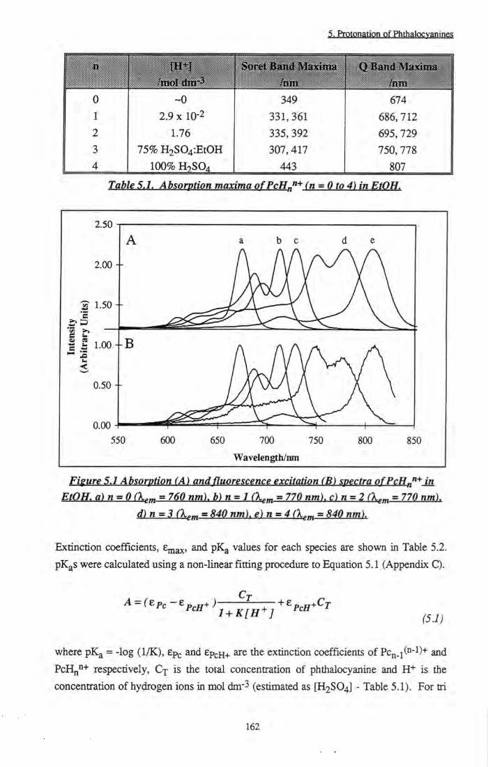
5. Protonation of Phthalocvanines
Q B;and Maximii i /nm
Q B;and Maximii i /nm
Q B;and Maximii i /nm
Q B;and Maximii i /nm
0 ~0 349 674 1 2.9 X 10⁻² 331,361 686.712 2 1.76 335,392 695,729 3 75% H2S04:EtOH 307,417 750,778
1 4 100% H7SO4 443 807
Table 5.1. Absorption maxima ofPcH„"+ fn = Oto 4) in EtOH.
2.50
2.00 +
^ 1.50 +
t 1.00
0.50 +
550 600 650 700 750
Wavelength/ran
800 850
Fieure 5.1 Absorption (A) and fluorescence excitation (B) specti-a ofPcH„"+ in EtOH. a)n = 0 (X,„ = 760 nm). b)n = l (X,^ = 770 nm). c)n = 2 (X,^ = 770 nm).
d)n=3 (X,^ = 840 nm). e)n = 4 (X,„ = 840 nm).
Extinction coefficients, Emax' values for each species are shown in Table 5.2.
pKgS were calculated using a non-linear fitting procedure to Equation 5.1 (Appendix C).
A = (£pc-ep^jj+) 1 + K[H^] (5.1)
where pK^ = -log (1/K), ep^ and ep£.H+ are the extinction coefficients of PCn.i("-^)+ and
PcHn"+ respectively, Cj is the total concentration of phthalocyanine and H+ is the
concentration of hydrogen ions in mol dm-3 (estimated as [H2SO4] - Table 5.1). For tri
162

5. Protppatipn of PhthaiQcyOTincs
and tetra protonation (n = 3 & 4) the concentration of acid required (75% and 100% H2S04:EtOH respectively) was too high for this equation to be valid. To analyse the formation of these species, Hammett acidity functions must be considered, however, no values for H2SO4 in EtOH have been published to date.
n
(ovms) i .ii-:.s.. . . . .s.A!. /am^Hiol-lcm-* 0 674 1 . 6 ± 0 . 1 x l 0 5 682 0.26 ±0.02 1 712 2.9 ± 0.3 8 .7±0 .2x 10^ 717 0.12 ±0.01
2 729 0.18 ±0 .05 1.1 ±0 .1 X 105 735 0.07 ±0.01 3 778 - 6.8 ± 0.4 x 10⁴ 788 0.03 ±0.005 4 807 - 1 . 8 ± 0 . 1 x l 0 5 820 0.01 ±0.005
Table 5.2. Ahsorbance and fluorescence data forPcHn^ (n = Oto 4) in EtOH.
Poiphyrin (P) molecules display similar behaviour at low pHf*^] Changes in the pH from 9.5, to 4.5, and finally 0.52 induced successive protonation of two inner ring nitrogens, i.e. P H 2 PH3+ <-4 PH42+. A shift to longer wavelengths was observed upon protonation. pK^ values of 5.8 and 2.3 (4.7 and 2.9)1^3] QJ-Q determined for each protonation step of haematoporphyrin TX^^^\
Protonation of the phthalocyanine ring resulted in a large decrease in the yield of radiative emission. Successive protonations were accompanied by shifts to longer wavelength of the emission spectrum (Figure 5.2) and large decreases in the fluorescence yield (Table 5.2). Fluorescence excitation spectra mimic the absorption specdra (Figure 5. IB), confirming the existence of a single species. The steady state fluorescence anisotropy was also found to be affected by protonation. Figure 5.3 shows tiie effect of excitation wavelength on tiie fluorescence anisotropy of fBu4ZnPc ([H+] ~ 0 mol dm-3) and PcH+ ([H+] = 0.022 mol dm-3 (0.03 mol dm-3 at 77 K)) in EPA at 77 K. Trace a is that of the parent phthalocyanine and fluctuates around r^y = 0.075. Trace b was produced by PcH+ and mimics the form of metal-free phthalocyanine (Section 4.4.4.1.2), passing from negative to positive values as the excitation wavelengths were scanned.
163
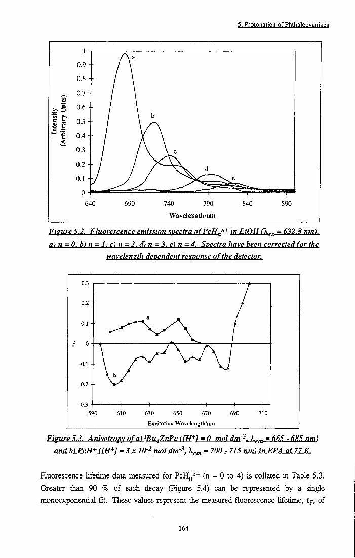
5. Protonation of Phthalocvanines
'E
c .t; I— x>
0.9 +
0.8 +
0.7 -h
0.6 +
640 690 740 790
Wavelength/nm
840 890
Figure 5.2. Fluorescence emission spectra ofPcH^"'^ in EtOH (X^^ = 632.8 nm). a) n = 0. b) n = 1. c) n = 2, d) n = 3. e) n = 4. Spectra have been corrected for the
waveleneth dependent response of the detector.
Excitation Wavelength/nm
Figure 5.3. Anisotropv of a) ^Bu^ZnPc (fH+1 = 0 mol dm'^. X^m = 665 - 685 nm) and b) PcH+ (fH+l = 3x lO'^ moldm'^. X^^ = 700 - 715 nm) in EPA at 77 K.
Fluorescence lifetime data measured for PcHn"+ (n = 0 to 4) is collated in Table 5.3.
Greater than 90 % of each decay (Figure 5.4) can be represented by a single
monoexponential fit. These values represent the measured fluorescence lifetime, tp, of
164

5. Protonation of Phthalocvanines
each species and decrease as the phthalocyanine ring is protonated. Small contributions from other decays are also observed and may be explained by the presence of other protonated species. A maximum error of + 10% may be assumed for these measurements.
Instniment Respoase Function
Time/as
Figure 5.4. Time resolved fluorescence decays of PcH„"'^ in EtOH, X^ = 638 nm. a) n=0 fX„„ = 680 nm). b)n=l (X.^ = 717 nm). c)n=2 (X„„ = 735 nm). din =3
(Xf.„ = 780 nm). e) n=4 fX^ = 830 nm). The instrument response function is also shown.
n Ti /ns T2 /ns x j /ns DW
(Amplitude, (Amplitude, (Amplitude,
Yield) Yield) Yield)
3.36 - - 1.36 1.85
(100,100)
1 2.83 1.085 - 1.16 1.99
(97.12,98.87) (2.88, 1.13)
2 1.84 0.9 - 1.36 1.91
(85.9, 92.55) (14.1,7.45)
3 1.32 2.49 - 1.34 1.98
(96.99, 94.43) (3.01, 5.57)
4 0.88 2.184 7.677 1.4 1.89
(99,26, 96.68) (0.49, 1.17) (0,25, 2,15)
Table 5.3. Fluorescence lifetimes ofPcHJ^ fn = 0to 4) in EtOH.
165
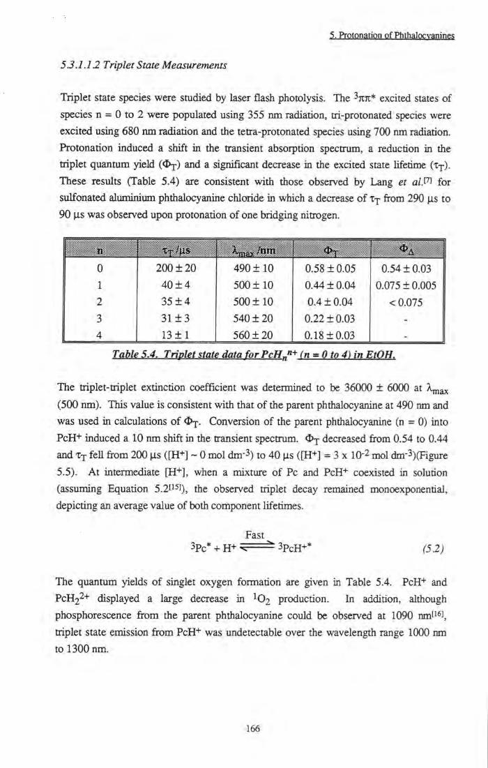
5. Protonation of Phthalocvanines
53.1.12 Triplet State Measurements
Triplet state species were studied by laser flash photolysis. The ^TITI* excited states of species n = 0 to 2 were populated using 355 nm radiation, tri-protonated species were excited using 680 nm radiation and the tetra-protonated species using 700 nm radiation. Protonation induced a shift in the transient absorption spectrum, a reduction in the triplet quantum yield and a significant decrease in the excited state lifetime (x-r). These results (Table 5.4) are consistent with those observed by Lang et a/.t' J for sulfonated aluminium phthalocyanine chloride in which a decrease of from 290 |is to 90 |is was observed upon protonation of one bridging nitrogen.
<I>T <J>A j
0 200 ± 2 0 490 ± 1 0 0.58 ±0.05 0.54 ±0.03 1 40 ± 4 500 ± 1 0 0.44 ± 0.04 0.075 ±0.005 2 3 5 ± 4 500 ± 10 0.4 ±0.04 < 0.075 3 3 1 ± 3 540 ± 20 0.22 ±0.03 -4 13 ± 1 560 ± 2 0 0.18 ±0.03 -
Table 5.4. Triolet state data for PcH„"+ fn = Oto 4) in EtOH.
The triplet-triplet extinction coefficient was determined to be 36000 ± 6000 at A^ax (500 nm). This value is consistent with that of the parent phthalocyanine at 490 nm and was used in calculations of ^j. Conversion of the parent phthalocyanine (n = 0) into PcH+ induced a 10 nm shift in the transient spectrum. O j decreased from 0.54 to 0.44 and Xj feU from 200 is ([H+] ~ 0 mol dm-3) to 40 |is ([H+] = 3 x 10⁻² mol dm-3)(Figure 5.5). At intermediate [H+], when a mixture of Pc and PcH+ coexisted in solution (assuming Equation 5.2ti^]). he observed triplet decay remained monoexponential, depicting an average value of both component lifetimes.
Fast 3pc*4 -H+^ 3pcH+'' (52)
The quantum yields of singlet oxygen formation are given in Table 5.4. PcH+ and PCH22+ displayed a large decrease in IO2 production. In addition, although phosphorescence from the parent phthalocyanine could be observed at 1090 nm' J, triplet state emission from PcH"*" was undetectable over the wavelength range 1000 nm to 1300 nm.
166
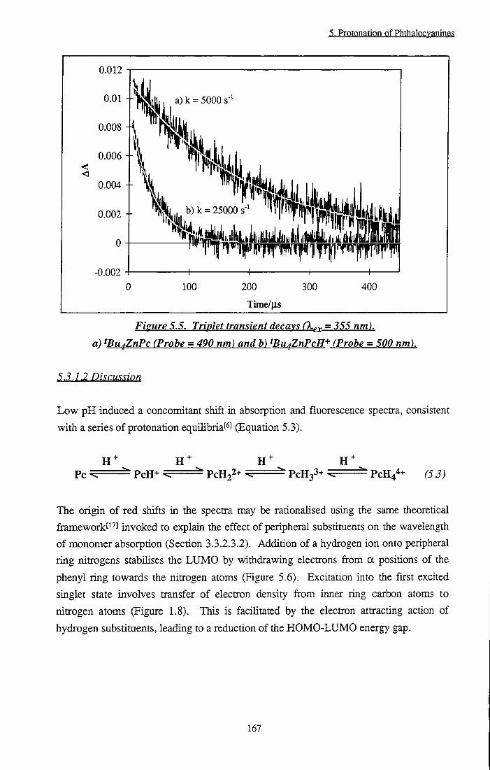
5. Protonation of Phthalocvanines
< <
0.012
0.01 -I-
0.008
0.006
0.004
0.002 -H
0
-0.002 -f 0
a) k = 5000 s-
I I
100 200 300
Time/us
400
Figure 5.5. Triplet transient decays (X^,^ = 355 nm).
a) fPu^ZnPc (Probe = 490 nm) and b) tBu^ZnPcH+ (Probe = 500 nm).
5.3.1.2 Discussion
Low pH induced a concomitant shift in absorption and fluorescence spectra, consistent
with a series of protonation equilibriat^] (Equation 5.3).
Pc PcH^ = ^ PCH22+ ^ PCH33+ ^ PCH44+ (5.3)
The origin of red shifts in the spectra may be rationalised using the same theoretical firameworkti"^] invoked to explain the effect of peripheral substituents on the wavelength of monomer absorption (Section 3.3.2.3.2). Addition of a hydrogen ion onto peripheral ring nitrogens stabilises the LUMO by withdrawing electrons fi-om a positions of the phenyl ring towards the nitrogen atoms (Figure 5.6). Excitation into the fu-st excited singlet state involves transfer of electron density from inner ring carbon atoms to nitrogen atoms (Figure 1.8). This is facihtated by the electron attracting action of hydrogen substituents, leading to a reduction of the HOMO-LUMO energy gap.
167

5. Protonation of Phthalocvanines
Figure 5.6. Effect of protonation on conjugation of the ring.
Similar shifts have been observed in the absorption spectra of protonated Schiff base metaUoporphyrinst'^l and attributed to a resonance effect of the withdrawing groups ratiier than delocalisation of the positive charge onto tiie ringli^l. This is not surprising since in these molecules, the positive charge is located on an imine moiety of the ring and, hence, is somewhat removed from conjugation of the ring. Large changes in the Soret band were also observed on protonation (Table 5.1). These are analogous to those induced by amino substituents on metallophthalocyaninet^^J in which a band was observed at 450 nm due to splitting of the Soret band. The theory of Morley et a/.t^'l predicts very small shifts of the Soret band upon peripheral substitution, thus it would appear that development of tiiis theorum is required to cover all Hgand binding cases. Splitting of Soret and Q bands may be attributed to removal of the degeneracy of transitions polarised along x and y directions. Binding of hydrogen to die phthalocyanine ligand lowered the symmetry of the complex from D4J1 to C2v (Figure 5.7) so that x and y transitions are no longer equivalent. This is corroborated by fluorescence anisotropy measurements. At hydrogen ion concentrations of 3 x lO'^ mol dm-3 (Figure 5.3), a similar spectrum to that measured for ^Bu4H2Pc (a D2h complex) was obtained. A negative anisotropy was observed at wavelengths below 690 nm, corresponding to excitation into a higher energy level than the emitting state. Using Equation 4.4, and the measured r^y of -0.2, the angle between transition dipole moments of absorbing and emitting states may be calculated to be 90° (X^^ = 605 nm). At wavelengths > 605 nm, state mixing caused the measured value for rgy to be higher (less negative) than its true value. An angle of 90° is consistent with removal of x and y degeneracy. At wavelengths above 690 nm a large positive anisotropy was measured (0.3 at 700 nm), i.e. excitation at this wavelength produced the emitting state directly. Effects on the viscosity of the solution due to the addition of acid (0.03 mol dm-3) gj-g negligible. With no added acid, the anisotropy had an average value of 0.075 although fluctuations with X^-^ were observed, consistent with observations for a fluorophore of D4h symmetry (Section 4.4.1.2).
168

5, Protpnation of Pht alQcvanings
N Zn N
H Bu
\ Figure 5,7. Stmmre oftBu^ZnPcH^
A reduction in photoactivity of tBu4ZnPc was observed as the concentration of
hydrogen ions increased. Table 5.5 shows radiative and non-radiative rate constants
calculated using measured quanmm yield and lifetime data. Simple relations used in
calculations are shown in the table.
0 7.2 ± 0 . 9 x 1 0 7 1.6 ± 0.2 X 10⁸ 0.16 ±0.05 4.4 ± 1 . 4 x 1 0 7
1 4.3 ± 0.6 X 10⁷ 1 .6±0.2x 10⁸ 0.44 ±0.04 1.6 ± 0 . 2 x 1 0 8
2 3.9 ± 0.7 X 10⁷ 2.2 ± 0.3 X 10⁸ 0.53 ±0.04 2.9 ± 0.4 X 10⁸
3 2.3 ± 0 . 4 x 1 0 7 1.7 ± 0.3 X 10⁸ 0.75 ±0.03 5.8 ± 0 . 6 x 1 0 8
4 1 .14±0.6x 10⁷ 2.0 +0.4 X 10⁸ 0.81 ±0.03 9.2 ± 1 X 10⁸
Table 5.5. Rate constants of radiative and non-radiative deactivation pathways of PcH„''+(n = 0to4)inEtOH.
As protonation of the ring increased, die rate of radiative deactivation of Si, kp*',
decreased, whilst that of internal conversion, k^ , increased. The rate of intersystem
crossing remained constant at 1.8 ± 0.25 x 10⁸ s'^ These trends are easily explained in
terms of fundamental photophysical behaviour and are directiy related to changes in AE
(Si <— SQ energy gap) upon protonation. The rate of radiative emissionl^U.pa] jg given
by Equation 5.4.
169

5, PrQtQngtiQn Qf Phthalocyanine?
_ 3 ki=A = 8%h<\> B (5.4)
where A is the Einstein coefficient for spontaneous emission, n is the refractive index of
the medium, v is the average wavenumber of emission and B is the Einstein transition
probability of absorption, given by |i2/6eo(h/27c)2, where | i is the transition dipole
moment and h is Plancks constant. Substitution for B into Equation 5.4 yields Equation
5.5 for the expression of kpO.
< v 6zo(hl2%y (55)
7
6
5
•33 2
1
0 1.5
y=3.9x-4.7
H i 2 2.5
1/< v"^>(/10"cm-^
21 -
20.5 - . B
20 -
19.5 - - A -a
- A
c 19 • y = -0.097X + 35
18.5 -
18 -
17.5 -
17 - h 1 1 V 1 1 1 140 145 150 155 160 165
AE/k J mol"'
170 175 180
Figure 5.8. Dependence ofkpO and kj^ on AE (Sj <- SQ) ofPcH„ n+
Figure 5.8A shows a plot of kpO vs l/<v-3>. A good linear correlation was obtained.
This confirms that the absorption intensity (determined by B) remained constant
170

g, ProtpnatiQn pf Phthalpcy^nine;
throughout the series and that the calculated decrease in the intrinsic rate of radiative emission of Sj (kpO) as protonation occurred is consistent with that predicted by theory.
Despite an increase in the intrinsic lifetime, XQ (XQ = 1/kpO), the measured lifetime of fluorescence decreased from 3 ns to 0.88 ns upon complete protonation (Table 5.3). This was due to a concurrent increase in intemal conversion pathways. Non-radiative transitions increase as the energy gap between states decreases according to the 'energy gap law'[231 (Equation 5.6).
--10^^ exp-aAE (5.6)
where a is a proportionality constant. Figure 5.8B shows a plot of Inkjc vs AE. Least squares analysis of the data yielded an intercept of 35, in reasonable agreement with the value predicted by Equation 5.6 of ln(10^3)^ i e 30. values are also in good agreement with previously published data that has been related to the energy of the singlet statef ^J. The energy gap law can be qualitatively explained in terms of Frank-Condon factors. As the energy gap between nuclear wavefunctions increases, the extent of overlap decreases and deactivation becomes more unfavourable. I f direct spin-orbit coupling to a vibrational level of T^ occurs, the rate of intersystem crossing may also be expected to depend on the energy difference between S and T^. kjsj, was found to remain constant at 1.8 ± 0.25 x 10^ s- as pH was lowered. This implies that AE between S and T^ remained approximately constant or that spin-orbit coupling to an upper excited triplet level, Tj, occurred.
Equilibria contributing to excited triplet state dynamics are presented in Figure 5.9. k j and k2 represent the triplet decay rate of unprotonated and monoprotonated phthalocyanine species, whilst, kf, k^ and kf*, k^* are the rates of H+ binding/dissociation in the ground and excited states respectively.
'Pc* + 3 (Pc-H
Pc + H Pc-H""
Figure 5.9. Triplet state equilibria o fPcH'*:
171

5. Prptonation of Phthalocvanines
Experimentally, single exponential decays were obtained (Figiu-e 5.5). This suggests that fast exchange between 3pc* and 3pcH+* occurred within the lifetime of the excited state such that kf* and kj,* are faster than k^ and k2 consistent with theoryt^^l. The equilibrium constant, K*, is represented by Equation 5.7.
[^Pc JLH"-]
The experimentally observed decay may be represented by the sum of two first order decays, -d[3pc* + 3pcH+*]/dt = ki[3pc*] + k2[3PcH+*]. Substituting for [3pcH+*] gives Equation 5.8.
3 * -(I + K*[H^])^^^-^ = (ki+k2K*[H^])[W] (5.8)
dt
However, -d[3pc*]/dt = kobs[3pc*], tiius the observed rate of decay, ko^g, is a first order
process that may be expressed by Equation 5.9.
1 + K*[H^]
At high [H+], K*[H+] » 1 tiierefore kobs ~ k2 - the rate of decay of 3pcH+* whilst at
low [H+], 1 » K*[H+] hence k^bs ~ k^ - tiie rate of decay of 3pc*. At intermediate
[H+J, an average value was observed which depended on the relative contribution of k j
andk2.
A large decrease in the yield of singlet oxygen formation was observed upon protonation. Such a large reduction was inconsistent with the measured decrease ui <I)-p and X j . This may be explained by lowering of the triplet energy to a value where energy transfer to ground state oxygen is no longer favourable. Lang et al.^'^^ have reported a substantial reduction in the rate of quenching, kg, by O2 of the triplet state of protonated chloro-aluminium phthalocyanine (kg = 6.6 x 10^ dm3 mol"^ s- ) relative to that of unprotonated phthalocyanine (kq = 1.6 x 10^ dm3 mol-1 s'^). It has been shown[2 3 that kq may be correlated with the energy gap between triplet state species participating in energy transfer. It may be inferred that, since the energy required to promote ground state molecular oxygen to its singlet state will remain constant, the
172

5. Protonation of Phthalocvanines
energy of ^Pc* must have decreased. It was not possible to corroborate this experimentally since phosphorescence of PcH"* could not be detected, however, it has been shown that kjsc, and therefore AE (Si -> T i ) , remained constant upon protonation, thus Ex of PcH+ may be estimated to be ~ 8400 cm"! (c.f 9090 cm"! for tBu4ZnPc), E A
= 7882 cm-l
(5.10)
The Sandros EquationP^J (Equation 5.10), may be used to estimate kg based upon
singlet and triplet state energies. Using the data above, we can expect a reduction in kq
by an order of magnitude,
5.3.2 ZnPcS2 inl% Triton X-100 solution
The UV/Vis absorption spectrum of a concentrated solution of ZnPcS2 in 1% Triton X-100 / H2O solution showed a characteristic monomer spectrum but was observed to produce a new absorbing species at 702 nm over a period of days or weeks (Figure 5.10). This spectrum bears a close resemblance to that reported by Yoon et al. in concentrated alcoholic solutions of sulfonated aluminium phthalocyanine chloridet'"!.
0,35 I
0,15
Waveler^h/nm
Fisure 5.10. UV/Visible absorption spectra of 'Fresh' (a) and 'Old' (days to weeks] (h) Solutions ofZnPcS-, in 1% Triton X-IOO/H^O solution.
173
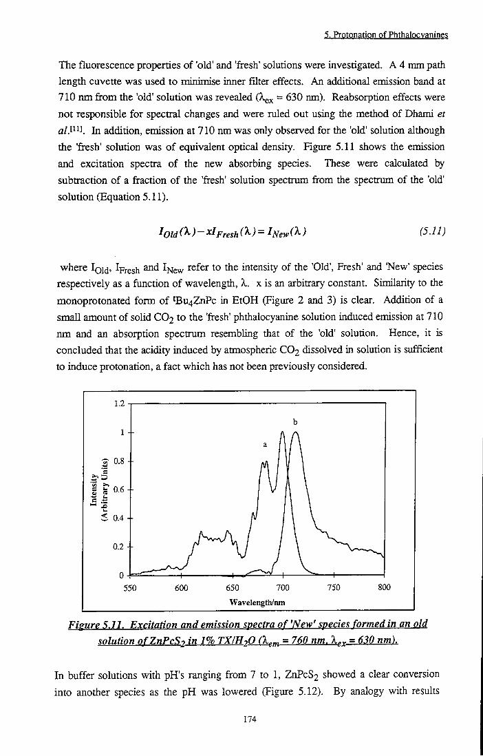
5. Protonation of Phthalocvanines
The fluorescence properties of 'old' and 'fresh' solutions were investigated. A 4 mm path length cuvette was used to minimise inner filter effects. An additional emission band at 710 nm from the 'old' solution was revealed (Xgx = 630 nm). Reabsorption effects were not responsible for spectral changes and were ruled out using the method of Dhami et a/.l ^J. In addition, emission at 710 nm was only observed for the 'old' solution although the 'fresh' solution was of equivalent optical density. Figure 5.11 shows the emission and excitation spectra of the new absorbing species. These were calculated by subtraction of a fraction of the 'fresh' solution spectrum from tiie spectrum of die 'old' solution (Equation 5.11).
I Old C^)- Fresh C^)- ^New ) (5.11)
where Igid. Ipresh ^New ^^^^^ the intensity of the 'Old', Fresh' and 'New' species respectively as a function of wavelength, X. x is an arbitrary constant. Similarity to the monoprotonated form of tBu4ZnPc in EtOH (Figure 2 and 3) is clear. Addition of a small amount of solid CO2 to the 'fresh' phthalocyanine solution induced emission at 710 nm and an absorption spectrum resembling that of the 'old' solution. Hence, it is concluded that the acidity induced by atmospheric CO2 dissolved in solution is sufficient to induce protonation, a fact which has not been previously considered.
1.2
0.8 + 'E
I s IS
< 0.4 l
0.2 +
550 600 650 700
Wavelength/nm
Figure 5.11. Excitation and emission spectra of 'New' species formed in an old
solution ofZnPcS2 in 1% TXIH2Qj2^,n = ^60 nm, X^x =
In buffer solutions with pH's ranging from 7 to 1, ZnPcS2 showed a clear conversion
into another species as the pH was lowered (Figure 5.12). By analogy with results
174

5. Protonation of Phthalocvanines
obtained for tBu4ZnPc this species was assigned to the PcH+ ionic form of phthalocyanine. Using Equation 5.1 the pK^ was calculated to be 4.4 ± 0.4.
0.035 --
0.03 -- Decreasing pH
E 0.025 + Decreasing pH
0.02 +
0.015 +
0.01 +
0.005 +
650 700
Wavelength/nm
800
Figure 5.12. Absorbance spectra of ZnPcS^ in buffers ranging from pH 7 to 1. pH=a) 7.06 b) 5.8 c) 53 d) 4.56 e) 4.17 f) 3.48 g) 3.38 h) 2.5 i) 2.1 i) 1.06.
Figure 5.13 shows the emission spectra of ZnPcS2 in buffers of various pH's. The inset shows the ratio of emission at X^^ of ZnPcS2 (679 nm) to that of monoprotonated phtiialocyanine emission (707 nm). A typical pH ciu^ve was obtained. Using the metiiod of Haugland et a/.I '' (Equation 5.12), a pK^ of 4.1 ± 0.5 was determined (Figure 5.14).
pH = pK^ + c[logl ^"""7 + log^ (5.12)
where R is the ratio of emission of two wavelengths (in this case 679/707 nm) with
minimum and maximum limiting values of R^jn and R ax respectively. I ^ and I ^ are the
intensity in acid and base respectively at the wavelength used for the denominator of R
(707 nm). Thus, pKg was calculated from the intercept minus log(Ia/I{)) (\.og(JiJi-^ = 0.6
for this study).
This value is within experimental error of that calculated using UVA^'is absorption data
and is comparable to that reported for protonation of haematoporphyrint'^l Protonation
175
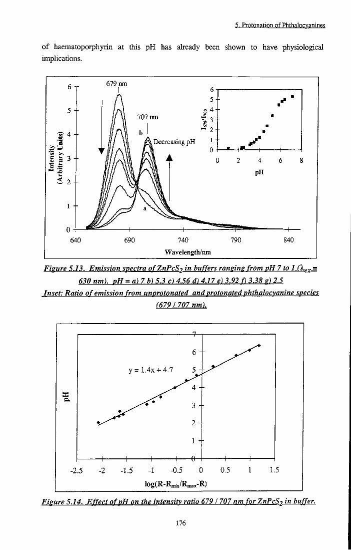
5. Protonation of Phthalocvanines
of haematoporphyrin at this pH has already been shown to have physiological implications.
5 +
e
S S5 3
S 2
1 +
640
679 nm 6
5
4
3
2
^ Decreasing pH ^ "I" 0
707 nm
4
pH
690 740
Wavelength/nm 790 840
Figure 5.13. Emission spectra o/ZwPcS? in buffers ranging from pH 7 to 1 fX,,^ =
630 nm). pH = a)7 b) 5.3 c) 4.56 d) 4.17 e) 3.92 f) 3.38 e) 2.5 Inset: Ratio o f emission from unprotonated and protonated phthalocvanine species
(6791707 nm).
a.
1—
6 -
y = 1.4X + 4.7 5 -
4 -
3 -
h- \ 1 V-
2 -
1 -
— e - 1 \
-2.5 -2 -1.5 -1 -0.5 0 0.5 1 1.5
Iog(R-R„^/R„^x-R)
Figure 5.14. Effect ofpH on the intensity ratio 679 1 707 nm for ZwPcSo in buffer.
176

g, Protpngtipn pf Phthal(?cyanings
Table 5.6 compares the photophysical properties of tBu4ZnPc (n = 0, 1), ZnPcS2 (n = 0, 1) and AIPCS2 (n = 0, 1) in 1% TX/H2O solution. Clearly, although die propensity of each phthalocyanine to protonate differs roarkedly, the resulting decrease in the photophysical properties is similar.
n • # x t j /iis
ZnPcS2 0 - 0.16 + 0.03 0.55 ±0.05 330 ± 3 0 0.56 ±0.05
1 4.4 ± 0 . 4 0.1 ±0 .02 0.17 ±0.02 100 ± 1 0 <0.05
tBu4ZnPc 0 - 0.14 ±0 .03 0.48 ±0.05 330 ± 3 0 0.53 ±0.05
1 -0.110.2 0.085 ±0.015 0.27 ±0.03 3 0 ± 5 0.17 ±0 .2
AIPCS2 0 - 0.48 ±0 .04 0.22 ±0.02 615 ± 7 0 0.2 ±0.02
1 -0.2 + 0.2 0.14 ±0.02 0.06 ±0.03 7 2 ± 10 0.09 ±0.03
Table 5.6. Comparison of photovh\sical properties of monovrotonated species of
'Bu^ZnPc. ZnPcS2 andAlPcS2 in 1% Triton X.IOO/H2O solution.
5.3.3 Solid State Measurements
Figure 5.15 shows the diffuse reflectance spectra of ZnPcS2 and tBu4ZnPc adsorbed onto cellulose. An additional peak was observed in the spectrum of ZnPcS2 at 705 mn. Identical spectra were obtained using microcrystalline cellulose or filter paper, however, silica based samples were characterisatic of monomeric phthalocyanine. Exposure of cellulose based samples to ammonia vapour removed the peak at 705 nm such that spectra resembled those recorded when sihca was employed. The remission fimction of ZnPcS2 on 100% cotton fabric was measured (Figure 5.15, Inset). This spectrum is different to those on both cellulose and silica, featuring a broad, structureless band superimposed onto monomeric absorption bands.
177
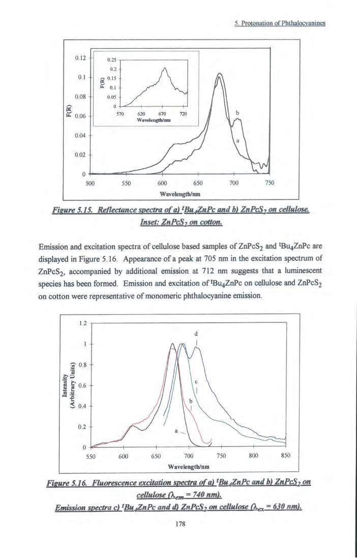
5. Protonation of Phthalocvanines
n 0.15
0,08 -
0,06
0.04 4-
500 550 600 650
Wavelcngth/nm
Fieure 5.15. Reflectance spectra of a) ^BufZnPc and b) ZnPcS-> on cellulose.
Inset: ZnPcS2 on cotton.
Emission and excitation spectra of cellulose based samples of ZnPcS2 and *Bu4ZnPc are
displayed in Figure 5 .16. Appearance of a peak at 705 nm in the excitation spectmm of
ZnPcS2, accompanied by additional emission at 712 nm suggests that a luminescent
species has been formed. Emission and excitation of tBu4ZnPc on cellulose and ZnPcS2 on cotton were representative of monomeric phthalocyanine emission.
S 0.6 i
550 600 650 700 750
Wavelength/nm
850
Fieure 5.16. Fluorescence excitation spectra of a) ^Bu fZnPc and b) ZnPcS-, on cellulose (k„„ = 740 nm).
Emission spectra c) ^BU fZnPc and d) ZnPcS-> on cellulose A.^^ = 630 nm).
178

5. Protonation of Phthalocvanines
Diffuse reflectance flash photolysis experiments revealed that the transient absorption spectrum of ZnPcS2 on cellulose was shifted by -10 nm from 490 nm to 500 nm with respect to that recorded using silica as the substrate. Decays fitted monoexponential kinetics and yielded lifetimes of 138 ± 15 |is on silica and 216 ± 20 |is on cellulose. Incorporation of tBu4ZnPc onto cellulose and silica substrates resulted in identical transient spectra centred at 490 nm.
By analogy to results discussed in Sections 5.3.1 and 5.3.2, the red shifted absorption peak observed by adsorption onto cellulose may be attributed to protonated phthalocyanine species. Singlet state measurements are also consistent with this theory. It has been shown (yide infra) that ZnPcS2H+ in 1% TX/H2O emits at 707 nm, or 580 cm"l to lower energy than the parent phthalocyanine. Solid state results are in good agreement with this, exhibiting an emission band at 712 nm, red shifted by 511 cm-^ Shifts in the transient absorption spectrum were identical to those observed for monoprotonated phthalocyanine species in ethanol. Triplet state lifetimes of cellulose based samples were longer than those prepared on silica. This is surprising considering lifetimes measured in solution state (330 |is and 100 \is for ZnPcS2 and ZnPcS2H+ respectively) which indicate that protonated species have a much faster decay rate. In solid state work it is known that diffusion of oxygen across cellulose is hindered (Section 3.3.4), tiius it is likely that the measured lifetime has been artificially enhanced, counteracting a decrease induced by molecular changes. As for solution state measurements, Pc* + H+ v ^ PcH''"* equilibria were rapid compared with deactivation processes.
Two possibilities present themselves as the reason behind protonation. Cellulose is a
large molecule with numerous sites available which are suitable for hydrogen bonding to
adsorbates (Figure 5.17)^^1.
CHpOH . L
CHpOH CHoOH 1
OH ^ h \
OH ^ H \
A ^ o - ^
OH
Figure 5.17. Structure of cellulose.
179
5. Protonation of Phthalocvanines
It is plausible that hydrogen bonding to a bridging methine nitrogen of the phthalocyanine ring would induce the results described above. Alternatively, excess free hydrogen ions in cellulose may be responsible for protonation. If hydrogen bonding was responsible, cotton fabric would be expected to produce the same effect as powdered cellulose or filter paper. However, spectra recorded on cotton cloth were comparable to those recorded for AIPCS2 on muslin by WilkinsonE^?] and Oldhamt^^l and are characteristic of a mixture of monomeric and aggregated phthalocyanine species. This implies that excess hydrogen ions which have been removed from the cloth by some process of manufacture, e.g. washing, must be the cause of protonation. Exposure of samples to ammonia vapour removed any traces of protonated phthalocyanine species. The implication of this is that excess hydrogen ions are removed via protonation of ammonia, i.e. NH3 + H+ v NH4+. Ammonia is a strong base (pKa(NH4+) = 9.25^31]) and binds hydrogen ions more readily than phthalocyanine (pK^ = 4.3).
5.4 Biological Relevance
Protonation of sensitisers for use in photodynamic destruction of tumour tissue may have important consequences regarding the efficacy of the dye in vivo. As a consequence of large lactic acid concentrations produced as a biproduct of anaerobic glycolysis, the pH of tumour tissue has been demonstratedt^^] ^^ry from 5.85 to 7.68, up to 1.15 units lower than that of healthy tissue (extracellular pH = 7.43, inffacellular pH = 7.00). Disulfonated zinc phtiialocyanine has been shown to have a pK^ value of 4.3 ± 0.5 in aqueous buffered solution (vide infra), thus at pH's of this magnitude a significant proportion of dye molecules would be expected to exist as the protonated form. Meso tetra 4-(sulfonatophenyl) porphyrin (TPPS4) demonstrated this property in RR 1022 epithelial cells which were shown to have a pH low enough to encourage protonation of this phthalocyaninel^^l A dramatic reduction in the propensity of protonated zinc phthalocyanines to produce cytotoxic, singlet oxygen species has been demonstrated. A significant shift in the absorption window to longer wavelength was also observed. This may have a detrimental effect on the yield of excited state molecules produced by the wavelength of the exciting light. Indeed, the in vivo absorption spectrum of disulfonated aluminium phthalocyanine has been shownf '*] to be red shifted by 15 nm with respect to the spectrum in aqueous solution. A shift of this direction and magnitude was consistent with the therapeutic efficiency of this phthalocyanine causing MS2 fibrosarcoma and L1210 leukaemic cell death. The authors speculated that this phenomenon must be due to a modification of the chemical structure of dye molecules. Protonation of the ring is one possible explanation for these effects. Despite negative effects of protonation, Lin et al. observed an increase in the photodynamic efficiency of
180

5. Protonation of Phthalocvanines
Nile Blue derivative at low pH's in the presence of the ionophore, nigericint'*!. A similar effect was observed by a different group when chloroaluminium phthalocyanine was used as sensitiserl^^l. It has since been shown that the positive action of nigericin was not due to its ability to lower intracellular pH, nevertheless, in the absence of nigericin, cells were more susceptible to photodynamic therapy at low pH (6.0)f^^l. It follows that other mechanisms such as the effect of pH on the locaUsation and retention of sensitisers must also be considered. The effect of pH on the localisation of porphyrin sensitisers has been studied in detail by PottierHl and Braultt^l. Both groups suggest that the ionic distribution of haematoporphyrin wiU be significantly different at pH = 6.5 (tumour pH) than at pH = 7.6. At the pH of tumour tissue, approximately 44% of porphyrin species will exist as the non-ionic, H2P form, compared with just 2% at neutral pH. Regarding this simply, retention in interstitial regions may be enhanced due to the lower solubility of non-ionic species and consequent resistance to removal by lymphatic drainage.
I f transport across cell membranes is considered, neutral molecules wiU penetrate into the cell more readily than ionic species due to the net negative charge surrounding plasma membranes. Once inside the cell, a higher environmental pH is experienced by the dye and the ionic distribution alters accordingly effectively trapping the phthalocyanine. This results in intracellular localisation of the dye molecules. A study by Bohmerl^^], in which enhanced cellular uptake of haematoporphyrin was observed at low pH corroborates this theory. An alternative explanation was made concerning reduced binding of porphyrin molecules to albumin delivery proteins.
Further experimental studies are required to evaluate the effects of in vivo pH on the efficacy of substituted zinc phthalocyanines. It is possible that protonation of phthalocyanine species (leading to cationic molecules) may promote association with cell membranes and a consequent increase in dye retention. This may counteract to some extent the detrimental effects of protonation on the photophysical propenies of zinc phthalocyanines, viz: reduced O 7 and O^.
181
5. Protonation of Phthalocvanines
5.5 Conclusion
It has been shown that protonation of zinc phthalocyanine molecules at the azomethine bridges causes a significant reduction ia photoactivity. As the pH of the solution was lowered using H2SO4, a red shift of the absorption and fluorescence spectta accompanied by a concurrent decrease in the fluorescence quantum yield was observed. The quantum yields of triplet production and singlet oxygen sensitisation were also reduced upon protonation. Solutions of ZnPcS2 in 1% TX/H2O and on solid cellulose substrates were found to protonate under surprisingly mild conditions. It is concluded that bands due to the 'dimer' in the absorption spectrum reported by Yoon et a/.fi°l were in fact protonated phthalocyanine.
5.6 References
[1] Barrett A.J., Kennedy J.C., Jones A., Nadeau P and Pottier R.H., The effect of tissue and cellular pH on the selective biodistribution of porphyrin-type photochemotherapeutic agents: A volumetric titration study., / . Photochem. Photobiol. B:Biol., 6, 309-323,1990.
[2] Pottier R., Laplante J.P. and Chow Y.A., Photofrins: A spectral study.. Can. J. Chem., 63, 1463,1985.
[3] Brault D., Physical chemistry of porphyrins and their interactions with membranes: The Importance of pH., J. Photochem. Photobiol., B:Biol., 6, 79-86, 1990.
[4] Lin C.W. and Shulok J.R., Enhancement of Nile Blue derivative-induced photocytoxicity by nigericin and low cytoplasmic pH., Photochem. Photobiol., 63, 143-146, 1994.
[5] Gaspard S., Verdaguer M. and Viovy R., Protonation des phthalocyanines., / .
Chem. Res. (M), 3064-3072, 1979. [6] lodko S.S., Kaliya O.L., Kondratenko N.V., Luk'yanets E.A., Popov V. I . and
Yapupol'skii L .M. , Quantitative characteristics of the stagewise protonation of phthalocyanines., Zhur. Obshch. Khim., 53, 901-903, 1983.
[7] Lang K., Wagnerova D.M., Engst P. and Kubat P., Influence of protonation on the reactions of triplet state sulfonated chloro-alunndnum (HI) phthalocyanine with
dioxygen., / . Chem. Soc. Faraday Trans., 88, 677-680, 1992. [8] Savitsky A.P., Lopatin K.V., Golubeva N.A., Poroshina M.Y., Chemyaeva E.B.,
Stepanova N.V., Solovieva L . I . and Lukyanets E.A., pH dependence of fluorescence
and absorbance spectra of free sulfonated aluminum phthalocyanine and its conjugate
with monoclonal antibodies., J. Photochem. Photobiol. B.Biol., 13, 327-333, 1992.
182

5. PrQtongtiQn pf Phthalocyanine?
[9] Simpson M.S.C., The photophysics of sulphonated aluminium phthalocyanines as sensitisers for Photodynamic Therapy (PDT)., PhD Thesis, Imperial College, London, 1994.
[10] Yoon M.J., Cheon Y.J. and Kim D.H., Absorption and fluorescence spectroscopic studies on dimerization of chloroaluminium (III) phthalocyanine tetrasulphonate in aqueous alcohoUc solutions., / . Photochem. Photobiol. B.Biol., 58, 31-36, 1993.
[11] Dhami S., De Mello A.J., Rumbles G., Bishop S.M., PhiUips D. and Beeby A.,
Phthalocyanine fluorescence at high concentrations: Dimers or reabsoq)tion effect?, Photochem. Photobiol., 61, 341-346, 1995.
[12] Pottier R., In vitro and m vivo fluorescence monitoring of photosensitisers., J. Photochem. Photobiol. B.-Biol., 6, 103-109, 1990.
[13] Brault D., Vever-Bizet C. and Le Doan T., Spectrofluorimetric study of porphyrin incorporation into membrane models - evidence for pH effects., Biochim.
Biophys. Acta., 857,238, 1986. [14] Pottier R.H., Kennedy J.C., Chow Y.F.A. and Cheung F., The pK^ values of
haematoporphyrin IX as determined by absorbance and fluorescence spectroscopy.,
Can. J. Chem., 33, 57,1988. [15] Barltrop J.A. and Coyle J.D., Principles of Photochemistry, p49, Wiley and
Sons, Bristol, 1978. [16] Vincett P.S., Voigt E.M. and Rieckhoff K.E., Phosphorescence and fluorescence
of phthalocyanines., / . Chem. Phys., 55, 4131, 1971. [17] Morley J.O. and Charlton M.H., Theoretical investigation of the structure and
spectra of zinc phthalocyanines., / . Phys. Chem., 99,1928,1995. [18] Ward B., Callahan P.M., Young R., Babcock G.T. and Chang O.K., Red shifts in
the optical spectra of porphyrin schiff bases upon protonation., J. Am. Chem. Soc,
105, 634,1983.
[19] Ward B., Chang C.K. and Young R., Spectral shifts upon reversible
modifications of CHO peripheral substiments in porphyrin, chlorin and
bacteriochlorin. A phenomenological explanation for the red shift of protonated schiff
base., / . Am. Chem. Soc, 106, 3943,1984.
[20] Mikhalenko S.A., Derkacheva V.M. and Luk'yanets E.A., Phtiialocyanines and
related compounds. XIX Tetra and octa amino substituted phthalocyanines., / . Gen. Chem. (USSR), 51, 1405, 1980.
[21] Kober E.E., Caspar J.V., Lumpkin R.S. and Meyer T.J., Application of the
energy gap law to excited state decay of osmium(n)-polypyridine complexes:
Calculation of relative nonradiative decay rates from emission spectral profiles., / .
Phys. Chem., 90, 3722, 1986.
183

5. Protonation of Phthalocvanines
[22] Atkins P.W., Physical Chemistry, p465, 4th Edn., Oxford University Press, Oxford, 1990.
[23] Turro N.J., Modern Molecular Photochemistry, pl83. University Science Books, CaUfomia, 1991.
[24] Barltrop J.A. and Coyle J.D., Principles of Photochemistry, p93, Wiley and Sons, Bristol, 1978.
[25] Barltrop J.A. and Coyle J.D., Principles of Photochemistry, pl26, Wiley and Sons, Bristol, 1978.
[26] Ford W.E., Rihter B.D. and Rodgers M.A.J., Quenching of naphthalocyanine triplets by O2: Application of the Sandros Equation., J. Am. Chem. Soc, 111, 2362, 1982.
[27] Whitaker J.E., Haugland R.P. and Prendergast E.G., Spectral and photophysical studies of benzo[c]xanthene dyes: Dual emission pH sensors.. Anal. Biochem., 194, 330, 1991.
[28] Kroschwitz J., Encyclopaedia of Polymer Science and Engineering, pl23, Wiley and Sons, New York, 1990.
[29] Wilkinson F. and Willsher C, Detection of transient absorption in a dyed cotton fabric and in semicinductor powders by diffuse reflectance laser flash photolysis., / .
Chem. Soc, Chem. Commun., 142,1985. [30] Oldham T.C., Spectroscopic Investigations of Photodynamic Mechanisms in Cell
Cultures., PhD Thesis, Imperial College, London, 1996. [31] Alberty R.A. and SHbey R.J., Physical Chemistry, p255, WUey and Sons, New
York, 1996. [32] Kessel D., Localisation phenomena: pH effects in Photodynamic Therapy of
Neoplastic Disease, p63, CRC Press, Boston, 1990. - and references tiierein. [33] Schneckenburger H., Gschwend M.H., Sailer R., Ruck A. and Surauss W.S.L.,
Time resolved pH dependent fluorescence of hydrophilic porphyrins in solution and in cultivated cells., / . Photochem. Photobiol. B.Biol., 27, 251, 1995.
[34] Cubeddu R., Canti G., Musolino M. , Pifferi A., Taroni P. and Valentini G., In vivo absorption spectrum of disulphonated aluminium phthalocyanine in a murine tumour model., / . Photochem. Photobiol. B.Biol., 34, 229, 1996.
[35] Vames M.E., Clay M.E., Freeman K., Antunez A.R. and Oleinick N.L., Enhancement of photodynamic ceU killing (with chloroalmninium phthalocyanine) by treatment of V79 cells with tiie ionophore nigericin.. Cancer Res., 50, 1620, 1990.
[36] Vames M.E., Bayne M.T. and Bright G.R., Reduction of intracellular pH is not the mechanism for the synergistic interaction between photodynamic therapy and nigericin., Photochem. Photobiol., 64, 853,1996.
184

5- Protonation pf PhthalQcygnine?
[37] Bohmer R.M. and Forstyn G., Uptake of haematoporphyrin derivative by normal and malignant cells: Effect of serum, pH, temperature and cell size.. Cancer Res., 45, 5328, 1985.
185

6.1 Introduction
6.1.1 Ligand Binding
The effect of axial ligands on the photophysics of aluminium and zinc phtiialocyanines has not been widely studied and there have been relatively few publications on the subject. It is generally accepted that triplet state sensitiser species are important intermediates in the mechanism of PDT cell destruction (Section 1.2). Thus, any factors which affect photophysical parameters, in particular the qiiantum yield or lifetime of the triplet state and the rate of intersystem crossing, may have important consequences on the effectiveness of the sensitiser. Work has been carried out to study the effect of axial ligands on the photophysics of disulfonated aluminium phthalocyanine (AIPCS2) by PhiUips' group at Imperial College, London!']. During their studies, it was noticed that addition of fluoride ions to AIPCS2 solution had a significant effect on the absorption spectrum and also on the rate of intersystem crossing, kis^. The wavelength of maximum absorption, was blue shifted by 4-6 imi (solvent dependent), indicating an increase in the HOMO-LUMO energy gap. Fluorescence and intemal conversion rates, kf and kjj. respectively, were unaffected, however kjgc increased, resulting in a larger yield of triplet state species. Ben-Hur et al. have reported similar results concerning the effect of F" on AlPcSn^l. Clearly, interaction between fluoride ions and aluminium phthalocyanine occurs. It has been suggested that F" replaces an axial OH" ligand to form [AIPCS2F], causing the observed changes in the photophysical propenies. Exchange of H2O/D2O as axial hgand to AIPCS2 has also been observed to induce photophysical changel . The actual process by which axial ligands influence photophysical processes is not fuUy understood. In studies of ruthenium phthalocyanine by Ferraudil' 5 and Doeffl^l, the relaxation rates of '^lai* and the kinetics of axial hgand exchange are investigated. Ferraudi suggests tiiat perturbations in the electronic structure of the phthalocyanine occur due to the electron withdrawing properties of the ligand. An increase in the lifetime of ^TtTi* states was observed which cortelated with the electron withdrawing tendency of the axial Hgand (4 t-butylpyridine < pyridine < dimethylformamide < dimethylsulfoxide < CO). This was ascribed to changes in the energy gap between excited and ground states. A similar theory has been proposed by Nappal ^ to explain
6- Ligand Binding and Electron Transfer Prppenig?
the results of his experiments on the complexation of the halogens to zinc tetraphenylporphyrin. A red shift of the absorption spectrum was reported which increased in the order F- < Q* < Br . The shift increased as the electron donating properties of the Ugand increased. It is believed that complexation of these ligands resulted in a greater transfer of charge onto the porphyrin ring.
Ben-Hur et a/.t' -f J have observed that the addition of fluoride ions in vivo inhibited photodynamic action of aluminium, gallium and zinc phthalocyanines. This is surprising considering the beneficial effects of fluoride on phthalocyanine triplet state production. It has been suggestedt'l that the fluorocomplex of AIPCS2 has a reduced binding affinity for proteins and biological membranes. Serum albumins and low density lipoproteins are believed to play an important role in the transport of photodynamic sensitisers from the site of injection to receptor sites witiiin malignant cells. Thus, reduced binding to protein may lead to reduced localisation within cells and a consequent decrease in photodynamic efficacy in vivo.
It has been demonstrated tiiat hydrophilic dyes tend to bind to albuminst o] whilst hydrophobic sensitisers show a preference for lipoproteins!'i). In order to be effective, once the drug has reached necrotic tissue, transfer to receptor sites within the tumour must occur. I f binding between protein and drug is too strong, transfer will fail and the sensitiser rendered less effectively^. Albumin is a helical, globular protein, containing several loops and troughs where hydrophobic amino acid residues are situatedf'^] xhese constitute potential binding sites for hydrophobic molecules. Weaker binding by hydrophobic sensitisers to water soluble albumins may therefore prove to be beneficial to the photodynamic process by increasing the conceno-ation of sensitiser transferred to tumour receptor sites.
6.1.2 Electron Transfer Properties 6.1.2.1 Theory
When a molecule absorbs radiation to generate an excited electronic state there is a
profound change in the reduction/oxidation potential of the compound, hence this may
lead to a change in its redox chemistry which may allow it to enter into electron transfer
reactions. Absorption of light leads to the promotion of an electron into a higher energy
level, leaving an electron 'hole' in the original molecular energy level. The molecule is a
better electron acceptor in its excited state and the redox potential of the couple
(Egx/Ered) is raised by an amount equal to the energy of the excited state. At the same
time, the reduction potential of the excited state is raised as its ability to donate an
187

6. Ligand Binding and Elecp-pn Transfer Prpperpe?
electron increases. It is, therefore, possible for phthalocyanines in excited singlet or triplet states to undergo chemical reactions that do not occur in ground state molecules. Electron transfer quenching reactions are a prime example of this and have been widely observed (Section 6.1.2.2). The theory of electron transfer reactions was developed by Marcust'3],[i4] j j g postulated that transfer occurs vidthin the constraints of the Franck-Condon principle, i.e. nuclei remain motionless for the duration of the transfer process. The atomic configuration of reactant and product encounter pairs may be considered to fluctuate due vibrational motion of the complex and rearrangement of surrounding solvent molecules. Each encounter complex has an analogous potential energy profile, however, due to differences in the electronic configuration of reactants and products bond lengths and solvent orientation differ, thus, each profile occurs in a different region of space (Figure 6.1).
A
Potential Energy
Reactants
Products
Atomic Configuration
Figure 6.1. Potential energy profile of electron transferl^^K
At the point where the two surfaces intersect, the encounter complexes differ only in the
position of an electron and transfer may occur. Marcus related the rate of reaction, kg,
to the activation energy, AG*, of the transfer via a quadratic equationf'^] Application of
this equation using various values of AG, the free energy change for reaction, predicts an
'inverted region' in the rate of electron transfer. When AG is endothermic (positive) little
or no reaction is predicted. As AG becomes negative, i.e. exothermic in nature, kg is
predicted to increase to a maximum value before decreasing in highly exothermic
reactions due to overlap between potential energy curves being possible only at high
energies.
This hypothesis has been expanded by Rehm and Wellert'^J who demonstrated that
Marcus theory is a useful guide for sUghtly positive or negative values of AG but found
188

6. Ligand Binding and Electron Transfer Properties
no evidence for the inverted region. In their experiments, a plateau was obtained where diffusion control is dominant, placing a maximum value on the rate of quenching. Fluorescence quenching via electron transfer may be represented by the scheme depicted in Figure 6.2.
^Sens^-i- ^Q 7 ^Sens* + ^Q ^ ^Sens ...^Q 0 , \ 1. -d Encounter
Complex
^ \^Sens''...^Q") Radical Pair
k , = Rate of Diffusion of Reactants d
k _^ = Rate of Dissociation of Encounter Complex
k = Rate of Electron Transfer (Activation Controlled Rate) a
k = Rate of Reverse Electron Transfer -a
^deac" Deactivation of Radical Ion Pair including Back Electron ^ ' Transfer to Ground State (krev) and Radical Dissociation (kdiss )
deac
Figure 6.2. Processes involved in fluorescence quenching by electron transferf^^l
AG, the free energy change for electron transfer, may be calculated using Equation 6.1
and used in Equation 6.2 to calculate the theoretical rate of elecd-on transferfi^].
AG=96J62(E(D^ I D ) - E ( A I A~ ) - 4 4KeQea
)-AE, (6.1)
E is the reduction potential of donor, D and acceptor. A, Eq q is the energy required to produce the excited state, 96.62 is the Faraday Constant, eg is the elementary charge, a is the encounter distance, e is the dielectric constant of die solvent and Eq is the relative permittivity of a vacuum. The term, eo /47ieoea, describes die free enthalpy change upon bringing two radical ions to the encounter distance.
kQ = 1 + - •(exp(—-) + exp(—))
d'^deac RT RT
(6.2)
189

6. Ligand Binding and Elecp-pn Transfer Propertie?
where is the rate of diffusion through the solvent medium, AV^ = k^.^, kjjgaj. is the rate of deactivation of the radical pair, R is the gas constant, T is the temperature and AG* is the activation free enthalpy. Rehm and Weller assumed AG* to be a monotonous function of AG (Equation 6.3).
A G * = ( ( ^ / H M J * ( 0 ) ) ' / I ' - , ^ (63)
AG*(0) is the activation enthalpy when AG = 0 and was shown to be 10 kJ mol"^ in
acetonitrile solution.
6,i.2.2 Electron Transfer Reactions of Phthalocyanines
Excited states of phtiialocyanines have been shown to act as both electron donors and
electron acceptors, i.e. in a reducing or oxidising capacity. Darwent and co-workers"''!
have investigated quenching of singlet and triplet states of AIPCS2 using methyl viologen
(MV2+), benzyl viologen (BV2+) and anthraquinone 2,6 disodium sulfonate (AQDS) as
electron acceptors. It was found that AQDS, MV2+ and BV2+ reacted to quench
fluorescence originating from the singlet state of AIPCS2. Flash photolysis studies
showed the triplet state to be unaffected by the addition of these acceptor species. By
comparison, Ohnof'^J describes triplet state quenching of ZnPc by 1,4-naphthalocyanine,
1,4-benzoquinone, trinitrofluorenone and organic oxidants at rates which may be
correlated to the reduction potentials of each acceptor. Quenching of 7uu* states of
ruthenium phthalocyanine (RuPc) species by nitroaromatic compounds and viologen salts
has also been reported!'^l.
In addition, phthalocyanines may be reduced by electron donors to form phthalocyanine
radical anionst ' l-PO]. Aramendia^i] has carried out a considerable amount of work
concerning quenching of excited states of substituted zinc phthalocyanines by amine
donors in micellar solution. Detection of ZnPc"- radical species is reported. Darwent
and co-workerst^''] investigated electron acceptor properties of aluminium
phthalocyanine. When benzohydroquinone (BQH2) was used as an electron donor,
products corresponding to both singlet and triplet state reaction were detected. This is
unusual since singlet state electron transfer induces a short lived radical pair (Equation
6.4) which has a spin allowed back reaction. Two processes are in competition:
i) Dissociation from the geminate ion pair to give separate radical ions.
ii) Back reaction to regenerate the starting materials in the ground state.
190

6. Ligand Binding and Electron Transfer Properties
Back reaction occurs rapidly and, consequently, electron transfer products from the singlet state are rarely detected. It was suggested that the extended lifetime in this case was due to the influence of two deprotonation steps inherent in encounter complex formation (Equation 6.4).
AlPcS2(Si) + BQH2 (AIPCS2-...BQ-) + 2H+ (6.4)
Polar solvents stabilise radical species, thus aiding dissociationt zi. Reaction products
resulting from triplet state (^KK*) quenching are commonly observedf23].124],[25]
case, back electron transfer is forbidden due to the spin multiphcity of the geminate ion
pair. Consequentiy, free radical formation is favoured.
The mechaiusm of PDT has not yet been fiilly established and the extent to which
electron transfer reactions contribute to cell destruction is not clear. The importance of
singlet oxygen as an intermediate has been stressed by many authors, however, biological
studies by Ben-Hur provide conflicting evidence. In a studyl l regarding
photosensitisation of Chinese hamster cells by hydrophilic phthalocyanines, lack of
reaction enhancement on substitution of H2O by D2O introduces doubts concerning the
involvement of singlet oxygen in the process. Yet in another studyt l, cytotoxic effects
of the superoxide radical anion are dismissed. Clearly there is confusion between in vivo
and in vitro studies. Further investigation into electron transfer reactions of sensitisers is
required in both homogeneous and heterogeneous media.
6.1.3 Aim
The aim of this study was to research the interaction of substituted zinc phthalocyanines
with simple organic and biological molecules in a ligand binding or electron transfer
capacity. Changes induced in the photophysical properties of substituted zinc
phthalocyanines upon binding of axial ligands have been investigated and used to assess
binding of 'Bu4ZnPc, ZnPcS2 and CIO to bovine serum albumin. The propensity of
ZnPc's to act as electron acceptors has been studied using amine donors such as
diazabicyclo[2,2,2]octane (DABCO) and tetramethyl-p-phenylenediamine (TMPD).
Following this, electron transfer reactions involving biologically important species,
specifically ascorbic acid and selected amino acids, have been investigated witii a view to
improving understanding of the role of Type I reactions in vivo.
191

6. Ligand Binding and Electron Transfer Properties
<?.2 Materials and Methods 6.2.1 Materials
Phthalocyanines. Tetra tertbutyl zinc phthalocyanine (tBu4ZnPc) and disulfonated zinc
phthalocyanine (ZnPcS2) were prepared by Dr. A. Beeby and used as received
(unpublished results). 1,4,8,11,15,18,22,25-octadecyl zinc phthalocyanine (CIO) was a
gift from Prof. M . J. Cook, University of East Anglia^sJ. 2,3,9,10,16,17,23,24-Octakis
[4',5'-bis(hexylthio)tetrathia-fulvalen-4-ylmethoxymethyl] phthalocyanine (Pc(TTF)8),
2,14,26,38-Tetrakis[4,5-bis(hexylthio)-1,3-dithiol-2-ylidene]
4H,6H, lOH, 18H,22H,28H,30H,34H,36H,40H,42H,46H,48H-
tetrakis([l,3]dithiolo[4,5-c]dioxecino) [7,8-b;7',8'-k;7",8"-t;7"',8"'-c'] phthalocyanine
(Pc(TTF)4) and tetrathiafulvalene (TTF) were prepared by Prof. M . R. Biyce's group^^].
University of Durham and used as received (Figure 6.3).
Amines. 1, 4-diazabicyclo [2,2,2] octane (Sigma, >99% purity), N , N dimethylaniline
(Aldrich, 99% purity), 1, 4 dimethoxybenzene (Aldrich, 99% purity) were used as
received. l,4-N,N,N',N'-tetramethylphenylenediamine (TMPD, Aldrich, 98% purity) was
recrystallised from hexane before use.
Amino Acids. N-acetyl L-tryptophanamide, N-acetyl L-tyrosinamide, N-^OC L-
methionine and L-cysteine were purchased from Sigma and used as received.
Others. Tetrabutylammonium cyanide (Aldrich, 96% purity), ascorbic acid (Sigma),
ascorbic acid palmitate (Sigma, 96% purity) and bovine serum albumin (Sigma, >99%
purity) were used as received.
Solvents. Solvents were Analar grade or better and were used without further
purification. Phosphate buffer saline (PBS) was purchased from Sigma and prepared
using distilled water. Triton X-100 (TX) was purchased from Sigma.
192

6. Ligand Binding and Electron Transfer Properties
x > = = < :
I
HiaCgS"^ S S -
PcfTTF)
H i 3 C g S S C g H i 3
X > = < X
o o
N-H H
C6H13S SCgHi3
Pc(TTF)4
Figure 6.3. Structures ofPc(TTF)g andPc(TTF)^.
193

6. Ligand Binding and Electron Transfer Properties
6.2.2 Methods
Equilibrium constants for binding, K, were determined using the Hildebrand-BenesiP°J equation (Equation 6.5) and assuming an equilibrium A + B ^ AB.
[A]o[B]o_ 1 ^ [BJo ^^^^
where [A]Q and [B]Q are the initial concentrations of species A and B respectively, AA is the observed change in absorbance and and are the extinction coefficients of complex, AB and reactant, A. AA was measured for a series of concentrations of B and K obtained by dividing the gradient of a graph of [A]Q['B]Q/AA VS [BJQ by the intercept.
Micellar phthalocyanine solutions were prepared using the methods described in Section 3.2. Except where specified otherwise, protein studies were carried out in 1% Triton X-100 / phosphate buffer saline ( 1 % TX/PBS) solution in order to facilitate contact between hydrophobic and hydrophilic phases. The electron transfer properties of TTF in micellar solution were also investigated in Triton X-100 solution. Due to the relative insolubility of TTF ia 1% w/v TX/H2O solution, the concentration of TX was increased to 10% w/v TX/H2O. Under these conditions, it was not possible to measure the concentration of TTF directiy so the extinction coefficient, e, was determined in acetonitrile solution (£350 = 1900 dm^ mol'^ cm-l) and used as an estimate for the value of £350 in surfactant solution. Knowing this, and the absorbance of TTF 10% TX/H2O solution at 360 nm, the concentration of micellar solutions of TTF could be determined.
Irradiation studies were performed using a xenon lamp (Bentham 605) and filter (RG610, Comar Instruments, UK.) combination. Samples were irradiated with stirring for a time interval, t and transferred directly to an absorption spectrometer (ATI Unicam UV2-100) for spectral measurements.
All electrode potentials are quoted vs the normal hydrogen electrode. Literature values
have been converted to this scale using E^ (Ag/AgCl) = 0.222 V vs NHE and E^, SCE
(Standard Calomel Electrode) = 0.24 V vs NHE.
Photophysical measurements were performed using the techniques described and
explained in Chapter 2 (pp 42-71).
194

Ligand Binding and Electron Transfer Properties
6.3 Results and Discussion
6.3.1 Ligand Binding
6.3.1.1 Cyanide Ions and Tetrahydrofuran
Figure 6.4A shows the effect o f 0.01 mol dm'^ CN" ions on the absorption spectrum of
ZnPcS2 in MeOH. A clear shift o f the Q band from 666 nm to 672 nm was observed
accompanied by splitting o f the Soret band. In Figure 6.4B a gradual shift was observed
as cyanide concenfrations were varied from 0 mol dm"^ to 0.01 mol dm'3. At
concenfrations greater than this no ftirther shift was detected. By analogy to results
reported regarding the effect o f fluoride on the absorption spectrum of disulfonated
aluminium phthalocyaninef'] (AIPCS2) these changes were assigned to binding o f CN"
ions to ZnPcS2 as axial ligands (Equation 6.6).
ZnPcSj + CN" K
ZnPcSzCN" (6.6)
Hildebrand-Benesi analysis (Figure 6.5) was performed at several wavelengths and
yielded a binding constant, K = 800 ± 200 dm^ moh^
7 OOE-07
6.00E-07 - y = 5.50E-05x + 5.3E-08 I ?t = 400nm,K= 1000 ±300 dm^ mol-'
5.OOE-07 -
< < 4.00E-07 -
g" 3.OOE-07 -= 1.9E-05X + 2.7E-08
A, = 655 nm,
2.00E-07 -K = 700 ± 150 dm^ mol-'
1.OOE-07 -^ . . ^ - - - - - - - e i : : ! ! ^ y^L4E-05x+I .8E-O8
0 nnp+nn ' X = 680 nm, K = 780 ± 50 dm^ mol"'
0 0.002 0.004 0.006 0.008 0.01
[CN-]/mol
Figure 6.5. Hildebrand-Benesi plots ofZnPcS^ + CN- in MeOH.
195

6. Ligand Binding and Electron Transfer Properties
0.25
c OS o
.A <
0.2 +
0.15
0.1 +
0.05
0
A
a
300 400 500 600 700 800
0.12
0.1
« 0.08 c
O .Q
0.06 t
0.04
0.02 +
0
Spectral Change
570 590 610 630 650 670 Wavelength/nm
690 710
Figure 6.4. UVIVisible absorption spectra ofZnPcS2 in MeOH + CA ' ions A. a. 0 mol dm-^ b. 0.01 mol dm'^
B. a. 0 mol dm'^ b. 5 x 7Q- mol dnr^ c.lx lO'^ mol dm-3 d. 2.5 x lO'^ mol dm-3 e.5x 10-3 mol dm'^ f . 7.5 x lO'^ mol dm'^ g. 0.01 mol dm'^.
This value is somewhat lower than that reported by Bishop et a/.f^l for fluoride binding to
AIPCS2 (4200 dm^ mol'^) but is comparable to results obtained by Ben-Hur et alP-^ also
regarding binding of F" to AlPcS^ (K = 500 dm^ mol"^). At the midpoint of conversion
to ZnPcS2CN% [CN-] = 1/K (assuming formation of a 1:1 complex). A calculated
196

6. Ligand Binding and Electron Transfer Properties
cyanide concentration of 1.25 x 10⁻³ mol dm-3 for a K value of 800 dm^ mol'^ is comparable to the experimental value of 1.1 x 10- mol dm-3, supporting the result obtained. Cyanide binding to ZnPcS2 corroborates Bishop's theory of complexation between hard/hard and soft/soft ligands and metal ions since CN" and Zn2+ are both considered to be predominantly 'hard' speciest^i). It has been demonstrated that triplet state and singlet oxygen yields of ZnPcS2 are unaffected by binding of cyanide ions^^i Shifts of the absorption spectrum such as this may have important implications with respect to in vivo application of photodynamic therapy. The irradiation wavelength selected for treatment is important for maximum efficiency and binding to biological substrates within the patient may lead to a shift of the optimal wavelength. In the case of phthalocyanines, a 5 nm shift of results in a 20% decrease in the absorbance. Gomer et alP^^ have observed that the in vitro action spectrum for destruction of Chinese hamster ovary (CHO) cells sensitised by haematopoiphyrin derivative (HpD) was shifted to higher wavelengths by ~8 nm to 630 nm with respect to the absoiption spectrum in ceil growth medium. It was shown that the action spectrum was in fact consistent with the absorption spectrum of HpD bound to CHO cells. In whole tumours the situation is different once more and the in vivo action spectrum of HpD has been reported to peak at 625 nmP'*]. The in vivo action spectra of phthalocyanines have also been found to demonstrate a shift with respect to their in vitro absorption spectra. Cubeddut35] has shown that the absorption spectrum of AIPCS2 was shifted by -15 nm to 685 nm in vivo and was consistent with therapeutic efficacy of the dye.
The photophysical properties of a phthalocyanine may be affected by axial ligand binding.
For example, the fluorescence yield, Op, triplet yield, ^ and triplet lifetime, Zj of
ZnPc(CMe(C02Me)2)4 were determined to be significantly different in a coordinating
solvent than in a non-coordinating medium. Table 6.1 depicts values measured in THE
and CHCI3.
/nitt B l CHCI3 THE
676.5 671
0.22 ±0.02
0.24 ± 0.02 0.45 ±0.05
0.65 ± 0.06
280 ± 3 0 420 ± 4 0
0.33 ±0.05 0.11 ±0.06 1
Table 6.1. Effect ofTHF on thephotophvsical properties ofZnPc(CMefC02Me)2)-f.
It is likely that these changes were caused by binding of THE to ZnPc(CMe(C02Me)2)4 as an axial ligand. The precise mechanism by which axial metal ligands induce
photophysical change is not generally understood. The results described above indicate
that contributions from two separate processes may be responsible - a decrease in
197

6. Ligand Binding and Electron Tr^ngf^r Properties
internal conversion pathways, kj^, and an increase in the rate of intersystem crossing, kisc- In the absence of time resolved data to characterise fluorescence lifetimes it is not possible to calculate the rate of each process. Studies performed by Phillips' group at Imperial College, London have demonstrated that the nature of the axial ligand is important. Exchange of the axial H2O ligands on AIPCS2 in aqueous solution for D2O was observed to produce an increase in the triplet yields solely as a result of a decrease in ki(.. This was ascribed to a decrease in Franck-Condon factorst^l By contrast, F" ions induced a decrease in Op of AIPCS2 and an increase in <I>j and t j simply through an increase in the rate of intersystem crossing, k^^. kj^, was not affected. The reasons behind this behaviour are unclear although it is proposed that changes in the S -> T^ energy gap may play some role.
0.3.1.2 Bovine Serum Albumin
The binding of bovine serum albumin (BSA) to substituted zinc phthalocyanines was investigated using changes in photophysical properties as indicators of complexation. Figure 6.6 shows monomerisation of ZnPcS2 in PBS upon addition of BSA. In 1% TX/PBS solution, ^Bu4ZnPc and ZnPcS2 were monomeric, whilst CIO was sUghtly aggregated (Chapter 3). BSA (5 x 10" mol dm^ ) induced a 1 nm red shift in the absorption maximum (kmsx) of ZnPcS2 from 672 nm to 673 nm. Addition of BSA had no effect on the ground state absorption spectra of CIO or 'Bu4ZnPc in 1% TX/PBS. These results are comparable to those obtained by Davilat^^l relating to aqueous solutions of trisulfonated aluminium phthalocyanine chloride (AIPCS3) and tetrakis 4-sulfonatophenyl dichloro tin porphyrin (SnTsPP). AIPCS3 showed a 2 nm red shift upon addition of protein whilst the only indication of binding in the case of SnTsPP was a subtle broadening of the Soret band. Small changes in the electronic structure such as these indicate very weak binding between these phthalocyanines and water soluble BSA protein. Results obtained in this study indicate a similar weak binding between ZnPcS2 and BSA but are inconclusive for Bu4ZnPc and CIO.
198

6. Ligand Binding and Electron Transfer Properties
Wave!ength/nm
Figure 6.6. Disa^ereeaHon ofZnPcS2 in PBS upon addition of BSA a. ZnPcS2 in PBS b. ZnPc5_. in PBS + 5x 1(H mol dm'^ BSA.
Sample ;^r,''^'m'"m
t r Sample . . -
ZnPcS2 0 . 1 7 1 0 . 0 2 0.55 ± 0.05 0.28 ± 0 . 0 5 340 ± 4 0 0.53 ± 0.05
+BSA 0.17 ± 0 . 0 2 0.52 ± 0 . 0 5 0.31 ± 0 . 0 5 350 ± 4 0 0.48 ± 0 . 0 5
tBu4ZnPc 0.15 ± 0 . 0 2 0.48 ± 0 . 0 5 0.37 ± 0.05 310 ± 2 5 0.5 ± 0.05
+BSA 0.15 ± 0 . 0 2 0.48 ± 0.05 0.37 ± 0.05 340 ± 4 0 0.5 ± 0 . 0 5
C I O 0.080 ± 0 . 0 0 5 0.57 ± 0 . 0 5 0.35 ± 0.05 110 ± 3 0 0.5 ± 0.05
+BSA 0.080 ± 0 . 0 0 5 0.52 ± 0 . 0 5 0.4 ± 0 . 0 5 125 ± 30 0.47 ± 0 . 0 5 1
Table 6.2. Effect of bovine serum albumin on the photophysical properties of
summed zinQphthalQcymines in 1% TX/PBS,
Table 6.2 collates the fluorescence yield, <I>F, triplet state yield, <I>T, triplet state lifetime, i j and yield of singlet oxygen formation, <I>A, measured for each phthalocyanine in the
absence and in the presence of BSA. Clearly, BSA had no effect on the photophysical efficiency of zinc phthalocyanines. These results differ from those obtained in a study by Davila[361 in which triplet state and singlet oxygen formation by ZnPcS3 were reported to increase upon binding to protein. This was ascribed to disaggregation of ZnPcS3 in PBS upon addition of protein. Since ZnPcS2 was shown to be monomeric under the conditions used in our study, phenomena due to disaggregation may be discounted.
199
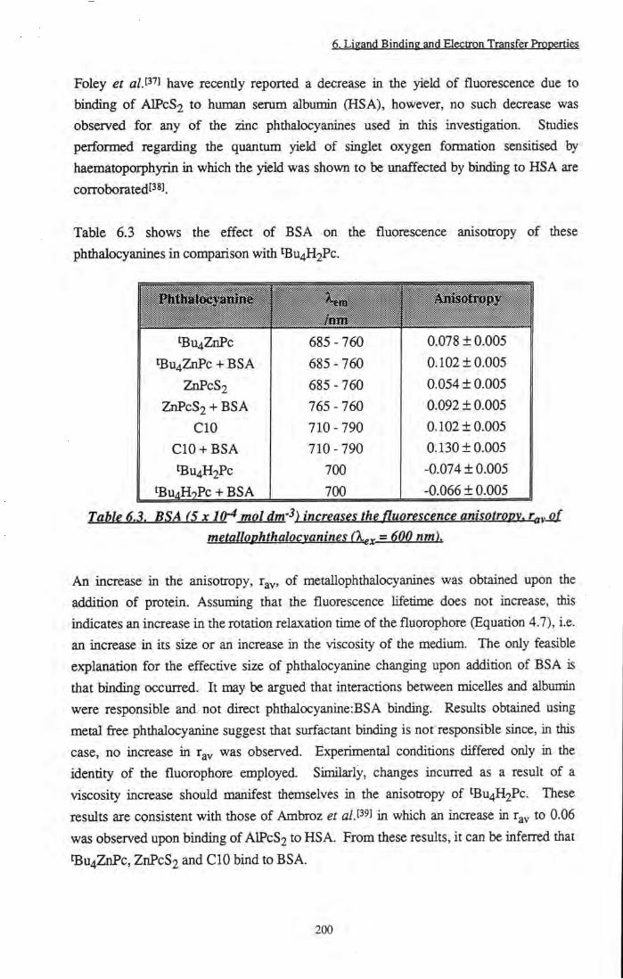
6. Ligand Binding and Electron Transfer Properties
Foley et a/.P' J have recently reported a decrease in the yield of fluorescence due to binding of AIPCS2 to human serum albumin (HSA), however, no such decrease was observed for any of the zinc phthalocyanines used in this investigation. Studies performed regarding the quantum yield of singlet oxygen formation sensitised by haematoporphyrin in which the yieki was shown to be unaffected by binding to HSA are corroboratedPsi.
Table 6.3 shows the effect of BSA on the fluorescence anisotropy of these phthalocyanines in comparison with U4H2Pc.
1 PJithaloej 'aniRe i ^^^^^^^^^^^^^^^^^^^^^ hxm ^^^^^^^^^^^^^^^^^^^^^
L ' hxm tBu4ZnPc 685 - 760 0.078 ± 0 . 0 0 5
^U4ZnPc + BSA 685 - 760 0.102 ± 0 . 0 0 5
ZnPcS2 685 - 760 0.054 ± 0 . 0 0 5
ZnPcS2 + BSA 765 - 760 0.092 ± 0 . 0 0 5
C I O 710 - 790 0.102 ± 0 . 0 0 5
C I O + BSA 710 - 790 0.130 ± 0 . 0 0 5
tBu4H2Pc 700 -0.074 ± 0 . 0 0 5
1 tBu4H7Pc -1- BSA 700 -0.066 ± 0 . 0 0 5
BSA X J (H mQl 4m-^) incrm?? th? fluomC^Tu;^ mmtrQPY, TgyM met(illQphthalQQy(inin^^ (X^x = ^00
An increase in the anisotropy, r y, of metallophthalocyanines was obtained upon the addition of protein. Assuming that the fluorescence lifetime does not increase, this indicates an increase in the rotation relaxation time of the fluorophore (Equation 4.7), i.e. an increase in its size or an increase in the viscosity of the medium. The only feasible explanation for the effective size of phthalocyanine changing upon addition of BSA is that binding occurred. It may be argued that interactions between micelles and albumin were responsible and not direct phthalocyanine:BSA binding. Results obtained using metal fi^e phthalocyanine suggest that surfactant binding is not responsible since, in this case, no iacrease in r y was observed. Experimental conditions differed only in the identity of the fluorophore employed. Similarly, changes incurred as a result of a viscosity increase should manifest themselves in the anisotropy of ^Bu4H2Pc. These results are consistent with those of Ambroz et a/.P'l in which an increase in r y to 0.06 was observed upon binding of AIPCS2 to HSA. From these results, it can be inferred that tBu4ZnPc, ZnPcS2 and C I O bind to BSA.
200

6. Ligand Binding and Electron Transfer Properties
It has been reported that quenching of phthalocyanine triplet state species by molecular oxygen is inhibited upon binding to proteint''°]'t'*^l Davila and Harrimanl^^J have shown
0.025
0.020
0.015 --
0.010 --
0.005
0.000
-0.005
-10
0.025
0.020 --
0.015 --
0.010
0.005
0.000
-0.005
k = 270000 s" 0.020 +
0.010 +
0.005
0.000
-0.005 -0.5 0 0.5 1 1.5 2 2.5 3 3.5 4 4.5
Tiine/|iS
k i = 41900 s-
k2= 3200
10 30
Time/us
50 70 90
k = 3100 s -1
««., I it.iAiiijMiiiiLidtyi,flnJili J U J l
-0.5 0 0.5 1 1.5 2 2.5 3 3.5 4 4.5
Time/p,s
Figure 6.7. Triplet transient decay of^Bu^ZnPc in 1% TX/PBS + 4x10-^ mol dm'^
BSA. Irradiation by A. 4 laser shots B. 30 laser shots C. 44 laser shots
(X^^ = 638 nm. ~ 0.2 ml/pulse). Exponential fits and residuals are shown.
201

6. Ligand Binding and Electron Transfer Properties
that the triplet state lifetime of various phthalocyanines was affected by binding to human serum albumin. Biexponential decays were obtained which were assigned to binding of sensitisers to different sites available within the protein which protect the dye from oxygen by varying amounts. It was proposed that extended lifetimes were due to binding to an inner site, which protected excited states from quenching by oxygen. For bulky phthalocyanines with axial ligands, such as AlPc, no evidence of an extended Ufetime was observed, suggesting that binding occurred exclusively to exposed sites on the surface of the protein. In an attempt to corroborate binding of hydrophobic zinc phthalocyanines to BSA, reaction of ^tot* states of ZnPcS2, ^Bu4ZnPc and CIO with O2 in the presence of BSA was smdied. The lifetime, Xj, was found to be dependent on the number of laser pulses used to excite the sample. Figure 6.7 shows the triplet transient decay recorded after 4, 30 and 44 laser pulses of ~ 0.2 mJ/pulse. After four shots (Sample B), a lifetime of 4 ± 2 jis was recorded for an oxygen concentration of 2.6 x 10"^ mol dm"^ (aerated sample). This increased to 325 ± 25 |j.s after 44 shots (Sample C) and is comparable to that measured in the absence of oxygen (310 |j.s - Table 6.2). At an intermediate number of pulses a biexponential decay was obtained (Sample B), indicative of decay from two distinct types of triplet species. Agitation of Samples B and C regenerated the short lived transient of Sample A.
The absorption spectra of die solutions were recorded before and after irradiation.
Evidence of increased absorption between 310 and 330 nm was observed. This is
representative of photooxidation of the tryptophan residue of BSA to N-
formylkynurenine (NFK)['*2].['*3] - a well known product of singlet oxygen reaction with
tryptophan. To investigate this further, a controlled smdy of photooxidation of BSA
sensitised by ^Bu4ZnPc was performed. Formation of NFK was recorded by monitoring
the absorbance of the solution at 322 nm. As Figure 6.8 shows, a steady increase in 322
nm absorption was observed followed by a plateau region. Photooxidation sensitised by
CIO and ZnPcS2 gave similar results. Concurrent photodegradation of the
phthalocyanines was observed. This explains the reason behind the plateau region of
Figure 6.8 where production of NFK reaches a maximum. Specifically,
photodegradation of phthalocyanine resulted in the production of ever-decreasing
amounts of singlet oxygen - the active species involved in oxidation of BSA - ±us , the
rate of production of NFK decreased until the concentration of singlet oxygen was
insufficient to induce further oxidation of tryptophan within the BSA.
202

6. Ligand Binding and Electron Transfer Properties
2.3
2.1
1.9
1.7
1.5
1.3
1.1
0.9
0.7 -1- 'Bu4ZnPc in 1% TX/PBS
0.5
'Bu4ZnPc + 5 X 10- M BSA in 1% TX/PBS
\ \ 1 \ 1 1 \ 0 100 200 300 400 500 600 700 800
Time/s
Figure 6.8. Increase in absorbance at 322 nm for ^Bu^ZnPc + 5 x IQ-^mol dm'^ BSA upon irradiation by red light (X > 610 nm). Effect of irradiation on a phthalocvanine
only solution is also shown.
Figure 6.8 also shows the change in absorbance at 322 nm of a phthalocyanine only solution. A decrease was observed which confirms that growth of absorption at 322 nm in the presence of BSA was not due to formation of photodegradation products of the phthalocyanine. Photooxidation of tryptophan is not a new or uncommon phenomenon and has been smdied in detail by several groupst ' l't' ^l'f'' ].
In order to study the effect of BSA on phthalocyanine triplet state properties in the absence of photooxidation, 0.01 mol dm-^ NaN^ was added to quench singlet oxygen species as they were produced (kg = 3.8 x 10^ dm^ mol'^ s-'^^'^'^K i.e. XA = 0.25 [is.
Figure 6.9 shows an example of triplet state quenching by ground state oxygen under
these conditions. In contrast to the results described above, monoexponential decays
were recorded at all concentrations of oxygen and were independent of irradiation
frequency.
203

6. Ligand Binding and Electron Transfer Properties
450000
400000 --
350000 -
300000 --
^ 250000 --
y = 2E+09x-H9000
200000 +
150000 +
100000 +
50000 +
0.00005 0.0001 0.00015
[02]/moIdm-
0.0002
Figure 6.9. CIO triplet state quenching bv in 1% TX/PBS. = 638 nm. ~ 0.2 mJ/pulse)
Table 6.4 collates quenching rate constants determined for each phthalocyanine and the effect of BSA.
1' PfaMoc nme
tBu4ZnPc 1.9 ±0 .2
tBu4ZnPc-i-BSA 1.4 ±0 .1
ZnPcS2 1.2 ±0 .2
ZnPcS2+BSA 1.0 ±0 .1
CIO 1.4 ±0 .2
ClO+BSA 1.9 ±0 .3
TdHe 6,4, Rate constants Qf triplet state quenching fey Qj^ygen jn the presence of
5xl0^mQldm-3BM.
Since kg was unaffected by the presence of BSA, it is apparent that BSA has no significant protective effect on the quenching of zinc phthalocyanine triplet state species by oxygen. It may be concluded, therefore, that the protective effects described above (Figure 6.7) were not a consequence of Pc:BSA binding and may be due to interaction with oxidised protein species. Several explanations present themselves. Firstly, it is
204

6. Ligand Binding and Electron Transfer Properties
possible that whilst binding to parent BSA is very weak (if it is present at all), oxidation of tryptophan residues to NEK may promote binding either directly or through conformational changes of the protein. However, shifts in sensitiser ground state absorption spectra were not observed, thus, this explanation is unlikely. Secondly, and much more simply, it is possible that oxidation of protein molecules in the illuminated volume depleted the available supply of oxygen, hence reducing quenching (k = 3100 s-^). Agitation of the sample regenerated the oxygen concentration and the triplet state species was quenched once more (k = 270000 s ). Alternatively, it is possible that photoinduced cross-linking of BSA occurred to create a protective network surrounding phthalocyanine, thus shielding excited states from deactivation. It is known that cross-linking of proteins occurs following amino-acid oxidationt' ^LK?!, thus this may be a viable explanation, however, further work is required to confirm the reason for these results.
From the experimental observations described above, it is not possible to confirm
Pc:BSA binding of tBu4ZnPc or CIO. Fluorescence anisotropy measurements suggest an
interaction, however, this has not been corroborated by any of the photophysical
measurements performed. No effect on the absorption, fluorescence or triplet state
properties was observed and triplet state quenching by oxygen was unaffected by the
presence of BSA.
205

6. Ligand Binding and Electron Transfer Properties
6.3.2 Electron Transfer Reactions 6.3.2.1 Simple Organic Quenchers 6.32.1.1 Results
Electron transfer reactions of excited state phthalocyanines with several simple amine
and sulfur based quenchers have been investigated (Figure 6.10).
OMe
Q NMec
OMe
o
NMe,
o NMe2
1, 4 Dimethoxybenzene 1,4 Tetramethylphenylenediamine E (Q^-IQ) = 1.59 V E(Q +-/Q) = 1.02 V
Dimethylaniline E(Q+-/Q) = 0.4V
Diazabicyclo[2, 2,2] octane E(Q^-IQ) = 0.81 V
Tetrathiafulvalene E(Q-^-/Q) = 0.59 V
Figure 6.10. Structure of simple organic quenchers.
a) Diazabicyclo[2,2,2]octane (DABCO)
The fluorescence yield of ZnPc in 1% pyr/tol solution was unaffected by DABCO at concentrations below 0.01 mol dm^^, however, a blue shift of the emission maximum, A^ax' fro^ ^ observed. A 1 nm hypsochromic shift of the ground state absorption spectrum was also observed (672 nm 671 nm). Addition of further DABCO resulted in a steady decrease in the yield of fluorescence emission. A^ax remained constant at 679 nm and no further shift in the energy of the Sj <— SQ transition was detected. A Stem-Volmer analysisf ^^ (Equation 6.7) was performed to determine a rate constant, kq, for the quenching process.
0) 0 _ l + kQ%p[Q] (6.7)
206

6. Ligand Binding and Electron Transfer Properties
where <I>o is the yield of emission at [Q] = 0 mol dm-3, is the yield at a quencher concentration, Q, I C Q is the rate constant for quenching and Tp is the fluorescence lifetime at [Q] = 0 mol dm-3. Figure 6.11 shows the linear plot obtained.
1.6
1.5 +
1.4 +
1.3 +
1.2
1.1 +
y = 5.6x + 1
0.02 0.04 0.06
[DABCOj/moldm-^
0.08 0.1
Figure 6.11. Stem-Volmer plot showing quenching ofZnPc emission bv DABCO in 1% pvr/tol.
T f = 3.5 ns. = 630 nm.
I C Q was calculated to be 1.6 ± 0.1 x 10^ dm^ mol'^ s"i. A fluorescence lifetime study of the quenching by DABCO was carried out, and a value of kQ = 1.2 ± 0.1 X 10^ dm3 mol"l s" determined. This value is an order of magnitude lower than the diffusion limited rate in toluene (1.1 x 10^^ dm^ mol'^ s'^). tp was unaffected by 0.01 mol dm-3 DABCO.
It is imlikely that quenching was due to an energy transfer process since the singlet energy of DABCO is much greater than that of the phthalocyanine. The results described above may be explained by the reaction scheme depicted in Figure 6.12. Kj , represents the equilibrium constant for complex formation and all other terms are defmed in Section 6.1.2.1.
207

6. Ligand Binding and Electron Transfer Properties
DABCO. hv ZnPc ZnPc.DABCO- ^ZnPc.DABcd ± ^(DABCO...ZnPc.DABCO)*
DABCO Encounter Complex
DABCO
\ DABCO*'... ^DABCOZnPc") *
Ion Pair
diss
DABCO"^' + DABCOZnPc'
Solvated Ions
Figure 6.12. Formation of DABCO...ZnPc.DABCO ion pair upon irradiation of ZnPc in 1% pxritol in the presence of> 0.01 mol dm-3 DABCO.
Addition of small amounts of DABCO lead to the formation of a ground state complex -DAB CO. ZnPc - in which a molecule of DABCO has replaced the pyridine as the axial ligand on the phthalocyanine . Evidence for this comes from the shift in the absorption spectrum. The fluorescence lifetime was unaffected by Mgand exchange (0.01 mol dm"^ DABCO). It is possible that at concentrations of 0.01 mol dm-^ two opposing effects must be considered - a longer lifetime due to complex formation and reduction of this lifetime due to quenching - leading to a net zero change. Alternatively, the electrode potential of ZnPc may be changed by DABCO binding such that ZnPc.DABCO is less susceptible to reduction than ZnPc.py. Upon addition of further quantities of DABCO, photo-induced electron transfer from free DABCO molecules to ZnPc.DABCO was observed. This may be assumed to follow the reaction profile of Rehm and Wellerl^^). Phthalocyanine radical anions could not be observed by nanosecond flash photolysis. This was for two reasons. Firstiy, toluene is a non-polar solvent, hence solvation of radical products is poor and energetically unfavourable, i.e. k^[^^ is small. Secondly, the ion pair resulting from singlet state quenching has a spin allowed back reaction. Consequentiy, k gy is large and observation of radicals produced by singlet state quenching is rare. Triplet state quenching by DABCO was not observed.
208
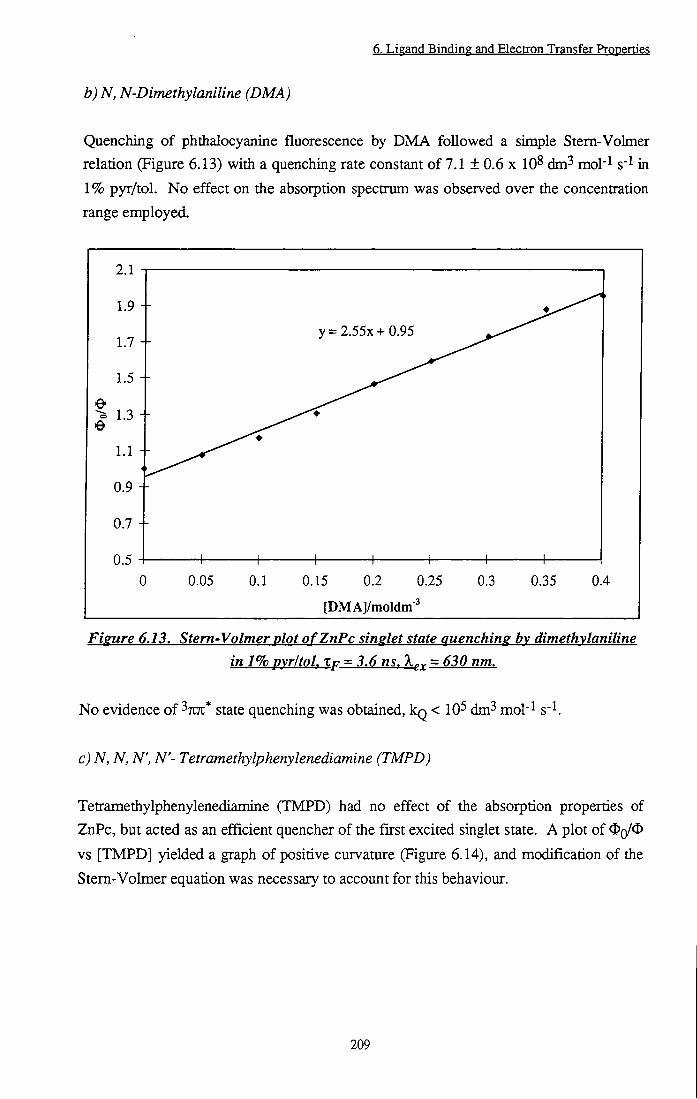
6. Li^and Binding and Electron Transfer Properties
b) N, N-Dimethylaniline (DMA)
Quenching of phthalocyanine fluorescence by DMA followed a simple Stem-Volmer relation (Figure 6.13) with a quenching rate constant of 7.1 ± 0.6 x 10^ dm^ mol'^ s" in 1% pyr/tol. No effect on the absorption spectrum was observed over the concentration range employed.
2.1
1.9 +
1.7
1.5 --
1.3 --
1.1 --
0.9
0.7 +
0.5
y = 2.55x + 0.95
+ 0 0.05 0.1 0.15 0.2 0.25 0.3 0.35 0.4
[DMAJ/moldm-^
Figure 6.13. Stem-Volmer plot o fZnPc singlet state quenching bv dimethvlaniline in 1% pyr/tol, Xp = 3.6 ns, = 630 nm.
No evidence of ^7ui* state quenching was obtained, kg < 10^ dm^ mol'^ s" .
c) N, N, N', N'- Tetramethylphenylenediamine (TMPD)
Tetramethylphenylenediamine (TMPD) had no effect of the absorption properties of ZnPc, but acted as an efficient quencher of the first excited singlet state. A plot of ^Q/^ vs [TMPD] yielded a graph of positive curvature (Figure 6.14), and modification of the Stem-Volmer equation was necessary to account for this behaviour.
209

6. Ligand Binding and Electron Transfer Properties
8
7
6 +
5
4 +
+ 0 0.005 0.01 0.015 0.02 0.025 0.03 0.035 0.04
[TMPD]/moldm-^
Figure 6.14. Stem-Volmer plot of ZnPc fluorescence quenching by TMPD in 1% pvr/tol. Tf = 3.5 ns. X^^ = 630 nm. Fit o f static quenching model.
Curved Stem-Volmer plots can usually be accounted for using one of three explanations:
i) Ground state complex formation ii) Static quenching
iii) Finite sink approximation
Ground state complex formation can result in an increase in rate constant, kq, if the complex formed between fluorophore and quencher is non-fluorescent. Complex formation is usually indicated by a shift in the UVA^is spectmm, therefore, in this case can be dismissed.
Static quenching[5 l'[52] occurs at high concentrations, when the fluorophore and
quenching molecule are already within the interaction sphere of volume, V, on excitation.
Under these conditions, diffusion is not required and quenching occurs almost
instantaneously. The probability of this situation arising depends on the volume of
interaction, V and the concentration of quencher molecules, [Q]- I f tiie quencher is
randomly distributed, then the probability of static quenching is given by the Poisson
distribution, e' tQJ. Thus, the Stem-Volmer equation may be modifiedt^^l to give:
210
6. Ligand Binding and Electrpn Transfer Prppertig?
^ = l + Ksv[Q]e^^Q^ (6.8)
where Kgy is the Stem-Volmer constant, kqXp. Application of this equation to the
experimental data gave a good fit (Figure 6.14). A quenching constant of 1.5 ± 0.1 x
IQlO dm^ mol-1 S'^ and a volume of interaction, V of 5.5 dm^ mol'^ were determined. V
represents the volume surrounding a fluorophore in which quenching may occur without
the need for diffusion controlled collision of both reactants. The value determined in this
study (5.5 dm^ mol'^) is comparable to that reported by Zengt^i] static quenching of
hydroxyl dimethyl carbazole by a-tocopherol (3.7 dm^ mol'l).
The finite sink approximation[531.[54],[55] is a modification of the Smoluchowski, Collins
and Kimball (SCK) expression for the rate constant of a dynamic quenching system.
This is based on a solution of the time independent flux equation[5''' within the limits
[Q](°°) at r(oo) and [Q](R) at R, where R is the reactant encounter distance. This leads
to Equation 6.9 (the reader is referred to References 53 and 54 for derivation of this
equation).
^ 4%NDR + ka
where N is Avogadro's constant, D is the sum of the diffusion coefficients of the
reactants, and k^ is the activation energy conQ-olled rate constant describing quenching at
a distance R. In the case of efficient quenching. Equation 6.9 fails to account for a
dependence of kg on the quencher concentration. An increase of k g with [Q] may be
due to static effects described earlier or to effects introduced by an initial time
dependence of the concentration gradient. These can be eliminated by assuming that
only the first encounter between fluorophore and quencher molecule is of importance,
thereby defining an initial average separation, XQ (sink radius). The diffusive region of
interest will therefore lie within the limits R < r < tq and the flux equation can be
integrated using the boundaries [Q](ro) at TQ and [Q](R) at R to give Equation 6.10.
^'^NDRk, ^^^^^
4%NDR + ka (l-R/r^ )
The sink radius, TQ has been defined as the most probable nearest neighbour separation.
Substitution of the appropriate distribution for TQ (TQ = (27cN[Q])'l/3) into the inverse of
211

6. Ligand Binding and Electron Transfer Properties
Equation 6.10 and division by the fluorescence lifetime in the absence of quencher, tf, yields the altemative Stem-Volmer relationship:
where
-1 fi -1 ( 2 % N / 3 KSV =K'^SV - \ (6.11)
K 4izNDRXpka
5^ 4TINDR + k„ ^^^^ (6.12)
Thus, the quenching coefficient, kg was obtained from the intercept of a plot of Ksv'^ vs [Q}^^^ (Figure 6.15). kq using this approximation was calculated to be 1.2 ± 0.1 x 10^^ dm^ mol"l s" .
0.022
0.02
> 0.018
0.016 +
0.014 +
0.012
y = -0.037X + 0.028
0.2 0.25 0.3 0.35
[TMPDJ^'^/mol^'^dm-'
0.4 0.45
Figure 6.15. Application of finite sink approximation to TMPD quenching data.
For comparison, the rate obtained by fitting a straight line to the data acquired at low concentrations of TMPD was determined. In this case, kq = 1.5 ± 0.1 x 10^^ dm^ mol"l
Flash photolysis investigations of 7U7t* states of ZnPc in 1% pyr/tol solution revealed
efficient quenching of the triplet state by TMPD (Figure 6.16). A quenching rate of 3.7
212

6. Ligand Binding and Electron Transfer Properties
± 0 . 3 x 10^ dm3 mol-1 s" was obtained from the gradient of a plot of k (rate of triplet state deactivation) vs [TMPD].
60000
50000 +
40000
- | 30000
20000 +
10000 +
y = 3.7E+08X + 5600
+ 0 0.00002 0.00004 0.00006 0.00008 0.0001 0.00012
[TMPD]/moldm-^
Fisure 6.16. ^ZnPc* Triplet state quenching bv TMPD in 1% pvr/tol.
Triplet state quenching may be assumed to follow a mechanism analogous to that proposed for singlet state quenching (Figure 6.2). Due to the spin forbidden nature of back electron transfer by ion pairs formed though triplet state electron transfer, kj^^ is slow and the rate of dissociation, k^^^ may be expected to dominate. However, the non-polar nature of toluene inhibited ion pair separation and recombination occurred; thus no evidence of phthalocyanine radical anions was observed.
d) 1,4 Dimethoxybenzene (DMB)
The absorption, fluorescence and triplet state characteristics of ZnPc in 1% pyr/tol were unaffected by DMB. It was concluded that complex formation between ground or excited states of phthalocyanine and dimethoxybenzene and electron transfer quenching of singlet or triplet state species by DMB did not occur.
213

6. Ligand Binding and Electron Transfer Properties.
e) Tetrathiafuhalene (TTF) i) Hgrnpgeneopg gplytipn
Tetrathiafulvalene (TTF) was found to efficientiy quench the first excited singlet state of
iBu4ZnPc in 1% pyr/tol, kq = 1.1 ± 0.1 x 10^0 dm^ mol"! s'l, c.f. k^ j f j = 1.1 x 10^0 dm^
mol"l s'^in toluene. By contrast, when octadecylzinc phthalocyanine was used as the
electron acceptor, no evidence of fluorescence quenching was observed. Electron
transfer quenching of fluorescence emission from ^Bu4H2Pc also yielded a diffusion
Umited quenching rate constant of 1.1 ± 0.1 x lO^^ dm^ mol-^ s' in toluene (Xp = 5.6
nsf^^l). TTF had no effect on the lifetime of triplet state species. The fluorescence yields
of coupled TTF-Pc complexes, Pc(TTF)8 and Pc(TTF)4 (Figure 6.3) were measured.
Values of < 10-^ and 1.7 ± 0.3 x 10⁻³ were obtained for Pc(TTF)8 and Pc(TTF)4,
respectively. These yields are negligible when compared with that determined for the
parent phthalocyanine, tBu4H2Pc ( % = 0.5). We propose[29], tiiat in compounds
Pc(TTF)8 and Pc(TTF)4, rapid intramolecular electron transfer occurred between the
covalentiy Unked TTF moieties to generate a charge transfer state, which rapidly
returned to the ground state, resulting in reduced fluorescence yields of these
compounds.
ii) Heterogeneous Solution
Having established that TTF will undergo redox reactions with excited singlet state
phthalocyanine in homogeneous solution, quenching kinetics in micellar solution (10%
T X / H 2 O ) were investigated. Stem-Volmer plots obtained when singlet states of
tBu4ZnPc and CIO in 1% TX/PBS were quenched by TTF in 10% T X / H 2 O are
displayed in Figure 6.17. The rate constants of electron transfer were estimated using
the gradient at low concentrations of quencher molecules. Under these conditions it may
be assumed tiiat the concentration of surfactant (TX) was constant and that = [ Q ] L ,
i.e. the total concentration of added quencher equals the concentration in the hpid phase.
The value obtained for tBu4ZnPc (kq = 1.8 ± 0.1 x 10^1 dm^ mol"! s"!) is an order of
magrutude greater than that obtained in homogeneous media
In this system, it was possible to measure a decrease in the yield of fluorescence of CIO
upon adding TTF (kq = 5.4 ± 1 x lO^^ dm^ mol-^ s" ). This value was unattainable in
1% pyr/tol solution. The large increase in kg in micellar solution may be explained by
the location of fluorophore and quencher. Due to tiieir hydrophobic nature,
phthalocyanines and TTF are both expected to be located within the micellar core,
allowing enhanced contact between the phthalocyanine and TTF, i.e. a much greater
214
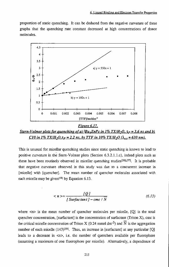
6. Ligand Binding and Electron Transfer Properties
proportion of static quenching. It can be deduced from the negative curvature of these graphs that the quenching rate constant decreased at high concentrations of donor molecules.
4.5
a)y = 550x+ 1
b) y = lOOx + 1
0.001 0.002 0.003 0.004 0.005 0.006 0.007 0.008
[TTFl/moldm'
Figure 6.17. Stem-Volmer plots for quenching of a) ^Bu^ZnPc in 1% TX/H2O. Xp = 3.6 ns and b)
CIO in 1% TX/H2O.XP = 2.2 ns. bv TTF in 10% TX/H2O (X^x = 630 nm).
This is unusual for micellar quenching studies since static quenching is known to lead to positive curvature in the Stem-Volmer plots (Section 6.3.2.l.l.c), indeed plots such as these have been routinely observed in miceUar quenching studiesf l-f ' J. It is probable that negative curvature observed in this study was due to a concurrent increase in [micelle] with [quencher]. The mean number of quencher molecules associated with each micelle may be givent^^l by Equation 6.13.
<n>= [QJ
[Surfactant] — cmc / N (6.13)
where <n> is the mean number of quencher molecules per micelle, [Q] is the total
quencher concentration, [surfactant] is the concentration of surfactant (Triton X), cmc is
the critical micelle concentration of Triton X (0.24 mmol dm'^) and N is the aggregation
number of each miceUe (143) 59). Thus, an increase in [surfactant] at any particular [Q] leads to a decrease in <n>, i.e. the number of quenchers available per fluorophore
(assuming a maximum of one fluorophore per micelle). Alternatively, a dependence of
215
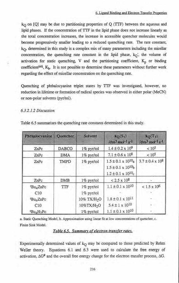
Ligan(j Binding and El^pqn Trangfgr Prppgrpgs
kg on [Q] may be due to partitioning properties of Q (TTF) between the aqueous and lipid phases. If the concentration of TTF in the lipid phase does not increase lineaiiy as the total concentration increases, the increase in accessible quencher molecules would become progressively smaller leading to a reduced quenching rate. The rate constant, kg, determined in this study is a complex mix of many parameters including the micellar concentration, the quenching rate constant in the lipid phase, kg', the volume of activation for static quenching, V and the partitioning coefficient, Kp or binding coefficientf^°J, K|,. It is not possible to determine these parameters without ftirther work regarding the effect of micellar concentration on the quenching rate.
Quenching of phtiialocyanine triplet states by TTF was investigated, however, no reduction in lifetime or formation of radical species was observed in either polar (MeCN) or non-polar solvents (pyr/tol).
63.2.12 Discussion
Table 6.5 siunmarises the quenching rate constants determined in this study.
Quencher Solvenr
ZnPc DABCO 1% pyr/tol 1.4 ± 0.2 X 10⁹ <105
ZnPc DMA 1% pyr/tol 7.1 ±0 .6 x 108 <105
ZnPc TMPD 1% pyr/tol 1.5 ±0 .1 X 1 0 %
1.5 + 0.1 xlQlOb 1.2 ±0 .1 X IQlOc
3.7 ± 0 . 4 x 1 0 8
ZnPc DMB 1% pyr/tol < 2 .5 X 108 -
tBu4ZnPc CIO
tBu4ZnPc CIO
tBU4H2Pc
TTF 1% pyr/tol 1% pyr/tol
10% T X / H 2 O
1 0 % T X / H 2 O
1% pyr/tol
1.1 ± 0 . 1 X lOlO
1 . 8 ± 0 . 1 X lOll 5 . 4 ± 1 X IQlO
1.1 ±0 .1 X 10^0
< 1.5 X 10⁶
a. Static Quenching Model, b. Approximation using linear fit at low concentrations of quencher, c.
Finite Sink Model.
Table 6.5. Summary of electron transfer rates.
Experimentally determined values of kg may be compared to those predicted by Rehm
WellCT theory. Equations 6.1 and 6.3 were used to calculate the free energy of
activation, AG* and the overall free energy change for the electron transfer process, AG.
216

6. Lig^nd Binding gnd gggtrpn Transfer Properties
Substituting these parameters into Equation 6.2 allowed the predicted value of kg to be calculated, was estimatedt ] using Equation 6.14.
8RT 3T1
(6.14)
where R is the gas constant (8.314 J Kr^ mol-i), T is the temperature (298 K) and TI is the viscosity of the solvent. The term k^AV^kfjeac was estimated as 0.25 and AG*(0) as 10 kJ mol-1 (the values determined by Weller for acetonitrile solution). Although, stricUy speaking, this value will vary witii solvent, the magnitude of the change is not expected to have a significant effect on the calculated value of kg, thus, this approximation is deemed reasonable. The reaction encounter distance, a was estimated as 1 nm. A further approximation that should be noted is that reported reduction potentials have been used which were not measured in the same solvents as those employed in this study. Table 6.6 collates calculated values for quaiching of ^Pc* species of ZnPc or tBu4ZnPc. The ground state redox potential of these phthalocyanines has been taken as E (ZnPc / ZnPc' ) = -0.6 Vl ) and the singlet state energy, AEQ^Q' ^
1.83 eVPO].
Quencher V v s N H E
TMPD 0.41161 -138 0.72 8.3 X 10⁹ 1.5 X IQlOa
TTF 0.59["] -120 0.83 8.1 x 10⁹ 1.1 X lOlOb
DABCO 0.81[6'H -99 1 8x10⁹ 1.4 x 10⁹
DMA 1.02116] -78.3 1.24 7.8 x 10⁹ 7.1 X 10⁸
DMB 1.59C161 -23.2 3.71 5xl09 < 2.5 X 10⁸
a. Calculated using static quaiching model, b. Phthalocyanine = ^U4ZnPc.
Table 6.6. Calculated quenching rates of^ZnPc* bv simple organic electron donors in 1% pyridine / toluene.
Comparison of these values with those obtained experimentally (Table 6.5) shows that some discrepancy exists between theoretically predicted values and data obtained. Weller suggested that a reaction proceeding within a factor of two of the theoretical rate constant may be assumed to follow an outer sphere electron transfer mechanism. TMPD and TTF fit this criterion well. The observed rates of quenching by DABCO, DMA and DMB are somewhat slower than predicted. Since it has been shown that DABCO forms a complex with ZnPc before quenching occurs, it is not surprising that in this case, kg was slower than that expected for pure electron transfer. In addition, calculated kg
217

6. Ligand Binding and Electron Transfer Properties
values are critically dependent on the electrode potentials chosen for quencher compounds and reduction of the ground state of phthalocyanine. Some variation exists in published values for electrode potentials; for example, values of -0.6 V[ 2] and -0.72 V[65] are reported for E (ZnPc / ZnPc--) and values for E (DABCO+- / DABCO) range from 0.81 V[^3] iQ 1 14 Y[66] Thus, inaccuracy in predicted values of kg is not surprising. Perhaps most significantiy, use of toluene as solvent may have a large effect on E for all species involved. In the main, Uterature values reported were determined in acetonitrile, a highly polar solvent, in which electron transfer properties are expected to be different to those in a non-polar solvent such as toluene. Poor solubUity of ZnPc in MeCN prevented experiments being performed in this solvent system.
Quenching of 3pc* by DABCO, DMA and TTF was not observed. Quenching by TMPD was a factor of thirty slower (3.7 x 10^ dm^ moV^ s'^) than the calculated diffusion controlled rate in toluene (1.1 x 10^^ dm^ mol'^ s ). The concentration of quencher molecules is in large excess over the concentration of excited phthalocyanine species, thus, changes in the concentration via reaction with singlet state species will be minimal. In addition, it may be shown using Equation 6.7 and the calculated rate constant for singlet state quenching (1.4 x 10^^ ^3 mol-^ s ) that at the concentrations of TMPD used for triplet state quenching (< 10" mol dmr^) < 1% of singlet state species have been quenched, hence, this is unlikely to account for such a large difference. A more plausible explanation is that triplet state quenching is activation conttolled as opposed to singlet state quenching which is diffusion controlled.
Studies comparing quenching of ZnPc with peripheral tertiary butyl and decyl hgands by TTF, have clearly demonstrated that tBu4ZnPc is a better electron acceptor than CIO. This may be for a number of reasons. Firstly, the energy required to produce die singlet state of CIO is lower than that of tBu4ZnPc, leading to a lower excited state reduction potential. The reduction potential of H^Bu4ZnPc)* was calculated[^''] to be 1.23 V whilst that of l(ClO)* was only 1.17 V (assuming the ground state reduction potential to be the same as ZnPc (-0.6 V)). However, substitution of these values into Equations 6.1 to 6.3 leads to kg (calc) in H 2 O = 4.5 x 10^ dm^ mol-^ s"! for tBu4ZnPc vs 4.4 x 10^ dm^ mol-^ s" for CIO. Thus, quenching rates would not be expected to be significantiy different. Alternatively, it is possible that during studies performed in micellar solution, each phthalocyanine may be located in a different region of the micelle, leading to varying accessibility to quencher molecules. Zhang et a/.f^s] have shown that die electron transfer rate from tyrosine to tetra sulfonated galhum phthalocyanine (GaPcS4) was greatly increased upon incorporation into micelles of cetylttimethylammonium chloride (CTAC). By contrast, that of disulfonated zinc phthalocyanine (ZnPcS2) was unaffected.
218
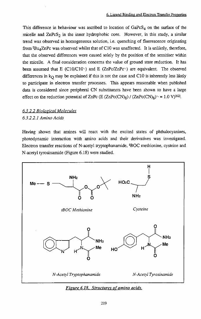
6. Ligand Binding and Eleptrpn Trangfgr Prpperp^
This difference in behaviour was ascribed to location of GaPcS4 on the surface of the micelle and ZnPcS2 in the inner hydrophobic core. However, in this study, a similar trend was observed in homogeneous solution, i.e. quenching of fluorescence originating from Bu4ZnPc was observed whilst that of CIO was unaffected. It is unlikely, therefore, that the observed differences were caused solely by the position of the sensitiser within the micelle. A final consideration concerns the value of ground state reduction. It has been assumed that E (ClO/ClO"-) and E (ZnPc/ZnPc" ) are equivalent. The observed differences in kg may be explained if this is not the case and CIO is inherendy less likely to participate in electron transfer processes. This appears reasonable when pubUshed data is considered since peripheral CN substituents have been shown to have a large effect on tiie reduction potential of ZnPc (E (ZnPc(CN)8) / (ZnPc(CN)8)-- = 1.0 V)[62].
6.3.2.2 Biological Molecules 6.3.2.2.1 Amino Acids
Having shown that amines will react with the excited states of phthalocyanines, photodynamic interaction with amino acids and their derivatives was investigated Electron transfer reactions of N-acetyl tryptophanamide, 'BGC methionine, cysteine and N-acetyl tyrosinamide (Figure 6.18) were studied.
H
NH2
Me
O O
H O 2 C .
7 y
tBOC Methionine
NH2
Cysteine
NH2
Me N. / M e
N-Acetyl Tryptophanamide N-Acetyl Tyrosinamide
Figure 6.18. Structures of amino acids.
219

6. Ligand Binding and Electron Transfer Prpp^rtigg
No evidence of quenching of ^Bu4ZnPc singlet state species was detected for any of the aminoacids under study in either methanol or 1% T X / H 2 O solution (kg < 1.4 x 10⁹ dm^ mol-1 s-1).
Studies of ^U4ZnPc triplet state quenching by ^BOC methionine in methanol revealed a non-linear reduction ia the lifetime of ^KK* species with increasing ^BOC methionine concentrations (Figure 6.19). No effect on the wavelength of maximum ground state absorption was observed at ^BOC methionine concentrations of 0.01 mol dm-3 so it was assumed that ground state complex formation did not occur. No evidence for the formation of radical species was obtained. The wavelength of maximum absorption of triplet species, X^^ = 490 ± 10 nm, was unaffected by the addition of 0.01 mol dm-3 ^BOC methionine.
10500
10000
9500
9000 -
8500 -
8000 +
7500 +
7000 +
— \ — 0.002
+ 0.004 0.006 0.008
[Methionine]/moldm'^
0.01 0.012
Figure 6.19. Increase in rate of decay of^KK* states offBu^ZnPc in MeOH upon addition of^BOC methionine. The fit of Equation 5.9 (substituting [Met] for fH+Rjo
the data is also shown.
Methionine has been shown to have a reduction potential of 1.34 V vs NHE, thus
Equation 6.1 predicts an endotheraiic electron transfer reaction with phthalocyanine. In
addition, no evidence of radical species was obtained, therefore, quenching of Xj by this
mechanism is unlikely. It is probable that methionine (Met) formed an excited state
complex (exciplex) with triplet state zinc phthalocyanine (^Pc*) which existed in
220

6. Ligand Binding and Electron Transfer Properties
eqiulibrium with unbound species (Figure 6.20). As the concentration of ^BOC methionine increased, the equihbrium shifted to the right, forming a complex with decay rate, k2. At zero concentration, the measured rate of decay was representative of that of the parent phthalocyanine, k j . Experimentally, two limiting cases, k^ and k2, were measured to be 6000 ± 300 s' and 9500 ± 500 s' respectively. These correspond to a hfetime of 100 |is for bound Pc.Met species (0.01 mol dm-3 ^BOC metiiionine) and 170 IIS for ^Bu4ZnPc (0 mol dm^^ ^BOC methionine). At intermediate concentrations of ^ O C methionine, the measured decay was representative of the average lifetime of bound and unbound phthalocyanine species. Monoexponential character was retained, indicating a fast binding equihbrium relative to the rate of triplet state deactivation (Section 5.3.1.2). Figure 6.19 shows tiie fit of Equation 5.9 (substituting [Met] for [H+]) to the measvu-ed data. A value of 770 dm^ mol*^ was calculated for K*.
Pc + Met Met ^Pc* + Met
1
F C Q + Met
± ^PcMet
PCQ + Met
Figure 6.20. Formation and decay of^Pc.Met* Complex.
Exciplex formation is encouraged by net stabilisation of the electrons which occupy the HOMO of both species. Consider complex formation in terms of molecular orbital theory[^9] Upon binding of two species, HOMO's and LUMO's interact to produce two new molecular orbitals, one destabUsed with respect to the originals and one stabilised (Figure 6.21). In ground state complexes, a net stabilisation of zero is obtained since two electrons are stabilised and two are desubilised. However, in excited state complexes, three electrons are stabihsed and only one is destabilised, leading to net stabilisation upon complex formation.
221

6. Ligand Binding and Electron Transfer Properties
H O M O L U M O
/ \ /
A AB
Ground State Complex Formation
Net Stabilisation = 0
1 \
H O M O
/ \
AB B
Excited Stale Complex Formation
Net Stabilisation = One Electron
Figure 6.21. Molecular orbital description of excivlex formation.
Flash photolysis studies of tryptophan, tyrosine and cysteine derivatives in MeOH or 1% T X / H 2 O solution showed tiie lifetime of ^Pc* to be unaffected by concentrations up to 0.01 mol dm-3 tryptophan and tyrosine and 0.5 mol dm-3 cysteine. This places maximum limits on the quenching rate of 1 x 10^ dm3 mol-^ s- and 1 x 10^ dm3 mol-^ s- respectively.
Using WeUer's equations (Section 6.1.2.1), theoretical quenching rate constants may be calculated (Table 6.8). Due to the polar nature of MeOH the electrostatic term of Equation 6.1 is very small (~ 0.04 eV) and was neglected in calculations. Considering these values, it is surprising that electron transfer from tryptophan, tyrosine and cysteine derivatives to singlet state phthalocyanine species was not observed. It might also be expected that cysteine would undergo reaction with triplet state phthalocyanine. Triplet state quenching by tryptophan and tyrosine species, however, is highly energetically unfavourable and consequentiy it is not surprising that evidence of reaction was not obtained.
222

6. Ligand Binding and Electron Transfer Properties
Cysteine^] Tyrosinet l
Tryptophanf l
Methioninef'O'
0.63 7.4 X 10⁹ 1.7 X 10⁷
0.93 5.9 X 10⁹ 5.4 X 10²
1.01 4.9 X 10⁹ 90
1.34 4.9 X 10⁷ -
Table 6.8. Calculated auenchine rates of tBu^ZnPc* by amino acids in MeOH.
Despite apparent contradiction with theory, these results are consistent with experimental
observation of other groups. Studies on the binding of AIPCS2 to hun^n serum albumin
by Foley et aZ.P''] have shown that the lifetime of the singlet state is affected by
complexation. Evidence of a component with a reduced fluorescence lifetime suggests
that the emission must be quenched in some way, possibly by amino acid residues on the
protein. However, quenching studies, showed no evidence of phthalocyanine singlet or
triplet state quenching by tryptophan, tyrosine, methionine, glutathione or histidine. A
rate constant of 7 x 10 dm^ raoV^ s" was measured for cysteine quenching of the triplet
stateP . No such quenching of excited states of zinc phthalocyanine by bovine serum
albumin has been recorded (Section 6.3.1.2). The photooxidation of tyrosine and
tryptophan by aluminium phthalocyanine (AlPc) and gallium phthalocyanine (GaPc) and
the mechanism of oxidation of tyrosine by GaPc and ZnPc have been investigated by
Ferraudit i] and Zhang' l respectively. They have shown that the propensity of
phtiialocyanine to undergo Type I reactions was largely dependent on the identity of the
central metal ion. ZnPc was the least reactive of these (k < 10 dm^ mol" s' ) and in
flash photolysis studies there was no evidence of radical species being produced. The
rate of photooxidation was also found to be highly dependent on the pH of the solution.
Maximum rates were obtained at pH's of 10 or 11 indicating that the phenoxide form of
tyrosine and the deprotonated form of tryptophan are more efficient electron donors. At
pH 7, the conditions employed in this study, the yield of radical ions was negligible.
632.2.2 Ascorbic Acid
Ascorbic acid, commonly known as Vitamin C, is an essential component in most
biological tissue (0.4 nmol dm-3)[72]. The possible role of ascorbic acid in photoinduced
Type I reactions was investigated by smdying its effect on the excited states of
tBu4ZnPc. Ascorbic acid palmitate, in which the 6-hydroxy group of ascorbic acid is
esterified by a fatty acid (Figure 6.22), was used to promote solubility in organic
solvents. The functional groups which influence electron transfer reactions of this
material are retained.
223

6. Ligand Binding and Electron Transfer Properties
CH2OH
H-
H3C(CH2)i4
1 H O
O-CH2
-OH 0 .
OH OH
Ascorbic Acid
OH OH
Ascorbic Acid Palmitate
Figure 6.22. Structures of ascorbic acid (AA) and ascorbic acid palmitate (AAPl
Homogeneous Solution
Quenching of excited singlet states of ZnPc in 1% pyr/tol by ascorbic acid palmitate
(AAP) was prohibited due to solubility factors. A maximum concentration of 2 x 10'^
mol diii"3 AAP was achieved. At this concentration, no effects on the ground state
absorption spectrum or fluorescence emission properties of phthalocyanine were
observed. In methanohc solution a concentration of 0.02 mol dm'^ was attained - again
fluorescence quenching was not observed (kg < 7.1 x 10^ dm^ mol'^ S'^).
AA and AAP in methanolic solution quenched the lifetime of ^K%* states of ' Bu4ZnPc.
Figure 6.23 shows the increase in the rate of deactivation of measured with increasing
concentration of AA and AAP. The presence of a new absorbing species at 570 nm and
600 nm with a lifetime of -2500 |is (Figure 6.24) was used to infer an electron transfer
process. The measured transient shows rapid decay of triplet state followed by
absorption and gradual deactivation to the ground state of a long Hved species. Figure
6.25 depicts the transient absorption spectrum of this species.
224
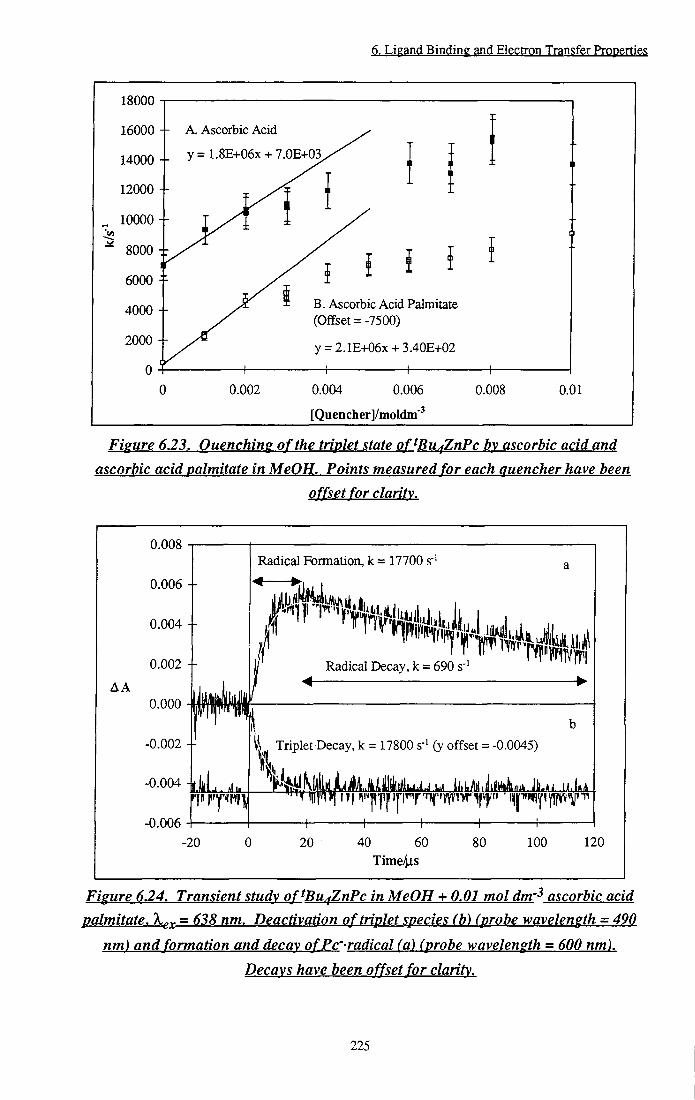
6. Li^and Binding and Electron Transfer Propenies
18000
16000 +
14000
12000 - -
10000 --
8000 : r ir
6000 - -
4000 - -
2000 - -0
A. Ascorbic Acid
y=1.8E+06x + 7.0E+03.
B. Ascorbic Acid Palmitate (Offset =-7500)
y = 2.1E+06x + 3.40E+02
0.002 0.004 0.006
[Quencher]/moIdm'
0.008 0.01
Figure 6.23. Quenching of the triplet state of^Bu^ZnPc by ascorbic acid and ascorbic acid palmitate in MeOH. Points measured for each quencher have been
o ffset for clarity.
0.008
0.006
0.004
A A
Radical Formation, k = 17700 sr^
< •ri
0.002 +
0.000
0.002 +
0.004
0.006
Radical Decay, k = 690 s"
\ ^ Triplet Decay, k = 17800 s"' (y offset = -0.0045)
H 1 \ h
-20 20 40 60 Time4is
80 100 120
Figure 6.24. Transient study of^Bu^ZnPc in MeOH + 0.01 mol dm'^ ascorbic acid
palmitate, X^x ~ Deactivation of triplet species (b) (probe wavelength = 490
nm) and formation and decay ofPc'-radical (a) (probe wavelength = 600 nm).
Decays have been offset for clarity.
225

6. Ligand Binding and Elecp-pn Transfer Prppertieg
0.0020 J
0.0018 --
0.0016 --
0.0014 --v u c 0.0012 --CS
iC u
0.0010 --o < 0.0008 --
0.0006 --
0.0004 --
0.0002 --
0.0000 560 570 580 590 600
Wavelength/nm 610 620
Figure 6.25. Transient absorption spectrum o f^Bu^ZnPc'-radical in MeOH. a) At = 300 \Ls. b)At = l ms Ck,^ = 638 nm).
Peaks at 575 nm and 600 nm were observed and by analogy to previously reported transient spectra assignment to ZnPc"- radical species was made. Aramendia' o] investigated photochemically induced electron transfer from amines to ZnPc - = 580 nm, Mackt ^J observed electron transfer from hydrazine to ZnPc upon irradiation -
at 319 nm, 432 nm, 570 nm, 635 nm and 956 nm and Clack[''3],[74] generated the radical anion of ZnPc electrochemically - A^^x 323 nm, 560 nm and 635 nm. The observed rate of quenching, kg, was estimated from the gradient at low concentrations of quencher. Rates of 1.8 ± 0.2 x 10^ dm^ mol"! s"! and 2.1 ± 0.2 x 10^ dm^ mol"! s"! were determined for AA and AAP respectively. As expected in a homogeneous medium, these values are within experimental error of one another.
AA/AAP have several ionic formst''^] which are dependent on the pH of the medium (Figure 6.26). In this study, the pH can be assumed to be ~7, thus the redox couple of interest is that of HA-/HA-. This has a reduction potential, E, of 0.74 V vs NHEf^^j
226

6. Ligand Binding and Electron Transfer Properties
H-CH2R
-OH
pKa = 4.0
CHgR -OH H-
CHgR -OH
OH OH
n^A
w OHO"
HA'
CH2R
O O
A
R = OH (Ascorbic Acid)
R=CH20(CH2)i4CH3
(Ascorbic Acid Palmitate)
HA
Fieure 626. Equilibria of ascorbic acid (Palmitate) and redox couples at vH = 7.
Using equations 6.1, 6.2 and 6.3, theoretical values for the free energy change for reaction, AG, the activation free energy, AG* and the overall rate of electron transfer, kg, may be calculated (k^ = 1.1 x 10^0 ^ 3 mol"! s"l in MeOH). These results are displayed in Table 6.9. Excellent agreement between tiie predicted and measured triplet state quenching rates was obtained: however, due to errors in reduction potentials employed, the predicted value of kg may vary by several orders of magnitude. It may also be inferred that in a suitable solvent system, singlet state quenching should occur.
, State Energy AG A G * 1 k<35 <Calc) kQ<Ex|}t)
Singlet Triplet
1.83 1.13
-47.34
20.3
2.03 24.4 1
7x10⁹
1.98 X 10⁶ 2 ± 0 . 3 x 10⁶
Table 6.9. Calculated quenching rates of^Bu^ZnPc* bv ascorbic acid in MeOH.
The measured value of triplet state quenching (2 x 10^ dm^ mol'^ s' ) is significantly
lower than would be expected for a diffusion controlled reaction. This implies that this
process is activation conti-oUed, i.e. ka of Figure 6.2 is the rate determining step.
227
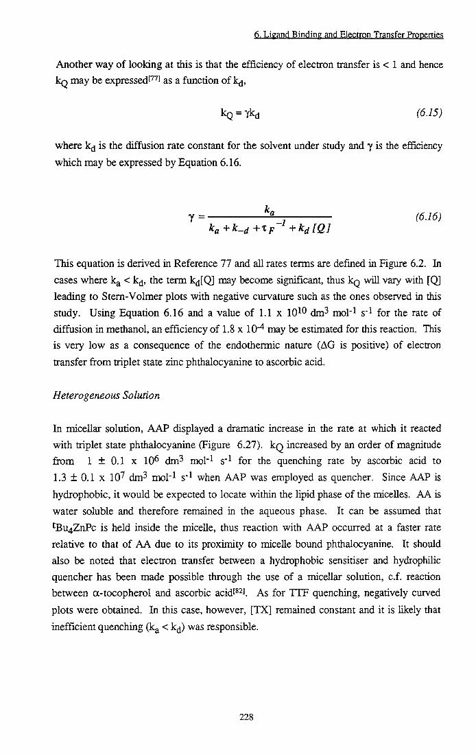
6. Ligand Binding and Electron Transfer Properties
Another way of looking at this is that the efficiency of electron transfer is < 1 and hence kq may be expressed!''''] as a function of k^,
kQ = 7kd (6.15)
where k^ is the diffusion rate constant for the solvent under study and 7 is the efficiency
which may be expressed by Equation 6.16.
Y = (6.16)
This equation is derived in Reference 77 and all rates terms are defined in Figure 6.2. In cases where k^ < k^, the term k£i[Q] may become significant, thus kg will vary with [Q] leading to Stem-Volmer plots with negative curvature such as the ones observed in this study. Using Equation 6.16 and a value of 1.1 x 10^^ dm^ mol'^ s' for the rate of diffusion in methanol, an efficiency of 1.8 x 10" may be estimated for this reaction. This is very low as a consequence of the endothermic nature (AG is positive) of electron transfer fi-om triplet state zinc phthalocyanine to ascorbic acid.
Heterogeneous Solution
In mdcellar solution, AAP displayed a dramatic increase in the rate at which it reacted with triplet state phthalocyanine (Figure 6.27). k q increased by an order of magnitude firom 1 ± 0.1 X 10^ dm^ mol"^ S' for the quenching rate by ascorbic acid to 1.3 ± 0.1 X 10" dm^ mol"l s" when AAP was employed as quencher. Since AAP is hydrophobic, it would be expected to locate within the lipid phase of the micelles. AA is water soluble and therefore remained in the aqueous phase. It can be assumed that tBu4ZnPc is held inside the micelle, thus reaction with AAP occurred at a faster rate relative to that of AA due to its proximity to micelle bound phthalocyanine. It should also be noted that electron transfer between a hydrophobic sensitiser and hydrophilic quencher has been made possible through the use of a micellar solution, c.f. reaction between a-tocopherol and ascorbic acidf^^i ^s for TTF quenching, negatively curved plots were obtained. In this case, however, [TX] remained constant and it is likely that inefficient quenching (k^ < k^j) was responsible.
228

6. Ligand Binding and Electron Transfer Properties
70000
60000
50000
40000
30000
20000
10000
A Ascorbic Acid Paknitate
y = 1.30E-H07X+2550
B. Ascorbic Acid
= 1E+06X + 3300
0.002 0.004 0.006
[Quencher]/moldm'
0.008 0.01
Figure 6.27. Comparison of^Bu^ZnPc triplet state quenching by a) ascorbic acid
palmitate and b) ascorbic acid in 1% TXIH2O solution.
The triplet state of sulfonated aluminium phthalocyanine by ascorbic acid in phosphate buffer saline has been reported by Ben-Hurt''^] to quench triplet state species (kg = 5 x 10'^ dm^ mol'l s' ) to produce a transient radical that absorbed at 572 nm and 620 nm This corroborates the results above. It was also shown that the rate of photohaemolysis of red blood cells increased upon addition of ascorbic acid implying that photodamage to neoplastic tissue may occur via a Type I mechanism mediated by Vitamin C or that the protective effect of Vitamin C towards membrane damage!'' ] has been reduced.
6.4 Conclusion
It has been demonstrated that axial ligands may bind to zinc phthalocyanines causing
changes in ground state absorption characteristics and excited singlet and triplet state
properties. Cyanide ions have been shown to cause a significant red shift in the ground
state absorption spectrum of ZnPcS2. The equihbrium constant of binding was
determined to be 800 dm^ mol"! in MeOH. Binding of THF to ZnPc(CMe(C02Me)2)4
induced an increase in the quantum yield and lifetime of triplet state species. It is likely
that these changes were caused by a combination of decreased internal conversion
pathways and increased intersystem crossing rates. It is suggested that in vivo Ugand
binding may account for shifts in reported action spectra of sensitisers relative to their in vitro absorption spectra.
229

6. Ligand Binding and Electron Transfer Properties
Interaction of peripherally substituted zinc phthalocyanines with bovine serum albumin (BSA) has been investigated. No direct spectroscopic evidence for binding of hydrophobic sensitisers, tBu4ZnPc or CIO was observed. ZnPcS2 displayed clear changes in ground state absorption spectra recorded in PBS or 1% TX/PBS upon addition of BSA, however, no effect on its fluorescence, triplet state or singlet oxygen quantum yields was recorded. This impUes that binding was weak, such that the electronic structure of the phthalocyanine was not perturbed. Ruorescence anisotropy measurements provided indirect evidence for binding. A significant increase in the steady state anisotropy of ZnPc's was recorded upon addition of BSA, suggesting an increase in the effective size of the fluorophore, i.e. binding to protein is implied. In contrast to literature reports, phthalocyanine triplet state quenching by oxygen was not decreased by the presence of BSA, however, some interaction between oxidised protein species and zinc phthalocyanine is suggested. Reasons for this behaviour are unclear and further work in this area is required. Evidence regarding binding of zinc phthalocyanines to BSA remains inconclusive.
It has been demonstrated that ZnPc's undergo electron transfer reactions with several electron donors, in particular with amine containing donor molecules. Reduced fluorescence yields of phthalocyanines with chemically bound tetrathiafulvalene hgands have been shown to be a result of intramolecular electron transfer quenching. In addition, it has been demonstrated that the nature of peripheral substituents may affect the propensity of a phthalocyanine to undergo Type I reactions. With respect to photodynamic therapy this may be important since the mechanism of ceU destruction is still unclear. The significance of singlet oxygen as an intermediate has been largely accepted, however, there is still debate over the role of Type 1 reactions in causing cell necrosis. In addition, photodegradation of phthalocyanines which as observed in vivo[*°l may be due to Type I reactions since it is known that in the main, phthalocyanines are stable to singlet oxygent^i^. ability of a sensitiser to undergo electron transfer
reactions with biological substrates such as proteins or DNA may, therefore, prove to be vital if we are to fuUy understand the processes involved in PDT. It has been found that Type I reactions with ascorbic acid may play an important role in the photodynamic process, however, electron transfer between zinc phthalocyanines and amino acids residues is unlikely to be significant.
230

6. Ligand Binding and Electron Transfer Properties
6.$ References
[I] Bishop S.M., Beeby A., Meunier H., Parker A.W., Foley M.S.C. and Phillips D., The photophysics of disulphonated metallophthalocyamnes upon complexation with fluoride., / . Chem. Soc. Faraday Trans., 92,2689,1996.
[2] Rosenthal I . , Shafirovich V.Y., Geacintov N.E., Ben-Hur E. and Horowitz B., The photochemical properties of fluoroaluminum phthalocyanine., Photochem. Photobiol, 60, 215-220, 1994.
[3] Foley M.S.C., The photophysics of sulphonated aluminium phthalocyanines for Photodynamic Therapy., PhD Thesis, Imperial College, London, 1994.
[4] Ferraudi G.J. and Prasad D.R., Excited state redox properties of phthalocyanines: Influence of the axial ligand in the rates of relaxation and electron transfer quenching of the lowest excited ^TITC* excited state., / . Chem. Soc, Dalton Trans., 2137-2140, 1984.
[5] Doeff M . and Sweigart D.A., Axial Ugand substitution reactions of ruthenium 11
phthalocyanine.,/norg. Chem., 20, 1683-1687, 1981. [6] Nappa M . and Valentine J.S., The influence of axial hgands on metalloporphyrin
visible absorption spectra: Complexes of tetraphenylporphyrinatozinc, / . Am. Chem. Soc, 100,100-116, 1978.
[7] Ben-Hur E., Hoe R.C., Ormondt H.V., Dubbehnan T.M.A.R. and Steveninck J.V., Photodynamic inactivation of retroviruses by phthalocyanine: the effects of sulphonation, metal Ugand and fluoride., / . Photochem. Photobiol. B:Biol., 13, 145-
152, 1992. [8] Ben-Hur E., Malik. Z., Dubbelman T.M.A.R., Margaron P., Ali H. and Van Lier
J.E., Phthalocyanine induced photohaemolysis: Structure activity relationship and the effect of fluoride., Photochem. Photobiol., 58, 351-355, 1993.
[9] Ben-Hur E., Dubbehnan T.M.A.R. and Steveninck J.V., The effect of fluoride on binding and photodynamic action of phthalocyanines with proteins., Photochem.
Photobiol., 54,703-707, 1991. [10] Reddi E., Transport modalities of photodynamic agents for tumours., S.P.I.E.,
2078, 246, 1994. [II] Jori G., Beltramini M. , Reddi E., Salvato B., Pagnan A., Ziron L., Tomio L. and
Tsanov T., Evidence for a major role of plasma-lipoproteins as haematopoiphyrin earners in vivo.. Cancer Lett., 24, 291-297,1984.
[12] Carter D.C., He X.M. , Munson S.H., Twigg P.D., Gemert K.M., Broom B.M. and Miller T.Y., 3-Dimensional structure of human serum albumin.. Science, 244, 1195, 1989.
231

6. Ligand Binding gnd Elecp-pn Trgngfgr Prppgrtig?
[13] Marcus R.A., Chemical and electrochemical electron transfer theory., Ann. Rev. Phys. Chem., 15, 155, 1964.
[14] Marcus R.A., On the theory of oxidation-reduction reactions involving electron transfer. I * . , J. Chem. Phys., 24,965, 1956.
[15] Gilbert A. and Baggot J., Essentials of Molecular Photochemistry, pi84, Blackwell Scientific Publications, Oxford, 1991.
[16] Rehm D. and Weller A., Kinetics of fluorescence quenching by electron and H-atom transfer., Israeli. Chem., 8, 259, 1970.
[17] Darwent J.R., McCubbin I . and Porter G., Photoreduction of methyl viologen sensitised by sulphonated phthalocyanines in micellar solution., / . Chem. Soc. Faraday Trans. 2, 78, 903-910,1982.
[18] Ohno T., Kato S. and Lichtin N.N., Electron transfer in the quenching of triplet states of zinc phthalocyanine and methylene blue by the use of Fe(in), Co(in) and organic oxidants., 5M//. Chem. Soc. Jpn., 55,2753,1982.
[19] Prasad D.R. and Ferraudi G., Electron transfer quenching vs exciplex-mediated quenching of the low-lying excited states in phthalocyanines., Inorg. Chem., 22, 1672-1674, 1983.
[20] Aramendia P.F., Energy and charge transfer photosensitised by phthalocyanines in micelles., EPA Newsletter, 52, 6-20,1994.
[21] Daraio M.E., Volker A., Aramendia P.F. and San Roman E., Reaction of zinc phthalocyanine excited states with amines in cationic micelles., Langmuir, 12, 2932, 1996.
[22] DeGraziano J.M., MacPherson A.N., Lidell P.A., Noss L., Sumida J.P., Seely G.R., Lewis I.E., Moore A.L., Moore T. A. and Gust D., Solvent dependence of photoinduced electron transfer in poiphyrin dyads., New J. Chem., 20, 839,1996.
[23] Nyokong T., Gasyna Z and Stillman M. , Phthalocyanine % cation radical species: Photochemical and electrochemical preparation of [ZnPc-l]+- in solution., Inorg. Chem., 26, 548-553, 1987.
[24] Ferraudi G., Oishi S. and Muraldiharan K., The photochemical properties of Rh(ni) phthalocyanine cation radicals., / . Phys. Chem., 88, 5261-5264, 1984.
[25] Ohno T., Kato S., Yamada A. and Tanno T., Electron transfer reactions of the photoexcited triplet state of chloroaluminium phthalocyanine with aromatic amines, benzoquinones and coordination compounds of iron(ll) and iron(in)., / . Phys. Chem., 87, 775-781,1983.
[26] Ben-Hur E. and Rosenthal I . , Photosensitisation of Chinese hamster cells by water soluble phthalocyanines., Photochem. PhotobioL, 43, 615-619, 1986.
232

6. Ligand Binding and Electron Transfer Properties
[27] Ben-Hur E., Carmichael A., Riesz P. and Rosenthal I . , Photochemical generation of superoxide radical and the cytotoxicity of phthalocyanines.. Int. J. Radiat. Biol., 48, 837,1985.
[28] Cook M.J., Chambrier L, Cracknell S.J., Mayes D.A. and Russell D.A. Octa
alkyl Zinc Phthalocyanines: Potential photosensitisers for use in the photodynamic therapy of cancer., Photochem. Photobiol., 62, 542-545, 1995.
[29] Wang C, Bryce M.R., Batsanov A.S., Stanley C.F., Beeby A. and Howard J.A.K., Synthesis, spectroscopy and electrochemistry of phthalocyanine derivatives functionalised with four and eight peripheral tetrathiafulvalene units., / . Chem. Soc, Perkin Trans., 2,1671, 1997.
[30] Rossotti F. and Rossotti H., The Determination of Stability Constants, p277, McGraw Hil l Book Company, New York, 1961.
[31] Greenwood N.N. and Eamshaw A., Chemistry of the Elements, pl065, Pergamon Press, UK, 1984.
[32] PhUhps D., The photochemistry of sensitisers for photodynamic therapy.. Pure Appl. Chem., 67, 117, 1995.
[33] Gomer C.J., Doiron D.R., Rucker N., Razum N.J. and Fountain S.W., Action spectrum (620 - 640 nm) for hematoporphyrin derivative induced cell killing., Photochem. Photobiol., 39, 365, 1984.
[34] Star W.M., In vivo action spectra, absorption and fluorescence excitation spectra of photosensitisers for photodynamic therapy., / . Photochem. Photobiol. B.Biol., 28, 101, 1995.
[35] Cubeddu R., Canti G., Musolino M. , Pifferi A., Taroni P. and Valentini G., In vivo absorption spectrum of disulphonated aluminium phthalocyanine in a murine tumour model., J. Photochem. Photobiol. B.Biol., 34,229, 1996.
[36] Davila J. and Harriman A., Photoreactions of macrocycHc dyes bound to human serum albumin., Photochem. Photobiol., 51,9, 1990.
[37] Foley M.S.C., Beeby A., Parker A.W., Bishop S.M. and Phillips D., Excited triplet state photophysics of the sulphonated aluminium phthalocyanines bound to human serum albumin., / . Photochem. Photobiol. B.Biol., 38, 10, 1997.
[38] Doiron D.R. and Gomer C.J., Porphyrin Localisation and Treatment of Tumours, p373, Alan R Liss, New York, 1984.
[39] Ambroz M. , MacRobert A.J., Morgan J., Rumbles G., Foley M.S.C. and Phillips D., Time resolved fluorescence spectroscopy and intracellular imaging of disulphonated aluminium phthalocyanine., Photochem. Photobiol. B.Biol., 22, 105, 1994.
233

6. Ligand Binding and Electron Transfer Properties
[40] Lang K., Wagnerova D.M., Engst P. and Kubat P., Quenching of the triplet state of metallocyanines by dioxygen in the presence of bovine semm albumin., Zeit. Physik. Chem., 187, 214, 1994.
[41] Reddi E., Lambert C.R., Jori G. and Rodgers M.A.J., Photokinetic and photophysical measurements of the sensitised photooxidation of the tryptophyl residue in N-Acetyl tryptophanamide and in HSA., Photochem. PhotobioL, 45, 345-351, 1987.
[42] Creed D., The photophysics and photochemistry of the near UV absorbing amino acids - 1. Tryptophan and its simple derivatives., Photochem. PhotobioL, 39, 537, 1984.
[43] Walrant P. and Santus R., N-Fomiylkynurenine, a tryptophan photooxidation product as a photodynamic sensitiser., Photochem. Photobiol., 19, 411, 1974.
[44] Reddi E., Rodgers M.A.J., Spikes J.D. and Jori G., The effect of medium polarity on the hematoporphyrin-sensitised photooxidation of L-tryptophan., Photochem. Photobiol., 40,415,1984.
[45] Rossi E., Van de Vorst A. and Jori G., Competition between the singlet oxygen and electron transfer mechanisms in the poiphyrin-sensitised photooxidation of L-tryptophan in aqueous micellar media., Photochem. Photobiol., 34,447, 1981.
[46] Langlois R., Brasseur N., Wagner J.R. and Van Lier I.E., Biological activities of phthalocyanines - IV. Type I I sensitised photooxidation of L-tryptophan and cholesterol by sulfonated metallo phthalocyanines., Photochem. Photobiol., 44, 117, 1986.
[47] Mashiko S., Suzuki N. , Koga S., Nakano M. , Goto T., Ashino T., Mizumoto L. and Inaba H., Measurement of rate constants for quenching singlet oxygen with a cypridina luciferin analog (2-methyl-6-paramethoxyphenyl 3, 7 dihydroimidazole [1, 2-A] pyrazin-3-one) and sodium azide., / . Biolum. Chemilum., 6, 69,1991.
[48] Vincent S.H. and Muller-Eberhard U., Effects of poiphyrins on proteins of cytosol and plasma. In vitro photo-oxidation and cross-linking of proteins by naturally occurring and synthetic porphyrins., J. Laboratory and Clinical Med., 110, 475, 1987.
[49] Dubbehnan T.M.A.R., De Goeij A.F.P.M. and Steveninck J.V., Photodynamic effects of protoporphyrin on human erythrocytes. Nature of the cross-hnking of membrane proteins., Biochim. Biophys. Acta., 511,141,1978.
[50] Turro N.J., Modern Molecular Photochemistry, p246. University Science Books, California, 1991.
[51] Zeng H. and Durocher G., Analysis of fluorescence quenching in some antioxidants firom non-linear Stem Volmer plots., / . Lumin., 63,75,1995.
234

6. Ligand Binding and Electron Transfer Properties
[52] Sung J., Shin K.J. and Lee S., Theory of diffusion influenced fluorescence quenching. Effects of static quenching on the Stem Volmer curve., Chem. Phys., 167, 17, 1992.
[53] Keizer J., Nonlinear fluorescence quenching and the origin of positive curvature
in Stem Volmer plots., J. Am. Chem. Soc, 105,1494,1983. [54] Stevens B., Application of a finite initial separation limit to the concentration
gradient model of fluorescence quenching in Uquids., Chem. Phys. Lett., 134, 519,
1987. [55] Stevens B., Biver I I I C.J. and McKeitiian D.N., Parameterization of diffusion
limited electron transfer quenching in the finite sink approximation., Chem. Phys. Lett., 187, 590,1991.
[56] Dr. A. Beeby, Unpublished Data. [57] Lakowitz J.R., Topics in Fluorescence Spectroscopy, 2, p53. Plenum Press, New
York, 1991. [58] Kalyansundarum K., Photochemistry in microheterogeneous systems, p56,
Academic Press, London, 1987. [59] Kalyansundarum K., Photochemistry in microheterogeneous systems, p26.
Academic Press, London, 1987. [60] Blatt E. and Sawyer W.H., Depth dependent fluorescent quenching in micelles
and membranes., Biochem. Biophys. Acta, 822,43,1985. [61] Atkins P.W., Physical Chemistry, p848. Edition 4, Oxford University Press,
Oxford, UK., 1990. [62] Leznoff C.C. and Lever A.B.P., Phthalocyanines. Properties and Applications.,
1, p305, VCH Publishers, New York, 1989. [63] Devonport W., Blower M.A., Bryce M.R. and Goldenberg L.M., A redox active
tetrathiafulvalene 920 pseudorotaxane: spectrochemical and cychc voltammetric studies of the highly reversible complexation/decomplexation process., / . Org. Chem., 62, 885, 1997.
[64] Fox M.A. and Chanon M . , Photoinduced Electron Transfer, C: Photoinduced Electron Transfer Reactions. Organic Substrates, p6, Elsevier, New York, 1988.
[65] Mack J. and Stillman M . , Photochemical formation of the anion-radical of zinc phthalocyanine and analysis of the absorption and magnetic circular-dichroism spectral data - assignment of the optical-spectrum of [ZnPc(-3)]'., / . Am. Chem. Soc, 116, 1292, 1994.
[66] Zhu Q.Q., Schnabel W. and Jacques P., Photoreduction of thioxanthen-9-one and benzophenone by amines. Electrical conductivity and Hght emission studies., / . Chem. Soc. Faraday Trans, 87, 1531, 1991.
235
6. Ligand Binding and Electron Transfer Properties
[67] Coyle J.D., HiU R.R., Roberts D.R., Light,Chemical Change and Life: A Source Book in Photochemistry, p364. Open University Press, Milton Keynes, 1982.
[68] Zhang X.F. and Xu H.J., Mechanism of photosensitised oxidation of tyrosine by gallium or zinc phthalocyanine in homogeneous and heterogeneous media., / . Photochem. Photobiol. B:Biol., 24, 109-116,1994.
[69] Turro N.J., Modern Molecular Photochemistry, pi38, University Science Books, California, 1991.
[70] Marciniak B., Bobrowski K. and Hug G.L., Quenching of triplet states of aromatic ketones by sulphur containing aminoacid in solution. Evidence for electron transfer., J. Phys. Chem., 97.11937, 1993.
[71] Ferraudi G., Arguello G.A., Al i H. and Van Lier J.E., Type-I and Type-II sensitised photooxidation of amino-acid by phthalocyanines - a flash photochemical study., Photochem. Photobiol., 47, 657-660, 1988.
[72] Berger T.M., PoHdori M.C., Dabbagh A., Evans P.J., HaUiwell B., Morrow J.D., Roberts n L.J. and Frei B., Antioxidant activity of vitamin C in iron overloaded human plasma., / . Biol. Chem., 272, 15656, 1997.
[73] Clack D.W. and Yandle J.R., Electronic spectra of the negative ions of some metal phthalocyanines.,/norg. Chem., 11,1738, 1972.
[74] Minor P.C., Gouterman M . and Lever A.B.P., Electronic spectra of phthalocyanine anions and cations., Inorg. Chem., 24,1894-1900, 1985.
[75] Creutz C , The complexities of ascorbate as a reducing agent., Inorg. Chem., 20,
4449, 1981. [76] Tur'yan Y. I . and Kohen R., Formal redox potentials of the dehydro-L-ascorbic
acid/L-ascorbic acid system., / . Electroanal. Chem., 380, 273, 1995. [77] Eftink M.R. and Ghrron C.A., Fluorescence quenching studies with proteins..
Anal. Biochem., 114, 199, 1981. [78] Rosenthal I . and Ben-Hur E., Ascorbate assisted phthalocyanine sensitised
photohaemolysis of human erythrocytes.. Int. J. Radiat. Biol., 62, 481, 1992. [79] Bisby R.H. and Parker A.W., Reactions of the a tocopherol radical in micellar
solutions studied by nanosecond laser flash photolysis., Febs Lett., 290, 205, 1991. [80] Ruck A., Beck G., Bactor R., Akgun N. , Gschwend M.H. and Steiner R.,
Dynamic fluorescence changes during photodynamic therapy in vivo and in vitro of hydrophilic A1(III) phthalocyanine tetrasulphonate and lipophilic Zn(n) phthalocyanine administered in hposomes., / . Photochem. Photobiol. B.Biol., 36, 127, 1996.
[81] Rotomskis R., Streckyte G. and Bagdonas S., Phototransformations of sensitisers. 1) Significance of the nature of the sensitiser in the photobleaching process
236

6. Ligand Binding and Electron Transfer Propenies
and photoproduct formation in aqueous solution., / . Photochem. Photobiol. B.Biol., 39, 167, 1997.
[82] Bisby R.H. and Parker A.W., Reactions of the alpha-tocopheroxyl radical in micellar solutions studied by nanosecond laser flash photolysis., Febs Lett., 290, 205, 1991.
237

SUMMARY
* Two novel, P-tetra substituted zinc phthalocyanines have been synthesised. The
photophysical properties of ZnPc were not significandy affected by the nature of the
peripheral substituents.
* The fluorescence anisotropy of metal free and 2dnc phthalocyanines is dependent
upon the exact experimental conditions, i.e. emission wavelength, excitation
wavelength, solvent and temperature.
* The photophysical properties of zinc phthalocyanines were greatiy affected by the
nature of their environment.
- The aggregation state of ZnPc(CHMeC02H)4 displayed a remarkable
sensitivity to the ionic strength of non-aqueous solution.
- CIO aggregated at 77 K in EPA. The aggregate emitted fluorescence at 760 nm
and generated triplet state species upon irradiation by 355 nm or 638 nm Hght. This
is believed to be the first fluorescent phthalocyanine dimer bound solely by Van der
Waals forces to be reported.
- Low pH induced protonation of the azomethane bridges of the phthalocyanine
ring. Protonated species, PcHn"+, displayed large changes in their photophysical
properties with respect to the parent phthalocyanine including a dramatic reduction
in their ability to generate singlet oxygen. Protonation of ZnPcS2 occurred at
physiologically important pH's and may have important implications for
Photodynamic Therapy.
* Binding of bovine serum albumin to substituted zinc phthalocyanines has been
investigated, however, results were inconclusive.
4> Zinc phdialocyanines have been shown to act as electron acceptors in the presence of
cenain amine or sulphur based donors. Electron transfer reactions were shown to be
important between ascorbic acid and phthalocyanine, however, reaction between
aminoacids - tryptophan, tyrosine, methionine and cysteine - and the phthalocyanines
was not observed.
238
APPENDIX A
A NOVEL SWITCH FOR SINGLET OXYGEN MEASUREMENTS
A.l Introduction
Measurement and detection of singlet oxygen, IO2 , has been important for many years. Singlet oxygen is a highly reactive species that is believed to be responsible for the destruction of biological media such as cells and tissues. In certain applications, for example, the photodynamic therapy of cancer (PDT), this is the required effect and production and reaction of I O 2 must be optimised for maximum benefit. However, in other situations, e.g. skin sensitisation by pharmaceuticals, singlet oxygen is an undesirable side effect to be avoided. The ability to measure the properties of ^02, i.e. its quantum yield and lifetime, with precision and accuracy is therefore of great interest. There are two methods by which ^©2 can be detected. Chemical trapping or quenching which involves an added acceptor species such as diphenyUsobenzylfuran (DPBF) or P-Carotene respectivelyfi^. This is sometimes unsatisfactory since the added species may interfere with the chemistry in other ways. A second, more convenient and reliable method, was developedf^l around 1980. Direct detection of singlet oxygen phosphorescence at 1270 nm was made possible through the use of a highly sensitive germanium photodiode coupled to a transient digitiser. However, when certain sensitisers are investigated, in particular those with red shifted absorption and emission bands such as the phthalocyanines, measurement of ^02 emission still causes problems due to the presence of a large spike at the start of the observed transient emission decayP-' l. It is thought that this phenemenon results from scattered laser Ught, sensitiser fluorescence and possibly some fluorescence from bandpass filters used to select 1270 nm lightf^l. As a result, the traces recorded by the transient digitiser show the electronic effects and the I O 2 signal can become distorted, preventing rehable measurements of TA and <I>A by this technique. Previous attempts to minimise this effect include careful choice of solventt^I and the use of interference filters to reduce unwanted emission wavelengthsl^l. Neither solution is completely satisfactory.
In this study, it has been demonstrated that the observed effects are due to overloading
of electronic circuits in the amplifier of a digital storage oscilloscope, DSO. The
development of an electronic switch, based on a DG 403DJ analogue switch, which may
be used to eliminate fluorescence contributions is also reported. Using a variable time
gate to prevent the large spike due to fluorescence entering the DSO amplifier allows
saturation of the oscilloscope ampKfier to be avoided, thus ^02 detection is improved.

A. NQV^I Switch for Singlet Qxyggn Measurement?
A,2 Materials and Method^ A.2.1 Materials
Several substances were used to test the performance of the switch. Rose Bengal (93 %) was purchased from Aldrich. Styryl 9 was purchased from Lambda Physik and AIPCS2 was a gift from Imperial College, London. Solutions were prepared in Analar methanol.
A.2J Experimental
All experimental studies were performed at the Rutherford Appleton Laboratory Central Laser Facility.
Excitation of the sample was carried out using the 355 nm output of an excimer (Lumonix HE 460 (XeCl))) pumped dye laser (Lambda Physik FL3002) with a typical pulse duration of 10 ns and energy within the range 0.1-0.5 mJ per pulse. Light was delivered to the sample using a 5 mm diameter Hquid Ught guide. The sample was held in a 10 mm pathlength quanz cuvette, and the absorbance was adjusted to 0.3 at the excitaton wavelength. The emission was collected at 90" to the excitation beam and 1270 nm radiation selected using an interference filter with a 20 nm bandpass (Infra-Red Engineering). Selected radiation was incident upon the cooled germanium photodiode/amplifier (NorthCoast EO-817P). In order to obtain the fastest response time from the detector it was operated in the high speed mode, "H" (tnse ~ 200 ns, FWHM = 400 ns). Output of the detector was either transferred directiy to a digital storage oscilloscope (Tektronics 2432A) or via the electronic switch box described below. Traces were the average of sixteen shots and were transferred to a computer for analysis by non-linear least squares methods.
Synchronisation of the firing of the laser and the oscilloscope was achieved using a Stanford digital signal generator (DG 535). The same generator was also used to provide a TTL pulse to operate the switching device (Figure A . l ) .
240
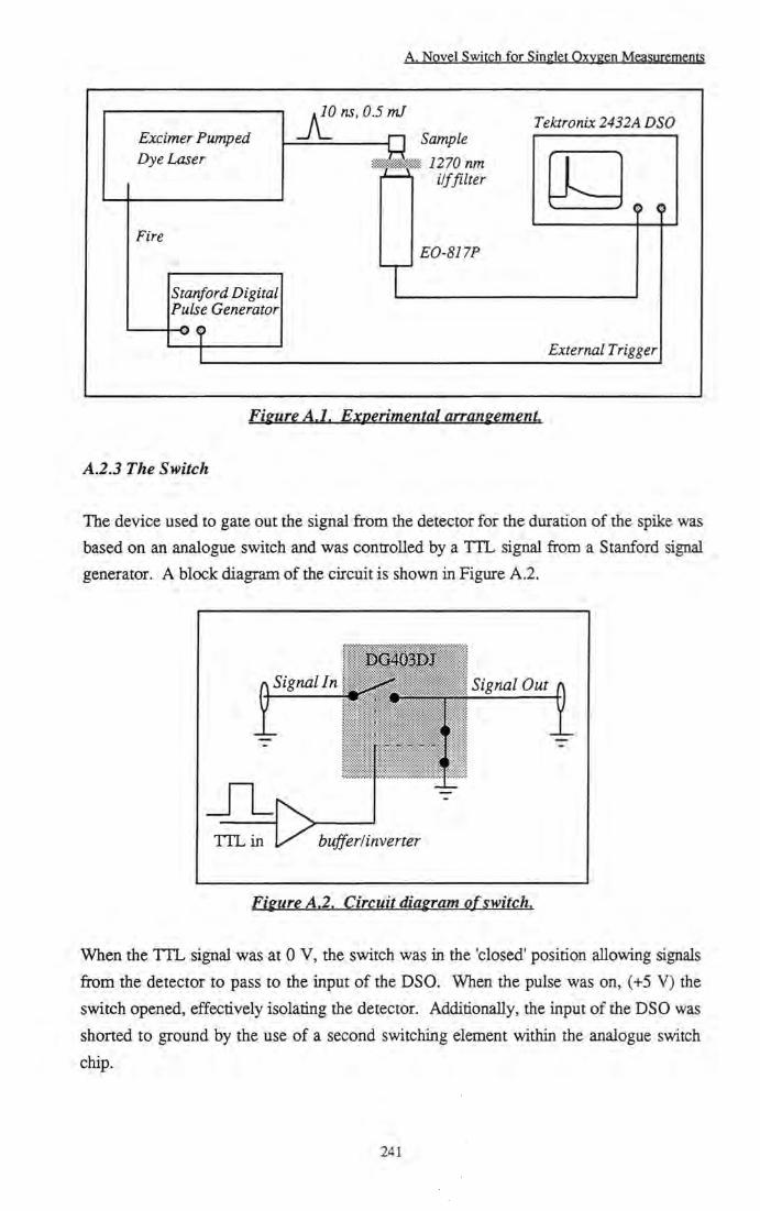
A. Novel Switch for Singlet Oxygen Measurements
Excimer Pumped Dye Laser
JC 10 ns, 0.5 ml
Fire
Stanford Digital Pulse Generator
<)
Sample 1270 nm ilffllter
EO-817P
Tektronix 2432A DSO
^ P 0
External Trigger
Figure A.l. Experimental arrangement
A.2.3 The Switch
The device used to gate out the signal from the detector for the duration of the spike was based on an analogue switch and was controlled by a TTL signal from a Stanford signal generator. A block diagram of the circuit is shown in Figure A.2.
DG403m
f Signal In Signal Out
T T L i n 0
•
buffer/inverter
Figure A.2. Circuit diagram of switch.
When the T T L signal was at 0 V, the switch was in the 'closed' position allowing signals from the detector to pass to the input of the DSO. When the pulse was on, (+5 V) the switch opened, effectively isolating the detector. Additionally, the input of the DSO was shorted to ground by the use of a second switching element within the analogue switch chip.
241

A. Novel Switch for Singlet Oxygen Measurements
A.3 Results and Discussion A.3.1 Electrical properties
The first stage of the study was to investigate the electrical properties of the switch. It is important that switching the amplifier on in this way should not introduce any spurious signals that noay influence the decay trace of ^02- As Figure A.3 shows, the switch does not perturb the baseline of the oscilloscope trace on a timescale over which the singlet oxygen decay may typically be observed. Furthermore, it was confirmed that radio frequency emission from the excimer laser HV discharge did not induce any signal in the measured background baseline.
0.2 +
r
o -0.1
0.2 +
Time /lis
Figure A.3. Physical properties of the switch.
Large 'switching transients' can be observed. Subsequent examination of these on a
faster timescale (200 ns/div) reveals that any distortions caused by switching signals are
insignificant 1 |is after the amplifier is turned on. Hence they should cause httie
interference with the measurements of ^02. Figure A.4 shows the distortion and
recovery of the baseline as the amplifier is switched off and again as it is turned on. The
gate width is 400 ns. It can be clearly seen that ringing due to the switching has
completely died away before distortions due to the 'on' signal begin.
242

A. Novel Switch for Singlet Oxygen Measurements
0.03
0.02 +
0.01
Voltage o
-0.01 +
-0.02 4-
-0.03
-V-
2 Time l\is
Figure A.4.
Decay trace of switching transient as the switch closes following a 400 ns gate.
A.3.2 Performance
Having shown that operation of the switch should not interfere with I O 2 emission 1 |is
after the gate is closed, its performance was investigated. A strong fluorescence signal at
1270 nm was generated by the excitation of a near infrared laser dye, Styryl-9 in
methanol. The fluorescence lifetime of this dye is short (<10 ns) compared to the
response of the Ge photodiode/amplifier (200 ns) and the dye does not sensitise singlet
oxygen. Using a vertical scale of 0.5 V/div, the 4 V luminescence signal lies well within
the range of the oscilloscope amplifier, saturation does not cause a problem and the trace
is displayed in Figure A.5a.
However, when a more sensitive scale was used, such as that required to detect I O 2 emission (1 mV/division), large amounts of ringing were observed and a negative offset
was obtained (Figure A.5b) which recovered slowly over several milliseconds. This
saturation occurred due to the 4 V peak of the pulse exceeding the 'overdrive' capacity of
the front end amplifier of the DSO. Recovery of this amplifier occurred on a millisecond
timescale.
243
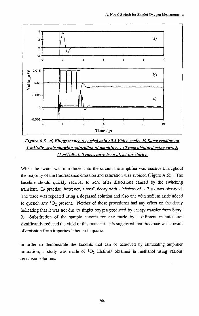
A. Novel Switch for Singlet Oxygen Measurements
> 0.015
0.005
-0.005
Time /as
Figure A.5. a) Fluorescence recorded using 0.5 VIdiv. scale, b) Same reading on 1 mV/div. scale showing saturation of amplifier, c) Trace obtained using switch
(1 mV/div.). Traces have been offset for clarity.
When the switch was introduced into the circuit, the amplifier was inactive throughout the majority of the fluorescence emission and saturation was avoided (Figure A.5c). The baseline should quickly recover to zero after distortions caused by the switching transient. In practise, however, a small decay with a lifetime of ~ 7 jis was observed. The trace was repeated using a degassed solution and also one with sodium azide added to quench any present. Neither of these procedures had any effect on the decay indicating that it was not due to singlet oxygen produced by energy transfer from Styryl 9. Substitution of the sample cuvette for one made by a different manufacturer significantly reduced the yield of this transient. It is suggested that this trace was a result of emission from impurites inherent in quartz.
In order to demonstrate the benefits that can be achieved by eliminating amplifier
saturation, a study was made of lifetimes obtained in metiianol using various
sensitiser solutions.
244

A. Novel Switch for Singlet Qxygen Measurement?
0.006 +
0.005 +
0.004 +
0.003 +
0.002 +
0.001 +
-0.001
0.002 +
-10 10 20
Time /\is
30 40
Figure A.6, Overlap of decays sensitised by Rose Bengal with and without the switch.
Rose Bengal exhibits very littie fluorescence leakage through the interference filter and has only a small spike at the start of the singlet oxygen decay, making it an ideal candidate to establish the effects of the switch on a singlet oxygen decay. Figure A.6 shows that use of the switch does not perturb measurement of either the yield of ^©2 or the profile of the decay. Analysis of singlet oxygen decays obtained with variable gate times yielded a lifetime value of 9.8 ± 0.3 |i,s (Literature value = 10.4 jis)'^!. Quenching of the singlet oxygen by Rose Bengal has not been take into account for this data, (kq = 4 ± 0.5 X 10' mol"l dm^ s" unpublished data).
Phthalocyanines are known to have a large fluorescence spike at the start of their I O 2 decay which interferes with any subsequent analysis. Use of the switch showed a significant improvement in both die profile and calculated lifetime of the singlet oxygen decay. With Styryl 9, a negative offset was obtained as a result of amplifier saturation when the detector was attached directiy to the DSO. Analysis of the decay (singlet oxygen convolved with the amplifier recovery) revealed an average lifetime of 7.9 ± 0.4 |is - clearly different from the quoted literature value of 10.4 fis. Introduction of the switch removed the negative offset and significandy improved the lifetime value to 11.2 ± 0.4 |Lis. (Figure A.7).
245

A. Novel Switch fpr Singlet Qxygen Measurement?
0.03
0.025 +
0.02 +
0.015 -h
o 0.01 +
0.005 +
0
-0.005
a)
b)
5 10 15
Time /\LS 20 25
Figure A.7. decay sensitised b\ phthalocyanine without (a) and with (b) the switch (offset for clarity).
The most dramatic results were obtained when a mixture of Rose Bengal and Styryl 9 was used. Saturation of the amplifier occurred to such a degree that it was impossible to detect singlet oxygen (Figure A.8A(a)). Use of the switch allowed detection and analysis of singlet oxygen decays with the correct lifetime of 10.1 ± 0.2 |is (Figure A.8B).
246
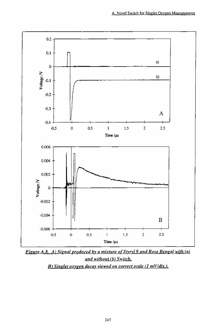
A. Novel Switch for Singlet Oxygen Measurements
0.2
0.1
3 o >
-0.1 +
-0.2
-0.3
-0.4 -0.5
0.006
0.004 +
0.002 +
an S >
-0.002
-0.004 +
-0.006 -0.5
0.5
0.5
1 1.5
+ 1 1.5
Time/|J5
a)
b)
A
2.5
B
2.5
Figure A.8. A) Signal produced by a mixture of Stvrvl 9 and Rose Bengal with (a) and without (b) Switch.
B) Singlet oxygen decay viewed on correct scale (1 mVldiv.).
247

A. Novel Switch for Singlet Oxygen Measurements
A.4 Conclusions
It has been shown that problems in detecting I O 2 produced by certain sensitisers are caused by saturation of the input amplifier of the digital storage oscilloscope following intense fluorescence emission. A rapid switch has been developed to overcome the problem. Input to the oscilloscope input amplifier is controlled via a TTL pulse avoiding saturation as a result of large voltage spikes. Convolution with amplifier recovery due to saturation and decay of I O 2 has been removed to allow calculation of lifetimes to within 10 % of established literature values.
A.5 References
[1] Bensasson R.V., Land E.J. and Truscott T.G., Excited States and Free Radicals in Biology and Medicine, Oxford University Press, New York, pp 118-142, 1993.
[2] Byteva I .M. and Gurinovitch G.P., Sensitised luminescence of oxygen in
solutions. / . Lumin. 21, 17-20,1979. [3] Bishop S., Preparation and properties of phthalocyanine photosensitisers for
PDT. PhD Thesis, Imperial College, London, 1993. [4] Beeby A., Bishop S., Parker A.W., Simpson M.S.C. and Phillips D., Time
resolved studies of singlet oxygen. EPSRC Central Laser Facility Annual Report, RAL-92-020, 227, 1992.
[5] Ford W.E., Rihter B.D., Kenney M.E. and Rodgers M.A.J., Photoproperties of alkoxy-substituted phthalocyanines with deep red optical absorbance. Photochem. Photobiol. 50,277-282, 1989.
[6] Michaeli A. and Feitelson J., Reactivity of singlet oxygen toward aminoacids and
peptides., Photochem. Photobiol., 59,284-289, 1994. [7] Nather D.U., Gilchrist J.R., Gensch T. and Roder B., Temporal and spectral
separation of singlet oxygen luminescence from near infrared emitting photosensitisers., Photochem. Photobiol., 57, 1056, 1993.
[8] Oldham T.C., Beeby A., Parker A.W., Stanley C.F., Telfer A., Barber A. and Phillips D., Improved Instrumentation For The Time-Resolved Detection Of Singlet Oxygen Luminesence., CRC Rutherford Appleton Laboratory Annual Report, RAL-
TR-96-076, 126,1995-1996. [9] Rodgers M.A.J., Solvent induced deactivation of singlet oxygen - additivity
relationships in non-aromatic solvents. / . Am. Chem. Soc, 105, 6201, 1982.
248
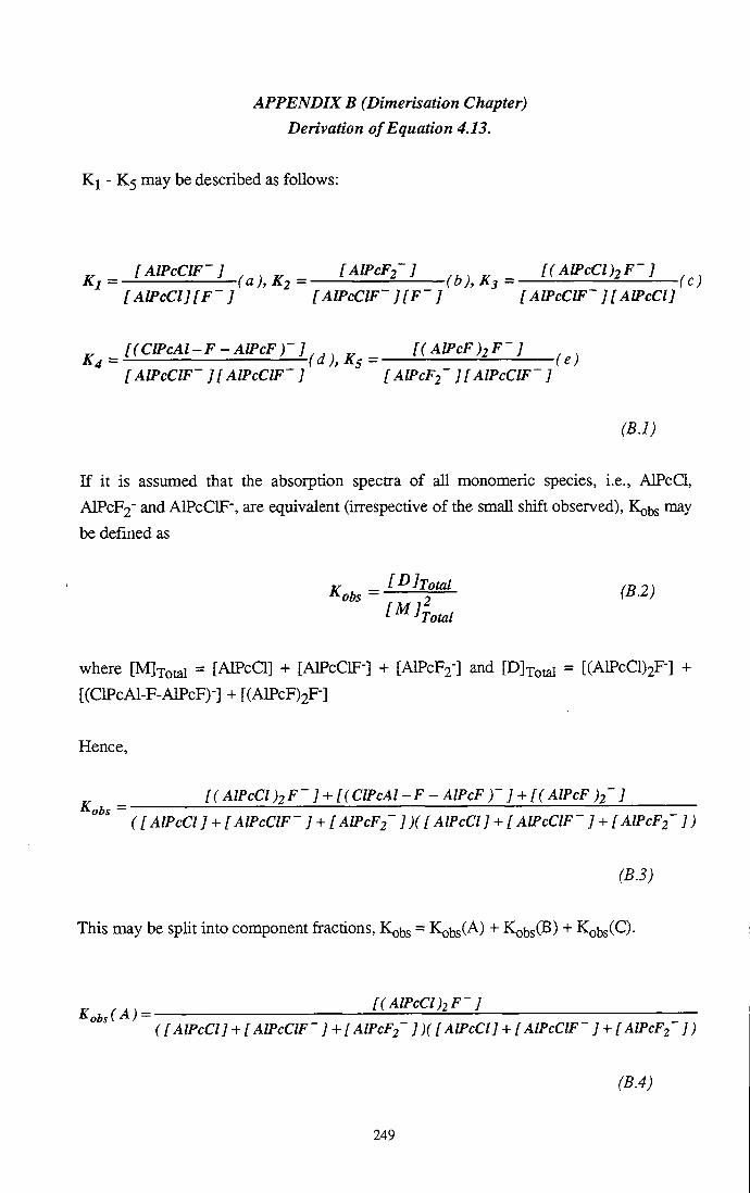
APPENDIX B (Dimerisation Chapter) Derivation of Equation 4.13.
K i - K5 may be described as follows:
„ [AlPcClF-] „ _ [AIPCF2-] „ _ [(AlPcCl)2F-] Kj = (a), K2 = (b), K3 = (c)
[AIPCCIJIP-] [AlPcClF~ ][F-] [AlPcClF~ ][AlPcCl]
^ [(ClPcAl-F-AlPcF)-]^,^ ^ [(AlPcF)2F-J K4 = f a ), A 5 = f e)
[AlPcClP- ][AlPcClF~ ] [ AlPcF2~ ][ AlPcClp-]
(B.l)
If it is assumed that the absorption spectra of aU monomeric species, i.e., AIPcCl,
A I P C F 2 " and AlPcClF', are equivalent (krespective of the small shift observed), KQ^J may
be defined as
rr _ [I^]Total / D 0 \
l^rTotal
where [MJxotal = [AlPcCl] + [AlPcOF"] + [ A I P C F 2 - ] and [Dlxotal = [(AlPcCl)2F-] +
[(ClPcAl-F-AlPcF)-] + [(AlPcF)2F-]
Hence,
[(AlPcCl)2F~] + [(ClPcAl-F - AlPcF ) ' ] + [( AlPcF )2~ ]
(I AlPcCl ] + [ AlPcClF -] + [ A/PcF2~ ] )( [ AlPcCl ] + [ AlPcClF ' ] + [ AIPCF2 ] )
(B.3)
This may be split into component fractions, Kobs = Kobs(A) + KobsOB) + Kobs(Q-
^ . . . [( AlPcCl )2F-] ([AlPcCl] + [ AlPcClF-] + [ AlPcF2~ ])([ AlPcCl] + [ AlPcClF-] + [ AlPcFz ] )
(B.4)
249

J, [(ClPcAl-F-AlPcF)-] ( [ AlPcCl] + [AlPcClF-] + [ AlPcF2~ ])([ AlPcCl] + [ AlPcClF-] + [ AlPcF2~ ])
(B.5)
K ( o - [(AlPcF)2-]
([AlPcCl] + [AlPcClF-] + [AlPcF2~ ])( [AlPcCl] + [AlPcClp-] + [AIPCF2' ])
(B.6)
From Equations B . l a and B. lb , KQbs(A) may be expressed as,
^ l(AlPcCl)2F-]
[ AlPcCl ](l + Ki[F-] + KiK2[F-r )[ AlPcClF ' ]( + 1 + K2[F ]) Ki[F-]
(B.7)
Rearranging,
J, . . . [(AlPcCl)2F-]Ki[F-]
[ AlPcCl][ AlPcClF~ ](1 + Kj [F~ ] + KjK2[F-]^ r
Finally, substituting for Equation B. lc ,
Kots ( A ) = MULEZI (B.9) (l + Ki[F-] + KiK2[F-]^/
Similarly, using Equation B . l a and b, KQ^JSOB) may be expressed as
i . . , , ( B ) ^ [(ClPcAl-F-AlPcF)-] ^,,0)
[ AlPcClF-]^( + 1 + K2[F-])^ Ki[F-J
Rearranging and substituting for Equation B . l d gives
250
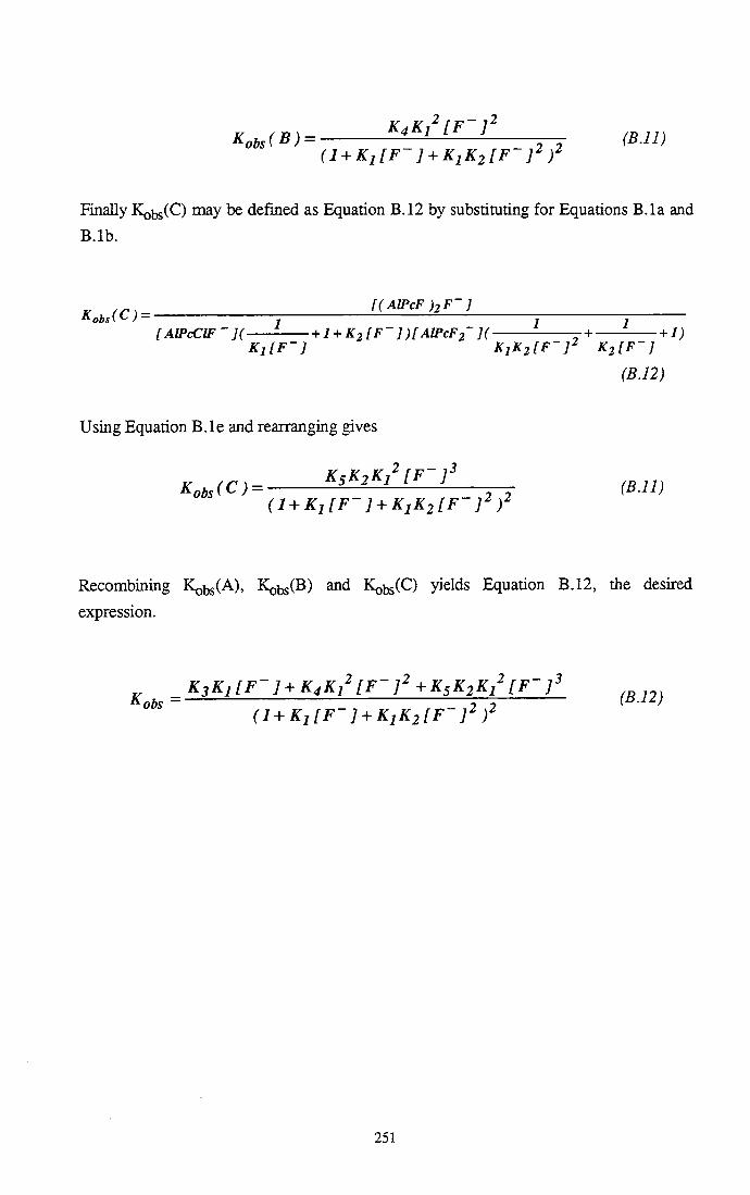
Kots (B) = K4Ki'[F-]' ^^^^^ (l + Ki[F-] + KiK2[F-]^ /
Finally Kobs(C) may be defined as Equation B.12 by substituting for Equations B.la and
B.lb.
^ [(AlPcF)2F-]
[AlPcClF-]( + I + K2 [F-])[AlPcF2~]( j + + 1) K i f p - ] KiKilF-]^ KilF-]
(B.12)
Using Equation B.le and rearranging gives
Kots (C) = ^sK2K^'[F-f ^^^^^ (l + Ki[F-] + KjK2[F-]^)^
Recombining Kobs(A), K^bsCB) and Kobs(C) yields Equation B.12, die desired
expression.
„ K3Ki[F-] + [F-]'+ K5K2Ki^ [ F ' f Kobs = n (^-i^)
(l + Ki[F-] + KiK2[F-]^)^
251

APPENDIX C (Protonation Chapter) Derivation of Equation 5.1
Considering each protonation to be a separate step, the equilibrium may be expressed by Equation C. l .
Pc + H^ <r^-^PcH^ (C.1)
where Pc = PcHn.i"-l+ and PcH+ = PcHnn+-
Assuming conservation of mass, the total concentration of phthalocyanine species is given by Equation C.2 and the equilibrium constant, K by Equation C.3.
CT=[PC] + [PCH^] ^^2)
[PcH^]
~[Pc][H-]
At any given wavelength, the absorbance, A(^), will be due to both PcHn.j"-! and
PcHn""*" species, i.e..
A = ZpJPc] + Zp^jj^[PcH^] (C.4)
Substituting for Equation C.3 gives
A = e p , ( - ^ ^ ) ^ E , ^ ^ . C r - ^ ^ ^ l + K[H^] 1 + K[H^] (C5)
Rearranging,
1 + K[H^] ^'^^ (C.6)
Thus K may be calculated by non-linear fitting of Equation C.6 to a plot of A vs [H+].
252

PUBLICATIONS
1. Beeby A., Parker A.W. and Stanley C.F., Elimination of fluorescence contributions to singlet oxygen measurements using a novel electronic switch, / . Photochem. Photobiol. B:Biol., 37, 267,1997.
2. Wang C , Bryce M.R., Batsanov A.S., Stanley C.F., Beeby A. and Howard J.A.K., Synthesis, spectroscopy and electrochemistry of phthalocyanine derivatives functionalised with four and eight peripheral tetrathiafulvalene units.. Accepted by / . Chem. Soc, Perkin Trans. 2, 1997.
3. Oldham T.C., Beeby A., Parker A.W., Stanley C.F., Telfer A., Barber A. and Phillips D., Improved Instrumentation For The Time-Resolved Detection Of Singlet Oxygen Luminesence., CRC Rutherford Appleton Laboratory Annual Report, RAL-TR-96-076,126,1995-1996.
4. Stanley C.F. and Beeby A., Photophysical Properties of Zinc Phthalocyanines in Relation to their Use as Sensitisers for Photodynamic Therapy., Aspects of Spectroscopy VII, Institute of Physics Spectroscopy Group., Durham, UK., August 21 - 22,1997. - Oral Presentation.
5. Stanley C.F. and Beeby A., The Photophysical Properties of Phthalocyanine Dimers., 25th Annual Meeting, American Society for Photobiology., St. Louis, Missouri, July 5 - 10,1997. - Poster Presentation.
6. Stanley C.F. and Beeby A., Photophysical Properties of Zinc Phthalocyanines at
Low pH., 12th International Congress on Photobiology, ICP' 96., Vienna,
Austria, September 1 - 6,1996. - Poster Presentation.
253

CONFERENCES
1. Aspects of Spectroscopy VI I , Institute of Physics Spectroscopy Group., Durham, UK., August 21 - 22, 1997. - Oral Presentation - C. F. Stanlev and A. Beeby, Photophysical Properties of Zinc Phthalocyanines in Relation to their Use as Sensitisers for Photodynamic Therapy.
2. 25tii Annual Meeting, American Society for Photobiology., St. Louis, Missouri, July 5 - 10, 1997. - Poster Presentation - C. F. Stanlev and A. Beeby, The Photophysical Properties of Phthalocyanine Dimers.
3. 12th International Congress on Photobiology, ICP' 96., Vienna, Austria,
September 1 - 6, 1996. - Poster Presentation - C. F. Stanlev and A. Beeby,
Photophysical Properties of Zinc Phthalocyanines at Low pH.
4. 6th Congress of the European Society for Photobiology., Cambridge, UK,
September 3 - 8,1995.
5. EPSRC Summer School in Laser Spectroscopy, University of East AngUa, UK,
September 4 - 10,1994.
6. North-Eastem Universities Graduate Symposia-University of Newcastie, 1997. University of Sunderland, 1996. University of Durham, 1995.
254

SEMINARS
1996 -1997
1. Professor G. Olah, Crossing Conventional Lines in my Chemistry of the Elements, Loker Hydrocarbon Research, USA.
2. Professor D. Knight, The Purpose of Experiments - A Look at Davy and Faraday, Durham University, Durham.
3. Professor D. Phillips, A Little Light Relief, Imperial College, London. 4. Professor Sir J. Black, What I Learned as a Medicinal Chemist, James Black
Foundation. 5. Professor R. H. Grubbs, Olefin Metathesis using Ruthenium Based Complexes,
Caltech, California. 6. Professor H . Ringsdorf, Function Based on Organisation, Johannes Gutenberg-
Universitat, Mainz, Germany.
7. Dr. K. Reid, Probing Dynamical Processes with Photoelectrons, Nottingham University, Nottingham.
8. Professor J. Eamshaw, Surface Light Scattering: Ripples and Relaxation, Belfast University, Belfast.
9. Professor K. Muller-Dethlefs, Chemical Application of Very High Resolution ZEKE Photoelectron Spectroscopy, York University, York.
10. Dr. J. Clarke, What We Can Learn From Polymer Movie Clips, UMIST, Manchester.
11. Dr. T. Ryan, Making Hairpins from Rings and Chains, UMIST, Manchester.
1995 -1996
1. Dr. D. M. Davies, Chemical Reactions in Organised Systems: Peracid reactivity in Surfactant Micelles and Cyclodextrin Hosts, University of Northumbria,
Newcastle.
2. Professor B. Robinson, Chemistry of Smart Fluids, University of East Anglia,
Norwich.
3. Professor I . Soutar, A Water of Glass. Luminescence Studies of Water Soluble Polymers, Lancaster University, Lancaster.
4. Professor J. Saykally, Terahertz Laser Spectroscopy of Water Clusters; Towards a Genuine Molecular Model of the Liquid, Berkeley, Califomia.
5. Dr. J. Penfold, Soft Soaps and Surfaces, ISIS Facility, Rutherford Appleton
Laboratory, Oxford.
255

6. Dr. Garry Rumbles, Laser Cooling in the Condensed Phase: the Tale of a Novel Observation, Imperial College, London.
7. Professor J. W. Emsley, Liquid Crystals: More then Meets the Eye, Southampton University, Southampton.
8. Professor E. W. Randall, New Perspectives in NMR Imaging, Queen Mary and Westfield College, London.
9. Professor V. Balzani, Supramolecular Photochemistry, University of Bologna, Italy.
1994.1995
1. Professor N. L. Owen, Determining Molecular Structure - the INADEQUATE NMR Way, Brigham Young University, Utah.
2. Professor N. Bartlett, Some Aspects ofAg(II) and Ag(III) Chemistry, University of California.
3. Dr. G. Rumbles, Real or Imaginary 3rd Order Non-linear Optical Materials? Imperial College, London.
4. Dr. T. Cosgrove, Polymers Do it at Interfaces, Bristol University, Bristol. 5. Dr. D. O'Hare, Synthesis and Solid State Properties of Poly-, Oligo- and
Multidecker Metallocenes, Oxford University, Oxford. 6. Dr. M . Schroder, Redox Active Macrocyclic Complexes: Rings, Stacks and
Liquid Crystals, University of Edinburgh, Edinburgh.
7. Professor R. Bonnett, Chemical Aspects of PDT, Queen Mary and Westfield College, London.
8. Dr. A. Beeby and Dr. A. Hughes, Photochemistry: A Demonstration Lecture, University of Durham, Durham.
9. Dr. P. G. Edwards, The Manipulation of Electronic and Structural Diversity in Metal Complexes - New Ligands for New Properties, University of Wales, Cardiff.
256