Synthesis, characterization and enhanced photocatalytic activity of TiO2/SiO2 nanocomposite in an...
9
This article appeared in a journal published by Elsevier. The attached copy is furnished to the author for internal non-commercial research and education use, including for instruction at the authors institution and sharing with colleagues. Other uses, including reproduction and distribution, or selling or licensing copies, or posting to personal, institutional or third party websites are prohibited. In most cases authors are permitted to post their version of the article (e.g. in Word or Tex form) to their personal website or institutional repository. Authors requiring further information regarding Elsevier’s archiving and manuscript policies are encouraged to visit: http://www.elsevier.com/copyright
Transcript of Synthesis, characterization and enhanced photocatalytic activity of TiO2/SiO2 nanocomposite in an...

This article appeared in a journal published by Elsevier. The attachedcopy is furnished to the author for internal non-commercial researchand education use, including for instruction at the authors institution
and sharing with colleagues.
Other uses, including reproduction and distribution, or selling orlicensing copies, or posting to personal, institutional or third party
websites are prohibited.
In most cases authors are permitted to post their version of thearticle (e.g. in Word or Tex form) to their personal website orinstitutional repository. Authors requiring further information
regarding Elsevier’s archiving and manuscript policies areencouraged to visit:
http://www.elsevier.com/copyright

Author's personal copy
Progress in Organic Coatings 72 (2011) 453– 460
Contents lists available at ScienceDirect
Progress in Organic Coatings
jou rn al h om epage: www.elsev ier .com/ locate /porgcoat
Synthesis, characterization and enhanced photocatalytic activity of TiO2/SiO2
nanocomposite in an aqueous solution and acrylic-based coatings
A. Mirabedinia, S.M. Mirabedinib,∗, A.A. Babaloua, S. Pazokifardb
a Chemical Engineering Department, Sahand University of Technology, P.O. Box 51335-1996, Tabriz, Iranb Iran Polymer & Petrochemical Institute, Colour & Surface Coatings Department, P.O. Box 14965-115, Tehran, Iran
a r t i c l e i n f o
Article history:Received 9 January 2011Received in revised form 23 May 2011Accepted 6 June 2011
Keywords:PhotocatalyticTiO2 nanoparticlesSol–gel treatmentSelf-cleanOrganic coatings
a b s t r a c t
In this study, TiO2/SiO2 nanocomposites were synthesized via a sol–gel route by adding tetraethylorthosil-icate (TEOS) to a solution containing different molar ratios of Degussa P25 TiO2 nanoparticles. FTIR, TGA,EDAX and XRD techniques were used to characterize the modified nanoparticles. Photocatalytic activityof the nanoparticles in an aqueous solution and into the acrylic based coating was evaluated using colourcoordinate data measurements, SEM analysis, gloss measurements and FTIR spectroscopy, in the pres-ence of Rhodamine B (Rh.B) dyestuff, as a pollutant model, before and after exposure to the UVA (340 nm)irradiation and compared to their unmodified counterparts.
The results showed that silica grafting effectively reduced the photocatalytic activity of the TiO2
nanoparticles as evidenced by absorption spectra and colour changes of Rh.B aqueous solutions duringthe UVA irradiation. The results revealed the effectiveness of sol–gel route for preparation of TiO2/SiO2
nanocomposites. The optimum result was obtained with 1% molar ratio of TiO2:TEOS. Addition ofTiO2/SiO2 nanocomposites into the acrylic based coating revealed reduction of photo-degradation ofRh.B compared to untreated nanoparticles. Finally, inclusion of TEOS treated TiO2 nanoparticles into theaqueous organic coatings, provides photocatalytic property and as a result, it can possibly be consideredfor self-cleaning coatings.
© 2011 Elsevier B.V. All rights reserved.
1. Introduction
Titanium dioxide has been widely used as a white pigment in abroad variety of applications, such as coatings, cosmetics, foodstuffsand extensive potential applications, due to its unique physical andchemical properties. It is well known that TiO2 in anatase crystallineform, which is biologically and chemically inert, can be used as anexcellent photo-catalyst material in the hydrophilic self-cleaningcoatings and widespread photocatalytic purposes under UV/Visiblelight irradiation [1–4]. When a photocatalytic anatase TiO2 havinga band gap of 3.26 eV is exposed to an UV light source with a wave-length less than 380 nm, the pairs of electron–holes are created. Theelectrons are promoted from the filled valence band to an equalor higher energy level conduction band, which can move freely.The pairs of electron–holes are very reactive and might adsorbedspecies (reactants) on the particle surface, thus photo-oxidationreactions happen [5–7]. The activation of photocatalytic TiO2 byUV light can be written as Eq. (1) [6]:
TiO2 + h� → h+ + e− (1)
∗ Corresponding author. Tel.: +98 21 4458 0040; fax: +98 21 4458 0023.E-mail address: [email protected] (S.M. Mirabedini).
where h�, h+ and e− are absorbed photon energy, hole and electron,respectively. The schematic of the photocatalytic oxidation processusing TiO2 particles as a catalyst is illustrated in Fig. 1 [6].
However, photocatalytic TiO2 particles cannot be incorporatedor deposited on the organic coatings, as they may oxidize the poly-meric matrix. Therefore, surface modification of TiO2 particles isrecommended to adjust their photocatalytic activity [8], dimen-sional stability [9] and wet-out between resin and particles [5].Among the various techniques that are used to adjust photocat-alytic activity of TiO2 particles, the silane treatment method offersdefinite advantages such as simplicity and low cost and also lowprocessing temperature [9,10]. As this regard, a large number ofreports deal with the application of titania–silica materials as cat-alysts in photocatalytic processes [10–13].
It is well known that TiO2 reveals outstanding photocatalyticactivity for oxidative degradation of environmental pollutants[13–18]. The photocatalytic performance of TiO2 depends on; crys-talline phases, specific surface area, particle size, morphology orheat treatment conditions [14]. The main application of photocat-alytic activity in organic coatings is in decomposition of organiccontaminants of water and air [13,19]. Illuminated anatase TiO2also exhibited super hydrophilic properties, which were exploitedfor various applications such as self-cleaning, anti-fogging andantimicrobial effects in coatings [16,17].
0300-9440/$ – see front matter © 2011 Elsevier B.V. All rights reserved.doi:10.1016/j.porgcoat.2011.06.002

Author's personal copy
454 A. Mirabedini et al. / Progress in Organic Coatings 72 (2011) 453– 460
Fig. 1. The schematic of TiO2 UV photo-excitation process (R = reduction;O = oxidation) [6].
Despite the large number of works in this field, up to date,only some articles have been published on the usage of photo-catalytic TiO2 particles in the organic coatings with the aim ofachieving self-cleaning property [18–20]. The presence of photo-catalytic TiO2 in a binder which contains mainly C–C bonds, leads tothe binder structure degradation. Ultraviolet radiation that causesphoto-catalysis supplies the energy needed for breaking C–C bonds[12]. The replacement of untreated TiO2 by surface-treated ones inbinders will inhibit photo-reduction of the particle by ultravioletradiation, therefore reduces its activity against radiation and as aresult, it can inhibit binder destruction.
In this study, TiO2/SiO2 nanocomposites were prepared viaa two-stage, sol–gel route using different molar ratios of TiO2nanoparticles and tetraethoxysilane as precursors. The treatednanoparticles were characterized using conventional techniques.Photocatalytic activity of TiO2 nanoparticles in distilled water andinto a water based acrylic coating was evaluated, in the presenceof Rh.B dyestuff as a model.
2. Experimental
2.1. Materials
TiO2 nanoparticles (P-25, with an average particle size of 30 nm)and tetraethylorthosilicate, TEOS, 98 wt.% were obtained fromEvonik Degussa GmbH and Fluka, respectively. Acrylic emulsionresin, Mowilith® LDM 7729, 48 wt.% solid content, was obtainedfrom Celanese Emulsions Co. Rhodamine B (Rh.B) and texanol waspurchased from Ciba Gigy and Eastman Chemical Co., respectively.All other chemicals were of reagent grade and used without furtherpurification.
2.2. Surface modification of TiO2 nanoparticles
The synthesis procedure was based on a two-stage sol–gelmethod of acid hydrolysis and basic condensation at ambient tem-perature [8,10]. Three various levels of TiO2 nanoparticles (0.5,1.0 and 2.0 mol.%) were dispersed in 40 ml isopropanol by the aidof sonication (Bandelin, HD3200, KE-76 probe) for 15 min underpower of 70 W, 0.7 s pulse on and 0.3 s pulse off, and were thenstirred for a further 1 h.
2.5 ml distilled water, 2.5 ml HCl and 35 ml isopropanol werethen added to the mixtures, and stirred at 1000 rpm for a further1 h. pH of the mixture was measured between 2 and 3. Properamount of TEOS (molar ratio isopropanol/TEOS = 2 and molar ratioH2O/TEOS = 1.2) was added dropwise to the mixture under stirringwithin 15 min and were stirred for a further 16 h at a dark place.Equivalent amount of TEOS for surface treatment of nanoparticleswas obtained by means of elemental analysis (CHN).
Treated nanoparticles were then filtered under suction andphysically adsorbed TEOS compounds on the modified surface of
nanoparticles were washed several times with distilled water andfinally dried in a low pressure oven at 100 ◦C.
2.3. TiO2/SiO2 nanocomposite characterization
FTIR spectroscopy was performed in KBr pellet on a BrukerEQUINOX 55 LSI01 FTIR spectrometer, collecting 16 scans inthe 400–4000 cm−1 range with 4 cm−1 resolution. The thermalbehaviour of the nanoparticles (after drying in 100 ◦C for 3 h) wasevaluated using a TGA-PL-1500 under O2 atmosphere from roomtemperature to 700 ◦C with 10 ◦C min−1 heating rate. Energy dis-persive X-ray spectroscopy (EDAX) was taken from the compressednanoparticles and with Dot-mapping method by INCA250 instru-ment; Oxford Co. Samples were prepared with the same methodthat was used for EDAX analysis with a LSI01-3 SPECAC press instru-ment under 12 bar pressure. X-ray diffraction (XRD) patterns of thenanoparticles were recorded on a Shimadzu XRD-6000 with flowintensity and voltage of 50 mA and 30 kV, in the range 20–70◦.
2.4. Preparation of acrylic nanocomposite coatings
0.5 g of P25 or treated TiO2 nanoparticles (1 mol.%) were directlyadded to the 25 ml distilled water (containing 0.25 g L−1 Rh.B) anddispersed using ultrasonic irradiating for 20 min. The dispersionswere then added to the 50 g emulsion resins and 0.5 g coalescingagent, mixed for further 60 min at a constant rate of 1000 rpm.
Coating samples with a wet thickness of 200 ± 5 �m wereapplied on the degreased glass substrate using a film applicator(Model 352, Erichsen Co.). The applied films were then allowedto dry at 23 ± 2 ◦C for about a week. The freestanding films werealso prepared by application of the coating samples on the PTFEsheets with a wet film thickness of 200 ± 1 �m. The films were thenleft to dry at room temperature (23 ± 2 ◦C) for about one week anddetached from the sheets easily.
2.5. TiO2/SiO2 nanocomposite photocatalytic activity
The aqueous dispersions were prepared by adding 0.65 g L−1
untreated or treated TiO2 nanoparticles, 0.025 g L−1 Rh.B and0.12 mol L−1 sodium dodecyl sulfate, in distilled water. The dis-persions were then exposed to the UVA irradiation (340 nm,0.89 W m−2) in a QUV chamber at 60 ◦C for various time intervals upto 8 h. The photocatalytic degradation of Rh.B in the dispersions wasmonitored using UV–Visible spectrophotometer, CECIL CE9200 inwavelengths 200–700 nm, via measuring the intensity of the typicalabsorbance peak at wavelength of 543 nm.
Photocatalytic activity of TiO2 nanoparticles into the coatingfilms was studied using colour coordinate data measurements,before and during exposure to the UVA irradiation. Photocatalyticperformance of the paint films containing 1 wt.% of P25 and treatedTiO2 (1 mol.%) was studied, in the presence of Rh.B dyestuff as amodel. The sample’s films were exposed to the UVA light (340 nm,0.89 W m−2) in a Q-Panel, QUV/Spray chamber, at 60 ◦C for dif-ferent time intervals up to 240 min. The colour coordinates weremeasured in accordance with ASTM D 65 standard practice using aMiniscan XE Plus colourimeter, from Hunter lab Co., illuminant D65,in angle 10◦. Total colour difference, �E, as a function of the UVAexposure time was calculated from CIE (Commission Internationalde l’é clairage) L*a*b* 1976 formula (Eq. (2)) [21]:
�E∗ab =
√(�L∗)2 + (�a∗)2 + (�b∗)2 (2)
where L* is on the black–white (L* = 0 for black, L* = 100 for white)axis, a* is on the red–green axis, and b* is on the yellow–blue axis.
SEM analysis (Cambridge Stereo scan S360 SEM, with Aucoating), gloss measurements (Nova gloss at 20◦) and FTIR-ATR

Author's personal copy
A. Mirabedini et al. / Progress in Organic Coatings 72 (2011) 453– 460 455
Fig. 2. FTIR spectra of (a) neat TiO2, TEOS treated with; (b) 0.5 mol.%, (c) 1.0 mol.%and (d) 2.0 mol.% TiO2, respectively.
spectroscopy were used to examine the effect of UVA irradiationon the surface properties of the acrylic based coatings containinguntreated and treated TiO2 nanoparticles.
3. Results and discussion
3.1. Characterization of TiO2/SiO2 nanocomposites
Fig. 2 shows the FTIR spectra of neat and differently treatedTiO2 nanoparticles. The assignments for the main FTIR bands of theuntreated and treated nanoparticles are listed in Table 1. A broadabsorption peak of molecular water at around 3400 cm−1 and asharp band at around 1625 cm−1, observed in both untreated andtreated TiO2 nanoparticles, are significantly intense [26,28]. The
Table 1Characteristic absorption peaks achieved from FTIR spectra of P25 and TEOS treatedTiO2 nanoparticles.
No. Wavenumber(cm−1)
Functionality (group)
1 400–800 Crystalline TiO2 [22]2 450, 780 and 1080 Bending vibrations of Si–O or SiOH [22,23]3 955 Vibration modes (Ti–O–Si) [24]4 1079 Asymmetric stretching vibration (Si–O–Si) [24]5 1570 Main characteristic peaks of Si–O bonds [24,25]6 1625 Vibration band of –OH [26,27]7 3400 Vibration band of –OH [26,27]
intensities of absorption peaks due to O–H group near 1620 and3400 cm−1 increased with an increase of the silica amount and itshows similar tendency with the results reported in the literature[27,29]. The absorption peak at around 1079 cm−1 is considerablybroad toward larger wavelength regions and getting broad andgreater, respectively, with increasing of TiO2 mol.%. This peak canbe assigned to Si–O–Si asymmetric stretching vibration and con-firms the silica grafting of TiO2 nanoparticles [30]. FTIR analysisindicates that the SiO2 is chemically bonded with the TiO2 nanopar-ticles, because of the absorption peak assigned to Ti–O–Si vibrationmodes at around 955 cm−1 [31]. Neat TiO2 have broad bands inthe range of 400–800 cm−1 which can be apparent in crystallineTiO2 [22]. Moreover, the peaks common for all silicate structures ataround 1080, 780, and 450 cm−1, due to one of the stretching andbending vibrations of Si–O or SiOH, were observed [22].
TGA technique was used to evaluate variations in the weightof a sample as a function of increasing temperature. Fig. 3 showsthe TGA thermographs of untreated and differently treated TiO2nanoparticles. TGA thermographs reveal that the weight% of neatTiO2 slightly decreases from 50 up to about 100 ◦C, and then sharplydecreases from 310 to 550 ◦C. As can be seen from Fig. 3(b–d), the
Fig. 3. TGA thermographs of nanoparticles after 3 h drying at 100 ◦C; (a) neat TiO2, TEOS treated with; (b) 0.5 mol.%, (c) 1.0 mol.% and (d) 2.0 mol.% TiO2.

Author's personal copy
456 A. Mirabedini et al. / Progress in Organic Coatings 72 (2011) 453– 460
Cou nt300
0 1 2 3 4 5 6 7Energy (keV )
Ti
Ti
Ti
Ti
TiTi
Si
O
O
O
Ti
Ti
Ti
C
C
C
(a)
(b)
(c)
Si
Si
(d) Ti Ti C Ti
O
Fig. 4. EDAX spectra of TiO2 nanoparticles; (a) P25, (b) 0.5 mol.% TiO2, (c) 1 mol.%TiO2 and (d) 2 mol.% TiO2.
various mol.% of TiO2 grafted nanoparticles show sharp weight loss,beginning at around 270 ◦C, continued till 650 ◦C. By decreasingTiO2 mol.%, more weight loss can be observed due to more SiO2grafting on the TiO2 nanoparticles.
TGA thermographs are separated in three zones: the first zone(from 50 ◦C to about 210 ◦C) corresponds mainly to the removalof physically adsorbed water and isopropanol [32]. For untreatedsample, the weight loss is about 0.5 wt.% and for differently treatedsamples are up to 2.7 wt.%. In the second weight loss degreasingzone, in temperature ranging from 210 to 450 ◦C, weight loss iscaused mainly by degradation of Si-compounds on TiO2 surface,and other organic intermediates. For untreated nanoparticles, rel-atively slight weight loss after 340 ◦C can be attributed to watermolecules formed from condensation of hydroxyl groups on theparticle’s surface (TiOH) [27]. In the last zone (about 550 ◦C), thesamples are almost stable and free of any organic composition sinceslightly weight change is observed due to de-hydroxylated at hightemperatures [28].
Fig. 4 illustrates EDAX spectra of untreated and treated TiO2nanoparticles, which indicates different graftings of Si element onTiO2 nanoparticles in different sol–gel processing conditions. Therelative nanoparticle compositions, as derived from EDAX spectra,are listed in Table 2. EDAX analysis of TEOS treated nanoparticlesclearly revealed the presence of Si element, suggesting the silicagrafting on the TiO2 nanoparticles. It is also shown that the Si con-tent of the treated nanoparticles was increased with increasing theratio of TEOS/TiO2 nanoparticles.
XRD patterns of untreated and silica-coated titanium dioxidewith different mole ratios of TEOS/TiO2 nanoparticles are shownin Fig. 5. In the P25 XRD spectrum, three primary diffraction sig-nals can be observed at 2� of 25.2◦, 37.8◦ and 48.0◦, which are
Table 2Elemental composition of P25 and TiO2 nanoparticles treated with differentamounts of TEOS obtained from EDAX analysis.
Sample code Component concentration (wt.%)
Ti Si O C
P25 52.10 – 45.17 2.730.5 mol.% TiO2 8.88 25.34 55.53 9.211.0 mol.% TiO2 10.32 19.97 63.85 10.892.0 mol.% TiO2 15.62 14.61 56.41 8.01
706050403020102 Theta
a)
b)
c)
d)
(110)
(200) (101 ) (211) (112 ) (301)
(202)
Fig. 5. X-ray diffraction patterns of the samples; (a) untreated TiO2, TEOS treatedwith; (b) 0.5 mol.%, (c) 1.0 mol.% and (d) 2.0 mol.% of TiO2.
assigned to diffraction from (1 0 1), (0 0 4) and (2 0 0) planes ofanatase crystalline form, respectively. It is reported that P25 isa totally crystalline material with an anatase/rutile ratio 81/19[25]. As it can be seen, by decreasing mole ratio of TiO2/TEOS, thediffraction signal of anatase TiO2 (1 0 1) decreased, which is relatedto a decrease in the ratio of the crystalline anatase phase to theentire composition of the particle. XRD analysis indicates the sil-ica compound layer absorbed (either physically and/or chemically)on the surface of TiO2 nanoparticles and by decreasing ratio ofTiO2/TEOS, the intensity of amorphous silica layer on the surfaceof TiO2 nanoparticles increased.
3.2. Photocatalytic activity of TiO2 nanoparticles
Visual appearance of Rh.B dyestuff in isopropanol dispersionsduring 2 h UVA irradiation in presence of untreated and treatednanoparticles is illustrated in Fig. 6. Obviously, the discolourationefficiency of P25 is slightly higher than that of treated nanoparticlecounterparts, during UVA irradiation. UV–Vis nanoparticle spectraof prepared suspensions of Rh.B in the presence of modified andunmodified TiO2 nanoparticles are shown in Fig. 7(a and b), respec-tively. As it can be seen from the Fig. 7a, the maximum absorptionpeak (at 543 nm) regularly reduces during the UVA irradiation timeas a result of lower concentration of Rh.B. Since there are no addi-tional peaks emerging in the UV–Vis spectra in the course of theexperiment either using P25 or treated nanoparticles, throughoutthe UVA irradiation, Rh.B can be changed in products that do notreveal any absorption in the UV–Vis wavelength, therefore, photo-bleaching and de-colourization, can be the main mechanisms ofphotodegradation [33].
The colour difference (�E) results of acrylic films containingmodified and unmodified TiO2 nanoparticles prior to and during8 h exposure to the UVA irradiation are shown in Fig. 8. It is evi-dent that surface treatment TiO2 nanoparticles with TEOS couplingagent, reduces �E values of the films; therefore, photocatalyticactivity of TiO2 is reduced. From the Fig. 8, it is clear that in the pres-ence of P25, the degradation of Rh.B is faster than that of its silanetreated counterpart. The Rh.B dyestuff was completely degradedafter 8 h exposure to the UVA irradiation and a colourless solutionwas obtained, while this was extended to more than 16 h for treatednanoparticles. Clearly, surface treatment of nanoparticles reducesthe effective surface area of TiO2 available to Rh.B by which Rh.B isdegraded. Untreated TiO2 nanoparticles provide a higher degrada-tion rate of Rh.B than untreated counterpart because of the higher

Author's personal copy
A. Mirabedini et al. / Progress in Organic Coatings 72 (2011) 453– 460 457
Fig. 6. Discolouration of Rh.B in the presence of P25 and SiO2/TiO2 nanoparticlesduring 2 h UVA irradiation.
0.75
1
1.25
1.5
1.75
200 300 400 500 60 0 700
Wav elength (nm)
a
b
Abs
orba
nce
(%)
P25 TiO2 Aft er 8 h UVA ExposureP25 TiO2, Before UVA ExposureP25 TiO2 Aft er 3h UVA Exposure
545.3 nm1.478 A
323.3 nm1.374 A
207.8 nm1.70 1 A
345.2 nm1.428 A
272 .9 nm1.493 A
207.5 nm1.615 A
543.1 nm1.12 5 A
0
0.25
0.5
0.75
1
1.25
200 300 40 0 500 600 70 0
Wave leng th (nm)
Abs
orba
nce
(%)
Tr.TiO 2, After 8 h UVA Expo sureTr. TiO 2, Before UVA Expo sureTr.TiO 2, After 3 h UVA Expo sure
369.8 nm0.12 5 A
207.5 nm1.154 A
542.3 nm0.847 A
542.3 nm0.60 9 A
275.0 nm0.44 3 A
Fig. 7. Absorption spectra of; (a) P25 TiO2 and Rh.B, (b) TiO2/SiO2 nanoparticles andRh.B, during UVA irradiation.
0
5
10
15
20
25
30
0 60 12 0 180 24 0 30 0 360 42 0 48 0
UVA Exposure Time (min)
Del
ta E
P25 & Acryli c
Tr.P25 & Acryli c
Fig. 8. Colour difference (�E) of coating films containing 1 wt.% untreated and sil-ica treated TiO2 nanoparticles, during UVA exposure time, in the presence of Rh.Bdyestuff.
crystallinity and higher surface areas than silica treated nanoparti-cles. Higher surface areas of untreated TiO2 nanoparticles resultedin larger amounts of Rh.B adsorbed on the nanoparticle surfaceleading to the higher degradation rates of dyestuff as comparedto the silica treated nanoparticles. That, in turn, reduces the elec-tron and hole (e−/h+) recombination, as a result more electronsand holes are available, which diffuse to the nanoparticle surfaceto react with the dyestuff molecules adsorbed on the surface at thefaster rate [27,34].
3.3. Coating performance under UVA irradiation
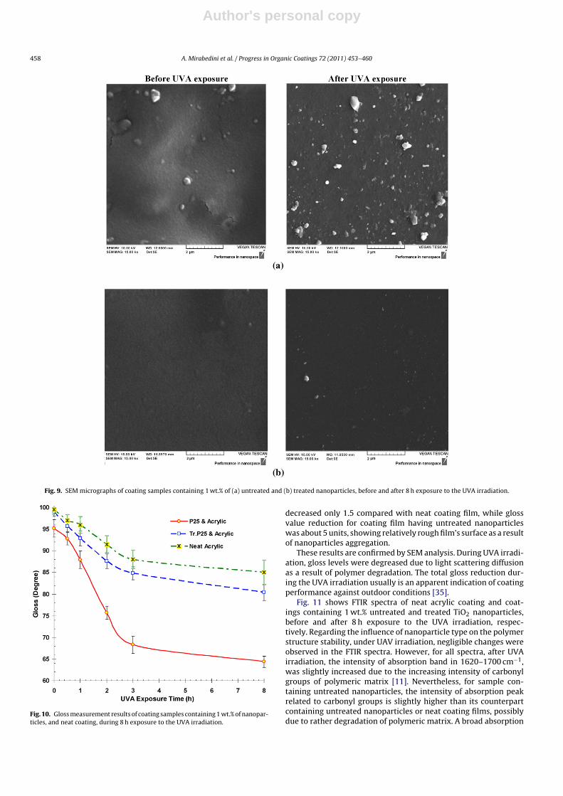
SEM micrographs of coating’s samples containing 1 wt.% ofuntreated and treated nanoparticles are shown in Fig. 9(a and b),before and after 8 h exposure to the UVA irradiation. As it canbe observed, for sample containing untreated nanoparticles, rel-atively large particle aggregates with the size of 0.2–0.6 �m witha non-uniform distribution appeared on the surface of the sam-ples. However, with TEOS treatment of nanoparticles, the size ofparticle aggregates on the surface of the coating film reduced toabout 0.1 �m and less and more uniform distribution of nanoparti-cles was also achieved, as compared to its untreated counterparts.However, in order to evaluate the homogeneity and distribution ofthe nanoparticles to the entire volume of the film, further studiesare needed.
It is clear from the SEM analysis, that the sample having treatednanoparticles revealed a relatively appropriate performance duringexposure to the UVA irradiation. After exposure to the UVA irradi-ation, rarely small holes are observed on the surface of the samplecontaining silica treated nanoparticles. However, coating samplecontaining untreated nanoparticles, the UVA irradiation perfor-mance was getting worse and chalking phenomenon is observedon the surface and the number and size of pores and defects aredramatically increased. These results revealed that TEOS graftingmodifies the photocatalytic activity of TiO2 nanoparticles. It seemsthat silica layer on the TiO2 particles acts as a barrier and delay C–Cbond on the polymeric matrix. This means, it is possible to use mod-ified TiO2 nanoparticles in the organic coatings, with slight furtherpolymer degradation.
Gloss measurement results of coating samples containing 1 wt.%of nanoparticles are shown in Fig. 10, during 8 h exposure to theUVA irradiation. Gloss measurement results also indicate the rela-tively homogeneity of the distribution of the silica nanoparticles onthe surface of coating film compared to its untreated counterparts,as gloss measurement values directly associated to the rough film’ssurface. The results reveal that by inclusion of treated nanopar-ticles within the coating film, the gloss measurement value was
Author's personal copy
458 A. Mirabedini et al. / Progress in Organic Coatings 72 (2011) 453– 460
Fig. 9. SEM micrographs of coating samples containing 1 wt.% of (a) untreated and (b) treated nanoparticles, before and after 8 h exposure to the UVA irradiation.
Fig. 10. Gloss measurement results of coating samples containing 1 wt.% of nanopar-ticles, and neat coating, during 8 h exposure to the UVA irradiation.
decreased only 1.5 compared with neat coating film, while glossvalue reduction for coating film having untreated nanoparticleswas about 5 units, showing relatively rough film’s surface as a resultof nanoparticles aggregation.
These results are confirmed by SEM analysis. During UVA irradi-ation, gloss levels were degreased due to light scattering diffusionas a result of polymer degradation. The total gloss reduction dur-ing the UVA irradiation usually is an apparent indication of coatingperformance against outdoor conditions [35].
Fig. 11 shows FTIR spectra of neat acrylic coating and coat-ings containing 1 wt.% untreated and treated TiO2 nanoparticles,before and after 8 h exposure to the UVA irradiation, respec-tively. Regarding the influence of nanoparticle type on the polymerstructure stability, under UAV irradiation, negligible changes wereobserved in the FTIR spectra. However, for all spectra, after UVAirradiation, the intensity of absorption band in 1620–1700 cm−1,was slightly increased due to the increasing intensity of carbonylgroups of polymeric matrix [11]. Nevertheless, for sample con-taining untreated nanoparticles, the intensity of absorption peakrelated to carbonyl groups is slightly higher than its counterpartcontaining untreated nanoparticles or neat coating films, possiblydue to rather degradation of polymeric matrix. A broad absorption

Author's personal copy
A. Mirabedini et al. / Progress in Organic Coatings 72 (2011) 453– 460 459
0
0.4
0.8
1.2
1.6
2
2.4
5001000150020002500300035004000Wavenu mber (cm-1)
Tran
smitt
ance
(%)
(a)
(b)
(c)
Fig. 11. FTIR-ATR spectra of; (a) neat acrylic coating, (b) coating containing 1 wt.%,and (c) coating having 1 wt.% silica treated TiO2 nanoparticles, before (solid line)and after (dashed line) 8 h exposure to the UVA irradiation.
peak of molecular water due to O–H group at region 3400 cm−1 anda sharp band at around 1625 cm−1 are relatively intense, and theseabsorption peaks to some extent increased after UVA irradiation[26,27]. Fig. 11(d) shows relatively intense absorption bands in therange of 400–800 cm−1, in FTIR spectrum of coating sample havinguntreated nanoparticles, which can be obvious in crystalline TiO2particles on the surface of coating’s sample [22].
4. Conclusion
This work reports the preparation, characterization and photo-catalytic activity of silica treated TiO2 nanoparticles. The resultsrevealed the effectiveness of sol–gel route for preparation ofTiO2/SiO2 nanocomposites. The optimum result was obtained with1% molar ratio of TiO2:TEOS. Addition of TiO2/SiO2 nanocom-posites into the acrylic based coating revealed adjustment ofphoto-degradation of Rh.B compared with untreated counterparts.Inclusion of TEOS treated TiO2 nanoparticles into the aqueousorganic coatings, provides photocatalytic property and due to thelow polymer degradation, under UAV irradiation. Silica on theTiO2 particles acts as a barrier layer and delay C–C bond on thepolymeric matrix. The potential application of these silica treatedTiO2 nanoparticles into the self-clean coating materials seems war-ranted.
Acknowledgement
The authors wish to acknowledge support from the researchwork reports in this paper from Iran Polymer and PetrochemicalInstitute and University of Sahand, Tabriz.
References
[1] M.H. Habibi, S. Tangestaninejad, B. Yadollahi, Photocatalytic mineralisation ofmercaptans as environmental pollutants in aquatic system using TiO2 suspen-sion, Appl. Catal. B: Environ. 33 (2001) 57–63.
[2] K.T. Ranjit, E. Joselevich, I. Willner, Enhanced photocatalytic degradation of�-donor organic compounds by N,N′-dialkyl-4,4′-bipyridinium-modified TiO2
particles, J. Photochem. Photobiol. A: Chem. 99 (1996) 185–189.[3] T. Yamaki, T. Sumita, A. Miyashita, Preparation of epitaxial TiO2 films by PLD
for photocatalyst applications, J. Cryst. Growth 237–239 (2002) 574–579.[4] I.P. Parkin, R.G. Palgrave, Self-cleaning coatings, J. Mater. Chem. 15 (2005)
1689–1695.[5] S.K. Ghosh, Functional Coatings, Wiley-VCH Verlag GmbH & Co. KGaA, Wein-
heim, 2006, p. 9.[6] J. Zhao, X. Yang, Photocatalytic oxidation for indoor air purification: a literature
review, Build. Environ. 38 (2003) 645–654.[7] J. Chen, C. Poon, Photocatalytic construction and building materials: from fun-
damentals to applications, Build. Environ. 44 (2009) 1899–1906.[8] A. Jaroenworaluck, W. Sunsaneeyametha, N. Kosachan, R. Stevens, Charac-
teristics of silica-coated TiO2 and its UV absorption for sunscreen cosmeticapplications, Surf. Interface Anal. 38 (2006) 473–477.
[9] M. Sabzi, S.M. Mirabedini, J. Zohuriaan-Mehr, M. Atai, Surface modification ofTiO2 nano-particles with silane coupling agent and investigation of its effect onthe properties of polyurethane composite coating, Prog. Org. Coat. 65 (2009)222–228.
[10] I.A. Siddiquey, T. Furusawa, M. Sato, K. Honda, N. Suzuki, Control of thephotocatalytic activity of TiO2 nanoparticles by silica coating with polydi-ethoxysiloxane, Dyes Pigments 76 (2008) 754–759.
[11] N.S. Allen, M. Edge, A. Ortega, G. Sandoval, C.M. Liauw, J. Verran, J. Strat-ton, R.B. McIntyre, Degradation and stabilisation of polymers and coatings:nano versus pigmentary titania particles, Polym. Degrad. Stab. 85 (2004) 927–946.
[12] N.S. Allen, M. Edge, J. Verran, J. Stratton, J. Maltby, C. Bygott, Photocatalytictitania based surfaces: environmental benefits, Polym. Degrad. Stab. 93 (2008)1632–1646.
[13] T. Yuranova, V. Sarria, W. Jardim, J. Rengifo, C. Pulgarin, Photocatalytic discol-oration of organic compounds on outdoor building cement panels modifiedby photoactive coatings, J. Photochem. Photobiol. A: Chem. 188 (2007) 334–341.
[14] J. Zhu, J. Yang, Z.F. Bian, J. Ren, Y.M. Liu, Y. Cao, H.X. Li, H.Y. He, K.N. Fan,Nanocrystalline anatase TiO2 photocatalysts prepared via a facile low temper-ature nonhydrolytic sol–gel reaction of TiCl4 and benzyl alcohol, Appl. Catal. B:Environ. 76 (2007) 82–91.
[15] Z. Liu, X. Zhang, T. Murakami, A. Fujishima, Sol–gel SiO2/TiO2 bilayer filmswith self-cleaning and antireflection properties, Sol. Energy Mater. Sol. Cells92 (2008) 1434–1438.
[16] A. Nakajima, K. Hashimoto, T. Watanabe, K. Takai, G. Yamauchi, A. Fujishima,Transparent superhydrophobic thin film with self-cleaning properties, Lang-muir 16 (2000) 7044–7047.
[17] M. Zhou, J. Yu, B. Cheng, Effects of Fe-doping on the photocatalytic activity ofmesoporous TiO2 powders prepared by an ultrasonic method, J. Hazard. Mater.B 137 (2006) 1838–1847.
[18] Y. Shi-ying, C. You-yuan, Z. Jian-guo, C. Ying-jie, Enhanced photocatalytic activ-ity of TiO2 by surface fluorination in degradation of organic cationic compound,J. Environ. Sci. 19 (2007) 86–89.
[19] T. Maggos, J.G. Bartzis, M. Liakou, C. Gobin, Photocatalytic degradation of NOx
gases using TiO2-containing paint: a real scale study, J. Hazard. Mater. 146(2007) 668–673.
[20] L. Sikong, B. Kongreong, D. Kantachote, W. Sutthisripok, Photocatalytic activityand antibacterial behavior of Fe3+-doped TiO2/SnO2 nanoparticles, J. EnergyRes. 1 (2010) 120–125.
[21] K. McLaren, The development of the CLE 1976 (L*a*b*) uniform-colour spaceand colour difference formula, J. Soc. Dyers Colourists 92 (1976) 338– 341.
[22] Z. Liu, R.J. Davis, Investigation of the structure of microporous Ti–Si mixedoxides by X-ray, UV reflectance, FT-Raman, and FT-IR spectroscopies, J. Phys.Chem. 98 (1994) 1253–1261.
[23] E. Liden, L. Bergstroem, M. Persson, R. Carlsson, Surface modification and disper-sion of silicon nitride and silicon carbide powders, J. Eur. Ceram. Soc. 7 (1991)361–368.
[24] G. Lin, J.A. Siddiqui, R.M. Ottenbrite, Surface modification of inorganic oxideparticles with silane coupling agent and organic dyes, Polym. Adv. Technol. 12(2001) 285–292.
[25] M. Behzadnasab, S.M. Mirabedini, K. Kabiri, S. Jamali, Corrosion performanceof epoxy coatings containing silane treated ZrO2 nanoparticles on mild steel in3.5% NaCl solution, Corros. Sci. 53 (2011) 89–98.
[26] H.K. Park, D.K. Kim, C.H. Kim, Effect of solvent on titania particle formationand morphology in thermal hydrolysis of TiCl4, J. Am. Ceram. Soc. 80 (1997)743–749.
[27] K.Y. Jung, S.B. Park, Enhanced photoactivity of silica-embedded titania particlesprepared by sol–gel process for the decomposition of trichloroethylene, Appl.Catal., B 25 (2000) 249–256.
[28] C. Deng, P.F. James, P.V. Wright, Poly(tetraethylene glycol malonate)e titaniumoxide hybrid materials by sol–gel methods, J. Mater. Chem. 8 (1998) 153– 159.
[29] J. Aguado, R. van Grieken, M.-J. Lopez-Munoz, J. Marugan, A comprehen-sive study of the synthesis, characterization and activity of TiO2 and mixedTiO2/SiO2 photocatalysts, Appl. Catal. A: Gen. 312 (2006) 202–212.
[30] A Duran, C. Serna, V. Fornes, J.M. Fernandez-Navarro, Structural considerationsabout SiO2 glasses prepared by sol–gel, J. Non-Cryst. Solids 82 (1986) 69–77.
[31] F.B. Li, X.Z. Li, C.H. Ao, S.C. Lee, M.F. Hou, Enhanced photocatalytic degradationof VOCs using Ln3+–TiO2 catalysts for indoor air purification, Chemosphere 59(2005) 787–800.

Author's personal copy
460 A. Mirabedini et al. / Progress in Organic Coatings 72 (2011) 453– 460
[32] J.M. Kim, S.M. Chang, S.M. Kong, K.-S. Kim, J.S. Kim, W.-S. Kim, Control ofhydroxyl group content in silica particle synthesized by the sol-precipitationprocess, Ceram. Int. 35 (2009) 1015–1019.
[33] S. Pazokifard, S.M. Mirabedini, M. Esfandeh, M. Mohseni, Z. Ranjbar, Silane graft-ing of TiO2 nanoparticles: dispersibility and photoactivity in aqueous solutions,Surf. Interface Anal. (2011), doi:10.1002/sia.3767, wileyonlinelibrary.com.
[34] M. Moonsiri, P. Rangsunvigit, S. Chavadej, E. Gulari, Effects of Pt and Ag on thephotocatalytic degradation of 4-chlorophenol and its by-products, Chem. Eng.J. 97 (2004) 241–248.
[35] D.G. Weldon, Failure Analysis of Paints and Coatings, revised edition, John Wiley& Sons, Ltd., Chichester, 2009, p. 30.