Bone-like Material, Synthesis, Optimization and Characterization
Upload
khangminh22Category
view
1download
0
�����������������
Citation: Naganthran, A.;
Verasoundarapandian, G.; Khalid,
F.E.; Masarudin, M.J.; Zulkharnain,
A.; Nawawi, N.M.; Karim, M.; Che
Abdullah, C.A.; Ahmad, S.A.
Synthesis, Characterization and
Biomedical Application of Silver
Nanoparticles. Materials 2022, 15, 427.
https://doi.org/10.3390/ma15020427
Academic Editors: Paweł
Piotr Pomastowski and
Viorica Railean-Plugaru
Received: 29 September 2021
Accepted: 15 December 2021
Published: 6 January 2022
Publisher’s Note: MDPI stays neutral
with regard to jurisdictional claims in
published maps and institutional affil-
iations.
Copyright: © 2022 by the authors.
Licensee MDPI, Basel, Switzerland.
This article is an open access article
distributed under the terms and
conditions of the Creative Commons
Attribution (CC BY) license (https://
creativecommons.org/licenses/by/
4.0/).
materials
Review
Synthesis, Characterization and Biomedical Application ofSilver NanoparticlesAshwini Naganthran 1, Gayathiri Verasoundarapandian 1 , Farah Eryssa Khalid 1 , Mas Jaffri Masarudin 2 ,Azham Zulkharnain 3 , Norazah Mohammad Nawawi 4,5, Murni Karim 6,7 , Che Azurahanim Che Abdullah 8,9
and Siti Aqlima Ahmad 1,10,*
1 Department of Biochemistry, Faculty of Biotechnology and Biomolecular Sciences, Universiti Putra Malaysia,Serdang 43400, Selangor, Malaysia; [email protected] (A.N.); [email protected] (G.V.);[email protected] (F.E.K.)
2 Department of Cell and Molecular Biology, Faculty of Biotechnology and Biomolecular Sciences, UniversitiPutra Malaysia, Serdang 43400, Selangor, Malaysia; [email protected]
3 Department of Bioscience and Engineering, Shibaura Institute of Technology, College of Systems Engineeringand Science, 307 Fukasaku, Saitama 337-8570, Japan; [email protected]
4 Institute of Bio-IT Selangor, Universiti Selangor, Jalan Zirkon A7/A, Seksyen 7, Shah Alam 40000, Selangor,Malaysia; [email protected]
5 Centre for Foundation and General Studies, Universiti Selangor, Jalan Timur Tambahan,Bestari Jaya 45600, Selangor, Malaysia
6 Department of Aquaculture, Faculty of Agriculture, Universiti Putra Malaysia, Serdang 43400, Selangor,Malaysia; [email protected]
7 Laboratory of Sustainable Aquaculture, International Institute of Aquaculture and Aquatic Sciences,Universiti Putra Malaysia, Port Dickson 71050, Negeri Sembilan, Malaysia
8 Department of Physics, Faculty of Science, Universiti Putra Malaysia, Serdang 43400, Selangor, Malaysia;[email protected]
9 Material Synthesis and Characterization Laboratory, Institute of Advanced Technology, Universiti PutraMalaysia, Serdang 43400, Selangor, Malaysia
10 Laboratory of Bioresource Management, Institute of Tropical Forestry and Forest Products (INTROP),Universiti Putra Malaysia, Serdang 43400, Selangor, Malaysia
* Correspondence: [email protected]
Abstract: Silver nanoparticles (AgNPs) have been employed in various fields of biotechnology dueto their proven properties as an antibacterial, antiviral and antifungal agent. AgNPs are generallysynthesized through chemical, physical and biological approaches involving a myriad of methods. Aseach approach confers unique advantages and challenges, a trends analysis of literature for the AgNPssynthesis using different types of synthesis were also reviewed through a bibliometric approach. Asum of 10,278 publications were analyzed on the annual numbers of publication relating to AgNPsand biological, chemical or physical synthesis from 2010 to 2020 using Microsoft Excel appliedto the Scopus publication database. Furthermore, another bibliometric clustering and mappingsoftware were used to study the occurrences of author keywords on the biomedical applications ofbiosynthesized AgNPs and a total collection of 224 documents were found, sourced from articles,reviews, book chapters, conference papers and reviews. AgNPs provides an excellent, dependable,and effective solution for seven major concerns: as antibacterial, antiviral, anticancer, bone healing,bone cement, dental applications and wound healing. In recent years, AgNPs have been employed inbiomedical sector due to their antibacterial, antiviral and anticancer properties. This review discussedon the types of synthesis, how AgNPs are characterized and their applications in biomedical field.
Keywords: physical; chemical; biological; AgNPs; design and synthesis methods of silver nanoparticles;biomedical properties of AgNPs; biomedical applications; action mechanism
Materials 2022, 15, 427. https://doi.org/10.3390/ma15020427 https://www.mdpi.com/journal/materials

Materials 2022, 15, 427 2 of 43
1. Introduction
Nanotechnology is one of the fastest growing field and its application can be appliedin various industry sectors. This technology helps to downsize bigger size materials withunique properties to atomic size and these are called nanoparticles (NPs). NPs are particlesthat ranges between 1 to 100 nm. These NPs are unique due to their small size, large surfacearea to volume ratio, high carrier capacity, high reactivity and easy variation of surfaceproperties [1]. The distinctive properties of smaller-sized AgNPs also enable them to beapplied in wide range of functions [2]. Currently, silver nanoparticles (AgNPs) have beenwidely used in agriculture, commercial, medical and industry applications [3]. AgNPsare used as an additive in vaccine adjuvant, anti-diabetic agent, wound and bone healing,biosensors and anticancer therapy in medical applications [4].
In agriculture, AgNPs are incorporated in nanopesticides and nanofertilizers [5]. TheseNPs are unique and have different surface to volume-ratio based on the type of synthesis.Smaller AgNPs has a larger surface to volume ratio, which release more silver cations, hencethey are more effective as antimicrobial agents as compared to a bigger sized AgNPs [6].Bigger AgNPs, which are more than 100 nm in size, have smaller surface to volume ratio andare usually used in drug delivery due to the quantity of drug delivered [7]. This size of theparticles is essential for the transport through the certain membranes in the human body.Generally, nanoparticles have been produced through various preparations, includingphysical, chemical and biological routes. The physical and chemical synthesis of AgNPs isoftentimes hazardous and is less cost-effective [8,9] whilst biological synthesis, conversely,are able to produce AgNPs with a higher yield, solubility, stability and biocompatibility [9].
Studies utilizing AgNPs have been of great interest in recent years, with this furtheredby the development of antibiotic resistance in many microbial pathogens over time [10].Nanomaterials have increasingly become a staple in biomedicine, which lead to formationof nanobiotechnology [11]. The application of nanomaterials in medicine is still in researchand development stage. Interestingly, silver (Ag) has been widely used in medical treatmentand management with variety of diseases since ancient times. AgNPs also are one of thenoble metallic NPs that have gained the most attention and exhibited the highest-levelcommercialization, which recorded 55.4% of the nanometals [12]. AgNPs and silver-basedcompounds are well known in this field due to their microbial killing potential [13,14].Currently, AgNPs play roles primarily in non-conventional and enhanced biomedicalapplications such as wound dressing, drug delivery, tissue scaffoldings and proactivecoating applications. In this regard, the AgNPs are considered to possess intrinsic featuressuch as attractive chemical and physical functionality, non-toxic nature, wide spectrumof bactericidal properties, anticancer properties, and therapeutic abilities [15]. Hence, theapplicability of AgNPs have increased in nanotechnology, biomedicine, and environment,thus there is a need for development of cost-effective method for the biosynthesizedAgNPs [16].
2. General Synthesis Routes of AgNPs
The synthesis of AgNPs, like for most nanomaterials, is mainly divided into threedifferent processes: chemical, physical and biological synthesis (Figure 1). Physical syn-thesis refers to synthesis of AgNPs from bulk materials to nanostructures using variousphysical forces [7]. Agglomeration is facilitated by the absence of capping or stabilizingagents. As a result, deterring it poses a significant challenge. Additionally, this synthesisrequires external energy and sophisticated equipment.
Chemical and biological synthesis assembles single atoms and molecules into largernanostructure to produce AgNPs. Chemical and biological synthesis also refers to synthesisof AgNPs by molecular components by nucleation and followed by growth. The chemicaland biological methods can obtain AgNPs by reducing the precursor salt. Various shapesof AgNPs can be obtained quickly by chemical synthesis, but these AgNPs have limitationsin medical application due to its toxicity. The synthesis involved usage of harsh chemicaland may be harmful to the environment. Hence, to overcome this issue, the biological

Materials 2022, 15, 427 3 of 43
synthesis was introduced as an alternative method. It is reported that biopolymers used inAgNPs synthesis were fucoidan, chitosan, ascorbic acid, levan, cellulose, polyphenols andpolypeptides [17–20]. Most of the biopolymers used in the synthesis of AgNPs play thedual role of reducing and stabilizing agent except the starch was used as a capping agent.Thin layer of biopolymers coat the surface of AgNPs, which acts as stabilizing and cappingagent. The modified surface showed improved biocompatibility, intracellular uptake fordrug delivery and longer stability [21].
Figure 1. Synthesis of AgNPs.
Bibliometric analysis is defined as a method of statistical evaluation of publications.It applies mathematical and statistical tools to measure the inter-relationships and influ-ence of publications within the scientific community [22,23]. This method can providea macroscopic overview of how the publications have had an impact on the scientificcommunity, which can be determined by the number of times an article has been cited byother authors [24]. The design study of bibliometric analysis maps out the academic outputas citation information to assess the impacts of an article [25]. This bibliometric analysisalso allows for the representation of information in ways that make relationships moreobvious and easier to understand leading to new insight and discovery [26]. Previously,De Souza et al. [27] published a bibliometric study on photosynthesized nanoparticles, andFeng and Chen [28] analyzed the bibliometric on nanocatalyst. The bibliometric studiesare used to improve the understanding of a research topic, main issues, developments,identifying leading research groups, networking and leading countries in nanotechnology.
A bibliometric analysis was carried out to analyze the co-occurrence of keywords.Data base construction was carried out using the Microsoft Excel applied to the Scopuspublication database (https://www.scopus.com/ accessed on 26 July 2021). A thematicsearch was limited to the years 2010 to 2020 pertained within Scopus database using thesearch term combination (Figure 2): ‘Synthesis’ AND ‘silver nanoparticles’ AND ‘biologicalsynthesis’, ‘Synthesis’ AND ‘silver nanoparticles’ AND ‘chemical Synthesis’, ‘Synthesis’AND ‘silver nanoparticles’ AND ‘physical synthesis’. The search yielded 951 physicalsynthesis publications, 7650 chemical synthesis publications and 1677 biological synthesispublications, according to the database. All the data were downloaded on the 26 July 2021.The most popular publication was chemical synthesis, followed by biological synthesis andlastly physical synthesis. Figure 2 also concluded that the number of total publicationsincreases by year except there is a decrease in year 2017.
The occurrences of author keywords on the biomedical applications of AgNPs wereevaluated by using a bibliometric clustering and mapping software, VOSviewer version1.6.15 (Centre for Science and Technology Studies, Leiden University, Leiden, The Nether-lands) [29,30]. The cluster networks were developed using the keywords “biomedical ANDapplication AND biosynthesis AND silver nanoparticles” from Scopus database (Figure 3).A collection of 224 documents were sourced from articles, reviews, book chapters, confer-ence papers and reviews. The similarity could be inferred using this mapping approach

Materials 2022, 15, 427 4 of 43
based on the strength of co-occurrence or linkage. A stronger co-occurrence frequency isdesigned to evaluate keyword correlation. The minimal frequency of keyword occurrenceswas attained with a threshold of five and defined to only 25 keywords. These keywordsrepresented as the terms or words mentioned in the titles, keywords, and abstracts for morethan five times in overall 224 documents.
Figure 2. Annual numbers of publications relating to the search terms “Silver nanoparticles” and“Biological/Chemical/Physical” in the Scopus publication database between 2010 and 2020 (Englishlanguage only).
Figure 3a depicts the clusters formed around the central theme of the interestedtopic. Each keyword is represented by varying sizes of nodes (circle). The number ofdocuments published between two keywords measured by the total strength of link (TLS).Thickness of line connecting one node to another, indicates the keyword’s co-occurrences.The strength of the correlation link is determined by the distance between each keyword. Astronger relationship is revealed by a smaller distance, vice versa [29,31,32]. The overlayvisualization map is straightforward, and the impact of each keyword was presented froma decade literature collection (Figure 3b) [33]. The author keywords were displayed by thenodes on a network map with a colour gradient based on the software’s assessments for theaverage publishing year. Keywords were distributed according to average publication yearfrom earliest to recent with blue (lowest score) to red (highest score) colour ranges. Keywordanalysis could be performed to discover the underlying areas in the study of biomedicalapplications of biosynthesis of AgNPs. This would represent as an enhanced resource forscientific research and efficiently extrapolate the core of subjects in a disciplinary field ofstudy [34,35].
The classification of keywords into topic areas or research themes is represented byclusters. Four clusters were illustrated in Figure 3a. Cluster 1 (yellow) with 8 occurringkeywords linked with “antimicrobial activity” (25 occurrences), “antibacterial activity”(19 occurrences), “AgNPs” (13 occurrences) and “antioxidant activity” (11 occurrences).The most common analytical tools for AgNPs characterization were identified in the clusteras “TEM”, “SEM”, “FTIR” and “XRD”. The largest node formed in Cluster 2 (green) was“silver nanoparticles” which also acts as the central theme in this research topic. The nodewas linked by “antibacterial”, “anticancer”, “antimicrobial”, “antioxidant” and “cytotox-icity” keywords. The terms “antifungal activity”, “biosynthesis”, “gold nanoparticles”,“plant extracts” and “metallic nanoparticles” highly linked to “biomedical applications”(8 occurrences) were demonstrated in Cluster 3 (blue).

Materials 2022, 15, 427 5 of 43
Figure 3. Research trend clusters mapping. (a) Network visualization of author keywords co-occurrences (b) Network visualization of author’s keywords cluster analysis across the annualaverage publications on the biomedical applications of AgNPs published between 2010 and 2020.Abbreviations: Fourier-transform infrared spectroscopy (FTIR), Transmission electron microscopy(TEM), Scanning electron microscopy (SEM), and X-ray diffraction (XRD).

Materials 2022, 15, 427 6 of 43
Moreover, in the Cluster 4 (red) illustrated the keywords “green synthesis”, “nanopar-ticles”, “characterization”, and “toxicity” commonly occurred in the field of “nanotech-nology” (10 occurrences). In Figure 3b, the keyword “FTIR” (7 occurrences) in turquoisecolour was term frequently mentioned in articles around the year 2013. The keyword“silver nanoparticles” had the greatest number of occurrences (103) and published by arti-cles between 2016 and 2018. Meanwhile, the keywords, “cytotoxicity”, “AgNPs”, “goldnanoparticles” and “metallic nanoparticles” were regularly contributed in publicationstowards the year 2020. Considering AgNPs with an outstanding therapeutic potential,much research work has focused into studying the complex processes of their biologicalfunctions, possible hazardous consequences, and applications of biomedical technologiesfor enhanced clinical testing. Figure 4 displays the advantages and disadvantages ofall three types of synthesis. The chemical synthesis was preferred due to production ofcontrolled shape and size of AgNPs [36,37].
Figure 4. The advantages and disadvantages of three types of synthesis (adapted from Mukherjeeand Patra, [38], Simões et al. [39] and Xu et al. [4]).
2.1. Chemical Route of Synthesis of AgNPs
The chemical method is the most conventional way to synthesize AgNPs. The mediumof synthesis can be organic or aqueous solvent. Chemical synthesis involves the use oftoxic and hazardous chemicals that can be potentially harmful to the environment. Thechemicals used in synthesis can be flammable and non-biodegradable [40]. These toxicchemicals may be absorbed on the nanoparticles and lead to toxic and adverse effect if usedin medical applications [41], example of reducing chemicals used in the chemical synthesisare citrate, ascorbate, element hydrogen and borohydride. Example of chemical synthesisincludes chemical reduction, microemulsion technique, sonochemical and microwaveassisted synthesis.
2.1.1. Chemical Reduction
The most common method used in chemical synthesis is chemical reduction by organicand inorganic agents. This method is commonly used due to its simplicity. The reducingagents are sodium citrate, sodium borohydride, polyethylene glycol (PVP) and Tollenreagent. These agents are used for reduction of oxidation state from Ag+ to Ag0 in theaqueous and non-aqueous solutions. The introduction of these reducing agents lead toa rapid rate of reaction, which produced a large amount of metal nuclei that producesextremely small particles [42]. A slow rate of reaction will lead to agglomeration of particles.The reduction in silver nitrate (AgNO3) takes place in the presence of stabilizer to shield

Materials 2022, 15, 427 7 of 43
the growth of NPs through aggregation. It was reported that a specific shape of AgNPscan be synthesized using ascorbic acid, thiosulfate, sodium citrate and polyethylene glycolas reducing agents. The AgNPs produced by these agents are spherical [43]. The usageof a stronger reducing agent produces a smaller AgNPs [44]. Tsuji et al. [45] reportedthat large AgNPs were produced when weak reducing agent, trisodium citrate was used.The surfactants used were citrate, polyvinyl alcohol, cetyltrimethylammonium bromide,and PVP. Surfactants are used to protect particles from agglomeration and to stabilizethe particles. Surfactants can also control morphology of synthesized AgNPs [6,46,47].Dang et al. [48] reported that a surfactant such as PVP can act as a size controller throughelectrostatic attraction between the ester bond of surfactant with Ag, thus helping tostabilize and keep the AgNPs smaller in size.
A study by Suriati et al. [42] carried out an investigation on the reduction of AgNO3by ascorbic acid and trisodium citrate as surfactant. They concluded that the increaseof trisodium citrate concentration produced more uniform quasi-spherical shape AgNPs.In contrast, when ascorbic acid concentration was increased, a slight change in shape ofsynthesized AgNPs were observed, from quasi-spherical to polygonal shape. It was alsofound that increased concentration of trisodium citrate resulted in decrease of AgNPs size,while increased concentration of ascorbic acid showed the opposite result. Ho and Nga [49]have used sodium citrate as reducing agent to synthesize AgNPs. They found that chemicalreduction without any surfactant or stabilizer produced AgNPs with good distributionand 20 nm in size. In other studies, AgNPs were synthesized using chemical reductionmethod with reducing agent, sodium borohydride and stabilizing agent, PVP and sodiumchloride [50]. The synthesized AgNPs were 20 nm in size.
2.1.2. Microemulsion Technique
Microemulsion is the synthesis of AgNPs when surfactant is used to thermodynam-ically stabilize the dispersion of two immiscible liquids such as oil in water or water inoil or water in superficial carbon dioxide, a mixture of oil, surfactant and water or oil,co-surfactant, surfactant, and water. The AgNPs synthesized are uniform in size andshape. Many types of surfactants such as anionic, cationic, and non-ionic surfactants areavailable to form microemulsion for AgNPs synthesis. Examples of anionic surfactantused are sodium dodecylbenzene sulfonate (SDS), bis(2-ethyhexyl) sulfosuccinate, laurylsodium sulphate, cationic surfactant are PVP and cetyltrimethylammonium bromide andthe non-ionic surfactant is Triton X-100 [43].
AgNPs are synthesized in a two-phase organic aqueous system [51]. Interphaseinterference and the intensity of interphase transport between phases are mediated byammonium salt, which affects the rate of interaction between metal precursors and reducingagents. Metal clusters are stabilized in a non-polar aqueous medium and transferred to anorganic medium using a stabilizing agent.
Reyes et al. [52] demonstrated the microemulsion synthesis of AgNPs using tolueneas organic phase, AgNO3 solution and a mixture of surfactants sodium dodecyl sul-fate/sodium bis (2-ethylhexyl) sulfosuccinate. Studies were done using different con-centrations of precipitating agent, sodium borohydride and dosing times. All mediumwith higher concentrations of precipitating agents produced worm like nanostructures,whereas the lowest concentration produced a mixture of AgNPs and worm like nanoparti-cles. In another study, Das et al. [53] reported the synthesis of cubic AgNPs with the size of17.89 ± 8.74 nm using biosurfactant extracted from Pseudomonas aeruginosa MKVIT3 andborohydrate as reducing agent. Similarly, Chen et al. [54] reported that microemulsionmethod was used to synthesize AgNPs with silver acetate and reducing agent oleylamineat 70 ◦C. The synthesis produced highly monodisperse AgNPs from 10–20 nm and storagestability of 6 months.

Materials 2022, 15, 427 8 of 43
2.1.3. Sonochemical Method
Sonochemical method is the synthesis of AgNPs by ultrasonic radiation, which pro-duced a local hot spot. This method was initially used to synthesize iron NPs, but ithas since been applied to the synthesis of a variety of different metals, including Ag [55].There are several methods for sonochemical synthesis, the most important of which arethe formation, growth, and collapse of bubbles [56]. When solutions are exposed to ul-trasonic radiation, acoustic fields may cause the bubbles in the solution to implode. Thecollapse of the cavitation bubble generates a shock wave, which has a rapid impact onthe particles’ surface [57]. Temperature, pH, pressure, microjet speed, and cooling rateall influence the synthesis [4]. With this method, the AgNO3 solution and nitriloacetatemixture produced spheres, rods, and dendrite shaped AgNPs. The advantages of thismethod include its simplicity and the ability to control the size of AgNPs by using differentprecursor concentrations [58].
Kumar et al. [59] used 1 mM AgNO3 with different starch concentrations (25 and 30 mg)as reducing and stabilizing agent. Result showed that 25 mg starched AgNPs were smallerthan 30 mg, due to aggregation of starched AgNPs at higher concentration.Elsupikhe et al. [60] reported the synthesis of AgNPs in stabilizing agent, κ-carrageenanwith different concentration of AgNO3 using sonochemical method. The number of AgNPssynthesized increased as the concentration of AgNO3 increased. In another study, Patilet al. [61] used sonochemical synthesis to develop AgNPs coated cotton fabrics. The mixtureof AgNO3, hexamethylenetetramine as reducing agent and PVP as the polymer stabilizer.The hydrodynamic diameters of AgNPs were found increase from 32 to 144 nm as theirradiation time increased 30 to 90 mins. Kuntyi et al. [62] demonstrated sonochemicalsynthesis using silver sacrificial anode with the sodium polyacrylate solution. The parame-ters tested were temperature and sodium polyacrylate concentration. The rate of synthesisincreases as the temperature and concentration increase.
2.1.4. Microwave Assisted Synthesis
Microwave heating was discovered in the early 1940s and is now used in a variety ofapplications in the food industry. Microwave assisted synthesis has recently gained atten-tion due to its superiority in synthesizing AgNPs when compared to conventional heating.Microwave-assisted synthesis can also be performed when a mixture is mixed thoroughlyand microwaved. Microwaves generate homogeneous heating, resulting in the formation offine, narrow and uniform sizes of nanocrystals. According to Nadagouda et al. [63], AgNPssynthesized via microwave assisted synthesis produced AgNPs which are smaller, narrowsize distribution and have higher degree of crystallization compared to AgNPs synthesizedvia conventional oil bath heating. This method demonstrated improved control over thenucleation and growth steps in the synthesis of AgNPs [64]. Additionally, microwave-assisted synthesis can provide rapid and uniform heating, reducing the time required forsynthesis. Microwave-assisted synthesis can also be used to accelerate the synthesis processfollowing plant-mediated biosynthesis. Renugadevi et al. [65] synthesized AgNPs usingbiological synthesis, extract of Baliospernum montanum followed by microwave heating.Similarly, AgNPs were synthesized using aqueous rhizome extract of Alpinia galanga medic-inal plant with the aid of microwave irradiation [66]. Abboud et al. [67] synthesized AgNPsusing aqueous onion (Allium cepa) extract with the aid of microwave heating.
Navaladian et al. [68] synthesized AgNPs by decomposing silver oxalate in a glycolmedium using microwave assisted synthesis and PVP was used as the capping agent. Thesynthesis was carried out using microwave for 60 and 75 s and it produced AgNPs withthe size of 5–6 nm and around 30 nm, respectively. Kumar et al. [69] demonstrated thesynthesis of AgNPs using glucose and starch as stabilizers. The conversion rate from Agions to AgNPs was recorded 99 ± 1% and AgNPs synthesized were spherical and lessthan 10 nm in size. Another study demonstrated the synthesis of AgNPs using ethanolas a reducing agent and PVP as a stabilizing agent [70]. The AgNO3 solution in ethano-lic medium was microwaved and produced spherical AgNPs with mean diameter of

Materials 2022, 15, 427 9 of 43
10 ± 5 nm. Chung et al. [71] reported the microwave assisted synthesis of AgNPs usingstarch as a stabilizing agent and ribose and arabinose as reducing agents. Both reducingagent ribose and arabinose produced AgNPs from 15 to 80 nm in size. A complete conver-sion was achieved by L-arabinose, D-arabinose and D-ribose were after 1 min, 2 to 8 minand 2 to 5 min respectively.
2.2. Physical Route of Synthesis of AgNPs
Physical method for synthesis of AgNPs includes evaporation-condensation, laserablation, solvated metal atom deposition (SMAD) method and ball milling and gammairradiation [72–75]. The most important physical methods are evaporation-condensationand laser ablation [76,77].
2.2.1. Evaporation–Condensation
The evaporation–condensation method requires the use of an atmospheric pressuretube furnace or a small ceramic heater to synthesize AgNPs. This technique is frequentlyused to synthesize AgNPs. The evaporation-condensation process is comprised of threemajor steps: (a) Material is evaporated or sublimated to form a vapour phase; (b) Materialis transported from the source to the substrate; and (c) Particles and/or films are formedthrough nucleation and then followed by growth. The vapour rapidly cools, forming smallAgNPs in high concentrations [76]. Additionally, this method required a specific kilowattof power from a typical furnace and a certain amount of time to reach a stable temperature.Additionally, the synthesis of AgNPs requires the use of radiation as a reducing agentrather than a hazardous chemical [72]. This method was used to synthesize nanospheresfrom a variety of metal materials, including Ag [78]. The disadvantages of evaporation-condensation include the lengthy duration of the process and the enormous amount ofenergy required [15].
In a study conducted by Kruis et al. [78], evaporation- condensation method involvesheating of mixture of AgNO3 and sodium acetate in a tube furnace. This resulted in conver-sion of the liquid mixture into gas state, and lastly condensed into AgNPs after the coolingprocess. The size of AgNPs produced was 3 to 50 nm. Raffi et al. [79] demonstrated thesynthesis of AgNPs with an evaporation-condensation method using inert gas, helium inthe process chamber. The authors concluded that smaller AgNPs with less agglomerationsproduced at a lower evaporation temperature and inert gas pressure. The AgNPs producedwere spherical and 9–32 nm in size. In another study, Jung et al. [80] reported the synthe-sis of AgNPs using ceramic heater, which can reach up to 1500 ◦C to synthesize AgNPsthrough evaporation-condensation method. The result showed that polydisperse AgNPswere synthesized from a constant temperature of the heater surface. The size of AgNPsproduced was 6.2 to 21.5 nm. In addition, the AgNPs were spherical and non-agglomerated.Similarly, Harra et al. [81] have synthesized AgNPs using furnace at temperatures between1300 and 1400 ◦C and the vapor was diluted with nitrogen, N2 gas. The size of AgNPsproduced was 50, 90 and 130 nm at different temperature of synthesis.
2.2.2. Laser Ablation
The laser ablation method is a method to obtain metal colloids without using chemicalreagents. In this method, intense laser pulses are focused on Ag target immersed in asolvent [82]. AgNPs could be synthesized via laser ablation of metallic bulk materials insolution [83]. Other than that, the length of chain in alcohol also plays a role in size of Ag-NPs synthesized. The alcohol with a longer chain length from C-3 to C-5 synthesized morestable and smaller particles than short chain alcohols such as methanol and ethanol [84].The characteristics of the AgNPs formed are influenced by a few factors including the wave-length of laser, the duration of laser pulses (femto-, pico- or nanoseconds), the laser fluence,ablation time effective medium and the presence of surfactants [51]. The longer the timingof ablation, the higher the concentration of AgNPs until saturation value is reached [85].Increasing surfactant concentration can also produce smaller AgNPs [51]. A study showed

Materials 2022, 15, 427 10 of 43
that femtosecond laser pulses produced AgNPs with narrower size distribution comparedto nanosecond laser pulses [45]. After the ablation, the liquid environment only containsAgNPs without other ions, reducing agents and compounds [86].
Amendola et al. [87] synthesized AgNPs in acetonitrile and N, N-dimethylformamideusing a bulk metal with laser ablation. The synthesis produced AgNPs surrounded by acarbon shell or included in a carbon matrix. Tajdidzadeh et al. [88] have synthesized AgNPsin ethylene glycol and chitosan solution. The method used chitosan solution producedsmaller AgNPs with a longer stability compared to AgNPs in ethylene glycol. In a similarstudy, AgNPs were synthesized with isopropanol coating using laser beam have had severalmonth stabilities [89]. Alhamid et al. [90] have produced AgNPs in distilled water by laserablation with continuous shots for 13 h. Menazea [91] investigated the synthesis AgNPswith different liquid media such as deionized water, distilled water, dimethylformamideand tetrahydrofuran. The result reported that AgNPs synthesized in deionized water havehigher ablation, stability and antibacterial efficiency than other media. Rhim et al. [92]studied the preparation of AgNPs by a laser ablation method in 5% PVP and a square silverplate was used in process.
2.2.3. Solvated Metal Atom Deposition (SMAD) Method
SMAD occurs when a bulk metal is evaporated under a vacuum to produce atoms orsmall clusters of Ag, while controlling the aggregation of these clusters [93]. The vapour ofthe metal then co-condensed with vapours of organic solvents such as acetone to form NPsin solution using a physical method. A metal wire under vacuum is then electrically heatedto achieve the evaporation of metal. The resulting solution would contain only colloidsand solvent with no byproducts.
The size of produced nanocrystals can be tuned by varying variables such as solventpolarity and heating rate [94]. The advantages of this method include the absence ofa reduction step, the ability to use a wide variety of metal-solvent combinations, theavoidance of toxic organometallic compounds, the achievement of very high dispersionof zero valent metals, the frequent encounter of unusual metal morphology that is highlyreactive, and the secure attachment of metal particles to the catalyst support.
The AgNPs synthesized were in various sizes and spherical in shape. Similarly, Baudotet al. [95] reported the AgNPs and other metal NPs were synthesized using SMAD methodwith chitosan was used as a capping agent. The AgNPs synthesized with chitosan resultingin an average size of 6 ± 1.3 nm.
2.2.4. Ball Milling
Mechanical milling is another term for ball milling. John Benjamin and his colleaguesat the International Nickel Company invented high-energy ball-milling in the late 1960s [96].Ball milling is a type of physical synthesis that involves dropping a ball into a containerand rotating it horizontally, which produces smaller AgNPs. Smaller AgNPs have ahigher surface energy, which results in particle aggregation [4]. There are several typesof mechanical mills that are used in synthesis: vibratory, attritor, planetary, and uniballmills [55]. The dispersion quality is determined by the milling time, rotational speed, andball size [74]. Diffusivity and phase of synthesized AgNPs are temperature dependentduring the ball milling process [97]. The quality of ball-milled products is influenced bya variety of factors, including the milling speed, time, temperature, atmosphere, processcontrol agent, ball to powder ratio, and the size and size distribution of the grindingmedium [98].
Cryomilling involves high energy ball milling and able to produce AgNPs in largequantities at low temperature (−160 ± 10 ◦C). AgNPs produced were 4–8 nm in sizeand the Ag powder collected was cooled down by LN2 [99]. Rak et al. [100] synthesizedAgNPs using lignin as reducing agent and polyacrylamide polymer as support polymer.The mixture of lignin, polyacrylamide and AgNPs was milled for 1.5 h and the AgNPsproduced were 1 to 30 nm. Khayati and Janghorban [97] demonstrated the synthesis of

Materials 2022, 15, 427 11 of 43
AgNPs graphite as reducing agent using mill. AgNPs synthesized were 14 nm in size withthe presence of process control agents.
2.2.5. Gamma Irradiation
Gamma irradiation has high energy, which can be used to synthesize AgNPs [101].Gamma irradiation is a method by which the Ag+ solution induces the reduction of Ag+
into metallic Ag. The colour of irradiated samples changes from colourless to golden yellowfor irradiated Ag [102]. In addition, the reducing agent used can be uniformly distributedin the solution and the AgNPs synthesized are highly pure and stable [103].
Rao et al. [104] reported the production of AgNPs using gamma radiation and gumacacia as a protecting agent. Smaller AgNPs produced due to higher concentration ofgum because it contributes to enhance steric stabilization. Leawhiran et al. [105] hadprepared antibacterial hydrogel wound dressing contained AgNPs using polyvinyl alcohol(PVA), gelatine and AgNO3 solution through gamma irradiation. Eghbalifam et al. [106]investigated the synthesis of AgNPs in the mixture of PVA and sodium alginate underdifferent gamma ray doses. A higher amount of AgNPs with smaller size was formed asthe irradiation dose increased. However, it was observed that no PVA hydrogel was formedup to 15 kGy radiation. In another study, Madhukumar et al. [107] described the synthesisof AgNPs in aqueous silk fibroin solution obtained from Bombyx mori silk using differentgamma irradiation dose. The results showed that 30 kGy, 40 kGy and 50 kGy irradiatedsamples produced 20, 33 and 22 nm in size of AgNPs respectively. Pratiwi et al. [101]synthesized AgNPs with PVA mixture and 10 mM AgNO3 solution under different gammairradiations, which were 0, 0.5, 1 and 2 kGy. The authors concluded that the higher radiationdose increase absorption intensity.
2.3. Biological Route of Synthesis of AgNPs
Biological synthesis of NPs is the simplest, non-toxic, and yet most ecofriendly methodfor producing high-quality NPs. In comparison to chemical and physical synthesis, bio-logical synthesis of AgNPs is advantageous. To synthesize NPs via biological synthesis,safe, non-toxic, and environmentally friendly reagents are used. Additionally, AgNPssynthesized in a single step, such as biological synthesis, have a higher degree of stability,diversity, and adequate dimensions [7]. Biological route synthesis requires living organismssuch as bacteria, fungi, plants and algae. In addition, extracts from organisms can actas reducing and capping agents in AgNPs synthesis [108]. The reduction of Ag ions isfacilitated by biomolecules such as enzymes, proteins, polysaccharide, amino acid andvitamins that can be found in the extracts and they are biodegradable.
2.3.1. Bacterial-Based Biosystems
Bacterial-based biosystems have been reported to synthesize AgNPs either intra-cellularly or extracellularly. AgNPs produced by microorganisms can be classified intotwo distinct categories, depending on the location where they were produced. AgNPsgenerally can be produced by bacteria by bioreduction process, in which reduction ofAgNO3 precursor from Ag+ ions to Ag0 forming AgNPs by Nicotinamide adenine dinu-cleotide (NADH)-dependent enzymes [109]. Singh et al. [110] synthesized AgNPs usingPseudomonas sp. THG-LS1.4. Thomas et al. [111] synthesized AgNPs intracellularly usingOchrobactrum anhtropi. However, extracellular biosynthesis mode was preferred due to easyrecovery of AgNPs.
The synthesized AgNPs exhibit a range of shapes and sizes (Table 1). The type ofbacteria used, the temperature, pH, and substrate concentration all have an effect on theshape and size. Numerous researchers have demonstrated that bacteria are capable ofsynthesizing silver and other NPs. The synthesis of AgNPs is a bioreduction process [109].Studies revealed that bacteria produce extracellular reductase enzymes to reduce silver ionsto AgNPs. Researchers revealed NADH-dependent enzymes involved in bioreduction and

Materials 2022, 15, 427 12 of 43
the reductase enzymes receive their electrons from NADH. The NADH is then oxidizedinto NAD+ ions.
Table 1. Reported size and shape of AgNPs synthesized by microorganisms.
Name of Bacteria Size (nm) Shape References
Bacillus cereus 10–30 spherical [112]Nocardiopsis sp. MBRC-1 45 ± 0.15 spherical [113]
Stenotrophomonas 40–60 multi-shaped [114]Acinetobacter calcoaceticus 8–12 spherical [115]
Escherichia coli 20–50 spherical [116]Leuconostoc lactis 35 spherical [117]
Haemophilus influenzae 80–101 spherical [118]Cyanobacterium Oscillatoria limnetica 3.30–17.97 quasi-spherical [119]
Cyanobacteria De sertifilum sp. 5–26 spherical [120]Sphingobium sp. MAH 11 7–22 spherical [121]
Bacillus licheniformis 7–22 spherical [122]Klebsiella pneumonia 26.84–44.42 spherical [123]
Pseudomonas stutzeri AG259 200 Triangle, hexagonand spherical [124]
Proteus mirabilis PTCC 1710 10–20 spherical [125]Lysinibacillus xylanilyticus strain
MAHUQ-40 8–30 spherical [126]
Bacillus sp. AZ1 7–31 spherical [127]Streptomyces sp. 09 PBT 005 198–595 spherical [128]Exiguobacterium sp. KNU1 4.4 spherical [129]
Shewanella sp. ARY1 38 spherical [130]Lactobacillus sp. strain LCM5 13.84 ± 4.56 spherical [131]
Bacillus sp-Brevibacillusborstelensis_MTCC10642 5–15 cubical [132]
Wang et al. [133] investigated the synthesis of AgNPs using Bacillus methylotrophicus.They reported that the AgNPs synthesis was carried out after 48 h of incubation at 28 ◦C.The synthesized NPs were tested against Candida albicans, Escherichia coli, Salmonella entericaand Vibrio parahaemolyticus. The results showed a better inhibition growth compared toantibiotics. Similar study was done by Karunakaran et al. [134], by performing synthesisof AgNPs using Aztobacter vinelandii culture extracts and spherical NPs with an averagesize of 20–70 nm were produced. These AgNPs exhibited a strong antioxidant activityand antibacterial properties against pathogens such as S. aureus, E. coli, S. fradiae and S.marcescens. The author also suggested that these AgNPs can be efficiently used in thedevelopment of nanomedicine. These AgNPs are efficiently used in health, food, andpharmaceutical industry.
Otari et al. [135] synthesized AgNPs using Rhodococcus sp. after 10 h incubationat room temperature. The AgNPs synthesized were spherical shape with 10–12 nm insize and they showed both bactericidal and bacteriostatic activity against wide range ofbacteria. In another study, endophytic bacterium Bacillus siamensis C1 was isolated froma medicinal plant Coriansrum sativum and AgNPs synthesized were about 25 to 50 nm insize [136]. These NPs showed a strong inhibitory property against rice bacterial leaf blightand bacterial brown stripe as well as promotes plant growth. Penicillium brevicompactum(MTCC-1999) was used to synthesize AgNPs, which were found to be spherical in shapeand ranging in size from 30 to 50 nm [137]. The AgNPs exhibited excellent antibacterialand anticancer activity, with IC50 values of 70 and 50 µg/mL after 24 and 48 h, respectively.Wpij et al. [138] investigated the synthesis of AgNPs using an actinobacterial reductant. Thebiosynthesized AgNPs demonstrated antibacterial activity against E. coli, K. pneumoniae, P.aeruginosa, and S. aureus, as well as anticancer activity against RAW 264.

Materials 2022, 15, 427 13 of 43
2.3.2. Use of Fungi
Fungi are well-known for their ability to decompose organic matter. Recent dataindicate that approximately 5.1 million fungal species exist [139]. Around 6400 bioactivesubstances are produced by filamentous fungi and other fungal species on a microscopicscale [140]. Fungi can synthesize AgNPs intra- or extracellularly, depending on the loca-tion of the synthesized AgNPs. Certain fungi are capable of synthesizing AgNPs in bothways [141]. Fungi reduce Ag+ ions to Ag0 forming AgNPs. NADH and NADH-dependentnitrate reductase are involved [142,143]. Fungi secretes extracellular enzymes such asprotease, cellulase, chitinase and β-glucosidase to degrade cellulose, protein, starch, hemi-cellulose, and animal compounds as a food source [144]. Fungi such as lignocellulolyticfungi obtained their source of carbon and energy by secreting extracellular hydrolase andoxidase enzymes to break down lignocellulosic materials, such as cellulose, lignin andhemicellulose [145]. Fungi are fancied as they secrete a large number of proteins andenzymes, which directly fasten and increase the synthesis of AgNPs [146,147]. Due to theability to tolerate and metal bioaccumulate, fungi have attracted more attention in thebiological production of AgNPs [143]. The biosynthesis involving fungi is usually freefrom toxic chemicals. Most fungi have high wall-binding and intracellular metal uptake,whereas several species grow fast, thus making it easy to culture and keep them in a labora-tory [148]. Filamentous fungi have gained more attention compared to other fungi becausethe AgNPs synthesized by them have good morphological characteristics, stability, and awide range of applications. Fungi have the ability to synthesize nanoparticles both intra-and extracellularly. Intracellular synthesis results in nanoparticles that are smaller thanthose synthesized extracellularly [149]. Intracellular is preferred for a variety of reasons,which includes impurities contamination from enzymes samples can be easily extracted, asthe intracellular enzymes generally require fewer purification steps [150]. Some metal ionscould induce specific proteins, which can hydrolyze the ions.
In a study, Aspergillus sydowii was used to synthesize AgNPs, with spherical shape andsize ranging from 1 to 24 nm [151]. These NPs showed antiproliferative and antifungal prop-erties against clinical pathogenic fungi, which can be applied in biomedical applications.Beauveria bassiana fungus biosynthesized AgNPs with a mixed size and shape, rangingfrom 10 to 50 nm with the mixture shapes of circular, triangular, and hexagonal shape [152].It was concluded that polyphenolic components were released by fungus used for NPssynthesis and growth. Polyphenols also act as a capping agent in the synthesis [153].
Husseiny et al. [154] synthesized AgNPs using fungi, Fusarium oxysporum and evalu-ated the effects on cancer cells. In addition, these AgNPs were also effective as antimicrobialpotential against E. coli and S. aureus. El-Alziz et al. [155] studied the synthesis of AgNPsusing fungus Fusarium solani isolated from wheat and these NPs have antifungal effectagainst different species of phytopathogenic fungi that contaminates wheat, barley, andmaize seeds. Sundaravadivelan and Padmanabhan [156] investigated use of synthesizedAgNPs from Trichoderma harzianum to control insect vectors. Authors have concluded thatthe synthesized AgNPs were effective against controlling the pupae and larvae of Aedesaegypti. Authors also suggested that these NPs could be used as an inexpensive approachto control A. aegypti. Elgorban et al. [157] demonstrated a study using fungus Aspergillusversicolor to synthesize AgNPs. An anti-pest study was conducted against Sclerotinia sclero-tiorum and Botrytis cinerea in strawberry plants using the AgNPs synthesized. The resultconcluded that AgNPs showed greatest effect against B. cinerea.
A green synthesis utilizing the filamentous fungus Aspergillus fumigatus produced Ag-NPs with a diameter of 5–25 nm [158]. Gajbhiye et al. [159] synthesized polydisperse spher-ical AgNPs ranging in size from 20 to 60 nm and evaluated their antifungal activity againstPhoma glomerata, Phoma herbarum, Fusarium semitectum, Trichoderma sp., and Candida albicans.The combination of fluconazole and synthesized AgNPs inhibited C. albicans the most, fol-lowed by P. glomerata and Trichoderma sp., with a smaller increase in fold area of inhibition,but no enhancement against P. herbarum or F. semitectum. Vigneswaran et al. [160] reportedthat Aspergillus flavus synthesized monodispersed AgNPs with a size of 8.92 ± 1.61 nm

Materials 2022, 15, 427 14 of 43
and used them to study the bioabsorption of Ag as NPs. The AgNPs synthesized werefound to be more than three months stable in water. Another study demonstrated thatthe endophytic fungus Botryosphaeria rho-dina synthesizes spherical AgNPs ranging in sizefrom 2 to 50 nm. It was reported that synthesized AgNPs exhibited in vitro anticancerefficacy against A-549 cells with an LC50 of 40 g/mL [161].
Numerous reports have indicated that NADPH-dependent reductase and electrontransfers may be involved in the synthesis of AgNPs. Hulikere and Joshi [162] reported thateven small molecules such as phenolics may be involved in the synthesis of AgNPs whenmarine endophytic fungi, Cladosporium cladosporioides species, are used. This was confirmedby the authors’ FT-IR analyses. S. aureus, S. epidermis, B. subtilis, E. coli, and Candida albicanswere all inhibited by the AgNPs synthesized. Kosbashigawa et al. [163] used Trametes trogiito synthesize AgNPs and obtained a wide variety of AgNPs sizes. Additionally, the authorshypothesized that several ligninolytic enzymes are involved in this biosynthesis process.
2.3.3. Use of Plants
Biological synthesis methods that utilize plants as an AgNPs synthesizer are superiorto other biological methods because plants are abundant in comparison to other biologicalresources [164]. Additionally, when compared to biological synthesis using microorgan-isms, using plants to synthesize AgNPs reduces isolation costs, culture media costs, andenvironmental contamination [165,166]. It has been demonstrated to have a slower kineticand a greater ability to manipulate crystal growth and stabilization. Green plants provide arapid, eco-friendly, cost-effective, non-pathogenic, and one-step method for AgNPs synthe-sis [167]. AgNPs are synthesized from plant extracts including leaves, stems, roots, seeds,and latex [168–170]. The shape, size, and concentration of AgNPs produced by plantsare determined by their chemical composition and concentration, AgNO3 concentration,extraction solvent used, extraction time and temperature, and reaction time and tempera-ture [171]. Nalvolthula et al. [172] summarized the synthesis of AgNPs, which involves afew types of biomolecules present in the flower extracts of Ixora coccinea (Figure 5).
Figure 5. Biomolecules in flower extracts of Ixora coccinea involved in the synthesis of AgNPs.
Plant extracts contain biomolecules such as polysaccharides, tannins, alkaloids, aminoacids, vitamins, polyphenols, terpenoids and saponins, which are eco-friendly and havemedicinal values, that involve in reduction and stabilization of Ag ions [173].Perera et al. [174] stated that biomolecules such as tannins, flavonoids and many sug-ars act as reducing agents whereas other biomolecules act as capping or stabilizing agentduring AgNPs synthesis. The high demand of AgNPs can be resolved by the large-scaleproduction of AgNPs, which takes place in the plants. The plants leaf is preferred comparedto the whole plant in the synthesis of extracellular AgNPs [175].
The proposed hypothetical mechanism for the synthesis of AgNPs (Figure 6) involvesplants or extracts of plants containing enzymes and bioreductant molecules that act asreductants when combined with metal to form AgNPs. For instance, a plant extractcontaining a reducing enzyme will be capable of reducing AgNO3 to silver and nitrateions. Additionally, the complex network of enzymes and antioxidant metabolites actssynergistically to protect the cell and its components from oxidative damage [164].

Materials 2022, 15, 427 15 of 43
Figure 6. Overview of steps of synthesize mediated by plant or plant extract (adapted fromNadeem et al. [176]).
The volume of plant extract, the silver concentration, the pH, the reaction time, andthe pH value of the mixture all significantly influence the size and shape of AgNPs pro-duced [177]. The pH of a mixture has a significant effect on the NP composition. Changesin pH value result in a charge shift in plant metabolism, which has an effect on the chelatingand reducing capacity. This can result in variations of the morphology, dimension, andyield of the synthesis. By increasing the pH of the reaction mixture, small and uniformsized particles are produced [178–180]. The shape of the AgNPs produced can also bealtered from nearly spherical to spherical by adjusting the pH [181].
Concentration of AgNO3 also plays an important in AgNPs synthesis. Smaller size ofAgNPs produced as the concentration of AgNO3 were increased [182–184]. Temperature isalso one of the essential factors affecting the properties of AgNP during synthesis. Hightemperature improves the rate of nucleation, which certainly produces smaller AgNPs.Different metabolites have different nature and reduction capability. Polyphenol familyis a group that possess all good characteristics compared to other available metabolites.The characteristics are sufficient molar concentration, high reducing/antioxidant powerand direct involvement in heavy metal detoxification mechanism in plants. The most abun-dant polyphenols are flavonoids sub-class. Flavonoids usually have different biologicalfunctions such as protection against ultraviolet (UV) radiation and phytopathogens, strongantioxidant activity and flower and fruit coloration [185]. Types of metal ions used alsoaffects the NPs production. The metal ions with higher electrochemical potential are likelyto condense faster [186]. For example, Ag ions have a better ionizing potential due to theirsmaller size compared to Au ions; therefore, it condenses faster.
Jha et al. [187] synthesized AgNPs (2–5 nm) using plant extracts of Bryophyllum sp.,Cyperus sp. and Hydrilla sp. It was confirmed that the reduction was due to phytochemi-cals such as flavones, quinones and organic acid present in plant tissues. In other study,Solanum indicum Linn was reported to synthesize AgNPs, which showed larvicidal activityagainst Cx. pipiens as well as antibacterial activity against S. aureus, E. coli, P. mirabilisand Shigella flexneri [188]. Solanum nigrum and Clitoria ternatea were reported to synthe-size small AgNPs and exhibited antibacterial properties against S. aureus, B. subtilis andS. pyogene [189]. NPs synthesized by Clitoria ternatea extract were smaller in size andexhibited higher antimicrobial activity against nosocomial pathogens compared to NPs syn-thesized by Solanum nigrum, Citrus sinensis, Centella asiatica, Syzygium cumini and Solanumtricobatum, which were irregular shape AgNPs of average size 41, 42, 53 and 52 nm. TheseAgNPs were found effective against P. aeruginosa [190]. Amaranthus gangeticus Linn leafextract was able to synthesize globular shape AgNPs, which exhibited antifungal prop-erties as well as antibacterial properties against both gram-positive and gram-negativebacteria [191]. Velmurugan et al. [192] demonstrated the synthesis of AgNPs by Prunusyedoensis and found that the synthesized NPs were more effective against skin bacteria thancommercial AgNPs.

Materials 2022, 15, 427 16 of 43
Saratale et al. [193] reported on the synthesis of AgNPs using Punica granatum leaf ex-tract (PGE), which resulted in spherical NPs measuring 35 to 60 nm in diameter. The biosyn-thesized AgNPs exhibited antidiabetic activity by inhibiting a-amylase and a-glucosidase.Additionally, AgNPs demonstrated significant anticancer activity when tested againsthuman liver cancer cells (IC50; 70 lg/mL). In another study, AgNPs were prepared usingan extract of the Ipomoea pes-caprae plant, and the AgNPs demonstrated significant antibac-terial activity against P. aeruginosa, E. coli, and Bacillus, as well as anticancer activity againstMCF-7 cancer cells. The MTT assay confirms the anticancer activity, with an IC50 of 78 g ofAgNPs/mL [194].
The plant, Brassica nigra was used to synthesize 10 to 50 nm of spherical shape AgNPs.The AgNPs synthesized showed a strong antifungal and antimicrobial properties as wellas anticancer activity at 100 µg/mL (IC50 of 55 µg/mL) on cancerous cells and was dosedependent [195]. Kanmani and Scleeva [196] reported the synthesis of AgNPs usingLinumisitatissimum extract produced needle shaped AgNPs. The AgNPs produced showedantidiabetic activity with a maximum inhibition of 79.84% in alpha-amylase assay and58.86% for alpha-glucosidase at 100 µg/mL.
2.3.4. Use of Algae
Algae have fast growth rate, cost effective scale-up and easy to harvest. Algae are theearliest and most abundant photosynthetic organisms on the earth [197]. AgNPs can besynthesized in a variety of shapes, including rods, wires, hexagons, spheres, cubes, andpentagons [4]. Additionally, algae are abundant in resources such as pigment, peptides,proteins, and secondary metabolites, which act as nano-biofactories [198]. Green algae(Chlorophyceae), blue-green algae (Cyanophyceae), red algae (Rhodophyceae), and brown algae(Phaeophyceae) are all types of algae [12,199,200]. Algae synthesize AgNPs in two distinctways: intracellularly and extracellularly [120]. Intracellular mode occurs within the celland does not require any pretreatment. Respiration, photosynthesis, and nitrogen fixationare likely to be involved in the synthesis. NADPH or a NADPH-dependent reductase maybe used as the reducing agent. Proteins containing amino groups or cysteine residues serveas stabilizing and capping agents, as do sulfated polysaccharides [201].
Three steps are required for AgNPs synthesis using algae, as shown in Figure 7:(i.) heating or boiling water or organic solvent and algal extract for a specified duration,(ii.) preparing a metal precursor solution, and (iii.) incubating the precursor with algalextract under controlled conditions with continuous stirring. The colour change of theAgNO3-algal extract mixture was then monitored to determine whether AgNPs wereformed [119,202,203]. Extracellular mode occurs external to the cell and entails pretreat-ment steps such as blending and washing. Metabolites, pigments, ions, various proteins(enzymes), and non-protein structures such as RNA, DNA, antioxidants, hormones, andlipids all contribute to the process.
Figure 7. Overview of steps of synthesize mediated by algae extract (adapted from Vincy et al. [202]).
Spirulina platensis is a blue-green alga, the major contributor of AgNPs. S. platensis isfree floating, filamentous cyanobacteria that synthesized spherical AgNPs (2–8 nm) [204].The algae also contain 60–70% vegetable protein, which is rich in essential beta carotene,iron, amino acid, natural vitamins, and essential fatty acid, which involves in capping
Materials 2022, 15, 427 17 of 43
and reduction of NPs. These kind of AgNPs are efficiently used in health, food, andpharmaceutical industry.
Husain et al. [205] screened thirty cyanobacterial species for extracellular synthesis ofAgNPs. The results showed all 30 strains were able to synthesize AgNPs, with variety ofsizes. Majority NPs synthesized were spherical. Cylindrospernum stagnale was the best strainproducing smallest AgNPs with the size of 38 to 40 nm. Microcheate was the fastest strainin synthesizing AgNPs, with the timing of 30 h. Similarly, Singh et al. [206] had reportedcyanobacterium Leptolyngbya was able to synthesize spherical AgNPs with average size of20–35 nm. These NPs showed antibacterial properties against both gram-positive (S. subtilis)and gram-negative (E. coli) bacteria. In addition, these NPs also showed enhancement inseed germination and early seedling development of wheat. Therefore, these NPs mighthave potential in pharmaceutical and agricultural industries. Other studies have demon-strated the isolation several cyanobacteria from Muthupet mangroves, including Spirulina,Oscillatoria, Microcoleus, Alphanocapsa, Phormidium, Gloecaps and Synechococcus [207]. TheAgNPs synthesized were spherical in shape and size about 40 to 80 nm. Microcoleus sp.was reported to have enhanced antibacterial activity against pathogens such as Salmonellatyphi, Vibrio cholera, Proteus vulgaris, Streptococcus sp., Bacillus subtilis, Escherichia coli andStaphylococcus aureus.
More than 20 different species of green micro algae species are exploited for AgNPssynthesis and almost all micro algae species can synthesize various size and morphology ofAgNPs [208]. Rajkumar et al. [209] had conducted biosynthesis of AgNPs using microalgaeChlorella vulgaris and the AgNPs obtained were about 55 nm in size. C. vulgaris also showedthe potential of photocatalytic dye degradation and this can be applied in environmentalbioremediation to remove harmful dyes from industries. Another study used microgreenalgae, Scenedesmus sp., to synthesize NPs both intracellularly and extracellularly, yield-ing AgNPs with an average size of 15–20 nm and 5–10 nm, respectively. Additionally,Scenedesmus sp. exhibited a high level of antimicrobial activity against gram-negative andgram-positive bacteria [210]. Ulva fasciata is a significant green macroalga which is ableto synthesize AgNPs and demonstrating antimicrobial efficacy [211]. Besides that, otherspecies of green macro alga that able to generate AgNPs were Chaetomorpha linum, Gracilariacorticate, Gracilaria edulis [201,212,213]. In another study, AgNPs biosynthesized by redalgae, Gelidium amansii reported could minimize the micro-fouling. AgNPs synthesizedby brown algae like Padina pavania were 49 to 86 nm in size and various shape like rectan-gle, polyhedral, hexagonal, triangular, and spherical [214]. Another brown algae, Padinatetrastromatica, was reported to synthesis spherical AgNPs, which exhibited antimicrobialactivity against B. subtilis, K. planticola, Bacillus sp. and Pseudomonas sp.
Venkatesan et al. [215] described a green synthesis of AgNPs using the marine algaeEcklonia cava, which resulted in spherical AgNPs with an average size of around 43 nm.Antioxidant activity, antibacterial activity against E. coli and S. aureus, and anticanceractivity against human cervical cancer cells were demonstrated for the synthesized AgNPs.Sargassum muticum, another brown algae, was used to synthesize AgNPs, yielding 40–65 nmNPs with spherical and hexagonal shapes. At doses of 25 and 50 g/mL of AgNPs, theproduced AgNPs demonstrated effective anticancer properties against the breast cancer cellline (MCF7) [216]. Kiran and Murugesan [217] demonstrated the synthesis of AgNPs usingthe marine alga Colpomenia sinuosa, with the particles ranging in size from 54 to 65 nmand having a cubic shape. Antidiabetic activity of biosynthesized AgNPs against alpha-glucosidase and alpha-amylase showed 90.50 ± 0.10 and 94.30 ± 0.10, respectively atconcentration of 1 mg/mL.
2.4. Characterization of AgNPs
The unique properties of AgNPs will determine their potential and application. Avariety of measurement techniques can be used to characterize AgNPs nanoparticles. Re-searchers employ a variety of classification schemes. Occasionally, researchers classifiedthe characterization of nanoparticles they prepared according to their structural, optical, or

Materials 2022, 15, 427 18 of 43
electrical properties. Nanoparticle structural characterization is further subdivided intomorphology, crystal structure, and composition. Additionally, there are classificationsfor spectroscopy and microscopy techniques. UV-Visible (UV-Vis), Fourier Transforma-tion Infrared (FTIR), Dynamic Light Scattering (DLS), Energy Dispersive X-Ray Analysis(EDX), and Photoluminescence (PL) are examples of spectroscopy techniques that arefrequently used to characterize nanoparticles. The scanning electron microscope (SEM),the transmission electron microscope (TEM), the atomic force microscope (AFM), and thehigh-resolution transmission electron microscope (HRTEM) are all examples of microscopictechniques that are frequently used by material science researchers. Several of the char-acterizations listed above are required for biosynthesis-based nanoparticles. Among thelisted techniques, FTIR, DLS, XRD and TEM are thoroughly explained in the followingsubsection. The required comprehensive characterization techniques for the biosynthesisof AgNPs are depicted in Figure 8.
Figure 8. Methods of characterization of biosynthesized AgNPs.
2.4.1. Fourier Transform Infrared Spectroscopy (FTIR) Analysis
FTIR is a method utilizing infrared light to characterize the structure of matter at themolecular scale. This method displays various chemical bonding in a sample or materialsas well as detects contaminants in a material, identifies oxidation and decomposition andfinds additives [95]. A typical FTIR includes IR source, mirrors, beam splitter, detectorand a computer. The IR radiation emitted from the source hits the beam splitter is partlydirected to two different mirrors. The two mirrors are the moving mirror, which moves ata constant velocity during data acquisition, and a stationary mirror. The IR beam reflectsand recombines at the beam splitter then passed through the sample. The infrared thatpasses through the sample is usually 10,000 to 100 cm−1. The radiation is the absorbed bythe sample and converted to rotational or vibrational energy before reaching the detector.The data obtained are processed by a computer to transform the interferogram producedinto an IR spectrum.
Recent studies on characterization of AgNPs synthesized by Cannonball leaves andthe FTIR showed three prominent peaks at 2927, 1631 and 1383 cm−1 [217]. The sharpand strong peak at 1631 cm−1 corresponded to stretching vibration of (NH) C = O group,peaks at 2927 was methoxy compounds and 1383 cm−1 was C-C and C-N stretching.Another study was on the characterization of AgNPs using FTIR reported that manyabsorption bands observed [218]. There were bands at 3422 cm−1, which related to thepresence of alcohol and phenol. There were bands at 2921 and 2856 cm−1 representedaromatic compounds, and 1450 and 1043 cm−1 represented amine stretch vibration ofproteins. Studies of characterizing AgNPs synthesized by Trichoderma longibrachiatum usingFTIR showed many bands, which were at 1634.92, 2156.94 and 3269.31 cm−1. The band1632.92 cm−1 represents the stretching vibrations of primary amines [219].

Materials 2022, 15, 427 19 of 43
2.4.2. Dynamic Light Scattering (DLS)
DLS is also known as Quasi-Elastic Light Scattering (QELS) or Photon CorrelationSpectroscopy [220]. DLS is a technique for determining the size and distribution of hydro-dynamic particles in a range of sizes. Currently, DLS is one of the quickest and most popularmethods for determining the size of particles in the range of 1 nm to 1 µm [70,221,222].Additionally, it is cost-effective and timesaving, as it is a rapid analyzer. The DLS techniqueis used to determine changes in the intensity of scattered light from a suspension or solutioncaused by Brownian motion. Brownian motion is the random movement of particles in azigzag pattern from any direction. According to Brownian motion analysis, larger particlesmove slower, encompass a shorter distance, and scatter more light than smaller particles.The size and shape of the macromolecules influence the hydrodynamic diameters [223].The larger particles scatter more light than the smaller ones, whereby even small amountsof aggregates or dust particles could shift the particle size distribution to a larger value.The velocity of the Brownian motion is defined by the translation diffusion coefficient (D)and can be converted into a particle size using Stokes-Einstein equation [224]. The equationis as below:
D = kBT3ηπd
(1)
where D = diffusion coefficient (m2s−1), d = hydrodynamic diameter, kB = Boltzmann’sconstant (1.38 × 10⁻²³ NmK−1), T = temperature (K), η = solvent viscosity (N s m−2).
The polydispersity index (PDI) is used in size distribution range of the particles withvalues lie between 0 and 1. The value 0 indicates the presence of highly homogeneous NPs,while and 1 indicates the presence of highly heterogeneous NPs population [225].
A typical DLS includes a laser, detector, digital signal processor correlator and com-puter. The laser is emitted from the laser light source and illuminates the sample in thecell in the DLS instrument. The scattered light signal is then collected using one of twodetectors, one at a 90◦ scattering angle and one at a 173◦ scattering angle. The presence ofboth detectors provides greater flexibility in terms of selecting measurement conditions.Particles can be dispersed in a wide variety of liquids, and only the refractive index andviscosity of the liquid are required to interpret the measurement result. The obtained opticalsignal exhibits change due to the particles’ relative positions changing randomly, and agraph is generated.
Elamawi et al. [220] reported DLS result of filtered AgNPs produced from Trichodermalongibrachiatum; 10 g fungal biomass and 15 g fungal biomass showed three peaks and twopeaks with the presence of 39.3, 4.4, 1.5 nm and 41.7 as well as 4.9 nm, respectively. Mohantaet al. [226] reported that DLS resulted from AgNPs synthesized by Protium serratum showed75.56 ± 0.46 nm in size, whereas Guilger-Casagrande et al. [143] reported AgNP-TS andAgNP-T NPs synthesized by Trichoderma harzianum representing hydrodynamic diametersof 57.02 ± 1.75 and 81.84 ± 0.67 nm, respectively. NPs synthesized using Trichodermaharzianum (AgNP-TS) whereas without enzymatic stimulation (AgNP-T) by the cell wall ofSclerotinia sclerotiorum. The AgNPs synthesized by Enicostemma axillare leaf extract showed25 to 80 nm in size and dispersity index (PDI) of 0.412 [227].
2.4.3. X-ray Diffraction (XRD)
X-ray diffraction is one of the most extensive conventional techniques to characterizeNPs. XRD is a technique used to determine the crystallographic structure and morphol-ogy, which include the crystalline structure, lattice parameters, nature of the phase andcrystalline size. The number of constituents influences the increase or decrease in intensity.Changing the atoms in the unit cell causes the changing of diffraction intensity. X-rays areelectromagnetic radiation similar to light. However, X-rays have a much shorter wave-length compared to light. They are produced when deceleration of electrically chargedparticles occurs [228]. The peak positions indicate the translational symmetry shape andsize of the particles while the peak intensity indicates the position of atoms located andelectron density inside the unit cell. This method is unsuitable for amorphous materials
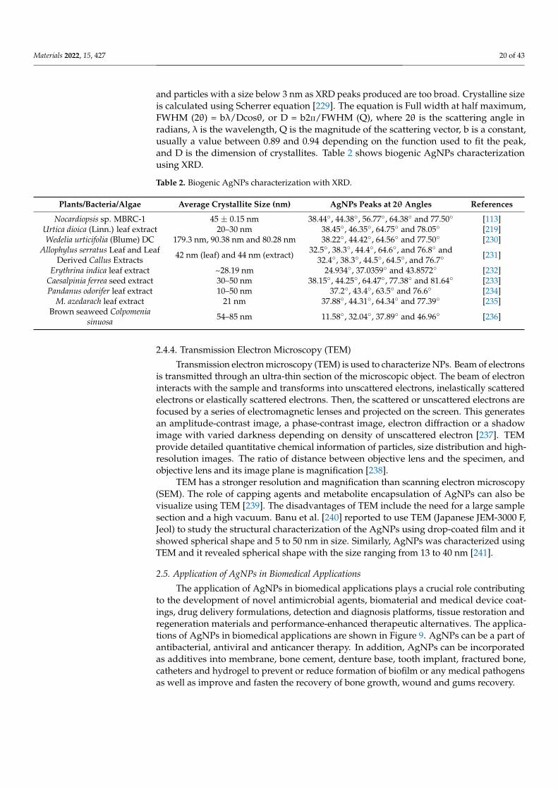
Materials 2022, 15, 427 20 of 43
and particles with a size below 3 nm as XRD peaks produced are too broad. Crystalline sizeis calculated using Scherrer equation [229]. The equation is Full width at half maximum,FWHM (2θ) = bλ/Dcosθ, or D = b2п/FWHM (Q), where 2θ is the scattering angle inradians, λ is the wavelength, Q is the magnitude of the scattering vector, b is a constant,usually a value between 0.89 and 0.94 depending on the function used to fit the peak,and D is the dimension of crystallites. Table 2 shows biogenic AgNPs characterizationusing XRD.
Table 2. Biogenic AgNPs characterization with XRD.
Plants/Bacteria/Algae Average Crystallite Size (nm) AgNPs Peaks at 2θ Angles References
Nocardiopsis sp. MBRC-1 45 ± 0.15 nm 38.44◦, 44.38◦, 56.77◦, 64.38◦ and 77.50◦ [113]Urtica dioica (Linn.) leaf extract 20–30 nm 38.45◦, 46.35◦, 64.75◦ and 78.05◦ [219]Wedelia urticifolia (Blume) DC 179.3 nm, 90.38 nm and 80.28 nm 38.22◦, 44.42◦, 64.56◦ and 77.50◦ [230]
Allophylus serratus Leaf and LeafDerived Callus Extracts 42 nm (leaf) and 44 nm (extract) 32.5◦, 38.3◦, 44.4◦, 64.6◦, and 76.8◦ and
32.4◦, 38.3◦, 44.5◦, 64.5◦, and 76.7◦ [231]
Erythrina indica leaf extract ~28.19 nm 24.934◦, 37.0359◦ and 43.8572◦ [232]Caesalpinia ferrea seed extract 30–50 nm 38.15◦, 44.25◦, 64.47◦, 77.38◦ and 81.64◦ [233]Pandanus odorifer leaf extract 10–50 nm 37.2◦, 43.4◦, 63.5◦ and 76.6◦ [234]
M. azedarach leaf extract 21 nm 37.88◦, 44.31◦, 64.34◦ and 77.39◦ [235]Brown seaweed Colpomenia
sinuosa 54–85 nm 11.58◦, 32.04◦, 37.89◦ and 46.96◦ [236]
2.4.4. Transmission Electron Microscopy (TEM)
Transmission electron microscopy (TEM) is used to characterize NPs. Beam of electronsis transmitted through an ultra-thin section of the microscopic object. The beam of electroninteracts with the sample and transforms into unscattered electrons, inelastically scatteredelectrons or elastically scattered electrons. Then, the scattered or unscattered electrons arefocused by a series of electromagnetic lenses and projected on the screen. This generatesan amplitude-contrast image, a phase-contrast image, electron diffraction or a shadowimage with varied darkness depending on density of unscattered electron [237]. TEMprovide detailed quantitative chemical information of particles, size distribution and high-resolution images. The ratio of distance between objective lens and the specimen, andobjective lens and its image plane is magnification [238].
TEM has a stronger resolution and magnification than scanning electron microscopy(SEM). The role of capping agents and metabolite encapsulation of AgNPs can also bevisualize using TEM [239]. The disadvantages of TEM include the need for a large samplesection and a high vacuum. Banu et al. [240] reported to use TEM (Japanese JEM-3000 F,Jeol) to study the structural characterization of the AgNPs using drop-coated film and itshowed spherical shape and 5 to 50 nm in size. Similarly, AgNPs was characterized usingTEM and it revealed spherical shape with the size ranging from 13 to 40 nm [241].
2.5. Application of AgNPs in Biomedical Applications
The application of AgNPs in biomedical applications plays a crucial role contributingto the development of novel antimicrobial agents, biomaterial and medical device coat-ings, drug delivery formulations, detection and diagnosis platforms, tissue restoration andregeneration materials and performance-enhanced therapeutic alternatives. The applica-tions of AgNPs in biomedical applications are shown in Figure 9. AgNPs can be a part ofantibacterial, antiviral and anticancer therapy. In addition, AgNPs can be incorporatedas additives into membrane, bone cement, denture base, tooth implant, fractured bone,catheters and hydrogel to prevent or reduce formation of biofilm or any medical pathogensas well as improve and fasten the recovery of bone growth, wound and gums recovery.

Materials 2022, 15, 427 21 of 43
Figure 9. Applications of AgNPs in Biomedical Applications.
AgNP applications may be used to combat the alarming and emerging problem ofpathogenic drug resistance. Additionally, AgNPs exhibit a broad antibacterial activityagainst both gram-positive and gram-negative bacteria, which is advantageous for biomedi-cal applications. Both pathogens can adhere to a surface, resulting in the growth of a biofilm.AgNPs were used to penetrate and disperse the biofilm, resulting in the pathogens beingreleased from the infection surface (Figure 10). According to another study, when AgNPsare applied to cells, they penetrate the cell and cause abnormal cell function, structuraldamage, and ultimately cell death [242]. A study by Neihaya and Zaman [243] reportedthe changes from black colonies into pink colonies after the application of biosynthesizedAg, which was concluded due the loss of biofilm formation ability. Similarly, anotherbiosynthesized Ag disrupts 80% of Uropathogenic E. coli (UPEC) biofilms [244]. As aresult, AgNPs could be excellent, dependable, and effective solution for major concerns:antibacterial, antiviral, anticancer therapy, bone healing, bone cement, dental applicationsand wound healing.
Figure 10. Formation of biofilm and AgNPs application (adapted from [245]).
2.5.1. AgNPs for Antibacterial Activities
Antibiotics have been used to treat antibacterial infections for years; however, dueto inconsiderate, misuse, and multiple use of antibiotics, multidrug-resistant (MDR) mi-croorganisms have emerged. Apart from that, many antibiotics have lost their efficacydue to the evolution of resistant strains. As a result, this has posed a serious threat to theglobal human population, as more than 60% of bacteria that cause nosocomial infectionsare now resistant to at least one of the most commonly used antibiotics [246]. Multidrug-resistant infections in humans are difficult to treat and result in a lengthy hospital stay [247]

Materials 2022, 15, 427 22 of 43
S. enteritidis, K. pneumoniae, E. coli, S. aureus, and P. aeruginosa are just a few of the species thatcause infections in biomedical settings. However, the application of AgNPs has emergedas a potential solution to the multidrug resistance problem. AgNPs are a cost-effectiveand efficient way to combat multidrug-resistant bacteria. Bacteria can be gram-positiveor gram-negative. AgNPs can be efficient in inhibiting both gram-positive and gram-negative bacteria. Figure 11 shows the possible mechanism of actions of AgNPs towardsgram-negative and gram-positive bacterial cells.
Figure 11. The possible mechanism (ROS activation) towards gram-positive and gram-negative(adapted from Pandey et al. [248]).
Gram-negative bacteria have a thin peptidoglycan layer with periplasmic membraneand an additional outer membrane, whereas Gram-positive bacteria have a thick pep-tidoglycan layer with periplasmic membrane. Gram positive bacteria are reported tobe more resistant to AgNPs [249,250]. The effect of AgNPs on E. coli, P. aeruginosa, andS. aureus was investigated, and it was concluded that gram-negative strains such as E. coliand P. aeruginosa are more susceptible to cell wall damage than S. aureus, a gram-positivestrain with a thick cell wall [251].
Huq and Akter [252] described the synthesis of AgNPs using the supernatant of Massilia sp.MAHUQ-52 and their antimicrobial activity against multidrug-resistant pathogens, Klebsiella pneu-moniae and Salmonella enteritidis. These pathogens were tested against AgNPs synthesizedin the laboratory and six additional antibiotics. S. enteritidis was found to be resistant tofive antibiotics tested and to have an inhibition zone of 16.8 ± 0.9 mm for biosynthesizedAgNPs. The AgNPs exhibited inhibition zones with a diameter of 17.6 ± 0.5 mm againstK. pneumoniae, whereas the other five antibiotics exhibited no antibacterial efficacy. Theauthors concluded that the inhibition is due to AgNPs rupturing the cell wall and causingcell death. Huq [253] investigated the antimicrobial efficacy of AgNPs synthesized byPseudo-duganella eburnea MAHUQ-39.
The antibiotic-resistant human pathogens used were E. coli, S. aureus and P. aeruginosa.MICs of P. aeruginosa and S. aureus were 6.25 µg/mL and 200 µg/mL and 100 µg/mLwhereas MBCs of P. aeruginosa and S. aureus were 50 µg/mL and 200 µg/mL, respec-tively. Nanda et al. [254] studied on the biosynthesis of AgNPs using S. aureus and theirantibacterial activity against human pathogens such as methicillin-resistant S. epidermis,methicillin-resistant S. pyogenes, S. typhi and K. pneumoniae. MRSA and MRSE are bacteriafound to be resistant to a wide range of broad-spectrum antibiotics. The inhibition zonesfor MRSE was 18 mm, followed by MRSA was 17.5 mm and S. pyogenes was 16 mm. It wasalso reported that all gram-positive pathogens were inhibited by AgNPs. In another study,Du et al.’s [255] in vitro results of the application of biosynthesized AgNPs using Novosph-ingobium sp. THG-C3 as a potential treatment/prevention against human pathogens, P.
Materials 2022, 15, 427 23 of 43
aeruginosa, V. cholera, S. aureus, K. pneumoniae, E. coli and S. typhi. The results showedthat biosynthesized AgNPs showed an effective antibacterial activity against S. aureus, K.pneumoniae and S. typhi with zone of inhibition of 12, 7 and 7 mm, respectively. There aremore literatures available for in-vitro testing against human pathogens (Table 3).
Table 3. In-vitro testing against human pathogens.
Organism Source Size and Shape Test Organism References
Origanum heracleoticum L. Bacteria Spherical; 30–40 nm E. coli, P. aeruginosa, S. aureus, S.pneumoniae, K. pneumoniae [256]
Sporosarcina koreensis DC4 Bacteria Spherical; 102 nmV. parahaemolyticus, E. coli, S.
enterica, B. anthracis, B. cereus andS. aureus
[257]
Dodonaea viscosa extract Plant Spherical; 16 nm E. coli, K. pneumoniae, P. fluorescens,S. aureus, B. subtilis [258]
Marine Pseudomonas sp. H64 Bacteria Spherical; 3–22 nmB. subtilis, S. faecalis, S. aureus, E.
coli, A. hydrophila, V.parahaemolyticus
[259]
Aloe arborescens Plant Spherical; 38 ± 2 nm P. aeruginosa, S. aureus [260]Acorus calamus Plant Spherical; 20–35 nm S. aureus, E. coli [261]Fusarium scirpi Fungus Quasi-spherical; 2–20 nm E. coli [244]
Artemisia marschalliana extract Plant Spherical; 5–50 nm S. aureus, B. cereus, A. baumannii [262]Moringa oleifera Plant Spherical; 8 nm K. pneumoniae, S. aureus [263]
Caulerpa racemose Algae Spherical and fewtriangular; 5–25 nm S. aureus, P. mirabilis [264]
Leptolyngbya strain JSC-1 Algae Spherical; 5–50 nm S. aureus, E. coli [265]
Penicillium oxalicum strain LA-1 Fungus Spherical; 52.26 nm K. pneumoniae, V. cholerae, E. coli, M.luteus, M. smegmatis, B. subtilis [266]
Aspergillus niger Fungus Spherical; 1–10 nmC. krusei, C. parapsilosis, C.
tropicalis, A. flavus, A. fumigates, S.aureus, P. aeruginosa, E. coli
[267]
Alga Syridia fusiformis AlgaeSpherical, Triangle,Pseudo-spherical,
rectangle; 5–50 nmK. pneumoniae, S. aureus [268]
Ipomoea asarifolia Plant Spherical; 20–60 nm B. subtilis, S. aureus, E. coli, K.pneumoniae [269]
2.5.2. AgNPs for Antiviral Activities
There have been recent outbreaks of an infectious disease as a result of the presence ofa newly evolving pathogenic virus. Viruses are capable of infecting both eukaryotes andprokaryotes. Virus replication requires living cells, which they damage or even destroy.Systemic infections and severe complications can occur as a result of acute and chronicconditions that develop following the viral infection. Viruses are found in the air, water,and, most commonly, the soil. The newly evolving virus has developed resistance tocurrently available antiviral drugs, prompting researchers to investigate new drugs andalternative treatment options. As a result, AgNPs have been used as an antiviral agent in thebiomedical field. Additionally, biogenic AgNPs are an antiviral agent against HIV-1 [270],herpes virus (HSV-1 and HSV-2) [271], and dengue virus (DEN-2) [272]. The mechanismby which a virus infects a cell is depicted in Figure 12 along with the role of AgNPs as anantiviral agent.
Although there are a few types of research on the effect of AgNPs towards viruses,the details of the interactions are scarce. However, this can be due to the complexity ofvirus structure that contribute to the limited knowledge of AgNPs mechanism towardsthe viruses. Salleh et al. [273] stated that there are two ways in which how AgNPs interactwith pathogenic virus; (1) Bind to the outer coat of virus and inhibit the attachment ofvirus towards cell receptors and (2) Bind to the DNA or the RNA of virus and inhibitreplication or propagation of the virus. Table 4 shows biosynthesized AgNPs and theantiviral properties towards different types of pathogenic virus.

Materials 2022, 15, 427 24 of 43
Figure 12. The antiviral activities of AgNPs [273].
Table 4. The antiviral properties of biogenic AgNPs.
Virus Family Source of AgNPs Size of AgNPs Mechanism References
Herpes simplex virustype 1 and type 2(HSV-1 & HSV-2)
Hepesviridae Dryopteris species, Musaparadisiaca, Catharanthus
roseus, Selaginellabryopteris, Syzygium
cumini
4–31 nm Block interaction ofviral cells
[274]
Human parainfluenzavirus type 3 (HPIV3) Paramyxoviridae
Zika virus Flaviviridae Rhazya stricta 20–40 nm Penetrate theinfectious agent [275]
Dengue virus (DEN-2) FlaviviridaeBruguiera cylindrica 30–70 nm Inhibitory effect on viral
RNA synthesis [276]
Carica papaya leaf 10–35 nm Inhibition ofviral replication [277]
Leucas aspera and Hyptissuaveolens 7–22 nm
Capping facilitated surfaceactivity makes these AgNPs
a tool for vector control[278]
HSV-1, HAV-10 andCoxB4 virus
Herpesviridae Lampranthus coccineus 10.12–27.89 nm
Interact with herpes simplexthymidine kinase, hepatitis
A 3c proteinase andCoxsackie virusB4 3c protease
[279]
Malephora lutea F.Aizoaceae 8.91–14.48 nm
Interact with herpes simplexthymidine kinase, hepatitis
A 3c proteinase andCoxsackie virusB4 3c protease
[279]
Respiratory SyncytialVirus (RSV) Pneumoviridae Curcuma longa 0.23 nm
Prevent the virus fromentering cells and inhibition
of viral replication[280]
HIV-1 Retroviridae Rhizophora lamaeckii 12–28 nm HIV-1 reverse transcriptaseinhibitory activity [270]
Herpes Simplex Virus(HSV-I,II) Herpesviridae Sargassum withtii - Prevent the virus from
entering cells [271]

Materials 2022, 15, 427 25 of 43
In addition, Sharma et al. [281] used the medicinal plants Andrographis paniculate,Phyllanthus niruri and Tinospora cordifolia to synthesize AgNPs and tested their antiviralproperties against chikungunya virus. Based on DLS, the average sizes of AgNPs synthe-sized by A. paniculate, P. niruri and T. cordifolia produced was 68.06, 28.38 and 37.10 nm,respectively. It was reported that A. paniculate AgNPs exhibited the highest antiviral prop-erties against chikungunya virus. In another study, Sreekanth et al. [282] demonstrated thebiosynthesis of AgNPs using extract of Panax ginseng roots and tested against influenzavirus. The AgNPs produced are spherical and 5 to 15 nm in size. It was reported that onlyslight anti-influenza was detected at concentrations of 0.005, 0.01 and 0.15 M and increasedat concentration of 0.02 and 0.25 M of AgNPs.
2.5.3. AgNPs for Anticancer Therapy
Cancer and its secondary complications account for an alarming number of deathsworldwide, owing to insufficient efficacy, severe side effects from currently availablechemotherapeutic agents, and poor prognosis. Breast cancer (13.7%) is the most frequentlyreported type of cancer worldwide, followed by colorectal cancer with 11% [283]. Secondarycomplications associated with chemotherapies used to treat these two leading cancersinclude infection with bacteria, viruses, and fungi, as well as a compromised immunesystem. The conventional treatment of cancer, which includes surgery, radiation therapy,and chemotherapy, results in ineffective anticancer therapy, owing to the treatment’sdecreased selectivity and specificity [284]. Additionally, cytotoxic drugs used to treatbreast cancer, such as doxorubin, cis-plation, and bleomycin, have drawbacks and areineffective [285]. As a result, nanoparticles such as AgNPs have the potential to act asanti-cancer nanomedicines (Table 5).
Nguyen and colleagues [286] reported on the treatment of hepatocellular carcinoma(HepG2) and breast cancer (MCF-7) with AgNPs synthesized in the presence of Ganodermalucidum (GL). Additionally, they demonstrated that spherical nanoparticles of AgNPs/GLwith a physical size of 10.72 nm effectively inhibited the proliferation of HepG2 and MCF-7cells. In another study, Narasimha et al. [287] synthesized AgNPs from Eucalyptus globulusL. leaf extract and used them to treat colorectal cancer cell lines (HCT116). AgNPs wereproduced with a physical size of 20 nm and a spherical shape. The AgNPs arrested thecell cycle, increased the expression of apoptotic genes, and decreased the expression ofantiapoptotic, inflammatory, and stem cell markers in HCT116 cells.
In another similar study, the Acalypha indica Linn AgNPs inhibits 40% of humanbreast cancer cells (MDA-MB-231) [288]. Deeb et al. [289] reported the in vivo and in vitrostudies using AgNPs biosynthesized with Arthrospira platenses, Microcystis aeruginosa andChlorella vulgarisactive metabolites against breast cancer. Results showed the safety usageof bio-AgNPs was 0.1 mg/mL on PBMCs cells and 1.5 mg/mL on the tested mice. Thebiogenic AgNPs showed dose-dependent cytotoxicity against HepG-2, CaCo-2 and MCF-7cell lines. The Arthrospira bio-AgNPs displayed a delay in tumour growth and prolongedsurvival in mice model, which explained that the treatment induced cellular apoptosisin cancer cell by down regulating of surviving, MMP7, TGF and Bcl2 genes expressions.Gajendran et al. [290] reported the inhibition of 50% of proliferation of human breast cellline (MCF7) after 24 h incubation using 20 µg/mL AgNPs produced by Danura inoxia. TheAgNPs can result in cell membrane integrity, apoptosis, oxidative stress and reduced DNAsynthesis. The AgNPs (5 µg/mL) biosynthesized by Dendrophthoe falcata were sphericaland 5 to 45 nm in size and caused MCF-7 to lose their 50% viability [291].

Materials 2022, 15, 427 26 of 43
Table 5. The anticancer results of green synthesized AgNPs.
Organism Source of AgNPs Cancer Cell Line IC50 Value Size and Shape of AgNPs References
Mixture of Curcuma longa andZingiber officinale Plant HT-29 150 µg/mL Spherical; 20–51 nm [292]
Solanum trilobatum Plant MCF-7 30 µg/mL Spherical; 12.5–41.9 nm [293]Aspergillus terreus ITCC 9932 Fungus MCF-7 25.24 ± 0.990 µg/mL Spherical; 25 nm [294]
Aspergillus nigerAspergillus michelle
Aspergillus japonicusFungus MCF-7
2.46 µg/mL3.12 µg/mL1.47 µg/mL
Varying in sizesVarying in sizes
~100 nm[295]
Agaricus bisporus Fungus MCF-7 50 µg/mL Spherical; 8–20 nm [296]Endophytic bacterium Bacteria MCF-7 50 µg/mL Spherical; 83–176 nm [297]Dimocarpus longan Plant PC3 10 µg/mL Cubical; 9–32 nm [298]
Ginkgo biloba Plant HeLA, SiHa Dose dependent Spherical; 40 ± 1.2 nm [299]Punica granatum Plant A5449 5 µg/mL Spherical; 6–45 nm [300]
Detarium microcarpum Plant HeLa, PANC-1 31.5 µg/mL,84 µg/mL
Spherical, circular,rectangular;
62–103 nm (TEM)[301]
Punica granatum Plant HeLa 100 µg/mL 46.1 nm [302]Fagonia indica Plant MCF-7 12.35 µg/mL Spherical;10–60 nm [303]
Alternanthera sessilis Plant PC3 6.85 µg/mL Spherical; 30–50 nm [304]
Oscillatoria limnetica Bacteria MCF-7, HCT-116 6.147 µg/mL,5.369 µg/mL
Quasi-spherical;3.30–17.97 nm [119]
Rhizopus stolonifer Fungus EAC, HT-29 2.15 µg/mL, 2 µg/mL Spherical; 5–50 nm [240]
Kanipandian et al. [241] performed a study in which they used biosynthesized AgNPswith a size range of 13–40 nm to induce mitochondrial-dependent apoptosis in lungadenocarcinoma. Cell cycle arrest was induced by the spherical AgNPs. Ferreira et al. [305]conducted in vitro and in vivo studies with biogenic AgNPs produced by Fusarium sp.In vitro treatment with IC50 decreased clonogenic survival, whereas treatment with IC25had a negligible effect on clonogenic survival.
2.5.4. AgNPs for Bone Healing
Bone is an active tissue with self-repairing capability. However, this capability isusually compromised when infection occurs in bone defects. Large defects resulted fromtumour resection, genetic malformation or severe trauma can be replaced or restored byimplanting bone grafts [306]. Implant failure is mainly associated with infection and oftencauses financial burden, patient suffering and even fatalities [307,308]. In addition, it wasalso reported that implant related infection can result in amputation in patients [309].
Around 50 to 60% of the infections commonly caused by Staphylococcus spp. [310].These bacteria are commonly treated with application of antibiotic, but as time goes by,these organisms gained resistant towards antibiotics used. In addition, they have abilityto rapidly form biofilm, within which they are protected from antibiotics including, thenatural antibiotics [311]. In this application, AgNPs are used due to their antimicrobialeffect in tumour prostheses, trauma implant, combined with hydroxyapatite coatings andbone cement [312].
Orthopaedic infections are typically responsible for bone destruction and implantloosening [313]. AgNPs incorporated into crystallised hydroxyapatite (HA) or titaniumscaffolds demonstrated significant antibacterial activity against both bacteria [314]. Felixand Muthu [315] discovered that AgNPs impregnated with bioscaffolds improve bonehealing.
2.5.5. AgNPs for Bone Cement
Bone cement is used in orthopaedic surgery to secure devices in dental and arthroplastysites, as well as to join fractured bones together. The surgery involved the replacement of adamaged body part with an artificial one due to an acute injury or degenerative disease.The most frequently performed operations were disc replacements in the knee, hip, andspine [316]. According to data from the National Healthcare Safety Network, infectionsoccurred in up to 2.3% joint replacement surgeries in the United States and with the highest

Materials 2022, 15, 427 27 of 43
infection rate occurring in 15% of ankle replacement surgeries. Antibiotics were tradition-ally administered orally or released from bone cement to prevent infections [317]. However,antibiotic use has resulted in the development of antibiotic-resistant bacteria. As a result, anovel approach was required, and thus the development of a non-antibiotic technique oc-curred [316]. Maybe AgNPs were incorporated into bone cement for antibacterial purposeswithout impairing the material’s cytotoxicity [318,319].
2.5.6. AgNPs for Dental Applications
Plaque formation is a contributing factor to the development of dental diseases. Theoral cavity is home to a variety of microorganisms. Polymicrobial communities growand form biofilms in the oral cavity. These biofilms are capable of causing a varietyof local diseases, including peri-implant and periodontal diseases, which can result inimplant failure or tooth loss [320]. Peri-implants are associated with severe complicationsfollowing implant replacement [321]. Peri-implantitis mucositis occurred in up to 50%of cases and peri-implantitis occurred in up to 43% [322]. Lang et al. [323] reportedthat there is no difference in the formation of bacterial biofilms on tooth and implantsurfaces, but that surface roughness may have an effect. The polymicrobial communitiesincludes Streptococcus sanguis, Streptococcus oralis, Streptococcus mitis, Eikenella corrodens andVeillonella atypica.
Some dental biomaterials were incorporated with AgNPs to reduce biofilm formation.Thomas et al. [324] biosynthesized AgNPs using Bacillus amyloliquefaciens SJ14 cultureand extract of Curcuma aromatica rhizome and incorporated in Polymethyl methacrylate(PMMA), showed inhibition against cariogenic bacteria, S. mutans from colonizing dentalrestorative material. The biosynthesized AgNPs incorporated PMMA can be applied as adental material. Modifications of PMMA surface by incorporating an antibacterial agentis to avoid or minimize microbial attachment and their posterior colonization [325]. TheAgNPs were biosynthesized using Geranium maculatum leaves. AgNPs is more effectiveagainst pathogens like Candida albicans [326].
In preventive dentistry, AgNPs were used to infiltrate carious lesions and precipitate,leading to enamel hardening. Al-Nerabieah et al. [327] reported in vivo studies usingNano-silver Fluoride with green tea extract (NSF-GTE) to arrest cavitated lesions and teethin preschool children was used as subject. The study recorded 67.4% effective in arrestingdentin carious lesions in both posterior and anterior primary teeth for six months. Mineraltrioxide aggregate (MTA) is one of the materials of choice for the repair of root perforationsand was introduced by Torabinejad et al. [328]. Many studies have reported MTA wasmixed with the various additives. In a study done by Bahador et al. [329], in vitro result ofthe application of AgNPs as a potential mixture of mineral trioxide aggregate (MTA) toprevent the infection of Porphyromonas gingivalis. Results showed that IMTA containing12% and 25% completely inhibited the proliferation of P. gingivalis in a dose dependentmanner in root perforations. Another study reported the application AgNPs biosynthesizedby aqueous leaf extract of Mangifera indica in dental restoration applications [330]. TheAgNPs reinformed glass ionomer cement (GIC) enhanced the mechanical strength of theconventional dental implant materials and act as a surface coating to act against S. aureusand E. coli. Paul et al. [331] investigated the use of AgNPs synthesized by white pepperoleoresin in testing against oral pathogens such as S. aureus, S. mutans and Pseudomonas sp.Results reported that biosynthesized AgNPs inhibited all three oral pathogens. Similarly,Umai et al. [332] reported the testing of AgNPs biosynthesized by Olea europaea and testedagainst major oral pathogens such as C. albicans and S. mutans. The AgNPs inhibited thegrowth and biofilm formation of S. mutans and C. albicans. The biosynthesized AgNPs canbe beneficial as a surface coating of conventional dental implants to prevent infections.In another, Halkai et al. [333] suggested that AgNPs synthesized by fungus Fusariumsemitectum showed effective antibacterial activity against endo-perio pathogens such as P.gingivalis, B. pumilus and E. faecalis. The biosynthesized AgNPs has the potential to be asolution in the management of endodontic, periodontal, and combined lesion.

Materials 2022, 15, 427 28 of 43
2.5.7. AgNPs for Catheters
Central venous catheters (CVCs) are used for a variety of purposes, including moni-toring hemodynamic, administering medications, providing nutritional support, adminis-tering intravenous fluids, and administering blood products [334]. Forssman developedone of the earliest techniques for CVC in 1929 and was awarded the 1956 Nobel Prizein Medicine for his work in this field [335]. However, these catheters have the potentialto introduce infection into the bloodstream [336]. In clinics, the morbidity and mortal-ity rates associated with catheter-related bloodstream infection (CRBSI) increased [337].Infection occurs when bacteria attach to the surface of CVCs and grow to form biofilmsunder favourable conditions [338]. According to a research study, CRBSI are caused by avariety of Staphylococcus aureus strains, with 82 percent of them being methicillin resistantstrains capable of forming biofilms and bacterial dispersion [339]. Hence, many approacheshave been administrated to reduce CRBSI. The Institute for Healthcare Improvement hadrecommended a promising approach, the use of central line bundles. These bundles consistof five different components of care, which include optimal catheter site selection, handhygiene, maximal barrier precautions, chlorhexidine skin antisepsis and daily review ofcentral line necessity [340]. However, alternative method such as impregnating or coatingcatheters with AgNPs internally and externally also was introduced. Recent studies hadreported AgNPs-modified catheters as nontoxic medical equipment with inhibitory effectsagainst infection related issues by releasing AgNPs [338,341,342].
LewisOscar et al. [343] demonstrated in vitro study of AgNPs synthesized by Spir-ulina platensis tested against biofilm formation of P. aeruginosa. The authors reported that100 µg/mL showed the maximum inhibition, 85.63% of biofilm formation and can be a po-tential coating medical device such as urinary catheter. Similarly, Rahuman et al. [344] stud-ied the inhibition of urinary tract infection pathogen using biosynthesized silver coated uri-nary catheter. The AgNPs coated urinary catheter showed excellent antibacterial activity to-wards S. aureus and P. aeruginosa as well as the highest biofilm inhibition, with 85.8% againstP. aeruginosa.
2.5.8. AgNPs for Wound Healing
Wounds develop as a result of skin tissues being cut, punctured, torn, or burned inresponse to stimuli or trauma. There are two types of wounds: acute and chronic, whichrefer to the length of time required for healing and other complications [345]. Woundhealing necessitates extensive communication between the extracellular matrix and theskin’s various cellular constituents. Additionally, wound healing requires the involvementof a variety of factors, including coagulation factors, connective tissue, cytokines, thevascular system, growth factors, and cell types. Failure to treat the wound properly willresult in a chronic wound. As a result, nanotechnology-based approaches to wound therapywere developed. Nanomaterials can be used as a drug delivery vehicle for wound healingor as a delivery agent for wound repair [346].
It has been demonstrated that Fusarium oxysporum biosynthesis of AgNP is accuratein vivo. The produced AgNPs have a diameter of between 20 and 40 nm and are thencomplexed with Enox for 28 days of treatment in an in vivo burn wound model. BothAgNP and AgNP-Enox demonstrated wound healing rates of between 89 and 95%. TheAgNP-Enox group had an increase in urea levels, indicating an increase in proteolysis dueto inflammation. There was no evidence of bleeding or toxic effects observed in terms ofhaemoglobin and biochemical parameters. Krishnan et al. [347] synthesized biosyntheticbiogenic cubical from Brevibacillus brevis KN8(2) culture filtrate and evaluated it in vivoon diabetic mice for early wound healing. The biogenic cubical AgNPs inhibited MMP-2and MMP-9 mRNA and protein expression in wounded granulation tissues, resulting inrapid wound healing. When compared to the control group, mice treated with AgNPsdemonstrated a rapid decrease in wound area, complete epithelialization, and a significantincrease in fibroblast and collagen deposition (no AgNPs). When the plant extract ofPeltophorum Pterocarpum was tested for wound healing activity against fibroblast 3T3,
Materials 2022, 15, 427 29 of 43
it produced AgNPs with a diameter of 20 to 50 nm and a spherical shape. Following48 h of exposure to AgNPs loaded with hydrogel, increased cell migration in fibroblastcells following the scratch, there was a noticeable increase in the number of fibroblasts,suggesting that they migrated or proliferated. When fibroblasts differentiate into myo-fibroblasts, contractile elements are expressed. Furthermore, it promotes wound healing.Using an aqueous leaf extract of Ardisia solanacea, we synthesized AgNPs with a diameterof 21 nm. We performed in vitro wound healing assays using normal fibroblast cell lines,BJ-5Ta, and observed the healing process using phase contrast microscopy [348]. Thewound healing activity of AgNPs was found to be beneficial to the study’s results.
Garg et al. [349] demonstrated the therapeutic potential of biogenic AgNPs synthesizedfrom Arnebia nobilis root extract hydrogel. The healing capacity of AgNPs with a diameterof 40 to 70 nm and a spherical shape was investigated using an excision wound model.The hydrogel formulation significantly improved wound contraction and closure duringthe first and second weeks. After 14 days, the albino rats healed wounds 9.34% fasterthan the control group, but the control group healed wounds 1.78% faster than the albinogroup after 21 days. In another study, Dhapte et al. [350] used biogenic AgNPs derivedfrom aqueous Bryonia laciniosa leaves extract to conduct in vitro and in vivo experiments.AgNPs synthesized at a size of 153 nm appeared to possess antibacterial activity againstgram-negative and gram-positive bacteria. According to research conducted on rats, thecontrol group experienced a 47.0–72.19% reduction in the size of the wound, whereas thecommercially available preparation of “Silver Sulfadiazine” healed 78.1–31.35% of thewounds. Additionally, 92.54 ± 3.67% of the wound was treated with AgNPs gel. AgNPgel-treated mice demonstrated rapid wound epithelialization, indicating that it may beused in biomedical wound healing. Similarly, biogenic silver nanoparticle-based gel wasprepared in gellan gum and demonstrated approximately 98% wound healing [351].
3. Conclusions
AgNPs can be synthesized using various method and biological synthesis is the mostpreferred method in producing AgNPs used in biomedical fields. Biomedicine is a branch ofmedical science that related to development of diagnostic modalities, treatment for currentand emerging treat to human. Biomedical pathogenic organism is an organism that cancause disease in a host (person) that can bring disease or sickness to host (humankind) [352].The most common methods of AgNPs characterization are UV-Vis, FTIR, DLS, XRD andSEM analysis. The applications of AgNPs in biomedical field increase by years as they areefficient and effective against biomedical pathogens. The application of nanotechnology,specifically AgNPs, could be one of the best solutions in disease control, infection control,anticancer therapy, dental applications, coating system and wound healing and many morebiomedical applications with minimal or no side effects.
Author Contributions: Conceptualization, S.A.A., M.J.M., A.N. and A.Z.; writing—original draftpreparation A.N., G.V. and F.E.K.; software, A.N., G.V. and F.E.K.; writing—review and editing,S.A.A., M.J.M., A.Z., N.M.N., M.K. and C.A.C.A.; supervision, S.A.A., M.J.M., A.Z., N.M.N., M.K.and C.A.C.A. All authors have read and agreed to the published version of the manuscript.
Funding: This project was financially supported by Universiti Putra Malaysia and Malaysia Ministryof Higher Education (MOHE) under the research grant attached to N.M. Nawawi, S.A. Ahmad, andM.M. Abd Karim (FRGS/1/2020/STG01/UNISEL/02/4). G.V. is funded by a personal scholarship(GRF) from Universiti Putra Malaysia. F.E.K. is funded by a personal scholarship from Majlis AmanahRakyat (MARA).
Institutional Review Board Statement: Not applicable.
Informed Consent Statement: Not applicable.
Data Availability Statement: Not applicable.
Acknowledgments: The authors would like to thank Universiti Putra Malaysia, the Shibaura Instituteof Technology and Universiti Selangor.

Materials 2022, 15, 427 30 of 43
Conflicts of Interest: The authors declare no conflict of interest.
References1. Uthaman, A.; Lal, H.; Thomas, S. Fundamentals of Silver Nanoparticles and Their Toxicological Aspects. In Polymer Nanocomposites
Based on Silver Nanoparticles. Engineering Materials; Lal, H.M., Thomas, S., Li, T., Maria, H.J., Eds.; Springer: Cham, Switzerland,2021; pp. 1–24. [CrossRef]
2. Dikshit, P.K.; Kumar, J.; Das, A.K.; Sadhu, S.; Sharma, S.; Singh, S.; Gupta, P.K.; Kim, B.S. Green synthesis of metallic nanoparticles:Applications and limitations. Catalysts 2021, 11, 902. [CrossRef]
3. De Silva, C.; Mohammad Nawawi, N.; Abd Karim, M.M.; Abd Gani, S.; Masarudin, M.J.; Gunasekaran, B.; Ahmad, S.A. Themechanistic action of biosynthesised silver nanoparticles and its application in aquaculture and livestock, industries. Animals2021, 11, 2097. [CrossRef] [PubMed]
4. Xu, L.; Wang, Y.Y.; Huang, J.; Chen, C.Y.; Wang, Z.X.; Xie, H. Silver nanoparticles: Synthesis, medical applications and biosafety.Theranostics 2020, 10, 8996–9031. [CrossRef] [PubMed]
5. Anand, R.; Bhagat, M. Silver nanoparticles (AgNPs): As nanopesticides and nanofertilizers. MOJ Biol. Med. 2019, 4, 19–20.[CrossRef]
6. Raza, M.A.; Kanwal, Z.; Rauf, A.; Sabri, A.N.; Riaz, S.; Naseem, S. Size- and shape-dependent antibacterial studies of silvernanoparticles synthesized by wet chemical routes. Nanomaterials 2016, 6, 74. [CrossRef]
7. Lee, S.H.; Jun, B.H. Silver nanoparticles: Synthesis and application for nanomedicine. Int. J. Mol. Sci. 2019, 20, 865. [CrossRef]8. Gurunathan, S.; Kalishwaralal, K.; Vaidyanathan, R.; Venkataraman, D.; Pandian, S.R.K.; Muniyandi, J.; Hariharan, N.; Eom, S.H.
Biosynthesis, purification and characterization of silver nanoparticles using Escherichia coli. Colloids Surf. B. 2009, 74, 328–335.[CrossRef]
9. Gurunathan, S.; Park, J.H.; Han, J.W.; Kim, J.H. Comparative assessment of the apoptotic potential of silver nanoparticlessynthesized by Bacillus tequilensis and Calocybe indica in MDA-MB-231 human breast cancer cells: Targeting p53 for anticancertherapy. Int. J. Nanomed. 2015, 10, 4203–4223. [CrossRef]
10. Hutchison, J.E. Greener nanoscience: A proactive approach to advancing applications and reducing implications of nanotechnol-ogy. ACS Nano 2008, 2, 395–402. [CrossRef]
11. Saji, V.S.; Choe, H.C.; Yeung, K.W.K. Nanotechnology in biomedical applications: A review. Int. J. Nano Biomater. 2010, 3, 119–139.[CrossRef]
12. Agnihotri, S.; Mukherji, S.; Mukherji, S. Size-controlled silver nanoparticles synthesized over the range 5–100 nm using the sameprotocol and their antibacterial efficacy. RSC Adv. 2014, 4, 3974–3983. [CrossRef]
13. Slawson, R.M.; Van Dyke, M.I.; Lee, H.; Trevors, J.T. Germanium and silver resistance, accumulation, and toxicity in microorgan-isms. Plasmid 1992, 27, 72–79. [CrossRef]
14. Zhao, G.; Stevens, S.E., Jr. Multiple parameters for the comprehensive evaluation of the susceptibility of Escherichia coli to thesilver ion. Biometals 1998, 11, 27–32. [CrossRef]
15. Almatroudi, A. Silver nanoparticles: Synthesis, characterisation and biomedical applications. Open Life Sci. 2020, 15, 819–839.[CrossRef]
16. Singh, P.; Kim, Y.J.; Singh, H.; Wang, C.; Hwang, K.H.; Farh, M.E.A.; Yang, D.C. Biosynthesis, characterization, and antimicrobialapplications of silver nanoparticles. Int. J. Nanomed. 2015, 10, 2567–2577. [CrossRef]
17. Leung, T.C.Y.; Wong, C.K.; Xie, Y. Green synthesis of silver nanoparticles using biopolymers, carboxymethylated-curdlan andfucoidan. Mater. Chem. Phys. 2010, 121, 402–405. [CrossRef]
18. Ahmed, K.B.A.; Kalla, D.; Uppuluri, K.B.; Anbazhagan, V. Green synthesis of silver and gold nanoparticles employing levan, abiopolymer from Acetobacter xylinum NCIM 2526, as a reducing agent and capping agent. Carbohydr. Polym. 2014, 112, 539–545.[CrossRef]
19. Regiel-Futyra, A.; Kus-Liskiewicz, M.; Sebastian, V. Development of noncytotoxic silver-chitosan nanocomposites for efficientcontrol of biofilm forming microbes. RSC Adv. 2017, 7, 52398–52413. [CrossRef]
20. Ahmad, M.B.; Tay, M.Y.; Shameli, K.; Hussein, M.Z.; Lim, J.J. Green synthesis and characterization of silver/chitosan/polyethyleneglycol nanocomposites without any reducing agent. Int. J. Mol. Sci. 2011, 12, 4872–4884. [CrossRef]
21. Palem, R.R.; Ganesh, S.D.; Kronekova, Z.; Sláviková, M.; Saha, N.; Saha, P. Green synthesis of silver nanoparticles and biopolymernanocomposites: A comparative study on physico-chemical, antimicrobial and anticancer activity. Bull. Mater. Sci. 2018, 41, 1–11.[CrossRef]
22. Lee, I.S.; Lee, H.; Chen, Y.H.; Chae, Y. Bibliometric analysis of research assessing the use of acupuncture for pain treatment overthe past 20 years. J. Pain Res. 2020, 13, 367–376. [CrossRef]
23. De Moya-Anegón, F.; Chinchilla-Rodríguez, Z.; Vargas-Quesada, B.; Corera-Álvarez, E.; Muñoz-Fernández, F.J.; González-Molina,A.; Herrero-Solana, V. Coverage analysis of Scopus: A journal metric approach. Scientometrics 2007, 73, 53–78. [CrossRef]
24. Schoenaker, D.A.J.M.; Mishra, G.D.; Callaway, L.K.; Soedamah-Muthu, S.S. The role of energy, nutrients, foods, and dietarypatterns in the development of gestational diabetes mellitus: A systematic review of observational studies. Diabetes Care 2016, 39,16–23. [CrossRef] [PubMed]
25. Shuaib, W.; Khan, M.S.; Shahid, H.; Valdes, E.A.; Alweis, R. Bibliometric analysis of the top 100 cited cardiovascular articles. Am.J. Cardiol. 2015, 115, 972–981. [CrossRef] [PubMed]

Materials 2022, 15, 427 31 of 43
26. Wallace, D.P.; Fleet, C.V. Knowledge into Action: Research and Evaluation in Library and Information Science; Libraries Unlimited:Santa Barbara, CA, USA, 2012; p. 39.
27. De Souza, F.G. Bibliometric analysis of the hot theme “phytosynthesized nanoparticles”. Arch. Biomed. Eng. Biotechnol. 2020, 4,1–5. [CrossRef]
28. Feng, L.; Chen, Q. Bibliometric analysis of the synthesis of nanocatalyst (1999-2018). IOP Conf. Ser. Earth Environ. Sci. 2020, 558,1–8. [CrossRef]
29. Van Eck, N.J.; Waltman, L. Software survey: VOSviewer, a computer program for bibliometric mapping. Scientometrics 2009, 84,523–538. [CrossRef]
30. Van Eck, N.J.; Waltman, L. Visualizing Bibliometric Networks. In Measuring Scholarly Impact; Springer International Publishing:New York, NY, USA, 2014; pp. 285–320. [CrossRef]
31. Van Eck, N.J.; Waltman, L. VOSviewer Manual: Manual for VOSviewer Version 1.6.7. 2018. Available online: https://www.vosviewer.com/documentation/Manual_VOSviewer_1.6.7.pdf (accessed on 25 July 2021).
32. Fabregat-Aibar, L.; Barberà-Mariné, M.G.; Terceño, A.; Pié, L. A Bibliometric and visualization analysis of socially responsiblefunds. Sustainability 2019, 11, 2526. [CrossRef]
33. Muritala, B.A.; Sánchez-Rebull, M.V.; Hernández-Lara, A.B. A Bibliometric analysis of online reviews research in tourism andhospitality. Sustainability 2020, 12, 9977. [CrossRef]
34. Roberts, R.J. Bibliometrics: An obituary for the impact factor. Nature 2017, 546, 600. [CrossRef]35. Pulsiri, N.; Vatananan-Thesenvitz, R. Improving systematic literature review with automation and bibliometrics. In Proceedings of
the PICMET 2018—Portland International Conference on Management of Engineering and Technology: Managing TechnologicalEntrepreneurship: The Engine for Economic Growth, Honolulu, HI, USA, 19–23 August 2018. [CrossRef]
36. Dos Santos, C.A.; Seckler, M.M.; Ingle, A.P.; Gupta, I.; Galdiero, S.; Galdiero, M.; Gade, A.; Rai, M. Silver nanoparticles:Therapeutical uses, toxicity, and safety issues. J. Pharm. Sci. 2014, 103, 1931–1944. [CrossRef]
37. Raji, V.; Chakraborty, M.; Parikh, P.A. Synthesis of starch-stabilized silver nanoparticles and their antimicrobial activity. Part. Sci.Technol. 2012, 30, 565–577. [CrossRef]
38. Mukherjee, S.; Patra, C.R. Biologically synthesized metal nanoparticles: Recent advancement and future perspectives in cancertheranostics. Future Sci. OA 2017, 3, FSO203. [CrossRef]
39. Simões, M.F.; Ottoni, C.A.; Antunes, A. Biogenic metal nanoparticles: A new approach to detect life on mars? Life 2020, 10, 28.[CrossRef]
40. Kowshik, M.; Ashtaputre, S.; Kharrazi, S.; Vogel, W.; Urban, J.; Kulkarni, S.K.; Paknikar, K.M. Extracellular synthesis of silvernanoparticles by a silver-tolerant yeast strain MKY3. Nanotechnology 2003, 14, 95–100. [CrossRef]
41. Gudikandula, K.; Charya Maringanti, S. Synthesis of silver nanoparticles by chemical and biological methods and their antimicro-bial properties. J. Exp. Nanosci. 2016, 11, 714–721. [CrossRef]
42. Suriati, G.; Mariatti, M.; Azizan, A. Synthesis of silver nanoparticles by chemical reduction method: Effect of reducing agent andsurfactant concentration. Int. J. Automot. Mech. Eng. 2014, 10, 1920–1927. [CrossRef]
43. Güzel, R.; Erdal, G. Synthesis of Silver Nanoparticles. In Silver Nanoparticles; Maaz, K., Ed.; IntechOpen: London, UK, 2018;pp. 1–20. [CrossRef]
44. Barani, H.; Mahltig, B. Microwave-assisted synthesis of silver nanoparticles: Effect of reaction temperature and precursorconcentration on fluorescent property. J. Clust. Sci. 2020. [CrossRef]
45. Tsuji, M.; Gomi, S.; Maeda, Y.; Matsunaga, M.; Hikino, S.; Uto, K.; Tsuji, T.; Kawazumi, H. Rapid transformation from sphericalnanoparticles, nanorods, cubes, or bipyramids to triangular prisms of silver with PVP, citrate, and H2O2. Langmuir 2012, 28,8845–8861. [CrossRef]
46. Kvítek, L.; Panácek, A.; Soukupová, J.; Kolár, M.; Vecerová, R.; Prucek, R.; Holecová, M.; Zboril, R. Effect of surfactants andpolymers on stability and antibacterial activity of silver nanoparticles (NPs). J. Phys. Chem. C 2008, 112, 5825–5834. [CrossRef]
47. Sadeghi, B.; Garmaroudi, F.S.; Hashemi, M.; Nezhad, H.R.; Nasrollahi, A.; Ardalan, S.; Ardalan, S. Comparison of the anti-bacterialactivity on the nanosilver shapes: Nanoparticles, nanorods and nanoplates. Adv. Powder Technol. 2012, 23, 22–26. [CrossRef]
48. Dang, T.M.D.; Le, T.T.T.; Fribourg-Blanc, E.; Dang, M.C. Influence of surfactant on the preparation of silver nanoparticles byPolyol method. Adv. Nat. Sci. Nanosci. Nanotechnol. 2012, 3, 1–4. [CrossRef]
49. Ho, V.T.; Nga, D.T. Synthesis of silver nanoparticles via chemical reduction and its anti-bacterial activities in wastewater ofshrimp pond. Int. J. Eng. Res. Technol. 2016, 5, 1–5.
50. Nur, S.U.; Anung, P.; Enny, L.; Endang, S.; Hotman, L.; Triani, W.; Siska, F. Critical parameters of silver nanoparticles (AgNPs)synthesized by sodium borohydride reduction. Res. J. Chem. Environ. 2018, 22, 179–183.
51. El-Nour, K.M.M.; Eftaiha, A.; Al-Warthan, A.; Ammar, R.A.A. Synthesis and applications of silver nanoparticles. Arab J. Chem.2010, 3, 135–140. [CrossRef]
52. Reyes, P.Y.; López, R.G.; Espinoza, J.A.; Treviño, M.E.; Saade, H. Synthesis of silver nanoparticles by precipitation in bicontinuousmicroemulsions. J. Nanomater. 2010, 2010, 948941. [CrossRef]
53. Das, M.; Patowary, K.; Vidya, R.; Malipeddi, H. Microemulsion synthesis of silver nanoparticles using biosurfactant extractedfrom Pseudomonas aeruginosa MKVIT3 strain and comparison of their antimicrobial and cytotoxic activities. IET Nanobiotechnol.2016, 10, 411–418. [CrossRef]

Materials 2022, 15, 427 32 of 43
54. Chen, S.; Ju, Y.; Guo, Y.; Xiong, C.; Dong, L. In-site synthesis of monodisperse, oleylamine-capped Ag nanoparticles throughmicroemulsion approach. J. Nanopart. Res. 2017, 19, 88. [CrossRef]
55. Renganathan, S. Green Synthesis of Ecofriendly Nanoparticles and Their Medical Applications. In Environmental SustainabilityUsing Green Technologies, 1st ed.; Sivasubramaniam, V., Ed.; CRC Press: Boca Raton, FL, USA, 2016; pp. 79–102. [CrossRef]
56. Gedanken, A. Sonochemistry and its application to nanochemistry. Curr. Sci. 2003, 85, 1720–1722.57. Jiang, L.P.; Xu, S.; Zhu, J.M.; Zhang, J.R.; Zhu, J.J.; Chen, H.Y. Ultrasonic-assisted synthesis of monodisperse single-crystalline
silver nanoplates and gold nanorings. Inorg. Chem. 2004, 43, 5877–5883. [CrossRef]58. Suslick, K.S.; Hyeon, T.; Fang, M.; Cichowlas, A.A. Sonochemical preparation of nanostructured catalysts. Mater. Sci. End. Sci.
1995, 204, 197–212. [CrossRef]59. Kumar, B.; Smita, K.; Cumbal, L.; Debut, A.; Pathak, R.N. Sonochemical synthesis of silver nanoparticles using starch: A
comparison. Bioinorg. Chem. Appl. 2014, 2014, 1–7. [CrossRef]60. Elsupikhe, R.F.; Shameli, K.; Ahmad, M.B. Sonochemical method for the synthesis of silver nanoparticles in κ-carrageenan from
silver salt at different concentrations. Res. Chem. Intermed. 2015, 41, 8515–8525. [CrossRef]61. Patil, H.A.; Shushilkumar, A.J.; More, V.B.; Kalas, D.S.; Pramod, S.P. Novel one step sonosynthesis and deposition technique to
prepare silver nanoparticles coated cotton textile with antibacterial properties. Colloid J. 2019, 81, 720–727. [CrossRef]62. Kuntyi, O.; Shepida, M.; Sozanskyi, M.; Sukhatskiy, Y.; Mazur, A.; Kytsya, A.; Bazylyak, L. Sonoelectrochemical synthesis of silver
nanoparticles in sodium polyacrylate solution. Biointerface Res. Appl. Chem. 2021, 11, 12202–12214. [CrossRef]63. Nadagouda, M.N.; Speth, T.F.; Varma, R.S. Microwave-assisted green synthesis of silver nanostructures. Acc. Chem. Res. 2011, 44,
469–478. [CrossRef]64. Seku, K.; Gangapuram, B.R.; Pejjai, B.; Kadimpati, K.K.; Golla, N. Microwave-assisted synthesis of silver nanoparticles and their
application in catalytic, antibacterial and antioxidant activities. J. Nanostructure Chem. 2018, 8, 179–188. [CrossRef]65. Renugadevi, K.; Aswini, V.; Raji, P. Microwave irradiation assisted synthesis of silver nanoparticle using leaf extract of Baliosper-
mum montanum and evaluation of its antimicrobial, anticancer potential activity. Asian J. Pharm. Clin. Res. 2012, 5, 283–287.66. Joseph, S.; Mathew, B. Microwave assisted biosynthesis of silver nanoparticles using the rhizome extract of Alpinia galanga and
evaluation of their catalytic and antimicrobial activities. J. Nanopart. Res. 2014, 2014, 1–9. [CrossRef]67. Abboud, Y.; Eddahbi, A.; El Bouari, A.; Aitenneite, H.; Brouzi, K.; Mouslim, J. Microwave-assisted approach for rapid and green
phytosynthesis of silver nanoparticles using aqueous onion (Allium cepa) extract and their antibacterial activity. J. NanostructureChem. 2013, 3, 84. [CrossRef]
68. Navaladian, S.; Viswanathan, B.; Varadarajan, T.K.; Viswanath, R.P. Microwave-assisted rapid synthesis of anisotropic Agnanoparticles by solid state transformation. Nanotechnology 2008, 19, 1–7. [CrossRef]
69. Kumar, S.V.; Bafana, A.P.; Pawar, P.; Rahman, A.; Dahoumane, S.A.; Jeffryes, C.S. High conversion synthesis of <10 nm starch-stabilized silver nanoparticles using microwave technology. Sci. Rep. 2018, 8, 1–10. [CrossRef]
70. Pal, A.; Shah, S.; Devi, S. Microwave-assisted synthesis of silver nanoparticles using ethanol as a reducing agent. Mater. Chem.Phys. 2009, 114, 530–532. [CrossRef]
71. Chung, D.S.; Kim, H.; Ko, J.; Lee, J.; Hwang, B.; Chang, S.; Kim, B.; Chung, S.J. Microwave synthesis of silver nanoparticles usingdifferent pentose carbohydrates as reducing agents. J. Chem. Chem. Eng. 2018, 12, 1–10. [CrossRef]
72. González-Pedroza, M.G.; Argueta-Figueroa, L.; García-Contreras, R.; Jiménez-Martínez, Y.; Martínez-Martínez, E.; Navarro-Marchal, S.A.; Marchal, J.A.; Morales-Luckie, R.A.; Boulaiz, H. Silver nanoparticles from annona muricata peel and leaf extracts asa potential potent, biocompatible and low cost antitumor tool. J. Nanomater. 2021, 11, 1273. [CrossRef]
73. Singh, R.; Schmitt, J.J.; Knoedler, J.J.; Occhino, J.A. Management of a vesicovaginal fistula using holmium laser ablation. Int.Urogynecol. J. 2016, 27, 969–971. [CrossRef]
74. Tola, O.H.; Oluwole, O.I.; Omotayo, A.B. Synthesis and characterization of silver nanoparticles from ecofriendly materials: Areview. Int. J. Eng. Res. Technol. 2020, 9, 782–795.
75. El-Batal, A.; Haroun, B.M.; Farrag, A.A.; Baraka, A.; El-Sayyad, G.S. Synthesis of silver nanoparticles and incorporation withcertain antibiotic using gamma irradiation. Bri. J. Pharm. Res. 2014, 4, 1341–1363. [CrossRef]
76. Iravani, S.; Korbekandi, H.; Mirmohammadi, S.V.; Zolfaghari, B. Synthesis of silver nanoparticles: Chemical, physical andbiological methods. Res. Pharm. Sci. 2014, 9, 385–406.
77. Mughal, S.; Shahzad, A.F.; Tahir, M.U.; Ayub, A.R. Role of silver nanoparticles in colorimetric detection of biomolecules. Biomed.Nurs. 2019, 5, 31–47. [CrossRef]
78. Kruis, F.E.; Fissan, H.; Rellinghaus, B. Sintering and evaporation characteristics of gas-phase synthesis of size-selected PbSnanoparticles. Mater. Sci. Eng. B Solid. State. Mater. Adv. Technol. 2000, 69, 329–334. [CrossRef]
79. Raffi, M.; Rumaiz, A.K.; Hasan, M.M.; Shah, S.I. Studies of the growth parameters for silver nanoparticle synthesis by inert gascondensation. J. Mater. Res. 2007, 22, 3378–3384. [CrossRef]
80. Jung, J.H.; Cheol Oh, H.; Soo Noh, H.; Ji, J.H.; Soo Kim, S. Metal nanoparticle generation using a small ceramic heater with a localheating area. J. Aerosol Sci. 2006, 37, 1662–1670. [CrossRef]
81. Harra, J.; Mäkitalo, J.; Siikanen, R.; Virkki, M.; Genty, G.; Kobayashi, T.; Kauranen, M.; Mäkelä, J.M. Size-controlled aerosolsynthesis of silver nanoparticles for plasmonic materials. J. Nanopart. Res. 2012, 14, 1–10. [CrossRef]
82. Jayaprakash, N.; Suresh, R.; Rajalakshmi, S.; Raja, S.; Sundaravadivel, E. An assortment of synthesis methods of silver nanoparti-cles: A review. Asian J. Chem. 2019, 31, 1405–1412. [CrossRef]

Materials 2022, 15, 427 33 of 43
83. Sylvestre, J.P.; Kabashin, A.V.; Sacher, E.; Meunier, M.; Luong, J.H.T. Nanoparticle size reduction during laser ablation in aqueoussolutions of cyclodextrins. In Proceedings of the Photon Processing in Microelectronics and Photonics III, San Jose, CA, USA,25–29 January 2004. [CrossRef]
84. Werner, D.; Hashimoto, S.; Tomita, T.; Matsuo, S.; Makita, Y. Examination of silver nanoparticle fabrication by pulsed-laserablation of flakes in primary alcohols. J. Phy. Chem. C 2008, 112, 1321–1329. [CrossRef]
85. Mukherjee, P.; Roy, M.; Mandal, B.P.; Dey, G.K.; Mukherjee, P.K.; Ghatak, J.; Tyagi, A.K.; Kale, S.P. Green synthesis of highlystabilized nanocrystalline silver particles by a non-pathogenic and agriculturally important fungus T. asperellum. Nanotechnology2018, 19, 1–7. [CrossRef]
86. Chen, Y.H.; Yeh, C.S. Laser ablation method: Use of surfactants to form the dispersed Ag nanoparticles. Colloids Surf. APhysicochem. Eng. Asp. 2002, 197, 133–139. [CrossRef]
87. Amendola, V.; Polizzi, S.; Meneghetti, M. Free silver nanoparticles synthesized by laser ablation in organic solvents and their easyfunctionalization. Langmuir 2007, 23, 6766–6770. [CrossRef]
88. Tajdidzadeh, M.; Azmi, B.Z.; Yunus, W.M.M.; Talib, Z.A.; Sadrolhosseini, A.R.; Karimzadeh, K.; Gene, S.A.; Dorraj, M. Synthesisof silver nanoparticles dispersed in various aqueous media using laser ablation. Sci. World J. 2014, 2014, 1–7. [CrossRef]
89. Sportelli, M.C.; Clemente, M.; Izzi, M.; Volpe, A.; Ancona, A.; Picca, R.A.; Palazzo, G.; Cioffi, N. Exceptionally stable silvernanoparticles synthesized by laser ablation in alcoholic organic solvent. Colloids Surf. A Physicochem. Eng. Asp. 2018, 559, 148–158.[CrossRef]
90. Alhamid, M.Z.; Hadi, B.S.; Khumaeni, A. Synthesis of silver nanoparticles using laser ablation method utilizing Nd:YAG laser.AIP Conf. Proc. 2019, 2202, 020013. [CrossRef]
91. Menazea, A.A. Femtosecond laser ablation-assisted synthesis of silver nanoparticles in organic and inorganic liquids mediumand their antibacterial efficiency. Radiat. Phys. Chem. 2020, 168, 1–13. [CrossRef]
92. Rhim, J.; Wang, L.; Lee, Y.; Hong, S. Preparation and characterization of bio-nanocomposite films of agar and silver nanoparticles:Laser ablation method. Carbohydr. Polym. 2014, 103, 456–465. [CrossRef]
93. Smetana, A.B.; Klabunde, K.J.; Sorensen, C.M. Synthesis of spherical silver nanoparticles by digestive ripening, stabilization withvarious agents, and their 3-D and 2-D superlattice formation. J. Colloid Interf. Sci. 2005, 284, 521–526. [CrossRef]
94. Rao, C.N.P.J.T.; Kulkarni, G. Nanocrystals: Synthesis, properties and applications. Angewandte Chemie Int. Ed. 2009, 6, 951–952.95. Baudot, C.; Tan, C.M.; Kong, J.C. FTIR spectroscopy as a tool for nano-material characterization. Infrared Phys. Technol. 2010, 53,
434–438. [CrossRef]96. Benjamin, J.S. Dispersion strengthened superalloys by mechanical alloying. Metall. Trans. 1970, 1, 2943–2951. [CrossRef]97. Khayati, G.R.; Janghorban, K. The nanostructure evolution of Ag powder synthesized by high energy ball milling. Adv. Powder
Technol. 2012, 23, 393–397. [CrossRef]98. Suryanarayana, C. Mechanical alloying and milling. Prog. Mater. Sci. 2001, 46, 1–184. [CrossRef]99. Kumar, N.; Biswas, K.; Gupta, R.K. Green synthesis of Ag nanoparticles in large quantity by cryomilling. RSC Adv. 2016, 6,
111380–111388. [CrossRef]100. Rak, M.J.; Frišcic, T.; Moores, A. One-step, solvent-free mechanosynthesis of silver nanoparticle-infused lignin composites for use
as highly active multidrug resistant antibacterial filters. RSC Adv. 2016, 6, 58365–58370. [CrossRef]101. Pratiwi, N.I.; Handayani, W.; Imawan, C. Clean synthesis of silver nanoparticles by radiochemical methods for antimicrobial
materials. In AIP Conference Proceedings; AIP Publishing LLC: Melville, NY, USA, 2020; p. 2314.102. Ali, I.; Meligi, G.A.; Akl, M.R.; Saleh, A. Influence of γ -ray irradiation doses on physicochemical properties of silver polystyrene
polyvinyl pyrrolidone nanocomposites. Mater. Chem. Phys. 2019, 226, 250–256. [CrossRef]103. El-Batal, A.I.; Amin, M.A.; Shehata, M.M.K.; Hallol, M.M.A. Synthesis of silver nanoparticles by Bacillus stearothermophilus using
gamma radiation and their antimicrobial activity. World Appl. Sci. J. 2013, 22, 1–16. [CrossRef]104. Rao, Y.N.; Banerjee, D.; Datta, A.; Das, S.K.; Guin, R.; Saha, A. Gamma irradiation route to synthesis of highly re-dispersible
natural polymer capped silver nanoparticles. Radiat. Phys. Chem. 2010, 79, 1240–1246. [CrossRef]105. Leawhiran, N.; Pavasant, P.; Soontornvipart, K.; Supaphol, P. Gamma irradiation synthesis and characterization of
AgNP/gelatin/PVA hydrogels for antibacterial wound dressings. J. Appl. Polym. Sci. 2014, 131, 1–11. [CrossRef]106. Eghbalifam, N.; Frounchi, M.; Dadbin, S. Antibacterial silver nanoparticles in polyvinyl alcohol/sodium alginate blend produced
by gamma irradiation. Int. J. Biol. Macromol. 2015, 80, 170–176. [CrossRef]107. Madhukumar, R.; Byrappa, K.; Wang, Y.; Sangappa, Y. Effect of gamma irradiation on synthesis and characterization of bio-
nanocomposite SF/Ag nanoparticles. Radiant. Eff. Defects S. 2018, 172, 915–921. [CrossRef]108. Kumar, A.; Mazumdar, R.S.; Dhewa, T. Biological synthesis of silver nanoparticles by using Viola serpens extract. Asian Pac. J. Trop.
Dis. 2016, 6, 223–226. [CrossRef]109. Langopati, N.; Gotau, M.A.; Tsoukleris, G.; Pavlatau, E.A. Biogenic synthesis of silver nanoparticles with antimicrobial properties.
Nanomed. Nanotechnol. 2020, 5, 1–12. [CrossRef]110. Singh, H.; Du, J.; Singh, P.; Yi, T.H. Extracellular synthesis of silver nanoparticles by Pseudomonas sp. THG-LS1.4 and their
antimicrobial application. J. Pharm. Anal. 2018, 8, 258–264. [CrossRef]111. Thomas, R.; Janardhanan, A.; Varghese, R.T.; Soniya, E.V.; Mathew, J.; Radhakrishnan, E.K. Antibacterial properties of silver
nanoparticles synthesized by marine Ochrobactrum sp. Braz. J. Microbiol. 2014, 45, 1221–1227. [CrossRef]

Materials 2022, 15, 427 34 of 43
112. Prakash, A.; Sharma, S.; Ahmad, N.; Ghosh, A.; Sinha, P. Synthesis of Agnps by Bacillus cereus bacteria and their antimicrobialpotential. J. Biomater. Nanobiotechnol. 2011, 02, 155–161. [CrossRef]
113. Manivasagan, P.; Venkatesan, J.; Senthilkumar, K.; Sivakumar, K.; Kim, S.K. Biosynthesis, antimicrobial and cytotoxic effect ofsilver nanoparticles using a novel Nocardiopsis sp. MBRC-1. BioMed Res. Int. 2013, 2013, 287638. [CrossRef]
114. Malhotra, A.; Dolma, K.; Kaur, N.; Rathore, Y.S.; Mayilraj, S.; Choudhury, A.R. Biosynthesis of gold and silver nanoparticles usinga novel marine strain of Stenotrophomonas. Bioresour. Technol. 2013, 142, 727–731. [CrossRef]
115. Singh, R.; Wagh, P.; Wadhwani, S.; Gaidhani, S.; Kumbhar, A.; Bellare, J.; Chopade, B.A. Synthesis, optimization, and charac-terization of silver nanoparticles from Acinetobacter calcoaceticus and their enhanced antibacterial activity when combined withantibiotics. Int. J. Nanomed. 2013, 8, 4277–4290. [CrossRef]
116. Kushwaha, A.; Singh, V.K.; Bhartariya, J.; Singh, P.; Yasmeen, K. Isolation and identification of E. coli bacteria for the synthesis ofsilver nanoparticles: Characterization of the particles and study of antibacterial activity. Eur. J. Exp. Biol. 2015, 5, 65–70.
117. Saravanan, C.; Rajesh, R.; Kaviarasan, T.; Muthukumar, K.; Kavitake, D.; Shetty, P.H. Synthesis of silver nanoparticles usingbacterial exopolysaccharide and its application for degradation of azo-dyes. Biotechnol. Rep. 2017, 15, 33–40. [CrossRef]
118. Ajah, H.A.; Hassan, A.S.; Aja, H.A. Extracellular biosynthesis of silver nanoparticles using Fusarium graminearum and theirantimicrobial activity. J. Glob. Pharma Technol. 2018, 10, 683–689.
119. Hamouda, R.A.; Hussein, M.H.; Abo-elmagd, R.A.; Bawazir, S.S. Synthesis and biological characterization of silver nanoparticlesderived from the cyanobacterium Oscillatoria limnetica. Sci. Rep. 2019, 9, 1–17. [CrossRef]
120. Hamida, R.S.; Abdelmeguid, N.E.; Ali, M.A.; Bin-Meferij, M.M.; Khalil, M.I. Synthesis of silver nanoparticles using a novelcyanobacteria desertifilum sp. Extract: Their antibacterial and cytotoxicity effects. Int. J. Nanomed. 2020, 15, 49–63. [CrossRef]
121. Akter, S.; Huq, M.A. Biologically rapid synthesis of silver nanoparticles by Sphingobium sp. MAH-11T and their antibacterialactivity and mechanisms investigation against drug-resistant pathogenic microbes. Artif. Nanomed. Biotechnol. 2020, 48, 672–682.[CrossRef]
122. Van Tan, L.; Tran, T.; Thi, V.D. Biosynthesis of silver nanoparticles from Bacillus licheniformis TT01 isolated from quail manurecollected in Vietnam. Processes 2021, 9, 584. [CrossRef]
123. Saleh, M.N.; Khoman Alwan, S. Bio-synthesis of silver nanoparticles from bacteria Klebsiella pneumonia: Their characterizationand antibacterial studies. J. Phys. Conf. Ser. 2020, 1664, 012115. [CrossRef]
124. Klaus, T.; Joerger, R.; Olsson, E. Microbially fabricated. Proc. Natl. Acad. Sci. USA 1999, 96, 13611–13614. [CrossRef]125. Samadi, N.; Golkaran, D.; Eslamifar, A.; Jamalifar, H.; Fazeli, M.R.; Mohseni, F.A. Intra/extracellular biosynthesis of silver
nanoparticles by an autochthonous strain of Proteus mirabilis isolated from photographic waste. J. Biomed. Nanotechnol. 2009, 5,247–253. [CrossRef]
126. Huq, M.A. Biogenic silver nanoparticles synthesized by Lysinibacillus xylanilyticus MAHUQ-40 to control antibiotic-resistanthuman pathogens Vibrio parahaemolyticus and Salmonella Typhimurium. Front. Bioeng Biotechnol. 2020, 8, 1–11. [CrossRef] [PubMed]
127. Deljou, A.; Goudarzi, S. Green extracellular synthesis of the silver nanoparticles using thermophilic Bacillus sp. AZ1 and itsantimicrobial activity against several human pathogenetic bacteria. Iran. J. Biotechnol. 2016, 14, 25–32. [CrossRef] [PubMed]
128. Kumar, P.S.; Balachandran, C.; Duraipandiyan, V.; Ramasamy, D.; Ignacimuthu, S.; Al-Dhabi, N.A. Extracellular biosynthesisof silver nanoparticle using Streptomyces sp. 09 PBT 005 and its antibacterial and cytotoxic properties. Appl. Nanosci. 2015, 5,169–180. [CrossRef]
129. Tamboli, D.P.; Lee, D.S. Mechanistic antimicrobial approach of extracellularly synthesized silver nanoparticles against grampositive and gram negative bacteria. J. Hazard. Mater. 2013, 260, 878–884. [CrossRef]
130. Mondal, A.H.; Yadav, D.; Mitra, S.; Mukhopadhyay, K. Biosynthesis of silver nanoparticles using culture supernatant of shewanellasp. ARY1 and their antibacterial activity. Int. J. Nanomed. 2020, 15, 8295–8310. [CrossRef]
131. Matei, A.; Matei, S.; Matei, G.M.; Cogălniceanu, G.; Cornea, C.P. Biosynthesis of silver nanoparticles mediated by culture filtrateof lactic acid bacteria, characterization and antifungal activity. EuroBiotech. J. 2020, 4, 97–103. [CrossRef]
132. Kumar, A.; Ghosh, A. Biosynthesis and characterization of silver nanoparticles with bacterial isolate from Gangetic-alluvial soil.Int. J. Biotechnol. Biochem. 2016, 12, 95–102.
133. Wang, C.; Kim, Y.J.; Singh, P.; Mathiyalagan, R.; Jin, Y.; Yang, D.C. Green synthesis of silver nanoparticles by Bacillus methylotrophi-cus, and their antimicrobial activity. Artif. Cells Nanomed. Biotechnol. 2016, 44, 1127–1132. [CrossRef]
134. Karunakaran, G.; Jagathambal, M.; Gusev, A.; Torres, J.A.L.; Kolesnikov, E.; Kuznetsov, D. Rapid biosynthesis of AgNPs using soilbacterium Azotobacter vinelandii with promising antioxidant and antibacterial activities for biomedical applications. JOM 2017, 69,1206–1212. [CrossRef]
135. Otari, S.V.; Patil, R.M.; Ghosh, S.J.; Thorat, N.D.; Pawar, S.H. Intracellular synthesis of silver nanoparticle by actinobacteria and itsantimicrobial activity. Spectrochim. Acta A Mol. Biomol. Spectrosc. 2015, 136B, 1175–1180. [CrossRef]
136. Ibrahim, E.; Fouad, H.; Zhang, M.; Zhang, Y.; Qiu, W.; Yan, C.; Li, B.; Mo, J.; Chen, J. Biosynthesis of silver nanoparticles usingendophytic bacteria and their role in inhibition of rice pathogenic bacteria and plant growth promotion. RSC Adv. 2019, 9,29293–29299. [CrossRef]
137. Majeed, S.; Abdullah, M.S.; Nanda, A.; Ansari, M.T. In vitro study of the antibacterial and anticancer activities of silvernanoparticles synthesized from Penicillium brevicompactum (MTCC-1999). J. Taibah Univ. Sci. 2016, 10, 614–620. [CrossRef]

Materials 2022, 15, 427 35 of 43
138. Wypij, M.; Jedrzejewski, T.; Trzcinska-Wencel, J.; Ostrowski, M.; Rai, M.; Golinska, P. Green synthesized silver nanoparticles:Antibacterial and anticancer activities, biocompatibility, and analyses of surface-attached proteins. Front. Microbiol. 2021, 12, 1–17.[CrossRef]
139. Blackwell, M. The fungi: 1, 2, 3... 5.1 million species? Am. J. Bot. 2011, 98, 426–438. [CrossRef] [PubMed]140. Berdy, J. Bioactive Microbial Metabolites. J. Antibiot. Res. 2005, 58, 1–26. [CrossRef] [PubMed]141. Ahmad, B.; Mukherjee, P.; Senapati, S.; Mandal, D.; Khan, M.I.; Kumar, R.; Sastry, M. Extracellular biosynthesis of silver
nanoparticles using the fungus Fusarium oxysporum. Colloids Surf. B 2003, 28, 313–318. [CrossRef]142. Zomorodian, K.; Syedmohammad, P.; Arman, S.; Pouyan, M.; Keyvan, P.; Mohammad Javad, R.; Ali Arabi, M. Biosynthesis and
characterization of silver nanoparticles by Pseudomonas pseudoalcaligenes. Res. J. Biotechnol. 2021, 16, 123–127.143. Guilger-Casagrande, M.; de Lima, R. Synthesis of Silver Nanoparticles Mediated by Fungi: A Review. Front. Bioeng. Biotechnol.
2019, 7, 1–16. [CrossRef] [PubMed]144. Wang, W.; Li, Y.; Wang, H.; Zu, Y. Differences in the activities of eight enzymes from ten soil fungi and their possible influences
on the surface structure, functional groups, and element composition of soil colloids. PLoS ONE 2014, 9, 1–12. [CrossRef]145. Sánchez-Corzo, L.D.; Álvarez-Gutiérrez, P.E.; Meza-Gordillo, R.; Villalobos-Maldonado, J.J.; Enciso-Pinto, S.; Enciso-Sáenz, S.
Lignocellulolytic enzyme production from wood rot fungi collected in chiapas, mexico, and their growth on lignocellulosicmaterial. J. Fungi 2021, 7, 450. [CrossRef]
146. Vahabi, K.; Mansoori, G.A.; Karimi, S. Biosynthesis of silver nanoparticles by fungus Trichoderma Reesei (A route for large-scaleproduction of AgNPs). Insciences J. 2011, 1, 65–79. [CrossRef]
147. Alghuthaymi, M.A.; Almoammar, H.; Rai, M.; Said-Galiev, E.; Abd-Elsalam, K.A. Myconanoparticles: Synthesis and their role inphytopathogens management. Biotechnol. Biotechnol. Equip. 2015, 29, 221–236. [CrossRef]
148. Castro-Longoria, E.; Vilchis-Nestor, A.R.; Avalos-Borja, M. Biosynthesis of silver, gold and bimetallic nanoparticles using thefilamentous fungus Neurospora crassa. Colloids Surf. B. 2011, 83, 42–48. [CrossRef]
149. Mughal, B.; Zaidi, S.Z.J.; Zhang, X.; Hassan, S.U. Biogenic nanoparticles: Synthesis, characterisation and applications. Appl. Sci.2021, 11, 2598. [CrossRef]
150. Robinson, P.K. Enzymes: Principles and biotechnological applications. Essays Biochem. 2015, 59, 1–41. [CrossRef]151. Wang, D.; Xue, B.; Wang, L.; Zhang, Y.; Liu, L.; Zhou, Y. Fungus-mediated green synthesis of nano-silver using Aspergillus sydowii
and its antifungal/antiproliferative activities. Sci. Rep. 2021, 11, 1–9. [CrossRef]152. Tyagi, S.; Tyagi, P.K.; Gola, D.; Chauhan, N.; Bharti, R.K. Extracellular synthesis of silver nanoparticles using entomopathogenic
fungus: Characterization and antibacterial potential. SN Appl. Sci. 2019, 1, 1–9. [CrossRef]153. Vivek, R.; Thangam, R.; Muthuchelian, K.; Gunasekaran, P.; Kaveri, K.; Kannan, S. Green biosynthesis of silver nanoparticles from
Annona squamosa leaf extract and its in vitro cytotoxic effect on MCF-7 cells. Process. Biochem. 2012, 47, 2405–2410. [CrossRef]154. Husseiny, S.M.; Salah, T.A.; Anter, H.A. Biosynthesis of size-controlled silver nanoparticles by Fusarium oxysporum, their
antibacterial and antitumor activities. Beni-Suef. Univ. J. Basic Appl. Sci. 2015, 4, 225–231. [CrossRef]155. El-aziz, A.; Mahmoud, M.A.; Metwaly, H.A. Biosynthesis of Silver Nanoparticles Using Fusarium Solani. Dig. J. Nanomater. Bios.
2015, 10, 655–662.156. Sundaravadivelan, C.; Padmanabhan, M.N. Effect of mycosynthesized silver nanoparticles from filtrate of Trichoderma harzianum
against larvae and pupa of dengue vector Aedes aegypti L. Environ. Sci. Pollut. Res. 2014, 21, 4624–4633. [CrossRef]157. Elgorban, A.M.; Aref, S.M.; Seham, S.M.; Elhindi, K.M.; Bahkali, A.H.; Sayed, S.R.; Manal, M.A. Extracellular synthesis of silver
nanoparticles using Aspergillus versicolor and evaluation of their activity on plant pathogenic fungi. Mycosphere 2016, 7, 844–852.[CrossRef]
158. Bhainsa, K.C.; D’Souza, S.F. Extracellular biosynthesis of silver nanoparticles using the fungus Aspergillus fumigatus. Colloids Surf.B Biointerf. 2006, 47, 160–164. [CrossRef]
159. Gajbhiye, M.; Kesharwani, J.; Ingle, A.; Gade, A.; Rai, M. Fungus-mediated synthesis of silver nanoparticles and their activityagainst pathogenic fungi in combination with fluconazole. Nanomed. Nanotechnol. Biol. Med. 2009, 5, 382–386. [CrossRef][PubMed]
160. Vigneshwaran, N.; Ashtaputre, N.M.; Varadarajan, P.V.; Nachane, R.P.; Paralikar, K.M.; Balasubramanya, R.H. Biological synthesisof silver nanoparticles using the fungus Aspergillus flavus. Mater. Lett. 2007, 61, 1413–1418. [CrossRef]
161. Akther, T.; Mathipi, V.; Kumar, N.S.; Davoodbasha, M.; Srinivasan, H. Fungal-mediated synthesis of pharmaceutically activesilver nanoparticles and anticancer property against A549 cells through apoptosis. Environ. Sci. Pollut. Res. 2019, 26, 13649–13657.[CrossRef] [PubMed]
162. Hulikere, M.M.; Joshi, C.G. Characterization, antioxidant and antimicrobial activity of silver nanoparticles synthesized usingmarine endophytic fungus- Cladosporium cladosporioides. Process. Biochem. 2019, 82, 199–204. [CrossRef]
163. Kobashigawa, J.M.; Robles, C.A.; Martínez Ricci, M.L.; Carmarán, C.C. Influence of strong bases on the synthesis of silvernanoparticles (AgNPs) using the ligninolytic fungi Trametes trogii. Saudi J. Biol. Sci. 2019, 26, 1331–1337. [CrossRef]
164. Prasad, R. Synthesis of silver nanoparticles in photosynthetic plants. J. Nanoparticles 2014, 2014, 963961. [CrossRef]165. Sharma, V.K.; Yngard, R.A.; Lin, Y. Silver nanoparticles: Green synthesis and their antimicrobial activities. Adv. Colloid Interface
Sci. 2009, 145, 83–96. [CrossRef]166. Singhal, G.; Bhavesh, R.; Kasariya, K.; Sharma, A.R.; Singh, R.P. Biosynthesis of silver nanoparticles using Ocimum sanctum (Tulsi)
leaf extract and screening its antimicrobial activity. J. Nanoparticle Res. 2011, 13, 2981–2988. [CrossRef]

Materials 2022, 15, 427 36 of 43
167. Huang, J.; Li, Q.; Sun, D.; Lu, Y.; Su, Y.; Yang, X.; Wang, H.; Wang, Y.; Shao, W.; He, N.; et al. Biosynthesis of silver and goldnanoparticles by novel sundried Cinnamomum camphora leaf. Nanotechnology 2007, 18, 105104. [CrossRef]
168. Ansari, M.A.; Alzohairy, M.A. One-pot facile green synthesis of silver nanoparticles using seed extract of phoenix dactylifera andtheir bactericidal potential against MRSA. Evid. Based Complement. Alternat. Med. 2018, 2018, 1860280. [CrossRef]
169. Rupiasih, N.N.; Aher, A.; Gosavi, S.; Vidyasagar, P.B. Green synthesis of silver nanoparticles using latex extract of Thevetiaperuviana: A novel approach towards poisonous plant utilization. J. Phys. Conf. Ser. 2013, 423, 1–8. [CrossRef]
170. Sigamoney, M.; Shaik, S.; Govender, P.; Krishna, S.B.N.; Sershen. African leafy vegetables as bio-factories for silver nanoparticles:A case study on Amaranthus dubius C Mart. Ex Thell. S. Afr. J. Bot. 2016, 103, 230–240. [CrossRef]
171. Vanlalveni, C.; Lallianrawna, S.; Biswas, A.; Selvaraj, M.; Changmai, B.; Rokhum, S.L. Green synthesis of silver nanoparticlesusing plant extracts and their antimicrobial activities: A review of recent literature. RSC Adv. 2021, 11, 2804–2837. [CrossRef]
172. Nalvolthula, R.; Merugu, R.; Pratap Rudra, M.P. Phytochemical analysis, synthesis, antitumor and antimicrobial activity of silvernanoparticles using flower extracts of Ixora coccinea. Int. J. Chem. Tech. Res. 2014, 7, 2374–2380.
173. Kulkarni, N.; Muddapur, U. Biosynthesis of metal nanoparticles: A review. J. Nanotechnol. 2014, 2014, 1–8. [CrossRef]174. Perera, K.M.K.G.; Kuruppu, K.A.S.S.; Chamara, A.M.R.; Thiripuranathar, G. Characterization of spherical Ag nanoparticles
synthesized from the agricultural wastes of Garcinia mangostana and Nephelium lappaceum and their applications as a photocatalyzer and fluorescence quencher. SN Appl. Sci. 2020, 2, 1–24. [CrossRef]
175. Akintelu, S.A.; Bo, Y.; Folorunso, A.S. A review on synthesis, optimization, mechanism, characterization, and antibacterialapplication of silver nanoparticles synthesized from plants. J. Chem. 2020, 2020, 1–12. [CrossRef]
176. Nadeem, M.; Abbasi, B.H.; Younas, M.; Ahmad, W.; Khan, T. A review of the green syntheses and anti-microbial applications ofgold nanoparticles. Green Chem. Lett. Rev. 2017, 10, 216–227. [CrossRef]
177. Bhaumik, J.; Thakur, N.S.; Aili, P.K.; Ghanghoriya, A.; Mittal, A.K.; Banerjee, U.C. Bioinspired nanotheranostic agents: Synthesis,surface functionalization, and antioxidant potential. ACS Biomater. Sci. Eng. 2015, 1, 382–392. [CrossRef]
178. Sathishkumar, G.; Gobinath, C.; Karpagam, K.; Hemamalini, V.; Premkumar, K.; Sivaramakrishnan, S. Phyto-synthesis of silvernanoscale particles using Morinda citrifolia L. and its inhibitory activity against human pathogens. Colloids Surf. B. 2012, 95,235–240. [CrossRef]
179. Ortega-Arroyo, L.; Martin-Martinez, E.S.; Aguilar-Mendez, M.A.; Cruz-Orea, A.; Hernandez-Pérez, I.; Glorieux, C. Green synthesismethod of silver nanoparticles using starch as capping agent applied the methodology of surface response. Starch/Staerke 2013,65, 814–821. [CrossRef]
180. Ashraf, S.; Abbasi, A.Z.; Pfeiffer, C.; Hussain, S.Z.; Khalid, Z.M.; Gil, P.R.; Parak, W.J.; Hussain, I. Protein-mediated synthesis,pH-induced reversible agglomeration, toxicity and cellular interaction of silver nanoparticles. Colloids Surf. B. 2013, 102, 511–518.[CrossRef]
181. Philip, D. Honey mediated green synthesis of silver nanoparticles. Spectrochim. Acta A Mol. Biomol. Spectrosc. 2010, 75, 1078–1081.[CrossRef]
182. Bar, H.; Bhui, D.K.; Sahoo, G.P.; Sarkar, P.; De, S.P.; Misra, A. Green synthesis of silver nanoparticles using latex of Jatropha curcas.Colloids Surf. A 2009, 339, 134–139. [CrossRef]
183. Patil, R.S.; Kokate, M.R.; Jambhale, C.L.; Pawar, S.M.; Han, S.H.; Kolekar, S.S. One-pot synthesis of PVA-capped silver nanoparti-cles their characterization and biomedical application. Adv. Nat. Sci. Nanosci. Nanotechnol. 2012, 3, 1–7. [CrossRef]
184. Anigol, L.B.; Charantimath, J.S.; Gurubasavaraj, P.M. Effect of Concentration and pH on the size of silver nanoparticles synthesizedby green chemistry. Org. Med. Chem. Int. J. 2017, 3, 1–5. [CrossRef]
185. Ferreira, S.J.; Sonnewald, U. The mode of sucrose degradation in potato tubers determines the fate of assimilate utilization. Front.Plant. Sci. 2012, 3, 1–18. [CrossRef]
186. Haverkamp, R.G.; Marshall, A.T. The mechanism of metal nanoparticle formation in plants: Limits on accumulation. J. Nanopart.Res. 2009, 11, 1453–1463. [CrossRef]
187. Jha, A.K.; Prasad, K.; Prasad, K.; Kulkarni, A.R. Plant system: Nature’s nanofactory. Colloids Surf. B 2009, 73, 219–223. [CrossRef][PubMed]
188. Alhag, S.K.; Ghramah, H.A.; Al-Keridis, L.A.; Al-Mekhlafi, F.A.; Ibrahim, E.H.; Khan, K.A. Biogenic synthesis of silver nanoparti-cles (AgNPs) using Solanum Indicum Linn. Indian J. Pharm. Educ. 2021, 55, S202–S208. [CrossRef]
189. Krithiga, N.; Jayachitra, A.; Rajalakshmi, A.; Gopal, P. Study on antioxidant and antimicrobial activities of the selected medicinalplants. J. Ethnolbiol. Ethnomedicine 2014, 1, 1–12.
190. Logeswari, P.; Silambarasan, S.; Abraham, J. Ecofriendly synthesis of silver nanoparticles from commercially available plantpowders and their antibacterial properties. Sci. Iran. 2013, 20, 1049–1054. [CrossRef]
191. Kolya, H.; Maiti, P.; Pandey, A.; Tripathy, T. Green synthesis of silver nanoparticles with antimicrobial and azo dye (Congo red)degradation properties using Amaranthus gangeticus Linn leaf extract. J. Anal. Sci. Technol. 2015, 6, 1–7. [CrossRef]
192. Velmurugan, P.; Cho, M.; Lim, S.-S.; Seo, S.-K.; Myung, H.; Bang, K.-S.; Sivakumar, S.; Cho, K.-M.; Oh, B.-T. Phytosynthesis ofsilver nanoparticles by Prunus yedoensis leaf extract and their antimicrobial activity. Mater. Lett. 2015, 138, 272–275. [CrossRef]
193. Saratale, R.G.; Shin, H.S.; Kumar, G.; Benelli, G.; Kim, D.S. Exploiting antidiabetic activity of silver nanoparticles synthesizedusing Punica granatum leaves and anticancer potential against human liver cancer cells (HepG2). Artif. Cells Nanomed. Biotechnol.2018, 46, 211–222. [CrossRef]

Materials 2022, 15, 427 37 of 43
194. Veeramani, S.; Ernest Ravindran, R.; Preethi, R.; Cyrril, J.; Kirubanandan, S.; Renganathan, S. Silver nanoparticles—Greensynthesis with Aq. Extract of stems Ipomoea Pes-Caprae, characterization, antimicrobial and anti-cancer potential. Int. J. Med. NanoRes. 2018, 5, 1–9. [CrossRef]
195. Ansar, S.; Alkhudhayr, B.M.A.; Alsubki, R. Synthesis of biogenic silver nanoparticles from polyphenolic Brassica nigra and theirpotential antifungal, antibacterial, antioxidant, and anticancer activities. Int. Food Res. J. 2021, 28, 317–325.
196. Kanmani, R.; Scleva, I.P. Antioxidant and antidiabetic activities of biologically synthesized silver nanoparticles using Linumusitatissimum Extract. Orient J. Chem. 2021, 37, 1235–1241. [CrossRef]
197. Fawcett, D.; Verduin, J.J.; Shah, M.; Sharma, S.B.; Poinern, G.E.J. A Review of current research into the biogenic synthesis of metaland metal oxide nanoparticles via marine algae and seagrasses. Int. J. Nanosci. 2017, 2017, 1–15. [CrossRef]
198. Patel, V.; Berthold, D.; Puranik, P.; Gantar, M. Screening of cyanobacteria and microalgae for their ability to synthesize silvernanoparticles with antibacterial activity. Biotechnol. Rep. 2015, 5, 112–119. [CrossRef]
199. Sahoo, P.C.; Kausar, F.; Lee, J.H.; Han, J.I. Facile fabrication of silver nanoparticle embedded CaCO3 microspheres via microalgae-templated CO2 biomineralization: Application in antimicrobial paint development. RSC Adv. 2014, 4, 32562–32569. [CrossRef]
200. Khanna, P.; Kaur, A.; Goyal, D. Algae-based metallic nanoparticles: Synthesis, characterization and applications. J. Microbiol.Methods 2019, 163, 1–24. [CrossRef] [PubMed]
201. Kannan, R.R.R.; Stirk, W.A.; Van Staden, J. Synthesis of silver nanoparticles using the seaweed Codium capitatum P.C. Silva(Chlorophyceae). S. Afr. J. Bot. 2013, 86, 1–4. [CrossRef]
202. Vincy, W.; Mahathalana, T.J.; Sukumaran, S. Algae as a sources for synthesis of nanoparticles-A review. Int. J. Latest Trends Eng.Technol. 2017, 5.
203. Prasad, T.N.V.K.V.; Kambala, V.S.R.; Naidu, R. Phyconanotechnology: Synthesis of silver nanoparticles using brown marine algaeCystophora moniliformis and their characterisation. J. Appl. Phycol. 2013, 25, 177–182. [CrossRef]
204. Doshi, H.; Ray, A.; Kothari, I.L. Bioremediation potential of live and dead Spirulina: Spectroscopic, kinetics and SEM studies.Biotechnol. Bioeng. 2007, 96, 1051–1063. [CrossRef]
205. Husain, S.; Sardar, M.; Fatma, T. Screening of cyanobacterial extracts for synthesis of silver nanoparticles. World J. Microbiol.Biotechnol. 2015, 31, 1279–1283. [CrossRef]
206. Singh, Y.; Kaushal, S.; Sodhi, R.S. Biogenic synthesis of silver nanoparticles using cyanobacterium: Leptolyngbya sp. WUC 59cell-free extract and their effects on bacterial growth and seed germination. Nanoscale Adv. 2020, 2, 3972–3982. [CrossRef]
207. Sudha, S.S.; Rajamanickam, K.; Rengaramanujam, J. Microalgae mediated synthesis of silver nanoparticles and their antibacterialactivity against pathogenic bacteria. Indian J. Exp. Biol. 2013, 51, 393–399.
208. Chaudhary, R.; Nawaz, K.; Khan, A.K.; Hano, C.; Abbasi, B.H.; Anjum, S. An overview of the algae-mediated biosynthesis ofnanoparticles and their biomedical applications. Biomolecules 2020, 10, 1498. [CrossRef]
209. Rajkumar, R.; Ezhumalai, G.; Gnanadesigan, M. A green approach for the synthesis of silver nanoparticles by Chlorella vulgarisand its application in photocatalytic dye degradation activity. Environ. Technol. Innovat. 2021, 21, 101282. [CrossRef]
210. Jena, J.; Pradhan, N.; Nayak, R.R.; Dash, B.P.; Sukla, L.B.; Panda, P.K.; Mishra, B.K. Microalga Scenedesmus sp.: A potentiallow-cost green machine for silver nanoparticle synthesis. J. Microbiol. Biotechnol. 2014, 24, 522–533. [CrossRef]
211. El-Rafie, H.M.; El-Rafie, M.H.; Zahran, M.K. Green synthesis of silver nanoparticles using polysaccharides extracted from marinemacro algae. Carbohydr. Polym. 2013, 96, 403–410. [CrossRef]
212. Priyadharshini, R.I.; Prasannaraj, G.; Geetha, N.; Venkatachalam, P. Microwave-mediated extracellular synthesis of metallic silverand zinc oxide nanoparticles using macro-algae (Gracilaria edulis) extracts and its anticancer activity against human PC3 cell lines.Appl. Biochem. Biotechnol. 2014, 174, 2777–2790. [CrossRef]
213. Bhimba, B.V.; Devi, J.S.; Nandhini, S.U. Green synthesis and cytotoxicity of silver nanoparticles from extracts of the marinemacroalgae Gracilaria corticata. Indian J. Biotechnol. 2015, 14, 276–281.
214. Abdel-Raouf, N.; Al-Enazi, N.M.; Ibraheem, I.B.M.; Alharbi, R.M.; Alkhulaifi, M.M. Biosynthesis of silver nanoparticles by usingof the marine brown alga Padina pavonia and their characterization. Saudi J. Biol. Sci. 2019, 26, 1207–1215. [CrossRef]
215. Venkatesan, J.; Kim, S.K.; Shim, M.S. Antimicrobial, antioxidant, and anticancer activities of biosynthesized silver nanoparticlesusing marine algae ecklonia cava. Nanomaterials 2016, 6, 235. [CrossRef]
216. Supraja, N.; Dhivya, J.; Prasad, T.N.V.K.V.; David, E. Synthesis, characterization and dose dependent antimicrobial and anti-cancerous efficacy of phycogenic (Sargassum muticum) silver nanoparticles against breast cancer cells (MCF 7) cell line. Adv. NanoRes. 2018, 6, 183–200. [CrossRef]
217. Kiran, M.V.; Murugesan, S. Biosynthesis and characterization of silver nanoparticles from marine macroscopic brown seaweedColpomenia sinuosa (Mertens ex Roth) derbes and solier. J. Adv. Chem. Sci. 2020, 6, 663–666. [CrossRef]
218. Devaraj, P.; Renganathan, A.; Prachi, K.; Chirom, A. Synthesis and characterization of silver nanoparticles using cannonballleaves and their cytotoxic activity against Mcf-7 cell line. J. Nanotechnol. 2013, 3, 1–5. [CrossRef]
219. Jyoti, K.; Baunthiyal, M.; Singh, A. Characterization of silver nanoparticles synthesized using Urtica dioica Linn. leaves and theirsynergistic effects with antibiotics. J. Radiat. Res. Appl. Sci. 2016, 9, 217–227. [CrossRef]
220. Elamawi, R.M.; Al-Harbi, R.E.; Hendi, A.A. Biosynthesis and characterization of silver nanoparticles using Trichoderma longi-brachiatum and their effect on phytopathogenic fungi. Egypt. J. Biol. Pest. Co. 2018, 28, 1–11. [CrossRef]

Materials 2022, 15, 427 38 of 43
221. Raval, N.; Maheshwari, R.; Kalyane, D.; Youngren-Ortiz, S.R.; Chougule, M.B.; Tekade, R.K. Importance of physicochemicalcharacterization of nanoparticles in pharmaceutical product development. In Basic Fundamentals of Drug Delivery, 1st ed.; Tekade,R.K., Ed.; Elsevier Inc: Amsterdam, The Netherlands, 2018; pp. 369–400. [CrossRef]
222. Babick, F. Dynamic light scattering (DLS). In Characterization of Nanoparticles: Measurement Processes for Nanoparticles, 1st ed.;Hodoroaba, V., Unger, W.E.S., Shard, A.G., Eds.; Elsevier: Amsterdam, The Netherlands, 2019; pp. 137–172. [CrossRef]
223. Stetefeld, J.; McKenna, S.A.; Patel, T.R. Dynamic light scattering: A practical guide and applications in biomedical sciences.Biophys. Rev. 2016, 8, 409–427. [CrossRef] [PubMed]
224. Li, Z.; Wang, Y.; Shen, J.; Liu, W.; Sun, X. The measurement system of nanoparticle size distribution from dynamic light scatteringdata. Opt. Lasers Eng. 2014, 56, 94–98. [CrossRef]
225. Kumar, A.; Dixit, C.K. Methods for characterization of nanoparticles. In Advances in Nanomedicine for the Delivery of TherapeuticNucleic Acids, 1st ed.; Nimesh, S., Chandra, R., Gupta, Eds.; Woodhead Publishing: Sawston, UK, 2017; Volume 1, pp. 44–58.[CrossRef]
226. Mohanta, Y.K.; Panda, S.K.; Bastia, A.K.; Mohanta, T.K. Biosynthesis of silver nanoparticles from Protium serratum and investigationof their potential impacts on food safety and control. Front. Microbiol. 2017, 8, 1–10. [CrossRef]
227. Raj, S.; Chand Mali, S.; Trivedi, R. Green synthesis and characterization of silver nanoparticles using Enicostemma axillare (Lam.)leaf extract. Biochem. Biophys. Res. Commun. 2018, 503, 2814–2819. [CrossRef]
228. Mourdikoudis, S.; Pallares, R.M.; Thanh, N.T.K. Characterization techniques for nanoparticles: Comparison and complementarityupon studying nanoparticle properties. Nanoscale 2018, 10, 12871–12934. [CrossRef]
229. Joshi, M.; Bhattacharyya, A.; Ali, S.W. Characterization techniques for nanotechnology applications in textiles. Indian J. Fibre Text.Res. 2008, 33, 304–317.
230. Rather, M.; Pandian, J.; Sundarapandian, S.M.; Yogamoorthi, A. Biosynthesis and characterization of silver nanoparticles usingleaf extract of Wedelia urticifolia (Blume) DC and evaluation of antibacterial efficacy. IOSR J. Pharm. Biol. Sci. 2017, 12, 14–23.[CrossRef]
231. Jemal, K.; Sandeep, B.V.; Pola, S. Synthesis, characterization, and evaluation of the antibacterial activity of Allophylus serratus leafand leaf derived callus extracts mediated silver nanoparticles. J. Nanomater. 2017, 2017, 1–11. [CrossRef]
232. Velavan, S.; Amargeetha, A. X-ray diffraction (XRD) and energy dispersive spectroscopy (EDS) analysis of silver nanoparticlessynthesized from Erythrina Indica Flowers. Nanosci. Technol. 2018, 5, 1–5. [CrossRef]
233. Soares, M.R.P.S.; Corrêa1, R.O.; Stroppa, P.H.F.; Marques, F.C.; Andrade, G.F.S.; Corrêa, C.C.; Brandão, M.A.F.; Raposo, N.R.B.Biosynthesis of silver nanoparticles using Caesalpinia ferrea (Tul.) Martius extract: Physicochemical characterization, antifungalactivity and cytotoxicity. PeerJ 2018, 6, 1–16. [CrossRef]
234. Hussain, A.; Alajmi, M.F.; Khan, M.A.; Pervez, S.A.; Ahmed, F.; Amir, S.; Husain, F.M.; Khan, M.S.; Shaik, G.M.; Hassan, I.Biosynthesized silver nanoparticle (AgNP) from pandanus odorifer leaf extract exhibits anti-metastasis and anti-biofilm potentials.Front. Microbiol. 2019, 10, 1–19. [CrossRef]
235. Jebril, S.; Khanfir Ben Jenana, R.; Dridi, C. Green synthesis of silver nanoparticles using Melia azedarach leaf extract and theirantifungal activities: In vitro and in vivo. Mater. Chem. Phys. 2020, 248, 1–25. [CrossRef]
236. Manam, V.K.; Subbiah, M. Biological synthesis of silver nanoparticles from marine alga Colpomenia sinuosa and its in vitroanti-diabetic activity. Am. J. Bio-Pharmacol. Biochem. Life Sci. 2014, 3, 1–7. [CrossRef]
237. Chouhan, N. Silver nanoparticles: Synthesis, characterization and applications. In Silver Nanoparticles—Fabrication, Characterizationand Applications; Intech Open: London, UK, 2018. [CrossRef]
238. Williams, D.B.; Carter, C.B. Transmission Electron. Microscopy: A Textbook for Materials Science; Springer: New York, NY, USA, 2009.239. Dawadi, S.; Katuwal, S.; Gupta, A. Current research on silver nanoparticles: Synthesis, characterization, and applications. J.
Nanomater. 2021, 202, 6687290. [CrossRef]240. Banu, A.; Gousuddin, M.; Yahya, E.B. Green synthesized monodispersed silver nanoparticles’ characterization and their efficacy
against cancer cells. Biomed. Res. Ther. 2021, 8, 4476–4482. [CrossRef]241. Kanipandian, N.; Li, D.; Kannan, S. Induction of intrinsic apoptotic signaling pathway in A549 lung cancer cells using silver
nanoparticles from Gossypium hirsutum and evaluation of in vivo toxicity. Biotechnol. Rep. 2019, 23, e00339. [CrossRef]242. Yang, X.; Chung, E.; Johnston, I.; Ren, G.; Cheong, Y.K. Exploitation of antimicrobial nanoparticles and their applications in
biomedical engineering. Appl. Sci. 2021, 11, 4520. [CrossRef]243. Neihaya, H.Z.; Zaman, H.H. Investigating the effect of biosynthesized silver nanoparticles as antibiofilm on bacterial clinical
isolates. Microb. Pathog. 2018, 116, 200–208. [CrossRef]244. Rodríguez-Serrano, C.; Guzmán-Moreno, J.; Ángeles-Chávez, C. Biosynthesis of silver nanoparticles by Fusarium scirpi and its
potential as antimicrobial agent against uropathogenic Escherichia coli biofilms. PLoS ONE 2020, 15, 1–20. [CrossRef] [PubMed]245. LewisOscar, F.; Vismaya, S.; Arunkumar, M.; Thajuddin, N.; Dhanasekaran, D.; Nithya, C. Algal Nanoparticles: Synthesis and
Biotechnological Potentials. In Algae—Organisms for Imminent Biotechnology; Thajuddin, N., Dhanasekaran, D., Eds.; IntechOpen:London, UK, 2016; pp. 158–182. [CrossRef]
246. Okeke, I.N.; Edelman, R. Dissemination of antibiotic-resistant bacteria across geographic borders. Clin. Infect. Dis. 2001, 33,364–369. [CrossRef] [PubMed]
247. Webb, G.F.; D’Agata, E.M.C.; Magal, P.; Ruan, S. A model of antibiotic-resistant bacterial epidemics in hospitals. Proc. Natl. Acad.Sci. USA 2005, 102, 13343–13348. [CrossRef] [PubMed]

Materials 2022, 15, 427 39 of 43
248. Pandey, J.K.; Swarnkar, R.K.; Soumya, K.K. Silver nanoparticles synthesized by pulsed laser ablation: As a potent antibacterialagent for human enteropathogenic Gram-positive and Gram-negative bacterial strains. Appl. Biochem. Biotechnol. 2014, 174,1021–1031. [CrossRef]
249. Feng, Q.L.; Wu, J.; Chen, G.Q.; Cui, F.Z.; Kim, T.N.; Kim, J.O. A mechanistic study of the antibacterial effect of silver ions onEscherichia coli and Staphylococcus aureus. J. Biomed. Mater. Res. 2000, 52, 662–668. [CrossRef]
250. Cavassin, E.D.; de Figueiredo, L.F.P.; Otoch, J.P. Comparison of methods to detect the in vitro activity of silver nanoparticles(AgNP) against multidrug resistant bacteria. J. Nanobiotechnology 2015, 13, 1–16. [CrossRef]
251. Khanna, K.; Panja, S.; Choudhuri, I.; Chakraborty, A.; Bhattacharyya, N. Evaluation of antibacterial activity and cytotoxicity ofgreen synthesized silver nanoparticles using Scoparia dulcis. Nano Biomed. Eng. 2015, 7, 128–133. [CrossRef]
252. Huq, M.A.; Akter, S. Biosynthesis, characterization and antibacterial application of novel silver nanoparticles against drugresistant pathogenic klebsiella pneumoniae and salmonella enteritidis. Molecules 2021, 26, 5996. [CrossRef]
253. Huq, M.A. Green synthesis of silver nanoparticles using pseudoduganella eburnea MAHUQ-39 and their antimicrobial mechanismsinvestigation against drug resistant human pathogens. Int. J. Mol. Sci. 2020, 21. [CrossRef]
254. Nanda, A.; Saravanan, M. Biosynthesis of silver nanoparticles from Staphylococcus aureus and its antimicrobial activity againstMRSA and MRSE. Nanomed. Nanotechnol. Biol. Med. 2009, 5, 452–456. [CrossRef]
255. Du, J.; Singh, H.; Yi, T.H. Biosynthesis of silver nanoparticles by Novosphingobium sp. THG-C3 and their antimicrobial potential.Artif. Cells Nanomed. Biotechnol. 2017, 45, 211–217. [CrossRef]
256. Rajendran, R.; Ganesan, N.; Balu, S.K.; Alagar, S.; Thandavamoorthy, P.; Thiruvengadam, D. Green synthesis, characterization,antimicrobial and cytotoxic effects of silver nanoparticles using Origanum Heracleoticum L. leaf extract. Int. J. Pharm.Sci. 2015, 7,288–293.
257. Singh, P.; Singh, H.; Kim, Y.J.; Mathiyalagan, R.; Wang, C.; Yang, D.C. Extracellular synthesis of silver and gold nanoparticles bySporosarcina koreensis DC4 and their biological applications. Enzyme Microb. Technol. 2016, 86, 75–83. [CrossRef]
258. Daniel, S.C.G.; Vinothini, G.; Subramanian, N.; Nehru, K.; Sivakumar, M. Biosynthesis of Cu, ZVI, and Ag nanoparticles usingDodonaea viscosa extract for antibacterial activity against human pathogens. J. Nanoparticle Res. 2013, 15, 1–10. [CrossRef]
259. Hassan, S.W.M.; Abd El-latif, H.H. Characterization and applications of the biosynthesized silver nanoparticles by marinePseudomonas sp. H64. J. Pure Appl. Microbiol. 2018, 12, 1289–1299. [CrossRef]
260. Kumar, S.S.D.; Houreld, N.N.; Kroukamp, E.M.; Abrahamse, H. Cellular imaging and bactericidal mechanism of green-synthesized silver nanoparticles against human pathogenic bacteria. J. Photochem. Photobiol. B Biol. 2018, 178, 259–269.[CrossRef]
261. Sudhakar, C.; Selvam, K.; Govarthanan, M. Acorus calamus rhizome extract mediated biosynthesis of silver nanoparticles and theirbactericidal activity against human pathogens. J. Genet. Eng. Biotechnol. 2015, 13, 93–99. [CrossRef] [PubMed]
262. Salehi, S.; Sadat Shandiz, S.A.; Ghanbar, F. Phytosynthesis of silver nanoparticles using artemisia marschalliana sprengel aerialpart extract and assessment of their antioxidant, anticancer, and antibacterial properties. Int. J. Nanomed. 2016, 11, 1835–1846.[CrossRef]
263. Bindhu, M.R.; Umadevi, M.; Esmail, G.A.; Al-Dhabi, N.A.; Arasu, M.V. Green synthesis and characterization of silver nanoparticlesfrom Moringa oleifera flower and assessment of antimicrobial and sensing properties. J. Photochem. Photobiol. B Biol. 2020, 205,111836. [CrossRef]
264. Kathiraven, T.; Sundaramanickam, A.; Shanmugam, N.; Balasubramanian, T. Green synthesis of silver nanoparticles using marinealgae Caulerpa racemosa and their antibacterial activity against some human pathogens. Appl. Nanosci. 2015, 5, 499–504. [CrossRef]
265. Zada, S.; Ahmad, A.; Khan, S. Biogenic synthesis of silver nanoparticles using extracts of Leptolyngbya JSC-1 that induce apoptosisin HeLa cell line and exterminate pathogenic bacteria. Artif. Cells Nanomed. Biotechnol. 2018, 46, S471–S480. [CrossRef] [PubMed]
266. Seetharaman, P.K.; Chandrasekaran, R.; Periakaruppan, R. Functional attributes of myco-synthesized silver nanoparticles fromendophytic fungi: A new implication in biomedical applications. Biology 2021, 10, 473. [CrossRef] [PubMed]
267. Sagar, G.; Ashok, B. Green synthesis of silver nanoparticles using Aspergillus niger and its efficacy against human pathogens.Library 2012, 2, 1654–1658.
268. Murugesan, S.; Bhuvaneswari, S.; Sivamurugan, V. Green Synthesis, Characterization of silver nanoparticles of a marine red algaSpyridia Fusiformis and their antibacterial activity. Int. J. Pharm. Sci. 2017, 9, 192. [CrossRef]
269. Khaled, J.M.; Alharbi, N.S.; Kadaikunnan, S. Green synthesis of Ag nanoparticles with anti-bacterial activity using the leaf extractof an African medicinal plant, Ipomoea asarifolia (Convolvulaceae). J. Clust. Sci. 2017, 28, 3009–3019. [CrossRef]
270. Kumar, S.D.; Singaravelu, G.; Ajithkumar, S.; Murugan, K.; Nicoletti, M.; Benelli, G. Mangrove-mediated green synthesis of silvernanoparticles with high HIV-1 reverse transcriptase inhibitory potential. J. Clust. Sci. 2017, 28, 359–367. [CrossRef]
271. Dhanasezhian, A.; Srivan, S.; Govindaraju, K.; Parija, P.; Sasikala, S.; Ramesh Kumar, M.R. Anti-herpes simplex virus (HSV-1and HSV-2) activity of biogenic gold and silver nanoparticles using seaweed Sargassum wightii. Indian J. Geo-Marine Sci. 2019, 48,1252–1257.
272. Sujitha, V.; Murugan, K.; Paulpandi, M. Green-synthesized silver nanoparticles as a novel control tool against dengue virus(DEN-2) and its primary vector Aedes aegypti. Parasitol. Res. 2015, 114, 3315–3325. [CrossRef]
273. Salleh, A.; Naomi, R.; Utami, N.D.; Mohammad, A.W.; Mahmoudi, E.; Mustafa, N.; Fauzi, B.M. The potential of silver nanoparticlesfor antiviral and antibacterial applications: A mechanism of action. Nanomaterials 2020, 10, 1566. [CrossRef]

Materials 2022, 15, 427 40 of 43
274. Gaikwad, S.; Ingle, A.; Gade, A. Antiviral activity of mycosynthesized silver nanoparticles against herpes simplex virus andhuman parainfluenza virus type 3. Int. J. Nanomed. 2013, 8, 4303–4314. [CrossRef]
275. Aziz, A.T.; Alshehri, M.A.; Alanazi, N.A. Phytochemical analysis of Rhazya stricta extract and its use in fabrication of silvernanoparticles effective against mosquito vectors and microbial pathogens. Sci. Total Environ. 2020, 700, 1–11. [CrossRef]
276. Murugan, K.; Dinesh, D.; Paulpandi, M. Nanoparticles in the fight against mosquito-borne diseases: Bioactivity of Bruguieracylindrica-synthesized nanoparticles against dengue virus DEN-2 (in vitro) and its mosquito vector Aedes aegypti (Diptera:Culicidae). Parasitol. Res. 2015, 114, 4349–4361. [CrossRef]
277. Bere, A.W.; Mulati, O.; Kimotho, J.; Ng’Ong’, A.F. Carica papaya leaf extract silver synthesized nanoparticles inhibit dengue type2 viral replication in vitro. Pharmaceuticals 2021, 14, 718. [CrossRef]
278. Elumalai, D.; Hemavathi, M.; Deepaa, C.V.; Kaleena, P.K. Evaluation of phytosynthesised silver nanoparticles from leaf extractsof Leucas aspera and Hyptis suaveolens and their larvicidal activity against malaria, dengue and filariasis vectors. Parasite Epidemiol.Control. 2017, 2, 15–26. [CrossRef]
279. Haggag, E.G.; Elshamy, A.M.; Rabeh, M.A. Antiviral potential of green synthesized silver nanoparticles of lampranthus coccineusand malephora lutea. Int. J. Nanomed. 2019, 14, 6217–6229. [CrossRef]
280. Yang, X.X.; Li, C.M.; Huang, C.H. Curcumin modified silver nanoparticles for highly efficient inhibition of respiratory syncytialvirus infection. Nanoscale 2015, 5, 1–9. [CrossRef]
281. Sharma, V.; Kaushik, S.; Pandit, P.; Dhull, D.; Yadav, J.P.; Kaushik, S. Green synthesis of silver nanoparticles from medicinal plantsand evaluation of their antiviral potential against chikungunya virus. Appl. Microbiol. Biotechnol. 2019, 103, 881–891. [CrossRef]
282. Sreekanth, T.V.M.; Nagajyothi, P.C.; Muthuraman. Ultra-sonication-assisted silver nanoparticles using Panax ginseng root extractand their anti-cancer and antiviral activities. J. Photochem. Photobiol. B Biol. 2018, 188, 6–11. [CrossRef]
283. Khan, M.S.; Tabrez, S.; Al-Okail, M.S.; Shaik, G.M.; Bhat, S.A.; Rehman, M.T.; Husain, F.M.; AlAjmi, M.F. Non-enzymatic glycationof protein induces cancer proliferation and its inhibition by quercetin; Spectroscopic, cytotoxicity and molecular docking studies.J. Biomol. Struct. Dyn. 2021, 9, 777–786. [CrossRef]
284. Liu, J.; Chen, Q.; Feng, L.; Liu, Z. Nanomedicine for tumor microenvironment modulation and cancer treatment enhancement.Nano Today 2018, 21, 55–73. [CrossRef]
285. Park, Y.H.; Hwang, C.; Kim, Y.; Lee, Y.; Jeong, D.; Cho, M. Antimicrobial effects of silver nanoparticles . Nanomedicine 2007, 3,95–101.
286. Nguyen, V.P.; Le Trung, H.; Nguyen, T.H.; Hoang, D.; Tran, T.H. Synthesis of biogenic silver nanoparticles with eco-friendlyprocesses using Ganoderma lucidum extract and evaluation of their theranostic applications. J. Nanomater. 2021, 2021, 6135920.[CrossRef]
287. Narasimha, V.R.; Latha, T.S.; Pallu, R.; Panati, K.; Narala, V.R. Anticancer activities of biogenic silver nanoparticles targetingapoptosis and inflammatory pathways in colon cancer cells. J. Clust. Sci. 2021, 1–17. [CrossRef]
288. Krishnaraj, C.; Muthukumaran, P.; Ramachandran, R.; Balakumaran, M.D.; Kalaichelvan, P.T. Acalypha indica Linn.: Biogenicsynthesis of silver and gold nanoparticles and their cytotoxic effects against MDA-MB-231, human breast cancer cells. Biotechnol.Rep. 2014, 4, 42–49. [CrossRef] [PubMed]
289. Deeb, N.; Abo-Eleneen, M.; Awad, O.; Aboshady, A. Arthrospira platensis mediated biosynthesis of silver nanoparticles as breast cancerproliferation and differentiation controlling agent: In-vitro and in-vivo safety approach; Research Square (Nano Express): Durham, NC,USA, 5 April 2021; pp. 1–25. [CrossRef]
290. Gajendran, B.; Chinnasamy, A.; Durai, P.; Raman, J.; Ramar, M. Biosynthesis and characterization of silver nanoparticles fromDatura inoxia and its apoptotic effect on human breast cancer cell line MCF7. Mater. Lett. 2014, 122, 98–102. [CrossRef]
291. Sathishkumar, G.; Gobinath, C.; Wilson, A.; Sivaramakrishnan, S. Dendrophthoe falcata (L.f) Ettingsh (Neem mistletoe): A potentbioresource to fabricate silver nanoparticles for anticancer effect against human breast cancer cells (MCF-7). Spectrochim. ActaPart. A Mol. Biomol. Spectrosc. 2014, 128, 285–290. [CrossRef] [PubMed]
292. Venkatadri, B.; Shanparvish, E.; Rameshkumar, M.R. Green synthesis of silver nanoparticles using aqueous rhizome extract ofZingiber officinale and Curcuma longa: In-vitro anti-cancer potential on human colon carcinoma HT-29 cells. Saudi J. Biol. Sci. 2020,27, 2980–2986. [CrossRef] [PubMed]
293. Ramar, M.; Manikandan, B.; Marimuthu, P.N. Synthesis of silver nanoparticles using Solanum trilobatum fruits extract and itsantibacterial, cytotoxic activity against human breast cancer cell line MCF 7. Spectrochim. Acta Part. A Mol. Biomol. Spectrosc. 2015,140, 223–228. [CrossRef]
294. Kumari, R.M.; Kumar, V.; Kumar, M.; Pareek, N.; Nimesh, S. Assessment of antibacterial and anticancer capability of silvernanoparticles extracellularly biosynthesized using Aspergillus terreus. Nano Express 2020, 1, 030011. [CrossRef]
295. Rajeswari, P.; Samuel, P.; Vijayakumar, J.; Selvarathinam, T.; Sudarmani, D.N.P.; Amirtharaj, K.; Deenathayalan, R. Green synthesisof silver nanoparticles by Aspergillus consortium and evaluating its anticancer activity against Human breast adenocarcinoma cellline (MCF7). Pharm. Biol. Eval. 2017, 4, 28. [CrossRef]
296. El-Sonbaty, S.M. Fungus-mediated synthesis of silver nanoparticles and evaluation of antitumor activity. Cancer Nanotechnol.2013, 4, 73–79. [CrossRef]
297. Ahmed, M.S.; Soundhararajan, R.; Akther, T. Biogenic AgNPs synthesized via endophytic bacteria and its biological applications.Environ. Sci. Pollut. Res. 2019, 26, 26939–26946. [CrossRef]

Materials 2022, 15, 427 41 of 43
298. He, Y.; Du, Z.; Ma, S. Biosynthesis, antibacterial activity and anticancer effects against prostate cancer (PC-3) cells of silvernanoparticles using Dimocarpus Longan Lour. Peel Extract. Nanoscale Res. Lett. 2016, 11, 1–10. [CrossRef]
299. Xu, Z.; Feng, Q.; Wang, M.; Zhao, H.; Lin, Y.; Zhou, S. Green biosynthesized silver nanoparticles with aqueous extracts of GinkgoBiloba induce apoptosis via mitochondrial pathway in cervical cancer cells. Front. Oncol. 2020, 10, 1–10. [CrossRef]
300. Annu, M.; Ahmed, S.; Kaur, G.; Sharma, P.; Singh, S.; Ikram, S. Evaluation of the antioxidant, antibacterial and anticancer (lungcancer cell line A549) activity of: Punica granatum mediated silver nanoparticles. Toxicol. Res. 2018, 7, 923–930. [CrossRef]
301. Adebayo, I.A.; Arsad, H.; Gagman, H.A.; Ismail, N.Z.; Samian, M.R. Inhibitory effect of eco-friendly naturally synthesized silvernanoparticles from the leaf extract of medicinal Detarium microcarpum plant on pancreatic and cervical cancer cells. Asian Pacific J.Cancer Prev. 2020, 21, 1247–1252. [CrossRef]
302. Sarkar, S.; Kotteeswaran, V. Green synthesis of silver nanoparticles from aqueous leaf extract of Pomegranate (Punica granatum)and their anticancer activity on human cervical cancer cells. Adv. Nat. Sci. Nanosci. Nanotechnol. 2018, 9, 1–10. [CrossRef]
303. Ullah, I.; Khalil, A.T.; Ali, M. Green-synthesized silver nanoparticles induced apoptotic cell death in MCF-7 breast cancer cells bygenerating reactive oxygen species and activating Caspase 3 and 9 enzyme activities. Oxid. Med. Cell Longev. 2020, 2020, 1–14.[CrossRef]
304. Firdhouse, M.J.; Lalitha, P. Biogenic silver nanoparticles—Synthesis, characterization and its potential against cancer inducingbacteria. J. Mol. Liq. 2016, 222, 1041–1050. [CrossRef]
305. Ferreira, L.A.B.; Garcia-Fossa, F.; Radaic, A.; Durán, N.; Fávaro, W.J.; de Jesus, M.B. Biogenic silver nanoparticles: In vitro andin vivo antitumor activity in bladder cancer. Eur. J. Pharm. Biopharm. 2020, 151, 162–170. [CrossRef]
306. Qing, Y.; Cheng, L.; Li, R.; Liu, G.; Zhang, Y.; Tang, X.; Wang, J.; Liu, H.; Qin, Y. Potential antibacterial mechanism of silvernanoparticles and the optimization of orthopedic implants by advanced modification technologies. Int. J. Nanomed. 2018, 13,3311–3327. [CrossRef]
307. Cochis, A.; Azzimonti, B.; Della Valle, C.; De Giglio, E.; Bloise, N.; Visai, L.; Cometa, S.; Rimondini, L.; Chiesa, R. The effect ofsilver or gallium doped titanium against the multidrug resistant Acinetobacter baumannii. Biomaterials 2016, 80, 80–95. [CrossRef][PubMed]
308. Gao, A.; Hang, R.; Huang, X.; Zhao, L.; Zhang, X.; Wang, L.; Tang, B.; Ma, S.; Chu, P.K. The effects of titania nanotubes withembedded silver oxide nanoparticles on bacteria and osteoblasts. Biomaterials 2014, 35, 4223–4235. [CrossRef] [PubMed]
309. Angelini, A.; Drago, G.; Trovarelli, G.; Calabrò, T.; Ruggieri, P. Infection after surgical resection for pelvic bone tumors: Ananalysis of 270 patients from one institution tumor. Clin. Orthop. Relat. Res. 2014, 472, 349–359. [CrossRef] [PubMed]
310. Tande, A.J.; Patel, R. Prosthetic joint infection. Clin. Microbiol. Rev. 2014, 27, 302–345. [CrossRef] [PubMed]311. Donlan, R.M.; Costerton, J.W. Biofilms: Survival mechanisms of clinically relevant microorganisms. Clin. Microbiol. Rev. 2002, 15,
167–193. [CrossRef] [PubMed]312. Brennan, S.A.; Fhoghlú, C.N.; DeVitt, B.M.; O’Mahony, F.J.; Brabazon, D.; Walsh, A. Instructional review: General orthopaedics
silver nanoparticles and their orthopaedic applications. Bone Joint J. 2015, 97, 582–589. [CrossRef] [PubMed]313. Soucacos, P.N.; Johnson, E.O.; Babis, G. An update on recent advances in bone regeneration. Injury 2008, 39, 1–4. [CrossRef]314. Marsich, E.; Bellomo, F.; Turco, G.; Travan, A.; Donati, I.; Paoletti, S. Nano-composite scaffolds for bone tissue engineering
containing silver nanoparticles: Preparation, characterization and biological properties. J. Mater. Sci. Mater. Med. 2013, 24,1799–1807. [CrossRef]
315. Felix, W.P.; Muthu, P. Bioscaffolds impregnated with Ormocarpum cochinchinense mediated Ag nanoparticles. In Proceedings ofthe International Conference on Electrical, Electronics, and Optimization Techniques (ICEEOT), Chennai, India, 3–5 March 2016.
316. Prokopovich, P.; Köbrick, M.; Brousseau, E.; Perni, S. Potent antimicrobial activity of bone cement encapsulating silver nanoparti-cles capped with oleic acid. J. Biomed. Mater. Res. Part. B Appl. Biomater. 2015, 103, 273–281. [CrossRef]
317. Tyllianakis, M.E.; Karageorgos, A.C.; Marangos, M.N.; Saridis, A.G.; Lambiris, E.E. Antibiotic prophylaxis in primary hip andknee arthroplasty. Comparison between cefuroxime and two specific antistaphylococcal agents. J. Arthroplasty 2010, 25, 1078–1082.[CrossRef]
318. Buckley, J.J.; Lee, A.F.; Olivi, L.; Wilson, K. Hydroxyapatite supported antibacterial Ag3PO4 nanoparticles. J. Mater. Chem. 2010,20, 8056–8063. [CrossRef]
319. Ewald, A.; Hösel, D.; Patel, S.; Grover, L.M.; Barralet, J.E.; Gbureck, U. Silver-doped calcium phosphate cements with antimicrobialactivity. Acta Biomater. 2011, 7, 4064–4070. [CrossRef]
320. Allaker, R.P. The use of nanoparticles to control oral biofilm formation. J. Dent. Res. 2010, 89, 1175–1186. [CrossRef]321. Sivolella, S.; Stellini, E.; Brunello, G.; Gardin, C.; Ferroni, L.; Bressan, E.; Zavan, B. Silver nanoparticles in alveolar bone surgery
devices. J. Nanomater. 2012, 2012, 1–12. [CrossRef]322. Zitzmann, N.U.; Berglundh, T. Definition and prevalence of peri-implant diseases. J. Clin. Periodontol. 2008, 35, 286–291.
[CrossRef]323. Lang, N.P.; Berglundh, T. Periimplant diseases: Where are we now?—Consensus of the Seventh European Workshop on
Periodontology. J. Clin. Periodontol. 2011, 38, 178–181. [CrossRef]324. Thomas, R.; Snigdha, S.; Bhavitha, K.B.; Babu, S.; Ajith, A.; Radhakrishnan, E.K. Biofabricated silver nanoparticles incorporated
polymethyl methacrylate as a dental adhesive material with antibacterial and antibiofilm activity against Streptococcus mutans. 3Biotech 2018, 8, 1–10. [CrossRef]

Materials 2022, 15, 427 42 of 43
325. Campos, V.; DeAlba-Montero, I.; Ruiz, F.; Butrón-Téllez Girón, C.; García-García, E.; Loredo-Tovías, M. Simple and rapid methodfor silver nanoparticles incorporation in polymethyl methacrylate (PMMA) substrates. Superf. y Vacío 2017, 30, 51–55. [CrossRef]
326. Acosta-Torres, L.S.; Mendieta, I.; Nuñez-Anita, R.E.; Cajero-Juárez, M.; Castaño, V.M. Cytocompatible antifungal acrylic resincontaining silver nanoparticles for dentures. Int. J. Nanomed. 2012, 7, 4777–4786. [CrossRef]
327. Al-Nerabieah, Z.; Arrag, E.A.; Comisi, J.C.; Rajab, A. Effectiveness of a novel nano-silver fluoride with green tea extract comparedwith silver diamine fluoride: A randomized, controlled, non-inferiority trial. Int. J. Dent. Oral Sci. 2020, 7, 753–761. [CrossRef]
328. Torabinejad, M.; Watson, T.F.; Pitt Ford, T.R. Sealing ability of a mineral trioxide aggregate when used as a root end filling material.J. Endod. 1993, 19, 591–595. [CrossRef]
329. Bahador, A.; Esmaeili, D.; Khaledi, A.; Ghorbanzadeh, R.J. An in vitro assessment of the antibacterial properties of nanosilverIranian MTA against Porphyromonas gingivalis. J. Chem. Pharm. Res. 2013, 5, 65–71.
330. Sundeep, D.; Vijaya Kumar, T.; Rao, P.S.S.; Ravikumar, R.V.S.S.N.; Gopala Krishna, A. Green synthesis and characterization of Agnanoparticles from Mangifera indica leaves for dental restoration and antibacterial applications. Prog. Biomater. 2017, 6, 57–66.[CrossRef] [PubMed]
331. Paul, R.P.; Roy, A.; Aafreen, M.; Shanmugam, R. Antibacterial activity of white pepper oleoresin mediated silver nanoparticlesagainst oral pathogens. J. Evol. Med. Dent. Sci. 2020, 9, 2352–2355. [CrossRef]
332. Umai, D.; Vikranth, A.; Meenambiga, S.S. A study on the green synthesis of silver nanoparticles from Olea europaea and its activityagainst oral pathogens. Mater. Today Proc. 2021, 44, 3647–3651. [CrossRef]
333. Halkai, K.R.; Mudda, J.A.; Shivanna, V.; Rathod, V.; Halkai, R. Evaluation of antibacterial efficacy of fungal-derived silvernanoparticles against Enterococcus faecalis. Contemp. Clin. Dent. 2018, 9, 45–48. [CrossRef] [PubMed]
334. Chen, Y.M.; Dai, A.P.; Shi, Y.; Liu, Z.J.; Gong, M.F.; Yin, X.B. Effectiveness of silver-impregnated central venous catheters forpreventing catheter-related blood stream infections: A meta-analysis. Int. J. Infect. Dis. 2014, 29, e279–e286. [CrossRef]
335. Sette, P.; Dorizzi, R.M.; Azzini, A.M. Vascular access: An historical perspective from Sir William Harvey to the 1956 Nobel prizeto André F. Cournand, Werner Forssmann, and Dickinson W. Richards. J. Vasc. Access 2012, 13, 137–144. [CrossRef]
336. Shah, H.; Bosch, W.; Hellinger, W.C.; Thompson, K.M. Intravascular Catheter-related bloodstream infection. Neurohospitalist 2013,3, 144–151. [CrossRef]
337. Lemaster, C.H.; Schuur, J.D.; Pandya, D.; Pallin, D.J.; Silvia, J.; Yokoe, D.; Agrawal, A.; Hou, P.C. Infection and natural history ofemergency department placed central venous catheters. Ann. Emerg. Med. 2010, 56, 492–497. [CrossRef] [PubMed]
338. Wu, K.; Yang, Y.; Zhang, Y.; Deng, J.; Lin, C. Antimicrobial activity and cytocompatibility of silver nanoparticles coated cathetersvia a biomimetic surface functionalization strategy. Int. J. Nanomed. 2015, 10, 7241–7252. [CrossRef]
339. Kumar, C.G.; Sujitha, P. Green synthesis of Kocuran-functionalized silver glyconanoparticles for use as antibiofilm coatings onsilicone urethral catheters. Nanotechnology 2014, 25, 1–16. [CrossRef] [PubMed]
340. Wang, H.; Tong, H.; Liu, H.; Wang, Y.; Wang, R.; Gao, H.; Yu, P.; Lv, Y.; Chen, S.; Wang, G.; et al. Effectiveness of antimicrobial-coated central venous catheters for preventing catheter-related blood-stream infections with the implementation of bundles: Asystematic review and network meta-analysis. Ann. Intensive Care 2018, 15, 71. [CrossRef]
341. Fufă, O.; Andronescu, E.; Grumezescu, V.; Holban, A.; Mogoanta, L.; Mogos, anu, G.; Socol, G.; Lordache, F.; Chifiriuc, M.;Grumezescu, A. Silver nanostructurated surfaces prepared by MAPLE for biofilm prevention. Biointerface Res. Appl. Chem. 2015,5, 1011–1017.
342. Mala, R.; Aglin, A.A.; Celsia, A.S.R.; Geerthika, S.; Kiruthika, N.; VazagaPriya, C.; Kumar, K.S. Foley catheters functionalised witha synergistic combination of antibiotics and silver nanoparticles resist biofilm formation. IET Nanobiotechnol. 2017, 11, 612–620.[CrossRef]
343. LewisOscar, F.; Nithya, C.; Dhamodharan, B.; Kumar, A.; Alharbi, N.; Thajuddin, N. Biofilm Inhibitory Effect of Spirulina platensisextracts on bacteria of clinical significance. Proc. Natl. Acad. Sci. India Sect. B Biol. Sci. 2015, 87, 1–9. [CrossRef]
344. Rahuman, H.B.H.; Dhandapani, R.; Palanivel, V. Bioengineered phytomolecules-capped silver nanoparticles using Carissa carandasleaf extract to embed on to urinary catheter to combat UTI pathogens. PLoS ONE 2021, 16, 1–27. [CrossRef]
345. Eming, S.A.; Martin, P.; Tomic-Canic, M. Wound repair and regeneration; Mechanism, signaling and translation. Sci. Transl. Med.2014, 6, 1–36. [CrossRef]
346. Hamdan, S.; Pastar, I.; Drakulich, S. Nanotechnology-driven therapeutic interventions in wound healing: Potential uses andapplications. ACS Cent. Sci. 2017, 3, 163–175. [CrossRef]
347. Krishnan, N.; Velramar, B.; Ramatchandirin, B. Effect of biogenic silver nanocubes on matrix metalloproteinases 2 and 9expressions in hyperglycemic skin injury and its impact in early wound healing in streptozotocin-induced diabetic mice. Mater.Sci. Eng. C 2018, 91, 146–152. [CrossRef]
348. Mohanta, Y.K.; Biswas, K.; Panda, S.K.; Bandyopadhyay, J.; De, D.; Jayabalan, R.; Bastia, A.K.; Mohanta, T.P. Phyto-assistedsynthesis of bio-functionalised silver nanoparticles and their potential anti-oxidant, anti-microbial and wound healing activities.IET Nanobiotechnol. 2017, 11, 1027–1034. [CrossRef]
349. Garg, S.; Chandra, A.; Mazumder, A.; Mazumder, R. Green synthesis of silver nanoparticles using Arnebia nobilis root extract andwound healing potential of its hydrogel. Asian J. Pharm. 2014, 8, 95–101. [CrossRef]
350. Dhapte, V.; Kadam, S.; Moghe, A.; Pokharkar, V. Probing the wound healing potential of biogenic silver nanoparticles. J. WoundCare 2014, 23, 431–441. [CrossRef]

Materials 2022, 15, 427 43 of 43
351. Al-Shmgani, H.S.A.; Mohammed, W.H.; Sulaiman, G.M.; Saadoon, A.H. Biosynthesis of silver nanoparticles from Catharanthusroseus leaf extract and assessing their antioxidant, antimicrobial, and wound-healing activities. Artif. Cells Nanomed. Biotechnol.2017, 45, 1234–1240. [CrossRef]
352. Pigłowski, M. Pathogenic and non-pathogenic microorganisms in the rapid alert system for food and feed. Int. J. Environ. Res.Public Health 2019, 16, 477. [CrossRef]
Copyright © 2022 FDOKUMEN