Ti3+-containing titania: Synthesis tactics and photocatalytic performance
Upload
independentCategory
view
1download
0
Superlattices and Microstructures 51 (2012) 877–885
Contents lists available at SciVerse ScienceDirect
Superlattices and Microstructures
j o u r n a l h o m e p a g e : w w w . e l s e v i e r . c o m / l o ca t e / s u p e r l a t t i c es
Synthesis and characterization of hydroxyapatite/titaniananocomposites using in situ precipitation technique
Mahnaz Enayati-Jazi a,⇑, Mehran Solati-Hashjin b,Ali Nemati a, Farhad Bakhshi b
a Department of Materials Engineering, Science and Research Branch, Islamic Azad University, Tehran, Iranb Department of Biomedical Engineering, Amirkabir University of Technology, Tehran, Iran
a r t i c l e i n f o
Article history:Received 20 November 2011Received in revised form 25 January 2012Accepted 10 February 2012Available online 20 February 2012
Keywords:Hydroxyapatite (HAp)Titania (TiO2)NanocompositesIn situ precipitation
0749-6036/$ - see front matter � 2012 Publisheddoi:10.1016/j.spmi.2012.02.013
⇑ Corresponding author. Tel.: +98 021 44865100E-mail address: [email protected] (M
a b s t r a c t
Hydroxyapatite/titania nanocomposites were successfully synthe-sized by in situ precipitation of precursor matters from hydroxyap-atite and titania at 70 �C with different hydroxyapatite/titaniaratios. X-ray diffraction, Fourier transform infrared spectroscopy,Brunauer–Emmett–Teller surface, scanning and transmission elec-tron microscopes were employed to characterize the preparednanocomposite powders. X-ray diffraction results indicated thathydroxyapatite and anatase (TiO2) were the major crystallinephases. By increasing the amount of titania nano-particles, Fouriertransform infrared spectroscopy revealed that (PO4)3� bands at567, 1033 cm�1 decreased. Brunauer–Emmett–Teller surfaceresults also showed a reduction in surface areas of nanocomposites.Transmission electron microscope observations revealed that theaspect ratio of hydroxyapatite/TiO2 nanocrystals increased byincreasing TiO2 proportion in nanocomposites. The observed nano-rod crystals tended to thin, elongated and plate-like in shape.
� 2012 Published by Elsevier Ltd.
1. Introduction
Hydroxyapatite (Ca10(PO4)6(OH)2), abbreviated as ‘‘HAp’’ is one of the most attractive ceramicmaterials for vertebrate and dental implant applications due to their compositional and biologicalsimilarity to native hard tissues [1–3]. HAp is the main mineral constituent of teeth and bones.According to the literature, HAp ceramics do not exhibit any cytoxic effects. Moreover, HAp candirectly bond to biocompatibility with hard tissues and also with the bone [4]. However, because of
by Elsevier Ltd.
; fax: +98 021 44865105.. Enayati-Jazi).

878 M. Enayati-Jazi et al. / Superlattices and Microstructures 51 (2012) 877–885
poor mechanical properties of pure HAp materials compared to those of natural bones, the use of HApis still rather limited in load-bearing applications [2,5,6]. Some attempts have been made to improveand ultimately obtain close to bone-like mechanical properties from HAp. In this regard, many rein-forcements such as alumina [7,8], zirconia [9,10], bioglass [11] and titania [1,3,12,13] have been usedin HAp materials.
Among the different HAp-based composites, HAp/TiO2 composites have attracted considerableattention in recent years, mainly due to the assumption that titania is able to enhance osteoblast adhe-sion and can induce cell growth [2,5,14,15]. It has been recently shown that adhesive and cohesivestrength of the HAp implants could be increased significantly by adding titania as the reinforcingagent [2,12,14].
Various processing techniques such as sol–gel [15], hydrothermal [16] and microwave hydrother-mal [5] have been utilized to synthesize HAp/TiO2 nanocomposites. Although all the aforementionedtechniques involve simplified preparation procedures, but obtaining a crystalline powder in most ofthese cases require a high-temperature heat treatment step and as a result increase in the productionexpenses. In contrast, HAp/TiO2 crystalline nanocomposites could be produced much cheaper byin situ precipitation technique at temperatures below 100 �C.
In this paper HAp/TiO2 nanocomposites were successfully synthesized using in situ precipitation ofprecursor matters from HAp and titania at 70 �C. In addition, the effect of different amounts of titaniaadditive was investigated on microstructure of HAp nanocomposites by scanning electron microscope(SEM) and transmission electron microscope (TEM). X-ray diffraction (XRD) and Fourier transforminfrared spectroscopy (FT-IR) were also employed to characterize the crystal structure and chemicalcomposition of the samples.
2. Materials and experimental procedure
2.1. Materials
Calcium hydroxide (Ca(OH)2) (Merck No. 3659763), orthophosphoric acid (H3PO4) (Merck No.1.00563.2500), titanium tetrachloride (TiCl4) (Merck No. 812382) and sodium hydroxide (NaOH)(Merck No. 6462) were obtained from Merck Chemicals and used as-received.
2.2. In situ synthesis of HAp and HAp–TiO2 nanocomposites
Hydroxyapatite nanopowders were prepared by solution precipitation method using Ca(OH)2 andH3PO4 as the starting materials and NaOH solution as the pH adjusting agent. Initially a 0.4 M suspen-sion of calcium hydroxide Ca(OH)2 was prepared and stirred using a magnetic stirrer. Then, a certainamount of H3PO4 solution (0.24 M) (the Ca/P ratio was kept the same as the stoichiometric value forHAp, 1.67) was added dropwise to the Ca(OH)2 dispersed medium. Following that, the pH of mixturesuspension was adjusted to 11 by adding NaOH solution (2 M). After thoroughly mixing the reactants,the mixed solution was stirred at room temperature overnight. Subsequently suspension was centri-fuged at 4500 rpm for 15 min and repeatedly washed three times with distilled water. The preparedprecipitate was dried in an oven at 90 �C for 14 h.
In order to prepare aqueous TiOCl2 solution for using as a stock solution, transparent titanium tet-rachloride had been cooled below 0 �C was placed in a reaction container at constant temperature(0 �C), and then ice pieces of distilled water were added to the container for the hydrolysis reactionto occur. During the reaction, yellow cake, as an intermediate product, was formed and then eventu-ally dissolved with the continuous addition of ice pieces to form a yellow, aqueous TiOCl2 solution, viathe following reaction [17]:
TiCl4 þH2O ! TiOCl2 þ 2HCl: ð1Þ
In the production of the HAp/TiO2 nanocomposites, 1 M of aqueous TiOCl2 solution and 0.24 M ofH3PO4 solution were added dropwise to 0.4 M of calcium hydroxide Ca(OH)2 suspension. The suspen-sion’s temperature was simultaneously increased to 70 �C and the pH of suspension was then adjusted

M. Enayati-Jazi et al. / Superlattices and Microstructures 51 (2012) 877–885 879
to 11 by adding 2 M of NaOH solution. After the complete mixing of the reactants at 70 �C for 1 h, thesuspension was cooled down to room temperature. In the next step, the mixed suspension was stirredat room temperature overnight. The precipitated HAp/TiO2 was removed from solution by centrifuga-tion method with rotation speed of 4500 rpm for 15 min and then was washed three times with dis-tilled water. The precipitates prepared in this step were dried in an oven at 90 �C for 14 h.
X-ray diffraction was conducted (using an UNISANTIS XMD 300 X-ray diffractometer operating at50 kV, 1 mA, Cu Ka radiation, k = 0.15406 nm). Additionally, in order to support the results obtainedfrom XRD, Fourier transform infrared spectra of the samples were recorded (using a Thermo NicoletNexus 870 FT-IR spectrometer operating at a resolution of 4 cm�1 and in the range of 450–3700 cm�1).
Microstructure of the samples was studied using scanning and transmission electron microscopes.Morphological of HAp/TiO2 nanocomposites was carried out (by using a PHILIPHS XL30 scanning elec-tron microscope operating at 25 kV). Furthermore, TEM studies were performed (by employing a PHI-LIPHS EM208 instrument, using an accelerating voltage of 100 kV).
The Brunauer–Emmett–Teller surface area (SBET) of the samples was analyzed by nitrogen adsorp-tion (in a Micromeritics BELSORP-MINI II nitrogen adsorption apparatus). All samples were degassedat 77 K prior to nitrogen adsorption measurements. The BET surface area was determined by multi-point BET method using the adsorption data in the relative pressure (P/P0) range of 0–0.5.
3. Results and discussion
Fig. 1 shows the XRD patterns of HAp/TiO2 nanocomposites with different amounts of TiO2 (10, 15and 20 wt.%) recorded in the 2h range of 10–70�. In all of curve, the peaks at 26.5�, 31.9�, 32.8�, 34.2�,39.8�, 47.7� and 49.5� are consistent with the (210), (112), (300), (202), (310), (312) and (321)Bragg reflections of hydroxyapatite, respectively [18]. Meanwhile XRD peaks at 2h = 25.8, 37.7, 48,53.8 and 55 can be assigned to the diffraction of (101), (004), (200), (105) and (211) ‘‘plains’’ of ana-tase TiO2 [19].
No additional phases are detected in these nanocomposites samples. This means that there hadbeen no reaction between HAp and titania compounds. It seems that under this experimental condi-tion, HAp is stable. While TiOCl2 is hydrolyzed, nucleated and then crystallized as TiO2 anatase [5,16].
In order to obtain complementary information in the presence of HAp or anatase, FT-IR analysiswas also conducted. Fig. 2 illustrates FT-IR results obtained for different samples (10, 15 and20 wt.% TiO2), in the spectral region from 450 to 3700 cm�1. Based on the previous reports, all thepeaks located at bands 472, 567, 960 and 1033 cm�1 belong to phosphate groups [4,20]. For the
Fig. 1. XRD patterns of HAp–TiO2 nanocomposites (10, 15 and 20 wt.% TiO2).

Fig. 2. FT-IR spectra obtained from various HAp–TiO2 nanocomposites (10, 15 and 20 wt.% TiO2).
880 M. Enayati-Jazi et al. / Superlattices and Microstructures 51 (2012) 877–885
OH�group of HAp, peak positions are 636 and 3575 cm�1 [4,6,20]. The bending mode of H2O bond at1648 cm�1 as well as its stretching mode in the range of 3000–3700 cm�1, which is almost overlappedby the O–H vibration mode of HAp at 3570 cm�1, are all observed in the FT-IR analysis of the samples[4–6,20]. The formation of these bands indicates that structural water is present in HAp/TiO2 nano-composites. This observation is well justified as the reaction has been carried out in an aqueousmedium.
Adsorption bands available at 874, 1420 and 1465 cm�1 correspond to carbonate adsorption[5,6,20]. It reveals that carbonate ions are present in the structure of HAp/TiO2 nanocomposites pro-duced. A possible explanation for this phenomenon is that atmospheric CO2 might have been adsorbedat ambient temperature. The absorption peaks at 550 and 700 cm�1 are the characteristic vibrationpeaks of the bond Ti–O, which confirms the formation of TiO2 [5].
It can also be seen in this figure that the intensities of (PO4)3� adsorption bands at 567 and1033 cm�1 decreases as the amount of titania in the samples increases. Meanwhile it can be deducedfrom the figure that the peaks at 1648 cm�1 as well as 3000–3700 cm�1 identified as H2O adsorptionbands do not change when the composition of samples vary.
In addition to the above mentioned peaks, few more peaks are also detected at around 2000 and2359 cm�1. These peaks are not the characteristic peaks of the samples. FT-IR analysis of KBr pellet(free run) reveals that these peaks could belong to KBr itself [6].
SEM photographs of HAp nanocomposites with different amounts of TiO2 are shown in Fig. 3. Theoverall evaluation of the morphology of the samples indicates a suitable and uniform distribution ofsmall particles within the large agglomerates of HAp/TiO2 nanocomposites.
All the samples were analyzed by using wavelength dispersive X-ray spectroscopy (WDX) to revealthe distribution of different phases. Microstructure of nanocomposites containing 10, 15 and 20 wt.%titania and their corresponding WDX spectra are illustrated in Fig. 4. The WDX intensity collected fromHAp and TiO2 nanoparticles shows the presence of Ca and Ti atoms, respectively. The WDX patternsobtained for different HAp/TiO2 nanocomposites reveals that there exists a homogenous distributionof HAp and TiO2 phases. This phenomenon is probably because in situ synthesis of HAp and TiO2 com-pounds has been happened.
TEM micrographs of the hydroxyapatite/titania nanocomposites (with 10, 15 and 20 wt.% TiO2)prepared by in situ precipitation technique are seen in Fig. 5. From the TEM micrographs it couldbe understood that the amount of titania in the samples had an influence on the aspect ratio of crystalparticles in nanocomposites produced.
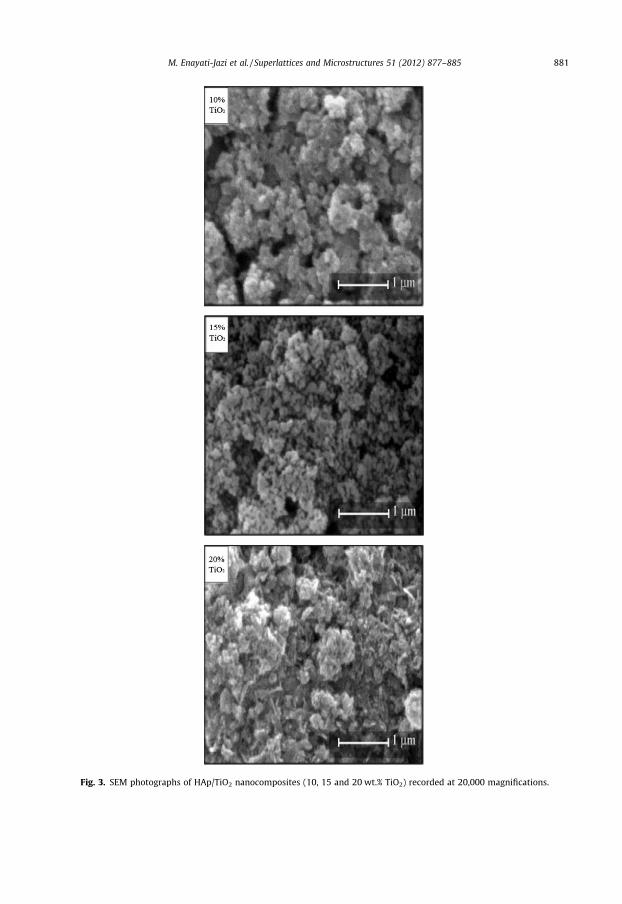
Fig. 3. SEM photographs of HAp/TiO2 nanocomposites (10, 15 and 20 wt.% TiO2) recorded at 20,000 magnifications.
M. Enayati-Jazi et al. / Superlattices and Microstructures 51 (2012) 877–885 881
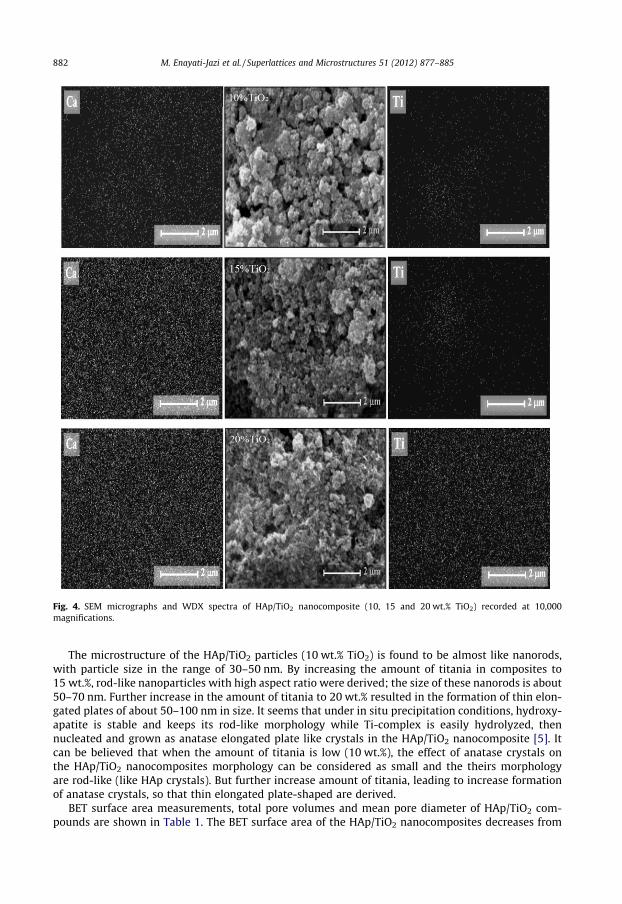
Fig. 4. SEM micrographs and WDX spectra of HAp/TiO2 nanocomposite (10, 15 and 20 wt.% TiO2) recorded at 10,000magnifications.
882 M. Enayati-Jazi et al. / Superlattices and Microstructures 51 (2012) 877–885
The microstructure of the HAp/TiO2 particles (10 wt.% TiO2) is found to be almost like nanorods,with particle size in the range of 30–50 nm. By increasing the amount of titania in composites to15 wt.%, rod-like nanoparticles with high aspect ratio were derived; the size of these nanorods is about50–70 nm. Further increase in the amount of titania to 20 wt.% resulted in the formation of thin elon-gated plates of about 50–100 nm in size. It seems that under in situ precipitation conditions, hydroxy-apatite is stable and keeps its rod-like morphology while Ti-complex is easily hydrolyzed, thennucleated and grown as anatase elongated plate like crystals in the HAp/TiO2 nanocomposite [5]. Itcan be believed that when the amount of titania is low (10 wt.%), the effect of anatase crystals onthe HAp/TiO2 nanocomposites morphology can be considered as small and the theirs morphologyare rod-like (like HAp crystals). But further increase amount of titania, leading to increase formationof anatase crystals, so that thin elongated plate-shaped are derived.
BET surface area measurements, total pore volumes and mean pore diameter of HAp/TiO2 com-pounds are shown in Table 1. The BET surface area of the HAp/TiO2 nanocomposites decreases from

Fig. 5. TEM micrographs of the HAp/TiO2 nanocomposite (10, 15 and 20 wt.% TiO2).
M. Enayati-Jazi et al. / Superlattices and Microstructures 51 (2012) 877–885 883
127 to 94 m2/g as the TiO2 content of the samples increases from 10 to 20 wt.%. The decrease in surfacearea of nanocomposites by adding titania is probably due to its affect on enhancing the grain growth
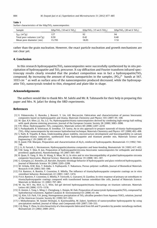
Table 1Surface characteristics of the HAp/TiO2 nanocomposites.
Sample HAp/TiO2 (10 wt.% TiO2) HAp/TiO2 (15 wt.% TiO2) HAp/TiO2 (20 wt.% TiO2)
SBET (m2/g) 127 92 94Total pore volumes (cm3/g) 0.50 0.42 0.40Mean pore diameter (nm) 15.795 18.05 17.8
884 M. Enayati-Jazi et al. / Superlattices and Microstructures 51 (2012) 877–885
rather than the grain nucleation. However, the exact particle nucleation and growth mechanisms arenot clear yet.
4. Conclusion
In this research hydroxyapatite/TiO2 nanocomposites were successfully synthesized by in situ pre-cipitation of hydroxyapatite and TiO2 precursor. X-ray diffraction and Fourier transform infrared spec-troscopy results clearly revealed that the product composition was in fact a hydroxyapatite/TiO2
compound. By increasing the amount of titania nanoparticles in the samples, (PO4)3� bands at 567,1033 cm�1 as well as surface area of the nanocomposites produced decreased, while the hydroxyap-atite TiO2 nanocrystals tended to thin, elongated and plate-like in shape.
Acknowledgements
The authors would like to thank Mrs. M. Salehi and Mr. R. Tahmasebi for their help in preparing thispaper and Mrs. N. Jafari for doing the XRD experiments.
References
[1] E. Fidancevska, G. Ruseska, J. Bossert, Y. Lin, A.R. Boccaccini, Fabrication and characterization of porous bioceramiccomposites based on hydroxyapatite and titania, Materials Chemistry and Physics 103 (2007) 95–100.
[2] W. Que, K.A. Khor, J.L. Xu, L.G. Yu, Hydroxyapatite/titania nanocomposites derived by combining high-energy ball millingwith spark plasma sintering processes, Journal of the European Ceramic Society 28 (2008) 3083–3090.
[3] F.N. Oktar, Hydroxyapatite–TiO2 composites, Materials Letters 60 (2006) 2207–2210.[4] S. Pushpakanth, B. Srinivasan, B. Sreedhar, T.P. Sastry, An in situ approach to prepare nanorods of titania–hydroxyapatite
(TiO2–HAp) nanocomposite by microwave hydrothermal technique, Materials Chemistry and Physics 107 (2008) 492–498.[5] S. Nath, R. Tripathi, B. Basu, Understanding phase stability, microstructure development and biocompatibility in calcium
phosphate–titania composites, synthesized from hydroxyapatite and titanium powder mix, Materials Science andEngineering C 29 (2009) 97–107.
[6] H. Jiand, P.M. Marquis, Preparation and characterization of Al2O3 reinforced hydroxyapatite, Biomaterials 13 (1992) 744–748.
[7] J. Li, B. Fartash, L. Hermansson, Hydroxyapatite/alumina composites and bone-bonding, Biomaterials 16 (1995) 417–422.[8] Y.M. Sung, Y. Shin, R. Jun, Preparation of hydroxyapatite/zirconia bioceramic nanocomposites for orthopedic and dental
prosthesis applications, Nanotechnology 18 (2007) 601–607.[9] R. Quan, D. Yang, X. Wu, H. Wang, X. Miao, W. Li, In vitro and in vivo biocompatibility of graded hydroxyapatite-zirconia
composite bioceramic, Material Science. Materials in Medicine 19 (2008) 183–187.[10] G. Georgiou, J.C. Knowles, J.E. Barralet, Dynamic shrinkage behavior of hydroxyapatite and glass-reinforced hydroxyapatite,
Journal of Materials Science 39 (2004) 2205–2208.[11] M. Uchida, H.M. Kim, T. Kokubo, S. Fujibayashi, T. Nakamura, Porous polymer scaffolds, Journal of Biomedical Materials
Research A 64 (2003) 583–590.[12] P.A. Ramires, A. Romito, F. Cosentino, E. Milella, The influence of titania/hydroxyapatite composite coatings on in vitro
osteoblast behavior, Biomaterials 22 (2001) 1467–1474.[13] P.A.A. Ramires, F. Cosentino, E. Milella, P. Torricelli, G. Giavaresi, R. Giardino, In vitro response of primary rat osteoblasts to
titania/hydroxyapatite coatings compared with transformed human osteoblast-like cells, Journal of Materials Science.Materials in Medicine 13 (2002) 797–801.
[14] W. Xu, W.Y. Hu, M.H. Li, C. Wen, Sol–gel derived hydroxyapatite/titania biocoatings on titanium substrate, MaterialsLetters 60 (2006) 1575–1578.
[15] H. Anmin, L. Tong, L. Ming, C. Chengkang, L. Huiqin, M. Dali, Preparation of nanocrystals hydroxyapatite/TiO2 compound byhydrothermal treatment, Applied Catalysis B: Environmental 63 (2006) 41–44.
[16] S.J. Kim, S.D. Park, Y.H. Jeong, Homogeneous precipitation of TiO2 ultrafine powders from Aqueous TiOCl2 solution, Journalof the American Ceramic Society 82 (1999) 927–932.
[17] I. Mobasherpour, M. Soulati Heshajin, A. Kazemzadeha, M. Zakeri, Synthesis of nanocrystalline hydroxyapatite by usingprecipitation method, Journal of Alloys and Compounds 430 (2007) 330–333.
[18] C.Q. Ning, Y. Zhou, In vitro bioactivity of a biocomposite fabricated from HA and Ti powders by powder metallurgy method,Biomaterials 23 (2002) 2909–2915.

M. Enayati-Jazi et al. / Superlattices and Microstructures 51 (2012) 877–885 885
[19] A. Ahmad, G.H. Awan, S. Aziz, Synthesis and application of TiO2 nanoparticles Pakistan Engineering Congress, in: 70thAnnual Session Proceedings, 2006, pp. 404–412.
[20] I. Manjubala, A. Wosez, C. Pilz, M. Rumpler, N. Fratzl-Zelman, P. Roschger, J. Stample, P. Fratzl, Biomimetic mineral-organiccomposite scaffolds with controlled internal architecture, Journal of Materials Science. Materials in Medicine 16 (2005)1111–1119.
Copyright © 2022 FDOKUMEN