Synthesis and characterization of hollow α-Fe2O3 sub-micron spheres prepared by sol–gel
7
Hyperfine Interact (2011) 202:131–137 DOI 10.1007/s10751-011-0353-1 Synthesis and characterization of hollow α-Fe 2 O 3 sub-micron spheres prepared by sol–gel Lizbet León · Angel Bustamante · Ana Osorio · G. S. Olarte · Luis De Los Santos Valladares · Crispin H. W. Barnes · Yutaka Majima Published online: 30 August 2011 © Springer Science+Business Media B.V. 2011 Abstract In this work we report the preparation of magnetic hematite hollow sub- micron spheres (α-Fe 2 O 3 ) by colloidal suspensions of ferric nitrate nine-hydrate (Fe(NO 3 ) 3 ·9H 2 O) particles in citric acid solution by following the sol–gel method. After the gel formation, the samples were annealed at different temperatures in an oxidizing atmosphere. Annealing at 180 ◦ C resulted in an amorphous phase, without iron oxide formation. Annealing at 250 ◦ C resulted in coexisting phases of hematite, maghemite and magnetite, whereas at 400 ◦ C, only hematite and maghemite were found. Pure hematite hollow sub-micron spheres with porous shells were formed after annealing at 600 ◦ C. The characterization was performed by X-ray diffraction (XRD), Mössbauer spectroscopy (MS) and scanning electron microscopy (SEM). Keywords Sol–gel method · Mössbauer spectroscopy · Hematite · Maghemite L. León (B ) · A. Bustamante · A. Osorio · G. S. Olarte Universidad Nacional Mayor de San Marcos, Lima, Perú e-mail: [email protected] L. De Los Santos Valladares · C. H. W. Barnes Cavendish Laboratory, University of Cambridge, J.J Thomson Av., Cambridge CB3 0HE, UK L. De Los Santos Valladares e-mail: [email protected] L. De Los Santos Valladares · Y. Majima Materials and Structures Laboratory, Tokyo Institute of Technology, 4259 Nagatsuta-cho, Midori-ku, Yokohama 226-8503, Japan Y. Majima CREST, Japan Science and Technology Agency JST, 4259 Nagatsuta-cho, Midori-ku, Yokohama 226-8503, Japan
-
Upload
independent -
Category
Documents
-
view
0 -
download
0
Transcript of Synthesis and characterization of hollow α-Fe2O3 sub-micron spheres prepared by sol–gel
Hyperfine Interact (2011) 202:131–137DOI 10.1007/s10751-011-0353-1
Synthesis and characterization of hollowα-Fe2O3 sub-micron spheres prepared by sol–gel
Lizbet León · Angel Bustamante · Ana Osorio ·G. S. Olarte · Luis De Los Santos Valladares ·Crispin H. W. Barnes · Yutaka Majima
Published online: 30 August 2011© Springer Science+Business Media B.V. 2011
Abstract In this work we report the preparation of magnetic hematite hollow sub-micron spheres (α-Fe2O3) by colloidal suspensions of ferric nitrate nine-hydrate(Fe(NO3)3·9H2O) particles in citric acid solution by following the sol–gel method.After the gel formation, the samples were annealed at different temperatures in anoxidizing atmosphere. Annealing at 180◦C resulted in an amorphous phase, withoutiron oxide formation. Annealing at 250◦C resulted in coexisting phases of hematite,maghemite and magnetite, whereas at 400◦C, only hematite and maghemite werefound. Pure hematite hollow sub-micron spheres with porous shells were formedafter annealing at 600◦C. The characterization was performed by X-ray diffraction(XRD), Mössbauer spectroscopy (MS) and scanning electron microscopy (SEM).
Keywords Sol–gel method · Mössbauer spectroscopy · Hematite · Maghemite
L. León (B) · A. Bustamante · A. Osorio · G. S. OlarteUniversidad Nacional Mayor de San Marcos, Lima, Perúe-mail: [email protected]
L. De Los Santos Valladares · C. H. W. BarnesCavendish Laboratory, University of Cambridge,J.J Thomson Av., Cambridge CB3 0HE, UK
L. De Los Santos Valladarese-mail: [email protected]
L. De Los Santos Valladares · Y. MajimaMaterials and Structures Laboratory, Tokyo Institute of Technology,4259 Nagatsuta-cho, Midori-ku, Yokohama 226-8503, Japan
Y. MajimaCREST, Japan Science and Technology Agency JST,4259 Nagatsuta-cho, Midori-ku, Yokohama 226-8503, Japan

132 L. León et al.
1 Introduction
The production of magnetic nanostructures is of current interest due to theirpromising applications in biomedicine [1, 2], bioengineering [3], nanodevices [4] andmagnetorheology [5]. In the case of iron oxides nanostructures, the formation ofantiferromagnetic hematite (α-Fe2O3) [6], ferrimagnetic maghemite (γ-Fe2O3) [7]and ferromagnetic magnetite (Fe3O4) [8] has been identified during their productionby thermal oxidation [9]. Recently, hollow iron-oxide-based microstructures have at-tracted high interest because of their potential applications in photonic crystals, drugdelivery, catalysis and encapsulation [10]. Various techniques such as polystyrene-templates [11], sono-chemical [12], solid-state reaction [13], hydrothermal [14] andthe surfactant-assisted sorvo-thermal [15, 16] method have been reported for thepreparation of iron oxides hollow structures. However, faster, cleaner and template-free methods are desired to prepare hollow iron-based structures. Sol–gel is aversatile technique used for the preparation of iron oxides nanostructures [17–20]. Inthis technique, different nanostructures can be obtained after thermal oxidizing thegel precursors previously obtained by hydrolysis and condensation of metal-oxides inalcohol or aqueous medium [21]. In this paper, we report the preparation of hollowhematite submicron spheres by the sol–gel method. Hematite was chosen because ofits abundance in nature, non-toxicity, cheap-cost and stability in ambient conditions.We studied the best annealing temperature of the sol–gel precursor to obtain uniformand high yield hollow α-Fe2O3 sub-microspheres.
2 Experimental
The preparation started with the suspension of colloidal ferric nitrate nine-hydrate(Fe(NO3)3.9H2O) particles (200 ml) and mono hydrated citric acid (C6H8O7.H2O)(0.1 and 0.2 M concentrations) in 800 ml of distilled water following the sameprocedure reported elsewhere [22]. Citric acid is commonly used because of itseffects in the formation, growth and shape of iron oxides nanostructures [23]. Thesolutions were agitated in a magnetic stirrer at 350 rpm and 70◦C for a periodof 48 h to evaporate the acid and water residuals. The gels were then stored fortwo days at 40◦C to completely eliminate any residuals and possible impuritiesformed by hydrolysis. For thermal oxidation, the precursors were introduced in atubular furnace (LENTON LTF-PTF Model 16/610) to be annealed in oxygen flowat different temperatures between 180◦C to 600◦C. The furnace was programmedto reach the required temperatures after 1 h 30 min, to remain constant for 12 h,and finally cool down at a rate of 1.4◦C /min. The crystallizations of the sampleswere studied by X-ray diffraction (XRD). The data was collected from 20◦ to 65◦(0.02◦ steps) using a powder universal diffractometer Bruker D8 Focus with Cu Kα
radiation (1.5406 Å). The Mössbauer spectra were recorded at room temperature inthe transmission geometry, using a 57Co in Rh source with average activity 25 mCiand the isomer shifts were measured relative to the metal α-Fe. The morphologicalanalysis was performed using a scanning electron microscope (SEM–XL30 SFEG).
Synthesis and characterization of hollow α-Fe2O3 sub-micron spheres 133
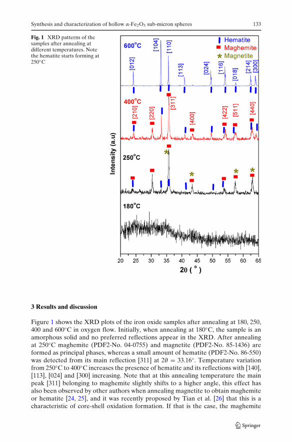
Fig. 1 XRD patterns of thesamples after annealing atdifferent temperatures. Notethe hematite starts forming at250◦C
3 Results and discussion
Figure 1 shows the XRD plots of the iron oxide samples after annealing at 180, 250,400 and 600◦C in oxygen flow. Initially, when annealing at 180◦C, the sample is anamorphous solid and no preferred reflections appear in the XRD. After annealingat 250◦C maghemite (PDF2-No. 04-0755) and magnetite (PDF2-No. 85-1436) areformed as principal phases, whereas a small amount of hematite (PDF2-No. 86-550)was detected from its main reflection [311] at 2θ = 33.16◦. Temperature variationfrom 250◦C to 400◦C increases the presence of hematite and its reflections with [140],[113], [024] and [300] increasing. Note that at this annealing temperature the mainpeak [311] belonging to maghemite slightly shifts to a higher angle, this effect hasalso been observed by other authors when annealing magnetite to obtain maghemiteor hematite [24, 25], and it was recently proposed by Tian et al. [26] that this is acharacteristic of core-shell oxidation formation. If that is the case, the maghemite
134 L. León et al.
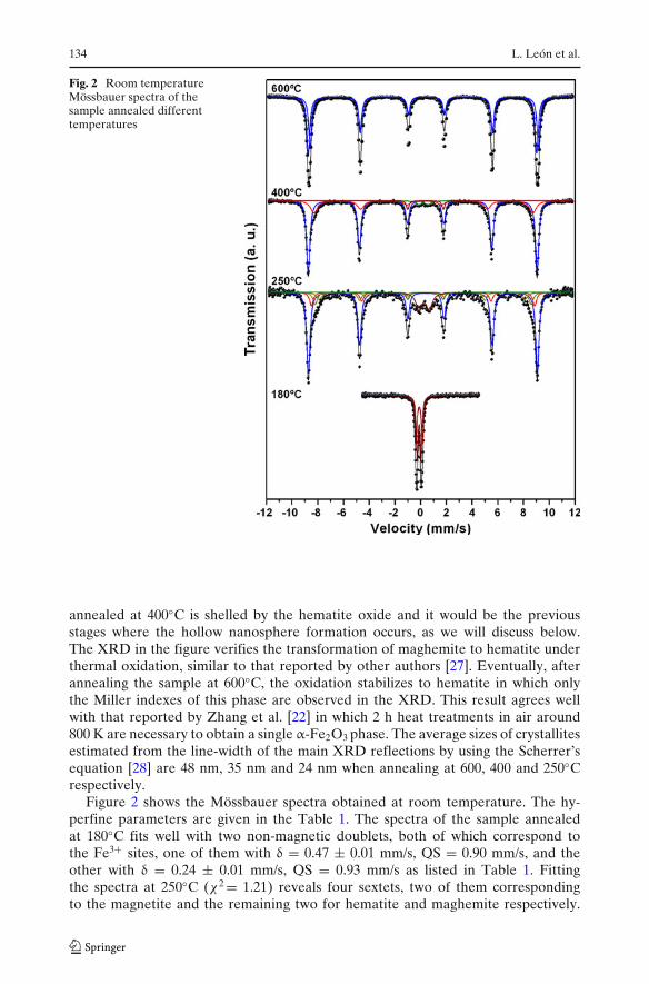
Fig. 2 Room temperatureMössbauer spectra of thesample annealed differenttemperatures
annealed at 400◦C is shelled by the hematite oxide and it would be the previousstages where the hollow nanosphere formation occurs, as we will discuss below.The XRD in the figure verifies the transformation of maghemite to hematite underthermal oxidation, similar to that reported by other authors [27]. Eventually, afterannealing the sample at 600◦C, the oxidation stabilizes to hematite in which onlythe Miller indexes of this phase are observed in the XRD. This result agrees wellwith that reported by Zhang et al. [22] in which 2 h heat treatments in air around800 K are necessary to obtain a single α-Fe2O3 phase. The average sizes of crystallitesestimated from the line-width of the main XRD reflections by using the Scherrer’sequation [28] are 48 nm, 35 nm and 24 nm when annealing at 600, 400 and 250◦Crespectively.
Figure 2 shows the Mössbauer spectra obtained at room temperature. The hy-perfine parameters are given in the Table 1. The spectra of the sample annealedat 180◦C fits well with two non-magnetic doublets, both of which correspond tothe Fe3+ sites, one of them with δ = 0.47 ± 0.01 mm/s, QS = 0.90 mm/s, and theother with δ = 0.24 ± 0.01 mm/s, QS = 0.93 mm/s as listed in Table 1. Fittingthe spectra at 250◦C (χ2 = 1.21) reveals four sextets, two of them correspondingto the magnetite and the remaining two for hematite and maghemite respectively.
Synthesis and characterization of hollow α-Fe2O3 sub-micron spheres 135
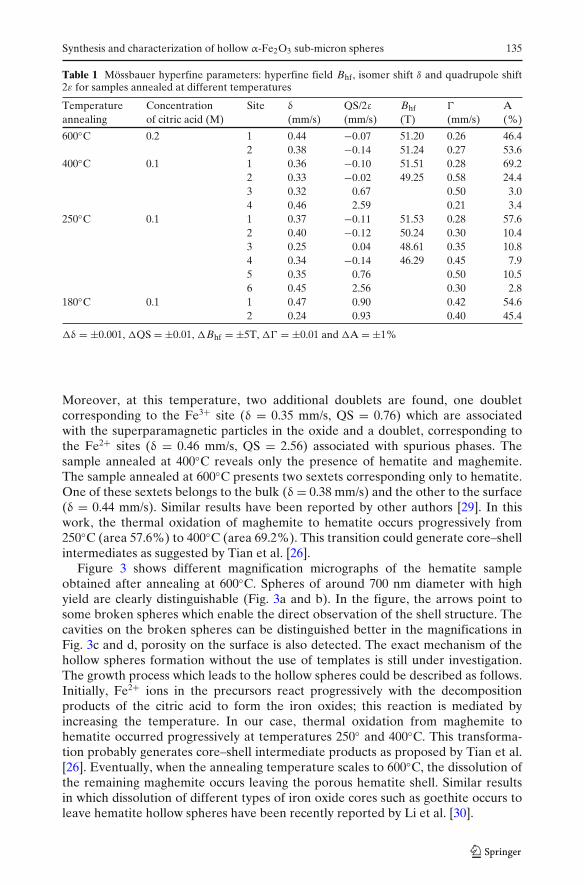
Table 1 Mössbauer hyperfine parameters: hyperfine field Bhf, isomer shift δ and quadrupole shift2ε for samples annealed at different temperatures
Temperature Concentration Site δ QS/2ε Bhf � Aannealing of citric acid (M) (mm/s) (mm/s) (T) (mm/s) (%)
600◦C 0.2 1 0.44 −0.07 51.20 0.26 46.42 0.38 −0.14 51.24 0.27 53.6
400◦C 0.1 1 0.36 −0.10 51.51 0.28 69.22 0.33 −0.02 49.25 0.58 24.43 0.32 0.67 0.50 3.04 0.46 2.59 0.21 3.4
250◦C 0.1 1 0.37 −0.11 51.53 0.28 57.62 0.40 −0.12 50.24 0.30 10.43 0.25 0.04 48.61 0.35 10.84 0.34 −0.14 46.29 0.45 7.95 0.35 0.76 0.50 10.56 0.45 2.56 0.30 2.8
180◦C 0.1 1 0.47 0.90 0.42 54.62 0.24 0.93 0.40 45.4
�δ = ±0.001, �QS = ±0.01, �Bhf = ±5T, �� = ±0.01 and �A = ±1%
Moreover, at this temperature, two additional doublets are found, one doubletcorresponding to the Fe3+ site (δ = 0.35 mm/s, QS = 0.76) which are associatedwith the superparamagnetic particles in the oxide and a doublet, corresponding tothe Fe2+ sites (δ = 0.46 mm/s, QS = 2.56) associated with spurious phases. Thesample annealed at 400◦C reveals only the presence of hematite and maghemite.The sample annealed at 600◦C presents two sextets corresponding only to hematite.One of these sextets belongs to the bulk (δ = 0.38 mm/s) and the other to the surface(δ = 0.44 mm/s). Similar results have been reported by other authors [29]. In thiswork, the thermal oxidation of maghemite to hematite occurs progressively from250◦C (area 57.6%) to 400◦C (area 69.2%). This transition could generate core–shellintermediates as suggested by Tian et al. [26].
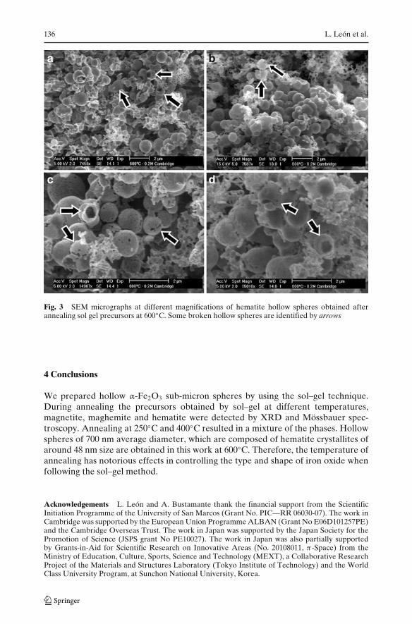
Figure 3 shows different magnification micrographs of the hematite sampleobtained after annealing at 600◦C. Spheres of around 700 nm diameter with highyield are clearly distinguishable (Fig. 3a and b). In the figure, the arrows point tosome broken spheres which enable the direct observation of the shell structure. Thecavities on the broken spheres can be distinguished better in the magnifications inFig. 3c and d, porosity on the surface is also detected. The exact mechanism of thehollow spheres formation without the use of templates is still under investigation.The growth process which leads to the hollow spheres could be described as follows.Initially, Fe2+ ions in the precursors react progressively with the decompositionproducts of the citric acid to form the iron oxides; this reaction is mediated byincreasing the temperature. In our case, thermal oxidation from maghemite tohematite occurred progressively at temperatures 250◦ and 400◦C. This transforma-tion probably generates core–shell intermediate products as proposed by Tian et al.[26]. Eventually, when the annealing temperature scales to 600◦C, the dissolution ofthe remaining maghemite occurs leaving the porous hematite shell. Similar resultsin which dissolution of different types of iron oxide cores such as goethite occurs toleave hematite hollow spheres have been recently reported by Li et al. [30].
136 L. León et al.
b
d
a
c
Fig. 3 SEM micrographs at different magnifications of hematite hollow spheres obtained afterannealing sol gel precursors at 600◦C. Some broken hollow spheres are identified by arrows
4 Conclusions
We prepared hollow α-Fe2O3 sub-micron spheres by using the sol–gel technique.During annealing the precursors obtained by sol–gel at different temperatures,magnetite, maghemite and hematite were detected by XRD and Mössbauer spec-troscopy. Annealing at 250◦C and 400◦C resulted in a mixture of the phases. Hollowspheres of 700 nm average diameter, which are composed of hematite crystallites ofaround 48 nm size are obtained in this work at 600◦C. Therefore, the temperature ofannealing has notorious effects in controlling the type and shape of iron oxide whenfollowing the sol–gel method.
Acknowledgements L. León and A. Bustamante thank the financial support from the ScientificInitiation Programme of the University of San Marcos (Grant No. PIC—RR 06030-07). The work inCambridge was supported by the European Union Programme ALBAN (Grant No E06D101257PE)and the Cambridge Overseas Trust. The work in Japan was supported by the Japan Society for thePromotion of Science (JSPS grant No PE10027). The work in Japan was also partially supportedby Grants-in-Aid for Scientific Research on Innovative Areas (No. 20108011, π-Space) from theMinistry of Education, Culture, Sports, Science and Technology (MEXT), a Collaborative ResearchProject of the Materials and Structures Laboratory (Tokyo Institute of Technology) and the WorldClass University Program, at Sunchon National University, Korea.
Synthesis and characterization of hollow α-Fe2O3 sub-micron spheres 137
References
1. Petri-Fink, A., Chatellain, M., Juilleret-Jeanneret, L., Ferrari, A., Hofmann, H.: Biomaterials 26,2685–2694 (2005)
2. Lee, S.-J., Jeong, J.-R., Shin, S.-C., Kim, J.-C., Chang, Y.-H., Chang, Y.-M., Kim, J.-D.: J. Magn.Magn. Mater. 272–276, 2432–2433 (2004)
3. Lubbe, A.S., Alexiou, C., Bergemann, C.: J. Surg. Res. 95, 200 (2001)4. Tadic, M., Markovic, D., Spasojevic, V., Kusigerski, V., Remškar, M., Pirnat, J., Jaglicic, Z.:
J. Alloys and Comp. 441, 291–296 (2007)5. De Los Santos Valladares, L., Llandro, J., Lee, D. W., Mitrelias, T., Palfreyman, J., Hayward,
T.J., Cooper, J., Bland, J.A.C., Barnes, C.H.W., Arroyo, J.L., Lees, M.: J. Mag. Magn. Mater.321, 2129–2134 (2009)
6. Amin, N., Arajs, S.: Phys. Rev. B. 35, 10 (1987)7. Dutta, P., Manivannan, A., Seehra, M.S.: Phys. Rev. B. 70, 174428 (2004)8. Liu, Q., Asare, K.O.: J. Colloid Interface Sci. 231, 401–403 (2000)9. Zboril, R., Mashlan, M., Petridis, D.: Chem. Mater. 14, 969–982 (2002)
10. Lou, X.W., Archer, L.A., Yang, Z.C.: Adv. Mater. 20, 3987 (2008)11. Shiho, H., Kawahashi, N.: J. Colloid Interf. Sci. 226, 91 (2000)12. Zhu, J.J., Xu, S., Wang, H., Zhu, J.M., Chen, H.Y.: Adv. Mater. 15, 156 (2003)13. Wang, J.S., Yang, J.K., Bao, Y.: Powder Technol. 145, 172 (2004)14. Liu, X.-M., Yin, W.-D., Miao, S.-B., Ji, B.-M.: Mat. Chem. Phys. 113, 518–522 (2009)15. Shen, D.H., Chen, D.R., Jiao, X.L., Zhao, Y.T.: J. Mater. Chem. 13, 2266 (2003)16. Lian, S.Y., Wang, E.B., Gao, L., Wu, D., Song, Y.L., Xu, L.: Mater. Res. Bull. 41, 1192 (2006)17. Sugimoto, T., Wang, Y., Itoh, H., Muramatsu, A.: Colloids Surf. A 134, 265–279 (1998)18. Sugimoto, T., Sakata, K.: J. Colloid Interface Sci. 152, 587 (1992)19. Sugimoto, T., Khan, M.M., Muramatsu, A.: Colloids Surf. A 70, 167 (1993)20. Sugimoto, T., Khan, M.M., Muramatsu, A., Itoh, H.: Colloids Surf. A. 79, 233 (1993)21. Hench, L.L., West, J.K.: Chem. Rev. 90, 33 (1990)22. Zhang, J., Rong, L.X., Liu, Y., Dong, B.Z.: Mat. Sci. Eng. A351, 224–227 (2003)23. Kandori, K., Kawashima, Y., Ishikawa, T.: J. Colloid Interf. Sci. 152, 284 (1992)24. Sun, Y.-K., Ma, M., Zhang, Y., Gu, N.: Colloids Surf A: Physicochem. Eng. Aspects 245, 15–19
(2004)25. Xu, J., Yang, H., Fu, W., Du, K., Sui, Y., Chen, J., Zeng, Y., Li, M., Zou, G.: J. Mag. Magn. Mat.
309, 307–311 (2007)26. Tian, Y., Wu, D., Jia, X., Yu, B., Zhan, S.: J. Nanomat. 837123 (2011)27. Haneda, K., Morrish, A.H.: J. Physique, Sup. 4, 38 (1977) C1-321–32328. Cullity, B.D.: Elements of X-ray Diffraction, 3rd edn., Prentice-Hall, Prentice-Hall International,
Upper Saddle River (2000)29. Bercoff, P.G., Bertorello, H.R.: Appl. Phys. A. 100, 1019–1027 (2010)30. Li, W., Guan, J.-G., Wang, W., Tong, G.-X., Fan, X.-A.: Mat. Chem. Phys. 118, 496–500 (2009)