Gummic acid stabilized γ-Fe2O3 aqueous suspensions for biomedical applications
8
FOR APPROVAL Hyperfine Interact DOI 10.1007/s10751-009-9922-y Gummic acid stabilized γ-Fe 2 O 3 aqueous suspensions for biomedical applications G. C. Papaefthymiou · I. Rabias · M. Fardis · E. Devlin · N. Boukos · D. Tsitrouli · G. Papavassiliou © Springer Science + Business Media B.V. 2009 Abstract Biomedical applications of magnetic nanoparticles depend critically on their preparation as aqueous colloidal suspensions, or ferrofluids, with long term stability under physiological conditions. Dispersion of the magnetic nanoparticles is generally achieved by the use of protein cages, polysaccharide, polypeptide and charged macromolecular coatings, which minimize interparticle magnetic interac- tions, particle agglomeration and precipitation. The synthesis and characterization of gummic-acid stabilized maghemite ferrofluids is reported. X-ray diffraction, transmission electron microscope and dynamic light scattering measurements give a γ-Fe 2 O 3 magnetic core diameter of 8 nm and a nanocomposite particle hydrody- namic diameter of 50 nm. Mössbauer and magnetization measurements indicate the presence of isolated, sterically stabilized superparamagnetic nanoparticles re- sistant to aging, and thus, promising agents for the production of novel magneto- pharmaceuticals. Keywords Superparamagnetism · Iron oxide · Maghemite · Gummic-acid · Ferrofluids · Magneto-pharmaceuticals 1 Introduction Ferrofluids are stable colloidal dispersions of magnetic particles in a carrier fluid having both liquid and magnetic properties. Engineering applications of ferrofluids abound in diverse areas of technology ranging from computers, magnetic inks for jet G. C. Papaefthymiou · I. Rabias · M. Fardis · E. Devlin · N. Boukos · D. Tsitrouli · G. Papavassiliou Institute of Materials Science, NCSR Demokritos, 15310 Athens, Greece G. C. Papaefthymiou (B ) Department of Physics, Villanova University, Villanova, PA 19085, USA e-mail: [email protected]
-
Upload
demokritos -
Category
Documents
-
view
4 -
download
0
Transcript of Gummic acid stabilized γ-Fe2O3 aqueous suspensions for biomedical applications

FOR APPROVAL
Hyperfine InteractDOI 10.1007/s10751-009-9922-y
Gummic acid stabilized γ-Fe2O3 aqueous suspensionsfor biomedical applications
G. C. Papaefthymiou · I. Rabias · M. Fardis · E. Devlin ·N. Boukos · D. Tsitrouli · G. Papavassiliou
© Springer Science + Business Media B.V. 2009
Abstract Biomedical applications of magnetic nanoparticles depend critically ontheir preparation as aqueous colloidal suspensions, or ferrofluids, with long termstability under physiological conditions. Dispersion of the magnetic nanoparticlesis generally achieved by the use of protein cages, polysaccharide, polypeptide andcharged macromolecular coatings, which minimize interparticle magnetic interac-tions, particle agglomeration and precipitation. The synthesis and characterizationof gummic-acid stabilized maghemite ferrofluids is reported. X-ray diffraction,transmission electron microscope and dynamic light scattering measurements give aγ-Fe2O3 magnetic core diameter of 8 nm and a nanocomposite particle hydrody-namic diameter of 50 nm. Mössbauer and magnetization measurements indicatethe presence of isolated, sterically stabilized superparamagnetic nanoparticles re-sistant to aging, and thus, promising agents for the production of novel magneto-pharmaceuticals.
Keywords Superparamagnetism · Iron oxide · Maghemite · Gummic-acid ·Ferrofluids · Magneto-pharmaceuticals
1 Introduction
Ferrofluids are stable colloidal dispersions of magnetic particles in a carrier fluidhaving both liquid and magnetic properties. Engineering applications of ferrofluidsabound in diverse areas of technology ranging from computers, magnetic inks for jet
G. C. Papaefthymiou · I. Rabias · M. Fardis · E. Devlin · N. Boukos ·D. Tsitrouli · G. PapavassiliouInstitute of Materials Science, NCSR Demokritos, 15310 Athens, Greece
G. C. Papaefthymiou (B)Department of Physics, Villanova University, Villanova, PA 19085, USAe-mail: [email protected]
FOR APPROVAL
G.C. Papaefthymiou et al.
printing, loudspeakers, semiconductors, motion sensors to petrochemicals [1, 2].More recently, aqueous ferrofluids have come under intense investigation for a widerange of applications in biotechnology and medicine as in enzyme immobilization,protein and gene separation, magnetic resonance imaging enhancement, hyperther-mia cancer therapy, controlled drug release and radiopharmaceutical applications[3]. Aqueous dispersions of iron oxide particles are stabilized by a hydrophilic doublelayer of surfactants or by charge, due to acid-base reactions at the surface or specificadsorption of ions. Thus, their long term colloidal stability against agglomerationand precipitation is ensured by the coating of the nanoparticles with appropri-ately charged macromolecules such as polymers or polysaccharides, which preventparticle-clustering through electrostatic and/or steric effects [4].
Herewith, we examine the magnetic properties of gummic acid stabilizedmaghemite nanoparticles. Gummic acid is a biocompatible macromolecule whicheasily adsorbs on the surface of maghemite. The results indicate that gummic acidpromotes the nucleation and growth of maghemite nanoparticles, while simultane-ously affording a thick coating layer, which imparts high magnetic anisotropy tothe nanoparticles and ensures the magnetic isolation of individual magnetic cores.The effectiveness of the resulting nanocomposite particles for hyperthermia cancertherapy applications is also evaluated.
2 Experimental
A dispersion of maghemite nanoparticles coated with gummic acid was synthesizedby the reaction of ferric chloride and ferrous chloride in the presence of NH3, usingco-precipitation method. The nominal molecular weight of the gummic acid usedwas 250,000 g/mol. The co-precipitation process involved mixing 30 ml of 0.66 MFeCl3 with 20 ml of 0.33 M FeCl2 and then adding 100 ml alkaline solution of NH3
(1 M) with 1 wt.% of gummic acid drop-wise. The stirring continued for 20 minunder nitrogen-gas atmosphere. The particles obtained were washed three timeswith ethanol and water using ultracentrifugation (5,000 rpm for 10 min at 10◦C) withnitrogen purged water. The ferrofluids were prepared at high pH and were constantlystirred during this fast one step reaction to avoid the appearance of larger particles.The pH of the final ferrofluid was decreased by carefully numbered titrations of0.01 M HCl to achieve neutrality followed by dispersion via continuous sonicationfor 10 min and repetitive filtering of the final colloid though a 0.2 μm porousmembrane [5].
Dry powders and aqueous suspensions (ferrofluid density = 1.018 g/ml) of thecoated magnetic nanoparticles were prepared and subsequently characterized by anumber of techniques including transmission electron microscopy (TEM) in a Jeol2011 electron microscope operated at 200 kV, Mössbauer spectroscopy (constantacceleration mode, 57Co(Rh) source calibrated with metallic iron at room temper-ature, in conjunction with Oxford Instruments cryostat), dynamic light scattering(DLS) performed by a Microtrac Series 9200 Ultrafine Particle Analyzer, X-raydiffraction (XRD) Crystallographic analysis recorded between 20◦ to 100◦ (2θ) at0.5◦ min−1 in a Siemens D500 diffractometer with Cu Kα radiation and magnetisationmeasurements (Quantum Design PPMS SQUID magnetometer). Specific Absorp-tion Rate (SAR) measurements were also carried out to determine the temperaturerise possible using a radiofrequency (RF) induction heater with 40 mg Fe2O3 per

FOR APPROVAL
Gummic acid stabilized γ-Fe2O3 aqueous suspensions
0 5 10 15 20 250
10
20
30
40
Num
ber
of P
artic
les
Diameter (nm)
(b)(a)
Fig. 1 a TEM image of aggregates of gummic-acid coated γ-Fe2O3nanoparticles (scale bar 20 nm);and b associated magnetic particle size distribution
Fig. 2 XRD pattern of gummic acid coated γ-Fe2O3 nanoparticles
milliliter sample in a moderate AC magnetic field with amplitude of 11 kA/m andfrequency 150 kHz.
3 Results and discussion
TEM analysis was used to observe the morphology, mean diameter and size distribu-tion of the magnetic particles, while DLS was used to determine the mean hydrody-namic size of the coated nanoparticles in the colloidal suspensions. Determination ofthe mean particle diameter, (8 ± 2) nm, was made by measuring 200 particles fromdifferent TEM micrographs, Fig. 1.
FOR APPROVAL
G.C. Papaefthymiou et al.
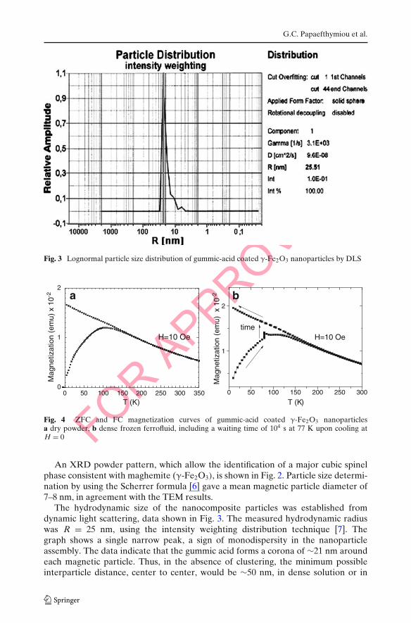
Fig. 3 Lognormal particle size distribution of gummic-acid coated γ-Fe2O3 nanoparticles by DLS
0 50 100 150 200 250 300 3500
1
2
Mag
netiz
atio
n (e
mu)
x 1
0-2
T (K)
H=10 Oe
ba
0 50 100 150 200 250 300
1
2
Mag
netiz
atio
n (e
mu)
x 1
0-2
T (K)
H=10 Oetime
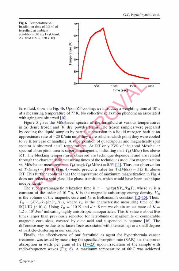
Fig. 4 ZFC and FC magnetization curves of gummic-acid coated γ-Fe2O3 nanoparticlesa dry powder, b dense frozen ferrofluid, including a waiting time of 104 s at 77 K upon cooling atH = 0
An XRD powder pattern, which allow the identification of a major cubic spinelphase consistent with maghemite (γ-Fe2O3), is shown in Fig. 2. Particle size determi-nation by using the Scherrer formula [6] gave a mean magnetic particle diameter of7–8 nm, in agreement with the TEM results.
The hydrodynamic size of the nanocomposite particles was established fromdynamic light scattering, data shown in Fig. 3. The measured hydrodynamic radiuswas R = 25 nm, using the intensity weighting distribution technique [7]. Thegraph shows a single narrow peak, a sign of monodispersity in the nanoparticleassembly. The data indicate that the gummic acid forms a corona of ∼21 nm aroundeach magnetic particle. Thus, in the absence of clustering, the minimum possibleinterparticle distance, center to center, would be ∼50 nm, in dense solution or in

FOR APPROVAL
Gummic acid stabilized γ-Fe2O3 aqueous suspensions
98,4
98,8
99,2
99,6
100,0
-15 -10 -5 0 5 10 15
98,4
98,8
99,2
99,6
100,0
98,0
98,5
99,0
99,5
100,0
96
97
98
99
100
97,5
98,0
98,5
99,0
99,5
100,0
-15 -10 -5 0 5 10 15
97,0
97,5
98,0
98,5
99,0
99,5
100,0
150K 150K
Tra
nsm
issi
on (
%)
a
RT
Dried ferrofluid
23% Superparamagnetic
11% Superparamagnetic
6% Superparamagnetic 6% Superparamagnetic
11% Superparamagnetic
20% Superparamagnetic
Velocity (mm/s)
250K
Frozen liquidferrofluid
b
78 K 78 k
Velocity (mm/s)
Fig. 5 Mössbauer spectra of a dense frozen ferrofluid and b dry powder samples of gummic-coatedmaghemite nanoparticles at various temperatures
freeze-dried forms of the ferrofluid. To determine whether interparticle magneticinteractions, known to lead to particle agglomeration and precipitation, were more orless neutralized at this distance the magnetic properties of the nanoparticle assemblywas examined using magnetization and Mössbauer measurements.
Figure 4a shows the zero field cooled (ZFC) and field cooled (FC) magnetizationin an applied field of 10 Oe. The characteristic appearance of the data, where themaximum of the ZFC curve, which occurs at 110 K, is well separated from thebifurcation or branching off temperature at about 150 K, is consistent with non-interacting, or weakly interacting, magnetic particles [8]. Also, the magnetization ofthe FC curve keeps increasing beyond the maximum of the ZFC curve. Thus, wemay identify the temperature at which the maximum of the ZFC curve occurs as asuperparamagnetic blocking temperature, TB, rather than a spin-glass-like freezingtemperature [9]. To further confirm the absence of spin-glass-like aging phenomena,ZFC/FC data were also collected on a sample of dense (40 mg Fe2O3/ml) frozen
FOR APPROVAL
G.C. Papaefthymiou et al.
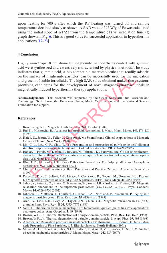
Fig. 6 Temperature vs.irradiation time of 0.3 ml offerrofluid at ambientconditions (40 mg Fe2O3/ml,AC field 105 G, 150 kHz)
20
30
40
50
60
70
Time [sec]
Tem
pera
ture
[oC
]
500 1000 1500 2000
ferrofluid, shown in Fig. 4b. Upon ZF cooling, we introduce a weighting time of 104 sat a measuring temperature of 77 K. No collective relaxation phenomena associatedwith aging are observed [10].
Figure 5 gives the Mössbauer spectra of the ferrofluid at various temperaturesin (a) dense frozen and (b) dry, powder forms. The frozen samples were preparedby cooling the liquid samples by partial submersion in a liquid nitrogen bath at anapproximate rate of −20 K/min until they were solid, at which point they were cooledto 78 K for ease of handling. A superposition of quadrupolar and magnetically splitspectra is observed at all temperatures. At RT only 23% of the total Mössbauerspectral absorption area is superparamagnetic, indicating that TB(Möss) lies aboveRT. The blocking temperatures observed are technique dependent and are relatedthrough the characteristic measuring times of the techniques used. For magnetizationvs. Mössbauer measurements TB(mag)/TB(Möss) = 0.35 [11]. Thus, our observationof TB(mag) = 110 K (Fig. 4) would predict a value for TB(Möss) = 315 K, aboveRT. This further confirms that the temperature of maximum magnetization in Fig. 4does not reflect a spin-glass-like phase transition, which would have been techniqueindependent.
The superparamagnetic relaxation time is τ = τ 0exp(KVm/kBT), where τ 0 is aconstant of the order of 10−9 s, K is the magnetic anisotropy energy density, Vm
is the volume of the magnetic core and kB is Boltzmann’s constant [12–15]. Thus,TB = (KVm/kB)/ln(τm/τ 0), where τm is the characteristic measuring time of theSQUID (∼10 s). Using TB = 110 K and d ∼ 8 nm we obtain an estimate of K =1.2 × 10⁵ J/m3 indicating highly anisotropic nanoparticles. This K value is about fivetimes larger than previously reported for ferrofluids of maghemite of comparablemagnetic core sizes, covered by oleic acid and suspended in heptane [16]. Thedifference may be due to surface effects associated with the coatings or a small degreeof particle-clustering in our samples.
Finally, the effectiveness of our ferrofluid as agent for hyperthermia cancertreatment was tested by measuring the specific absorption rate (SAR), i.e. the powerabsorption in watts per gram of Fe [17–23] upon irradiation of the sample withradio-frequency waves (Fig. 6). A maximum temperature of 66◦C was achieved
FOR APPROVAL
Gummic acid stabilized γ-Fe2O3 aqueous suspensions
upon heating for 700 s after which the RF heating was turned off and sampletemperature declined slowly as shown. A SAR value of 92 W/g of Fe was calculatedusing the initial slope of �T/�t from the temperature (T) vs. irradiation time (t)graph shown in Fig. 6. This is a good value for successful application in hyperthermiaapplications [17–23].
4 Conclusion
Highly anisotropic 8 nm diameter maghemite nanoparticles coated with gummicacid were synthesized and extensively characterised by physical methods. The studyindicates that gummic acid, a bio-compatible macromolecule that readily adsorbson the surface of maghemite particles, can be successfully used for the nucleationand growth of stable ferrofluids. The high SAR value obtained makes these systemspromising candidates for the development of novel magneto-pharmaceuticals inmagnetically induced hyperthermia therapy applications.
Acknowledgements This research was supported by the Greek Secretariat for Research andTechnology. GCP thanks the European Union, Marie Curie action, and the National ScienceFoundation for support.
References
1. Rosensweig, R.E.: Magnetic fluids. Sci. Am. 247, 136–145 (1982)2. Raj, K., Moskowitz, B.: Advances in ferrofluids technology. J. Magn. Magn. Mater. 149, 174–180
(1995)3. Häfeli, U., Schutt, W., Teller, J., Zborowski, M.: Scientific and Clinical Applications of Magnetic
Carriers. Plenum, New York (1998)4. Lin, C.-L., Lee, C.-F., Chiu, W.-Y.: Preparation and properties of poly(acrylic acid)oligomer
stabilized superparamagnetic ferrofluid. J. Colloid Interface Sci. 291, 411–420 (2005)5. Rabias, I., Fardis, M., Devlin, E., Boukos, N., Tsitrouli, D., Papavassiliou, G.: No aging phenom-
ena in ferrofluids: the influence of coating on interparticle interactions of maghemite nanoparti-cles. ACS Nano 2, 977–983 (2008)
6. Klug, H.P., Alexander, L.E.: X-ray Diffraction Procedures: For Polycrystalline and AmorphousMaterials, p. 992. Wiley, Hoboken (1974)
7. Chu, B.: Laser Light Scattering: Basic Principles and Practice, 2nd edn. Academic, New York(1992)
8. Prene, P., Troc, E., Jolivet, J.-P., Livage, J., Cherkaoui, R., Nogues, M., Dorman, J.-L., Fiorani,D.: Magnetic properties of isolated γ-Fe2O3 particles. IEEE Trans. Magn. 29, 2658 (1993)
9. Sahoo, S., Petracic, O., Binek, C., Kleemann, W., Sousa, J.B., Cardoso, S., Freitas, P.P.: Magneticrelaxation phenomena in the superspin-glass system [Co80Fe20/Al2O3]10. J. Phys., Condens.Matter 14, 6729–6736 (2002)
10. Johnson, T., Mattsson, J., Djurberg, C., Khan, F.A., Nordland, P., Svedlindh, P.: Aging in amagnetic particle system. Phys. Rev. Lett. 75, 4138–4141 (1995)
11. Xiao, G., Liou, S.H., Levy, A., Taylor, J.N., Chien, C.L.: Magnetic relaxation in Fe-(SiO2)
granular films. Phys. Rev., B 34, 7573–7577 (1986)12. Néel, L.: Théorie du traînage magnétique des ferromagnétiques en grains fins avec applications
aux terres cuites. Ann. Geophys. 5, 99 (1949)13. Brown, W.F., Jr.: Thermal fluctuations of a single-domain particle. Phys. Rev. 130, 1677 (1963)14. Brown, W.F., Jr.: Thermal fluctuations of a single-domain particle. J. Appl. Phys. 39, 993 (1968)15. Aharoni, A.: Relaxation processes in small particles. In: Dormann, J.L., Fiorani, D. (eds.) Mag-
netic Properties of Fine Particles, p. 3. Elsevier Science, North-Holland (1991)16. Millan, A., Urtizberea, A., Silva, N.J.O., Palacio, F., Amaral, V.S., Snoeck, E., Serin, V.: Surface
effects in maghemite nanoparticles. J. Magn. Magn. Mater. 312, L5 (2007)
FOR APPROVAL
G.C. Papaefthymiou et al.
17. Ma, M., Wu, Y., Zhou, J., Sun, Y., Zhang, Y., Gu, N.: Size dependence of specific powerabsorption of Fe3O4 particles in AC magnetic field. J. Magn. Magn. Mater. 268, 33–39 (2004)
18. Pankhurst, Q.A., Connolly, J., Jones, S.K., Dobson, J.: Applications of magnetic nanoparticles inbiomedicine. J. Phys. D Appl. Phys. 36, 167–181 (2003)
19. Ito, A., Matsuoka, F., Honda, H., Kobayashi, T.: Heat shock protein 70 gene therapy combinedwith hyperthermia using magnetic nanoparticles. Cancer Gene Ther. 10, 918–925 (2003)
20. Okawa, K., Sekine, M., Maeda, M., Tada, M., Abe, M.: Heating ability of magnetite nanobeadswith various sizes for magnetic hyperthermia at 120 kHz, a noninvasive frequency. J. Appl. Phys.99(1–3), 08H102 (2006)
21. Veiseh, O. et al.: Optical and MRI multifunctional nanoprobe for targeting gliomas, Nano Lett.5, 1003–1008 (2005)
22. Brezovich, I.A.: Low frequency hyperthermia. Med. Phys. Monogr. 16, 82–111 (1998)23. Hergt, R., Dutz, S.: Magnetic particle hyperthermia-biophysical limitations of a visionary tumour
therapy. J. Magn. Magn. Mater. 311, 187–192 (2007)