Pressure response of vacancy ordered maghemite (γ-Fe2O3) and high pressure transformed hematite...
9
Pressure response of vacancy ordered maghemite (g-Fe 2 O 3 ) and high pressure transformed hematite (a-Fe 2 O 3 ) Giovanni Hearne a,n , Vittoria Pischedda b a Department of Physics, University of Johannesburg, PO Box 524, Auckland Park, 2006 Johannesburg, South Africa b Laboratoire de Physique de la Mati ere Condense´e et Nanostructures, University Lyon 1 and CNRS, 69622 Villeurbanne Cedex, France article info Article history: Received 19 September 2011 Received in revised form 31 December 2011 Accepted 1 January 2012 Available online 10 January 2012 Keywords: Ferric iron oxides Vacancies High pressures M¨ ossbauer spectroscopy Topotactic transformation Spin reorientation abstract Combined XRD and M ¨ ossbauer effect spectroscopy studies to high pressures of 30 GPa of vacancy ordered maghemite are presented. The vacancy ordered superstructure is robust and remains intact up to the pressure-induced onset transition to hematite at 13–16 GPa. The pressure transformed hematite is shown to be crystallographically textured, unlike the randomised low pressure maghemite phase. This arises out of a pressure or stress instigated topotactic transformation of the cubic-spinel to hexagonal-corundum structure. The textured sample permits us to obtain information on the spin reorientation behavior of the pressure transformed hematite in compression and decompression sequences. Spin reorientation is restricted to 151 over wide pressure ranges, attributable to the effect of entrapped vacancies in the high pressure structure. Thus there are structural and magnetic peculiarities specific to pressure transformed hematite not evident in pressurized hematite starting material. These are triggered by the maghemite-hematite transformation. & 2012 Elsevier Inc. All rights reserved. 1. Introduction Maghemite (g-Fe 2 O 3 ), the fully oxidized structural analog of magnetite Fe 3 O 4 , is both of geophysical significance and of crucial importance to the magnetic recording and data storage industries. Other uses are as catalysts, in pigments as well as a large range of biomedical applications. There has been considerable interest in the magnetic– electronic pressure response of magnetite in the last several years. This has given rise to several publications and what appears to be false alarms based on the claims of an inverse-normal spinel structure change in the low pressure regime, see Baudelet et al. and references therein [1]. The pressure response of maghemite is less well studied, although there has been some X-ray diffraction (XRD) studies of nano-phase maghemite [2,3], and to the best of our knowledge one 57 Fe M¨ ossbauer effect spectroscopy (MS) study [4]. It is now established that in the bulk material a sluggish structural transi- tion at room temperature is onset at 10–15 GPa where the low pressure cubic spinel phase starts to transform to the well known corundum structure of hematite a-Fe 2 O 3 and reaches completion by 30–35 GPa [2,4]. Hematite as starting material has also been quite extensively studied in high pressure–temperature (P–T) experiments. Much of this is motivated by geo-science considerations of the behavior of these oxides under deep Earth conditions [5]. There has also been extensive interest in the sluggish structural transition at room temperature which is onset at 50 GPa and where a progressive transformation to the distorted corundum structure (Rh 2 O 3 -II type) occurs, concurrent with an insulator–metal tran- sition and magnetic collapse [6–8]. All of the above-mentioned oxides are magnetic insulators, typifying strongly correlated 3d transition metal compounds of the charger-transfer type. Pressure tuning the interplay between the spin, charge and even orbital degrees of freedom in these oxides has been considered very topical in recent years [1,6,9]. In this work we make a more detailed examination of the pressure-induced maghemite-hematite transition in a well charac- terized bulk sample [10]. The existence of the structural transforma- tion at pressure is well known for several years now, our work serves to emphasize the effects and consequences of the transformation on the high pressure phase which has not been delineated in previous studies. It is shown that the response of the pressure transformed hematite has structural and magnetic peculiarities that are not evident when hematite itself, as starting material, is cycled to high pressure and back to ambient conditions again. Thus these peculia- rities are supposed to be linked to the pressure or stress instigated structural transformation of the defect cubic spinel (maghemite) to corundum structure of hematite. Contents lists available at SciVerse ScienceDirect journal homepage: www.elsevier.com/locate/jssc Journal of Solid State Chemistry 0022-4596/$ - see front matter & 2012 Elsevier Inc. All rights reserved. doi:10.1016/j.jssc.2012.01.003 n Corresponding author. E-mail addresses: [email protected] (G. Hearne), [email protected] (V. Pischedda). Journal of Solid State Chemistry 187 (2012) 134–142
-
Upload
univ-lyon1 -
Category
Documents
-
view
1 -
download
0
Transcript of Pressure response of vacancy ordered maghemite (γ-Fe2O3) and high pressure transformed hematite...

Journal of Solid State Chemistry 187 (2012) 134–142
Contents lists available at SciVerse ScienceDirect
Journal of Solid State Chemistry
0022-45
doi:10.1
n Corr
E-m
Vittoria
journal homepage: www.elsevier.com/locate/jssc
Pressure response of vacancy ordered maghemite (g-Fe2O3) and highpressure transformed hematite (a-Fe2O3)
Giovanni Hearne a,n, Vittoria Pischedda b
a Department of Physics, University of Johannesburg, PO Box 524, Auckland Park, 2006 Johannesburg, South Africab Laboratoire de Physique de la Mati�ere Condensee et Nanostructures, University Lyon 1 and CNRS, 69622 Villeurbanne Cedex, France
a r t i c l e i n f o
Article history:
Received 19 September 2011
Received in revised form
31 December 2011
Accepted 1 January 2012Available online 10 January 2012
Keywords:
Ferric iron oxides
Vacancies
High pressures
Mossbauer spectroscopy
Topotactic transformation
Spin reorientation
96/$ - see front matter & 2012 Elsevier Inc. A
016/j.jssc.2012.01.003
esponding author.
ail addresses: [email protected] (G. Hearne),
[email protected] (V. Pischedda).
a b s t r a c t
Combined XRD and Mossbauer effect spectroscopy studies to high pressures of �30 GPa of vacancy
ordered maghemite are presented. The vacancy ordered superstructure is robust and remains intact up
to the pressure-induced onset transition to hematite at 13–16 GPa. The pressure transformed hematite
is shown to be crystallographically textured, unlike the randomised low pressure maghemite phase.
This arises out of a pressure or stress instigated topotactic transformation of the cubic-spinel to
hexagonal-corundum structure. The textured sample permits us to obtain information on the spin
reorientation behavior of the pressure transformed hematite in compression and decompression
sequences. Spin reorientation is restricted to �151 over wide pressure ranges, attributable to the
effect of entrapped vacancies in the high pressure structure. Thus there are structural and magnetic
peculiarities specific to pressure transformed hematite not evident in pressurized hematite starting
material. These are triggered by the maghemite-hematite transformation.
& 2012 Elsevier Inc. All rights reserved.
1. Introduction
Maghemite (g-Fe2O3), the fully oxidized structural analog ofmagnetite Fe3O4, is both of geophysical significance and of crucialimportance to the magnetic recording and data storage industries.Other uses are as catalysts, in pigments as well as a large range ofbiomedical applications.
There has been considerable interest in the magnetic–electronic pressure response of magnetite in the last severalyears. This has given rise to several publications and what appearsto be false alarms based on the claims of an inverse-normalspinel structure change in the low pressure regime, see Baudeletet al. and references therein [1].
The pressure response of maghemite is less well studied,although there has been some X-ray diffraction (XRD) studies ofnano-phase maghemite [2,3], and to the best of our knowledgeone 57Fe Mossbauer effect spectroscopy (MS) study [4]. It is nowestablished that in the bulk material a sluggish structural transi-tion at room temperature is onset at 10–15 GPa where the lowpressure cubic spinel phase starts to transform to the well knowncorundum structure of hematite a-Fe2O3 and reaches completionby 30–35 GPa [2,4].
ll rights reserved.
Hematite as starting material has also been quite extensivelystudied in high pressure–temperature (P–T) experiments. Much ofthis is motivated by geo-science considerations of the behaviorof these oxides under deep Earth conditions [5]. There has alsobeen extensive interest in the sluggish structural transition atroom temperature which is onset at �50 GPa and where aprogressive transformation to the distorted corundum structure(Rh2O3-II type) occurs, concurrent with an insulator–metal tran-sition and magnetic collapse [6–8].
All of the above-mentioned oxides are magnetic insulators,typifying strongly correlated 3d transition metal compounds ofthe charger-transfer type. Pressure tuning the interplay betweenthe spin, charge and even orbital degrees of freedom in theseoxides has been considered very topical in recent years [1,6,9].
In this work we make a more detailed examination of thepressure-induced maghemite-hematite transition in a well charac-terized bulk sample [10]. The existence of the structural transforma-tion at pressure is well known for several years now, our work servesto emphasize the effects and consequences of the transformation onthe high pressure phase which has not been delineated in previousstudies. It is shown that the response of the pressure transformedhematite has structural and magnetic peculiarities that are notevident when hematite itself, as starting material, is cycled to highpressure and back to ambient conditions again. Thus these peculia-rities are supposed to be linked to the pressure or stress instigatedstructural transformation of the defect cubic spinel (maghemite) tocorundum structure of hematite.

G. Hearne, V. Pischedda / Journal of Solid State Chemistry 187 (2012) 134–142 135
The pressure response of a high quality sample of maghemitewith an ordered vacancy sub-lattice is examined. A comparison ismade with other structural studies and the previous 57Fe MSstudy. This combined XRD structural and MS magnetic studyhelps to reveal the high degree of lattice preferred orientation inthe pressure transformed phase. There is crystallographic c-axisalignment and an associated magnetic texturing of moments,biased in the direction of the load axis. Decompression experi-ments evidence deformation of the local atomic environment ofthe Fe atom which affects the direction of the maximum value ofthe electric field gradient (EFG), normally assumed to always bedirected along the c-axis of the hexagonal unit cell. Probing thetextured hematite sample in compression–decompression cyclesallows us to obtain unique information on the pressure-responseof the spin reorientation angle. This spin reorientation, as a resultof competing magnetic anisotropies is shown to be quite differentto that associated with the Morin transition in pristine hematitestarting materials.
This manuscript is organized as follows : Section 2 has adescription of the XRD and 57Fe MS experiments; Section 3 hasthe results of the XRD pressure studies as well as results of theMossbauer experiments at pressure together with necessarybackground theory needed to interpret the data; Section 4combines the outcomes of the XRD and Mossbauer experimentsto rationalize the behavior of the pressure transformed hematiteand Section 5 culminates in the conclusions of this study.
2. Experimental
The sample has been obtained from the source in the referenceby Shmakov et al. [10]. As detailed in that reference it wasprepared as a high purity powder by thermal decomposition ofa-FeOOH. The sample has been very well characterized bysynchrotron XRD and electron microscopy and is the main subjectof the publication by Shmakov et al. [10]. Our XRD and 57Fe MSmeasurements at ambient conditions confirm the integrity of thesamples to be that of single phase maghemite (e.g., see Fig. 1).
Synchrotron XRD experiments at high pressure have beenperformed in angular dispersive mode (l¼0.7022 A) at theXRD1 beam line of the ELETTRA Synchrotron (Trieste, Italy) usinga miniature piston-cylinder type DAC with anvil culets of 400 mmdiameter. The sample has been loaded in a 200 mm diameter
Fig. 1. Selected powder XRD spectra of maghemite up to 32 GPa at room
temperature. The maghemite (g-Fe2O3) to hematite (a-Fe2O3) transformation is
onset in the region 14–19 GPa. The labels denote M (maghemite reflections), H
(hematite reflections), Pt (platinum), G (extra peaks: gasket or impurity). The
weak superstructure peaks from the vacancy ordering in maghemite are indicated
by arrows.
cavity in a stainless steel gasket pre-indented to 80 mm thickness.A piece of Pt foil has been used as internal pressure calibrant andwe loaded the sample chamber with liquid nitrogen as pressuretransmitting medium by an immersion technique involving theopening and sealing of the DAC to low pressures in a cryogenbath. The explored pressure range was from ambient pressure to30 GPa and back to ambient at intervals of about 2 GPa. Thediffractograms were collected at room temperature on a CCD areadetector and the images were integrated with the FIT2D softwarepackage [11]. These were subsequently analyzed by the GSAScode [12] to extract the cell parameters from a full-profile fittinganalysis.
Details of the MS experimental set-up for pressure work, onthe natural Fe abundance sample, has been delineated in thereference by Takele and Hearne [13]. A separate but similarpiston-cylinder DAC to the XRD work has been used. Similar tothe XRD study, the sample was loaded as loose randomizedpowder grains into a 250 mm diameter cavity of a Re gasketindented to a thickness of �30 mm. Silicone oil was used as thepressure transmitting medium. The average pressure was deter-mined from the fluorescence spectra of a few ruby balls distrib-uted in the cavity and the pressure distribution never exceeded5% of the average pressure. Data acquisition times at roomtemperature were typically 20–40 h to obtain 250,000 countsper channel for a spectrum and its mirror image in 1024 channels.Each spectrum was folded to remove baseline curvature priorto the fitting using the WINORMOS analyzing software [14].Lorentzian profiles have been used in fitting the sextet spectrainvolving theoretical considerations where the electric quadru-pole interaction is much smaller than the magnetic dipoleinteraction.
The relative intensities and line width in the typical sextetprofile obtained for a thin randomized powder sample of maghe-mite (10 mg/cm2) measured outside of the DAC compare favor-ably with the sample measured in the DAC after closure. This istaken to indicate that so-called thickness (line broadening) effectsare not significant for the pressurized sample, and thus the fittingusing the usual Lorentzian profiles is reliable [15].
3. Results
3.1. XRD pressure outcomes
Maghemite (g-Fe2O3) is the fully oxidized structural analog ofmagnetite (Fe3O4), but differs from the latter by the presence ofvacancies distributed on the cation sublattice, to ensure chargecompensation. Its formula can be written (Fe3þ) [Fe5/3
3þ&1/3] O4
where ( ) and [ ] designate tetrahedral and octahedral coordina-tion, respectively. Charge balance is attained by way of vacancies,symbolized by & and constituting �11% of the cation sites. Thevacancies can be distributed at random (resulting in a cubic unitcell whose space group is Fd-3m) or partially ordered on theoctahedral positions resulting in a change of the cubic cell (tospace group P4332) or, for the fully ordered structure, in theformation of a superlattice involving the vacancies such that theunit cell is best described as tetragonal (space group P41212) withlattice parameters a0 ¼b0Ea and c0E3a [16]. The lattice para-meters a0,b0,c0 are for the tetragonal structure while a is the latticeparameter for the cubic structure.
In our sample the presence of weak superstructure reflectionsin the diffraction pattern at ambient conditions, see Fig. 1,indicates ordering of cation vacancies and its tetragonal super-structure has been well described in previous work [10]. It may benoted that there is no indication of appreciable preferred orienta-tion effects. There is an increased contribution to the background
G. Hearne, V. Pischedda / Journal of Solid State Chemistry 187 (2012) 134–142136
of the diffraction pattern due to the sample location in the DACenvironment and we were not able to distinguish the very weaksuperlattice reflections (1 0 1) and (1 0 2) at 2yo61. These arefeatures of the tripling of the unit cell. In the analysis we aretherefore obliged to use a unit cell for the partially orderedstructure with a0 ¼b0 ¼a, c0Ea and space group P43212 (iso-morphic subgroup of P41212) for initial trial refinements. Therefined structural parameters derived from this space group arethen used in a constrained refinement under the P41212 spacegroup to derive the final set of structural parameters, as alreadyused by Greaves [17] and Jorgensen et al. [18].
Fig. 1 shows high pressure powder XRD patterns of maghemiteup to 32 GPa at room temperature. The original ordered vacancystructure of maghemite is maintained up to at least �14.5 GPawhere we can still distinguish the presence of the weak super-lattice reflections. At �19 GPa additional signatures, at 2y valuesof 151 and 191, are apparent. This suggests that the onsettransformation to a new structural phase ascribed to hematite(a-Fe2O3) is in the range 14–19 GPa. Maghemite phase signaturescoexist with the new structural phase to beyond �25 GPa andevolve into the background signal by �30 GPa.
Our results are in good agreement with transition pressuresreported in previous XRD studies [19], as well as the 57Fe MSpressure study [4]. On the other hand Jiang et al. [2] and Clarket al. [3] found phase transition pressures at �25 GPa and wellbeyond this, for bulk materials. These do not likely representonset transition pressures but values where the hematite signa-tures start to become significant. It may be noted that the phasetransition in g-Fe2O3 can be affected by the preparation methodof the specimen, metal ion doping, particle size, degree of crystal-linity of the sample etc.
The pressure-induced phase transformation from maghemiteto hematite is irreversible and upon decompression the highpressure form is preserved to ambient conditions. In Fig. 2 weshow the hematite patterns from 25.5 GPa to ambient pressurecollected upon decompression. Hematite crystallizes in the corun-dum-type structure with space group R-3c and has a rhombohedralprimitive unit cell [20]. It can be also described in a hexagonal unitcell setting containing six formula units, which is what we will referto in the rest of this manuscript [21]. The trigonal axis of the FeO6
octahedron is directed along the c-axis of the hexagonal unit cell. Itis along this c-axis that Fe atoms in adjacent layers share faces ofFeO6 octahedra [7,20].
Fig. 2. Powder XRD patterns of the high pressure transformed hematite during
decompression.
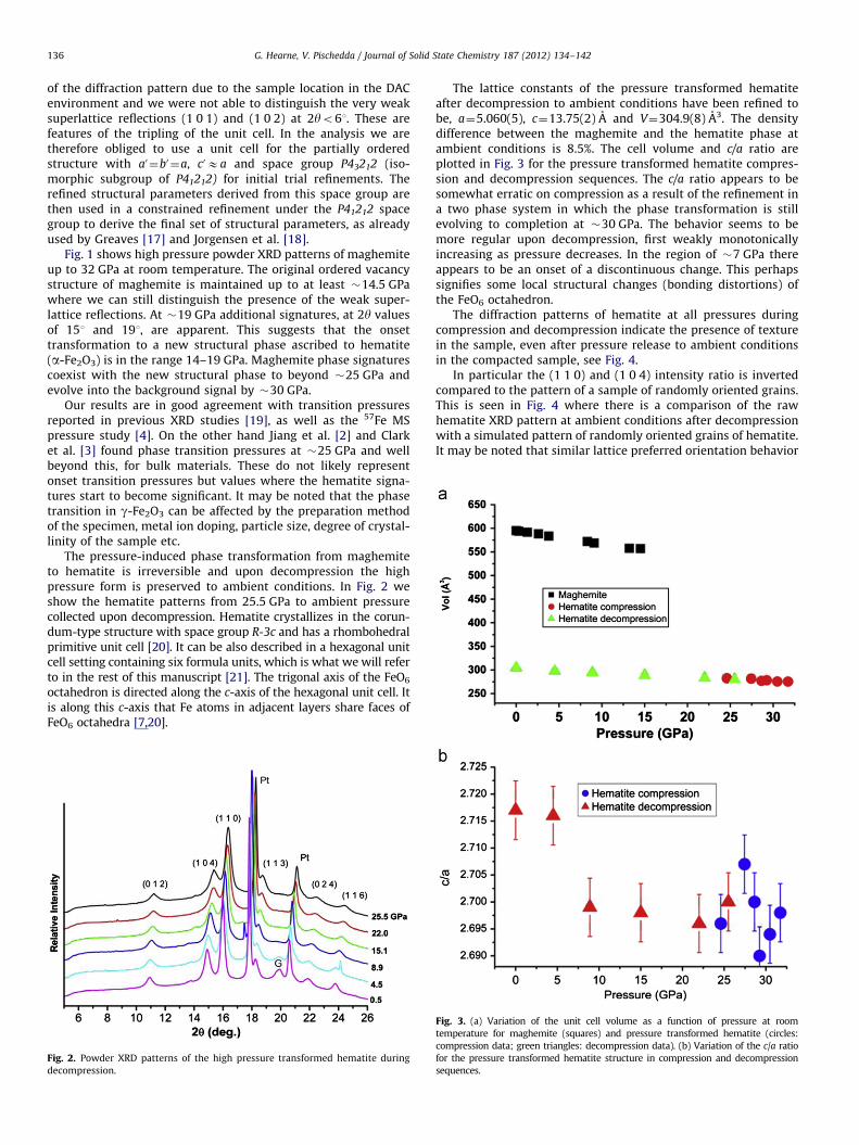
The lattice constants of the pressure transformed hematiteafter decompression to ambient conditions have been refined tobe, a¼5.060(5), c¼13.75(2) A and V¼304.9(8) A3. The densitydifference between the maghemite and the hematite phase atambient conditions is 8.5%. The cell volume and c/a ratio areplotted in Fig. 3 for the pressure transformed hematite compres-sion and decompression sequences. The c/a ratio appears to besomewhat erratic on compression as a result of the refinement ina two phase system in which the phase transformation is stillevolving to completion at �30 GPa. The behavior seems to bemore regular upon decompression, first weakly monotonicallyincreasing as pressure decreases. In the region of �7 GPa thereappears to be an onset of a discontinuous change. This perhapssignifies some local structural changes (bonding distortions) ofthe FeO6 octahedron.
The diffraction patterns of hematite at all pressures duringcompression and decompression indicate the presence of texturein the sample, even after pressure release to ambient conditionsin the compacted sample, see Fig. 4.
In particular the (1 1 0) and (1 0 4) intensity ratio is invertedcompared to the pattern of a sample of randomly oriented grains.This is seen in Fig. 4 where there is a comparison of the rawhematite XRD pattern at ambient conditions after decompressionwith a simulated pattern of randomly oriented grains of hematite.It may be noted that similar lattice preferred orientation behavior
Fig. 3. (a) Variation of the unit cell volume as a function of pressure at room
temperature for maghemite (squares) and pressure transformed hematite (circles:
compression data; green triangles: decompression data). (b) Variation of the c/a ratio
for the pressure transformed hematite structure in compression and decompression
sequences.

Fig. 4. Top panel shows simulated XRD pattern for randomly oriented hemati-
teþPt powder sample. Labels are hkl values of hematite, Pt (platinum) and G
(gasket) reflections. Bottom panel shows observed and calculated XRD patterns for
textured hematite at ambient conditions as recovered from high pressure. Rietveld
refinement has been performed using a March–Dollase semiempirical function to
model the lattice preferred orientation effect. The difference pattern is shown
below the fitted spectrum.
G. Hearne, V. Pischedda / Journal of Solid State Chemistry 187 (2012) 134–142 137
is evident in the XRD patterns of recent pressure studies compar-ing the transformation behavior of bulk and nano-phase maghe-mite. Yet these preferred orientation effects have not beenhighlighted or linked to the structural transformation [19].
To refine the structure of the recovered hematite sample wethen took into account a preferred orientation correction usingthe March–Dollase semi-empirical function available in GSAS[22]. The best fit (bottom panel of Fig. 4) is obtained with apreferred orientation of the c-axis of hematite such that there isbias along the incident beam (load axis) direction.
3.2. 57Fe MS pressure studies
3.2.1. Maghemite spectra
Fig. 5 shows the relatively high resolution Mossbauer spectrain the pressure regime up to �20 GPa. The two-site (tetrahedraland octahedral) feature sextet patterns of the spectra are evidentfrom the fitting up to �13 GPa. At low pressures close to ambientconditions these yield magnetic hyperfine fields, Bhf, of 49.4 T and51.0 T at tetrahedral and octahedral sites, respectively, in accor-dance with the literature [23]. Beyond 13 GPa a new sextetcomponent (shown shaded and having Bhf¼52.2 T) emerges,indicative of the onset of the second-order maghemite-hematitetransformation. This figure suggests that the onset of the phasetransition is in the range 13–16 GPa consistent with the XRD data
of Fig. 1; but is likely somewhat dependent on the experimentalconditions (particle size, pressure transmitting medium etc).
In this low pressure regime the spectra of maghemite havealso been fitted with a single sextet to check for the onset of linebroadening likely signifying the onset of the structural transition(bottom panel of Fig. 5). Such line broadening is seen to developat �13 GPa and is likely the onset pressure of the transformation.
At 16 GPa and beyond where the hematite abundance,deduced from the total absorption area of the spectral compo-nent, becomes appreciable (Z33%), unusual relative line inten-sities start to evolve for the sextet absorption profile. This is seenby focusing on the intensities of adjacent resonances of lines2 and 3 (ratio I2/I3) or lines 5 and 4 (ratio I5/I4), where I denotesthe absorption intensity or area (see Fig. 5).
3.2.2. Relative intensities of high pressure phase spectra and the
Morin transition
For any spectral component the ratio of these lines is depen-dent on the angle, g, between the g-ray propagation direction(which in this case is along the load axis) and the direction of Bhf
at the 57Fe nucleus (which is collinear with the atomic spinmoment). The ratio is given by [24,25],
R¼I2,5
I3,4¼
sin2 g14ð1þcos2 gÞ
, ð1Þ
R ranges from zero for g¼01 to 4 for g¼901 and has a value of2 at gE551, see Fig. 6(a). An assembly of spins randomly orientedin the sample is expected to give a sextet pattern with relativeintensities of 3:2:1:1:2:3. That is, the ratio RA¼2 and the sub-script A refers to an assembly of spins. Note that a value RA¼2 willalso be obtained for g¼551 for a single crystal sample or oneshowing preferred orientation. The absorption areas instead ofintensities are often used to compare the relative strengths of theresonance lines and are therefore needed to obtain the RA value.RA may involve errors due to so-called saturation effects when thesample thickness exceeds certain recommended values [15,26].The bottom panel of Fig. 5 shows that such effects are expected tocontribute negligible error in the evaluation of RA. That is, thesample measured in the DAC at ambient pressure yields similarspectral parameters to that of a thin absorber of �10 mg/cm2
measured outside of the DAC (open symbols).The fitting of the spectra at PZ19 GPa involves the pressure
transformed hematite component (as a majority phase exceeding50% abundance) and the maghemite components comprising thetetrahedral and octahedral sites of the spinel structure. Thehematite component at these high pressures has values forRAr1, well below that expected for an assembly of randomlyoriented spins (in which RA¼2). Obtaining the relative lineintensities for the minority phase (transforming maghemite)component are more ambiguous because of strong overlap ofinner lines 2, 3, 4, 5 (see Fig. 5) and two closely aligned andoverlapping octahedral and tetrahedral site spectral components.At these pressures we rather choose to model the transformingmaghemite component as a single broadened sextet. The RA valuefor this maghemite component may be constrained at �1.6similar to the value at the onset of the transformation at lowpressure (see bottom panel of Fig. 5) or left as a free fittingparameter. This does not impact significantly on the RA valueobtained for the pressure transformed hematite component inwhich RAE1. Our attention is focused on this pressure trans-formed hematite component.
Under ambient conditions hematite is known to undergo aspin reorientation at the so-called Morin transition of TM�260 K.This is a consequence of competing magneto-crystalline andmagnetic dipolar anisotropy energies KMC and KMD, respectively,

0
0.6
0.8
1.0
1.2
1.4
1.6
1.8
2.0
0.6
0.8
1.0
1.2
1.4
1.6
1.8
2.0linew
idth (mm
/s)RA
TIO
( I 2
.5 /
I 3.4
)Pressure (GPa)
maghemitesingle site fit
2 4 6 8 10 12 14
Fig. 5. (a) Mossbauer spectra of maghemite to pressures beyond the onset of the structural transition to hematite. Solid lines through the data (symbols) are the
theoretical fit. Hematite component is shown shaded at 16 and 19 GPa. Note the change in relative line intensities in the range 13–16 GPa when the hematite component
starts to evolve. Panel (b) shows single component fit to monitor the resonance absorption area ratio RA and linewidth of the pressurized maghemite. Open symbols are for
a thin sample measured outside of the DAC.
1 2 3 6
Bhfγ-ray
CollinearRA« 2
γ-ray
Bhf
PerpendicularRA=4
γ-rayBhf
RandomRA=2
00.00.51.01.52.02.53.03.54.0
RA =
I 2.5
/ I 3
.4
angle γ
1 2 3 6
1 2 3 6
1 2 3 5 6
QS > 0Antiferromagnetic AF
1 2 3 5 6
QS < 0WeaklyFerromagnetic WF
0-1.0-0.50.00.51.01.52.0
angu
lar t
erm
in Q
S
angle between VZZ and Bhf, θ
Vzz Bhf
Vzz
Bhf
4 5
4 5
4 5
4
10 20 30 40 50 60 70 80 90 20 40 60 80
4
Fig. 6. Cartoon depicting (a) relative line intensities and the relation between Bhf direction with respect to g-ray beam direction. (b) QS parameter values and associated
line positions and the relation between Vzz and Bhf.
G. Hearne, V. Pischedda / Journal of Solid State Chemistry 187 (2012) 134–142138
of opposite sign and with different temperature dependencies[21,27]. At T4TM, KMD dominates, and spins are aligned perpen-dicular to the c-axis (i.e., in the basal plane) in the hexagonalsetting. Spins in adjacent layers stacked along the c-axis areslightly canted toward each other and a weak ferromagnetic(WF) moment develops in the basal plane [28]. At ToTM, KMC
dominates, and spins align along the c-axis. and the system
is a collinear antiferromagnet (AF) [29,30]. The transition tem-perature TM is known to be quite sensitive to the presence ofimpurities and small particle size. It is obviously pressure depen-dent. This has been well documented in comparatively lowpressure studies to �10 GPa of early research on pressurizedhematite as starting material and in fact TM rises to roomtemperature by�5 GPa. [21] This means that at P45 GPa at
Admin
Typewritten Text
swop stick spectra
Admin
Typewritten Text
Admin
Typewritten Text
Admin
Typewritten Text

G. Hearne, V. Pischedda / Journal of Solid State Chemistry 187 (2012) 134–142 139
room temperature, moments start to align along the c-axis in anAF configuration of adjacent layers. Neutron diffraction work bothat ambient conditions and in a separate study up to �10 GPasuggests that the spin direction in the sub-lattices of the AF state(at ToTM) are inclined by not more than �201 away from thec-axis [20,31]. At room temperature and the very high pressuresreached in our investigation, �30 GPa at which T5TM, it isanticipated that the spins are even closer aligned to the c-axis,if not collinear with it.
Therefore in the pressure transformed hematite onset at13–16 GPa, for which RAr1, it may be inferred from Eq. (1) thatthe spin direction with respect to the load axis is gr401, seeFig. 6(a). There is thus preferred orientation of the magneticmoments (spin texture) in the load axis direction, consistent withthe crystallographic texture seen in the XRD study. This persiststo the highest pressure of �30 GPa, depicted in Fig. 7. It should benoted that such preferred orientation effects are not evident inthe low pressure maghemite phase for which RAZ1.6. At thehighest pressure of 30 GPa where full conversion to the hematitephase is completed RA¼1.0; thus spins are aligned at a 401 (cone)angle to the load axis direction [24]. This spin alignment persistsupon decompression to �7 GPa, see Fig. 7.
Upon decompression to ambient conditions there is an increase inintensity of lines 2 and 5 relative to the innermost lines 3 and 4. Avalue of RAE2.1 is obtained from the fitting. This means that themoments are either randomly oriented or re-aligned at an angle ofgE551 to the load axis (Fig. 6(a)). This spin alignment (magnetictexturing) behavior of the pressure transformed hematite needs to beconsidered in conjunction with the XRD results which evidenceappreciable lattice preferred orientation (crystallographic texture)effects. This will be discussed in the next Section 4.
3.2.3. Quadrupole shift parameter and spin reorientation
Since spin alignment features appear to be important in thepressurized sample, another important fitting parameter relatedto this is the quadrupole shift (QS) parameter. This is the
γ-ray
θ > 55°
γ ≈ 40°
VzzBhf
θ ≈ 0°
γ ≈ 40°
θ ≈ 90°
γ ≈ 55°
BhfVzz
Fig. 7. Mossbauer spectrum at the highest pressure as well as those taken upon
decompression. Depiction of the relative orientations of, g-ray beam, Vzz and Bhf
are also indicated. This is deduced from the QS value and ratio RA, assuming that
axial symmetry is maintained throughout. The g-ray beam coincides with the
compression (load axis). The angle between the g-ray beam and Bhf is g. The angle
between Vzz and Bhf is y.
interaction of the electric field gradient (EFG) as a result of thelocal (electronic and nearest neighbor atoms) charge environmentand the quadrupole moment. It reflects the site asymmetry of the57Fe local environment. The expression for QS of a magneticallyordered sample in which there is a magnetic hyperfine field Bhf atthe 57Fe nucleus is given by (see Kolk, pp. 43–44) [26]:
QS¼ 18e2qQ ð3cos2y�1þZ2 sin2ycos2fÞ: ð2aÞ
where e is the electron charge and eq� Vzz ¼ @2V=@z@z is the
maximum value of the EFG in the principle axis system (in whichthe EFG tensor is diagonal), V is the electric potential at thenuclear site, and the Q on the right hand side of the equation isthe nuclear quadrupole moment of the 57Fe nucleus. The asym-metry parameter Z¼ ½Vxx�Vyy�=Vzz has values 0rZr1. Theangular factor in Eq. (2) involves the polar angles y and f thatBhf makes with the direction of Vzz. This QS parameter has theeffect of shifting each of the resonance lines such that they are notsymmetrically centered around zero of velocity on the x-scale (seeFig. 6(b)).
The QS parameter is only pertinent to hematite whose primi-tive unit cell (rhombohedral) has lower than cubic symmetry. Inthe case of hematite, at ambient conditions, Vzz is directed alongthe c-axis in the coordinates of the hexagonal unit cell ( equiva-lent [1 1 1] direction in the rhombohedral setting ). This is thetrigonal axis of the FeO6 octahedron along which there is axialsymmetry and thus Z¼0 in Eq. (2) [21]. The expression for the QS
thus reduces to
QS¼ 18e2qQ ð3cos2y�1Þ: ð2bÞ
The QS parameter for the maghemite phase is usually expectedto be zero because of the cubic lattice symmetry of the spinel(Vzz¼0). The Fe atoms having nearest neighbor vacancies con-tribute slight resonance line broadening effects and normally QS
is set to zero in fitting spectra of the cubic lattice symmetrysituation of the spinel [32]. Recently it has been demonstratedthat the presence of nearest neighbor vacancies at the Fe sites inthe structure can render the QS to be finite [33]. Therefore this canbe modeled in high resolution MS spectra of maghemite. Howeveradditional line-broadening effects from the 57Co(Rh) source ofhigh specific activity, which is an imperative for the high pressureMS experiments, would render such a detailed analysis unreliablein our case. Slight pressure gradients and deviatoric stresses dueto a degree of non-hydrostaticity would also cause some linebroadening. Therefore in our analysis of the maghemite spectraQSE0 mm/s and resonance line broadening accounts for any siteasymmetry effects from the vacancies [4].
In hematite the spin reorientation through TM is manifested inthe angular factor in the QS of Eq. (2b), such that QS
(yE901)¼�0.20 mm/s for T4TM. This is about half the valueand of opposite sign to the case of QS (yE01)¼0.40 mm/s forToTM, see also Fig. 6(b) [21]. The QS parameter is thus often usedto determine whether hematite is in the spin canted WF state orcollinear AF state.
In this work the QS parameter will prove useful in ascertaining,in the pressure transformed hematite phase, how the spinmoments (Bhf) align with respect to Vzz from Eq. (2b). At ambientpressure this component of the EFG is biased along the crystal-lographic c-axis (which coincides with the trigonal axis of theFeO6 octahedron). This is in addition to monitoring how the spinsalign relative to the load axis using the relative absorptionintensities and Eq. (1).
The evolution of the QS parameter is represented in the labelsof Figs. 7 and 8 and is similar to that of a previous study. [4]As soon as hematite becomes the majority phase at PZ19 GPathe QS values increase monotonically from þ0.40 mm/s toþ0.50 mm/s at 30 GPa, in comparison to QSE�0.20 mm/s in

γ-ray
0.92
0.96
1.00
-9
0.95
1.00
-9
0.95
1.00
2nd compression7 GPaQS = -0.11 mm/s
Rel
. Tra
nsm
issi
on
2nd decompress.1 GPaQS = -0.20 mm/s
Velocity (mm/s)
3rd compression5 GPaQS = -0.13 mm/s
θ > 55°
γ ≈ 40°
θ ≈ 90°
γ ≈ 55°
VzzBhf
θ > 55°
γ ≈ 40°
Bhf
Vzz
-6 -3 0 3 6 9
-6 -3 0 3 6 9
Fig. 8. Mossbauer spectra of decompression–recompression cycles of pressure
transformed hematite.
G. Hearne, V. Pischedda / Journal of Solid State Chemistry 187 (2012) 134–142140
the canted WF state at Po5 GPa. The monotonic increase of QS
upon rising pressure is likely due to changes in Vzz because ofchanges in inter-atomic distances. The QS values of the pressuretransformed hematite are indicative of the collinear AF statesimilar to the value of hematite starting material below the Morintransition ToTM. At these high pressures TM is well above roomtemperature [21]. Recall that from the neutron diffraction pres-sure study it is anticipated that the spins are aligned at less than201 to the c-axis, if not collinear with it [31].
3.2.4. Decompression data and recompression
In the decompression sequence from 30 GPa the DAC wasdecompressed in small pressure steps; however, a Mossbauerspectrum was first recorded when the pressure was as low as�7 GPa. The QS is seen to be negative indicative of spins at anangle y4551 to Vzz, see Fig. 6(b). This would be considered as anapproach toward the canted WF state, similar to hematite startingmaterial at ambient pressure in which Vzz?Bhf. However note thatRAE1 in the pressure transformed sample decompressed to�7 GPa. This is similar to that at the highest pressure indicatingthat spin alignment is still biased along the load axis, see Fig. 7.
Thus the spins remain aligned close to the load axis (gE401)as it was at �30 GPa. According to the XRD decompression datathis is also the bias direction of the c-axis, as was the case at thehighest pressure. Yet the QS value has changed sign compared tothe high pressure value. This indicates that the angle y betweenBhf and Vzz undergoes some change compared to the highestpressure situation. It is not due to a reorientation of Bhf, as the RA
value has not changed. The change in QS must rather be ascribedto changes in the EFG (Vzz), that is, in the angular factor in Eq. (2)as will be discussed further in the next section.
After decompression to ambient conditions, the anvils wereslightly separated from the gasketed sample cavity and thesample left to relax for a few hours. Measurement of this samplestill compacted in the gasket cavity, yielded QS¼�0.21 mm/s(and RA�2.1) typical of hematite starting materials at roomtemperature where Bhf is at 901 to Vzz.
Following this recovery to ambient conditions, the sample wasrecompressed and decompressed to ambient conditions in sub-sequent sequences. Mossbauer spectra were recorded at eachpressure step and these are depicted in Fig. 8. However absorp-tion areas are quite different from the case of randomly orientedmagnetic moments [4]. The ratio RA reverts to the value thatoccurred at very high pressure in the pressure-induced transfor-mation sequence, RA�1. This is indicative of magnetic moments(Bhf) again aligned close to the load axis, g�401. We have notperformed pressure XRD studies on the recompressed sample.The Mossbauer data (RA values) do seem to suggest that c-axispreferred orientation along the load axis persists again in anyrecompression experiments.
4. Discussion
4.1. Nature of the maghemite to hematite phase transformation
Our XRD pressure studies evidence lattice preferred orienta-tion arising out of the maghemite-hematite transformation. Itconfirms what has been seen (but not identified as such) recentlyin pressure studies of a vacancy disordered maghemite startingmaterial [19]. It is perhaps important to note that when hematiteis pressurized as a starting material from the outset, this latticepreferred orientation does not seem to occur even to the highestpressures typical of our study [4,5,7]. This is then suggestive thatthe lattice preferred orientation is linked to the maghemite-hematite structural transformation, although it may also beadditionally driven by deviations from hydrostatic stress condi-tions at these high pressures. Possible future experiments shouldinvestigate the transformation when near ideal hydrostatic con-ditions can be maintained at such elevated pressures, e.g., byusing a helium pressure transmitting medium.
The behavior of the maghemite-hematite transition may becompared with the pressure transformation of Fe metal. Thisundergoes an analogous cubic (bcc)-hexagonal (hcp) latticestructural transformation and has been studied quite extensivelybecause of geophysical implications [34,35]. It has been shownthat the a-Fe develops preferred orientation under non-hydro-static stress conditions [35]. The preferred orientation of thea-phase is inherited by the e-phase (hcp) in accordance withthe Burgers orientation relationship. This is a relationship estab-lished by Burgers [36], in which (1 1 0)bcc planes would become(0 0 0 1)hex basal planes in a martensitic transformation, becauseof the structural relationship between bcc and hcp lattices [35].After the phase transition the e-phase displays a texture with thec-axis oriented in a direction orthogonal to the load axis direction.Upon further compression the c-axes rotate and become parallelto the load axis.
In our case, the maghemite does not seem to show a highdegree of lattice preferred orientation up to the onset of thestructural transition during compression. At least it is not aspronounced as what it is in the high pressure transformedhematite. This is deduced from the relative line intensities ofboth the XRD and Mossbauer spectra up to �13 GPa near theonset of the phase transition, see figs. 1 and 5. These lineintensities (or absorption areas) evolve from RA values of �1.8to �1.6 at the onset of the structural transition, compared withRA�1 in the pressure transformed sample.
The texture in the pressure transformed hematite may arise froma slip mechanism and a crystallographic plane inherited from themaghemite, as in the bcc-hcp transition of Fe metal, according to aBurgers orientation relationship [35]. Alternatively the transition maybe similar to the temperature induced maghemite-hematite transi-tion which proceeds topotactically, meaning that it occurs by a

G. Hearne, V. Pischedda / Journal of Solid State Chemistry 187 (2012) 134–142 141
restacking of various atomic planes rather than by wholesale recrys-tallization and consequent nucleation and growth [37,38]. It is thensupposed that the high pressure phase forms in a crystallographicorientation with the c-axis [0 0 0 1] unit cell direction (hexagonalsetting) developed along [1 1 1] directions of the cubic spinel unit cellof the low pressure phase [38,39]. In this scenario under compression,it may then be anticipated that c-axis alignment along the load axisdirection is favored.
4.2. Bulk modulus values
Turning to the mechanical properties of the phases, the bulkmodulus of the low and high pressure phases have been calcu-lated by a least-squares fit of the pressure–volume data to a thirdorder Birch–Murnaghan equation of state. The resulting zero-pressure bulk modulus is B0¼187(74) for maghemite andB0¼268(714) GPa for hematite (combining decompression andcompression data sets), with an assumption of a pressure deriva-tive B0
0 ¼4. The B0�187 GPa that we determined for g-Fe2O3 is ingood agreement with the values of 19076 GPa reported by Clarket al. [3] and slightly lower of the values of 203710 GPa and213710 reported by Jiang et al. [2] and Zhu et al. [19], respec-tively. It seems to be systematically lower than all previouslyreported B0 values, perhaps as a result of the vacancy orderedsuperstructure. The B0�268 GPa of our high-density a-Fe2O3 issomewhat higher than those of samples reported in the literature[2,7], namely B0�230 GPa. Clark et al. report a value of 299(730) GPa [3].
4.3. Decompression behavior
Upon decompression to �7 GPa the magnetic moments remainpinned along the same angle to the load axis (�401) as at the highestpressure. Recall that upon decompression the c-axis of the hexagonalunit cell also shows preferred orientation along the load axis, seeFig. 2. All of this is deduced from the relative intensities of the spectrallines in both the Mossbauer and XRD data, which do not change fromthe values at the highest pressure.
There is a need to understand the Mossbauer decompressiondata at �7 GPa where the QS¼�0.13 mm/s has changed sign.This must be due to the angular factor in Eq. (2), indicating thatVzz is changing direction or there is a change from axial symmetry(Z value affected) or both. Therefore upon decompression to�7 GPa it is the EFG that is affected and not the Bhf orientation.Assuming that axial asymmetry is retained (Z¼0), Eq. (2b), thechange in sign of the QS value is suggestive of Vzz becomingorthogonal to Bhf. The negative value of the QS indicates thaty4551 at this low pressure, see Fig. 7.
This is likely due to a distortion of the FeO6 octahedron. Adetailed study of the compressional behavior of hematite pres-surized as starting material [7], shows that such bond distortionsdo occur in this material. It is anticipated that this is happening inthe decompression of highly textured hematite of this study. Ahint of this is found in the behavior of the c/a ratio plotted inFig. 3, which has a compelling indication of a discontinuouschange onset at �7 GPa of the decompression sequence.
Upon further decompression to ambient conditions the XRDpatterns show that the hematite crystallites remain highly tex-tured as anticipated for a compacted material (with c-axis pre-ferred orientation along the load axis), but the magnetic momentsswing further away from the load axis (to �551) as inferred fromthe ratio RAE2, see Fig. 6(a). Thus from high pressure down toambient conditions the spin reorientation is only of the order of151. Therefore moments cannot be in the basal plane of thehexagonal unit cell if they were aligned close to the c-axis at thehighest pressure in the AF state as anticipated [31]. Recall that
this basal plane orientation is the case in hematite startingmaterial at ambient conditions. This is also what is interpretedto be the case in pressurized hematite starting materials whichhave been decompressed to ambient conditions [4]. In therecovered sample, Fig. 7, the value QS¼�0.20 mm/s is suggestiveof the moments now being near orthogonal to Vzz. We havealready indicated that Vzz is not necessarily along the samedirection it was at high pressure (which was presumably alongthe c-axis), as implied by the behavior of c/a in Fig. 3 which hintsat local structural distortions of the FeO6 octahedron.
4.4. Recompression behavior of pressure transformed hematite
Turning to the recompression experiments (Fig. 8) we see thatthere is a change from RAE2 at or near ambient pressure toRAE1 at �5 GPa in successive recompression–decompressioncycles. Presumably the sample is compacted and remains tex-tured after the first decompression to ambient conditions. Thischange in the RA ratio suggests a spin re-alignment of only 151(with respect to the load axis), Fig. 6(a).
It appears that in the pressure transformed hematite theknown 901 spin reorientation of hematite starting material,associated with the Morin transition, is inhibited over widedecompression and recompression ranges. It is suggested thatthis is due to retained residual strain in the pressure transformedhematite as a result of, or in combination with, defects or crystalimperfections in the material associated with the maghemite-hematite inversion. Similar effects have been seen in shockedhematite or as a function of nanophase grain size [40,41].Although in those cases it is the WF canted state (basal planealignment) that is stabilised, in which TM is suppressed to lowertemperatures from �250 K in hematite starting material atambient conditions. This transition is very sensitive to crystalimperfections because of how they change the magnetic aniso-tropy energies that drive the Morin transition [39,41,42].
5. Conclusion
In the pressure transformed phase a high degree of latticepreferred orientation is evident, in contrast to the low pressuremaghemite phase. There is crystallographic c-axis alignment andan associated magnetic texturing of moments, biased in thedirection of the load axis. Decompression data from XRD evidencedeformation of the local atomic environment of the Fe atom. Thisis expected to affect the direction of the maximum value of theelectric field gradient (EFG), normally assumed to always bedirected along the c-axis of the hexagonal unit cell.
Such a well textured hematite sample, likely arising out of thetopotactic structural transition allows us to obtain some informa-tion on the pressure-response of the spin reorientation behavior.Hematite pressurized as starting material at room temperatureshows spin reorientation from basal plane to near c-axis align-ment occurring over a wide compression range, that is, spinreorientation of �901 [4,21]. This corresponds to a rise in theMorin temperature to above room temperature. It is presumedthat upon decompression the same re-alignment occurs (nearly901 spin reorientation) but along a different pressure pathway.The previous 57Fe MS study of maghemite pressure transformedto hematite presumed the same scenario [4].
However, in this study we benefit more information from boththe crystallographic and magnetic texture effects revealed in theXRD and MS patterns of the same sample. This shows that such acomplete spin reorientation does not seem to occur in the highpressure transformed hematite when it is reverted back toambient conditions and when it is recompressed back to high

G. Hearne, V. Pischedda / Journal of Solid State Chemistry 187 (2012) 134–142142
pressures. Spin reorientation seems to be restricted to �151 andchanges of the MS parameters are rather suggestive of analteration of the direction of Vzz, due to local structural distortions(of the FeO6 octahedron ).
A combination of effects of retained residual lattice strain andcrystal imperfections arise out of the pressure-induced structuraltransformation. These are supposed to account for the strong outof plane component of the spins in the hematite sample recoveredto ambient conditions. The residual lattice strain or crystalimperfections, or both effects, possibly originate from the defec-tive (vacancy ordered or disordered) cubic spinel low pressurephase. In temperature transformed samples such vacancies willdiffuse out and play no further role. By contrast, under externallyapplied stress it is conceivable that such remnant defects wouldremain trapped within the lattice of the high pressure phase. Theyaffect spin reorientation behavior and possibly also the structuralpressure-response of the FeO6 octahedral environment, in com-parison to defect-free hematite. The absence of superstructurepeaks in the XRD pattern of this pressure transformed hematitesuggests that these entrapped defects (vacancies) are disorderedwithin the corundum lattice.
Acknowledgments
We thank E. Boldyreva and S.V. Tsybulya of the Chemistrydivisions at Novosibirsk State University for supplying the sam-ple. S.R. Naidoo assisted with the XRD work. This work alsoprofited from beam time provided by ELETTRA to an ICTP fundedprogram for developing nations. Funding has been derived fromboth the NRF-SA and FRC-UJ (Science) initiatives.
References
[1] F. Baudelet, S. Pascarelli, O. Mathon, J.-P. Itie, A. Polian, J.-C. Chervin, Phys.Rev. B 82 (2010). 140412 (1–4).
[2] J.Z. Jiang, J.S. Olsen, L. Gerward, S. Morup, Europhys. Lett. 44 (1998) 620.[3] S.M. Clark, S.G. Prilliman, C.K. Erdonmez, A.P. Alivisatos, Nanotech 16 (2008)
2813–2818.[4] T. Kawakami, S. Nasu, T. Tsutsui, T. Sasaki, T. Yamada, S. Endo, M. Takano,
T. Katamoto, J. Phys. Soc. Jpn. 72 (2003) 2640–2645.[5] S. Ono, K. Funakoshi, Y. Ohishi, E. Takahashi, J. Phys.: Condens. Matter 17
(2005) 269–276.[6] M.P. Pasternak, G.K. Rozenberg, G.Y. Machavariani, O. Naaman, R.D. Taylor,
R. Jeanloz, Phys. Rev. Lett. 82 (1999) 4663–4666.[7] G.K. Rozenberg, L.S. Dubrovinsky, M.P. Pasternak, O. Naaman, T.L. Bihan,
R. Ahuja, Phys. Rev. B 65 (2002). 064112 (1–8).
[8] J. Badro, G. Fiquet, V.V. Struzhkin, M. Somayazulu, H.K. Mao, G. Shen,T.L. Bihan, Phys. Rev. Lett. 89 (2002). 205504 (1–4).
[9] I.S. Lyubutin, A.G. Gavriliuk, Phys. Usp. 52 (2009) 989–1017.[10] A.N. Shmakov, G.N. Kryukova, S.V. Tsybulya, A.L. Chuvilin, L.P. Solovyeva,
J. Appl. Cryst. 28 (1995) 141–145.[11] A. Hammersley, Comput. Prog. FIT2D (ESRF, Grenoble 1998).[12] A.C. Larson, R.B.V. Dreele, General Structure Analysis System (GSAS), Los
Alamos National Lab Report LAUR 86-748.[13] S. Takele, G.R. Hearne, Nucl. Instrum. Methods B 183 (2001) 413–418.[14] R.A. Brand, WinNormos-for-Igor Users Manual, WissEl GmbH, Starnberg,
Universitat Duisburg, Duisburg, 2006.[15] G.T. Long, T.E. Cranshaw, G. Longworth, Moss. Effect. Ref. Data J 6 (1983)
42–49.[16] R. Grau-Crespo, A.Y. Al-Baitai, I. Saadoune, N.H.D. Leeuw, J. Phys.: Condens.
Matter 22 (2010). 255401 (1–8).[17] C. Greaves, J. Solid State Chem 49 (1983) 325–333.[18] J.-E. Jørgensen, L. Mosegaard, L.E. Thomse, T.R. Jensen, J.C. Hanson, J. Solid
State Chem. 180 (2007) 180–185.[19] H. Zhu, Y. Ma, H. Yang, C. Ji, D. Hou, L. Guo, J. Phys. Chem. Solids 71 (2010)
1183–1186.[20] A.H. Hill, F. Jiao, P.G. Bruce, A. Harrison, W. Kockelmann, C. Ritter, Chem.
Mater. 20 (2008) 4891–4899.[21] C.L. Bruzzone, R. Ingalls, Phys. Rev. B 28 (1983) 2430–2440.[22] W.A. Dollase, J. Appl. Crystallogr. 19 (1986) 267–272.[23] S.-J. Lee, S. Lee, New J. Phys. 8 (2006) 98. doi:10.1088/1367-2630/8/6/098.[24] U. Gonser, M. Ron, H. Ruppersberg, W. Kuene, A. Trautwein, Phys. Stat. Sol. (a)
10 (1972) 493–499.[25] H.-D. Pfannes, U. Gonser, Appl. Phys 1 (1973) 93–102.[26] B. Kolk, Studies of Dynamical Properties of Solids with the Mossbauer Effect,
Elsevier, Amsterdam, 1984.[27] J.O. Artman, J.C. Murphy, S. Foner, Phys. Rev. 138 (1965) 912–917.[28] F. Bødker, M.F. Hansen, C.B. Koch, K. Lefmann, S. Mørup, Phys. Rev. B 61
(2000) 6826–6838.[29] R.J. Harrison, S.A. McEnroe, P. Robinson, C.J. Howard, Am. Mineral. 95 (2010)
974–979.[30] A.H. Morrish, Canted Antiferromagnetism: Hematite, World Scientific, Singa-
pore, 1994.[31] J.B. Parise, D.R. Locke, C.A. Tulk, I. Swainson, L. Cranswick, Physica B 385–386
(2006) 391–393.[32] H.A. Annersten, S.S. Hafner, Z. Kristall. B 137 (1973) 320–340.[33] K.M. Spiers, D.M. Paganin, J.D. Cashion, J. Phys.: Condens. Matter 23 (2011)
1–6.[34] H.-R. Wenk, S. Matthies, R.J. Hemley, H.-K. Mao, J. Shu, Nature 405 (2000)
1044–1047.[35] S. Merkel, H.-R. Wenk, P. Gillet, H.-K. Mao, R.J. Hemley, Phys. Earth Planet.
Inter. 145 (2004) 239–251.[36] M.I. Burgers, Physica 1 (1934) 561–586.[37] J.D. Bernal, D.R. Dasgupta, A.L. Mackay, Nature 180 (1957) 645–647.[38] S. Kachi, K. Momiyama, S. Shimizu, J. Phys. Soc. Jpn. 18 (1963) 106–116.[39] D.J. Dunlop, O. Ozdemir, Rock Magnetism: Fundamentals and Frontiers,
Cambridge University Press, Cambridge, 1997.[40] D.L. Williamson, E.L. Venturini, R.A. Graham, B. Morosin, Phys. Rev. B 34
(1986) 1899–1907.[41] O. Ozdemir, D.J. Dunlop, T.S. Berquo, Geochem. Geophys. Geosys. 9 (2008)
Q10Z01. doi:10.1029/2008GC002110.[42] O. Ozdemir, D.J. Dunlop, J. Geophys. Res. 111 (2006). B12S03 (1–13).