Higher-order nonclassical properties of atom-molecule Bose-Einstein Condensate
Upload
independentCategory
view
0download
0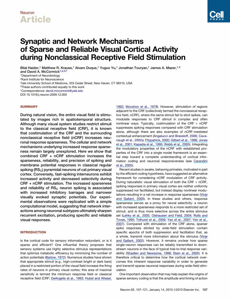
Neuron
Article
Synaptic and Network Mechanismsof Sparse and Reliable Visual Cortical Activityduring Nonclassical Receptive Field StimulationBilal Haider,1 Matthew R. Krause,1 Alvaro Duque,1 Yuguo Yu,1 Jonathan Touryan,1 James A. Mazer,1,3
and David A. McCormick1,2,3,*1Department of Neurobiology2Kavli Institute for NeuroscienceYale University School of Medicine, 333 Cedar Street, New Haven, CT 06510, USA3These authors contributed equally to this work
*Correspondence: [email protected]
DOI 10.1016/j.neuron.2009.12.005
SUMMARY
During natural vision, the entire visual field is stimu-lated by images rich in spatiotemporal structure.Although many visual system studies restrict stimulito the classical receptive field (CRF), it is knownthat costimulation of the CRF and the surroundingnonclassical receptive field (nCRF) increases neu-ronal response sparseness. The cellular and networkmechanisms underlying increased response sparse-ness remain largely unexplored. Here we show thatcombined CRF + nCRF stimulation increases thesparseness, reliability, and precision of spiking andmembrane potential responses in classical regularspiking (RSC) pyramidal neurons of cat primary visualcortex. Conversely, fast-spiking interneurons exhibitincreased activity and decreased selectivity duringCRF + nCRF stimulation. The increased sparsenessand reliability of RSC neuron spiking is associatedwith increased inhibitory barrages and narrowervisually evoked synaptic potentials. Our experi-mental observations were replicated with a simplecomputational model, suggesting that network inter-actions among neuronal subtypes ultimately sharpenrecurrent excitation, producing specific and reliablevisual responses.
INTRODUCTION
Is the cortical code for sensory information redundant, or is it
sparse and efficient? One influential theory proposes that
sensory systems use highly selective stimulus representations
that optimize metabolic efficiency by minimizing the number of
action potentials (Barlow, 1972). Numerous studies have shown
that appropriate stimuli (e.g., high-contrast bright or dark bars)
placed in a restricted portion of the visual field increase the firing
rates of neurons in primary visual cortex; this area of maximal
sensitivity is termed the minimum response field or classical
receptive field (CRF; DeAngelis et al., 1993; Hubel and Wiesel,
1962; Movshon et al., 1978). However, stimulation of regions
adjacent to the CRF (collectively termed the nonclassical recep-
tive field, nCRF), where the same stimuli fail to elicit spikes, can
modulate responses to CRF stimuli in complex and often
nonlinear ways. Typically, costimulation of the CRF + nCRF
suppresses spiking responses compared with CRF stimulation
alone, although there are also examples of nCRF-mediated
contextual enhancement (Angelucci and Bressloff, 2006; Cava-
naugh et al., 2002a; Fitzpatrick, 2000; Gilbert et al., 1996; Jones
et al., 2001; Kapadia et al., 1995; Webb et al., 2005). Integrating
the modulatory properties of the nCRF with established pro-
perties of the CRF into a single model framework is an essen-
tial step toward a complete understanding of cortical infor-
mation coding and neuronal responsiveness (see Carandini
et al., 2005).
Recent studies in awake, behaving primates, motivated in part
by the efficient coding hypothesis, have suggested an alternative
framework for considering nCRF modulation of CRF activity.
During naturalistic visual stimulation of both the CRF + nCRF,
spiking responses in primary visual cortex are neither uniformly
suppressed nor facilitated, but instead display nonlinear modu-
lations resulting in a net increase in response sparseness (Vinje
and Gallant, 2000). In these studies and others, response
sparseness serves as a proxy for neural selectivity: a neuron
with increased sparseness responds to a more restricted set of
stimuli, and is thus more selective across the entire stimulus
set (Lehky et al., 2005; Olshausen and Field, 2004; Rolls and
Tovee, 1995; Tolhurst et al., 2009; Yao et al., 2007; Yen et al.,
2007). Compared with stimulation of the CRF alone, sparser
spike responses elicited by wide-field stimulation contain
specific epochs of both suppression and facilitation that, as
a whole, transmit more information about the stimulus (Vinje
and Gallant, 2002). However, it remains unclear how sparse
single-neuron responses can be reliably transmitted to down-
stream neurons in the face of typical trial-to-trial response vari-
ability (Shadlen and Newsome, 1998; Stein et al., 2005). It is
therefore critical to determine how the cortical network over-
comes this inherent response variability in order to generate
and transmit sparse neuronal responses during wide-field stim-
ulation.
One important observation that may help explain the origins of
sparse sensory coding is that the amplitude and timing of action
Neuron 65, 107–121, January 14, 2010 ª2010 Elsevier Inc. 107
Neuron
Selective and Reliable Spiking in Visual Cortex
potentials depends critically upon the connectivity and activity
levels of presynaptic excitatory and inhibitory neuronal subtypes
(Azouz et al., 1997; Bruno and Sakmann, 2006; Contreras and
Palmer, 2003; Silberberg and Markram, 2007; Swadlow, 2003;
Yoshimura and Callaway, 2005). The net pattern of activity
across the distributed network of presynaptic neuronal subpop-
ulations is collectively visible in a single neuron’s membrane
potential fluctuations, where the amplitude and precise timing
of excitatory and especially inhibitory potentials is a critical factor
in determining exactly when a given pyramidal neuron spikes
(Gabernet et al., 2005; Haider and McCormick, 2009; Hasen-
staub et al., 2005; Higley and Contreras, 2006; Pouille and Scan-
ziani, 2001; Wehr and Zador, 2003).
Here, we report that wide-field naturalistic stimulation of clas-
sical regular spiking (RSC) pyramidal neurons in cat primary
visual cortex both increased the amplitude of inhibitory postsyn-
aptic potentials (IPSPs) and increased the trial-to-trial reliability
of excitatory postsynaptic potentials (EPSPs). These synaptic
events were mirrored by spiking activity recorded in specific
excitatory and inhibitory neuronal subpopulations. Injection of
these recorded PSP sequences into a simple computational
model revealed that while both changes in IPSPs and EPSPs
contributed to the sparseness and reliability of spiking
responses during CRF + nCRF stimulation, IPSPs were the pri-
mary cause of increased neuronal sparseness, while the EPSPs
greatly enhanced trial-to-trial spike reliability and predominantly
shaped the overall spiking response. These combined excitatory
and inhibitory network interactions may ultimately explain the
highly selective and more reliable neuronal activity observed in
visual cortex in response to naturalistic sensory inputs.
RESULTS
Stimulus Presentation and Experimental ApproachWe performed extracellular and intracellular recordings in cat
primary visual cortex during presentation of naturalistic movie
sequences masked by circular apertures of two sizes. One
restricted the movie to the CRF (as defined quantitatively, see
Experimental Procedures and Supplemental Experimental
Procedures available online), while the other was larger and
resulted in costimulation of both the CRF and the surrounding
nCRF. Note that in both configurations visual stimulation within
the CRF was identical—only the size of the aperture was
changed (from a radius of 1X CRF to 3X CRF).
We first present results obtained from electrophysiologically
identified classical regular spiking (RSC) neurons, the most abun-
dant and most frequently recorded cortical neuronal subtype
(Nowak et al., 2003).
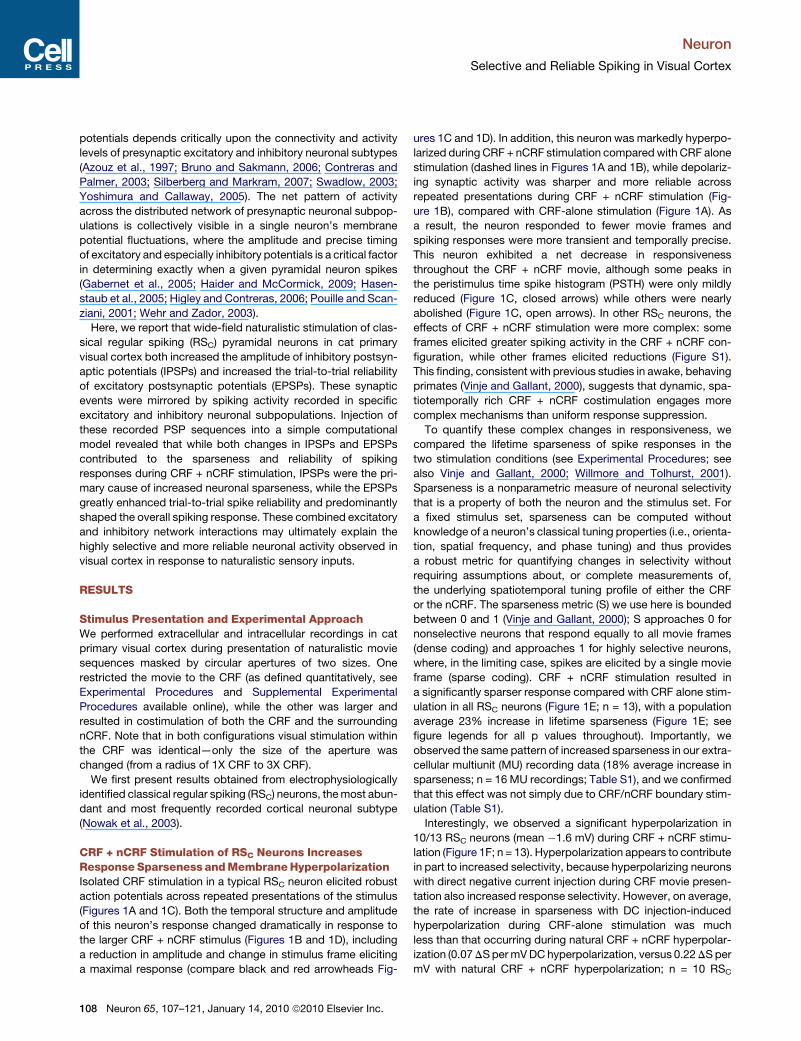
CRF + nCRF Stimulation of RSC Neurons IncreasesResponse Sparseness and Membrane HyperpolarizationIsolated CRF stimulation in a typical RSC neuron elicited robust
action potentials across repeated presentations of the stimulus
(Figures 1A and 1C). Both the temporal structure and amplitude
of this neuron’s response changed dramatically in response to
the larger CRF + nCRF stimulus (Figures 1B and 1D), including
a reduction in amplitude and change in stimulus frame eliciting
a maximal response (compare black and red arrowheads Fig-
108 Neuron 65, 107–121, January 14, 2010 ª2010 Elsevier Inc.
ures 1C and 1D). In addition, this neuron was markedly hyperpo-
larized during CRF + nCRF stimulation compared with CRF alone
stimulation (dashed lines in Figures 1A and 1B), while depolariz-
ing synaptic activity was sharper and more reliable across
repeated presentations during CRF + nCRF stimulation (Fig-
ure 1B), compared with CRF-alone stimulation (Figure 1A). As
a result, the neuron responded to fewer movie frames and
spiking responses were more transient and temporally precise.
This neuron exhibited a net decrease in responsiveness
throughout the CRF + nCRF movie, although some peaks in
the peristimulus time spike histogram (PSTH) were only mildly
reduced (Figure 1C, closed arrows) while others were nearly
abolished (Figure 1C, open arrows). In other RSC neurons, the
effects of CRF + nCRF stimulation were more complex: some
frames elicited greater spiking activity in the CRF + nCRF con-
figuration, while other frames elicited reductions (Figure S1).
This finding, consistent with previous studies in awake, behaving
primates (Vinje and Gallant, 2000), suggests that dynamic, spa-
tiotemporally rich CRF + nCRF costimulation engages more
complex mechanisms than uniform response suppression.
To quantify these complex changes in responsiveness, we
compared the lifetime sparseness of spike responses in the
two stimulation conditions (see Experimental Procedures; see
also Vinje and Gallant, 2000; Willmore and Tolhurst, 2001).
Sparseness is a nonparametric measure of neuronal selectivity
that is a property of both the neuron and the stimulus set. For
a fixed stimulus set, sparseness can be computed without
knowledge of a neuron’s classical tuning properties (i.e., orienta-
tion, spatial frequency, and phase tuning) and thus provides
a robust metric for quantifying changes in selectivity without
requiring assumptions about, or complete measurements of,
the underlying spatiotemporal tuning profile of either the CRF
or the nCRF. The sparseness metric (S) we use here is bounded
between 0 and 1 (Vinje and Gallant, 2000); S approaches 0 for
nonselective neurons that respond equally to all movie frames
(dense coding) and approaches 1 for highly selective neurons,
where, in the limiting case, spikes are elicited by a single movie
frame (sparse coding). CRF + nCRF stimulation resulted in
a significantly sparser response compared with CRF alone stim-
ulation in all RSC neurons (Figure 1E; n = 13), with a population
average 23% increase in lifetime sparseness (Figure 1E; see
figure legends for all p values throughout). Importantly, we
observed the same pattern of increased sparseness in our extra-
cellular multiunit (MU) recording data (18% average increase in
sparseness; n = 16 MU recordings; Table S1), and we confirmed
that this effect was not simply due to CRF/nCRF boundary stim-
ulation (Table S1).
Interestingly, we observed a significant hyperpolarization in
10/13 RSC neurons (mean �1.6 mV) during CRF + nCRF stimu-
lation (Figure 1F; n = 13). Hyperpolarization appears to contribute
in part to increased selectivity, because hyperpolarizing neurons
with direct negative current injection during CRF movie presen-
tation also increased response selectivity. However, on average,
the rate of increase in sparseness with DC injection-induced
hyperpolarization during CRF-alone stimulation was much
less than that occurring during natural CRF + nCRF hyperpolar-
ization (0.07 DS per mV DC hyperpolarization, versus 0.22 DS per
mV with natural CRF + nCRF hyperpolarization; n = 10 RSC
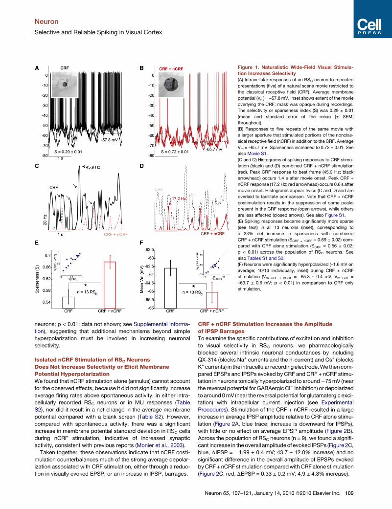
Figure 1. Naturalistic Wide-Field Visual Stimula-
tion Increases Selectivity
(A) Intracellular responses of an RSC neuron to repeated
presentations (five) of a natural scene movie restricted to
the classical receptive field (CRF). Average membrane
potential (Vm) = –57.8 mV. Inset shows extent of the movie
overlying the CRF; mask was opaque during recordings.
The selectivity or sparseness index (S) was 0.29 ± 0.01
(mean and standard error of the mean [± SEM]
throughout).
(B) Responses to five repeats of the same movie with
a larger aperture that stimulated portions of the nonclas-
sical receptive field (nCRF) in addition to the CRF. Average
Vm = –65.7 mV. Sparseness increased to 0.72 ± 0.01. See
also Movie S1.
(C and D) Histograms of spiking responses to CRF stimu-
lation (black) and (D) combined CRF + nCRF stimulation
(red). Peak CRF response to best frame (45.9 Hz; black
arrowhead) occurs 1.4 s after movie onset. Peak CRF +
nCRF response (17.2 Hz; red arrowhead) occurs 0.6 s after
movie onset. Histograms appear twice (C and D) and are
overlaid to facilitate comparison. Note that CRF + nCRF
costimulation results in the suppression of some peaks
present in the CRF response (open arrows), while others
are less affected (closed arrows). See also Figure S1.
(E) Spiking responses became significantly more sparse
(see text) in all 13 neurons (inset), corresponding to
a 23% net increase in sparseness with combined
CRF + nCRF stimulation (SCRF + nCRF = 0.69 ± 0.02) com-
pared with CRF alone stimulation (SCRF = 0.56 ± 0.02;
p < 0.01) across the population of RSC neurons. See
also Tables S1 and S2.
(F) Neurons were significantly hyperpolarized (–1.6 mV on
average; 10/13 individually, inset) during CRF + nCRF
stimulation (Vm CRF + nCRF = –65.3 ± 0.4 mV; Vm CRF =
–63.7 ± 0.6 mV; p < 0.01) in comparison to CRF only
stimulation.
Neuron
Selective and Reliable Spiking in Visual Cortex
neurons; p < 0.01; data not shown; see Supplemental Informa-
tion), suggesting that additional mechanisms beyond simple
hyperpolarization must be involved in increasing neuronal
selectivity.
Isolated nCRF Stimulation of RSC NeuronsDoes Not Increase Selectivity or Elicit MembranePotential HyperpolarizationWe found that nCRF stimulation alone (annulus) cannot account
for the observed effects, because it did not significantly increase
average firing rates above spontaneous activity, in either intra-
cellularly recorded RSC neurons or in MU responses (Table
S2), nor did it result in a net change in the average membrane
potential compared with a blank screen (Table S2). However,
compared with spontaneous activity, there was a significant
increase in membrane potential standard deviation in RSC cells
during nCRF stimulation, indicative of increased synaptic
activity, consistent with previous reports (Monier et al., 2003).
Taken together, these observations indicate that nCRF costi-
mulation counterbalances much of the strong average depolar-
ization associated with CRF stimulation, either through a reduc-
tion in visually evoked EPSP, or an increase in IPSP, barrages.
CRF + nCRF Stimulation Increases the Amplitudeof IPSP BarragesTo examine the specific contributions of excitation and inhibition
to visual selectivity in RSC neurons, we pharmacologically
blocked several intrinsic neuronal conductances by including
QX-314 (blocks Na+ currents and the h-current) and Cs+ (blocks
K+ currents) in the intracellular recording electrode. We then com-
pared EPSPs and IPSPs evoked by CRF and CRF + nCRF stimu-
lation in neurons tonically hyperpolarized to around�75 mV (near
the reversal potential for GABAergic Cl� inhibition) or depolarized
to around 0 mV (near the reversal potential for glutamatergic exci-
tation) with intracellular current injection (see Experimental
Procedures). Stimulation of the CRF + nCRF resulted in a large
increase in average IPSP amplitude relative to CRF alone stimu-
lation (Figure 2A, blue trace; increase is downward for IPSPs),
with little or no effect on average EPSP amplitude (Figure 2B).
Across the population of RSC neurons (n = 9), we found a signifi-
cant increase in the overall amplitude of evoked IPSPs (Figure 2C,
blue, DIPSP = �1.99 ± 0.4 mV; 43.7 ± 12.0% increase) and no
significant difference in the overall amplitude of EPSPs evoked
by CRF + nCRF stimulation compared with CRF alone stimulation
(Figure 2C, red, DEPSP = 0.33 ± 0.2 mV; 4.9 ± 4.3% increase).
Neuron 65, 107–121, January 14, 2010 ª2010 Elsevier Inc. 109
Figure 2. Wide-Field Visual Stimulation Selectively Increases the Amplitude of Inhibitory Postsynaptic Potentials
(A) Average inhibitory postsynaptic potentials (IPSPs) recorded during 12 presentations of a naturalistic movie to the CRF (black traces) and to the CRF + nCRF
(blue). QX-314 and Cs+ in micropipette. Upper and lower thin traces indicate ± SEM. Dashed vertical line indicates movie onset, downward deflections indicate
IPSPs. Some IPSP barrages increase greatly (closed arrows), while others change little (open arrows). CRF + nCRF stimulation significantly increases average
IPSP amplitude (compared with average response during CRF stimulation) in this cell by –2.73 ± 0.51 mV (p < 0.01; recorded at 0 mV).
(B) In contrast to IPSPs, EPSPs (recorded at –75 mV) did not significantly differ in amplitude between the two conditions (–0.41 ± 0.61 mV; p > 0.1). The neuron
shown here is the same neuron as shown in Figures 1A and 1B after spike inactivation.
(C) Population differences in EPSPs (red) and IPSPs (blue) evoked with CRF + nCRF stimulation, compared with CRF alone stimulation. All nine neurons were
determined to be RSC before spike inactivation. CRF + nCRF evoked IPSP barrages were significantly larger on average (blue, –1.99 ± 0.4 mV; 43.7 ± 12.0%
increase; p < 0.01) while EPSP barrages were not (0.33 ± 0.2 mV; 4.9 ± 4.3% increase; p > 0.1). Values are mean ± SEM.
Neuron
Selective and Reliable Spiking in Visual Cortex
CRF + nCRF Stimulation of Fast-Spiking InterneuronsDecreases Selectivity and Increases ResponseAmplitudeThe finding of increased visually evoked IPSPs barrages in RSC
neurons during CRF + nCRF stimulation suggests that this
change is partially driven by increased activity in one or more
subtypes of cortical inhibitory neurons. The only subtype of
inhibitory neuron unambiguously identifiable under our recording
conditions is the fast-spiking (FS) interneuron (Nowak et al.,
2003). Recordings from electrophysiologically identified FS
cells (Figure 3C inset) revealed increased firing rates and
decreased response sparseness during CRF + nCRF stimulation
(Figures 3A–3C and 4E). As with changes in RSC spiking activity,
CRF + nCRF stimulation elicited nonlinear changes in FS inter-
neuron activity (Figures 3B and 3C open arrows), as echoed by
nonlinear modulation of IPSPs recorded in RSC neurons (see
Supplemental Information).
CRF + nCRF Stimulation of Thin-Spike Regular-SpikingNeurons, like FS Interneurons, Results in DecreasedResponse Sparseness and Increased ResponseAmplitudeWhile searching for FS interneurons, we preferentially recorded
from neurons with thin action potentials, this being one of
several—but not the only—defining characteristic of FS interneu-
rons (Nowak et al., 2003). As a result, we also recorded from
a substantial fraction of neurons with unusually thin action poten-
tials that nonetheless exhibited spike frequency adaptation to
current pulse injection (Figure 4D, inset) with sustained firing rates
<200 Hz during the pulse. These neurons have previously been
110 Neuron 65, 107–121, January 14, 2010 ª2010 Elsevier Inc.
shown to belong to a subclass of regular spiking neurons termed
thin-spike regular-spiking neurons (RSTS; Figure S2; Nowak et al.,
2003). Intracellular labeling of these neurons revealed them to be
spiny pyramidal neurons (Figure S2), consistent with previous
reports (Nowak et al., 2003). RSTS cells are distinct from chatter-
ing neurons (which also display thin spikes) in that they do not
discharge intrinsic bursts of action potentials (Nowak et al.,
2003). The majority (11/15 or 73%) of RSTS neurons, like FS inter-
neurons, showed significantly increased firing rates (see below)
and decreased response sparseness during CRF + nCRF stimu-
lation (Figures 3D–3F).
FS and RSTS Neurons Are Functionally Distinctfrom RSC NeuronsNot only did these three classes of cells (RSC, FS, and RSTS)
exhibit differences in their response to wide-field visual stimula-
tion, but they also displayed unique biophysical properties. As
previously reported, both FS and RSTS neurons exhibit signifi-
cantly narrower spike widths (0.19 ± 0.01 and 0.24 ± 0.02 ms
at half height, respectively) and significantly faster membrane
time constants (t) compared with RSC neurons (Table S3; Nowak
et al., 2003). In addition, the power-law relationship between
visually evoked membrane potential changes and firing rate (An-
derson et al., 2000; Miller and Troyer, 2002) was significantly
steeper for FS neurons compared with either RSTS or RSC
neurons (Figure S3). This predicts that FS neurons will exhibit
greater nonlinear increases in firing rate for the same net depola-
rizing synaptic input than either RSC or RSTS pyramidal neurons.
Simple and complex cells were represented with similar
frequency across all three cell types (Table S3). We also
Figure 3. Fast-Spiking Interneurons and Thin-Spike Regular-Spiking Neurons Become More Active and Less Sparse during CRF + nCRF
Stimulation
(A) Intracellular responses of an electrophysiologically identified FS interneuron (inset, shows sustained firing rate >300 Hz in response to current pulse) during ten
trials of CRF stimulation (black).
(B) CRF + nCRF stimulation (red) elicits larger responses, compared with the CRF configuration (closed arrows).
(C) PSTHs from 15 repeated trials of CRF (black) and CRF + nCRF presentations (red) reveal elevated PSTH peaks (closed arrows), and the appearance of new
peaks (open arrow) during wide-field stimulation. FS interneuron population (n = 5 intracellular, n = 4 extracellular) significantly decreased response sparseness
(12%) with CRF + nCRF stimulation (SCRF = 0.48 ± 0.007; SCRF + nCRF = 0.43 ± 0.007; p < 0.01). Values are mean ± SEM.
(D) Intracellular response of an RSTS neuron (inset, adapting firing pattern to current pulse, rate�100 Hz, spike width at half height 0.25 ms) during five trials of CRF
stimulation (black).
(E) Response of same neuron to five trials of CRF + nCRF stimulation (red). Note increased action potential response (closed arrows) and addition of new
responses (open arrow).
(F) PSTH across 15 trials of CRF stimulation (black) and CRF + nCRF stimulation (red) reveals elevated PSTH peaks (closed arrow), along with addition of peaks
(open arrows) during wide-field stimulation. Inset, RSTS neuron population (n = 12 intracellular, 3 juxtacellular) significantly decreased sparseness (7% average
decrease; SCRF = 0.66 ± 0.006; SCRF + nCRF = 0.62 ± 0.005; p < 0.01) during CRF + nCRF stimulation. See Figure S2 for RSTS neurons, and Table S3 for biophysical
and functional response properties of cell classes. Values are mean ± SEM.
Neuron
Selective and Reliable Spiking in Visual Cortex
observed that the size of the CRF was significantly larger and the
response latency significantly shorter in FS and RSTS neurons
than in RSC neurons (Table S3). Together, these results demon-
strate that a neuron’s unique biophysical properties (i.e.,
neuronal subtype) along with its spatial and temporal integrative
properties are predictive of the response to wide-field visual
stimulation.
CRF + nCRF Stimulation of RSC Neurons IncreasesTrial-to-Trial Reliability of Evoked Synaptic and ActionPotential ResponsesThroughout our experiments, we repeatedly observed that trial-to-
trial response reliability of both subthreshold membrane potential
fluctuations and spike times increased during CRF + nCRF stimu-
lation (i.e., decreased variability across repeated trials; Figures 4A
and 4B; see alsoFigures 1A and 1B).Toquantifysubthreshold trial-
to-trial response reliability, we performed a pair-wise cross corre-
lation analysis of membrane potential (Vm) responses recorded on
each trial versus every other trial, within each RSC neuron, sepa-
rated by experimental condition (following digital spike removal,
see Supplemental Experimental Procedures). In RSC neurons we
observed an 81.4 ± 12.2% increase in mean membrane potential
reliability and a 298.1 ± 45.7% increase in spike train reliability
across trials during wide-field stimulation (Figure 4C). A similar
pattern of increased sparseness simultaneous with increased
trial-to-trial reliability was observed in our MU recordings (2-fold
reliability increase; n = 16 recordings; Figure S4).
Changes in Spiking Activity Reflect Changesin the Amplitude and Reliability of EPSPs and IPSPsEvoked by Wide-Field StimulationWe next examined whether the increase in membrane potential
reliability with wide-field stimulation resulted in increased reli-
ability of EPSPs, IPSPs, or both (see Figure 2). As expected,
Neuron 65, 107–121, January 14, 2010 ª2010 Elsevier Inc. 111

Figure 4. Correlated Activity in Subthreshold and
Spiking Responses in Distinct Excitatory Networks
Drives Increased Reliability of Visual Responses
during Wide-Field CRF + nCRF Stimulation
(A) Response of an RSC neuron to five natural movie
presentations to the CRF (current pulse response, inset).
Note the trial-to-trial variability of membrane potential
(Vm) response.
(B) Same neuron, responses to five trials of CRF + nCRF
movie. Across all 20 trials, there was a 21% increase in
the reliability of Vm across trials (inset; RVm = 0.68) and a
163% increase in reliability of spike responses (RSpikes =
0.29) compared with CRF stimulation (inset in 4A;
RVm = 0.56; RSpikes = 0.11; p < 0.01 for both comparisons).
Sparseness across all trials also significantly increased
(p < 0.01).
(C) Trial-to-trial membrane potential response reliability
of RSC neuron population (n = 13) significantly increases
with CRF + nCRF stimulation (Vm RCRF = 0.26 ± 0.01; Vm
RCRF + nCRF = 0.31 ± 0.01; p < 0.01) in parallel with
increased reliability of spike responses in these same
neurons (spikes RCRF = 0.12 ± 0.01; spikes RCRF + nCRF =
0.18 ± 0.01; p < 0.01). Values are mean ± SEM. See also
Figure S4 for similar results in MU recordings.
(D) Isolated EPSPs significantly increase reliability (by
70%) with CRF + nCRF stimulation (EPSP RCRF = 0.16 ±
0.01; EPSP RCRF + nCRF = 0.27 ± 0.02; p < 0.01), while
IPSP reliability does not significantly change (IPSP RCRF =
0.14 ± 0.01; IPSP RCRF + nCRF = 0.13 ± 0.01; p > 0.1).
(E) Normalized firing rates of both FS and RSTS neurons
increase significantly with CRF + nCRF stimulation
(22.8 ± 6.3% and 26.8 ± 12.5%, respectively; p < 0.01,
sign test), while normalized firing rates of RSC neurons
decrease significantly with CRF + nCRF stimulation
(–21.2 ± 13.4%; p < 0.01, sign test). Firing rates normalized
to CRF alone average firing rates for each neuron (FS:
7.6 ± 1.8 Hz; RSTS: 2.8 ± 0.7 Hz; RSC: 1.8 ± 1.2 Hz).
(F) RSTS neurons significantly decrease their spike-train
reliability (black and red, left) with CRF + nCRF stimulation,
(RCRF = 0.37 ± 0.02; RCRF + nCRF = 0.30 ± 0.02; p < 0.01)
while FS neurons maintain high spike-train reliability
(black and blue, right) with CRF + nCRF stimulation,
(RCRF = 0.29 ± 0.02; RCRF + nCRF = 0.28 ± 0.02; p > 0.1).
Neuron
Selective and Reliable Spiking in Visual Cortex
trial-to-trial reliability of both EPSPs and IPSPs was significantly
greater than expected from temporally shuffled data (data
not shown), but surprisingly, the peak IPSP reliability was unal-
tered between the two stimulus conditions (Figure 4D, black
and blue bars). However, for these same neurons, trial-to-trial
EPSP reliability significantly increased (by nearly 70%) during
CRF + nCRF stimulation (Figure 4D, black and red bars).
We wondered whether the unique properties of the different
neuronal subtypes discussed above (Table S3) could underlie
the increase in hyperpolarization (Figure 1F) and IPSP ampli-
tudes (Figure 2C) without an accompanying change in IPSP reli-
ability (Figure 4D). We re-examined the activity levels in each cell
type, and found the average firing rates of both FS and RSTS
neurons increased significantly during CRF + nCRF stimulation
(FS: 22.8 ± 6.3%; RSTS: 26.8 ± 12.5%; normalized by average
rate during CRF stimulation; Figure 4E, red). While not signifi-
cantly different from each other, these increases were signifi-
cantly greater than the change in normalized firing rate for RSC
neurons, which decreased significantly (�21.2 ± 13.4%) during
CRF + nCRF stimulation (Figure 4E, black).
112 Neuron 65, 107–121, January 14, 2010 ª2010 Elsevier Inc.
Interestingly, like IPSP reliability in RSC neurons, the high reli-
ability of FS interneuron spike trains was not altered by CRF +
nCRF stimulation (Figure 4F; black and blue). In addition, the
spike train reliability of RSTS neurons decreased significantly
during CRF + nCRF stimulation (Figure 4F; red and blue).
Note, however, that although RSTS neurons showed a relative
decrease in trial-to-trial reliability during CRF + nCRF stimula-
tion, RSTS neurons were consistently more reliable than RSC
neurons under similar stimulation conditions (cf. Figure 4C).
Increased Temporal Precision of RSC Spiking IsAssociated with Sharper Synaptic ResponsesThe results presented thus far suggest that increased EPSP reli-
ability in RSC neurons may be due to increased spiking reliability
in other RSC pyramidal neurons within the cortical network.
However, it is also possible that increased reliability arises
from increased temporal precision of spikes across the popula-
tion of RSC neurons. In many neurons, peaks in the PSTH
became sharper in the CRF + nCRF condition compared with
the CRF condition (e.g., Figures 1D, 4A, and 4B), suggesting

Figure 5. Temporal Precision of Spike Responses in RSC Neurons
Increases with CRF + nCRF Stimulation and Is Associated with Nar-
rowing of the Underlying Synaptic Events
(A) Width of the autocovariance function of a representative RSC neuron’s
PSTH is significantly (35%) narrower with combined CRF + nCRF stimulation
(red) compared with CRF alone stimulation (black). Across the population of
RSC neurons (n = 13), there was a significant narrowing (by 33%) of the average
event in the PSTH with combined CRF + nCRF stimulation (181.6 ± 15.6 ms,
red bar) compared with CRF alone stimulation (272.4 ± 23.9 ms, black bar;
p < 0.01). See also Figure S5 for interspike interval histograms. Values are
mean ± SEM.
(B) Spike-triggered average of Vm in these same neurons reveals a narrower
synaptic potential underlying spikes, and more rapid prespike trajectory
(from –179 ms to threshold) with CRF + nCRF stimulation compared with
CRF alone stimulation (dV/dt CRF = 0.062 ± 0.002 mV/ms; dV/dt CRF + nCRF =
0.073 ± 0.002 mV/ms; p < 0.01). Traces aligned at spike threshold voltage
before averaging (0 on ordinate). Inset shows that spike threshold is also
significantly lower with wide-field stimulation (Threshold CRF + nCRF =
�55.1 ± 0.2 mV; Threshold CRF = –54.2 ± 0.2 mV; p < 0.01). All data for
n = 13 RSC neurons (mean ± SEM).
Neuron
Selective and Reliable Spiking in Visual Cortex
an increase in temporal precision. To quantify this observation
we computed the change in width of the central peak of the
PSTH’s autocovariance function between the two stimulation
conditions (e.g., Desbordes et al., 2008). Because the typical
RSC PSTH exhibited a few peaks interrupted by periods of
relative silence (e.g., Figures 4A, 4B, and S2), this method
adequately captures the temporal extent of the average PSTH
event, which is the summed spike activity elicited by a few select
movie frames across trials.
The average half-width of the central peak of the PSTH auto-
covariance function (see Supplemental Experimental Proce-
dures) significantly decreased during CRF + nCRF stimulation
(87.5 ms; 35%) in a representative RSC neuron (Figure 5A, inset)
and by 33% in the RSC population (Figure 5A; n = 13). The
increase in PSTH precision was also associated with a reduced
mean and modal interspike interval, corresponding to an
increase in instantaneous firing rates for select portions of RSC
spike trains strongly driven by specific movie frames (Figure S5;
see also Figure S2).
Is increased spike time precision across trials accompanied
by a decrease in the width of the synaptic barrages triggering
spikes? We calculated the spike-triggered average (STA) of Vm
across the population of RSC neurons (n = 13) in response to
CRF and CRF + nCRF stimulation. Membrane potential STAs
were systematically sharper in the CRF + nCRF condition (Fig-
ure 5B; red versus black), and accompanied by an increase in
the average rate of change of Vm (dV/dt) for the rising phase of
Vm trajectory to spike threshold (Figure 5B). This change does
not appear to result from a hyperpolarization-induced increase
in driving force on EPSPs, because DC-induced hyperpolariza-
tion of RSC neurons during CRF alone presentation did not result
in the same degree of STA sharpening seen during CRF + nCRF
stimulation (n = 10 RSC neurons; data not shown).
Consistent with the finding of a more hyperpolarized mem-
brane potential prior to spike initiation and a faster prespike
dV/dt, actual spike thresholds (defined as the voltage at the
peak of the second derivative of Vm) were significantly lower
across the population of RSC neurons during CRF + nCRF stim-
ulation compared with CRF-alone stimulation (Figure 5B; see
also Azouz and Gray, 2003).
Interaction of Synaptic Excitation and Inhibition LargelyExplains Changes in Spike Responses duringCRF + nCRF StimulationOur experimental results demonstrate that in RSC neurons IPSPs
become stronger without changes in reliability during CRF +
nCRF stimulation, while conversely, EPSPs become more reli-
able with no change in average amplitude. How do these two
factors contribute to changes in spike train sparseness and
reliability?
Experimental isolation of EPSPs and IPSPs necessitates phar-
macological blockade of intrinsic conductances, making it
impossible to simultaneously record EPSPs, IPSPs, and spikes,
thereby preventing us from directly addressing this question (but
see Pospischil et al., 2007). Instead we turned to a simple leaky
integrate and fire (LIF) single-neuron model where we could
explore the relative contributions of recorded EPSPs and IPSPs
on the sparseness and reliability of spiking activity. Excitatory
and inhibitory conductances (Ge and Gi) were derived from our
recordings of isolated EPSPs and IPSPs, respectively, and
then simultaneously injected into the model LIF neuron at rest
(�65 mV; e.g., Figure 6C; see Supplemental Experimental
Procedures).
Neuron 65, 107–121, January 14, 2010 ª2010 Elsevier Inc. 113

Figure 6. Changes in Excitatory and Inhibitory Synaptic Barrages Drive Increased Sparseness and Reliability with Wide-Field Stimulation in
a Leaky Integrate and Fire Model Neuron
(A) Correction for input resistance (Rin) and capacitance (Cm) of the recorded neuron allows inference of synaptic currents (IPSC or EPSC) that underlie an indi-
vidual IPSP (left) or EPSP (right) amplitude-time series recorded in real neurons during CRF presentation. All traces in this figure were derived from data obtained
from the neuron illustrated in Figures 1 and 2.
(B) Injection of these IPSC or EPSC traces into a leaky integrate and fire (LIF) model with experimentally measured Rin and Cm reproduces the original recorded
IPSP (left) and EPSP (right) trace. Reconstructed example EPSP and IPSP amplitude-time series for CRF + nCRF stimulation shown in blue and red (lower traces).
(C) Excitatory and inhibitory conductances (Ge and Gi) derived from the reconstructed currents during CRF stimulation are injected into the LIF model cell at rest
(�65 mV).
(D) Matrix of Ge and Gi combinations that can be examined in the LIF model. Injection of Ge and Gi from the same conditions (e.g., within CRF or CRF + nCRF
stimulation) represents the control conditions (Da and Db). Mixing Ge and Gi obtained from different conditions represents our experimental manipulation
(Dc and Dd).
(Ea) LIF raster and PSTH in response to 60 simulated (E + I)CRF trials. Sparseness, S = 0.32 ± 0.002, spike-train reliability, RCRF = 0.33 ± 0.02. (Eb) LIF raster and
PSTH in response to 60 simulated (E + I)CRF + nCRF trials. Sparseness and spike-train reliability increase significantly (S = 0.70 ± 0.002, spike train RCRF + nCRF = 0.41
± 0.02; p < 0.01 for both comparisons to CRF simulations). Note nonlinear change in shape of PSTH: some peaks are enhanced (solid arrowheads) while others
are suppressed (open arrowhead). Correlation coefficient (r) of PSTH (E + I)CRF to PSTH (E + I)CRF + nCRF = 0.46 ± 0.02. (Ec) LIF raster and PSTH in response to 60
simulated (ECRF + ICRF + nCRF) trials. Sparseness increase significantly (S = 0.68 ± 0.002; p < 0.01) compared with (E + I)CRF. Spike-train reliability decreased signif-
icantly in comparison to (E + I)CRF simulations. (ECRF + ICRF + nCRF) R = 0.33 ± 0.02; p < 0.01. Correlation coefficient (r) of PSTH (ECRF + ICRF + nCRF) to PSTH
(E + I)CRF + nCRF = 0.47 ± 0.02. (Ed) LIF raster and PSTH in response to 60 simulated (ECRF + nCRF + ICRF) trials. Sparseness increased (S = 0.53 ± 0.002; p <
0.01), although significantly less than in (ECRF + ICRF + nCRF) simulation. However, spike-train reliability increased significantly in comparison to (ECRF + ICRF +
nCRF) simulation (p < 0.01), and was not significantly different from (E + I)CRF simulations, (ECRF + nCRF + ICRF) R = 0.40 ± 0.02; p > 0.1. Correlation coefficient
of PSTH (ECRF + nCRF + ICRF) to PSTH (E + I)CRF + nCRF = 0.97 ± 0.02, a significant (106%, p < 0.01) increase compared with (ECRF + nCRF + ICRF) simulations.
Neuron
Selective and Reliable Spiking in Visual Cortex
How well does combined Ge and Gi injection into the LIF model
replicate a real neuron’s spiking response? We examined this by
utilizing spiking responses recorded from an RSC neuron prior to
onset of action potential inactivation (Figure 1). Ge and Gi
conductance traces were derived for this same neuron based
on responses to the same movie, but after blockade of intrinsic
conductances, as described above (Figure 2). Ge and Gi were
derived by compensating for the resistive-capacitive properties
114 Neuron 65, 107–121, January 14, 2010 ª2010 Elsevier Inc.
of the recorded neuron at the actual holding potentials used to
record EPSPs and IPSPs (Figure 6A; see Nowak et al., 1997).
The full amplitude-time series of the underlying conductance
(Ge or Gi) was then calculated from the amplitudes of the recon-
structed synaptic currents and the instantaneous membrane
potential driving force. Each derived Ge and Gi time series was
then injected into an LIF model neuron matched in input resis-
tance and membrane time constant to the recorded neuron.
Neuron
Selective and Reliable Spiking in Visual Cortex
The model neuron’s membrane potential accurately replicated
the original IPSP and EPSP barrages, when injected at the same
holding potentials (cf. Figures 6A and 6B, top traces), confirming
the model’s basic validity. The same procedure was applied to
all single trial EPSP and IPSP responses in the CRF and CRF +
nCRF conditions, and used to construct a database of Ge and
Gi traces for CRF and CRF + nCRF stimulation. Individual Ge
and Gi conductance traces were drawn at random from the
database (within a given stimulus condition), and injected simulta-
neously into the model neuron at rest to generate simulated single
trial intracellular membrane potential and spike responses
(Figure 6C). This procedure was repeated for 60 unique combina-
tions of Ge and Gi sequences in each stimulus configuration.
As seen in Figure 6Ea, random combinations of Ge and Gi
sequences recorded during CRF-alone stimulation produce
simulated spike responses that are highly correlated with the
actual recorded spike response for the same neuron during
CRF stimulation (cf. Figure 1C; Model PSTH versus Actual
PSTH rCRF = 0.54 ± 0.006). The model neuron, like the real
neuron, showed a nonlinear change in the PSTH in response to
(E + I)CRF + nCRF injection (Figure 6Eb, arrows) and a dramatic
increase in response sparseness (compare Figures 6Ea and
6Eb and Figures 1C and 1D). Response sparseness of the model
neuron increased significantly during ‘‘wide-field’’ stimulation;
similarly, the trial-to-trial reliability of the spike trains increased
significantly in the (E + I)CRF + nCRF simulation. These computa-
tional results strongly suggest that increased spike train sparse-
ness and reliability (at rest) are largely accounted for by simply
combining the underlying excitatory and inhibitory conduc-
tances evoked by CRF + nCRF stimulation.
Simulations Support the Hypothesis that NetworkInhibition Drives Changes in Sparseness whileRecurrent Excitation Drives Changes in ReliabilityThe model was then used to examine the relative contributions of
excitation and inhibition upon sparseness and reliability during
CRF + nCRF stimulation. This was done by artificially pairing
Ge traces derived from CRF recordings with Gi traces derived
from CRF + nCRF recordings (Figure 6Dc; ECRF + ICRF + nCRF),
and vice versa. As evident in the PSTH, adding ICRF + nCRF trials
to ECRF trials significantly increased sparseness compared with
pairing of ICRF with ECRF trials, but did not significantly alter
spike-train reliability (Figure 6Ec).
However, when we paired Ge derived from CRF + nCRF
presentation with Gi derived from CRF recordings, we observed
a dramatic increase in the similarity of the PSTH to that con-
structed from the (E + I)CRF + nCRF trials, along with a significant
increase in the trial-to-trial reliability of the spiking responses
that was no different than the reliability of the spike trains in
the full (E + I)CRF + nCRF simulation. Conversely, the increase in
sparseness was significantly smaller than that observed in the
ECRF + ICRF + nCRF simulation. The results from this neuron
suggest that changes in Gi have a predominant effect on sparse-
ness, while changes in Ge have a predominant effect on spike
reliability and the similarity of PSTH structure to that occurring
normally under wide-field stimulation conditions.
We repeated these Ge and Gi LIF simulations for all of the
neurons from which we recorded EPSPs and IPSPs (n = 9;
same as in Figure 2C), and the simulation results strongly paral-
leled many of our experimental observations. First, in every case
individually, and across the population, coinjection of Ge and Gi
derived from CRF + nCRF stimulation produced significantly
sparser (34%) and more reliable (27%) spike trains in compar-
ison to injection of Ge and Gi derived from CRF stimulation alone
(Figures 7A and 7B).
Second, spikes occurring in the CRF + nCRF simulations were
accompanied by significant narrowing of the width of the
average synaptic conductance preceding each spike. Further-
more, jittering the exact timing of the excitatory-inhibitory rela-
tionship by as little as 20 to 50 ms significantly decreased
spike-train reliability and sparseness, respectively (Figure S6).
Most importantly, by artificially recombining the influences of
CRF versus CRF + nCRF induced excitation and inhibition, we
found that changes in Gi during CRF + nCRF stimulation had
a predominant effect on increasing sparseness (Figure 7A), while
changes in Ge during CRF + nCRF stimulation had a predominant
effect on increasing spike-train reliability (Figure 7B). Moreover,
changes in Ge largely dictated the shape of the overall PSTHs
compared with those obtained with (E + I)CRF + nCRF stimulation
(Figure 7C). These results indicate that the synchronous interac-
tion of Ge and Gi induced by combined CRF + nCRF stimulation
largely replicates the effects observed in our recordings, with
enhanced inhibition contributing to increased sparseness, which
in turn facilitates more reliable and precise recurrent cortical
excitation that determines the overall spiking response.
DISCUSSION
We have demonstrated here that wide-field naturalistic visual
stimulation—simultaneously engaging the CRF + nCRF—not
only increases response sparseness in cat primary visual cor-
tex, but also significantly increases trial-to-trial reliability and
temporal precision. These effects arise as a consequence of
complex interactions between excitatory and inhibitory mecha-
nisms mediated by distinct neuronal subtypes. Wide-field visual
stimulation simultaneously increased activity of FS inhibitory
interneurons and increased IPSP amplitudes in RSC pyramidal
neurons. At the same time, in the same population of RSC
neurons, wide-field stimulation produced an increase in the
trial-to-trial reliability of both action potentials and underlying
EPSPs. Interestingly, the injection of excitatory and inhibitory
conductances (derived from actual recordings of RSC neuron
responses) into a simple model replicated these findings. This
suggests that changes in visually evoked synaptic potentials
during wide-field stimulation were largely responsible for the
observed increases in action potential sparseness and reliability
in RSC neurons. The simulations also revealed that increased
amplitudes of visually evoked IPSPs predominantly drove
increased neuronal sparseness, while increased EPSP reliability
predominantly drove increased action potential reliability.
Functional Interactions between Inhibitoryand Excitatory Networks Determines ResponseSparseness and ReliabilityCortical neuronal responses are determined in large part by the
precise amplitude-time course of barrages of excitatory and
Neuron 65, 107–121, January 14, 2010 ª2010 Elsevier Inc. 115
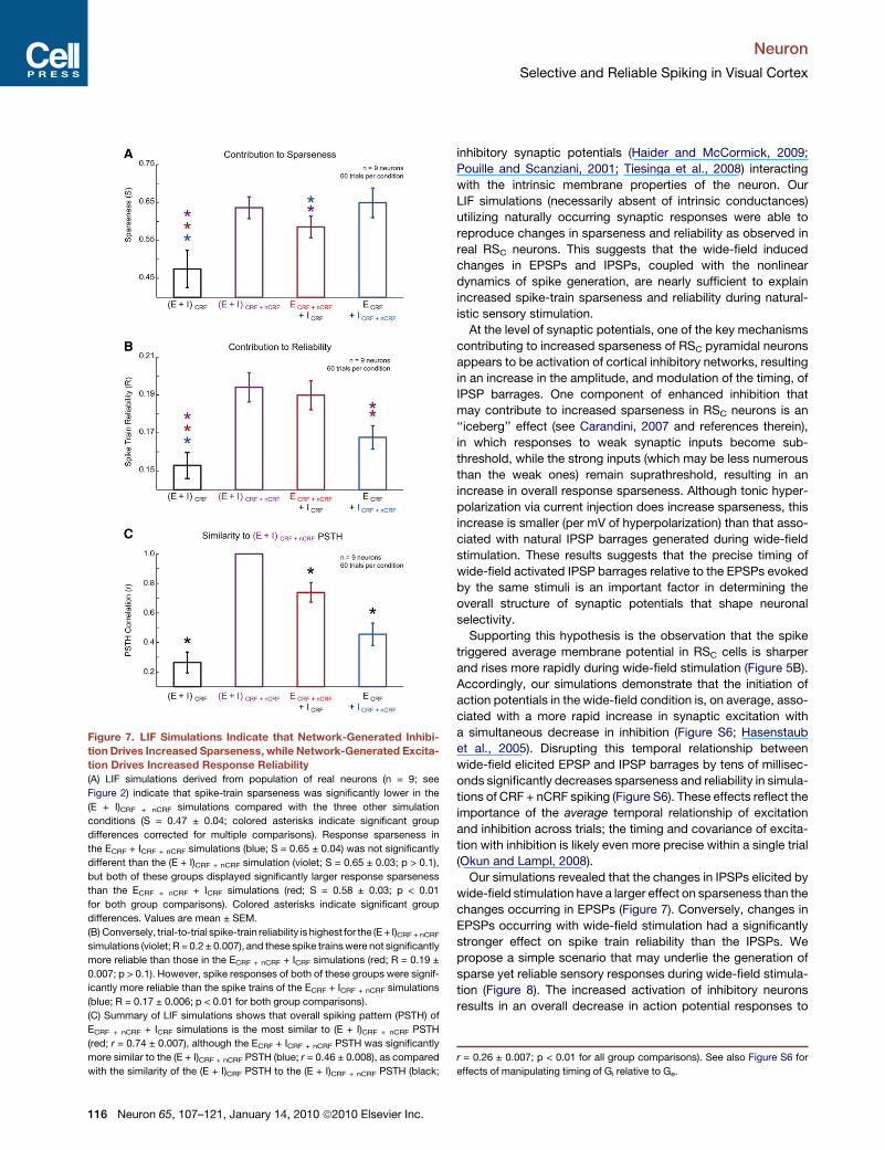
Figure 7. LIF Simulations Indicate that Network-Generated Inhibi-
tion Drives Increased Sparseness, while Network-Generated Excita-
tion Drives Increased Response Reliability
(A) LIF simulations derived from population of real neurons (n = 9; see
Figure 2) indicate that spike-train sparseness was significantly lower in the
(E + I)CRF + nCRF simulations compared with the three other simulation
conditions (S = 0.47 ± 0.04; colored asterisks indicate significant group
differences corrected for multiple comparisons). Response sparseness in
the ECRF + ICRF + nCRF simulations (blue; S = 0.65 ± 0.04) was not significantly
different than the (E + I)CRF + nCRF simulation (violet; S = 0.65 ± 0.03; p > 0.1),
but both of these groups displayed significantly larger response sparseness
than the ECRF + nCRF + ICRF simulations (red; S = 0.58 ± 0.03; p < 0.01
for both group comparisons). Colored asterisks indicate significant group
differences. Values are mean ± SEM.
(B) Conversely, trial-to-trial spike-train reliability is highest for the (E + I)CRF + nCRF
simulations (violet; R = 0.2 ± 0.007), and these spike trains were not significantly
more reliable than those in the ECRF + nCRF + ICRF simulations (red; R = 0.19 ±
0.007; p > 0.1). However, spike responses of both of these groups were signif-
icantly more reliable than the spike trains of the ECRF + ICRF + nCRF simulations
(blue; R = 0.17 ± 0.006; p < 0.01 for both group comparisons).
(C) Summary of LIF simulations shows that overall spiking pattern (PSTH) of
ECRF + nCRF + ICRF simulations is the most similar to (E + I)CRF + nCRF PSTH
(red; r = 0.74 ± 0.007), although the ECRF + ICRF + nCRF PSTH was significantly
more similar to the (E + I)CRF + nCRF PSTH (blue; r = 0.46 ± 0.008), as compared
with the similarity of the (E + I)CRF PSTH to the (E + I)CRF + nCRF PSTH (black;
Neuron
Selective and Reliable Spiking in Visual Cortex
116 Neuron 65, 107–121, January 14, 2010 ª2010 Elsevier Inc.
inhibitory synaptic potentials (Haider and McCormick, 2009;
Pouille and Scanziani, 2001; Tiesinga et al., 2008) interacting
with the intrinsic membrane properties of the neuron. Our
LIF simulations (necessarily absent of intrinsic conductances)
utilizing naturally occurring synaptic responses were able to
reproduce changes in sparseness and reliability as observed in
real RSC neurons. This suggests that the wide-field induced
changes in EPSPs and IPSPs, coupled with the nonlinear
dynamics of spike generation, are nearly sufficient to explain
increased spike-train sparseness and reliability during natural-
istic sensory stimulation.
At the level of synaptic potentials, one of the key mechanisms
contributing to increased sparseness of RSC pyramidal neurons
appears to be activation of cortical inhibitory networks, resulting
in an increase in the amplitude, and modulation of the timing, of
IPSP barrages. One component of enhanced inhibition that
may contribute to increased sparseness in RSC neurons is an
‘‘iceberg’’ effect (see Carandini, 2007 and references therein),
in which responses to weak synaptic inputs become sub-
threshold, while the strong inputs (which may be less numerous
than the weak ones) remain suprathreshold, resulting in an
increase in overall response sparseness. Although tonic hyper-
polarization via current injection does increase sparseness, this
increase is smaller (per mV of hyperpolarization) than that asso-
ciated with natural IPSP barrages generated during wide-field
stimulation. These results suggests that the precise timing of
wide-field activated IPSP barrages relative to the EPSPs evoked
by the same stimuli is an important factor in determining the
overall structure of synaptic potentials that shape neuronal
selectivity.
Supporting this hypothesis is the observation that the spike
triggered average membrane potential in RSC cells is sharper
and rises more rapidly during wide-field stimulation (Figure 5B).
Accordingly, our simulations demonstrate that the initiation of
action potentials in the wide-field condition is, on average, asso-
ciated with a more rapid increase in synaptic excitation with
a simultaneous decrease in inhibition (Figure S6; Hasenstaub
et al., 2005). Disrupting this temporal relationship between
wide-field elicited EPSP and IPSP barrages by tens of millisec-
onds significantly decreases sparseness and reliability in simula-
tions of CRF + nCRF spiking (Figure S6). These effects reflect the
importance of the average temporal relationship of excitation
and inhibition across trials; the timing and covariance of excita-
tion with inhibition is likely even more precise within a single trial
(Okun and Lampl, 2008).
Our simulations revealed that the changes in IPSPs elicited by
wide-field stimulation have a larger effect on sparseness than the
changes occurring in EPSPs (Figure 7). Conversely, changes in
EPSPs occurring with wide-field stimulation had a significantly
stronger effect on spike train reliability than the IPSPs. We
propose a simple scenario that may underlie the generation of
sparse yet reliable sensory responses during wide-field stimula-
tion (Figure 8). The increased activation of inhibitory neurons
results in an overall decrease in action potential responses to
r = 0.26 ± 0.007; p < 0.01 for all group comparisons). See also Figure S6 for
effects of manipulating timing of Gi relative to Ge.
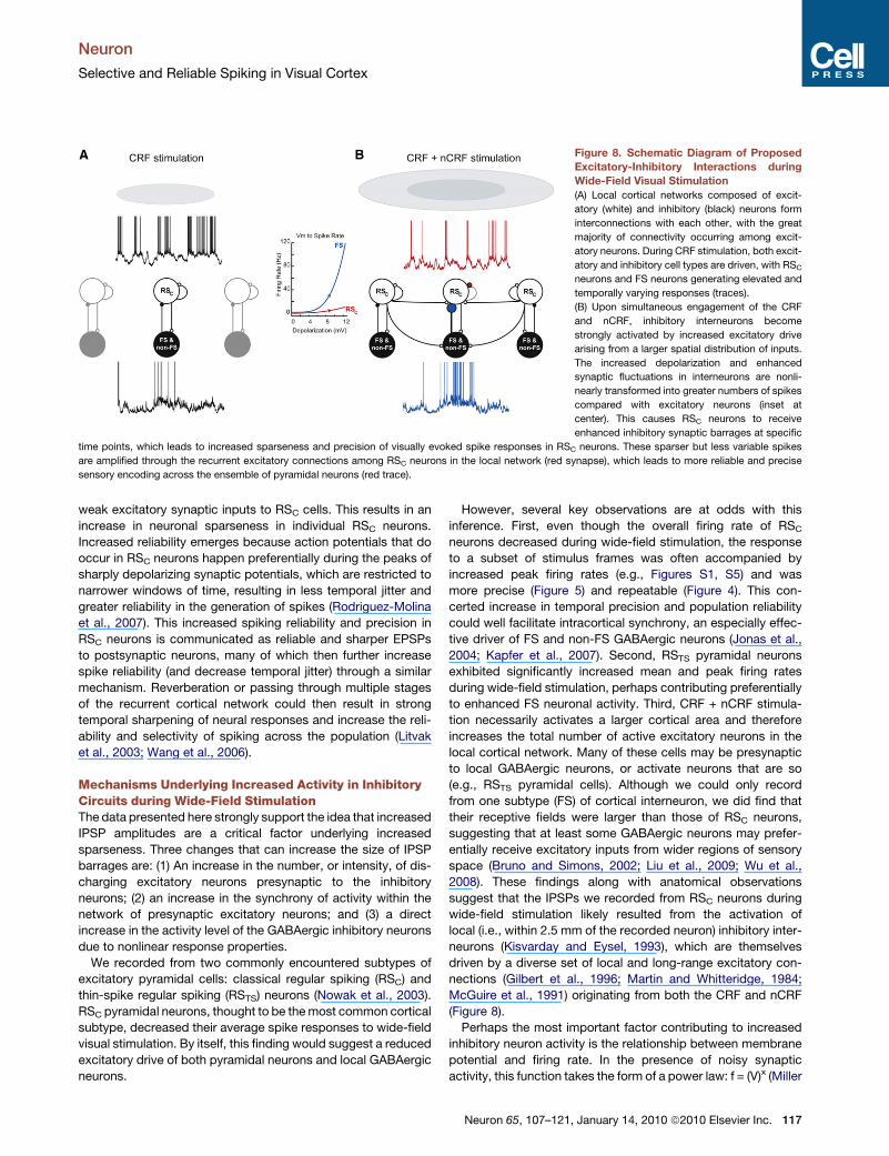
Figure 8. Schematic Diagram of Proposed
Excitatory-Inhibitory Interactions during
Wide-Field Visual Stimulation
(A) Local cortical networks composed of excit-
atory (white) and inhibitory (black) neurons form
interconnections with each other, with the great
majority of connectivity occurring among excit-
atory neurons. During CRF stimulation, both excit-
atory and inhibitory cell types are driven, with RSC
neurons and FS neurons generating elevated and
temporally varying responses (traces).
(B) Upon simultaneous engagement of the CRF
and nCRF, inhibitory interneurons become
strongly activated by increased excitatory drive
arising from a larger spatial distribution of inputs.
The increased depolarization and enhanced
synaptic fluctuations in interneurons are nonli-
nearly transformed into greater numbers of spikes
compared with excitatory neurons (inset at
center). This causes RSC neurons to receive
enhanced inhibitory synaptic barrages at specific
time points, which leads to increased sparseness and precision of visually evoked spike responses in RSC neurons. These sparser but less variable spikes
are amplified through the recurrent excitatory connections among RSC neurons in the local network (red synapse), which leads to more reliable and precise
sensory encoding across the ensemble of pyramidal neurons (red trace).
Neuron
Selective and Reliable Spiking in Visual Cortex
weak excitatory synaptic inputs to RSC cells. This results in an
increase in neuronal sparseness in individual RSC neurons.
Increased reliability emerges because action potentials that do
occur in RSC neurons happen preferentially during the peaks of
sharply depolarizing synaptic potentials, which are restricted to
narrower windows of time, resulting in less temporal jitter and
greater reliability in the generation of spikes (Rodriguez-Molina
et al., 2007). This increased spiking reliability and precision in
RSC neurons is communicated as reliable and sharper EPSPs
to postsynaptic neurons, many of which then further increase
spike reliability (and decrease temporal jitter) through a similar
mechanism. Reverberation or passing through multiple stages
of the recurrent cortical network could then result in strong
temporal sharpening of neural responses and increase the reli-
ability and selectivity of spiking across the population (Litvak
et al., 2003; Wang et al., 2006).
Mechanisms Underlying Increased Activity in InhibitoryCircuits during Wide-Field StimulationThe data presented here strongly support the idea that increased
IPSP amplitudes are a critical factor underlying increased
sparseness. Three changes that can increase the size of IPSP
barrages are: (1) An increase in the number, or intensity, of dis-
charging excitatory neurons presynaptic to the inhibitory
neurons; (2) an increase in the synchrony of activity within the
network of presynaptic excitatory neurons; and (3) a direct
increase in the activity level of the GABAergic inhibitory neurons
due to nonlinear response properties.
We recorded from two commonly encountered subtypes of
excitatory pyramidal cells: classical regular spiking (RSC) and
thin-spike regular spiking (RSTS) neurons (Nowak et al., 2003).
RSC pyramidal neurons, thought to be the most common cortical
subtype, decreased their average spike responses to wide-field
visual stimulation. By itself, this finding would suggest a reduced
excitatory drive of both pyramidal neurons and local GABAergic
neurons.
However, several key observations are at odds with this
inference. First, even though the overall firing rate of RSC
neurons decreased during wide-field stimulation, the response
to a subset of stimulus frames was often accompanied by
increased peak firing rates (e.g., Figures S1, S5) and was
more precise (Figure 5) and repeatable (Figure 4). This con-
certed increase in temporal precision and population reliability
could well facilitate intracortical synchrony, an especially effec-
tive driver of FS and non-FS GABAergic neurons (Jonas et al.,
2004; Kapfer et al., 2007). Second, RSTS pyramidal neurons
exhibited significantly increased mean and peak firing rates
during wide-field stimulation, perhaps contributing preferentially
to enhanced FS neuronal activity. Third, CRF + nCRF stimula-
tion necessarily activates a larger cortical area and therefore
increases the total number of active excitatory neurons in the
local cortical network. Many of these cells may be presynaptic
to local GABAergic neurons, or activate neurons that are so
(e.g., RSTS pyramidal cells). Although we could only record
from one subtype (FS) of cortical interneuron, we did find that
their receptive fields were larger than those of RSC neurons,
suggesting that at least some GABAergic neurons may prefer-
entially receive excitatory inputs from wider regions of sensory
space (Bruno and Simons, 2002; Liu et al., 2009; Wu et al.,
2008). These findings along with anatomical observations
suggest that the IPSPs we recorded from RSC neurons during
wide-field stimulation likely resulted from the activation of
local (i.e., within 2.5 mm of the recorded neuron) inhibitory inter-
neurons (Kisvarday and Eysel, 1993), which are themselves
driven by a diverse set of local and long-range excitatory con-
nections (Gilbert et al., 1996; Martin and Whitteridge, 1984;
McGuire et al., 1991) originating from both the CRF and nCRF
(Figure 8).
Perhaps the most important factor contributing to increased
inhibitory neuron activity is the relationship between membrane
potential and firing rate. In the presence of noisy synaptic
activity, this function takes the form of a power law: f = (V)x (Miller
Neuron 65, 107–121, January 14, 2010 ª2010 Elsevier Inc. 117

Neuron
Selective and Reliable Spiking in Visual Cortex
and Troyer, 2002). We found that FS interneurons exhibit a partic-
ularly steep power law relationship, with a relatively large expo-
nent compared with RSC neurons (Figure S3). As a result, FS
cells are likely to be more sensitive to and strongly activated
by the overall synaptic fluctuations provided by joint stimulation
of the CRF and nCRF (Figure 8).
One should keep in mind that although we only recorded from
FS interneurons, the effects of inhibition on RSC neurons arise
from a broad range of inhibitory neuron subtypes. Indeed, other
subtypes of interneurons also generate highly nonlinear
response enhancement to costimulation of multiple excitatory
pathways (e.g., Martinotti cells; Kapfer et al., 2007; Silberberg
and Markram, 2007). Clarification of the specific contributions
of the many inhibitory neuron subtypes to response selectivity
and reliability requires further investigation.
Implications of Increased Sparseness, Reliability,and PrecisionOne of the main observations of our study was that classical
regular spiking pyramidal neurons (RSC) simultaneously increase
their visual selectivity and reliability, while decreasing overall
firing rate, in response to wide-field visual stimulation. Similarly,
presentation of full field natural scenes in a temporal sequence
that replicates natural eye movements also generated synaptic
and action potential responses that are both reliable and sparse
in cat V1 (Yves Fregnac, Pierre Baudot, Manuel Levy, and Olivier
Marre, 2005, Cosyne meeting, abstract). This finding suggests
that, during natural vision, RSC neurons are more energetically
efficient (Niven and Laughlin, 2008), more selective (Figure 1D)
and reliable (Figure 4C), and consequently more informative per
spike (Vinje and Gallant, 2002). Our observations provide strong
support for the efficient sparse coding hypothesis (Barlow,
1972; Olshausen and Field, 2004; Vinje and Gallant, 2000, 2002).
The moment-to-moment demands of natural behavior depend
upon the reliability of neuronal responsiveness, and insights into
mechanisms that limit response variability are critical toward
understanding the nature of cortical computation. Indeed,
repeated presentations of identical stimuli can elicit highly vari-
able spike responses (Heggelund and Albus, 1978; Shadlen
and Newsome, 1998; Tiesinga et al., 2008). Although some
studies have shown that response reliability can be quite high
after controlling for factors such as eye movements or recording
mainly from input layers (Gur et al., 1997; Kara et al., 2000), in
general, the variability of cortical responses scales approxi-
mately with the mean (but see DeWeese and Zador, 2006;
Maimon and Assad, 2009). These conclusions are based on
recordings likely to be dominated by RSC neurons, because
they are by far the most commonly recorded cells in visual cortex
(but see Chen et al., 2008; Mitchell et al., 2007).
To our knowledge, the data presented here provide the first
mechanistic link between sparse sensory coding and increased
response reliability in visual cortex under naturalistic stimulus
conditions. These findings complement recent findings of sparse
cortical responses in several other species and sensory systems,
under a variety of experimental conditions (Greenberg et al.,
2008; Houweling and Brecht, 2008; Hromadka et al., 2008;
Waters and Helmchen, 2006). Although recent in vitro studies
have shown that response correlation necessarily increases as
118 Neuron 65, 107–121, January 14, 2010 ª2010 Elsevier Inc.
firing rates increase (de la Rocha et al., 2007), we show here
that sparse sensory responses, which exhibit increased effi-
ciency (i.e., fewer total action potentials), can be accompanied
by increased response reliability across trials. These findings
have broad implications for the nature of visual encoding, since
they demonstrate that increased sparseness (reflective of
increased stimulus selectivity) is also associated with higher
trial-to-trial response reliability to a more restricted set of stimuli.
Relationship to Previous Studies and Other SensorySystemsWe have identified a basic operational mode of visual cortical
circuits engaged by spatiotemporally rich wide-field stimulation,
as experienced during natural vision (David and Gallant, 2005;
Mazer and Gallant, 2003). Our work suggests that intracortical
inhibitory networks are critical for the generation of selective
and reliable visual responses during wide-field stimulation.
Specifically, we hypothesize that the intrinsic properties and
anatomical connectivity of FS and other types of interneurons,
as well as the RSTS subclass of pyramidal neuron, enable them
to rapidly integrate activity from horizontal connections and
shape the output of RSC pyramidal cells. We speculate that an
additional function of the excitatory-inhibitory network mecha-
nisms described here is to decorrelate neuronal responses
across cell classes (David and Gallant, 2005; Mazer and Gallant,
2003; Vinje and Gallant, 2000; Wang et al., 2003), ultimately
increasing coding bandwidth and efficiency (El Boustani et al.,
2009; Felsen et al., 2005; Olshausen and Field, 1996; Simoncelli,
2003; Vinje and Gallant, 2002), while simultaneously increasing
signal reliability across the population.
Our results are generally consistent with the large body of
extracellular recording literature indicating an overall suppres-
sion of CRF elicited responses with nCRF costimulation (Ange-
lucci and Bressloff, 2006; Bair et al., 2003; Cavanaugh et al.,
2002b; DeAngelis et al., 1994; Durand et al., 2007; Fitzpatrick,
2000; Jones et al., 2001; Webb et al., 2005), and increased
spiking selectivity driven by surround stimulation (Chen et al.,
2005; Okamoto et al., 2009). Our observations are also consis-
tent with studies showing that stimulus context modulates
both perceptual and neuronal sensitivity (Ito and Gilbert, 1999).
However, our results extend these findings by demonstrating
that wide-field visual stimulation with dynamic, spatiotemporally
rich stimuli drives highly specific network interactions among
distinct neuronal subtypes that ultimately lead to increases in
spike precision, spike reliability, and response sparseness in
the output of RSC pyramidal neurons.
Moreover, the results presented here during naturalistic stim-
ulation strongly implicate intracortical inhibitory potentials, at
least partially originating in FS interneurons, as a critical compo-
nent of these effects (cf. Anderson et al., 2001; Ozeki et al.,
2009). Although complex inhibitory modulations are not entirely
unexpected from a spatiotemporally rich and time-varying stim-
ulus, the network interactions described here among distinct
cortical neuronal subtypes that ultimately increase the reliability
and precision of the network response are not easily predicted
from existing studies of surround suppression. It is entirely
possible that the use of a spatiotemporally rich wide-field
(‘‘naturalistic’’) stimulus set puts the cortex in a more ‘‘transient’’
Neuron
Selective and Reliable Spiking in Visual Cortex
response regime that strongly engages inhibitory circuits
(Ozeki et al., 2009). Such dynamics certainly deserve further
investigation.
Finally, it is likely that the excitatory-inhibitory mechanisms
described here in visual cortex generalize across sensory sys-
tems, particularly under naturalistic stimulus conditions. Recent
studies of the rodent somatosensory system show that cortical
responses exhibit nonlinear modulation with multi-whisker stim-
ulation as compared with single whisker stimulation, and elicit
complex activity patterns across extended regions of barrel
cortex (Jacob et al., 2008). Studies of both mammalian auditory
cortex (Hromadka et al., 2008) and the avian song system (The-
unissen et al., 2001) indicate that neural responses are highly
selective for features present in natural sounds. Song generation
itself may be mediated by ‘‘ultra sparse’’ neuronal activity (Hahn-
loser et al., 2002). Although our results demonstrate the intracel-
lular and network mechanisms underlying enhanced selectivity
to ongoing wide-field visual stimulation, the exact interaction of
the spatiotemporal statistics of natural sensory stimulation with
the response properties of distinct neuronal subtypes merits
further investigation.
EXPERIMENTAL PROCEDURES
Animal Preparation and Electrophysiological Recordings
Briefly, young adult female cats were initially anesthetized with ketamine/xyla-
zine and then maintained on isoflurane vaporized in O2 for the duration of the
experiment. Standard surgical procedures for reducing respiratory and
cardiac pulsations were employed, and all experiments conformed to Yale
University IACUC standards. A craniotomy overlying Area 17 was performed,
the dura was dissected, and metal electrodes and/or beveled sharp glass
micropipettes (55–120 MU), filled with 2 M K+ acetate (for recording action
potentials) or filled with 25–50 mM Qx-314 and 2 M Cs+ acetate (to block
most intrinsic conductances; see Haider et al., 2006; Hasenstaub et al.,
2005) were advanced into the cortex. All action potential responses to natural
movies were recorded with zero current injection; EPSPs were recorded
near �80 mV and IPSPs were recorded near 0 mV.
Visual Stimulation
Neurons were characterized with computer assisted hand-mapping, then
quantitatively mapped using a two-dimensional (2D) sparse noise stimulus
(Jones and Palmer, 1987; Mazer et al., 2002) composed of light and dark
bars at each neuron’s preferred orientation on a linearized 19 inch CRT
(Siemens). Screen background was a uniform gray and bars were 100%
contrast. We designated the least-squares 2D circular fit of the half-maximal
spike response contour (or the half-maximal membrane potential response
in the experiments where spikes were inactivated) as the CRF. We then pre-
sented repeated segments of one of seven different long-duration (5–16 s;
without jumps or cuts) movies for 10–20 trials, and determined which frame
of the movie evoked the greatest number of spikes. We then selected this
frame and 1.5 s flanking each side of this frame to present as the 3 s ‘‘optimal’’
movie, as shown in all figures here. A mask of equal color and luminance with
the background occluded all portions of the movie save for a circle of diameter
equal to and centered over the CRF. This mask was enlarged to expose 3X the
CRF, and these stimuli were designated as the CRF + nCRF stimuli. In both
cases, the pixels presented in the CRF were identical over trials. Presentation
of CRF alone and CRF + nCRF stimuli were randomly interleaved. Movies were
digitized from commercial DVDs (Winged Migration, The Incredibles, Aeon
Flux), converted to grayscale and presented at 25–28 Hz.
Analysis
We quantified neural selectivity by computing lifetime response sparseness
(S), S = 1 � a, where a denotes the activity fraction, a = [Si (ri/n)]2/Si (ri2/n),
and ri is the response to the i-th frame of the movie, and n is the total number
of frames in the movie. Lifetime sparseness (S) is a metric of a single neuron’s
selectivity that is closely related to the kurtosis of the firing rate distribution. For
highly selective neurons, with maximal responses occurring primarily during
a single movie frame, the response distribution across all movie frames will
be highly peaked and S will approach 1.0 (Willmore and Tolhurst, 2001). Life-
time sparseness is different from population sparseness, which measures the
activity profile across an ensemble of neurons.
Real-time analysis and visual stimulation utilized custom written software in
Python (PyPE). All arithmetic means reported and plotted ± standard error of
the mean. All analyses, statistics and plots were generated with built-in and
custom functions in MATLAB (Mathworks). Unless explicitly noted, all p values
(a = 0.01) were calculated with the nonparametric Kruskal-Wallis analysis of
variance, with Tukey-Kramer correction in cases of multiple comparisons.
Throughout the main text and results, when statistical significance is
mentioned, p values and tests are presented in the appropriate portions of
the corresponding figure legends or tables, as relevant.
SUPPLEMENTAL INFORMATION
Supplemental Information includes six figures, three tables, one movie, and
Supplemental Experimental Procedures and can be found with this article
online at doi:10.1016/j.neuron.2009.12.005.
ACKNOWLEDGMENTS
The authors thank Flavio Frohlich and Kristy Sundberg for help during exper-
iments, and Carlos Maureira and Lionel Nowak for helpful suggestions. B.H.,
A.D., M.R.K., J.T., J.A.M., and D.A.M. performed experiments; B.H., M.R.K.,
J.T., and J.A.M., analyzed data; B.H., A.D., and M.R.K. performed histology;
Y.Y., B.H., and D.A.M. performed simulations; B.H., J.A.M. and D.A.M. wrote
the manuscript.
Accepted: November 25, 2009
Published: January 13, 2010
REFERENCES
Anderson, J.S., Lampl, I., Gillespie, D.C., and Ferster, D. (2000). The contribu-
tion of noise to contrast invariance of orientation tuning in cat visual cortex.
Science 290, 1968–1972.
Anderson, J.S., Lampl, I., Gillespie, D.C., and Ferster, D. (2001). Membrane
potential and conductance changes underlying length tuning of cells in cat
primary visual cortex. J. Neurosci. 21, 2104–2112.
Angelucci, A., and Bressloff, P.C. (2006). Contribution of feedforward, lateral
and feedback connections to the classical receptive field center and extra-
classical receptive field surround of primate V1 neurons. Prog. Brain Res.
154, 93–120.
Azouz, R., and Gray, C.M. (2003). Adaptive coincidence detection and
dynamic gain control in visual cortical neurons in vivo. Neuron 37, 513–523.
Azouz, R., Gray, C.M., Nowak, L.G., and McCormick, D.A. (1997). Physiolog-
ical properties of inhibitory interneurons in cat striate cortex. Cereb. Cortex 7,
534–545.
Bair, W., Cavanaugh, J.R., and Movshon, J.A. (2003). Time course and time-
distance relationships for surround suppression in macaque V1 neurons.
J. Neurosci. 23, 7690–7701.
Barlow, H.B. (1972). Single units and sensation: a neuron doctrine for percep-
tual psychology? Perception 1, 371–394.
Bruno, R.M., and Sakmann, B. (2006). Cortex is driven by weak but synchro-
nously active thalamocortical synapses. Science 312, 1622–1627.
Bruno, R.M., and Simons, D.J. (2002). Feedforward mechanisms of excitatory
and inhibitory cortical receptive fields. J. Neurosci. 22, 10966–10975.
Carandini, M. (2007). Melting the iceberg: contrast invariance in visual cortex.
Neuron 54, 11–13.
Neuron 65, 107–121, January 14, 2010 ª2010 Elsevier Inc. 119
Neuron
Selective and Reliable Spiking in Visual Cortex
Carandini, M., Demb, J.B., Mante, V., Tolhurst, D.J., Dan, Y., Olshausen, B.A.,
Gallant, J.L., and Rust, N.C. (2005). Do we know what the early visual system
does? J. Neurosci. 25, 10577–10597.
Cavanaugh, J.R., Bair, W., and Movshon, J.A. (2002a). Nature and interaction
of signals from the receptive field center and surround in macaque V1 neurons.
J. Neurophysiol. 88, 2530–2546.
Cavanaugh, J.R., Bair, W., and Movshon, J.A. (2002b). Selectivity and spatial
distribution of signals from the receptive field surround in macaque V1
neurons. J. Neurophysiol. 88, 2547–2556.
Chen, G., Dan, Y., and Li, C.Y. (2005). Stimulation of non-classical receptive
field enhances orientation selectivity in the cat. J. Physiol. 564, 233–243.
Chen, Y., Martinez-Conde, S., Macknik, S.L., Bereshpolova, Y., Swadlow,
H.A., and Alonso, J.M. (2008). Task difficulty modulates the activity of specific
neuronal populations in primary visual cortex. Nat. Neurosci. 11, 974–982.
Contreras, D., and Palmer, L. (2003). Response to contrast of electrophysio-
logically defined cell classes in primary visual cortex. J. Neurosci. 23, 6936–
6945.
David, S.V., and Gallant, J.L. (2005). Predicting neuronal responses during
natural vision. Network 16, 239–260.
de la Rocha, J., Doiron, B., Shea-Brown, E., Josic, K., and Reyes, A. (2007).
Correlation between neural spike trains increases with firing rate. Nature
448, 802–806.
DeAngelis, G.C., Ohzawa, I., and Freeman, R.D. (1993). Spatiotemporal orga-
nization of simple-cell receptive fields in the cat’s striate cortex. I. General
characteristics and postnatal development. J. Neurophysiol. 69, 1091–1117.
DeAngelis, G.C., Freeman, R.D., and Ohzawa, I. (1994). Length and width
tuning of neurons in the cat’s primary visual cortex. J. Neurophysiol. 71,
347–374.
Desbordes, G., Jin, J., Weng, C., Lesica, N.A., Stanley, G.B., and Alonso, J.M.
(2008). Timing precision in population coding of natural scenes in the early
visual system. PLoS Biol. 6, e324.
DeWeese, M.R., and Zador, A.M. (2006). Non-Gaussian membrane potential
dynamics imply sparse, synchronous activity in auditory cortex. J. Neurosci.
26, 12206–12218.
Durand, S., Freeman, T.C., and Carandini, M. (2007). Temporal properties of
surround suppression in cat primary visual cortex. Vis. Neurosci. 24, 679–690.
El Boustani, S., Marre, O., Behuret, S., Baudot, P., Yger, P., Bal, T., Destexhe,
A., and Fregnac, Y. (2009). Network-state modulation of power-law frequency-
scaling in visual cortical neurons. PLoS Comput. Biol. 5, e1000519.
Felsen, G., Touryan, J., Han, F., and Dan, Y. (2005). Cortical sensitivity to visual
features in natural scenes. PLoS Biol. 3, e342.
Fitzpatrick, D. (2000). Seeing beyond the receptive field in primary visual
cortex. Curr. Opin. Neurobiol. 10, 438–443.
Gabernet, L., Jadhav, S.P., Feldman, D.E., Carandini, M., and Scanziani, M.
(2005). Somatosensory integration controlled by dynamic thalamocortical
feed-forward inhibition. Neuron 48, 315–327.
Gilbert, C.D., Das, A., Ito, M., Kapadia, M., and Westheimer, G. (1996). Spatial
integration and cortical dynamics. Proc. Natl. Acad. Sci. USA 93, 615–622.
Greenberg, D.S., Houweling, A.R., and Kerr, J.N. (2008). Population imaging of
ongoing neuronal activity in the visual cortex of awake rats. Nat. Neurosci. 11,
749–751.
Gur, M., Beylin, A., and Snodderly, D.M. (1997). Response variability of
neurons in primary visual cortex (V1) of alert monkeys. J. Neurosci. 17,
2914–2920.
Hahnloser, R.H., Kozhevnikov, A.A., and Fee, M.S. (2002). An ultra-sparse
code underlies the generation of neural sequences in a songbird. Nature
419, 65–70.
Haider, B., and McCormick, D.A. (2009). Rapid neocortical dynamics: cellular
and network mechanisms. Neuron 62, 171–189.
Haider, B., Duque, A., Hasenstaub, A.R., and McCormick, D.A. (2006).
Neocortical network activity in vivo is generated through a dynamic balance
of excitation and inhibition. J. Neurosci. 26, 4535–4545.
120 Neuron 65, 107–121, January 14, 2010 ª2010 Elsevier Inc.
Hasenstaub, A., Shu, Y., Haider, B., Kraushaar, U., Duque, A., and McCor-
mick, D.A. (2005). Inhibitory postsynaptic potentials carry synchronized
frequency information in active cortical networks. Neuron 47, 423–435.
Heggelund, P., and Albus, K. (1978). Response variability and orientation
discrimination of single cells in striate cortex of cat. Exp. Brain Res. 32,
197–211.
Higley, M.J., and Contreras, D. (2006). Balanced excitation and inhibition
determine spike timing during frequency adaptation. J. Neurosci. 26, 448–457.
Houweling, A.R., and Brecht, M. (2008). Behavioural report of single neuron
stimulation in somatosensory cortex. Nature 451, 65–68.
Hromadka, T., Deweese, M.R., and Zador, A.M. (2008). Sparse representation
of sounds in the unanesthetized auditory cortex. PLoS Biol. 6, e16.
Hubel, D.H., and Wiesel, T.N. (1962). Receptive fields, binocular interaction
and functional architecture in the cat’s visual cortex. J. Physiol. 160, 106–154.
Ito, M., and Gilbert, C.D. (1999). Attention modulates contextual influences in
the primary visual cortex of alert monkeys. Neuron 22, 593–604.
Jacob, V., Le Cam, J., Ego-Stengel, V., and Shulz, D.E. (2008). Emergent prop-
erties of tactile scenes selectively activate barrel cortex neurons. Neuron 60,
1112–1125.
Jonas, P., Bischofberger, J., Fricker, D., and Miles, R. (2004). Interneuron
Diversity series: Fast in, fast out–temporal and spatial signal processing in
hippocampal interneurons. Trends Neurosci. 27, 30–40.
Jones, J.P., and Palmer, L.A. (1987). The two-dimensional spatial structure of
simple receptive fields in cat striate cortex. J. Neurophysiol. 58, 1187–1211.
Jones, H.E., Grieve, K.L., Wang, W., and Sillito, A.M. (2001). Surround
suppression in primate V1. J. Neurophysiol. 86, 2011–2028.
Kapadia, M.K., Ito, M., Gilbert, C.D., and Westheimer, G. (1995). Improvement
in visual sensitivity by changes in local context: parallel studies in human
observers and in V1 of alert monkeys. Neuron 15, 843–856.
Kapfer, C., Glickfeld, L.L., Atallah, B.V., and Scanziani, M. (2007). Supralinear
increase of recurrent inhibition during sparse activity in the somatosensory
cortex. Nat. Neurosci. 10, 743–753.
Kara, P., Reinagel, P., and Reid, R.C. (2000). Low response variability in simul-
taneously recorded retinal, thalamic, and cortical neurons. Neuron 27,
635–646.
Kisvarday, Z.F., and Eysel, U.T. (1993). Functional and structural topography
of horizontal inhibitory connections in cat visual cortex. Eur. J. Neurosci. 5,
1558–1572.
Lehky, S.R., Sejnowski, T.J., and Desimone, R. (2005). Selectivity and sparse-
ness in the responses of striate complex cells. Vision Res. 45, 57–73.
Litvak, V., Sompolinsky, H., Segev, I., and Abeles, M. (2003). On the transmis-
sion of rate code in long feedforward networks with excitatory-inhibitory
balance. J. Neurosci. 23, 3006–3015.
Liu, B.H., Li, P., Li, Y.T., Sun, Y.J., Yanagawa, Y., Obata, K., Zhang, L.I., and
Tao, H.W. (2009). Visual receptive field structure of cortical inhibitory neurons
revealed by two-photon imaging guided recording. J. Neurosci. 29, 10520–
10532.
Maimon, G., and Assad, J.A. (2009). Beyond Poisson: increased spike-time
regularity across primate parietal cortex. Neuron 62, 426–440.
Martin, K.A., and Whitteridge, D. (1984). Form, function and intracortical
projections of spiny neurones in the striate visual cortex of the cat. J. Physiol.
353, 463–504.
Mazer, J.A., and Gallant, J.L. (2003). Goal-related activity in V4 during free
viewing visual search. Evidence for a ventral stream visual salience map.
Neuron 40, 1241–1250.
Mazer, J.A., Vinje, W.E., McDermott, J., Schiller, P.H., and Gallant, J.L. (2002).
Spatial frequency and orientation tuning dynamics in area V1. Proc. Natl. Acad.
Sci. USA 99, 1645–1650.
McGuire, B.A., Gilbert, C.D., Rivlin, P.K., and Wiesel, T.N. (1991). Targets of
horizontal connections in macaque primary visual cortex. J. Comp. Neurol.
305, 370–392.

Neuron
Selective and Reliable Spiking in Visual Cortex
Miller, K.D., and Troyer, T.W. (2002). Neural noise can explain expansive,
power-law nonlinearities in neural response functions. J. Neurophysiol. 87,
653–659.
Mitchell, J.F., Sundberg, K.A., and Reynolds, J.H. (2007). Differential attention-
dependent response modulation across cell classes in macaque visual area
V4. Neuron 55, 131–141.
Monier, C., Chavane, F., Baudot, P., Graham, L.J., and Fregnac, Y. (2003).
Orientation and direction selectivity of synaptic inputs in visual cortical
neurons: a diversity of combinations produces spike tuning. Neuron 37,
663–680.
Movshon, J.A., Thompson, I.D., and Tolhurst, D.J. (1978). Spatial summation
in the receptive fields of simple cells in the cat’s striate cortex. J. Physiol.
283, 53–77.
Niven, J.E., and Laughlin, S.B. (2008). Energy limitation as a selective pressure
on the evolution of sensory systems. J. Exp. Biol. 211, 1792–1804.
Nowak, L.G., Sanchez-Vives, M.V., and McCormick, D.A. (1997). Influence of
low and high frequency inputs on spike timing in visual cortical neurons. Cereb.
Cortex 7, 487–501.
Nowak, L.G., Azouz, R., Sanchez-Vives, M.V., Gray, C.M., and McCormick,
D.A. (2003). Electrophysiological classes of cat primary visual cortical neurons
in vivo as revealed by quantitative analyses. J. Neurophysiol. 89, 1541–1566.
Okamoto, M., Naito, T., Sadakane, O., Osaki, H., and Sato, H. (2009). Surround
suppression sharpens orientation tuning in the cat primary visual cortex. Eur. J.
Neurosci. 29, 1035–1046.
Okun, M., and Lampl, I. (2008). Instantaneous correlation of excitation and
inhibition during ongoing and sensory-evoked activities. Nat. Neurosci. 11,
535–537.
Olshausen, B.A., and Field, D.J. (1996). Natural image statistics and efficient
coding. Network 7, 333–339.
Olshausen, B.A., and Field, D.J. (2004). Sparse coding of sensory inputs. Curr.
Opin. Neurobiol. 14, 481–487.
Ozeki, H., Finn, I.M., Schaffer, E.S., Miller, K.D., and Ferster, D. (2009). Inhib-
itory stabilization of the cortical network underlies visual surround suppres-
sion. Neuron 62, 578–592.
Pospischil, M., Piwkowska, Z., Rudolph, M., Bal, T., and Destexhe, A. (2007).
Calculating event-triggered average synaptic conductances from the
membrane potential. J. Neurophysiol. 97, 2544–2552.
Pouille, F., and Scanziani, M. (2001). Enforcement of temporal fidelity in pyra-
midal cells by somatic feed-forward inhibition. Science 293, 1159–1163.
Rodriguez-Molina, V.M., Aertsen, A., and Heck, D.H. (2007). Spike timing and
reliability in cortical pyramidal neurons: effects of EPSC kinetics, input
synchronization and background noise on spike timing. PLoS ONE 2, e319.
Rolls, E.T., and Tovee, M.J. (1995). Sparseness of the neuronal representation
of stimuli in the primate temporal visual cortex. J. Neurophysiol. 73, 713–726.
Shadlen, M.N., and Newsome, W.T. (1998). The variable discharge of cortical
neurons: implications for connectivity, computation, and information coding.
J. Neurosci. 18, 3870–3896.
Silberberg, G., and Markram, H. (2007). Disynaptic inhibition between neocor-
tical pyramidal cells mediated by Martinotti cells. Neuron 53, 735–746.
Simoncelli, E.P. (2003). Vision and the statistics of the visual environment.
Curr. Opin. Neurobiol. 13, 144–149.
Stein, R.B., Gossen, E.R., and Jones, K.E. (2005). Neuronal variability: noise or
part of the signal? Nat. Rev. Neurosci. 6, 389–397.
Swadlow, H.A. (2003). Fast-spike interneurons and feedforward inhibition in
awake sensory neocortex. Cereb. Cortex 13, 25–32.
Theunissen, F.E., David, S.V., Singh, N.C., Hsu, A., Vinje, W.E., and Gallant,
J.L. (2001). Estimating spatio-temporal receptive fields of auditory and visual
neurons from their responses to natural stimuli. Network 12, 289–316.
Tiesinga, P., Fellous, J.M., and Sejnowski, T.J. (2008). Regulation of spike
timing in visual cortical circuits. Nat. Rev. Neurosci. 9, 97–107.
Tolhurst, D.J., Smyth, D., and Thompson, I.D. (2009). The sparseness of
neuronal responses in ferret primary visual cortex. J. Neurosci. 29, 2355–2370.
Vinje, W.E., and Gallant, J.L. (2000). Sparse coding and decorrelation in
primary visual cortex during natural vision. Science 287, 1273–1276.
Vinje, W.E., and Gallant, J.L. (2002). Natural stimulation of the nonclassical
receptive field increases information transmission efficiency in V1. J. Neurosci.
22, 2904–2915.
Wang, X.J., Liu, Y., Sanchez-Vives, M.V., and McCormick, D.A. (2003). Adap-
tation and temporal decorrelation by single neurons in the primary visual
cortex. J. Neurophysiol. 89, 3279–3293.
Wang, S., Wang, W., and Liu, F. (2006). Propagation of firing rate in a feed-
forward neuronal network. Phys. Rev. Lett. 96, 018103.
Waters, J., and Helmchen, F. (2006). Background synaptic activity is sparse in
neocortex. J. Neurosci. 26, 8267–8277.
Webb, B.S., Dhruv, N.T., Solomon, S.G., Tailby, C., and Lennie, P. (2005). Early
and late mechanisms of surround suppression in striate cortex of macaque.
J. Neurosci. 25, 11666–11675.
Wehr, M., and Zador, A.M. (2003). Balanced inhibition underlies tuning and
sharpens spike timing in auditory cortex. Nature 426, 442–446.
Willmore, B., and Tolhurst, D.J. (2001). Characterizing the sparseness of neural
codes. Network 12, 255–270.
Wu, G.K., Arbuckle, R., Liu, B.H., Tao, H.W., and Zhang, L.I. (2008). Lateral
sharpening of cortical frequency tuning by approximately balanced inhibition.
Neuron 58, 132–143.
Yao, H., Shi, L., Han, F., Gao, H., and Dan, Y. (2007). Rapid learning in cortical
coding of visual scenes. Nat. Neurosci. 10, 772–778.
Yen, S.C., Baker, J., and Gray, C.M. (2007). Heterogeneity in the responses of
adjacent neurons to natural stimuli in cat striate cortex. J. Neurophysiol. 97,
1326–1341.
Yoshimura, Y., and Callaway, E.M. (2005). Fine-scale specificity of cortical
networks depends on inhibitory cell type and connectivity. Nat. Neurosci. 8,
1552–1559.
Neuron 65, 107–121, January 14, 2010 ª2010 Elsevier Inc. 121
Copyright © 2022 FDOKUMEN