surgical management of fracture both bones forearm in adults ...
132
i “SURGICAL MANAGEMENT OF FRACTURE BOTH BONES FOREARM IN ADULTS WITH LOCKING COMPRESSION PLATE” BY Dr. SANDEEP M.M.R. M.B.B.S., Dissertation submitted to the Rajiv Gandhi University of Health Sciences, Karnataka, Bangalore. In Partial fulfillment of the requirements for the degree of MASTER OF SURGERY IN ORTHOPAEDICS Under the guidance of Dr. R. RAMESH M.S., (Ortho) PROFESSOR DEPARTMENT OF ORTHOPAEDICS J.J.M. MEDICAL COLLEGE DAVANGERE – 577 004. 2012
-
Upload
khangminh22 -
Category
Documents
-
view
0 -
download
0
Transcript of surgical management of fracture both bones forearm in adults ...

i
“SURGICAL MANAGEMENT OF FRACTURE BOTH BONES FOREARM IN ADULTS WITH
LOCKING COMPRESSION PLATE”
BY Dr. SANDEEP M.M.R.
M.B.B.S.,
Dissertation submitted to the Rajiv Gandhi University of Health Sciences, Karnataka, Bangalore.
In Partial fulfillment of the requirements for the degree of
MASTER OF SURGERY IN
ORTHOPAEDICS
Under the guidance of
Dr. R. RAMESH M.S., (Ortho) PROFESSOR
DEPARTMENT OF ORTHOPAEDICS J.J.M. MEDICAL COLLEGE
DAVANGERE – 577 004.
2012

ii
RAJIV GANDHI UNIVERSITY OF HEALTH SCIENCES, KARNATAKA
DECLARATION BY THE CANDIDATE
I hereby declare that this dissertation/thesis entitled “SURGICAL
MANAGEMENT OF FRACTURE BOTH BONES FOREARM IN ADULTS
WITH LOCKING COMPRESSION PLATE” is a bonafide and genuine research
work carried out by me under the guidance of Dr. R. RAMESH M.S.(Ortho),
Professor, Department of Orthopaedics, J.J.M. Medical College, Davangere.

iii
CERTIFICATE BY THE GUIDE
This is to certify that this dissertation entitled “SURGICAL
MANAGEMENT OF FRACTURE BOTH BONES FOREARM IN ADULTS
WITH LOCKING COMPRESSION PLATE” is a bonafide research work done by
Dr.SANDEEP M.M.R in partial fulfillment of the requirement for the degree of
M.S. (Orthopaedics).

iv
ENDORSEMENT BY THE HOD,
THE PRINCIPAL/HEAD OF THE INSTITUTION
This is to certify that this dissertation entitled “SURGICAL
MANAGEMENT OF FRACTURE BOTH BONES FOREARM IN ADULTS
WITH LOCKING COMPRESSION PLATE” is a bonafide research work done
by Dr. SANDEEP .M.M.R. under the guidance of Dr. R. RAMESH M.S.(Ortho),
Professor, Department of Orthopaedics, J.J.M. Medical College, Davangere.

v
COPYRIGHT
Declaration by the Candidate
I hereby declare that the Rajiv Gandhi University of Health Sciences,
Karnataka shall have the rights to preserve, use and disseminate this dissertation /
thesis in print or electronic format for academic / research purpose.
© Rajiv Gandhi University of Health Sciences, Karnataka.

vi
ACKNOWLEDGEMENT
Ever since I began this dissertation, innumerable people have participated by
contributing their time, energy and expertise.
To each of them and to others I may have omitted to mention through
oversight, I owe a debt of gratitude for the help and encouragement.
I greatfully acknowledge the very valuable and supportive guidance of
Dr. R. RAMESH Professor, Department of Orthopaedics, J.J.M. Medical College,
Davangere, whose unstinted support has been indispensable for the writing of this
dissertation. I appreciate the encouragement for stimulating me to undertake this
dissertation.
I express my special thanks to Dr.G.NAGARAJ M.S., D.Ortho, Professor and
H.O.D., Department of Orthopaedics, J.J.M. Medical College, Davangere, for
equipping me with a sound knowledge of orthopaedics which has enabled me to
successfully complete this dissertation.
I am deeply indebted to all my teachers who have patiently imparted the
benefits of their research and clinical experience to me. My sincere thanks to
Dr.D.S.SATYENDRA RAO, Dr.M.R.JAYAPRAKASH, Dr.G.C.BASAVARAJ,
Dr.D.MAHESHWARAPPA, Dr.T.M.RAVINATH, Professor and Unit Heads and
Dr.PRABHU.B.BASAVANAGOUDA, Dr.KARIGOWDAR, Dr.NAGABHUSHA
N, Dr.PALAKSHAPPA, Dr.J.MANJUNATH, Dr.VIJAYAKUMAR KULAMBI,
Dr.MALLIKARJUNA REDDY, Dr.RUDRAMUNI Professors, Department of
Orthopaedics, J.J.M .Medical College, Davangere.
I am also thankful to Dr.SUBODH SHETTY, Dr.PRASANNA ANABERU
and Dr. J.RAGHUKUMAR Readers, Department of Orthopaedics and for their
whole hearted support and encouragement.
I am especially appreciative and indebted to Dr. J SRINATH, Assistant
Professor, Department of Orthopaedics, J.J.M .Medical College, Davangere.

vii
I owe a lot to my very encouraging and cheerful batchmates ASHOK
SAMPAGAR, CHETAN UMRANI, PRAMOD, NAVEEN, DAN JACOB,
PRABHUDEV , VAMSHI, SUSHEEL and BHARATH, two years with them have
been really amazing and their support has been inspiring despite the work pressures
we were facing together.I am bereft of words when it comes to thanking my seniors
MADHUSUDHAN, VEERBHADRA, IMTIAZ, VARUN and Late DIVAKAR for
all their help towards my dissertation work. I also thank my juniors for all their
support in carrying out my dissertation work. Thanks to you all for your love and
motivation.
I am extremely grateful to Dr. H.R. CHANDRASHEKAR M.D., Principal,
and Dr.H.GURUPADAPPA M.D., Director of Post-graduate studies & research,
J.J.M. Medical College, Davangere, for their valuable help and co-operation
I express my sincere thanks to Superintendent and Resident Medical Officers
of Chigateri General Hospital and Bapuji Hospital for their valuable help and co-
operation.
Each of the non-medical staff of the Department of Orthopaedics, have made
significant contribution to this work, for which I express my humble gratitude.
I am eternally grateful to my PARENTS Mr. RAMASWAMY M.M., Mrs.
JAYALAKSHMI R. my brother Mr. BUBESH R.R., and sister in law Mrs. PRIYA
my sister Mrs. SATHYAKALA D and brother in law Dr. DHANABHALAN for
their love, perseverance and understanding. They have been my pillar of support and
encouraged me in every step. This dissertation would have not been possible without
their blessings.
Very special thanks to my best buddy, SUHASINI K.A who has always been
around ever since and continues to be. Thanks for being considerate and all the help
towards my dissertation requirements.
My sincere thanks to all my post-graduate colleagues, and my friends for their
whole hearted support.
I am thankful to Mr.P.S.MAHESH, Chief Librarian, J.J.M. Medical College,
Davangere, for his co-operation. I also thank Mr. VIJYA KUMAR Physiotherapist.

viii
The value of this book is immeasurably enhanced by the meticulous data
processing work done by Mr. SANJEEV KUMAR G.P. of M/S. GUNDAL
Compu-Center.
Finally I thank all my patients who formed the back bone of this study without
whom this study would not have been possible.

ix
LIST OF ABBREVATIONS USED
AO - Arbeitsgemeinschaft fur Osteosynthese Fragen
ASIF - Association for the study of internal fixation
BP - Blood Pressure
BT/CT - Bleeding Time / Clotting Time
DCP - Dynamic Compression Plate
Deg - Degree
DVT - Deep Vein Thrombosis
ECG - El ectrocardi ography
FBS/PPBS - Fasting Blood Sugar / Postprandial Blood Sugar
Hb - Haemoglobin
HbsAg - Hepatitis B Virus Surface Antigen
HIV - Human Immunodeficiency Virus
IV - Intravenous
LCDCP - Limited Contact Dynamic Compression Plate
Lat - Lateral
M: F Ratio - Male: Female Ratio
No. Patients - Number of patients
ORIF - Open Reduction and Internal Fixation
OTA - Orthopedic Trauma Association
RR - Respiratory Rate
RTA - Road Traffic Accident
Wks - Weeks
# - Fracture
LCP - Locking compression plate

x
ABSTRACT
BACKGROUND AND OBJECTIVE:
The fractures of both bones forearm are one of the commonest fractures found and can be treated by different methods. The accepted management for fractures of both bones forearm is open reduction and internal fixation using compression plating. The present study is undertaken to verify the claims made by the authors of the new design of the plate (LCP) and to learn the techniques, advantages and complication of the new method of internal fixation of forearm fractures.
MATERIALS AND METHODS:
It is a prospective study which was carried out from October 2009 to September 2011 in Bapuji Hospital and Chigateri General Hospital attached to J.J.M. Medical College, Davangere. In this study period 20 cases of fracture both bones of forearm were treated by open reduction and internal fixation using Locking Compression Plate.
RESULTS:
In our series, majority of the patients were males, middle aged, with road traffic accidents being the commonest mode of injury, involving middle third. Transverse or short oblique fractures were most common. The fractures united in all 20 patients. Excellent or full range of mobility of elbow and wrist joints were present in 17 patients(85%), 3(15%) patients having good range of movements.
INTERPRETATION /CONCLUSION
The LCP of forearm fractures produce excellent results, the advantage being early mobilization, early union but the complication, duration of surgery and surgical techniques remains unchanged.
Mesh Key Words: shaft of both bones of forearm fractures, LCP, Open reduction and internal fixation.

xi
TABLE OF CONTENTS
PAGE NO
1. INTRODUCTION 01
2. OBJECTIVES 03
3. REVIEW OF LITERATURE 04
4. METHODOLOGY 57
5. RESULTS 65
6. DISCUSSION 83
7. CONCLUSION 91
8. SUMMARY 93
9. BIBLIOGRAPHY 94
10. ANNEXURES
ANNEXURE-I : PROFORMA 100
ANNEXURE-II : CONSENT FORM 103
ANNEXURE-III : MASTER CHART 104

xii
LIST OF TABLES
TABLE NO. TOPIC PAGE NO.
1 ARTERIES OF FOREARM 20
2 MUSCLES OF ANTERIOR COMPARTMENT OF FOREARM 23
3 MUSCLES OF THE LATERAL COMPARTMENT OF FOREARM 26
4 MUSCLES OF POSTERIOR COMPARTMENT OF FOREARM 27
5 AGE DISTRIBUTION 65
6 SEX DISTRIBUTION 65
7 SIDE AFFECTED 67
8 MODE OF INJURY 67
9 LEVEL OF FRACTURE 68
10 TYPE OF FRACTURE 68
11 ASSOCIATED INJURIES 70
12 DURATION OF FRACTURE UNION 71
13 CRITERIA FOR EVALUATION OF RESULTS 71
14 RESULTS 73
15 COMPLICATIONS 73
16 – 25 TABLES COMPARING THE RESULT OF PRESENT STUDY WITH PREVIOUS STUDIES 84 – 90
26 MASTER CHART 104

xiii
LIST OF GRAPHS
SL.NO. GRAPHS AND PIE CHARTS PAGE
1. AGE DISTRIBUTION 66
2. SEX DISTRIBUTION 66
3. MODE OF INJURY 67
4. LEVEL OF INJURY 69
5. TYPE OF FRACTURE 69
6. TIME OF UNION 72
7. RESULTS 72

xiv
LIST OF FIGURES
FIGURE NO.
FIGURES PAGE NO.
1 MEASUREMENT OF RADIAL BOW 16
2 ANATOMY OF FOREARM BONES 19
3 FOREARM MUSCLES– ANTERIOR
COMPARTMENT
25
4 FOREARM MUSCLES– POSTERIOR
COMPARTMENT
29
5 DISPLACEMENTS 31
6 AO/OTA CLASSIFICATION OF RADIUS AND ULNA DIAPHYSEAL FRACTURES
34
7 LCP MECHANISM 49 – 51
8 LCP IN MIPPO TECHNIQUE 53
LIST OF PHOTOGRAPHS
SL. NO. PAGE NO.
1 OPERATIVE PHOTOGRAPHS 63 - 64
2 CLINICAL AND RADIOLOGICAL PHOTOGRAPHS 75 - 82

Introduction

1
INTRODUCTION
Forearm bone fractures are commonly encountered in today’s industrial
era. Various treatment modalities were introduced from time to time and each
of them had some edge over the previous one. Continuing this process of
revolution and based on many years of experience with compression plating
and promising results obtained with so called internal fixation, an implant
system has been developed which combines the two treatment modalities.
Despite the combination of these different treatment techniques no
compromises were made with regard to application as a compression plate or as
a bridging device in the form of an internal fixation. LCP (Locking
compression plate) is a product of these combinations and is in line with the
latest plating techniques, the aim of which is to achieve the smallest surgical
incision and to preserve blood supply to the bone and adjacent soft tissues and
stability at the fracture site.
LCP has got features of both LC-DCP and a PC-Fix as it uses screw
heads that are conically threaded on the undersurface and create an angular
stable plate screw device.
This type of plate fixation relies on the threaded plate-screw interface to
lock the bone fragments in position and do not require friction between the
plate and bone as in conventional plating. The present study was undertaken to
evaluate the use of LCPs in fractures of forearm bones.

2
The functional outcome was certified using "Anderson et al, scoring
system". The variables taken into consideration were –
a. Union of the fracture,
b. Range of elbow and wrist movements4.
In conclusion, open reduction and internal fixation helps in perfect
fracture reduction, rigid fixation, better bone healing and early mobilization,
the normal functions of the hand can be achieved at the earliest.

Objectives

3
OBJECTIVES
• To study the functional outcome of treating diaphyseal fractures both
bones forearm with Locking Compression Plates.
• To study the duration of union with LCP.
• To study the complications of Locking Compression Plates.

Review of Literature

4
REVIEW OF LITERATURE
Fractures have been recognized and treated as long as recorded in
history. History of fracture and its knowledge dates back to Egyptian
Mummies of 2700 B. C5.
In olden days, surgeons merely used to fix two bone fragments in an
approximate alignment which resulted in mechanical failures owing to metal
reaction as well as to the inadequate design of screws and plates. In the today's
scenario evolution of the plates has taken place, reflecting clinical usage and
developments in engineering and material science6.
For thousands of years the only option for the management of fractures
was some form of external splintage. 5000 years ago the Egyptians used palm
bark and linen bandages for management of fractures. Clay and lime mixed
with egg white were used, but the material most commonly used has been, the
wood7.
In 1770, the first attempt at internal fixation took place in Toulouse,
France. It was Lapejode and Sicre, two surgeons who used brass wire for
cerclage of long bone fractures7.
In 1843, Malgaigne performed fracture stabilization using the Griffe.7
The term "Osteosynthesis" was coined by Albin Lambotte in 1894, a
Belgian surgeon regarded universally as father of modern internal and
external fixation. He devised an external fixator and different plates and
screws, together with surgical instruments7.

5
In 1912, Beckman advocated plate fixation of diaphyseal fractures. A
year later Nicholaysen described intramedullary nailing8.
In 1913, Schone described the principles of intramedullary nailing in
forearm fractures. Gilfillen devised metallic plate for fixation of fractures of
Radius and Ulna8.
Robert Danis, a surgeon in Brussels, published two books on
Osteosynthesis in 1932 and 1949. The books contained fascinating
observations on the use of rigid fixation devices.
In 1945, Mervyn Evans described the method to determine rotational
alignment in forearm fractures by the so called Tuberosity View.9
In 1949, Danis of Belgium was the first surgeons to report the use of
inter fragmentary compression by applying plates under tension along the
longitudinal axis of the bone7.
In 1958, Maurice E. Muller, a Swiss Surgeon, was inspired by Robert
Danis books and formed the AO group (Arbeitsgemeinschaft fur osteosynthe
sefragen), later on to be known in English speaking countries as the
Association for the study of internal fixation (ASIF). This group dedicated
itself to research into osteosynthesis, the design of appropriate instrumentation
for fracture surgery and the documentation7.
In 1959, Sage used the medullary forearm nail system. He had a
exhaustive review of 555 fractures repaired with medullary devices for both
radius and ulna10.
A study reported success using the compression plate in the treatment of
forearm fractures10.

6
In 1963, the removable compression device was developed by the AO
group. The maintainance of compression of the bone relied on the friction
between the plate undersurface and the surface of the plated bone11.
The first attempt at biological plating were at Biotzy and Weberpers in
196411.
In a study, they treated forearm fractures in adults using plates. They
believed that plate fixation as the most satisfactory treatment for forearm
fractures and can achieve good functional results with avoidable
complications12.
The dynamic compression plate (DCP) was developed in 1969 by Perren
and used successfully in humans by Allgower et al in the same year. Its
spherical geometry not only allowed self compression but also enabled the
maintenance of a congruent fit between the screw and the plate hole at different
angles of inclination. Thus, the plate was more adaptable to different situations
of internal fixation and could fulfill all the different plate functions13
In a study, the authors published their study on treatment of fractures of
the radius and ulna with compression plates. They concluded in this study, the
ASIF compression apparatus, is an excellent method for internal fixation of
fractures of the forearm and early mobilization was possible with compression
plating3
In a study, they published a wonderful series of cast bracing. They
avoided below and above joint immobilization and still documented excellent
functional results14.

7
In a study, it was noted that, 244 patients with 330 diaphyseal fractures
of radius and ulna which were treated with ASIF compression plates. The
overall union rate was 97. 9% for the radius and 96. 3% for the ulna. He
achieved excellent functional results in acute diaphyseal fractures of forearm
and advised minimal stripping of periosteum before plate application4.
In a study conducted on the animals to know the effect of compression
on bone with the help of measuring device that measured compression as bone
healing progressed ,they noted that there was loss of compression as the
fracture heals ,some amount of compression persisted even after bony union.
The fall in compression was due to the Haversian remodeling. They concluded
that compression and absolute rigidity of fracture ends that results from the
force applied is highly favorable for fracture healing15.
In a study, the authors analyzed 64 patients with fracture of radius and /
ulna fixed with AO compression plates. The purpose of their study was to
determine the effect of early post operative mobilization after rigid fixation16.
In a study, which was consisted of 1903 radial shaft fractures, 666 ulnar
shaft fractures, for 97% cases narrow DCP was used. They noted that there
were 3.2% non union and rest of them had good functional outcome. They
recommended the 3.5mm DCP for fixation of forearm fractures17.
In a study, the authors designed the dynamic compression unit(DCU),
precursor of LC-DCP. they modified the plate holes, the lower side of the
plate(oblique undercuts) and the distribution of the plate holes (even
distribution)11.

8
In a study, authors outlined the complication of forearm fractures in 87
diaphyseal radius/ulna fractures. Major complications occurred in 28% of
cases. Non union occurred in 93% of cases, noted in fractures treated with only
4 screws18.
They concluded that (i) plating with 4 screws may be inadequate
fixation for forearm fractures and at least 5 screws must be used to affix the
plate to either radius / ulna, (ii) The ulna remains the most difficult bone to
achieve primary healing. This may be due to torsional stresses that increase
during pronation and supination, (iii) Synostosis appears to be more common in
patients who sustain concomitant head injury and hence heterotrophic
ossification18.
In a study, they proposed the use of biodegradable polymeric materials,
so that the implant dissolves after a certain time in the body avoiding a second
operation for removal of implant. No such material has yet made available for
use with conventional techniques of internal fixation, which combines adequate
strength, ductility, maintenance of compression and degradability without
marked tissue reaction. Tissue tolerance and local effects on infection are still
unsolved problems19.
In a study, published an article regarding immediate fixation of open
fractures of the diaphysis of the forearm. They demonstrated immediate stable
plate fixation is a beneficial method of treatment of open fractures of forearm
and achieved excellent or good functional results in 85% of the series20.
In a retrospective study of 129 diaphyseal fractures of radius and ulna,
they used the 3.5mm and 4.5mm AO DCP. They noted 98% union and

9
excellent results in 92% of patients. They noted, immediate internal fixation
resulted in a low rate of complications. They concluded internal fixation with
3.5mm AO DCP provides excellent results for diaphyseal fractures of radius
and ulna21.
In a study, author developed the limited contact dynamic compression
plate (LC-DCP) to release the new concept of biological internal fixation.
The LC-DCP is technically a further development of DCP. The
symmetrical self-compressing plate hole and deletion of the elongated distance
between the innermost screw holes makes the LC-DCP more versatile for use
in any fracture type. Grooves on the under surface of the LC-DCP serve three
purposes.
i) Improved blood circulation by decreased damage to contact
between plates and bone.
ii) Allows for a small bone bridge beneath the plate at the most
critical area, which is otherwise weak due to a stress
concentration effect.
iii) More even distribution of the plate than in conventional plates22.
In a study, authors in their treatment of 9 clavicle fractures with 3.5mm
LC-DCP is a superior device endowed with several technical advantages which
makes it an ideally suited implant for satisfying the unique anatomical and
biomechanical requirements of internal fixation of the clavicle23.
In a study, they studied the effect of malunion on functional after plate
fixation of fractures of both bones of forearm in adults and concluded that

10
restoration of normal radial bow was necessary and related to the functional
outcome24.
In a study performed to check mechanical comparison of DCP, LC-DCP
and Point contact fixator (PC-fix) in cadaveric sheep tibiae and concluded
that the DCP has torsion and bending properties comparable with LC-DCP
and PC-Fix in fixation of simple transverse diaphyseal fractures25.
In a study, authors evaluated the mechanical behaviour of newly
developed plates at the junction between plate and bone for the LC-DCP and
PC-Fix under simulated physiological load and using the tension band principle
on the human femora. They found that slippage was more important for the LC-
DCP than for the PC-Fix, particularly at the proximal end of the plate and when
the screws were insufficiently tightened on the LC-DCP. Better stability was
obtained with PC-Fix26.
In a study, authors were carried a trial comparing the LC-DCP with PC-
Fix for forearm fractures. Their study concluded plating as the best method of
fixation for diaphyseal fractures of the forearm. Despite the differences in the
concept of fracture fixation, these two implants appear to be equally effective
for the treatment of diaphyseal forearm fractures.
In a study, authors concluded that the limited contact dynamic
compression plate is used to treat the displaced fractures of radius and ulna,
until other fixator device proven to be superior, the 3.5 limited contact dynamic
compression plate remains the gold standard for internal fixation of forearm
fractures27.

11
Later, in a study author developed the locking compression plate to
release the new concept of biological fixation.
In a study authors studied 45 forearm fractures treated by open
reduction and internal fixation with 3.5mm stainless steel LCPs. Radiographic
assessment was performed at 3,6,12 and 18 months. Two patients had delayed
union but none had nonunion.33% of the fractures were reduced
anatomically.56% of the fractures healed with no or minimal callus formation
and 44% with moderate callus formation. Mean healing time was 16 months.
The LCP is an effective bridging device used for treating comminuted
fractures.28
In a study authors conducted a prospective study in 30 adult patients of
forearm fractures. Follow up was done at 3,6,and 12 months. All the fractures
united with mean union time of 12.6 weeks. LCP is a stronger construct and by
preventing primary and secondary loss of reduction it does not alter the natural
course of healing of fracture, which is not possible with the use of DCP and
LCDCP.29
In a study authors conducted a retrospective study between LCP and
DCP. 9 fractures were treated with LCP and 10 fractures with DCP. They
observed that, as axial compression seems to be important in these fractures,
the LCP would seem to offer little, technical advantage over the standard
DCP.30
In a study authors conducted a study on stability of locking
compression plates in osteoporotic bone. For this study he selected 18 pairs of
fresh frozen human cadaver radii. Specimens had an average of 79 years and an

12
average bone mineral density of 0.393 g/cm2 as measured by DEXA scanning.
They observed that, use of an LCP leads to a more stable construct as compared
with the standard LC-DCP.31
Primary stability achieved with locking screw in a plate prevents
secondary displacement irrespective of the bone enabling good results in
osteoporotic bone and young patients.
Internal fixation with plates allows excellent control of fracture
fragment and there fore permits accurate restoration of anatomy which remains
the key principle in treating forearm fracture as it preserves maximal forearm
function37
With compression plate fixation, early active motion is possible10

13
ANATOMY OF FOREARM
The forearm fulfills an important role in the integrated function of the
upper extremity. It maintains a stable link between elbow and wrist, provides
an origin for many of muscles that insert on the hand, and allows rotation of
the wrist to position the hand more effectively in space32.
Acute injuries can involve different components of the forearm unit
simultaneously, thus necessitating the understanding of forearm anatomy for
planned reduction and surgical management.
EMBRYOLOGY33
Development of the limb buds.
• The forelimb bud appears about the 26th day (end of 4th week) and
hindlimb bud about the 28th day. The limbs become paddle-shaped after
about 4 days (5th week).
• Grooves between the future digits (digital rays) can be seen by the 36th day
(6th week).
• By the 50th day or so (8th week) the elbows and shoulder are established,
and the fingers are free.
• Rotation of limbs occurs during the 7th week.
• Cartilaginous models of bones start forming in the 6th week, and primary
centers of ossification are seen in many bones in the 8th week. They are
present in all long bones by the 12th week.

14
• Each limb bud is covered by the surface ectoderm and contains a
mesodermal core which is derived from the somatopleuric layer of the plate
under the inductive influence of the adjacent somites The core mesoderm of
the lateral plate differentiates to form the bones, ligaments, joints and
vasculature of the limbs, whereas the limb musculature is derived from the
mesodermal somites that migrate into the developing limb bud.
• The limb muscles are innervated by the branches from the ventral primary
rami of spinal nerves; C5 to T1 for the upper limb.
SKELETAL ANATOMY:
Normal function of the forearm requires intact skeletal structures
formed by radius and ulna, interosseous membrane, radioulnar joints and
normal soft tissue structures.
OSTEOLOGY OF THE FOREARM:
Ulna:
The ulna is the medial bone of the forearm and in homologous with
fibula of the lower limb. It has a upper end, a shaft and lower end (head). It's
size diminishes from upper to lower end. Upper end is strong, expanded and
has hook like projection called trochlear notch which articulates with trochlea
of humerus. It has two processes olecranon and coronoid processes and two
articular notches, trochlear and radial notches. Lower end is smaller and has
small rounded head. Styloid process projects downwards from the
posteromedial aspect of the head and gives attachment to ulnar collateral
ligament of the wrist.

15
The shaft of the ulna has interosseous, anterior and posterior borders.
The interosseous border continues above with the supinator crest, which gives
attachment to interosseous membrane. Posterior border begins at the posterior
aspect of olecranon and is subcutaneous throughout its length. Anterior border
is thick and rounded. Begins above and at the medial side of the tuberosity of
ulna and runs down to the base of styloid process. Ulna has three surfaces,
anterior, medial and posterior which serve for muscle attachments34.
Radius:
Radius is the lateral bone of forearm and in homologous with the tibia of
lower limb. It has upper end, a shaft and lower end.
Radius has a characteristic bow which has demonstrated to be important
for forearm rotation and must be accurately restored when this bone is
fractured. It has double curvature in both anteroposterior and lateral planes. It
has three borders, interosseous, anterior and posterior. Three surfaces,
anterior, posterior and lateral. The lower half is practically subcutaneous on
its lateral and dorsal aspect. It is cylindrical in upper half. The interosseous
border in its lower three fourth gives attachment to interosseous membrane34.
Lower end of the radius is the widest part of the radius and is four sided
on transverse section. It has projection laterally called styloid process. It has
carpal articular surface. Posteriorly, it has a dorsal tubercle, called Lister
tubercle, which is limited below by a smooth ridge to which the base of
articular disc of inferior radioulnar joint is attached34.
The maximum radial bow can be measured by standard lateral
radiographs with forearm in neutral rotation. A reference line is drawn from the

tip of the
maximum
perpendicu
percentage
of maximu
Failure to
associated
Upp
articulates
hollowed o
the head is
insertion t
tendon by
OSSIFICA
Ulna:
It o
secondary
bicipital tu
radial bo
ular line to
e distance
um radial b
restore the
with a los
per end o
with radi
out to form
s neck and
to biceps b
a bursa34.
ATION:
ossifies in
centers, on
uberosity t
ow is mea
o the refere
from the ti
bow along
e paramete
s of 20% o
Fig:1 Me
f radius h
ial notch o
m a shallow
tuberosity
brachii whi
n cartilage
ne for dist
16
o the ulnar
asured as
ence line d
ip of bicip
g the length
ers within a
or more of
asuremen
has head,
of the uln
w cup for a
y. The roug
ile its smoo
e from on
al end and
r most asp
the numb
drawn prim
ital tubero
h of refere
approximat
forearm ro
t Of Radia
neck and
na. The up
articulation
gh posterior
oth anterio
ne primary
d two for ol
ect of the
ber of mil
marily. It is
sity to the
ence line21
tely 4% of
otation37
al Bow
bicipital
pper surfac
n with the c
r part of th
or part is s
y center fo
lecranon. P
distal radi
llimeters a
s measured
point of th
(i.e., x/y
f the oppos
tuberosity
ce of the
capitellum.
he tuberosit
eparated fr
for the sha
Primary ce
us. The
along a
d as the
he apex
x 100).
site was
y. Head
head is
. Below
ty gives
rom the
aft and
nter for

17
the shaft appears in 8th week of intrauterine life. Secondary center for upper
end (growing end) appears at 7-9 years in females and 8-10 years in males and
unites with the shaft at 14th year in females and 17th year in males. Secondary
center for lower end appears in 5th year in female and 6 years in males, fuses
with shaft at 17th year in female and 18th year in male34.
Radius: Ossifies in cartilage from one primary center and two secondary centers.
primary center appears at 8th week for the shaft. Secondary center for upper
end appears at 3-4 years in females and 4-5 years in males, unites with the shaft
at 14thyear in females and 17th year in males. Secondary center of lower end
appears at 1-2 years and unites with shaft at 17th year in females and 19th year
in males. It is the growing end34.
The radioulnar articulations:
The radius and ulna are joined to each other at the superior and
inferior radioulnar joints. The two bones are also connected by the
interosseous membrane; which is sometimes said to constitute a middle
radioulnar joint.
a) Superior radioulnar joint:
The essential structure is the annular ligament which holds the head of
radius in place. The annular ligament is attached to the anterior and posterior
margins of radial notch of ulna and has no attachment to radius. Superiorly it
blends with the capsule at the lower margin of the cylindrical articular surface.
It is a pivot type of synovial joint.
Movement-pronation and supination of forearm

18
b) Inferior radioulnar joint:
It is closed distally by a triangular fibrocartilage which is attached to its
base to the ulnar notch of radius and by its apex to a fossa at the base of ulnar
styloid.
Movement-pronation and supination of forearm
c) Interosseous membrane:
This connects the borders of two bones. Its fibers run from radius
down to the ulna at an oblique angle and are supposed to have an effect
in transmitting thrust from the wrist to the elbow via lower end of radius
to upper end of ulna and to the humerus. It provides attachment to many
muscles of forearm. Interosseous membrane is relaxed in complete
pronation and supination, becomes taut while hand is midway between
pronation and in supination34.

19

20
TABLE 1: ARTERIES OF FOREARM35
Artery Origin Course
Radial Smaller terminal division of brachial artery in cubital fossa
Runs inferolaterally under cover of brachioradialis and distally lateral to flexor carpi radialis tendon; winds around lateral aspect of radius and crosses floor of anatomical snuff box to pierce fascia; ends by forming deep palmar arch with deep branch of ulnar artery
Ulnar Larger terminal branch of brachial artery in cubital fossa
Passes inferomedially and then directly, deep to pronator teres, palmaris longus, and flexor digitorum superficialis to reach medial side of forearm, passes superficial to flexor retinaculum at wrist and gives a deep palmar branch to deep arch and continues as superficial palmar arch.
Radial recurrent Lateral side of radial, just distal to its origin
Ascends on supinator and then passes between brachioradialis and brachialis.
Anterior and posterior ulnar recurrent
Ulnar, just distal to elbow joint
Anterior ulnar recurrent artery passes superiorly and posteriorly, ulnar collateral artery passes posteriorly to anastomoses with ulnar collateral and interosseous recurrent arteries.
Common interosseous
Ulnar, just distal to bifurcation of brachial artery
After a short course, terminates by dividing into anterior and posterior intersosseous arteries.
Anterior and posterior-interosseous
Common interosseous artery
Pass to anterior and posterior sides of interosseous membrane

21
NERVES OF FLEXOR COMPARTMENT:
The lateral cutaneous nerve of the forearm, the cutaneous continuation
of the musculocutaneous nerve, pierces the deep fascia above the elbow
lateral to the tendon of biceps and supplies the anterolateral surface of
the forearm. The medial cutaneous nerve of the forearm supplies front
and back of the medial part of the forearm36.
The superficial terminal branch of the radial nerve, the cutaneous
continuation of the main nerve, runs from the cubital fossa on the
surface of supinator, pronator teres tendon and flexor digitorum
superficialis, on the lateral side of forearm under cover of
brachioradialis. In the middle third of the forearm it lies beside and
lateral to radial artery. It then leaves the flexor compartment of the
forearm by passing backwards deep to the tendon of brachioradialis and
breaks into two or three branches.36
The median nerve leaves the cubital fossa between the two heads of
pronator teres. It passes deep to the fibrous arch of flexor digitorum
superficialis. Just above the wrist the nerve comes closer to the surface
between the tendons of flexor carpi radialis and flexor digitorum
superficialis, lying behind the tendon of palmaris longus. It supplies
muscular branches to pronator teres, flexor carpi radialis, palmaris
longus and flexor digitorum superficialis (lateral half), the nerve also
supplies the elbow and proximal radioulnar joints36.

22
Deep to flexor digitorum superficialis, the median nerve gives off an
anterior interosseous branch which runs down the artery of the same name and
supplies flexor digitorum profundus, flexor pollicis longus, pronator quadratus,
the inferior radioulnar, wrist and carpal joints36.
The ulnar nerve enters the forearm from the extensor compartment of
arm by passing between the two heads of flexor carpi ulnaris. The nerve lies
undercover of the flattened aponeurosis of flexor carpi ulnaris with the ulnar
artery to its radial side. It supplies flexor carpi ulnaris and ulnar half of flexor
digitorum profundus36.
NERVE OF EXTENSOR COMPARTMENT:
Posterior interosseous nerve:
The nerve appears in the extensor compartment after passing through the
supinator muscle. It passes downwards over the abductor pollicis longus origin
and dips down to reach the interosseous membrane were it passes between the
muscles as far as the wrist joint. Here it ends in a small nodule from which
branches supply the wrist joint. The nerve supplies the muscles which arise
from the common extensor origin and deep muscles of the extensor
compartment36.

23
TABLE 2: MUSCLES OF ANTERIOR COMPARTMENT OF FOREARM35
Name of the muscle
Origin Insertion Nerve supply
Nerve roots
Action
Pronator teres a)Humeral head b)Ulnar head
Medial epicondyle of humerus Medial border of coronoid process of ulna
Lateral aspect of shaft of radius
Median nerve
C6,C7 Pronation and flexion of forearm
Flexor carpi radialis
Medial epicondyle of humerus
Bases of second and third metacarpal bones
Median nerve
C6, C7 Flexes and abducts hand at wrist
Palmaris Longus
Medial epicondyle of humerus
Flexor retinaculum and palmar aponeurosis
Median nerve
C7.C8 Flexes hand
Flexor carpi Ulnaris a)Humeral head b) Ulnar head
Medial epicondyle of Humerus Medial aspect of olecranon process and posterior border of ulna
Base of fifth metacarpalbone
Ulnar nerve C8.T1 Flexes and Adducts hands at wrist joint
Flexor digitorum superficialis a)Humeroulnar head
Medial epicondyle of humerus; medial border of coronoid process of ulna
Middle phalanx of medial four fingers
Median nerve
C7.C8, Tl.
Flexes middle phalanx of fingers and assists in flexing phalanx and hand.

24
b) Radial head Oblique line on anterior surface of shaft of radius
Flexor pollicis longus
anterior surface of shaft of ulna
Distal phalanx of thumb
Anterior interosseous branch of median nerve
C8,T1 Flexes distal phalanx of thumb
Flexor digitorum profundus
Anteromedial surface of shaft of ulna
Distal phalanges of medial four fingers
Ulnar(medial half) and median (lateral half) nerves
C8.T1
Flexes distal phalanx of fingers; then assists in flexion of middle and proximal phalanx
Pronator quadratus
Anterior surface of shaft of ulna
Anterior surface of shaft of radius
Anterior interosseous branch of median
C8.T1 Pronates forearm

25

26
TABLE 3 : MUSCLES OF THE LATERAL COMPARTMENT OF THE
FOREARM
Name of the muscle
Origin Insertion Nerve supply
Nerve root
Action
Brachioradialis Lateral supracondylar ridge of humerus
Base of styloid process of radius
Radial nerve
C5,C6,C7 Flexes forearm at elbow joint, rotates forearm to the mid prone position
Extensor carpi radialis
Lateral supracondylar ridge of humerus
Posterior surface of base of second metacarpal bone
Radial nerve
C6,C7 Extends and abducts hand at wrist joint

27
TABLE 4: MUSCLES OF POSTERIOR FASCIAL COMPARTMENT
Name of the
muscle
Origin Insertion Nerve supply
Nerve root
Action
Extensor carpi
radialis brevis
Lateral
epicondyle of
humerus
Posterior
surface of base
of third
metacarpal
bone
Deep branch of
radial nerve
C7.C8 Extends and
abducts hand at
wrist joint
Extensor
digitorum
super ficialis
Lateral
epicondyle of
humerus
Middle and
distal
phalanges of
medial four
fingers
Deep branch of
radial nerve
C7.C8 Extends fingers and
hand
Extensor digiti
minimi
Lateral
epicondyle of
humerus
Extensor
expansion of
little finger
Deep branch of
radial nerve
C7.C8 Extends metacarpo
phalangeal joint of
little finger
Extensor carpi
ulnaris
Lateral
epicondyle of
humerus
Base of fifth
metacarpal
bone
Deep branch of
radial nerve
C7.C8 Extends and
adducts hand at
wrist joint
Anconeus Lateral
epicondyle
of humerus
Lateral surface
of olecranon
Radial nerve
C7,C8,T1 Extends
elbow joint
Supinator Lateral
epicondyle of
humerus,
annular ligament
Neck and shaft
of radius
Deep branch of
radial nerve
C5, C6 Supination of
forearm

28
and proximal
radioulnar joint, and ulna
Extensor
pollicis brevis
Posterior
surface of shaft
of radius
Base of
proximal
phalanx of
thumb
Deep branch of
radial nerve
C7.C8 Extends
metacarpophalange
al joints of thumb
Extensor
pollicls longus
Posterior
surface of shaft
of ulna
Base of distal
phalanx of
thumb
Deep branch of
radial nerve
C7.C8 Extends distal
phalanx of thumb
Extensor
indices
Posterior
surface of shaft
of ulna
Extensor
expansion of
index finger
Deep branch of
radial nerve
C7.C8 Extends
Metacarpophalangeal joint of Index
finger.

29

30
BIOMECHANICS OF FOREARM:
Ulna is relatively straight bone, but the radius is much more complex.
The ulna is a fixed strut around which the radius rotates in pronation and
supination.
The complexity of the angles and curves in the radius has to be
maintained, especially the lateral bow of the radius, for restoration of full
supination and pronation37
MECHANISM OF INJURY37
The mechanism of injury that cause fractures of radius and ulna are
myriad. By far the most common is high speed vehicular trauma.
1. Direct violence:
Automobile and motorcycle accidents result in some type of direct blow
to the forearm; other causes include fights in which one of the adversaries is
struck on the forearm with a stick. Gun shot wounds can also cause fracture of
both bones of forearm.
2. Indirect violence:
Fall on an outstretched hand results in most of these fractures. Most
forearm shaft fracture resulting from fall occurs in athletes and in fall from
heights.
DISPLACEMENTS 38:
Myriad displacements occur in fracture of both bones of forearm. The
muscle groups acting across the forearm cause complex deforming forces when
fractures are present.

The
the supina
functions,
radius and
In
above the
exert an u
distal fragm
In
combined
proximal f
mid prone
e radius an
tor, pronat
when the
d ulna by de
fracture of
insertion o
unopposed
ment gets p
fracture o
forces of
fragment b
position39
nd ulna are
tor teres an
ere is a fr
ecreasing t
f the uppe
of pronator
force that
pronated b
Fi
of the rad
f biceps a
by the pro
.
31
e connected
nd pronator
acture, the
the inteross
er radius,
teres, two
supinates
ecause of p
ig 5: Displ
dius locate
and supina
nator teres
d to each o
r quadratus
ese muscle
seous space
below the
strong mu
the proxim
pronator te
acements
ed distal
ator is som
s and the
other by th
s. In additi
es tend to
e.
insertion
uscles (bice
mal radial
eres and qu
to the pr
mewhat ne
proximal f
hree muscl
ion to their
approxim
of supina
eps and sup
fragment
uadratus38.
onator ter
eutralized
fragment a
les viz.,
r named
mate the
ator and
pinator)
and the
res, the
on the
assumes

32
In fracture of distal third radius, the distal fragment is pronated because
of pronator quadratus. Hence in closed treatment of fracture both bones
forearm, immobilization in desired position is mandatory. For upper third
fractures of radius, the forearm is to be immobilized in supination. For middle
third- mid pronation and for distal third-pronation of forearm. These
immobilization positions help in satisfactory union and good functional
results39.
The anatomical restoration of the double bow of the radius must be
maintained to achieve normal pronation and supination. Bone healing of both
radius and ulna is slow because of small contact surfaces at the fracture site
and is the reason why stable fixation of fragments is very important.
Intramedullary nailing straightens the radius with loss of curvatures leading to
cross union. Hence plating is considered to be the treatment of choice in
forearm fractures40.
The rotational alignment of the forearm is difficult to determine in the
ordinary anteroposterior and lateral X-ray. The "Bicipital tuberosity view"
recommended by Evans is helpful24. Because the surgeon has no hold on the
proximal fragment, the distal radial fragment has to be brought into correct
relationship with the proximal fragment. Ascertaining the rotation of
the proximal fragment from the Evans tuberosity view before reduction, gives
some idea of how much pronation or supination has to be done. The tuberosity
view is made with the X-ray tube tilted 20° towards the olecranon, with the
subcutaneous border of ulna flat on the cassette. The X-ray can be
compared with serial diagrams showing the prominence in supination. As an
alternative, a film of the opposite elbow can be taken at a given degree of

33
rotation for comparison. In this method full supination is referred to as 180°
and mid position 90° and full pronation as 0°
Since the normal range of pronation is by the radius crossing over the
ulna and compressing the deep flexor muscles between the two bones, anything
encroaching upon this space such as fibrous tissue, callus, edema or
haemorrhage will alter the compressibility of the flexor muscles and limit
pronation. It is therefore expected that in all the fractures of mid third
radius/ulna some loss of pronation will occur and will last for a considerable
time after union has occured41. Assessment of other factor limiting rotation is
therefore based on measurements of supination rather than pronation42.
The usual deformities encountered are rotation, angulation and over-
riding. Associated comminution may also be seen. Care must be taken to
include the elbow and wrist joint radiographs to ascertain any associated
dislocation or articular fractures.
However, because closed reduction is considered somewhat demanding
and unpredictable, most orthopaedic surgeons prefer open reduction and
internal fixation for fractures of both bones of forearm.

F
The
the simple
fractures49
Fig 6: A
CLA
FRACTU
e AO class
e fractures
9 The subty
AO/OTA C
ASSIFIC
URES OF
ification h
s. Type B
ypes of thes
lassificatio
34
ATION O
F BOTH B
has broadly
are wedg
se fracture
on of radiu
OF DIAP
BONES O
y classified
e fractures
s are show
us and uln
PHYSEA
OF FORE
d into three
s and Typ
wn in the Fi
nar diaphy
AL
EARM
e types. Ty
pe C are c
gure.
yseal fract
ype A is
complex
ures.

35
TREATMENT
The orthopaedician has a variety of options for treating the patient with
fracture of both bones of the forearm. They are cast immobilization,
intramedullary nailing, external fixation and plate fixation.
Conservative treatment of fracture both bones of forearm have met with
poor functional outcome. The role of intramedullary nailing of forearm
fractures is very limited in adults. It is fair to say that the vast majority of
fractures of both bones of forearm can be most effectively treated by accurate
anatomic reduction, rigid plate fixation and early mobilization of the soft
tissues37.
Indications for open reduction of fractures of the shafts of the radius and
ulna37.
1. All displaced fractures of radius and ulna in adults.
2. All isolated displaced fractures of the radius.
3. Isolated fractures of the ulna with angulation greater than 10°.
4. All monteggia fractures
5. All Galeazzi fractures
6. Fractures associated with compartment syndrome, regardless of the
degree of displacement.
7. Multiple fractures in the same extremity.
8. Pathologic fractures.

36
APPROACHES TO THE RADIUS 43:
ANTERIOR APPROACH: (Volar Henry’s approach)
It offers an excellent, safe exposure of the radius, uncovering the entire
length of the bone. The approach was first described by Henry, and his name
usually is associated with it.
Position of the patient:
Patient in supine position on the operating table, with the arm on the
arm board. Place a tourniquet on the arm and finally supinate the forearm.
Land marks:
Biceps tendon, brachioradialis muscle and styloid process of the radius.
Incision:
Make a straight incision from the anterior flexor crease of the elbow just
lateral to the biceps tendon down to the styloid process of the radius. The
length of incision depends on the amount of bone that needs to be exposed.
Internervous plane:
Proximally: Brachioradialis muscle (radial nerve) and
pronator teres (median nerve)
Distally: Brachioradialis muscle (radial nerve) and Flexor carpi radialis
muscle (median nerve)
Superficial surgical dissection:
After developing the plane, identify the superficial radial nerve running
on the undersurface of the brachioradialis and moving with it. The radial artery
lies beneath the brachioradialis in the middle part of forearm, the artery may
have to be mobilized and retracted medially to achieve adequate exposure.

37
Preserve the superficial radial nerve, which is a sensory nerve, also runs under
cover of the brachioradialis.
Deep surgical dissection:
Proximal third: The proximal third of the radius is covered by the supinator
muscle, through which the posterior interosseous nerve passes on its way to the
posterior compartment of the forearm. The posterior interosseous nerve is
vulnerable by this approach, hence fully supinate the forearm and perform
subperiosteal dissection.
Middle third: To reach anterior surface of the bone, covered by pronator teres
and flexor digitorum superficialis, pronate the arm so that the insertion of
pronator teres onto the lateral aspect of the radius is exposed. Detach this
insertion and strip the muscle off.
Distal third: To reach the bone, partially supinate the forearm and incise the
periosteum of the lateral aspect of the radius lateral to the pronator quadratus
and flexor pollicis longus. Continue the dissection subperiosteal lifting them
off the radius.
Dangers:
Nerves:
The posterior interosseous nerve: The key to ensure its safety is to
detach the insertion of supinator subperiosteally.
The superficial radial nerve.
Vessels:
The radial artery.
The recurrent radial arteries - leash of vessels that arise from radial
artery just below the elbow joint.

38
POSTERIOR APPROACH TO THE RADIUS 43: (Thompson approach)
It provides good access to the entire dorsal aspect of the radial shaft. It
is named after "Thompson" hence dorsal Thompson approach to radius.
Position: Patient supine on operating table with arm on the arm board. Pronate
the patient's forearm to expose the extensor compartment.
Landmarks: Lateral epicondyle of the humerus, Lister's tubercle.
Incision: Make either straight or gently curved incision, extending from a point
anterior to lateral epicondyle of the humerus to a point just distal to ulnar side
of the Lister's tubercle at the wrist.
Internervous plane:
Proximally:
Extensor carpi radialis brevis (Radial nerve) and
Extensor digitorum communis (Posterior interosseous nerve)
Distally:
Extensor carpi radialis brevis (Radial nerve) and
Extensor pollicis longus muscle (Posterior interosseous nerve)
Superficial surgical dissection: After developing the internervous plane,
incise the deep fascia. Distally, the abductor pollicis Iongus and extensor
pollicis brevis emerge between the two muscles. Continue the dissection
proximally, separating the two muscles to reveal the upper third of the shaft
of radius, which is covered by enveloping supinator.

39
Deep surgical dissection:
Proximal third: The supinator muscle cloaks the dorsal aspect of the upper
third of the radius; the posterior interosseous nerve runs within its substance
between the superficial and deep heads. Care should be taken not to injure the
posterior interosseous nerve. Identify the nerve and fully supinate the arm to
bring the anterior surface of radius into view. Detach the insertion of the
supinator muscle from anterior aspect of the radius. Strip the supinator off the
bone subperiosteally to expose the proximal third of the shaft of the radius.
Middle third: Two muscles, the abductor pollicis longus and the extensor
pollicis brevis, blanket this approach as they cross the dorsal aspect of the
radius before heading distally and radially across the middle third of the radius.
Make an incision along their superior and inferior borders, and then retract
them off the bone.
Distal third: Separating the extensor carpi radialis brevis from extensor
pollicis longus has already led directly onto the lateral border of the radius,
subperiosteal dissection leads to dorsal aspect of the bone.
Dangers:
Posterior interosseous nerve identifying and preserving the nerve in the
supinator muscle is the only means of ensuring that it will not be trapped
beneath the plate.
APPROACH TO ULNA 43:
Exposing the shaft of ulna is the simplest of all forearm
approaches, uncovering the entire length of the bone.

40
Position: Place the patient in supine position on the operating table with the
arm placed across the chest to expose the subcutaneous border of the ulna.
Landmarks: Subcutaneous border of ulna.
Incision: Linear, longitudinal incision over the subcutaneous border of the ulna.
Inter-nervous plane: Extensor carpi ulnaris (posterior interosseous nerve) and
Flexor carpi ulnaris (ulnar nerve).
Surgical dissection:
Incise down the subcutaneous border of ulna. Even though the bone feels
subcutaneous in its middle third, the fibers of Extensor carpi ulnaris muscle
nearly always have to be divided to reach the bone.
Incise the periosteum over the ulna longitudinally and dissect around
the bone in a subperiostal plane to reveal either the flexor or the extensor
aspects of the bone as needed.
Dangers: Ulnar nerve, which travels down the forearm under the flexor carpi
ulnaris, lies on the flexor digitorum profundus. The nerve is safe as long as the
flexor carpi ulnaris is stripped off the ulna subperiosteally. Nerve is most
vulnerable in its proximal dissections.
Vessels: The ulnar artery travels down the forearm with the ulnar nerve, lying
on its radial side. Hence vulnerable while dissecting the flexor carpi ulnaris43.

41
COMPLICATIONS
Infection:
Despite all attempts to prevent infection, some open fractures and closed
fractures treated by open reduction inevitably become infected. If infected,
care should be taken to combat infection by either appropriate antibiotics or
debridement. The principle of the treatment is that union of the fracture must
be obtained even in presence of the infection37.
Nerve injury:
They are uncommon in closed fracture of forearm bones. They are more
common in major compound wounds with extensive soft tissue loss. Posterior
interosseous nerve is at danger during dorsal (Thompson) approach to
proximal radius37.
Vascular injury:
The viability of the forearm and hand is not in jeopardy, if either the
radial or the ulnar artery is functioning. Radial artery poses a threat when
radius is approached anteriorly. It is rare to have both radial and ulnar artery
getting lacerated except in open fractures37.
Compartment syndrome:
This can occur either after trauma or after surgery on the forearm
bones. They are usually due to faulty hemostasis or closure of the deep fascia.
They can usually be avoided by releasing the tourniquet before wound closure
to make sure hemostasis is adequate, by closing only the subcutaneous tissue
and skin, and by using suction drains37.

42
Radioulnar synostosis:
Seen frequently in patients with either a crushing injury of forearm or a
head injury. The highest risk for synostosis is in proximal fractures treated
through single incision. Heterotropic ossification can occur in the interosseous
membrane in response to screws that are too long, if the screws impinge on the
radius. Excision of the synostosis, obliteration of the dead space with muscle,
prevention of hematoma formation and early mobilization are the goals in the
treatment of synostosis37.
Muscle and tendon entrapment and adherence:
Muscle tendon units get trapped between or adherent to the forearm
bones following fracture. They can restrict the motion of fingers, thumb, or
wrist. Release of entrapped muscle belly can result in return of full active
finger motion37.
Malunion:
Fracture both bones forearm results in malunion if neglected and can be
corrected by corrective osteotomy.
Non union:
They appear to have been caused by infection or errors in technique.
Accurate open reduction and rigid internal fixation will prevent these
complications.
Soft tissue contracture:
Contracture of the interosseous membrane, proximal radioulnar joint, or
distal radioulnar joint either in isolation or combination will result in

43
significant loss of forearm rotation. Prevention of contracture is achieved by
rigid fixation and early motion37.
PLATES
Plates are devices fastened to bone for the purpose of providing
fixation. They provide anatomical fracture reduction and stable fixation.
Regardless of their length, thickness, geometry, configuration/type of holes,
all plates are classified into four groups6.
1. Neutralization plate: It acts as a bridge, transmitting various forces from
one end of the bone to the other, bypassing the area of fracture. A plate used
in combination with a lag screw is also a neutralization plate.
2. Compression plate: The plate produces a locking force across a fracture site
to which it is applied. The effect occurs according to Newton's third law. The
bone under compression will have superior stability, improved milieu for bone
healing and early mobilization. Compression will result in a) Compaction of
the fracture to force together the inter digitating spicules of bone and increase
the stability of the construct. b) Reduction of the space between the
bone fragments to decrease the gap to be bridged by the new bone. c)
Protection of the blood supply through enhanced fracture stability d) resists the
tendency of the fragments to slide under torsion or shear.
3. Buttress plate: As the name suggests, is to strengthen (buttress) a weakened
area of cortex. The plate prevents the bone from collapsing during the healing
process. It has a large surface area which facilitates wider distribution of
load.

44
4. Condylar plate: It has been used in the treatment of intraarticular distal
femoral fractures. It maintains the reduction of the major intra articular
fragments, hence restoring the anatomy of the joint surface. It also rigidly fixes
the metaphyseal components to the diaphyseal shaft, permitting early
movement of the extremity. This plate functions as both neutralization and
buttressing.
Short coming of the DCP19
1. flat under surface:
The extensive contact of the undersurface of the plate with bone leads to
major interference with periosteal blood supply and plate induced
osteoporosis.the potential danger of such necrosis, is the possible formation of
a sequestrum from underneath the plate. At the time of plate removal from the
tension side of the bone, there is a notch in the bone, which behaves as a stress
riser and induce or facilitate a refracture.
2. Inclination of screw hole:
The geometry of DCP hole allows the screw to be tilted about 25O in
longitudinal axis. This has led in difficulties to lag oblique fractures through
the plate.
3. Distribution of plate holes:
The conventional round hole plates has an extended middle segment
without holes. This middle segment has led to difficulties when a fracture with
a zone of fragmentation has to be stabilized. Once the position of the plate is
chosen and the first hole drilled, because of the middle segment it becomes
impossible to shift the plate in the long axis of the bone. When plate bridges a

45
defect in the bone diaphyses, a fatigue fracture can occur because stress, due to
cyclic weight bearing and torsion loads, becomes concentrated at the exposed
plate holes.
4. Asymmetry of plates holes:
Symmetric plate holes allows compression in both directions. The plate
hole of DCP is asymmetric : the self compressing part of the plate hole is
located at end of the plate hole away from fracture.
5. Fragile lining:
Plates with rectangular cross section provoke the formation of a
comparatively thin bony wall along the length of the plate. If the ridges so
formed are thin they are easily nicked at the time of the plate removal. This not
only renders the bone less strong but may also act as a stress riser and
contributes to failure.
Solution to the problem of DCP19:
The LC-DCP : the LC-DCP is further development of DCP having
improved design offering the following advantages.
1 Structured under surface:
A groove in the undersurface of plates significantly improves the blood
supply of the plated bony segment. As a corollary to the improvement in the
periosteal blood supply and cortical blood supply the osteoporosis which was
said to have been result of the so called "stress shielding" has disappeared.
Grooves under surface of the plate allow for the formation of a small amount of
callus in the most critical area. A small bridge of callus in this "critical zone"
greatly adds to the strength of the bone.

46
UNDERCUT SCREW HOLES
Each screw holes has been fitted out with the oblique undercuts at both
ends. This allows a screw to be fitted with the 40 deg in each direction of the
long axis of bone. This has greatly eased the passing of large screws through
the plate, particularly when lagging short oblique fractures. Screw can be tilted
+/-7 deg in the transverse plane.
The LCDCP screw hole is made of two inclined and one horizontal
cylinder meeting at the same angle and permits compression at the both ends.
UNIFORM SPACING OF HOLES
The uniform spacing of the screw holes and the elimination of the
middle segment allow for the easy shifting of the plates in the long axis as well
as easy changes in the plate length. The enlarged cross section at the plate
holes and reduced cross section between holes offer a constant degree of
stiffness along the long axis of the plate. No stress concentration occurs at the
holes when the plate is exposed to a bending load or during the contouring. The
constant distribution of the stiffness along the plate offers advantages with
respect to avoiding failure.
TRAPEZOID CROSS SECTION
The trapezoid cross section of the plate with the smaller surface in
contact with the bone has resulted in the formation of the lower and broader
ridges of bone along the length of the plates. Such ridges are less likely to be
injured at the time of plate removal.
ADDITONAL NEW COMPRESSING PRINCIPAL
A basic spherical gliding principal of the screw within the screw hole of
the DCP has been preserved, but the screw hole has been redesigned so that this

47
feature is present at both ends of the hole. This improves the versatility of the
plate when complex fractures are being fixed. The holes allow 1.0 mm
displacement of the fragment if a load screw is inserted.
THE AVOIDANCE OF BONE DAMAGE AND IMPROVEMENTS
IN COMPRESSION
Whenever a lag screw is passed through a DCP screw hole to lag
fracture, it is subjected to a transitional force, which tends to shift the screw
head towards the buttress position as the screw is tightened. This may give risk
to one of the complication.
Either the screw head or thread may abut against the inner wall of the
plate hole. Thread of the screw may bite on one side into the wall of the sliding
hole which may reduce the large defect by as much as 37%. The use of an
elongated and undercut screw hole avoids these complications.
TITANIUM
This is known to be biological exceptionally inert and therefore it is well
tolerated as an implant material.
Before the application of the LC DCP, the plate has to be contoured /pre
bent and the fracture is anatomically reduced. The LC DCP 4.5mm is fixed to
the bone with 4.5mm cortical screws. The screw can be inserted into different
positions; neutral, load, and buttress. For this purpose special drill guides have
to be used the LC DCP drill guide 4.5 mm or the universal drill guide, 4.5
mm.26
THE LC DCP DRILL GUIDES
The 4.5 mm LC DCP drill guides will be similar to the DCP drill guide;
it has neutral (green) and a load (yellow) guide combined on a handle. The

48
inserts will only fit this special handle, which has been designed with the same
undercut plate to distinguish it from the DCP guide handle. The neutral guide
places the screw in a neutral position if the arrow points towards the fracture.
Turned 180 deg, the arrow pointing away from the fracture, the buttress
position is obtained. The (yellow) load guide places the screw in an eccentric
position for the compression. When the arrow points towards the fracture, the
displacements is 1.0 mm.
Because of the spherical end of the guides, they have a congruent fit in
the plate hole; also can be tilted 40deg in the longitudinal end +/- 7deg in the
transverse plane.
The LC DCP universal drill guide 4.5mm has two different sleeves
combined on a handle. The two sleeves are pre loaded by a spring in such a
way that the inner sleeve protrudes at the tip, by applying pressure the inner
sleeve pushed back into the outer sleeve. When the sleeve is placed in the plate
hole and pressure is applied the rounded ends of the outer sleeve follows the
inclined "cylinder" of the plate hole to neutral position.
The load position is obtained by placing, protruding inner sleeve in the
far end (away from the fracture) of the plate hole. Similarity, the buttress
position is obtained when this sleeve is placed in the end of the plate hole
nearer the fractures.26
2.1nternal fixator [LCP]
The concept of internal fixators was devised by a group of Polish
surgeons. Principles they used to design the implants were
• The screws should be fixed to the plate
• Compression b/w the plate & bone should be eliminated.

49
• The number of screws necessary for stable fixation should be reduced.
• Plate stability & interfragmentary compression should be preserved.
The new technology is more closely related to concept of pure splinting.
The function of screws in internal fixator is more akin to that of external
fixator pins.
1. The basic principle of the internal fixator is its angular stability, where
as stability of conventional plate osteosynthesis relies on friction caused
by compression between the bone & the plate. In contrast the principle
of fixation of angular stable devices is screw locking. Compression
between bone and plate is a voided, thereby biological integrity of
periosteum is maintained.
2. Friction transfers load tangentially between the implant surface and
bone in DCP, while in LCP the screws with threaded head acts as a peg
connecting the splint to bone.

50
3. Precise contouring of the fixator is not necessary, where as screw
tightening in poorly contoured conventional plates causes fracture mal-
alignment; the internal fixator holds the fragments in position. This
feature makes the internal fixator ideal for MIPO.
4. Locking of the screws in the LIFP and the very close proximity of the
plate to the bone allows for the use of monocortical screws .Damage to
the imtramedullary blood vessels by the application of conventional
bicortical screws is eliminated by the use of monocortical screws.
Locking Compression Plates- features
The Locking Compression Plates (LCP) have these LC-DCP features:
• 50° of longitudinal screw angulation
• 14° of transverse screw angulation
• Uniform hole spacing
• Load (compression) & neutral screw positions
The most promising idea to compensate was to merge a DCU (dynamic
compression unit) hole geometry of the DCP & LC-DCP with the conical
threaded hole of the PC-fix II & LISS, the result being the so called combihole.

51
The locking & compression holes allow placement of conventional cortex &
cancellous bone screws on one side or threaded conical locking screws on the
opposite side of each hole.
A. Threaded hole section for locking screws
B. DCU hole section for conventional screws
The locking head screw is captured in the threaded part of the
combihole, & provides angular & axial stability.
FIXATION PRINCIPLES
Bridge/Locked Plating Using Locking Screws
• Screws lock to the plate, forming a fixed-angle construct.
• Bone healing is achieved indirectly by callus formation when using
locking screws exclusively.
Maintenance of primary reduction
Once the locking screws engage the plate, no further tightening is
possible. Therefore, the implant locks the bone segments in their relative
positions regardless of degree of reduction.

52
Precontouring the plate minimizes the gap between the plate & the bone,
but an exact lit is not necessary for implant stability. This feature is especially
advantageous in minimally or less invasive plating techniques because these
techniques do not allow exact contouring of the plate to the bone surface.
Stability under load
By locking the screws to the plate, the axial force is transmitted over the
length of the plate. The risk of a secondary loss of the intraoperative reduction
is reduced.
Blood supply to the bone-Locking the screw into the plate does not generate
additional compression. Therefore, the periosteum will be protected & the
blood supply to the bone preserved.
The LCP with combination holes can be used, depending on the fracture
situation, as an internal fixator, as a compression plate or as an internal fixation
system combining both techniques.
I) LCP a conventional plating technique (compression method, principle of
absolute stability).
• Simple fractures in the diaphysis & metaphysis (if precise reduction is
required for functional outcome).
• Articular fractures
• Delayed or non-union.
• Closed wedge osteotomies.
The operative technique is much same as conventional plating. In case
of good bone quality, additional screws can be regular cortical screws, giving

53
stability by increasing fixation between plate & the bone. Three bi-cortical
conventional screws on each side of fracture are effective. In osteoporotic bone
stability is increased by using locking head screws.
II. LCP in a MIPO technique (internal fixator method, principle of relative
stability)
• Multifragmentary fractures in diaphysis & metaphysic
• Simple fractures in metaphysis & diaphysis (if non-precise
reduction is enough for functional outcome.
• Open-wedge osteotomies
• Periprosthetic fractures
• Secondary fractures after intramedullary nailing.
In bones of good quality, the use of unicortical locking head screws is
sufficient. However atleast 3 screws must be inserted on either side of fracture
in each main fragment.
In osteoporotic fractures, use of locking screws in strongly
recommended with atleast 3 screws in each manifestation, on either side of
fracture, of which at least one must be inserted bio-cortically.

54
III. LCP in a combination of both methods (compression method &
internal fixator method)
Articular fracture with a multifragmentary fracture extension into the
diaphysis: anatomical reduction & interfragmentary compression of the
articular component, bridging of the reconstructed joint block to the diaphysis.
Segmental fracture with two different fracture patterns (one simple & one
multifragmentary) conventional method & compression at simple fracture &
bridging technique, internal fixator principle for multifragmentary fracture.
The term 'combination' describes the combination of two biomechanical
principles i.e. use of combination of interfragmentary compression & the
internal fixator method (bridging).
BIOMECHANICAL & CLINICAL BENEFITS OF LCP:
• The plate & screws from one stable system & the stability of the fracture
depends on the stiffness of the construct. Locking the screw into the
plate to ensure angular as well as axial stability eliminates the
possibility for the screw to toggle, slide or be dislodged & thus strongly
reduces the risk of postoperative loss of reduction.
• Multiple angle stable screw fixation in the epi & metaphyseal region,
allows for fixation of many fractures that are not treatable with standard
devices.
• Improved stability in multifragmentary, complex fractures, which have
loss of medial/lateral buttress or have bone loss - double plating
avoided.

55
• The fixed angle stability avoids subsidence of fixation in metaphyseal
areas. This allows for less precise contouring of the plate, as fixation
depends on plate-screw construct rather than friction between plate bone
interface.
• Improved biology for healing. Fixation provided by the plate does not
depend on the compression between the plate & bone but on the fixation
of the screw to the plate & anchorage of the screw in the bone, the plate
no longer needs to make any contact with the underlying bone. The
immediate advantage of this is that there is absolutely no interference
with periosteal blood supply. Maintained bone perfusion decreases
infection rate, bone resorption, delayed & non-union, & secondary loss
of reduction.
• Better fixation in osteoporotic bone.
• No or less need for primary bone graft as more fractures fixed with
bridging technique with elastic fixation & also because of angle stable
constructs avoiding post operative collapse.
These benefits of LCP are seen especially in the following situation:
• Epi / metaphyseal fractures (short articular block, little bone mass for
purchase, angular stability).
• In situations where the MIPO technique is indicated or possible, because
accurate contouring of the plate is not mandatory.
• Fractures with severe soft tissue injuries.
• Fractures in osteoporotic bone.

56
DISADVANTAGES OF LCP
1. The surgeon has no tactile feedback as to the quality of screw purchase into
the bone as he tightens the screw. As the screw lock in the plate, all screws
abruptly stop advancing when the threads are completely seated in the plate
regardless of bone quality.
2. Current locking plate designs can be used to maintain fracture reduction but
not to obtain it. The fracture must be reduced & limb alignment, length &
rotation must be set properly before placement off any locked screws.
Inability of the surgeon to alter the angle of the screw within the hole &
still achieve a locked screw.
3. Any attempt to contour locked plates could potentially distort the screw
holes & adversely affect screw purchase.

Methodology

57
METHODOLOGY
The present study includes treatment of 20 cases of fracture both bones
of forearm by open reduction and internal fixation with 3.5 mm LCP between
August 2009 to July 2011 at Chigateri General Hospital and Bapuji
Hospital attached to J. J. M. Medical College, Davangere.
Inclusion and Exclusion criteria:
Inclusion Criteria:
• Patients with diaphyseal fractures of both bones of forearm
• Patients above the age of 18 years
• Patients fit for surgery
Exclusion Criteria:
• Compound fractures of forearm bones
• Patients not willing for surgery
• Patients medically unfit for surgery
On admission of the patient, a careful history was elicited from the
patient and/or attendants to reveal the mechanism of injury and the severity of
trauma. The patients were then assessed clinically to evaluate their general
condition and the local injury.
In general condition of the patient the vital signs were recorded.
Methodical examination was done to rule out fractures at other sites. Local
examination of injured forearm revealed swelling, deformity and loss of
function. Any nerve injury was looked for and noted.

58
Palpation revealed abnormal mobility, crepitus and shortening of the
forearm. Distal vascularity was assessed by radial artery pulsations, capillary
filling, pallor and paraesthesia at finger tips.
Radiographs of the radius and ulna i. e., anteroposterior and lateral
views, were obtained. The elbow and wrist joints were included in each view.
The limb was then immobilized in above elbow Plaster of Paris slab with sling.
The patient was taken for surgery after routine investigations and after
obtaining fitness towards surgery. The investigations are as follows: Hb%,
Urine for sugar, FBS, Blood urea, Serum creatinine, HIV, HBSAg and ECG.
Proximal radius was approached by dorsal Thompson incision and volar
Henry approach was used for middle and distal radius. A narrow 3.5mm LCP
was used and a minimum of 6 cortices were engaged with screw fixation in
each fragment.
INSTRUMENTS AND IMPLANTS USED IN LOCKING
COMPRESSION PLATING FOR FOREARM BONES:
1. Narrow 3. 5mm stainless steel LCP of varying length
2. 3. 5mm Drill sleeve for locking screws
3. 3. 5mm Drill sleeve system
4. Drill bits of 2. 7mm and 3. 5mm
5. Hand drill/Power drill
6. 3. 5mm counter sink
7. Tap for 3. 5mm cortical screw
8. Depth gauge

59
9. 3.5 mm locking screws
10. 3. 5mm cortical screws of varying sizes.
11. Hexagonal screw driver
12. Bending templates
13. Bending press/pliers
14. Sharp hook
15. General instruments like retractors, periosteal elevators, reduction
clamps, bone levers etc.
16. Pneumatic tourniquet.
Preoperative planning:
If evidence of compartment syndrome, surgery has to be done as soon
as possible.
Consent of the patient or relative was taken prior to the surgery.
Appropriate length of the plate to be used was assessed with the help of
radiographs.
A dose of tetanus toxoid and antibiotic were given preoperatively.
Preparation of the part was done before a day of surgery.
The injured forearm was immobilized in above elbow POP slab during
preoperative period.
Instruments to be used were checked before hand and sterilized.

60
Position:
Pneumatic tourniquet is recommended.
Patient supine on the operating table.
Henry’s approach-the arm is placed on an arm board with elbow straight
and forearm in supination.
Thompson approach-the arm is on the arm board, Elbow flexed and
forearm in mid pronation.
Incision:
Ulnar shaft: Parallel and slightly volar to the subcutaneous crest of the
ulna.
Radial shaft: Dorsal Thompson approach and Volar Henry's
approach.
OPERATIVE PROCEDURE:
Type of anaesthesia: General anaesthesia was used in 12 cases and
brachial block in 5 cases.
Pneumatic tourniquet was applied: Time noted.
Painting and draping of the part done.
The Radius was approached using either dorsal Thompson/Volar
Henrys approach . For proximal radius and mid shaft fractures, dorsal
Thompson approach was preferred and for distal radius fractures
Volar Henry's approach was preferred. Ulna was approached directly
over the subcutaneous border.
The bone which was less comminuted and more stable was fixed first
and later the other bone was fixed.10

61
After identifying the fracture ends, periosteum was not elevated and
fracture ends were cleaned.
With the help of reduction clamps fracture was reduced and held in
position. The plate was then applied after contouring if required.
A plate of at least 6 holes was chosen and longer plates were used in
spiral, segmental and comminuted fractures
For upper third radial fractures, the plate was fixed dorsally. For
middle third, the plate was fixed dorsolateral and for distal radial
fractures the plate was fixed on the volar aspect. In ulnar fractures,
plate was applied over the posterior surface of ulna19.
A drill sleeve for locking screw is fixed in the hole, near the fracture
site, and 2.7 mm drill bit is use to drill both the cortex of the bone, the
sleeve is removed and the screw length is measured with depth gauge
A 3.5 mm locking screws are then inserted, as the locking screws are
of self tapping, tapping of the screw hole is not done.
After adaptation of the fragments, a screw hole for axial compression
is drilled in the fragment which forms an acute angle near the plate.
Here the load guide is used with the arrow pointing towards the
fracture line to be compressed. At this position, a lag screw will be
inserted for axial compression.
The lag screw is applied by subsequently over drilling (3.5mm) the
near cortex to create a gliding hole. The lag screw and remaining
screws are inserted.

62
Once stable fixation is achieved and hemostasis secured
meticulously, the wound is closed in layers over a suction drain and
sterile dressing is applied.
After treatment:
Postoperatively a crepe bandage was applied over the affected forearm
and arm pouch was given. The patient was instructed to keep the limb elevated
and move their fingers and elbow joint. Suction drain was removed after 24-48
hours. Wound was inspected after 3-4 days postoperatively. Antibiotics and
analgesics were given to the patient till the time of suture removal.
Suture/staples removed on 10th postoperative day and check X-ray in
anteroposterior and lateral views were obtained.
Later patient were discharged after suture/staple removal with the
forearm in arm pouch and advised to perform shoulder, elbow, wrist and finger
movements. Patients were advised not to lift heavy weight or exert the affected
forearm.
Follow-up:
All the patients were followed up at monthly intervals for first 3 months
and evaluation was done based on "Anderson et al4” scoring system. Elbow
movements and wrist movements were noted and the union was assessed
radiologically.
The fracture was designated as united when there was presence of
periosteal callus bridging the fracture site and trabeculation extending across
the fracture line.

Operative Photographs

63
3.5mm LCP, Drill Sleeve, Locking General instruments Screws and Drill bit
Draping Volar approach for radius
Fracture fragments exposed Fixed with 6 holed 3.5mm And reduced LCP

64
Wound closed with sutures
Subcutaneous approach for Bone exposed and reduced
ulna
Fixed with 3.5mm LCP Wound closed

Results

65
RESULTS
The present study consists of 20 cases of fracture both bones of the
forearm. All the cases were openly reduced and internally fixed with 3.5mm
LCP. The study period was from August 2009 to July 2011
AGE DISTRIBUTION
The age of these patients ranged from 18-55 years with fracture being
most common in 2nd and 3rd decade and an average age of 33.5 years.
TABLE 5: AGE DISTRIBUTION
AGE NO. OF. PATIENTS PERCENTAGE
18-20 2 10
21-30 8 40
31-40 6 30
41-50 3 15
51-60 1 05
Total 20 100
SEX DISTRIBUTION:
Out of 20 patients, 14 patients (70%) were males and 6 patients (30%)
were females showing male preponderance because of working in factories,
fields, traveling and sports.
TABLE 6: SEX DISTRIBUTION
SEX NO.OF PATIENTS PERCENTAGE
Male 14 70
Female 6 30
Total 20 100

0
1
2
3
4
5
6
7
8No.of patients
18‐20
2
30
GRAPH-
GRAPH –
21‐30
8
66
1 : AGE D
– 2 : SEX D
0 31
8
Age in
DISTRIBU
DISTRIBU
‐40 4
6
n years
UTION
UTION
41‐50
3
70
51‐60
1
M
F
Male
Female

SIDE AFF
patients w
Sid
Righ
Lef
MODE OF
In
8(40%) pa
Mod
A
FECTED
There wer
with right fo
e affected
ht forearm
ft forearm
F INJURY
our study,
atients with
e of injury
RTA
Fall
Assault
Total
40
re 14(70%
orearm fra
TABL
Y
, there wer
h fall and o
TABLE
y
GRAPH
67
%) patients
cture.
E 7: SIDE
No.of p
8
1
re 10(50%)
nly 2(10%
E 8: MODE
No. of p
10
8
2
20
– 3 : MOD
10
with left f
E AFFECT
patients
8
2
) patients
) patients w
E OF INJU
atients
0
0
DE OF IN
forearm fra
TED
with road
with assaul
URY
P
NJURY
50
acture and
Percentag
40
60
traffic acc
lt.
Percentage
50
40
10
100
R
F
A
6(30%)
ge
cidents,
e
RTA
Fall
Assault

68
FRACTURE CHARACTERSTICS:
1) Clinical : All the fractures were closed injuries.
2) Level of fracture
Majority of the fractures were seen in the mid diaphysis of both bones of
forearm.14 (70%) patients had mid diaphysial fractures, 3 (15%) had proximal
third fractures and 3 (15%) patients had lower third fracture of both bones of
forearm
TABLE 9 : LEVEL OF FRACTURE
Level of fracture No. of patients Percentage
Proximal third 3 15
Middle third 14 70
Distal third 3 15
Total 20 100
3) Type of the fracture
Majority 72.5% of the fractures were transverse/short oblique. About
25% of fractures were comminuted and only 2.5% of segmental fractures were
present.
TABLE 10: TYPE OF THE FRACTURE
Type of fracture Radius Ulna Percentage
Transverse/short oblique 14 15 72.5
Comminuted 6 4 25
Segmental - 01 2.5
Total 20 20 100

0
2
4
6
8
10
12
14
16
No.of patients
G
Transversobliq
14
GRAPH –
GRAPH –
15
se/Short que
15
69
– 4 : LEV
5 : TYPE
15
70
Comminute
6
4
VEL OF IN
E OF FRAC
5
ed Seg
NJURY
CTURE
gmental
01
Proxima
Middle
Distal th
al third
third
hird
Radius
Ulna

70
ASSOCIATED INJURIES:
5 (25%) of the patients had associated injuries.
TABLE 11 : ASSOCIATED INJURIES:
Associated injuries No.of cases Percentage
Closed head injury 01 05
Unilateral pubic bone fracture 01 05
Olecranon fracture 01 05
Fracture of both bones of leg 01 05
Upsilateral fracture shaft humerus 01 05
Total 05 25
STATISTICS OF SURGERY:
14 of the 20 cases were operated under general anaesthesia and in other
6 patients brachial block was used. Dorsal Thompson approach for radius was
used in 5 patients and volar Henrys approach for radius was used in 15 patients.
Ulna was approached subcutaneously. Pneumatic tourniquet was used in all the
cases. Follow-up ranged from 5 months to 24 months.
DURATION OF FRACTURE UNION:
The fracture was considered as united when there were no subjective
complaints, radiologically when the fracture line was not visible.
Those fractures which healed after 6 months without an additional
operative procedure was considered as delayed union. Fractures which did not

71
unite after six months or that needed an additional operative procedure to unite
was considered as non-union.
all patients(100%) had sound union in less than 6 months, non of the
patients had delayed union or non union.
TABLE 12: DURATION OF FRACTURE UNION
CRITERIA FOR EVALUATION OF RESULTS:
"Anderson" et al scoring system (1975) 4
TABLE 13: CRITERIA FOR EVALUATION OF RESULTS
Results Union Flexion/Extension at elbow joint
Supination and pronation
Excellent Present <10 0 loss <25% loss
Satisfactory Present <20 0 loss <50% loss
Unsatisfactory Present >20 0 loss >50% loss
Failure Non union with or without loss of motion
Using the Anderson et al scoring system we had 17(85%) patients with
excellent results, 3 (15%) patients with satisfactory results.
Time of union No.of patients Percentage
<4 months 16 80
4-6 months 04 20
6 months-1year --- -----
Non union --- -----
Total 20 100

0
10
20
30
40
50
60
70
80
90PE
RCEN
TAGE
80
<4 mont
GRAPH
GRA
ths
15
0
72
H – 6 : TIM
APH – 7 :
20
4‐6 months
0
ME OF UN
RESULT
6 mont
85
NION
S
0
ths‐1year
0
Non unio
Excelle
Satisfac
Unsatis
Failure
on
nt
ctory
sfactory
s

73
TABLE 14: RESULTS
COMPLICATIONS:
TABLE 15: COMPLICATIONS
Intraoperative complications - There were no cases of intraoperative
complications.
Postoperative complications:
1. Superficial Infection: One patient developed superficial
infection.Infection was controlled with appropriate antibiotics after
culture and sensitivity report.
2. Posterior interosseous nerve injury: Immediate postoperative
(Proximal radius fracture fixation), one patient developed transient
posterior interosseous nerve injury. Patient was treated with static cock
up splint which recovered in a span of about 1 1/2 months.
Results No. of patients Percentage
Excellent 17 85
Satisfactory 3 15
Unsatisfactory - -
Failures - -
Complications No. of cases Percentage
Superficial infection 1 5
Posterior interosseous nerve injury 1 5
Total 2 10

74
DURATION OF SURGERY AND TOURNIQUET TIME:
In our study we noted the duration of surgery for fixation of both bones
forearm ranged from 60-90 min, with average time of 77 min. The tourniquet
time ranged from 40-60 min, with average time of 54 min.

Clinical & Radiological Photographs

75
At the time of injury Post operative
At 1 month After 3 months
After 6 months

76

77
At the time of injury Post – operative
After 1 month After 3 months
After 6 months After implant removal

Elb
C
bow extens
Supination
Case No.4 S
ion
n
78
Showing r
range of m
Elb
ovement
bow in flex
Pronati
xion
ion

79
At the time of injury Post operative
After 1 month After 3 months

80
Case No.5 Showing range of movement
Elbow Extension Elbow flexion
Pronation Supination

At
A
the time of
After 1 mo
f injury
nth
81
Po
Afte
ost operativ
er 3 month
ve
hs

82

Discussion

83
DISCUSSION
Fracture both bones of forearm presents a formidable challenge to the
orthopaedicians as the various muscle forces acting upon the fracture tend to
displace it. Hence to provide the functional rehabilitation of the upper limb,
anatomic reduction and rigid fixation is mandatory. This is achieved by open
reduction and internal fixation with dynamic compression plate and screws46.
The present study was undertaken to determine the efficacy of LCP in
the treatment of fractures of both bones of the forearm. A total of 20 patients
of fracture both bones of forearm were treated with open reduction and internal
fixation using 3.5mm LCP.
We evaluated our results and compared them with those obtained by
various other studies utilizing different modalities of treatment. Our analysis is
as follows.
1) Age distribution:
In our study, fracture was commoner in the second and third decade, with
average age of 33.5 years (18-55 years).
Our findings are comparable to the study made by Herbert Dodge (1972)
and Berton Moed (1986).
H.Nevile Burwell and A.D. Charnley in 1964 witnessed 50% of the
patients between second and third decade and an average of 44.8 years12.
1n 1972, Herbert S.Dodge and Gerald W.Cady found 24 years as the
average age in their series3.
Berton R.Moed(1986) found the average age was 22years20.

84
In 1989, Michael W.Chapman et al, series showed average age as
33years21
In 2003, Frankle Leung and Shew Ping Chow accounted an average age of
36 years2.
TABLE16: AGE DISTRIBUTION
Series Min. age(yrs) Max. age(yrs) Average(yrs)
H.N.Burwell 16 57 44.8
Herbert Dodge 13 59 24
Michael Chapman 13 79 33
Moed B.R 14 65 22
Frankie Leung 11 90 36
Present study 18 55 33.5
2) Sex distribution:
Our series had male preponderance with 70% male patients and 30%
female patients which was comparable to previous studies.
H.Dodge in his study noted about 89% males and 11% females3.
Michael Chapman noted about 78% males and 22% females21.
William in his series had 67% of males and 33% of females.47
Frankie Leung series showed 82.6% males and 17.4% of females2.

85
TABLE 17: SEX DESTRIBUTION
Series Males(%) Females(%)
H.Dodge 89 11
M.Chapman 78 22
Willium A.T 67 33
Frankie Leung 82.6 17.4
Present study 70 30
3) Mode of injury:
Moed B. R. et al, accounted 50% of his cases to road traffic accident,
20% due to industrial accident, 14% due to fall, 12% due to direct blow and 4%
due to gunshot injuries20.
Thomas Grace et al. Noted about 29 (45%) patients with automobile or
motorcycle accident, 14 (22%), in falls 2(3%), had gunshot wounds and
remainder had other miscellaneous types of injuries48.
Smith noted about 45% of his cases were due to RTA, 36% were due to
fall and 19% were due to industrial accidents49.
In our series 50% of cases had road traffic accidents, 40% had fall and
10% had direct blow (assault). Our series is comparable to Grace et al., and
Smith series.
TABLE 18: MODE OF INJURY
Series Accident (%) Fall (%) Direct blow/Miscellaneous (%)
Mode 70 14 16
Grace 45 22 33
Smith 45 36 19
Present study 50 40 10

86
4) Extremity affected:
H. N. Burwell and A. D. Charnley reported about 50% incidence of
fracture both bones in right arm12.
M. W. Chapman reported about 55% incidence of fractures of both
bones in right extremity21.
We accounted about 60% incidence of fracture both bones in left
extremity.
TABLE 19: EXTREMITY AFFECTED
5) Fracture anatomy:
a) Type of fracture:
M. W. Chapman et al, series noted about 53% of fractures as
comminuted and 47% were transverse/short oblique21.
Our series accounted 72.5% of fractures as transverse/short oblique and
27.5% were comminuted. The results were not comparable to the previous
studies, which can be attributed to low velocity trauma in our country.
TABLE 20: TYPE OF FRACTURE
Series Right (%) Left (%)
H.N.Burwell 50 50
M.W.Chapman 55 45
Present study 40 60
Series Transverse/Short Oblique Comminuted
Chapman 47% 53%
Present study 72.5% 27.5%

87
b) Level of fracture:
A.Sarmiento et al, noted about 84.6% of fracture both bones were in
middle third and 15.4% of cases had lower third fracture of both bones14.
H.S. Dodge and G.W. Cady documented 71.5% fracture both bones n
middle third, 21.5% in distal third and 7% in proximal third3.
M. W. Chapman et al noted about 59% and 40% of fractures in middle
third of Radius and ulna, 13% and 21% in proximal third of radius and ulna and
28% and 12% in lower third of radius and ulna respectively21.
Our series had 70% of fractures in middle third, 15% in proximal third
and 15% in lower third, comparable to previous studies.
TABLE 21: LEVEL OF FRACTURE
Series Proximal third Middle third Distal third
Sarmiento - 84.6% 15. 4%
Dodge 7% 71.5% 21. 5%
Present study 15% 70% 15%
6) Time of union:
Anderson's criteria for evaluation of union were taken into account. In
our series we had an average union time of 11.85 weeks with range of 8 to 20
weeks. We had 100% union of both radius and ulna.

88
TABLE 22: TIME FOR UNION
Series Union time(wks) Range(wks) Union
Anderson 7.4 5-10 97
Chapman 12 6-14 98
Mc Knee
Frankie Leung
10.7
17
5-18
8-36
97.3
100
Present series 11.85 8-20 100
The results of our present studies are comparable to the previous studies
7) Functional results:
The range of motion was determined and Anderson et al, scoring system
was used as a measure for the functional outcome.4
Anderson et al reported about 54 (50.9%) cases as excellent, 37 (34.9%)
satisfactory, 12 (11.3%) unsatisfactory and 2 (2.9%) failure.4
Chapman et al reported 36 (86%) cases as excellent, 3 (7%) satisfactory,
1 (2%) unsatisfactory and 2 (5%) failure21.
Frankie Leung reported 98% cases as excellent and 2% sarisfactory
results2.
In our series we had 17 (85%) cases with excellent results, 3 (15%)
satisfactory results.

89
TABLE 23: RESULTS
Series Excellent Satisfactory Unsatisfactory Failure
Anderson 50.9 34.9 11.3 2.9
Frankie 98 2 - -
Chapman 86 7 12 5
Present study 85 15 - -
Our series had 100% of excellent /satisfactory results which is
comparable to the previous studies.
8.Complications
TABLE 24 : COMPLICATIONS
Complications Anderson Chapman Present study
Infection 2.9% 2.5% 5%
Non union 2.9% 2.3% --
Posterior interosseous nerve injury 2% 1.5% 5%
Radioulnar synostosis 1.2% 2.3% --
In our series we had a case of superficial infection which resolved with
appropriate antibiotics.
A case of posterior interosseous nerve injury immediately following
surgery. Patient was treated conservatively and there was spontaneous
resolution of the nerve injury.

90
9. Duration Of Follow Up
TABLE 25: DURATION OF FOLLOW UP
Series Range Average
Anderson 4mts – 9 years 3 years
Chapman 6mts – 48mts 12mts
Moed 12mts – 9 years 3 years
Present study 5 – 24mts 12mts
We had a follow up which ranged from 5 months to 24 months with an
average mean of 12 months, which is comparable to Chapman series but other
series had longer follow up.
10) Duration of surgery and Tourniquet time:
The Duration of surgery ranged between 60-90 min, with an average of 77
min. The tourniquet time ranged from 40-60 min, with an average of 54 min
These findings could not be compared to the previous studies as there was no
data available.

Conclusion

91
CONCLUSION
The present study was conducted to assess the outcome of LCP plating
in fractures of both bones forearm.
We conclude -
• Fractures of both bones of forearm in adults are commoner in second and
third decade of life. Males predominant in the high incidence of fractures
due to manual working and outdoor activities.
• Majority of the fractures were transverse/short oblique in the middle shafts
of both bones forearm and were due to vehicle accidents/fall.
• The 3.5mm LCP, properly applied, is an excellent method for internal
fixation of fractures of the forearm bone.
• Use of tourniquet, separate incisions for radius and ulna and preservation of
the natural curves of radius will lesser the rate of complications.
• These fractures have to be fixed as early as possible and it is important to
achieve anatomical reduction and stable internal fixation for excellent
functional outcome.
• A minimum of 6 cortices has to be fixed on each fracture fragment.
• After LCP fixation, postoperative support, given in the form of arm pouch
in most instances, can be discontinued after the soft tissues have healed and
rapid return to full, painless motion can be anticipated.
• Most of the fracture united within 4 months.

92
• LCP plating of both bones forearm produces excellent results when applied
properly.
To obtain excellent results: proper preoperative planning, minimal soft
tissue dissection, adherence to AO principles, strict asepsis, proper
postoperative rehabilitation and patient education are mandatory

Summary

93
SUMMARY
Twenty cases of fractures of both bones forearm were treated by open
reduction and internal fixation with 3.5mm LCP. The follow up ranged from 5
months - 24 months.
Males were predominant with left forearm affection more than right.
Most of the fractures were due to road traffic accidents and fall.
The average age was 33.5 years with fracture being most common in
second and third decade.
Most of fractures both bones forearm were located in the middle third
and the fracture pattern, transverse/short oblique was commonest.
20(100%) Radius and 20(100%) ulna united within 6 months.
The results were based on Anderson et al, scoring system and in our study
there were 17 (85%) patients with excellent results, 3 (15%) patients with
satisfactory results.

Bibliography

94
BIBLIOGRAPHY
1. Morgan, William J. and Thomas P. Breen: Complex fractures of
forearm. Hand clin 1994, 10(3);375-390
2. F Leung – Locking compression plate in the treatment of forearm
fractures : a prospective study journal of orthopaedic surgery 2006
3. Leung F Chow SP:Locking compression plate in the treatment of
forearm fractures A prospective study. J Orthop Surg(Hong kong). 2006
Dec;14(3):291-4
4. L.D. Anderson, Sisk.D, Tooms.RE and Park W.I Compression
plate fixation in acute diaphyseal fractures of the radius and ulna
J. Bone Joint Surg. Am., Apr 1975; 57: 287.
5. Sevitt. Simon. Primary repair of fractures and compression fixation.
Chap-10 in Bone repair and fracture healing in man. Edinburgh.
Churchill Livingstone, 1981, 145-156pp.
6. Thakur A. J. Bone plates. Chapter-4 in the elements of fracture fixation,
Churchill Livingstone, New Delhi, 1997: 57-79pp.
7. Colton C.: History of Osteosynthesis. Chapter-2, in AO/ASIF
Instruments and implants 2nd edn, Texhammer R and C. Colton, Berlin,
Springer Verlag, 1994: 3pp.
8. Chandler R. N.: Principles of Internal Fixation. Chapter-3, in Fractures
in Adults, Vol. 1, 4th Edn., Rockwood C. A. Jr. et al, Philadelphia;
Lippincott Raven, 1996: 159pp.
9. Evans EM, Rotational deformity in the treatment of fracture of both
boneforearm, J Bone & Joint Surg,1945, 24: 373-379.

95
10. Crenshaw,, Andrew H.: Fractures of shoulder girdle, Arm and Forearm.
Chapter-49, in Campbells Operative Orthopaedics, Edt. Canale, S. Tery,
Mosby, 2003: 3042-3058.
11. Perren S.M ,the concept of biological plating using the limited contact
dynamic compression plate {LC-DCP},Injury,1991,22(1):1-41.
12. Burwell, H. N and Charnley. D. A. Treatment of Forearm fractures
in adults with particular reference to plate fixation, J. Bone & Joint
Surg.1964, 46-B(3), 404-424.
13. Perren.S.M. Physical and biological aspects of fracture healing with
special reference to internal fixation. Clin. orthop. 1979.138; 175-196pp.
14. Sarmiento, Augusto, Cooper. S .J. and Sinclair F. W. Forearm fractures.
J Bone & Joint Surg.1975:57-A (3): 297-304.
15. Hadden WA, Reschauer R, Seggl W. Results of AO plate fixation of
forearm shaft fractures in adults. Injury. 1983;15:44-52.
16. Grace J. G. and Eversmann W. W. J. R., Forearm fractures treated by
rigid fixation with early motion. J Bone &. Joint surg. 1980.68-A:
43-438.
17. Allgower, M., Ehrsam, R., Ganz, R. et al. Clinical experience with a
new compression plate "DCP". Acta Orthop. Scand. 1969 Suppl., v.125,
p.45-63,
18. Stern PJ, Drury WJ. Complications of plate fixation of forearm
fractures. Clin Orthop 1983;175:25-9.

96
19. Perren S.M, Basic Aspect of internal fixation,chapter-1, in Manual of
Internal Fixation, 3rd Edn., Muller.M.E, Allgower. M, Scheider.R,
Willenegger.H,Berlin,Springer Verlag, 1991:240-242.
20. Moed BR, Kellam JF, Foster RJ, Tile M, and Hansen ST
Immediate internal fixation of open fractures of the diaphysis of the
forearm
J. Bone Joint Surg. Am., Sep 1986; 68: 1008 - 1017.
21. Chapman MW, Gordon JE, and Zissimos AG
Compression-plate fixation of acute fractures of the diaphyses of the
radius and ulna. J. Bone Joint Surg. Am., Feb 1989; 71: 159 - 169.
22. Perren S.M,Klause K, Pohler O, Predieri M, Steinemann S, Gautier E,
Limited contact dynamic compression plate {LC-DCP}.Arch Orthop
Trauma Surg,109(6)1990: 304-310.
23. Mullaji A.B and J.B.Jupiter,Low contact dynamic compressionplate of
clavicle.Injury,1994,25(1): 41-45.
24. Schemitsch, Emil H. and. Richards. R.R .The effect of malunion on
functional outcome after plate fixation of fracture of both bones of
forearm in adults. J Bone & Joint surg, 1992; 74_A (7). 1068-1078.
25. Miclau T, A mechanical comparison of the DCP, LC-DCP and point
contact fixator. J Orthop Trauma, 1995:9(1): 17-22.
26. Borgeaud M, Cordey J, Leyvraz PE, Perren SM, Mechanical analysis of
the bone to plate interface of the LC-DCP and PC-Fix on human
femora. Inlury,2000,31(3):29-36.

97
27. Hertle R, Dominique A, Rothenflush. Fracture of shaft of radius and
ulna. Chapter 27. Rockwood and Green’s fracture in adults. Vol. 1 Ed.6,
Robert W Bucholz and James D. Heckman, Charles Court-Brown.
2006;979-980.
28. Leung F, Chow SP:" Locking compression plate in the treatment of
forearm fractures’ a prospective study. J Orthop Surg(Hong Kong). 2006
Dec;14(3):291-4
29. S. Sharma, H. Dang, V. Sharma & S. Sharma: Treatment of diaphyseal
forearm bone fractures by Locking compression Plate(LCP)." The
internet journal of orthopaedic surgery.2009 volume 11 no:1.
30. Charles Tjerk Stevens, Henk jan TEN DUIS: “Plate osteosynthesis of
simple forearm fractures: LCP versus DCP". Acta Orthop Belg 2008;
Vol. 74: 180-183.
31. Michael J Gardner MD:” Stability of locking compression plates in
osteoporotic bone. Hosp for special study, Newyork, USA(symposis);
32. Robin.R.R Current Concepts Review - Chronic Disorders of the Forearm
J. Bone Joint Surg. Am., Jun 1996; 78: 916 - 30.
33. Harald.E, Patricia.C. Pectoral girdle and upper limb.Grey’s anatomy,
39thedn.Standring.S editor. London: Churchil livingstone.2005.p 799-
942.
34. Gray. Henry: Osteology Chap-3 in Gray's Anatomy Edt. Williams, Peter
L et al., Norwich, Churchill Livingstone, 1989, 410-415pp.

98
35. Snell, Richard S. The Upper Limb. Chapter-9 in Clinical anatomy for
medical students, Philadelphia; Lippincott Williams & Wilkins, 2000:
448pp.
36. Sinnatamby, Chummy S.: Upper limb. Chapter-2, in Last's anatomy,
Edinburgh; Churchill Livingstone, 2000: 68-74
37. Richards RR. Fracture of the shafts of the radius and ulna. Chapter-21
In: Rockwood and Green’s fractures in adults, Bucholz RW and
Heckman JD, Charles MC. Philadelphia : Lippincott Williams and
Wilkins; 2001. p.869-915.
38. Benjamin A.Injuries of the Forearm chap-22 in Watson-Jones Fractures
& Joint Injuries. Edt. J. N. Wilson, 6th Ed, 2000. 650-709.
39. McRae, Ronald and Max Esser,: Injuries to the forearm bones. Chapter-
8, in Practical fracture treatment, 5th Edn., Edinburgh ;Churchill
Livingstone, 2002: 173-186.
40. Smith H. and Sage F.P.Medullary fixation of forearm fractures. J. Bone
Joint Surg. Am., Jan 1957; 39: 91 – 188
41. Schemitsch E. H. and Richards .R. R., The effect of malunion on
functional outcome after plate fixation of fractures of both bones
forearm in adults. J Bone Joint Surg, 1992: 74(7): 1068-78.
42. Patrick J, A study of supination and pronation with special reference to
the treatment of forearm fractures. J Bone & Joint Surg, 1946.28: 737-
748.

99
43. Hoppenfeld: The forearm. Chapter-4 in Surgical exposures in
orthopaedics, 2nd Edn, Philadelphia; Lippincott Raven
Publishers,1999:117-133.
44. Texhammer R.: AO/ASIF Instrumentation. Chapter-6 in AO/ASIF
Instruments and implants 2nd edn, Texhammer R and C. Colton, Berlin,
Springer Verlag, 1994: 84-86.
45. Texhammer R.: AO/ASIF Instrumentation. Chapter-6, in AO/ASIF
Instruments and implants 2nd edn, Texhammer R and C. Colton, Berlin,
Springer Verlag, 1994: 191pp.
46. Routt, Chip. M. L.Forearm fractures. Chapter-14, in Orthopaedic trauma
protocols, Sigvard T. Hausen, and Marc F. Swiontkowski, New York;
Raven Press, 1993: 121-124pp.
47. Teipner., William A. and. Mast W. J. Internal Fixation of Forearm
Diaphyseal fractures; Double plating versus single compression plating,
Orthop Clin of N-Am, 1980; 11(3): 381-391.
48. Grace. Thomas G et al Forearm Fractures. J Bone & Joint Surg 1980 ; 62-
A(3) : 433-438.
49. Smith JEM. Internal Fixation in the Treatment of Fractures of the shaft of
Radius and Ulna in Adults. J Bones &Joint Surg 1959, 41(B) No.1, 122-
131.
50. Agur, Anne M. R. and Lee M.J.: Upper limb. Chapter-6, in Grants atlas
of anatomy, 10th Edn., Philadelphia; Lippincott Williams and Wilkins,
1999: 492-493pp.

Annexures

100
ANNEXURE - I P R O F O R M A
SL. No : HOSPITAL:
NAME : I.P. No :
AGE/SEX : D. O. A :
OCCUPATION: D. O. S :
ADDRESS : D. O. D :
CONSULTANT:
HISTORY:
1. Presenting complaints
2. Mode of injury
3. Duration / side affected
4. Family / Personal history
EXAMINATION:
1. General physical examination:
Vitals - Pulse_____ Beats/min Temp______ °C
B. P;______ mm of Hg R.R--Cycles/min
2. Systemic examination
CVS PA
RS CNS
3. Local Examination:
Inspection: Attitude
Swelling
Deformity
Wounds
Others
Palpation: Local rise of temperature
Tenderness
Abnormal mobility
Crepitus

101
Measurements: Length of forearm Rt Lt
Movements; Elbow; Flexion Extension
Wrist: Supination Pronation
4. Associated injuries:
5. Complications:
INVESTIGATIONS:
Blood routine,
Urine routine
X-ray Forearm - AP
Lateral
Level of fracture
Type of fracture
TREATMENT:
1. Pre-operative:
Above elbow POP slab with sling
Antibiotics
Analgesics
2. Surgical procedure:
Type of anaesthesia - GA/brachial block
Duration of surgery
Tourniquet time
Approach - Thompson/Henry
Operative findings
Operative Complications - Excessive bleeding
Difficult reduction Stable/Unstable
3. Post operative:
Immobilisation
Antibiotics
Suture removal
Complications
4. Duration of Hospital stay

102
5.Follow up
6. Assessment of results:
ANDERSON et al CRITERIA
Excellent - Union + loss of < 10° Flexion/Extension + loss of < 25%
pronation / supination.
Satisfactory - Union + loss of < 20° Flexion/Extension + loss of <50%
pronation / Supination.
Unsatisfactory - Union + loss of > 30° Flexion / Extension + loss of
50% supination / pronation.
Failure - Nonunion with / without loss of motion
6. Complications:
Infection
Delayed union, Non union, Malunion
VIC (Volkmann's Ischaemia)
Nerve injury
Follow up
Radiograph
(Union)
Elbow flexion/extension
Wrist supination/pronation

103
ANNEXURE – II CONSENT FORM
FOR OPERATION/ANAESTHESIA
I_________ Hosp. No.______ in my full senses hereby give my
complete consent for ______ or any other procedure deemed fit which is
a diagnostic procedure / biopsy / transfusion / operation to be performed
on me / my son / my daughter / my ward_____age under any anaesthesia
deemed fit. The nature and risks involved in the procedure have been explained
to me to my satisfaction. For academic and scientific purpose the
operation/procedure may be televised or photographed.
Date:
Signature/Thumb Impression
of Patient/Guardian
Name:
Designation Guardian Relation ship
Full address

104
ANNEXURE – III : MASTER CHART
Sl.No Name Age Sex IP.no Hospital MOI Side Fracture site
Type of fracture
Assoc. injury
Surg. approach
Time of
Union in
weeks
ROM Compli-cation
Duration of
surgery in
minutes
Tourniquet
Results
Time in min
1 Meenakshi 23 F 20185 CGH RTA Rt M / 3rd R-Trans,
------- R-Henry
10 Full Nil 75 60 Excellent U-Trans U-Sub
2 Mohamad khalid 34 M 572315 BH RTA Rt M / 3rd R&U-
Trans -------- R-Henry
9 Full Nil 60 60 Excellent U-Sub
3 Anjanappa 52 M 578152 BH RTA Lt M /3rd R-Com #shaft
humerus Lt R-Henry
20 Good Nil 80 55 Satisfactory U-Trans U-Sub
4 Gowramma 45 F 21751 CGH Fall Lt M / 3rd Trans -------- R-Henry
10 Full Nil 70 60 Excellent U-Sub
5 Shivanna 32 M 582715 BH RTA Rt P /3rd Trans, --------- R-Thom
12 Full Nil 75 50 Excellent U-Sub
6 Gururaj 25 M 591512 BH RTA Lt D/ 3rd R-Trans
------- R-Henry
9 Full Nil 80 50 Excellent U-Com U-Sub
7 Manjunath 20 M 24726 CGH Fall Lt M /3rd Trans ------ R-Henry
11 Full Nil 70 60 Excellent U-Sub
8 Gurushanthappa 26 M 59276 BH RTA Lt P / 3rd R-Com
--- R-Thom
12 Full Superficial infection 90 55 Excellent
U-Trans U-Sub
9 Basvarajappa 35 M 27015 CGH Fall Rt M / 3rd Trans --- R-Henry
14 Full Nil 75 50 Excellent U-Sub
10 Rathnamma 42 F 597702 BH Assault Lt M / 3rd R-Com
--- R-Henry
16 Good Nil 85 60 Satisfactory U-Trans U-Sub
11 Ramesh 37 M 601027 BH Fall Rt D / 3rd Trans --- R-Henry
12 Full Nil 70 40 Excellent U-Sub
12 vinod 28 M 602116 BH RTA Lt M /3rd R-Com #Both bone
leg-Lt
R-Henry 10 Full Nil 80 50 Excellent
U-Trans U-Sub
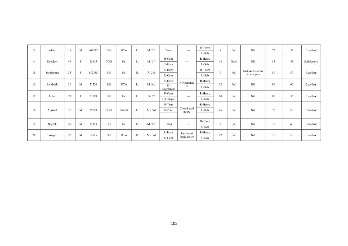
105
13 Akhil 19 M 609272 BH RTA Lt M / 3rd Trans --- R-Thom
8 Full Nil 75 55 Excellent U-Sub
14 Umadevi 47 F 28615 CGH Fall Lt M / 3rd R-Com
---- R-Henry
18 Good Nil 85 45 Satisfactory U-Trans U-Sub
15 Sarojamma 33 F 612355 BH Fall Rt P / 3rd R-Trans
--- R-Thom
9 Full Post.interosseous nerve injury 80 50 Excellent
U-Com U-Sub
16 Santhosh 24 M 51536 BH RTA Rt M /3rd R-Trans #Olecronon
Rt
R-Henry 12 Full Nil 90 60 Excellent U-
Segmental U-Sub
17 Usha 27 F 51998 BH Fall Lt D / 3rd R-Com
--- R-Henry
10 Full Nil 80 55 Excellent U-Oblique U-Sub
18 Aravind 36 M 29828 CGH Assault Lt M / 3rd
R-Tran Closed head
injury
R-Henry
16 Full Nil 75 50 Excellent U-Com, U-Sub
19 Nagesh 29 M 52113 BH Fall Lt M /3rd Trans --- R-Thom
9 Full Nil 70 60 Excellent U-Sub
20 Joseph 23 M 52715 BH RTA Rt M / 3rd R-Trans Unilateral
pubic bone# R-Henry
12 Full Nil 75 55 Excellent U-Com U-Sub

106
KEY TO MASTER CHART
BH - Bapuji hospital , Davangere
C -Comminuted
CGH -Chigateri general hospital , Davangere
F -Female
IP No. -In patient number
Lt -Left
D/3 -Distal third
M -Male
M/3 -Middle third
P/3 -Proximal third
MOI -Mode of injury
R -Radius
Rt -Right
ROM -Range of motion
RTA -Road traffic accident
Sl . No. -Serial number
Sub -Subcutaneous approach
Thom -Thompson approach
U -Ulna
# - Fracture