Reason for referral and consultation liaison psychiatry diagnoses
Upload
khangminh22Category
view
1download
0
Fracture liaison service
Citation for published version (APA):
Van den Berg, P. (2020). Fracture liaison service: optimizing care from a nurse practitioner's perspective.Ridderprint. https://doi.org/10.26481/dis.20201218pb
Document status and date:Published: 01/01/2020
DOI:10.26481/dis.20201218pb
Document Version:Publisher's PDF, also known as Version of record
Please check the document version of this publication:
• A submitted manuscript is the version of the article upon submission and before peer-review. There canbe important differences between the submitted version and the official published version of record.People interested in the research are advised to contact the author for the final version of the publication,or visit the DOI to the publisher's website.• The final author version and the galley proof are versions of the publication after peer review.• The final published version features the final layout of the paper including the volume, issue and pagenumbers.Link to publication
General rightsCopyright and moral rights for the publications made accessible in the public portal are retained by the authors and/or other copyrightowners and it is a condition of accessing publications that users recognise and abide by the legal requirements associated with theserights.
• Users may download and print one copy of any publication from the public portal for the purpose of private study or research.• You may not further distribute the material or use it for any profit-making activity or commercial gain• You may freely distribute the URL identifying the publication in the public portal.
If the publication is distributed under the terms of Article 25fa of the Dutch Copyright Act, indicated by the “Taverne” license above,please follow below link for the End User Agreement:
www.umlib.nl/taverne-license
Take down policyIf you believe that this document breaches copyright please contact us at:
providing details and we will investigate your claim.
Download date: 30 Jan. 2022

FRACTURE LIAISON SERVICE: Optimizing care from a nurse
practitioner’s perspective
Peter van den Berg

© copyright Peter van den Berg, Delft 2020
Printing: Ridderprint
Vormgeving: Publiss
ISBN: 978-94-6416-249-3
All rights reserved. No part of this publication may be reproduced, stored in a
retrieval system or transmitted in any form or by any means, electronic, mechanical,
photocopying, recording or otherwise, without prior permission of the author or the
copyright-owning journals for previous published chapters.

FRACTURE LIAISON SERVICE: Optimizing care from a nurse practitioner’s perspective
PROEFSCHRIFT
ter verkrijging van de graad van doctor aan de Universiteit Maastricht,
op gezag van de Rector Magnificus, Prof. dr. Rianne M. Letschert
volgens het besluit van het College van Decanen,
in het openbaar te verdedigen
op vrijdag 18 december 2020 om 10 uur
door
Peter van den Berg

PromotorenProf. Dr. J.P.W. van den Bergh
Prof. Dr. P.P.M.M. Geusens
Copromotor
Dr. D.H. Schweitzer (Reinier de Graaf Gasthuis Delft)
Beoordelingscommissie
Prof. dr. L.W. van Rhijn (voorzitter)
Prof. dr. L.P.S. Stassen
Prof. dr. M.C. Zillikens (Erasmus Medical Center Rotterdam)
Prof. dr. M.H. Emmelot (University Medical Center Utrecht)
Dr. S.P.G. Bours

ContentsList of abbreviations
Chapter 1 General Introduction
Chapter 2 Meeting international standards of secondary fracture
prevention: a survey on Fracture Liaison services in the
Netherlands. Osteoporos Int. 2015 Sep;26(9):2257-63.
doi: 10.1007/s00198-015-3117-y.
Chapter 3 Challenges and Opportunities to Improve Fracture Liaison Service
Attendance: Fracture Registration and Patient Characteristics
and Motivations. Osteoporos Int. 2019 Aug;30(8):1597-1606.
doi: 10.1007/s00198-019-05016-4.
Chapter 4 Characterization of Fracture Liaison Service Non-Responders
After Invitation by Home Visits and Questionnaires.
Osteoporos Int. 2020 May 13. doi:10.1007/s00198-020-05442-9.
Online ahead of print.
Chapter 5 Quantification of Calcium intake from Calcium-Dense Dairy
Products in a Dutch Fracture Cohort.
Nutrients. 2014 Jun 23;6(6):2404-18. doi:10.3390/nu6062404.
Chapter 6 The Use of Pulse-Echo Ultrasound in Women With a Recent Non-
Vertebral Fracture to Identify Those Without Osteoporosis and/
or a Subclinical Vertebral Fracture: A Pilot Study. Arch Osteoporos.
2020 Apr 14;15(1):56. doi:10.1007/s11657-020-00730-7.
Chapter 7 A dedicated Fracture Liaison Service telephone program and use
of bone turnover markers for evaluating 1-year persistence with
oral bisphosphonates. Osteoporos Int. 2018 Apr;29(4):813-824.
doi:10.1007/s00198-017-4340-5.
Chapter 8 Summary and conclusions
Chapter 9 General discussion and future perspectives
Nederlandse samenvatting
Valorization
Dankwoord
Curriculum vitae
11
25
43
75
95
117
139
165
173
193
198
202
207

6
List of abbreviationsAIRE Appraisal of Indicators, Research and Evaluation tool
ASBMR American Society of Bone and Mineral Research ASBMR)
AUROC Area under the ROC curve
BBS Berg Balance Scale
BMD Bone Mineral Density
BMI Body Mass Index
BTM Bone Turnover Marker
CBS Centraal Bureau voor de Statistiek
CFNCS Dutch National Food Consumption Survey
CI Confidence Interval
CSFII Continuing Survey of Food Intake by Individuals (USA)
CtF®-BPF Capture the Fracture® Best Practice Framework
CTh Cortical Thickness
DI Density Index
DXA Dual Energy X-ray Absorptiometry
ECTS European Calcified Tissue Society
EUGMS European Union Geriatric Medicine Society
EULAR European League against Rheumatism
FDA Food and Drug Administration (USA)
FFN Fragility Fracture Network
FLS Fracture Liaison Service
FRAX Fracture Risk Assessment Tool
GCP Good Clinical Practice
GI Gastro Intestinal
GP General Practitioner
HV Home Visit
IOF International Osteoporosis Foundation
IRR Inter Rater Reliability
IU International Unit
IV Intravenous
KPI Key Performance Indicator
LSC Least Significant Change

7
LSP Landelijk Schakel Punt (Dutch National Exchange Point Pharmacies-GPs.)
METC Medisch Ethische Toetsings Commissie (Medical Ethical Review Board)
N&NP/PA Nurses, Nurse Practitioners and Physician Assistants
NHANES National Health and Nutrition Examination Survey (USA)
NICE The National Institute for Health and Care Excellence (UK)
No PC No Phone Call
non-HV|Q no consent for HV or Q
NOV Nederlandse Orthopaedische Vereniging
NP Nurse Practitioner (Verpleegkundig Specialist)
NPV Negative Predictive Value
NS Not significant
NVF Non-vertebral fracture
NVNH Non-Vertebral Non-Hip (fractures)
NVT Nederlandse Vereniging voor Traumachirurgie
OR Odds Ratio
PA Physician Assistant
PC Phone Call
PDCA Plan-Do-Check-Act
P-EU Pulse-Echo Ultrasound
PINP Procollagen type 1 N Propeptide
PPV Positive Predictive Value
Q Questionnaire
QUS Quantitative Ultra Sound
RCT Randomized Controlled Trial
RDA Recommended Daily Allowance
RDI Recommended Daily Intake
RIVM Het Rijksinstituut voor Volksgezondheid en Milieu (Dutch National Institute for Public Health and the Environment)
ROC Receiver Operator Curve
SC Subcutaneous
s-CTX serum C-terminal telopeptide of type 1 collagen
SECOB SECondary Osteoporosis and metabolic Bone disorder
TFI Tilburg Frailty Indicator
List of abbreviations

8
TUG Timed Up and Go Test
UMCG University Medical Centre Groningen
V&VN Dutch National Council of Nurses (Verpleegkundigen & Verzorgenden in Nederland)
VF Vertebral Fracture
VF&O Vallen, Fracturen & Osteoporose (Dutch FLS Nurses and Nurse Practitioners Association)
VFA Vertebral Fracture Assessment
VMS Veiligheid Management Systeem (Safety Management System)
WHO World Health Organization
List of abbreviations

9
List of abbreviations

1010
1

11
General Introduction
CHAPTER 1

12
Chapter 1
Epidemiology of fractures
Bones have evolved throughout millions of years of evolution and perform several
tasks. Bones provide a framework for attachment of muscles to enable movements,
they protect soft tissues against external trauma, they serve as a reservoir for calcium
homeostasis, host blood cells for hematopoiesis, and immune cells and store energy,
such as fat in the bone marrow. Strong bones can resist load, such as running, jumping
and falls. Fractures occur when the load exceeds bone strength [1,2]. Thus, fractures
are the result of weakened bones, severe or repeated trauma’s or a combination of
both. In 50+ subjects the most frequent trigger for fractures is a fall or an accident, in
combination with postmenopausal and age-related bone loss. In adults, fracture risk
increases with age and remaining lifetime fracture risk in Caucasian men and women
of 50 years is >20% and >50% respectively [3]. With the aging of the population
worldwide more fractures are expected [4]. The number of all fractures per year
among the Dutch population older than 50 years is 120,000 according to data from
health insurance companies [5]. This burden comes with approximately € 600 million
expenses per year and will even rise with 50% by 2030 [6,7]. For the Netherlands it
seems that the burden in costs holds trend with that of other European countries.
However, due to Dutch demographics it seems that the expected increase in health
care budgets for fractures is not the highest in Europe [4]. Other European countries
like Austria, the Scandinavian countries and Belgium are already facing even higher
future costs and, as a consequence it will have negative effects on national health care
budgets [4]. Based on extensive prospective population and cohort studies, it has
become clear that the risk of subsequent fractures is particularly high within the first
years after a fracture in patients older than 50 years [8-13]. This high and immediate
risk of having subsequent fractures is referred to as ‘imminent fracture risk’ [14,15].
Osteoporosis has been reported in about a third of fracture patients who are older
than 50 years and accounts as one of the risk factors for subsequent fractures [5].
However, there is still a huge knowledge gap regarding this imminent subsequent
fracture risk in daily practice among both patients and health care professionals [16-19].
Fragility fractures are also associated with a short- and long-term excess mortality
[20,21]. In Dutch patients older than 50 years with a non-vertebral fracture (NVF),
the absolute mortality was 32% of which nearly a third (17%) occurred one-year post
fracture [22]. An increased risk for subsequent fractures and excess mortality after
initial fractures was shown in several studies [8,11,20-22]. These findings support the
effort to develop fracture prevention strategies soon after first fracture. Currently, the

13
General introduction
1
fracture liaison service (FLS) is considered the most effective organizational structure
for secondary fracture prevention [23-29].
Fracture Liaison Service (FLS) from an historical perspective
McLellan et al. introduced in 2003 a novel out-patient program for the evaluation
and management of patients with osteoporotic fracture called the Fracture Liaison
Service (FLS) [30]. In the Netherlands the first FLS related initiatives and outcomes
were reported from Groningen in 2004 and from Maastricht in 2007 [31,32].
The Delft FLS was founded in 2007 after in-depth discussions with local GPs and
nowadays this FLS is highly esteemed by patients, hospital specialists and general
practitioners. In the Dutch guideline on osteoporosis and fracture prevention (2011)
[33] it was recommended to evaluate all fracture patients of 50 years or older in
preferentially a nurse-led structured program and in 2012 a set of quality indicators
was developed for the evaluation of post-fracture care in Dutch Hospitals [34].
Internationally, the International Osteoporosis Foundation (IOF) started a global
initiative called “Capture the Fracture®’’ [35]. Capture the Fracture® used the FLS
concept as the fundament to prevent re-fractures propagating their so-called nurse-
coordinated multidisciplinary approach. These so-called coordinators’ or ‘local
champions’ approach consisted of a framework of 13 best practices necessary for
a structured combat against secondary fractures [36]. Today, the FLS initiative is
internationally endorsed by the International Osteoporosis Foundation IOF [25], the
American Society of Bone and Mineral Research ASBMR) [26], the European League
against Rheumatism (EULAR) [27] and the multidisciplinary Fragility Fracture Network
(FFN) [28]. Formal position statements by these international scientific societies
encouraged even more FLS initiatives abroad as well as in the Netherlands [37].

14
Chapter 1
Fig 1. FLS-care: nationwide Quality Assessment and the FLS fi ve-step approach
FLS-care: a fi ve-step approach
In order to strive for standardized and optimal FLS-care, a fi ve-step approach has
been proposed by van den Bergh et al. [23]. This approach was based on the
concept of a systematic, preferably coordinator-based, approach for identifi cation,
enrollment, evaluation, treatment and monitoring of patients with a fracture after
the age of 50 years. After identifi cation of patients, a detailed evaluation of medical
history, medication use, clinical risk factors, vitamin D status, dietary calcium
intake, known contributors to secondary osteoporosis and fall risk should then be
performed, together with assessment of BMD and VFA. Next, patients need to be
further evaluated for undiagnosed contributors to secondary osteoporosis and
metabolic bone disorders. Then a multifactorial intervention should follow, including
lifestyle management recommendations, calcium and/or vitamin D supplementation if
required, and treatment of underlying disorders. Specifi c anti-osteoporotic treatment
should be considered in line with national guidelines with organized follow-up of
patients after 3 months and annually, thereafter [33].
Gaps in secondary fracture prevention care
The recognition of the FLS as a well-defi ned structured outpatient clinic was a fact, but
gaps in knowledge of both patients and doctors have not yet been resolved. Dutch
healthcare is well organized and basic medical costs are covered by all Dutch health
care insurance companies. Hospitals are equipped to highest standards and care
processes and logistics are demanded by Dutch law. This unique situation provides the
opportunity to comply with national guidelines on osteoporosis, fracture prevention

15
General introduction
1
and falls endorsing to put maximum effort on secondary fracture prevention [33,34].
Unfortunately, there are still gaps in secondary fracture prevention care.
Attendance gap
In 2013, the International Osteoporosis Foundation (IOF) had recognized ‘FLS
attendance’ as an important marker of local FLS quality (www.capturethefracture.org).
FLS attendance was expected to be some 50% or even lower but exact data were
not available. In many countries, FLS initiatives were initiated, but attendance was
reported to be generally low and varying between 20-89% [37]. Patient identification
and selection differed markedly among FLSs, in terms of proportions of inpatients
and outpatients, age, inclusion of women and/or men and fracture selection (any
fracture or only patients with an NVF) [37]. The reasons for these low attendance rates
are unclear, but patient views and opinions about osteoporosis and the consequent
underestimation of subsequent fracture risk might be of strong influence [16–19].
In addition, besides person-related considerations, patients may be unintentionally
uninvited because of administrative issues [38].
Fracture risk evaluation gap
Fracture risk evaluation includes the evaluation of the presence of clinical risk factors
based on medical history and clinical evaluation (age, sex, body weight, personal
and family fracture history, diseases and medication use that increase fracture and
fall risk), DXA including VFA and laboratory tests to exclude underlying illness. The
Dutch guideline of 2011 recommend DXA to identify osteoporosis as well as VFA to
identify subclinical VFs and fall risk assessment [33]. Given the national guideline on
osteoporosis and fracture prevention in the Netherlands (2011) and the excellent
access to health services and the high hospital density throughout the country, the
Osteoporosis Quality Indicator was introduced to monitor in all Dutch hospitals. This
Quality Indicator, based on a so called Appraisal of Indicators, Research and Evaluation
(AIRE) tool [39,40], meaning a hospital-self-report on the proportion of patients of
50 years and older with a recent fracture that received a DXA within 6 months post
fracture. Over the last years, DXA was reported to be performed in 33% of patients
on average, with a wide range (5-100%) [34,41]. In addition, the CBS data collected in
2016 showed that DXA (with and without VFA) was performed in only 26% of 120,000

16
Chapter 1
people older than 50 years with a fracture [7,41]. This low proportion in spite of clear
recommendations according to national guidelines (2011) [33] underpins the notion
of a huge gap in our national fracture risk evaluation initiative. In addition, these
indicators do not provide in depth information on the quality of post-fracture care
beyond DXA measurements as described in the IOF standards and according to the
five-step approach of FLS care.
Treatment gap
Treatment gap is defined as the proportion of truly osteoporosis prescriptions
divided by the total number of osteoporotic fractures plus the degree of adherence
to medication. Both aspects need to be addressed by the FLS in line with Capture
the Fracture® Best Practice Framework and international consensus [36,26-29]. Not
starting anti-resorptive treatment was mainly related to insufficient medical advice,
attitudes towards medication use including concerns about side effects, and disease
awareness [16,42-45]. In patients that started treatment, the one-year adherence
to oral bisphosphonates in the general Dutch population was reported to be 43%
[46]. The persistence in using anti-osteoporosis drugs in patients with a fracture
was 75% after 1 year and 45% after 5 years [47], which is better than in the general
Dutch population but still points at low long term persistence percentages in these
high risk patients. Recently, it was suggested that individualized solutions, based
on collaboration between patient and healthcare provider are needed to improve
adherence and persistence in osteoporosis medication [48].
Nurse Initiatives regarding Fractures, Falls and Osteoporosis
At the time of the first introduction of FLS care in the Netherlands, there was no network
for health care professionals and, therefore an informal network was launched by a
small group of nurses with interest in osteoporosis and fractures. The recognition of
the complexity of secondary fracture prevention care and the identification of several
knowledge gaps led to a nationwide Dutch nursing initiative by the formal founding
of the national board of FLS specialized osteoporosis and fracture prevention nurses
(Dutch Osteoporosis Nurses Association VF&O) in 2008 [49]. The VF&O steering
board started to initiate organized education and training on-the-job programs to
improve the skills for FLS care, and national meetings twice a year. The vision of VF&O

17
General introduction
1
is to become “best on the job’’ through liaising protocol-based decision making, state-
of-the-art-expertise and multidisciplinary patient care. Apart from these activities,
there is a growing number of nurse practitioners who are traditionally registered
in the Dutch National Council of Nurses (V&VN) [50,51]. Part of this highly trained
group has the capability and skills to work in secondary fracture prevention care. In
contrast to nurses, nurse practitioners and physician assistants are legally authorized
to order diagnostic tests and to initiate medical treatments [52-54]. In parallel with
the upcoming organization of nurses in boards and national councils there an urgent
need was felt to establish an international vision on the daily role of nurses, nurse
practitioners and physician assistants in the field of secondary fracture prevention
[55-57]. Endorsed by the IOF in the Capture the Fracture® campaign “make the first
(fracture) the last” nurses should become entitled to become ‘local champions’ i.e. key
players in daily secondary fracture prevention practice [35,36]. Nurses who are well-
trained and capable to connect all input from disciplines (in and outside the hospital)
in favor of individual fracture patient care [58-59] and who take the lead to ensure
the highest FLS attendance as possible [60]. Protocols are mandatory and medical
consultation must be well organized. Indeed, best practice today means 50% FLS
attendance, despite the efforts and care of expert nurse and nurse practitioners in
many FLS facilities. Therefore, firm steps and original viewpoints are necessary to
further optimize secondary fracture prevention and it is a challenge for nurses, nurse
practitioners and physician assistants to show leadership in the organization of FLS
care. These ambitions form the basis of this dissertation on FLS care from a nurse
practitioner`s perspective.

18
Chapter 1
Aims and outline of this dissertation.
This dissertation has multiple aims regarding the five-step approach of FLS care.
1. To gather information on the quality of FLS care in the Netherlands, beyond
the standard Dutch DXA-based quality registrations.
2. To get a better understanding of patient characteristics and motivations
related to FLS attendance and non-attendance.
3. To study the performance of an ultrasound prescreening tool to exclude
FLS patients without osteoporosis or subclinical vertebral fractures for DXA
measurement and VFA.
4. To investigate whether FLS patients complied with recommendations for
daily calcium intake.
5. To introduce a dedicated telephone program and the use of bone
turnover markers for the evaluation of one-year persistence in using oral
bisphosphonates.
Outline
After the introduction in Chapter 1, we studied the Dutch FLS performance in Chapter
2 by developing a nationwide audit to assess the implementation of FLS quality
standards as formulated by the International Osteoporosis Foundation (IOF; CtF®
Best Practice Framework (BPF)) in Dutch hospitals.
In Chapter 3 we studied hospital registration and patient-related factors that were
associated with attendance and non-attendance in patients that were invited at the FLS.
In Chapter 4 we studied the reasons for non-attendance in identified FLS eligible
patients by a questionnaire and by home visits.
In Chapter 5 we studied whether the application of a non-ionizing peripheral Pulse
Echo Ultrasound device enables identification of women with recent non-vertebral
fractures at the FLS who would not need a DXA/VFA.
In Chapter 6 we studied traditional dairy intake in a FLS cohort aiming to analyze the
recommended daily calcium allowance (RDA).
In Chapter 7 we studied the impact of telephone calls at one-year of osteoporosis
medication and the adherence to medication was objectified by means of bone
markers and pharmacy delivery data.

19
General introduction
1
Chapter 8 summarizes the results of this dissertation.
Chapter 9 includes a general discussion based on findings in this dissertation and
provides future perspectives for clinical practice and research.

20
Chapter 1
References1. Charnley J. The closed treatment of common fractures. https://www.cambridge.org/core/books/
closed-treatment-of-common-fractures/9FBF0A141FD5A341743609B9CE9CA6CD.
2. Tornetta P, Ricci W, Court-Brown C, McQueen M, McKee M. Rockwood and Green’s Fractures in Adults (9th Edition). ISBN/ISSN 9781496386519 Publication Date March 27, 2019Sambrook P, Cooper C. Osteoporosis. Lancet. 2006 Jun 17;367(9527):2010-8.
3. Nguyen ND, Ahlborg HG, Center JR, Eisman JA, Nguyen TV. Residual lifetime risk of fractures in women and men. J Bone Miner Res. 2007 Jun;22(6):781-8.
4. Kanis JA, Borgström F, Compston J, Dreinhöfer K, Nolte E, Jonsson L, Lems WF, McCloskey EV, Rizzoli R, Stenmark J. SCOPE: a scorecard for osteoporosis in Europe. Arch Osteoporos. 2013;8:144. doi: 10.1007/s11657-013-0144-1.
5. Lötters FJ, van den Bergh JP, de Vries F, Rutten-van Mölken MP. Current and Future Incidence and Costs of Osteoporosis-Related Fractures in The Netherlands: Combining Claims Data with BMD Measurements. Calcif Tissue Int. 2016 Mar;98(3):235-43. doi: 10.1007/s00223-015-0089-z.
6. https://www.rivm.nl/publicaties/vergrijzing-en-toekomstige-ziektelast-prognose-chronische-ziektenprevalentie-2005-2025. Assessed 23-2-2020.
7. https://opendata.cbs.nl/statline/#/CBS/nl/navigatieScherm/zoeken?searchKeywords =osteoporose. Assessed 25-3-2020.
8. Bliuc D, Nguyen ND, Nguyen TV, Eisman JA, Center JR. Compound risk of high mortality following osteoporotic fracture and refracture in elderly women and men. J Bone Miner Res. 2013 Nov;28(11):2317-24. doi: 10.1002/jbmr.1968.
9. Balasubramanian A, Zhang J, Chen L, Wenkert D, Daigle SG, Grauer A, Curtis JR. Risk of subsequent fracture after prior fracture among older women. Osteoporos Int. 2019 Jan;30(1):79-92. doi: 10.1007/s00198-018-4732-1.
10. van Geel TA, van Helden S, Geusens PP, Winkens B, Dinant GJ. Clinical subsequent fractures cluster in time after first fractures. Ann Rheum Dis. 2009 Jan;68(1):99-102. doi: 10.1136/ard.2008.092775.
11. Huntjens KM, van Geel TA, van den Bergh JP, van Helden S, Willems P, Winkens B, Eisman JA, Geusens PP, Brink PR. Fracture liaison service: impact on subsequent non-vertebral fracture incidence and mortality. J Bone Joint Surg Am. 2014 Feb 19;96(4):e29. doi: 10.2106/JBJS.L.00223.
12. Gehlbach S, Saag KG, Adachi JD, Hooven FH, Flahive J, Boonen S, Chapurlat RD, Compston JE, Cooper C, Díez-Perez A, Greenspan SL, LaCroix AZ, Netelenbos JC, Pfeilschifter J, Rossini M, Roux C, Sambrook PN, Silverman S, Siris ES, Watts NB, Lindsay R. Previous fractures at multiple sites increase the risk for subsequent fractures: the Global Longitudinal Study of Osteoporosis in Women. J Bone Miner Res. 2012 Mar;27(3):645-53. doi: 10.1002/jbmr.1476.
13. Klotzbuecher CM, Ross PD, Landsman PB, Abbott TA 3rd, Berger M. Patients with prior fractures have an increased risk of future fractures: a summary of the literature and statistical syndissertation. J Bone Miner Res. 2000 Apr;15(4):721-39.
14. van Geel TA, Huntjens KM, van den Bergh JP, Dinant GJ, Geusens PP. Timing of subsequent fractures after an initial fracture. Curr Osteoporos Rep. 2010 Sep;8(3):118-22. doi: 10.1007/s11914-010-0023-2.
15. van Helden S, Cals J, Kessels F, Brink P, Dinant GJ, Geusens P. Risk of new clinical fractures within 2 years following a fracture. Osteoporos Int. 2006;17(3):348-54.
16. Alami S, Hervouet L, Poiraudeau S, Briot K, Roux C. Barriers to Effective Postmenopausal Osteoporosis Treatment: A Qualitative Study of Patients’ and Practitioners’ Views. PLoS One. 2016 Jun 29;11(6):e0158365. doi: 10.1371/journal.pone.0158365.

21
General introduction
1
17. Salminen H, Piispanen P, Toth-Pal E. Primary care physicians’ views on osteoporosis management: a qualitative study. Arch Osteoporos. 2019 Apr 26;14(1):48. doi: 10.1007/s11657-019-0599-9.
18. Neuner JM, Schapira MM. The importance of physicians’ risk perception in osteoporosis treatment decision making. J Clin Densitom. 2012 Jan-Mar;15(1):49-54. doi: 10.1016/j.jocd.2011.07.008.
19. Swart KMA, van Vilsteren M, van Hout W, Draak E, van der Zwaard BC, van der Horst HE, Hugtenburg JG, Elders PJM. Factors related to intentional non-initiation of bisphosphonate treatment in patients with a high fracture risk in primary care: a qualitative study. BMC Fam Pract. 2018 Aug 23;19(1):141. doi: 10.1186/s12875-018-0828-0.
20. Tran T, Bliuc D, van Geel T, Adachi JD, Berger C, van den Bergh J, Eisman JA, Geusens P, Goltzman D, Hanley DA, Josse RG, Kaiser SM, Kovacs CS, Langsetmo L, Prior JC, Nguyen TV, Center JR. Population-Wide Impact of Non-Hip Non-Vertebral Fractures on Mortality. J Bone Miner Res. 2017 Sep;32(9):1802-1810. doi: 10.1002/jbmr.3118.
21. Tran T, Bliuc D, Hansen L, Abrahamsen B, van den Bergh J, Eisman JA, van Geel T, Geusens P, Vestergaard P, Nguyen TV, Center JR. Persistence of Excess Mortality Following Individual Nonhip Fractures: A Relative Survival Analysis. J Clin Endocrinol Metab. 2018 Sep 1;103(9):3205-3214. doi: 10.1210/jc.2017-02656.
22. Huntjens KM, Kosar S, van Geel TA, Geusens PP, Willems P, Kessels A, Winkens B, Brink P, van Helden S. Risk of subsequent fracture and mortality within 5 years after a non-vertebral fracture. Osteoporos Int. 2010 Dec;21(12):2075-82. doi: 10.1007/s00198-010-1178-5.
23. van den Bergh JP, van Geel TA, Geusens PP. Osteoporosis, frailty and fracture: implications for case finding and therapy. Nat Rev Rheumatol. 2012 Jan 17;8(3):163-72. doi: 10.1038/nrrheum.2011.217.
24. Kanis JA, Johansson H, Odén A, Harvey NC, Gudnason V, Sanders KM, Sigurdsson G, Siggeirsdottir K, Fitzpatrick LA, Borgström F, McCloskey EV. Characteristics of recurrent fractures. Osteoporos Int. 2018 Aug;29(8):1747-1757. doi: 10.1007/s00198-018-4502-0.
25. https://www.iofbonehealth.org/ Assessed 25-3-2020.
26. Eisman JA, Bogoch ER, Dell R, Harrington JT, McKinney RE Jr, McLellan A, Mitchell PJ, Silverman S, Singleton R, Siris E. Making the first fracture the last fracture: ASBMR task force report on secondary fracture prevention. J Bone Miner Res. 2012 Oct;27(10):2039-46. doi: 10.1002/jbmr.1698.
27. Lems WF, Dreinhöfer KE, Bischoff-Ferrari H, Blauth M, Czerwinski E, da Silva J, Herrera A, Hoffmeyer P, Kvien T, Maalouf G, Marsh D, Puget J, Puhl W, Poor G, Rasch L, Roux C, Schüler S, Seriolo B, Tarantino U, van Geel T, Woolf A, Wyers C, Geusens P. EULAR/EFORT recommendations for management of patients older than 50 years with a fragility fracture and prevention of subsequent fractures. Ann Rheum Dis. 2017 May;76(5):802-810. doi: 10.1136/annrheumdis-2016-210289.
28. Dreinhöfer KE, Mitchell PJ, Bégué T, Cooper C, Costa ML, Falaschi P, Hertz K, Marsh D, Maggi S, Nana A, Palm H, Speerin R, Magaziner J; on behalf of: the Fragility Fracture Network (FFN); European Geriatric Medicine Society (EuGMS); European Federation of National Associations of Orthopaedics and Traumatology (EFORT); International Collaboration of Orthopaedic Nursing (ICON); International Geriatric Fracture Society (IGFS); International Osteoporosis Foundation (IOF). A global call to action to improve the care of people with fragility fractures. Injury. 2018 Aug;49(8):1393-1397. doi: 10.1016/j.injury.2018.06.032.
29. Conley RB, Adib G, Adler RA, Åkesson KE, Alexander IM, Amenta KC, Blank RD, Brox WT, Carmody EE, Chapman-Novakofski K, Clarke BL, Cody KM, Cooper C, Crandall CJ, Dirschl DR, Eagen TJ, Elderkin AL, Fujita M, Greenspan SL, Halbout P, Hochberg MC, Javaid M, Jeray KJ, Kearns AE, King T, Koinis TF, Koontz JS, Kužma M, Lindsey C, Lorentzon M, Lyritis GP, Michaud LB, Miciano A, Morin SN, Mujahid N, Napoli N, Olenginski TP, Puzas JE, Rizou S, Rosen CJ, Saag K, Thompson

22
Chapter 1
E, Tosi LL, Tracer H, Khosla S, Kiel DP. Secondary Fracture Prevention: Consensus Clinical Recommendations from a Multistakeholder Coalition. J Bone Miner Res. 2020 Jan;35(1):36-52. doi: 10.1002/jbmr.3877.
30. McLellan AR, Gallacher SJ, Fraser M, McQuillian C (2003). The fracture liaison service: success of program for the evaluation and management of patients with osteoporotic fracture. Osteoporos Int 14:1028–1034.
31. Hegeman JH, Oskam J, van der Palen J, Ten Duis HJ, Vierhout PA. The distal radial fracture in elderly women and the bone mineral density of the lumbar spine and hip. J Hand Surg Br. 2004 Oct;29(5):473-6.
32. van Helden S, Cauberg E, Geusens P, Winkes B, van der Weijden T, Brink P. The fracture and osteoporosis outpatient clinic: an effective strategy for improving implementation of an osteoporosis guideline. J Eval Clin Pract. 2007 Oct;13(5):801-5.
33. Dutch Institute for Healthcare Improvement CBO (2011) Richtlijn Osteoporose en Fractuurpreventie. www.diliguide.nl/document/1015/file/pdf/. (Dutch) Assessed 14-02-2019.
34. https://www.zorginzicht.nl/kwaliteitsinstrumenten/osteoporose-indicatoren assessed 9-4-2020
35. Akesson K, Marsh D, Mitchell PJ, McLellan AR, Stenmark J, Pierroz DD, Kyer C, Cooper C (IOF Fracture Working Group) (2013). Capture the Fracture®: a Best Practice Framework and global campaign to break the fragility fracture cycle. Osteoporos Int. 24(8):2135-52. doi: 10.1007/s00198-013-2348-z.
36. https://www.capturethefracture.org/ assessed 25-3-2020.
37. Vranken L, Wyers CE, van den Bergh JPW, Geusens PPMM. The Phenotype of Patients with a Recent Fracture: A Literature Survey of the Fracture Liaison Service. Calcif Tissue Int. 2017 Sep;101(3):248-258. doi: 10.1007/s00223-017-0284-1.
38. van den Berg P, van Haard PMM, Geusens PP, van den Bergh JP, Schweitzer DH. Challenges and opportunities to improve fracture liaison service attendance: fracture registration and patient characteristics and motivations. Osteoporos Int. 2019 Aug;30(8):1597-1606. doi: 10.1007/s00198-019-05016-4. Epub 2019 May 25.
39. Berg M, Meijerink Y, Gras M, Goossensen A, Schellekens W, Haeck J, Kallewaard M, Kingma H. Feasibility first: developing public performance indicators on patient safety and clinical effectiveness for Dutch hospitals. Health Policy. 2005 Dec;75(1):59-73.
40. de Koning, J.S., Kallewaard, M. & Klazinga, N.S. Prestatie-indicatoren langs de meetlat – het AIRE instrument. TVGW 85, 261–264 (2007). https://doi.org/10.1007/BF03078683
41. https://www.zorginstituutnederland.nl/ assessed 25-3-2020.
42. Grover ML, Edwards FD, Chang YH, Cook CB, Behrens MC, Dueck AC. Fracture risk perception study: patient self-perceptions of bone health often disagree with calculated fracture risk. Womens Health Issues. 2014 Jan-Feb;24(1):e69-75. doi: 10.1016/j.whi.2013.11.007.
43. Hiligsmann M, Cornelissen D, Vrijens B, Abrahamsen B, Al-Daghri N, Biver E, Brandi ML, Bruyère O, Burlet N, Cooper C, Cortet B, Dennison E, Diez-Perez A, Gasparik A, Grosso A, Hadji P, Halbout P, Kanis JA, Kaufman JM, Laslop A, Maggi S, Rizzoli R, Thomas T, Tuzun S, Vlaskovska M, Reginster JY. Determinants, consequences and potential solutions to poor adherence to anti-osteoporosis treatment: results of an expert group meeting organized by the European Society for Clinical and Economic Aspects of Osteoporosis, Osteoarthritis and Musculoskeletal Diseases (ESCEO) and the International Osteoporosis Foundation (IOF). Osteoporos Int. 2019 Nov;30(11):2155-2165. doi: 10.1007/s00198-019-05104-5. Epub 2019 Aug 7.
44. Wozniak LA, Johnson JA, McAlister FA, Beaupre LA, Bellerose D, Rowe BH, Majumdar SR. Understanding fragility fracture patients’ decision-making process regarding bisphosphonate treatment. Osteoporos Int. 2017 Jan;28(1):219-229. doi: 10.1007/s00198-016-3693-5.

23
General introduction
1
45. Gleeson T, Iversen MD, Avorn J, et al. Interventions to improve adherence and persistence with osteoporosis medications: a systematic literature review. Osteoporos Int. 2009; 20(12):2127-2134.
46. Netelenbos JC, Geusens PP, Ypma G, Buijs SJ. Adherence and profile of non-persistence in patients treated for osteoporosis--a large-scale, long-term retrospective study in The Netherlands. Osteoporos Int. 2011 May;22(5):1537-46. doi: 10.1007/s00198-010-1372-5.
47. Klop C, Welsing PM, Elders PJ, Overbeek JA, Souverein PC, Burden AM, van Onzenoort HA, Leufkens HG, Bijlsma JW, de Vries F. Long-term persistence with anti-osteoporosis drugs after fracture. Osteoporos Int. 2015 Jun;26(6):1831-40. doi: 10.1007/s00198-015-3084-3.
48. Cornelissen D, de Kunder S, Si L, Reginster JY, Evers S, Boonen A, Hiligsmann M; European Society for Clinical and Economic Aspect of Osteoporosis, Osteoarthritis and Musculoskeletal Diseases (ESCEO). Interventions to improve adherence to anti-osteoporosis medications: an updated systematic review. Osteoporos Int. 2020 May 1. doi: 10.1007/s00198-020-05378-0. [Epub ahead of print].
49. https://osteoporoseverpleegkundige.nl/ (Dutch) assessed 26-3-2020.
50. https://venvnvs.nl/venvnvs/ (Dutch) assessed 26-3-2020.
51. https://venvnvs.nl/venvnvs/information-in-english/ assessed 26-3-2020.
52. Boeijen ERK, Peters JWB, van Vught AJAH. Nurse practitioners leading the way: An exploratory qualitative study on the added value of nurse practitioners in outpatient care in the Netherlands. J Am Assoc Nurse Pract. 2019 Oct 3. doi: 10.1097/JXX.0000000000000307.
53. Chow S. Nurse Practitioner Fracture Liaison Role: A Concept Analysis. Orthop Nurs. 2017 Nov/Dec;36(6):385-391. doi: 10.1097/NOR.0000000000000399.
54. Rice P, Mehan U, Hamilton C, Kim S. Screening, assessment, and treatment of osteoporosis for the nurse practitioner: key questions and answers for clinical practice--a Canadian perspective. J Am Assoc Nurse Pract. 2014 Jul;26(7):378-85. doi: 10.1002/2327-6924.12134.
55. Fragility Fracture Nursing: Holistic Care and Management of the Orthogeriatric Patient [Internet]. Cham (CH): Springer; 2018. Chapter 1. Osteoporosis and the Nature of Fragility Fracture: An Overview. Van Oostwaard M. Editors Hertz K1, Santy-Tomlinson J2, editors. 2018 Jun 16.
56. Seuffert P, Sagebien CA, McDonnell M, O’Hara DA. Evaluation of osteoporosis risk and initiation of a nurse practitioner intervention program in an orthopedic practice. Arch Osteoporos. 2016;11:10. doi: 10.1007/s11657-016-0262-7.
57. Roh YH, Koh YD, Noh JH, Gong HS, Baek GH. Effect of health literacy on adherence to osteoporosis treatment among patients with distal radius fracture. Arch Osteoporos. 2017 Dec;12(1):42. doi: 10.1007/s11657-017-0337-0.
58. Raybould G, Babatunde O, Evans AL, Jordan JL, Paskins Z. Expressed information needs of patients with osteoporosis and/or fragility fractures: a systematic review. Arch Osteoporos. 2018 May 8;13(1):55. doi: 10.1007/s11657-018-0470-4.
59. Besser SJ, Anderson JE, Weinman J. How do osteoporosis patients perceive their illness and treatment? Implications for clinical practice. Arch Osteoporos. 2012;7:115-24. doi: 10.1007/s11657-012-0089-9.
60. Huntjens KM, van Geel TC, Geusens PP, Winkens B, Willems P, van den Bergh J, Brink PR, van Helden S. Impact of guideline implementation by a fracture nurse on subsequent fractures and mortality in patients presenting with non-vertebral fractures. Injury. 2011 Sep;42 Suppl 4:S39-43. doi: 10.1016/S0020-1383(11)70011-0.

2424
2

25
CHAPTER 2
Meeting international standards of secondary fracture prevention: A Survey on Fracture Liaison Services in the Netherlands
Peter van den Berg (1)*, Dave H. Schweitzer (2), Paul M.M. van Haard (3), Joop P. van
den Bergh (4) and Piet P. Geusens (5).
1. Dept. of Orthopaedics and Surgery, Reinier de Graaf Hospital, Delft,
The Netherlands.
2. Dept. of Internal Medicine, Reinier de Graaf Hospital, Delft, The Netherlands.
3. Dept. Medical Laboratories, Reinier de Graaf Hospital, Association of Clinical
Chemistry, Delft, The Netherlands.
4. Dept. of Internal Medicine, VieCuri Medical Centre Noord-Limburg and Dept.
of Internal Medicine, Subdivision Rheumatology, Maastricht University Medical
Center, Maastricht, The Netherlands.
5. Dept. of Internal Medicine, Subdivision Rheumatology, Maastricht University
Medical Center and Hasselt University , Belgium.
Corresponding author E-Mail: [email protected]
Tel.: +31-15-260-4926; Fax: +31-70-260-3828.
Keywords: IOF BPF Standards; FLS ;The Netherlands.
CHAPTER 2

26
Chapter 2
Mini-abstractThe fracture liaison service (FLS) is advocated to be effective for the prevention of
secondary fractures, but implementation is variable. A questionnaire based on the
IOF Capture the Fracture® FLS standards was used in the current study. The results
showed high compliancy with IOF standards in the Dutch responding hospitals.
Abstract
Introduction
The fracture liaison service (FLS) is advocated for the prevention of secondary fractures,
but its implementation varies between hospitals and countries. The present survey
applied the standards proposed by the International Osteoporosis Foundation (IOF)
to evaluate the implementation of FLSs in non-university hospitals in the Netherlands.
Methods
A questionnaire based on the IOF FLS standards was used in this study, requesting
the selection, evaluation and treatment data of patients older than 50 years with a
recent fracture.
Results
Of 90 invited hospitals, 24 (27%) fully responded, providing data of 24,468 consecutive
patients, corresponding with 25% of fracture patients in the Netherlands in the year
2012. After excluding skull and toe fractures and patients exceeding the upper age
limits applied by individual hospitals, 11,983 patient data were available for analysis.
The data showed high compliance (>90%) for fracture patient identification, invitation
for FLS, timing of assessment, identification of vertebral fractures, application of
national guidelines, evaluation of secondary osteoporosis, drug initiation when
indicated, communication with the general practitioner, application of follow-up
strategy, and 70% for fall prevention. The response rate was on average 49%.

27
Meeting international standards of secondary fracture prevention
2
Conclusions
The available data also showed that patients attending the FLS were evaluated,
treated and followed in high compliancy with IOF standards. Some standards are
open to different interpretations and may need further specification. The major
shortcoming in FLS practice was that patients invited to attend the FLS showed a low
response rate. None of the hospitals achieved the IOF standard patient response rate
of over 90%.
IntroductionThe fracture liaison service (FLS) is advocated as the most appropriate approach
for secondary fracture prevention [1,2,3]. This approach is managed by a central
coordinator, mostly a qualified osteoporosis nurse [4,5]. However, the implementation
of FLSs varies between hospitals [6] and countries [7,8]. The International Osteoporosis
Foundation (IOF) has proposed standards to evaluate the implementation and the
quality and performance of the FLS [9].
After a fracture, the risk of a subsequent fracture steeply increases [10,11]. Of all low
trauma fractures in patients over 50 years of age, subsequent fractures accounted
for 40% of fractures in women and 24% in men [12]. After any osteoporotic fracture
in patients over 60 years of age, 24% of women (31% of survivors) and 20% of men
(32% of survivors) had a subsequent fracture within 5 years [13]. Using composite
risk analysis that takes into account subsequent fracture risk and mortality, the risk of
subsequent fractures reached 40% in survivors of hip fracture during 5 year follow-
up [14], 80% of survivors of a hip fracture during 20 years follow-up [15], and 20% of
survivors of a hand or foot fracture during 5 year follow-up [16]. In addition, the risk of
mortality is increased after a first and subsequent osteoporotic fracture [17].
The aim of the FLS is to reduce the risk of subsequent fractures in high risk patients. The
first FLS was reported in Glasgow in 2003 [18]. It involved systematic identification of all
fracture patients, adequate evaluation of their fracture risk, diagnostic evaluation with
DXA and VFA and analysis for secondary osteoporosis or metabolic bone disorders,
followed by prescription of calcium and vitamin D and specific anti-osteoporosis
medication, if needed, as well as follow-up according to a 5-step approach [19].
In the Netherlands, several nationwide scientific committees have addressed the
importance of initiating a FLS in hospitals in order to achieve adequate secondary
fracture prevention. National guidelines on osteoporosis and fracture prevention

28
Chapter 2
strongly advocate the identification and examination of patients with a recent
fracture, including risk evaluation and differential diagnosis [20,21]. These initiatives
have contributed to the development of a FLS in most Dutch hospitals. At present 85
specialised FLS nurses are working in 68 non-university hospitals.
In 2012 the International Osteoporosis Foundation (IOF) launched the “Capture The
Fracture®” campaign for secondary fracture prevention [3]. In this campaign, the
IOF presented a Best Practice Framework (BPF) with 13 standards for evaluating the
performance of FLSs. In this study, we applied these 13 standards to evaluate the
implementation of FLS in non-university Dutch hospitals. As far as we know, no similar
study has been performed before.
Methods In the Netherlands, there are 90 FLSs in non-university teaching and non-teaching
hospitals, and 95 osteoporosis nurses are registered in the database of VF&O (the
Dutch association for nurses on Falls, Fractures and Osteoporosis); 85 of them are
working in 68 of the non-university hospitals, while the remaining 10 are working in
university hospitals or in other professional settings.
For evaluation of the FLSs, the authors of this survey designed a digital questionnaire
consisting of demographic questions and specific questions about the FLS organisation
and the 13 BPF standards[3], which were translated from the original English text into
Dutch. Next, all 90 non-university hospitals in the Netherlands were invited by mail to
participate in this survey, providing a web link that gave access to the questionnaire.
The questionnaire was sent to the 85 VF&O affiliated FLS coordinators working in 68
of the non-university hospitals, and to the physicians involved in osteoporosis care in
the remaining 22 non-university hospitals (Figure 1).
To ensure the anonymity of participating hospitals, the questionnaire was distributed
by DeVosJansen Research Association (DVJ), a professional, independent and
unrelated organisation. The results of the questionnaires were sent to DVJ using
internet-based Kinesis Survey software, and DVJ delivered the results anonymously to
the Research group for analysis. Only descriptive statistical analyses were performed.

29
Meeting international standards of secondary fracture prevention
2
Fig 1. Flowchart on inclusion of analysed FLSs
Invitation and Questionnaire
68 hospitals
nurse member of VF&O 23 responses
22 hospitals
not members of VF&O 1 response
Invitation for the study mailed to 90 non-university hospitals by Independent 0rganisation
(DVJ Research Association)
24 completed Questionnaires for analysis
Collection of data sent by DVJ to the Research Group Fracture & Osteoporosis clinics
ResultsFrom 90 hospitals, 24 FLSs (27%) returned completed questionnaires (Figure 1).
Another 6 questionnaires were incomplete and not suitable for analysis. Three
hospitals reported not to be able to extract sufficient data from their database, and
57 did not respond.
Demographics
Of the included hospitals, 16 (67%) were teaching hospitals and 8 were non-teaching
hospitals. The FLS organizing departments were the Trauma and Orthopaedic Surgery
in 12 hospitals (50%) and Internal Medicine, Geriatrics and Rheumatology in another
12 hospitals (50%). The lead clinician was either the Trauma and/or Orthopaedic
Surgeon in 9 hospitals (38%), 2 of which shared leadership with internal medicine, or a

30
Chapter 2
specialist in Internal Medicine in 15 hospitals (62%) (8 Endocrinologists, 1 Geriatrician
and 4 Rheumatologists and 2 physicians sharing leadership with surgeons).Of the
24 participating hospitals, 22 (92%) had a coordinating nurse. For this coordinator
at the FLS, they reported a median of 12 working hours/week (range 4-36 hours),
with additional secretarial help in 17 hospitals (71%) with a median of 9 working
hours /week (range 4-20 hours). The supportive presence of a physiotherapist was
reported in 7 hospitals. The FLS had been initiated between 2004-2006 in 6 hospitals
(25%), between 2007-2009 in 12 (50%), and between 2010-2012 in 6 (25%). DXA was
available in 23 hospitals (96%), and VFA in 16 hospitals (67%). (Table 1)
Table 1. Characteristics of the FLSs (following Demographics)
Yes NoPresence of a coordinating nurse 22 (92%) 2 (8%)Teaching hospital 16 (67%) 8 (33%)DXA scan available 23 (96%) 1 (4%)VFA available 16 (67%) 8 (33%)Secretarial services available 17 (71%) 7 (29%)
BPF Standard 1. Patient identification
All respondents had a system for tracking every patient who presented at their hospital
with a fracture; 9 used hospital administration records, and 17 used emergency
department records, with an overlap of 2 hospitals that reported the use of both
these facilities. A total of 24,468 patients with any recent fracture were identified in
year 2012, 18,001 between the age of 50 and 80 years, and 6,467 in patients older
than 80 years. This represented around 25% of all fracture patients older than 50
years in the Netherlands in that year. A median of 1,020 fracture patients were seen
per participating hospital (range 217-3,377).
BPF Standard 2. Patient evaluation
This standard focuses on the response rate, which depends on selection criteria and
invitation strategy. Therefore we asked for these factors, revealing that institutions
varied in the use of selection criteria, invitation strategy and response rate, but all
FLSs evaluated each and every responder. Of all the patients with recent fractures, the
following categories were excluded for invitation: skull fractures in all FLSs, toe fractures

31
Meeting international standards of secondary fracture prevention
2
in 3 FLS, patients >90 yrs in 9 FLSs, >85 years in 4 FLSs and >80 years in 1 FLS.
The process of invitation was coordinated by the osteoporosis nurse in 14 FLSs, (58%),
by the nurse practitioner/physician assistant in 3 hospitals (13%), by the physician in 3
hospitals (13%) and by the secretarial assistant in 6 FLSs (25%).
Six FLSs invited patients personally, during treatment in the plaster room or during the
orthopaedic outpatient clinic visit (25%). A combination of personal and subsequently
written invitation was reported in 5 FLSs (21%). Ten FLSs used written invitations
first (42%), 5 of these (21%) invited patients additionally by phone. Three FLSs (13%)
invited patients only by phone. In 6 FLSs, patients were invited within 1 month after
fracture, in 15 between 1 and 2 months and in 3 between 2 and 3 months.
Of the 24,468 fracture patients identified by the participating hospitals in year 2012,
11,983 attended the FLS (49%), with a median of 499 patients per FLS (range 112-
1,050). The response rate depended on age (54% in 50-80 yrs old, 34% in 80+) and
timing of invitation (47% after 1 month, 58% after 1-3 months). All responding patients
were subsequently assessed for future fracture risk at the respective FLSs.
BPF Standard 3. Post fracture assessment timing
In 11 FLSs (46%) patients actually visited the FLS for assessment within 8 weeks after
the fracture, in 11 FLSs (46%) they visited between 9 and 12 weeks after the fracture
and in 2 (8%) more than 13 weeks after the fracture.
BPF Standard 4. Vertebral Fracture
In all participating hospitals, all patients with clinical vertebral fractures, who attended
the FLS were assessed. Ten FLSs (42%) also reported a strategy to invite patients
with non-clinical vertebral fractures as reported by the radiologist. In 15 hospitals,
the FLSs reported the structural application of VFA at any BMD level in patients with
a non-vertebral fracture. Seven FLSs reported VFA assessment only in patients with
osteoporosis, and 6 reported VFA assessment in case of osteopenia and osteoporosis.
One FLS reported the structural application of X-ray of the vertebral column instead
of VFA. In 11,983 patients, 9,690 DXA scans (81%) were performed, and 7,045 of these
(73%) included VFA. Ten FLS (42%) reported also a strategy to invite patients with non-
clinical vertebral fractures as reported by the radiologist.

32
Chapter 2
BPF Standard 5. Assessment Guidelines
A total of 22 FLSs (92%) reported implementation of national guidelines concerning
osteoporosis and fracture prevention. Two FLSs did not specify which guidelines
were used.
BPF Standard 6. Secondary Causes of Osteoporosis
All FLSs assessed causes of known SECondary Osteoporosis and metabolic Bone
disorders (SECOB) through medical history. Ten FLSs performed laboratory tests on
all patients as standard procedure. In 22 FLSs, all the patients who required treatment
additionally underwent investigation for SECOB, while 2 FLSs reported performing
laboratory analyses only when secondary osteoporosis was clinically suspected.
BPF Standard 7. Fall Prevention Services
In 17 FLSs (71%), patients were evaluated for the presence of fall risk. Implementation
of national guidelines on fall prevention was reported by 14 FLSs (58%). The content
of fall risk evaluation was heterogeneous. The following evaluations were reported:
attention for mobility in general, including the use of alcohol, attention for visual
impairments and the use of crutches (38%) and evaluation of orthostatic hypotension
(8%). Three FLSs (13%) reported function and muscle tests, including the Timed Up
and Go test (TUG), the Berg Balance Scale (BBS) and a Hand Grip Strength test. Three
FLSs reported systematic referral to a falls prevention clinic (13%).
BPF Standard 8. Multifaceted health and lifestyle risk factor assessment
Multifaceted health and lifestyle risk factor assessment was performed in 22 FLSs,
of which 20 FLSs reported the health and lifestyle risk factors to the GP who was
responsible for subsequent referral to the appropriate practitioner for further
evaluation and treatment.
BPF Standard 9. Medication Initiation Standard
In all patients not on treatment at the time of fracture presentation, medication
was initiated at the FLS in 19 hospitals (79%) and initiated by the GP in 5 hospitals

33
Meeting international standards of secondary fracture prevention
2
(21%). For further follow-up prescription, all patients were referred to their GP. The
analysis of this standard did not include the prescription of calcium and vitamin D
supplements.
BPF Standard 10. Medication Review
A total of 22 FLSs reported to be following the national guidelines for patients already
in receipt of osteoporosis medication when they presented with a fracture. This
included review of medication compliance, considerations of alternative osteoporosis
medication and optimization of non-pharmacological interventions.
BPF Standard 11. Communication Strategy
A total of 22 FLSs communicated the results of all patients to the GPs, and 1 FLS did
so in 90% of the patients (1 FLS unknown). Of the 24 FLSs, 22 reported prescription
of osteoporosis medication, 21 reported BMD results, need for adequate calcium
and vitamin D intake and laboratory results, 20 reported primary osteoporosis
risk factors and lifestyle health risk factor assessment, 19 reported screening on
secondary osteoporosis, results of X-rays of the spine and the proposed follow-up
strategy. Seventeen reported on the duration of therapy, 15 FLSs reported fracture
risks and 13 reported VFA results, current drug treatment and fall risk factors.
Previous fractures/time since last fracture were reported by 11 FLSs and medication
compliance review was reported by 12 FLSs. The FRAX score was reported by 1 FLS as
standard procedure. (Table 2)
BPF Standard 12. Long-term Management
Eight FLSs reported a follow-up protocol in the FLS after 3 months, 7 a telephone
call (after 1-12 months) and 15 reported that further follow-up was delegated to the
patient’s GP.
BPF Standard 13. Database
All FLSs used local databases for inviting fracture patients, and 12 (50%) had a local
database on the results of examinations at the FLS. A national registry on fractures

34
Chapter 2
is available in the Netherlands and open to the public. All hospital-reported fractures
have specific codes which correspond with the insured coverage [22]. However, no
nationwide hospital data is available on patients evaluated in an FLS. This survey was
based on individual FLS databases that included around 25% of all fractures in the
Netherlands in year 2012.
Table 2. Various aspects communicated by the FLSs to GP (following IOF CtF®BPF standard 11)
Aspects Yes No1 Fracture risk score (FRAX) 1 (4%) 23 (96%)2 DXA/ BMD 21 (88%) 3 (12%)3 Vertebral column X-ray 19 (80%) 5 (20%)4 Primary Osteoporosis risk factors 20 (84%) 4 (16%)5 Screening on secondary Osteoporosis 19 (80%) 5 (20%6 Laboratory tests 21 (88%) 3 (12%)7 Fall risk factors 13 (54%) 11 (46%)8 Current drug treatment 13 (54%) 11 (46%)9 Medication compliance review 12 (50% 12 (50%)10 Follow-up Plan 19 (80%) 5 (20%)11 Follow up Plan: Duration of therapy 17 (70%) 7 (30%)12 Lifestyle health risk-factor assessment 20 (84%) 4 (16%)13 Former fractures/ Time since last fracture 11 (46%) 13 (54%)14 Calcium intake or supplement 21 (88%) 3 (12%)15 Vitamin D intake or supplement 21 (88%) 3 (12%)16 VFA 13 (54%) 11 (46%)17 Prescription osteoporosis medication 22 (92%) 2 (8%)18 Fracture risks 15 (62%) 9 (38%)
DiscussionThis large survey, which included one quarter of all Dutch fracture patients over
50 years of age in the year 2012, aimed at evaluating the implementation of FLS
according to the proposed international BPF standards in non-university hospitals
in the Netherlands. The survey reflects the huge clinical effort in each of the 24
participating hospitals, showing a median of 1,020 fracture patients per hospital, and
assessing a mean of 499 fracture patients per FLS.
All the evaluated FLSs scored above 90% for the following BPF standards: identification
of patients with a recent fracture in the hospitals, invitation for FLS, timing of
assessment, identification of vertebral fractures, application of national guidelines,
evaluation of secondary osteoporosis, drug initiation when indicated, communication
with the general practitioner, and application of follow-up strategy. Our data suggest

35
Meeting international standards of secondary fracture prevention
2
that the majority of Dutch FLSs are organized in line with the best model according to
Ganda Type A [2,7,8], including an osteoporosis nurse, all-encompassing service and
initiation of osteoporosis medication. These findings contrast with a previous survey
in the Netherlands, which evaluated a smaller cohort of FLSs/hospitals before the
year 2010 and found that patients were referred to the GP for treatment decisions
(Ganda Type B) [2,6].
One explanation for the observed high level of FLS care is that most of the issues in
the IOF standards were already included in national guidelines on osteoporosis and
fracture prevention in the Netherlands [20,21]. These guidelines strongly advocate 1/
to identify patients with a recent fracture, 2/ to evaluate their fracture risk using DXA,
VFA and clinical risk factors, 3/ to perform differential diagnosis, 4/ to decide about
secondary fracture prevention and 5/ to follow up treatment. Another explanation is
the longstanding experience of several Dutch hospitals with FLS, since 18 of the 24
participating hospitals had already started an FLS before the Dutch guidelines were
revised in 2011.
Some standards were applied with high variability in implementation. While some
FLSs performed evaluation of secondary osteoporosis in all patients, other FLSs only
did so in patients who required treatment or if secondary osteoporosis was clinically
suspected. Fall risk evaluation consisted of questionnaires alone in 12 FLSs (50%), or
in combination with evaluation of orthostatic hypotension (in 8%), and only 3 FLSs
(13%) reported function and muscle tests. Also, FLSs differed in performing either
vertebral fracture evaluation in all patients or only in patients with osteopenia or
osteoporosis.
The major shortcoming in practices of the FLSs was the low response rate of patients
who were invited to visit the FLS. A patient response rate of over 90%, which is
suggested as the highest BPF standard, was nowhere achieved by any FLS in the
current survey. With the exception of surveys in the UK, where response rates of 75
to 85% have been reported [23-26], response rates are usually lower (between 28
and 61%) in other countries and continents [27-32]. Further research is required to
understand the causes of these lower response rates and to identify ways to increase
them. Earlier studies have shown that systematic screening is more effective than
referral, that personal communication is more effective than written information
for patients, and that the FLS may be more effective than electronic messaging and
communication with GPs [33].
An important limitation of this survey is that the response was relatively low, with

36
Chapter 2
33 out of 90 hospitals responding (37%). Moreover, six of these hospitals delivered
incomplete data and 3 indicated not to be able to generate any data. As a consequence,
the analysis was based on 24 out of the 90 invited hospitals (27%). This could implicate
that well performing FLSs are overrepresented. Unfortunately, any information about
responding and non-responding FLS’s is lacking since anonymity was assured in this
survey. The self-reporting of participants about their own hospital may be a potential
bias although the majority of data were extracted from FLS derived databases.
This is the first attempt of a nationwide audit of standards of care delivered by the
FLS. However, our response rate emphasizes that FLSs have to be encouraged to
participate in these kind of surveys in order to improve our understanding how to
optimize secondary fracture prevention per country. It is also needed to analyze and
compare FLS implementation in other countries and among countries. Moreover,
country-specific Quality/Clinical Standards for FLS have been published in Canada
(adherent with the IOF BPF’s and the osteoporosis Canada Clinical Guidelines)[34]
and the UK were the 5IQ approach is advocated to quality improvement [35]. Each
initiative, albeit different from the IOF Capture the Fracture® will provide useful
information about the quality of FLS care on a global scale.
Of 68 FLSs run by osteoporosis nurses, 23 were analyzed (34%). In addition, of 22
services run by physicians, 1 participated in the analysis (5%). Therefore, the results
may be not representative for the FLS care throughout the Netherlands, even though
they are based on one quarter of all fractures in Dutch patients over 50 years of age.
Conclusion
In 24 of the 90 non-university hospitals in the Netherlands, we showed that patients
attending the FLS were evaluated, treated and followed in high compliance with
IOF standards. Some standards are open to different interpretations and will need
further specification. The major shortcoming in FLS practice in this survey was that
patients invited to attend the FLS showed a low response rate. Further research
should identify the causes of this low response rate and ways to increase it.
Responsibility
PVDB, DHS, JPVDB and PPG designed the study, analyzed and interpreted the data,
and wrote and drafted the manuscript. PVH prepared the data for statistical analysis

37
Meeting international standards of secondary fracture prevention
2
and supported the statistical interpretation of data and writing of the manuscript. All
authors had full access to all data including the numerical reports and tables in the
study, and all take responsibility for the integrity of the data and the accuracy of the
data analyses. PVDB and PPG are the guarantors.
Acknowledgments
The authors are grateful to Lisette van Hulst for her critical linguistic advice and to
Agnes Offenberg, president of the board of the Dutch Osteoporosis Nurses Society
VF&O, and Harry van den Broek, president of the board of the Dutch Osteoporosis
Patient Association for their enthusiastic support.
Conflicts of Interest
Peter van den Berg, Dave Schweitzer, Paul van Haard, Joop van den Bergh and Piet
Geusens declare that they have no conflict of interest.

38
Chapter 2
References1. Eisman JA, Bogoch ER, Dell R, Harrington JT, McKinney RE Jr, McLellan A, Mitchell PJ, Silverman
S, Singleton R, Siris E; ASBMR Task Force on Secondary Fracture Prevention (2012) Making the first fracture the last fracture: ASBMR task force report on secondary fracture prevention. J Bone Miner Res. 27(10):2039-46. doi: 10.1002/jbmr.1698. .
2. Ganda K, Puech M, Chen JS, Speerin R, Bleasel J, Center JR, Eisman JA, March L, Seibel MJ (2013) Models of care for the secondary prevention of osteoporotic fractures: a systematic review and meta-analysis. Osteoporos Int. 24(2):393-406. doi: 10.1007/s00198-012-2090-y.
3. http://share.iofbonehealth.org/WOD/2012/report/WOD12-Report.pdf (Accessed 12 December 2012)
4. Marsh D, Akesson K, Beaton DE, Bogoch ER, Boonen S, Brandi ML, McLellan AR, Mitchell PJ, Sale JE, Wahl DA; IOF CSA Fracture Working Group.(2011) Osteoporos Int. 22(7):2051-65. doi: 10.1007/s00198-011-1642-x.
5. Sander B, Elliot-Gibson V, Beaton DE, Bogoch ER, Maetzel A (2008) A coordinator program in post-fracture osteoporosis management improves outcomes and saves costs. J Bone Joint Surg Am. 90(6):1197-205. doi: 10.2106/JBJS.G.00980.
6. Huntjens KM, van Geel TA, Blonk MC, Hegeman J.H, van der Elst M, Willems P, Geusens PP, Winkens B, Brink P, van Helden SH (2011) Implementation of osteoporosis guidelines: a survey of five large fracture liaison services in the Netherlands. Osteoporos Int. 22(7):2129-35. doi: 10.1007/s00198-010-1442-8.
7. Kanis JA, Borgström F, Compston J, Dreinhöfer K, Nolte E, Jonsson L, Lems WF, McCloskey EV, Rizzoli R, Stenmark J (2013) SCOPE: a scorecard for osteoporosis in Europe. Arch Osteoporos. 8(1-2):144. doi: 10.1007/s11657-013-0144-1.
8. Svedbom A, Hernlund E, Ivergård, M Compston J, Cooper C, Stenmark J, McCloskey EV, Jönsson B, Kanis JA & the EU review panel of the IOF (2013) Osteoporosis in the European Union: a compendium of country-specific reports. Arch Osteoporos. 8:137 doi 10.1007/s11657-013-0137-0
9. Akesson K, Marsh D, Mitchell PJ, McLellan AR, Stenmark J, Pierroz DD, Kyer C, Cooper C (IOF Fracture Working Group) (2013). Capture the Fracture®: a Best Practice Framework and global campaign to break the fragility fracture cycle. Osteoporos Int. 24(8):2135-52. doi: 10.1007/s00198-013-2348-z.
10. van Geel TA, Nguyen ND, Geusens PP, Center JR, Nguyen TV, Dinant GJ, Eisman JA (2010) Development of a simple prognostic nomogram for individualising 5-year and 10-year absolute risks of fracture: a population-based prospective study among postmenopausal women. Ann Rheum Dis. 70(1):92-7. doi: 10.1136/ard.2010.131813.
11. Langsetmo L, Goltzman D, Kovacs CS, Adachi JD, Hanley DA, Kreiger N, Josse R, Papaioannou A, Olszynski WP, Jamal SA; CaMos Research Group (2009) Repeat low-trauma fractures occur frequently among men and women who have osteopenic BMD. J Bone Miner Res. 24(9):1515-22. doi: 10.1359/jbmr.090319.
12. Center JR, Bliuc D, Nguyen TV, Eisman JA (2007) Risk of subsequent fracture after low-trauma fracture in men and women. JAMA. 24;297(4):387-94.
13. Bliuc D, Nguyen ND, Nguyen TV, Eisman JA, Center JR. (2013) Compound risk of high mortality following osteoporotic fracture and refracture in elderly women and men. J Bone Miner Res. 28(11):2317-24. doi: 10.1002/jbmr.1968.
14. Huntjens KM, van Geel TA, van den Bergh JP, van Helden S, Willems P, Winkens B, Eisman JA, Geusens PP, Brink PR (2014) Fracture liaison service: impact on subsequent nonvertebral fracture incidence and mortality. J Bone Joint Surg Am. 19:96(4):e29. doi: 10.2106/JBJS.L.00223.

39
Meeting international standards of secondary fracture prevention
2
15. von Friesendorff M, Besjakov J, Akesson K (2008) Long-term survival and fracture risk after hip fracture: a 22-year follow-up in women. J Bone Miner Res. 23(11):1832-41. doi: 10.1359/jbmr.080606.
16. Huntjens KM, Kosar S, van Geel TA, Geusens PP, Willems P, Kessels A, Winkens B, Brink P, van Helden S (2010) Risk of subsequent fracture and mortality within 5 years after a non-vertebral fracture. Osteoporos Int. 21(12):2075-82. doi: 10.1007/s00198-010-1178-5.
17. Bliuc D, Alarkawi D, Nguyen TV, Eisman JA, Center JR (2014) Risk of Subsequent Fractures and Mortality in Elderly Women and Men With Fragility Fractures With and Without Osteoporotic Bone Density: The Dubbo Osteoporosis Epidemiology Study. J Bone Miner Res. 31. doi: 10.1002/jbmr.2393.
18. McLellan AR, Gallacher SJ, Fraser M, McQuillian C (2003) The fracture liaison service: Success of program for the evaluation and management of patients with osteoporotic fracture. Osteoporos Int. 14:1028–34
19. van den Bergh JP, van Geel TA, Geusens PP (2012) Osteoporosis, frailty and fracture: implications for case finding and therapy. Nat Rev Rheumatol. 8(3):163-72. doi: 10.1038/nrrheum.2011.217.
20. Dutch Institute for Healthcare Improvement CBO (2011) Richtlijn Osteoporose en Fractuurpreventie. www.diliguide.nl/document/1015/file/pdf/. (Dutch) Accessed 19 December 2011
21. https://www.nhg.org/standaarden/volledig/nhg-standaard-fractuurpreventie (Dutch) Accessed 25 December 2014
22. http://www.dutchhospitaldata.nl/Paginas/default.aspx (Dutch) Accessed 25 December 2014
23. McLellan AR, Wolowacz SE, Zimovetz EA, Beard SM, Lock S, McCrink L, Adekunle F, Roberts D (2011) Fracture liaison services for the evaluation and management of patients with osteoporotic fracture: a cost-effectiveness evaluation based on data collected over 8 years of service provision. Osteoporos Int. 22(7):2083-98. doi: 10.1007/s00198-011-1534-0.
24. Mitchell PJ (2013) Best practices in secondary fracture prevention: fracture liaison services. Curr Osteoporos Rep. 11(1):52-60. doi: 10.1007/s11914-012-0130-3
25. Gallacher SJ (2005) Setting up an osteoporosis fracture liaison service: background and potential outcomes. Best Pract Res Clin Rheumatol. 19(6):1081-94.
26. Vaile JH, Sullivan L, Connor D, Bleasel JF. (2013) A Year of Fractures: a snapshot analysis of the logistics, problems and outcomes of a hospital-based fracture liaison service. Osteoporos Int. 24(10):2619-25. doi: 10.1007/s00198-013-2357-y.
27. Dehamchia-Rehailia N, Ursu D, Henry-Desailly I, Fardellone P, Paccou J. (2014) Secondary prevention of osteoporotic fractures: evaluation of the Amiens University Hospital’s fracture liaison service between January 2010 and December 2011. Osteoporos Int. 25(10):2409-16. doi: 10.1007/s00198-014-2774-6.
28. Giles M, Van Der Kallen J, Parker V, Cooper K, Gill K, Ross L, McNeill S. (2011) A team approach: implementing a model of care for preventing osteoporosis related fractures. Osteoporos Int. 22(8):2321-8. doi: 10.1007/s00198-010-1466-0.
29. Eekman DA, van Helden SH, Huisman AM, Verhaar HJ, Bultink IE, Geusens PP, Lips P, Lems WF (2014) Optimizing fracture prevention: the fracture liaison service, an observational study. Osteoporos Int. 25(2):701-9. doi: 10.1007/s00198-013-2481-8.
30. Naranjo A, Ojeda-Bruno S, Bilbao Cantarero A, Quevedo Abeledo JC, Henríquez-Hernández LA, Rodríguez-Lozano C (2013) Results of a model of secondary prevention for osteoporotic fracture coordinated by rheumatology and focused on the nurse and primary care physicians. Reumatol Clin. 10(5):299-303. doi: 10.1016/j.reuma.2013.12.007.

40
Chapter 2
31. Premaor MO1, Pilbrow L, Tonkin C, Adams M, Parker RA, Compston J (2010) Low rates of treatment in postmenopausal women with a history of low trauma fractures: results of audit in a Fracture Liaison Service. QJM. 103(1):33-40. doi: 10.1093/qjmed/hcp154.
32. Majumdar SR, Lier DA, Leslie WD. (2013) Cost-effectiveness of two inexpensive postfracture osteoporosis interventions: results of a randomized trial. J Clin Endocrinol Metab. 98(5):1991-2000. doi: 10.1210/jc.2013-1034.
33. Aizer J, Bolster MB. (2014) Fracture liaison services: promoting enhanced bone health care. Curr Rheumatol Rep. 16(11):455. doi: 10.1007/s11926-014-0455-2.
34. http://www.osteoporosis.ca/wp-content/uploads/OC-Quality-Standards-ENG-Nov-2014.pdf (Accessed 13 March 2015)
35. http://training.nos.org.uk/login/index.php (Accessed 13 March 2015)

41
Meeting international standards of secondary fracture prevention
2

4242
3

43
Peter van den Berg (1)*, Paul M.M. van Haard (2), Piet P. Geusens (3), Joop P. van den
Bergh (4), Dave H. Schweitzer (5).
1. Dept. of Orthopedics and Traumasurgery, Fracture Liaison Service, Reinier de
Graaf Hospital, Delft, the Netherlands
2. Dept. of Medical Laboratories, Association of Clinical Chemistry, Reinier the
Graaf Hospital, Delft, the Netherlands
3. Dept. of Internal Medicine, Subdivision Rheumatology, Maastricht University
Medical Center, Maastricht, the Netherlands and Hasselt University, Hasselt,
Belgium
4. Dept. of Internal Medicine, VieCuri Medical Centre Noord-Limburg and Dept.
of Internal Medicine, Subdivision Rheumatology, Maastricht University Medical
Center, Maastricht, the Netherlands
5. Dept. of Internal Medicine and Endocrinology, Reinier the Graaf Hospital, Delft,
the Netherlands
* Corresponding author E-Mail: [email protected]
Tel.: +31 152604926; Fax: +31 152605982
Challenges and opportunities to improve fracture liaison service attendance: fracture registration and patient characteristics and motivations
CHAPTER 3

44
Chapter 3
DisclosuresPeter van den Berg: no disclosures
Paul M.M. van Haard: no disclosures
Piet P. Geusens: no disclosures
Joop P. van den Bergh: no disclosures
Dave H. Schweitzer: no disclosures
Conflicts of InterestPeter van den Berg, Paul van Haard, Piet Geusens, Joop van den Bergh and Dave
Schweitzer declare that they have no conflict of interest.
AcknowledgmentsWe are grateful to Mrs. Wil Aarssen and Mrs. Maria van Woerden for their excellent
secretarial services and to Mrs. Lisette van Hulst for her linguistic advice.
Contributions.PVDB, DHS and PVH are responsible for the study design. PVDB and DHS were the
authors, supported by PVH, who also performed all statistical analyses. PG and JVDB
supported the process with important scientific contributions.
Mini-abstract:This questionnaire-based study evaluated the reasons for attendance or non-
attendance at the fracture liaison service in patients with a recent fracture. Frailty,
male sex, living alone and low education were associated with non-attendance, and
the information perceived by the patient was associated with attendance.
Keywords: Non-attendance; FLS; Attendance.

45
Challenges and opportunities to improve fracture liaison service attendance
3
Abstract
Introduction
The purpose of this study was to evaluate hospital registration- and patient-related factors
associated with attendance or non-attendance to the Fracture Liaison Service (FLS).
Patients and Methods
Out of 1728 consecutive patients registered with a recent fracture at hospital entry,
and after exclusion of 440 patients because of death, residence in a nursing home,
already on osteoporosis treatment or recent DXA, 1288 received an FLS invitation.
We evaluated the hospital registration of fractures at entry and exit of the hospital.
A questionnaire was sent to all invited patients to evaluate factors related to non-
attendance (including age, gender, frailty, living alone, income, education, extrinsic
motivations (impact of perceived information) and intrinsic motivations (patient`s
own perceived views and opinions) and to attendance (personal impact of clinical
professionals` advice).
Results
There were 278 more hospital exit codes than entry codes. Of the 1288 invited patients
745 returned analyzable questionnaires (537 attenders and 208 non-attenders).
Non-attendance was associated with male gender (OR:2.08, 95% CI:1.35;3.21),
frailty (OR:1.62, CI:1.08;2.45), living alone (OR:2.05, CI:1.48;2.85), low education
(OR:1.82,CI:1.27;2.63), not interested in bone strength (OR:1.85 CI:1.33;2.63) and
being unaware of increased subsequent fracture risk (OR:1.75, CI:1.08;2.86).
Information perceived by the patient was significantly associated with attendance
(OR: 3.32, CI;1.75; 6.27).
Conclusion
Fracture entry registration inaccuracies, male gender, frailty, living alone, having low
general education or low interest in bone health and subsequent fracture risk were
independently associated with FLS non-attendance. Adequately perceived advice
(to have a bone densitometry and attend the FLS) was strongly associated with FLS
attendance.

46
Chapter 3
Introduction The Fracture Liaison Service (FLS) is widely considered the most effective model of
care for the prevention of subsequent fractures [1-4], and fully coordinated, intensive
strategies are the most effective approach to secondary fracture prevention [5] and
treatment adherence [6-7].
However, there is still a large evaluation and treatment gap in secondary fracture
prevention [1-3, 8, 9], in spite of available treatments to prevent subsequent fractures
[10].
The reasons for this low attendance are unclear, but patients` views and opinions
about osteoporosis and the consequent underestimation of subsequent fracture risk
might be of strong influence [11-14]. Besides person-related considerations there
may be administrative pitfalls to be evaluated.
Patient information is a key component of effective self-management [11], but it
was not always clear whether the studies involved primary or secondary fracture
prevention.
Also, patient characteristics can be involved in FLS attendance. Between FLSs, there
is a high variability of selected patients according to age or sex [12]. In a study in
the UK, socio-economic factors did not play a role in FLS participation [13]. Patient
characteristics that could play a role in not attending the FLS include illness perception,
frailty, living alone and low education [14-17]. We therefore evaluated the association
of administrative fracture registration, patient characteristics and patient information
with FLS non-attendance and attendance.
The first objective was to study the accuracy of the administrative fracture registration
process on the proportion of patients that was or was not invited to the FLS. The
second objective was to study the impact of demographic aspects, patients` views
and motivations on attending.
Patients and Methods
Patients
This study was performed in patients older than 50 year who presented because of a
clinical fracture between January 1st and December 31th in the year 2016 at the Reinier
de Graaf Hospital, Delft, the Netherlands.
Identification of patients with a recent fracture was based on the administrative

47
Challenges and opportunities to improve fracture liaison service attendance
3
fracture registration that was registered by the clinical professional at the time of
hospital entrance. Registrations were monthly reconciled by one of the FLS officers.
After exclusion of patients who had deceased, following patients were also not
invited according to the local FLS protocol: permanent residents in a nursing home
and patients with medical conditions in need of anti-osteoporotic treatments (not
including calcium or vitamin D) or who had a DXA within two years before fracture. All
remaining patients were subsequently invited to attend the FLS.
We used two strategies to identify these patients to invite them for a FLS visit, in line
with the Dutch Guideline on Osteoporosis and Fracture prevention as implemented
in our hospital protocol [18]. Strategy 1 was to invite personally as many patients
as possible by the clinical professional at the plaster department. Strategy 2 was to
send an invitation letter to all patients who were not personally contacted by the
clinical professional at the plaster department or who, in spite of this, had not made
an FLS appointment. To accomplish strategy 2, we screened all entry fracture code
registrations from the database of the Emergency Department once every month.
Questionnaire
In June 2017, all these patients, whether or not they had accepted the invitation
to attend the FLS, were send a questionnaire by post mail. The content of this
questionnaire is available in the Appendix.
The questionnaire consisted of 1/ Patient characteristics included age, sex, marital
status, education, country of birth, income and fracture location; 2/ Therapy: use of
calcium and vitamin D supplements and having osteoporosis treatment; 3/ Patient
views and opinions: Intrinsic motivations (5 questions): about fracture and bone,
general health and questions about patients` views on bone quality and extrinsic
motivations (11 questions): perceived advice to attend the FLS and have a DXA scan
and motivations why they had decided to attend or not to attend the DXA and FLS
and 4/ various aspects of frailty were evaluated, using the validated Tilburg Frailty
Indicator (TFI) questionnaire [19]. The TFI has a scoring range between 1 and 15,
frailty is defined by a score ≥ 5 but <10 and being very frail by a score ≥ 10 with the
highest limit of 15 points.
Motivations for attending the FLS were evaluated for extrinsic and intrinsic
factors. Extrinsic motivations were considered to arise from information by clinical
professionals, for example the inclination to follow instructions by the health

48
Chapter 3
professional to attend the FLS. Intrinsic motivations origin from self-reflection, such
as personal judgment to strive for a better bone health.
Administrative registration of patients with a recent fracture
Reimbursement of treatments in Dutch hospitals is based on administrative fracture
registration by the clinical professional. This is firstly done at the time of hospital
admission because of a fracture (“entry registration”) [20-22], using the hospital
electronic patient management system ChipSoft HiX 6.1[23]. In this study the FLS
invitation process was based on entry registrations from the Emergency Departments
database, which is common practice among many Dutch FLSs [8,9]. During the
process of fracture care, this entry registration is reconciled by the Hospital Financial
Dept., based on the automated collection of fracture registrations from all medical
departments. In case of incomplete entry registration the Hospital Financial Dept.
was entitled to make adjustments before the final registration was sent to the health
insurance company for reimbursement (“exit registration”). The fracture registration
accuracy was evaluated by comparing all entry fracture registration codes with all exit
fracture registration codes.
Fracture types (according to exit registrations) were subdivided into Non-Vertebral
Non-Hip (NVNH) minor fractures (n= 855, 43%), NVNH major fractures (n= 740, 37%),
hip fractures (n= 312, 15%) or clinical vertebral fractures (n= 99, 5%), based on the
level of subsequent fracture risk as previously reported [24].
Statistical Analysis
Data were analyzed using Statgraphics Centurion XVII software (Version 17.2.05 for
MS-Windows; Statpoint, Inc., Warrenton, VA, USA).
Analyses were performed on two age strata (< 70 and >=70 yr.) in line with the Dutch
VMS [25], gender, and on dichotomized variables of the 745 questionnaires. Medians
of age (yr.) with respect to gender were compared with the Wilcoxon rank-sum test
and the Micceri Normality test. Associations were assessed via crosstabulation, using
two column-dependent variables Attending (Yes/No), and Frailty (Yes/No) using TFI.
After bivariate crosstabulation, a multivariate logistic regression model was fitted to
identify any possible association between the binary outcome variable attending (Yes/
No) and independent predictive variables i.e. demographics as age and gender and

49
Challenges and opportunities to improve fracture liaison service attendance
3
extrinsic or intrinsic motivations. We tried to decipher model enhancing and significant
interactions applying R TREE package. The association was assessed by direction and
absolute value of adjusted residuals. Evidence of any association in the population from
which the sample was drawn was assessed with unconditional, uncorrected Pearson‘s
Chi-Squared test; if significant, the association was measured using Odds Ratios and 95%
Confidence Intervals. Multivariate Odds Ratios were estimated with logistic regression,
with non-attendance as dependent outcome and demographics, frailty, and motivations
as independent outcome variables after exclusion of those independent variables that
were not significant in the univariate analysis and after testing for interaction between
variables. Where applicable, 95% Confidence Intervals and correlation coefficients were
used. A p-value < .05 was considered statistically significant.
Ethics
The study was carried out in accordance with the declaration of Helsinki and the
guidelines of the International Conference on Harmonization Good Clinical Practice
(GCP) after a certificate of no objection approved by the regional Medical Ethical
Review Board (METC Zuidwest Holland) no. NL 17.109.
ResultsA total of 2,006 patients of 50 years or older had a validated exit fracture code
registration. As patients were only invited to attend the FLS based on entry fracture
code registrations, we calculated that 278 (14%) were missed and therefore had not
received the FLS invitation by post mail. Of the remaining 1728 patients, 440 patients
were excluded from invitation because they had died or were excluded based on our
local FLS protocol. (Fig.1). Therefore, only 1,288 patients were invited. This resulted
in 841 attenders and 447 not attenders meaning 65% of all invited patients (Fig. 1).
From these 1,288 patients we received 772 questionnaires (27 not analyzable). The
remaining 745 evaluable questionnaires came from 537 attenders (72%) and from
208 non-attenders (28%).

50
Chapter 3
Fig 1. Flowchart of FLS patients in 2016 based on entry and exit fracture code registration and response to questionnaires
Legends: DXA: Dual Energy X-ray Absorptiometry; FLS: Fracture Liaison Service.

51
Challenges and opportunities to improve fracture liaison service attendance
3
Table 1. Characteristics of 745 FLS patients that responded to the Questionnaire.
Pooled data for men and women ATTENDINGn=537 (72%)
NOT-ATTENDING n=208 (28%)
Demographics
Age at Fracture WomenMen
69 (50-96)414 (77%)123 (23%)
75 (50-95)143 (69%)65 (31%)
Fracture type: Minor fracture Major Fracture Hip Fracture Vertebral Fracture
207 (39%)250 (46%)54 (10%)26 (5%)
68 (32%)99 (48%)25 (12%)16 (8%)
Marital status: Living together (married/shared living)Not marriedDivorcedWidow/Widower
343 (64%)47 (9%)43 (8%)104 (19%)
98 (47%)18 (9%)17 (8%75 (36%)
Education: No responsePrimary/secondary school vs. High school/university
5 (1%)342 (64%)190 (35%)
4 (2%)154 (74%)50 (24%)
Country of birth: The NetherlandsIndonesiaSurinam and AntillesTurkey and Morocco
507 (94%)6 (1%)5 (1%)19 (4%)
194 (93%)2 (1%)06 (6%)
Income (in € per month): Low (€ 601 - € 1200)Normal (€ 1200 - € 1800)High (> € 2100) No response
67 (12%)96 (18%)275 (51%)99 (19%)
34 (16%)49 (24%)70 (33%)55 (27%)
Are you satisfied with your living/housing environment? Yes NoNo response
519 (97%)12 (2%)6 (1%)
200 (96%)6 (3%)2 (1%)
Use of calcium and vitamin D supplementation and use of osteoporosis medication
Use of calcium tablets? YesNoNo response
113 (21%)396 (74%)28 (5%)
62 (30%)137 (66%)14 (4%)

52
Chapter 3
Pooled data for men and women ATTENDINGn=537 (72%)
NOT-ATTENDING n=208 (28%)
Use of Vitamin D? YesNoDo not knowNo response
299 (56%)217 (40%)10 (2%)11(2%)
74 (36%)116 (56%)8 (4%)10 (4%)
Do you take oral bisphosphonates? Yes 106 (20%) 38 (18%)
Patients views and opinions
What caused your fracture Osteoporosis The fall The accident Bad physical condition Imbalance / dizziness No response
65 (12%)240 (45%)85 (16%)9 (2%)30 (6%)108 (19%)
23 (11%)79 (38%)36 (17%)3 (1%)23 (11%)82 (39%)
I think that my fracture risk is not increased although I had a fracture.YesNo/do not know
99 (18%)432 (82)
24 (12%)181 (88%)
Perceived advice to attend the FLSPerceived advice to attend=YesPerceived advice to attend= No
428 (80%) 109 (20%)
61 (29%) 147 (71%)
Taking interest in Bone Quality YesNoSomewhatNo response
63 (12%)236 (44%)229 (43%)9 (1%)
15 (7%)118 (57%)64 (31%)11 (5%)
Various aspects of health and frailty
How healthy is your lifestyle? HealthyNot healthy, not unhealthyUnhealthy
400 (74%)134 (25%)3 (1%)
138 (66%)65 (31%)5 (3%)
Self-reported Level of Health: GoodPoorNo response
408 (76%)101 (19%)28 (5%)
145 (70%)44 (21%)19 (9%)
Do you suffer from 2 or more chronic diseases? YesNoNo response
173 (32%)352 (66%)12 (2%)
72 (35%)131 63%)8 (2%)

53
Challenges and opportunities to improve fracture liaison service attendance
3
Pooled data for men and women ATTENDINGn=537 (72%)
NOT-ATTENDING n=208 (28%)
Do you take 4 or more tablets every day? YesNoNo response
179 (33%)336 (63%)22 (4%)
84 (40%)112 (54%)12 (6%)
Frailty (Tilburg Frailty Indicator scores)Women TFI score > 5 (frail)TFI score < 5 (not frail)Men: TFI score > 5 (frail)TFI score < 5 (not frail)
414 (77%)167 (40%) 247 (60%) 123 (23%) 16 (13%) 107 (87%)
143 (69%)60 (42%)83 (58%)65 (31%)24 (37%)41 (63%)
Legends:
Results are presented as median (range) or numbers (percentage). A Tilburg Frailty Indicator score > 5 represents frailty.
The characteristics of attenders and non-attenders are presented in table 1. Using
univariate analysis (Table 2), non-attendance was significantly associated with age >
70 years, being male, living alone, having low income or low education and being
frail. Out of the five questions concerning extrinsic and eleven questions concerning
intrinsic motivations one extrinsic motivation and two intrinsic motivations showed
significant associations. The extrinsic motivation “the clinical professional did not
advise me to go for DXA” was significantly associated with non-attendance as well as
the following two intrinsic motivations 1. “I am not interested in my bone strength” 2.
“I do not think that my fracture risk is increased after sustaining this fracture”. On the
other hand, patients` perceived advice by the clinical professional to go through DXA
and visit the FLS was significantly associated with attendance (Table 2 and 3).
In multivariate analysis, all factors that were significant in univariate analysis remained
significant, except age and low income (table 3). Non-attendance was independently
associated with being male, living alone, low education, frailty (OR: 1.62, CI: 1.08; 2.45),
no perceived advice to go through DXA and visit the FLS as well as “I am not interested
in my bone strength” and “I do not think that my fracture risk is increased after
sustaining this fracture” with non-attendance but not with age, neither as bivariate <70
>70 years groups, nor per decade, or categorized for highest versus lowest quartiles
or per age category in standard deviation subgroups (table 3). Perceived advice to
go through DXA and to visit the FLS was positively and significantly associated with

54
Chapter 3
attendance, with an OR of 3.32 (CI: 1.75; 6.27).
Table 2. Univariate Associations of Demographics, Extrinsic and Intrinsic Motivations and Frailty with FLS non-attendance and attendance.
Measure of Association:OR (95%CI)
Test of Association: Pearson’s Chi-squarep-value
FLS NON-ATTENDANCEDemographic factors contributing to be a non-attender MaleLiving aloneAge > 70 yr.Low income Low education
1.67 (1.17; 2.42)1.98 (1.43; 2.74)1.87 (1.35; 2.60)3.03 (2.00; 4.55) 3.03 (2.00; 4.55)
.002<.001<.001<.001<.001
Extrinsic Motivations contributing to be a non-attenderNo advice was perceived to have a DXA and to visit the FLS
9.1 (6.7; 12.5) <.001
Intrinsic Motivations contributing to be a non-attenderI am not interested in my bone strength* 2.08 (1.50; 2.94) <.001I do not think that my fracture risk is increased after sustaining this fracture**
1.72 (1.08; 2.86) .024
Frailty: Tilburg Frailty Indicator (TFI)Frailty 2.12 (1.51; 2.98) .002FLS ATTENDANCEExtrinsic Motivation contributing to be an attenderAdvice was perceived to have a DXA and to visit the FLS 5.9 (3.7; 9.1) <.001
Legends:
OR: Odds Ratio; 95%CI: 95% Confidence Interval, p-value < .05 is considered significant. Frailty: Tilburg Frailty Indicator (TFI) dichotomized: < 5 = not frail vs. > 5 = frail, Living alone: dichotomized Yes/No meaning living with other individual(s) vs. living alone; Age: dichotomized ≤ 70 yr. or > 70 yr.; Income: dichotomized highest monthly income > €2100 vs. < € 2100; Education: dichotomized high school or university grouped vs. other levels of education. Statistical analysis (applying R package: tree) revealed no interaction between variables.
*I am not interested in my bone strength; grouped data: I am not interested in my bone strength vs. somewhat interested and interested in my bone strength.
** I do not think that my fracture risk is increased after sustaining this fracture; grouped data Yes vs. No and I do not know.

55
Challenges and opportunities to improve fracture liaison service attendance
3
Table 3. Multivariate logistic regression of Demographics, Extrinsic and Intrinsic motivations and Frailty with FLS non-attendance or attendance.
Measure of Association:OR (95%CI)
Test of Association:Pearson’s Chi-squarep-value
FLS NON-ATTENDANCEDemographics contributing to be a non-attenderMaleLiving aloneLow education
2.08 (1.35; 3.21)2.05 (1.48; 2.85)1.82 (1.27; 2.63)
.002<.001.0014
Extrinsic Motivations contributing to be a non-attenderNo advice was perceived to have a DXA and to visit the FLS
3.23 (1.96; 5.56) <.001
Intrinsic Motivations contributing to be a non-attenderI am not interested in my bone strength.I think that my fracture risk is not increased although I had a fracture
1.85 (1.33; 2.63)1.75 (1.08; 2.86)
<.001.002
Frailty: Tilburg Frailty Indicator (TFI)Frailty 1.62 (1.08; 2.45) .002FLS ATTENDANCEExtrinsic Motivation contributing to be an attenderAdvice was perceived to have a DXA and to visit the FLS
3.32 (1.75; 6.27) <.001
Legends:
OR: Odds Ratio; 95%CI: 95% Confidence Interval, p-value < .05 is considered significant.

56
Chapter 3
Discussion The aim of our study was to gain more insight into reasons of FLS non-attending or
attending. We identified failures in administrative fracture entry registration as well as
being male, frail, having low general education, living alone and lack of extrinsic and
intrinsic motivations as independent risks for FLS non-attending. Extrinsic motivation
was an independent FLS attending factor.
There was an invitation gap of 14% in our hospital due to administrative errors. This
finding may provide opportunities to improve this as yet unidentified factor for non-
attending the FLS. According to a previously reported study this improvement may
have nationwide implications for FLS attendance and consequences for fracture
reduction and cost savings [6,26].
The Fracture Liaison Service (FLS) is considered the most effective approach for
secondary fracture prevention, but attendance is heterogeneous [1,8,9]. Therefore,
we evaluated the invitation process, including registration and patients’ personal views.
It is of note that there is no reported systemic audit on FLS registration. However,
there is literature on patients` views and considerations after fracture, highlighting
the importance of the lack of information and low awareness to be at high risk for
subsequent fracture. [11,27]. Since there are different degrees in FLS structures
and populations, studies have been conducted to look for common phenotypes
and socio-economic factors in order to better predict FLS non-attendance [5,16,17].
In addition to these factors we found being male, frail, living alone and being lower
educated independently and significantly contributed to non-attendance.
Being male revealed a significant factor for non-attending. This finding has been
previously published in another FLS study [28]. In a recent review on osteoporosis
in men it was found that after low trauma men are less likely to be screened due to
mans’ reluctance to be screened, although their lifetime risk of osteoporotic fractures
is between 13 and 25% after the age of 50 [29]. Although women have a higher risk for
a first fracture than men, the relative risk of a subsequent fracture is higher in men, so
that the absolute subsequent fracture risk is similar between men and women [30].
Frailty leads to a negative spiral with an increase in fall risk, hospital admissions and
death [31-33] and was a significant determinant of non-attendance in this study.
Frailty is operationalized into phenotypes via psychometric clinical descriptions
[34-36] or via a scoring system according to validated indicators [37]. In this study
the Tilburg Frailty Indicator (TFI) was used because the TFI is also designed to point
insight into demographic variables. TFI has been propagated previously for its robust

57
Challenges and opportunities to improve fracture liaison service attendance
3
validation properties, showing favorable outcomes for the relationship between
phenotypical and psychometric properties [37]. In an intentionally similar survey on
the use of osteoporosis medication after fracture and women’s motivation, it was
found that frailty was 44% according to the TFI [27]. Moreover, 25-50% of people
older than 85 years comply with the accepted definition of frailty [31,36,37]. In the
current study, frailty was found in 40% of non-attenders (42% women and 37%
men) who were younger (median 70 years). This underlines the importance of frailty
including its associated functional restrictions in the prediction of fracture risk. [38-
39]. Importantly, frailty and functional restrictions are not only associated with non-
attendance but also with high imminent fracture risk [38,39].
In this FLS study we found a univariate association between age (> 70 years) and non-
attendance but the association was not significant in the multivariate model, indicating
that other risk factors were dominant over age. This finding, although intuitively
perceived to be associated, is not new in the field of osteoporosis. For example, in the
Study of Osteoporotic Fractures among women of 65 years and older, age predicted
one-year subsequent risk of hip and non-vertebral fractures, however, this predictive
factor diminished after adjusting for functional and cognitive restrictions [39].
Low education was accounted as an independent associated factor of non-attendance
in support with two previous studies from Canada and the UK. These studies point
to the important role of someone’s capability for self-reflection, health literacy and
self-management [11,16].
Living alone was also found to be a significant determinant for non-attending. In
Western European countries e.g. The Netherlands, the number of adult people
living alone is gradually increasing. Recent Dutch data show 43% of the elderly adult
population to live alone (21% after divorce and 22% after death of a loved one [40].
Living alone should not be interpreted synonymous with loneliness but stands for
increasing odds of losing social contacts and also the reluctance to ask support in
general and for medical care [41].
Adequate motivation or the lack of it was a strong determinant associated with FLS
attendance or non-attendance. Moreover, motivating patients for FLS attendance by
health care professionals should take place shortly after sustenance of fracture [1,9].
Therefore, successful patient – health care professional interaction may well resolve
part of the widely observed treatment gap. In this perspective, interfering factors like
health professionals’ self-assurance and the need in general for up-to-date training
and uniformity in the field of fracture prevention remain essential [7]. The beneficial

58
Chapter 3
effect of face to face FLS promotion immediately post fracture has been shown
effective in the current and in a previous study [8]. More than a third of all fracture
registrations is attributable to osteoporosis [26]. Such a high proportion prompted
to the conviction that all available instruments should be deployed to prevent new
fractures as we have the insight, methods and medications [10,27]. Physicians, cast
technicians and FLS nurses / nurse practitioners are encouraged to embrace this part
of treatment to put effort in gaining patients‘ attention to accept treatment to prevent
subsequent fractures.
The relatively high response rate of 60% to this questionnaire study is remarkable.
Usually lower response rates of pencil and paper studies and of anonymous internet
surveys have been reported [42].
This study has limitations. In this retrospective study, information regarding fracture
treatment, either conservatively or surgery, exact time between fracture and
questionnaire and duration of in– and outpatient treatment, was not available.
In addition we were not able to correlate treatment modalities and complications
and time of rehabilitation to attendance. 3. To ensure statistical power demographics
were analyzed as groups i.e. Living alone, Income and Level of education. These latter
factors are probably different per country, region or neighborhood.
In conclusion, failures in administrative fracture entry registration as well as frailty,
male gender, having low general education, living alone and low interest in bone
health and subsequent fracture risk were independent determinants for FLS non-
attendance. Adequate motivation of patients by the healthcare professional shortly
after the fracture, or the lack of, was the strongest determinant associated with both
FLS attendance and non-attendance, respectively. We advocate to increase attention
on the fracture registration process and to put effort on a personal tailored approach
to help patients to make an informed decision. Apart from these aspects, alternative
strategies may be needed for those individuals who are frail, are living alone and have
low education in order prevent subsequent fractures.

59
Challenges and opportunities to improve fracture liaison service attendance
3
Appendix - QuestionnaireIn 2016 you were treated in the Reinier de Graaf Hospital for a broken bone.
In order to get the best possible picture of your health and the health of your bones
in particular, we would like you to fill in this questionnaire. In addition we find it
important in order to continue to improve our services. If you are unable to give an
answer to a question, you may skip it. Please make one choice per question unless
indicated otherwise.
1) When you think about your bones, how much are healthy bones and breaking
bones on your mind?
They´re on my mind a lot.
They´re somewhat on my mind
They´re not on my mind at all.
2) Has your mother and/or your father ever broken a hip?
Yes
No
I don´t know
3) In 2016 you experienced a bone fracture, please tick the bone you broke:
Collarbone
Lower arm / wrist
Middle of hand / finger
Upper arm / shoulder
Elbow
Vertebra
Rib
Pelvis
Hip / Upper leg
Lower leg / knee
Ankle
Middle of foot / toe

60
Chapter 3
4) Since you were 50, have you previously broken another bone?
(not counting the fracture in 2016)
Please indicate which bone that was. If you had not previously broken anything,
please go on to question 6.
Collarbone
Lower arm / wrist
Middle of hand / finger
Upper arm / shoulder
Elbow
Vertebra
Rib
Pelvis
Hip / Upper leg
Lower leg / knee
Ankle
Middle of foot / toe
5) What do you think was the cause of your bone fracture in 2016?
Please make a choice for each possibility.
Yes No I don´t know
Osteoporosis / Breakable bones
A fall
An accident (e.g. on the bike or in the car)
Physical weakness
Poor balance or dizziness
6) Since your bone fracture in 2016, has a doctor suggested possible treatment
options aimed at osteoporosis in order to prevent new fractures and the
possibility of a DEXA scan (bone density test)?
Yes
No
I don´t know

61
Challenges and opportunities to improve fracture liaison service attendance
3
7) Since your bone fracture in 2016, has a cast technician /nurse suggested possible
treatment options aimed at osteoporosis in order to prevent new fractures and
the possibility of a DEXA scan (bone density test)?
Yes
No
I don´t know
8) Has your GP spoken to you about `osteoporosis` (bone density loss)?
YesNoI don´t know
9) After your bone fracture were you invited to go to the Fracture and Osteoporosis
Out Patients´ Clinic at the Reinier de Graaf Hospital?
Yes
No
I don´t know
10) Have you had a DEXA scan (bone density test) done?
Yes
No
I don´t know
11) Have you been to the Fracture and Osteoporosis Out Patients´ Clinic ?
Yes
No
I don´t know

62
Chapter 3
12) Please will you indicate what made you decide to have a DEXA scan (bone density
test) done . (If you decided not to have a DEXA scan done, please go on to question
19)
The doctor or cast technician / nurse or other healthcare specialist advised me to have it doneI wanted to know how healthy my bones were but it doesn´t occur in my familyThere is a history of osteoporosis in my family and I was concerned
I had had more breaks and now I wanted to have it checked
I take prednisolone / medicine which can cause osteoporosis
I had an early menopause
I fall now and then because of epilepsy
I fall now and then because of dizziness or muscle weakness
I sometimes fall because of Multiple Sclerosis or Parkinson´s disease
13) Please indicate what made you decide not to go to the DEXA scan
(bone density test) ?
The doctor or cast technician / nurse or other healthcare specialist didn´t advise me to do thatThe doctor or cast technician / nurse or other healthcare specialist advised that indeed, but I haven´t have it done yetI don´t believe that a DEXA scan will produce any useful information
I am of the opinion that I don´t have osteoporosis: my bones are normal
I don´t need a DEXA scan, there is no osteoporosis: it was a heavy fallI don´t want to be made anxious: I don´t want to know the results of the DEXA scanI have already had that test before
I am already prescribed osteoporosis medication
I am already prescribed calcium tablets
I am already prescribed vitamin D
I am not physically able to go to the hospital on my own for this
I don´t want to bother my children/ carers to take me to hospital for this
I don´t want it to make my health costs to increaseOther:

63
Challenges and opportunities to improve fracture liaison service attendance
3
14) Which source of information is the most important, in order, for finding out about
the risk of breaking bones and osteoporosis (bone density loss)? (1=most important
7= least important)
Medical specialistGPNurse / Plaster-cast specialistPhysiotherapistOther healthcare specialists (pharmacist / chemist)Family and friendsMedia (TV. radio, internet, magazines)
15) Are you of the opinion that once someone has broken a bone there is a higher risk
to then break something else?
YesNoI don´t know
16) Are you of the opinion that treatment with medicine is possible against
osteoporosis?
YesNoI don´t know
17) Have you ever been prescribed any medicine against osteoporosis such as
alendronic acid, risedronic acid, denosumab or zoledronic acid?
Yes
No
I don´t know
18) If yes: in …….. (year)
19) How long have you used this osteoporosis medicine?
Less than 3 months
Between 3 months and ½ a year
Between ½ a year and 1 year
Between 1 year and 3 years
Longer than 3 years
Longer than 5 years

64
Chapter 3
20) Are you using this osteoporosis medicine (still) at the moment?
Yes
No
I don´t know
21) If you do not use this osteoporosis medicine (any more): what are your thoughts
on that? (You may tick more than one answer)
My doctor / healthcare specialist has not advised me to do that or prescribed thatMy doctor / healthcare specialist advised me to stopMy DEXA scan was good: I do not have osteoporosis and so no increased risk of bone fracturesI got a prescription but I haven´t picked it up yetI would rather not take any medicineI am afraid of getting side effects from this medicineAt the moment I am not interested in preventing fractures caused by osteoporosisI do not believe that this medicine works against preventing fracturesI have enough with calcium tablets and vitamin DI have a lot of physical exercise and get enough calcium from my foodI cannot afford the extra cost of the medicine.
22) Have you ever been prescribed calcium tablets?
YesNoI don´t know
23) Do you take calcium tablet at the moment?
YesNoI don´t know
24) Have you ever been prescribed vitamin D ?
YesNoI don´t know

65
Challenges and opportunities to improve fracture liaison service attendance
3
25) Do you take vitamin D at the moment?
YesNoI don´t know
26) How would you judge your general health before the bone fracture in 2016?
ExcellentVery goodGoodReasonablePoor
27) How would you judge your health at the moment?
ExcellentVery goodGoodReasonablePoor

66
Chapter 3
Tilburg Frailty Indicator
Gobbens RJJ, van Assen MALM, Luijkx KG, Wijnen-Sponselee MTh, Schols JMGA.
The Tilburg Frailty Indicator: psychometric properties. J Am Med Dir Assoc 2010;
11(5):344-355.
Part A Determinants of frailty
1. Which sex are you? 0 male 0 female
2. What is your age? ........years
3. What is your marital status? 0 married/living with partner
0 unmarried
0 separated/divorced
0 widow/widower
4. In which country were you born? 0 The Netherlands
0 Former Dutch East Indies
0 Suriname
0 Netherlands Antilles
0 Turkey
0 Morocco
0 Other,namely................
5. What is the highest level of education you have completed?
0 none or primary education
0 secondary education
0 higher professional or
university education

67
Challenges and opportunities to improve fracture liaison service attendance
3
6. Which category indicates your net monthly household income?
0 €600 or less
0 €601 - €900
0 €901 - €1200
0 €1201 - €1500
0 €1501 - €1800
0 €1801 - €2100
0 €2101 or more
7. Overall, how healthy would you say your lifestyle is?
0 healthy
0 not healthy, not unhealthy
0 unhealthy
8. Do you have two or more diseases and/or chronic disorders?
0 yes 0 no
9. Have you experienced one or more of the following events during the past year?
- the death of a loved one 0 yes 0 no
- a serious illness yourself 0 yes 0 no
- a serious illness in a loved one 0 yes 0 no
- a divorce or ending of an important intimate relationship
0 yes 0 no
- a traffic accident 0 yes 0 no
- a crime 0 yes 0 no
10. Are you satisfied with your home living environment?
0 yes 0 no

68
Chapter 3
Part B Components of frailty
B1 Physical components
11. Do you feel physically healthy? 0 yes 0 no
12. Have you lost a lot of weight recently without wishing to do so? (‘a lot’ is: 6 kg or
more during the last six months, or 3 kg or more during the last month)
0 yes 0 no
Do you experience problems in your daily life due to:
13. ...........difficulty in walking? 0 yes 0 no
14. ..........difficulty maintaining your balance? 0 yes 0 no
15. ..........poor hearing? 0 yes 0 no
16. ..........poor vision? 0 yes 0 no
17. ...........lack of strength in your hands? 0 yes 0 no
18. ...........physical tiredness? 0 yes 0 no
B2 Psychological components
19. Do you have problems with your memory? 0 yes 0 sometimes 0 no
20. Have you felt down during the last month? 0 yes 0 sometimes 0 no
21. Have you felt nervous or anxious during the last month?
0 yes 0 sometimes 0 no
22. Are you able to cope with problems well? 0 yes 0 no
B3 Social components
23. Do you live alone? 0 yes 0 no
24. Do you sometimes miss having people around you? 0 yes 0 sometimes 0 no
25. Do you receive enough support from other people? 0 yes 0 no

69
Challenges and opportunities to improve fracture liaison service attendance
3
* The TFI was translated into English using the method of back-translation.
Scoring Part B Components of frailty (range: 0 – 15) (Cut point: 5)
Question 11: yes = 0, no = 1
Question 12 – 18: no = 0, yes = 1
Question 19: no and sometimes = 0, yes = 1
Question 20 and 21: no = 0, yes and sometimes = 1
Question 22: yes = 0, no = 1
Question 23: no = 0, yes = 1
Question 24: no = 0, yes and sometimes = 1
Question 25: yes = 0, no = 1

70
Chapter 3
References1. Akesson K, Marsh D, Mitchell PJ, McLellan AR, Stenmark J, Pierroz DD, Kyer C, Cooper C (IOF
Fracture Working Group). Capture the Fracture®: a Best Practice Framework and global campaign to break the fragility fracture cycle. Osteoporos Int. 2013 24(8):2135-52. doi: 10.1007/s00198-013-2348-z.
2. Lems WF, Dreinhöfer KE, Bischoff-Ferrari H, Blauth M, Czerwinski E, da Silva J, Herrera A, Hoffmeyer P, Kvien T, Maalouf G, Marsh D, Puget J, Puhl W, Poor G, Rasch L, Roux C, Schüler S, Seriolo B, Tarantino U, van Geel T, Woolf A, Wyers C, Geusens P. EULAR/EFORT recommendations for management of patients older than 50 years with a fragility fracture and prevention of subsequent fractures. Ann Rheum Dis. 2017 May;76(5):802-810. doi: 10.1136/annrheumdis-2016-210289.
3. Eisman JA, Bogoch ER, Dell R, Harrington JT, McKinney RE Jr, McLellan A, Mitchell PJ, Silverman S, Singleton R, Siris E. Making the first fracture the last fracture: ASBMR task force report on secondary fracture prevention. J Bone Miner Res. 2012 Oct;27(10):2039-46. doi: 10.1002/jbmr.1698.
4. McLellan AR, Gallacher SJ, Fraser M, McQuillian C. The fracture liaison service: success of a program for the evaluation and management of patients with osteoporotic fracture. Osteoporos Int. 2003 Dec;14(12):1028-34.
5. Ganda K, Puech M, Chen JS, Speerin R, Bleasel J, Center JR, Eisman JA, March L, Seibel MJ (2013). Models of care for the secondary prevention of osteoporotic fractures: a systematic review and meta-analysis. Osteoporos Int. 24(2):393-406. doi: 10.1007/s00198-012-2090-y.
6. Luc M, Corriveau H, Boire G, Filiatrault J, Beaulieu MC, Gaboury I. Patient-Related Factors Associated with Adherence to Recommendations Made by a Fracture Liaison Service: A Mixed-Method Prospective Study. Int J Environ Res Public Health. 2018 May 9;15(5). pii: E944. doi: 10.3390/ijerph15050944.
7. Swart KMA, van Vilsteren M, van Hout W, Draak E, van der Zwaard BC, van der Horst HE, Hugtenburg JG, Elders PJM. Factors related to intentional non-initiation of bisphosphonate treatment in patients with a high fracture risk in primary care: a qualitative study. BMC Fam Pract. 2018 Aug 23;19(1):141. doi: 10.1186/s12875-018-0828-0. PMID: 30139341
8. Eekman DA, van Helden SH, Huisman AM, Verhaar HJ, Bultink IE, Geusens PP, Lips P, Lems WF. Optimizing fracture prevention: the fracture liaison service, an observational study. Osteoporos Int. 2014 Feb;25(2):701-9. doi: 10.1007/s00198-013-2481-8.
9. van den Berg P, Schweitzer DH, van Haard PM, van den Bergh JP, Geusens PP. Meeting international standards of secondary fracture prevention: a survey on Fracture Liaison Services in the Netherlands. Osteoporos Int. 2015 Sep;26(9):2257-63. doi: 10.1007/s00198-015-3117-y.
10. Khosla S, Hofbauer LC. Osteoporosis treatment: recent developments and ongoing challenges. Lancet Diabetes Endocrinol. 2017 Nov;5(11):898-907. doi: 0.1016/S2213-8587(17)30188-2.
11. Giangregorio L, Thabane L, Cranney A, Adili A, deBeer J, Dolovich L, Adachi JD, Papaioannou A. Osteoporosis knowledge among individuals with recent fragility fracture. Orthop Nurs. 2010 Mar-Apr;29(2):99-107. doi: 10.1097/NOR.0b013e3181d2436c. PMID: 20335769
12. Alami S, Hervouet L, Poiraudeau S, Briot K, Roux C. One Barriers to Effective Postmenopausal Osteoporosis Treatment: A Qualitative Study of Patients’ and Practitioners’ Views. PLoS One. 2016 Jun 29;11(6):e0158365. doi: 10.1371/journal.pone.0158365. eCollection 2016. PMID: 27355576
13. Ong T, Tan W, Marshall L, Sahota O. The relationship between socioeconomic status and fracture in a fracture clinic setting: data from the Nottingham Fracture Liaison Service. Injury. 2015 Feb;46(2):366-70. doi: 10.1016/j.injury.2014.10.002.

71
Challenges and opportunities to improve fracture liaison service attendance
3
14. Grover ML, Edwards FD, Chang YH, Cook CB, Behrens MC, Dueck AC. Fracture risk perception study: patient self-perceptions of bone health often disagree with calculated fracture risk. Womens Health Issues. 2014 Jan-Feb;24(1):e69-75. doi: 10.1016/j.whi.2013.11.007. PMID: 24439949
15. Besser SJ, Anderson JE, Weinman J. How do osteoporosis patients perceive their illness and treatment? Implications for clinical practice. Besser SJ, Anderson JE, Weinman J. Arch Osteoporos. 2012;7:115-24. doi: 10.1007/s11657-012-0089-9. PMID: 23225289
16. Raybould G, Babatunde O, Evans AL, Jordan JL, Paskins Z. Expressed information needs of patients with osteoporosis and/or fragility fractures: a systematic review. Arch Osteoporos. 2018 May 8;13(1):55. doi: 10.1007/s11657-018-0470-4.
17. Vranken L, Wyers CE, van den Bergh JPW, Geusens PPMM. The Phenotype of Patients with a Recent Fracture: A Literature Survey of the Fracture Liaison Service. Calcif Tissue Int. 2017 Sep;101(3):248-258. doi: 10.1007/s00223-017-0284-1.
18. Dutch Institute for Healthcare Improvement CBO (2011) Richtlijn Osteoporose en Fractuurpreventie. www.diliguide.nl/document/1015/file/pdf/. (Dutch) Assessed 14-02-2018
19. Gobbens RJ, van Assen MA, Luijkx KG, Wijnen-Sponselee MT, Schols JMJ. The Tilburg Frailty Indicator: psychometric properties. Am Med Dir Assoc. 2010 Jun;11(5):344-55. doi: 10.1016/j.jamda.2009.11.003.
20. https://www.nza.nl/english Assessed 01-03-2018
21. https://en.nvz-ziekenhuizen.nl/ Assessed 01-03-2018
22. https://werkenmetdbcs.nza.nl/downloadcentrum-ziekenhuiszorg/infomateriaal/ 9156-factsheet-dbc-systematiek/file (Dutch). Assessed 01-03-2018
23. https://chipsoft.com/?Source=https%3A%2F%2Fwww%2Echipsoft%2Enl%2F. Assessed 14-02-2018
24. Warriner A ea. Minor, major, low-trauma, and high-trauma fractures: what are the subsequent fracture risks and how do they vary? Curr. Osteoporos Rep. 2011 Sep;9(3):122-8. doi: 10.1007/s11914-011-0064-1.
25. https://www.vmszorg.nl/wp-content/uploads/2017/11/web_2009.0104_praktijkgids_kwetsbare_ouderen.pdf (Dutch) assessed 30-12-2018
26. Lötters FJ, van den Bergh JP, de Vries F, Rutten-van Mölken MP. Current and Future Incidence and Costs of Osteoporosis-Related Fractures in The Netherlands: Combining Claims Data with BMD Measurements. Calcif Tissue Int. 2016 Mar;98(3):235-43. doi: 10.1007/s00223-015-0089-z.
27. Boudreau DM, Yu O, Balasubramanian A, Wirtz H, Grauer A, Crittenden DB, Scholes D. A Survey of Women’s Awareness of and Reasons for Lack of Postfracture Osteoporotic Care. J Am Geriatr Soc. 2017 Aug;65(8):1829-1835. doi: 10.1111/jgs.14921.
28. Bours SP, van Geel TA, Geusens PP, Janssen MJ, Janzing HM, Hoffland GA, Willems PC, van den Bergh JP. Contributors to secondary osteoporosis and metabolic bone diseases in patients presenting with a clinical fracture. J Clin Endocrinol Metab. 2011 May;96(5):1360-7. doi: 10.1210/jc.2010-2135. Epub 2011 Mar 16. PMID: 21411547
29. Adler RA. Update on osteoporosis in men. Best Pract Res Clin Endocrinol Metab. 2018 Oct;32(5):759-772. doi: 10.1016/j.beem.2018.05.007.
30. Center JR. Fracture Burden: What Two and a Half Decades of Dubbo Osteoporosis Epidemiology Study Data Reveal About Clinical Outcomes of Osteoporosis. Curr Osteoporos Rep. 2017 Apr;15(2):88-95. doi: 10.1007/s11914-017-0352-5.
31. Clegg A, Young J, Iliffe S, Rikkert MO, Rockwood K. Frailty in elderly people. Lancet. 2013 Mar 2;381(9868):752-62. doi: 10.1016/S0140-6736(12)62167-9.
32. Li G, Thabane L, Papaioannou A, Ioannidis G, Levine MA, Adachi JD. An overview of osteoporosis

72
Chapter 3
and frailty in the elderly. BMC Musculoskelet Disord. 2017 Jan 26;18(1):46. doi: 10.1186/s12891-017-1403-x.
33. Blain H, Masud T, Dargent-Molina P, Martin FC, Rosendahl E, van der Velde N, Bousquet J, Benetos A, Cooper C, Kanis JA, Reginster JY, Rizzoli R, Cortet B, Barbagallo M, Dreinhöfer KE, Vellas B, Maggi S, Strandberg T; EUGMS Falls and Fracture Interest Group; European Society for Clinical and Economic Aspects of Osteoporosis and Osteoarthritis (ESCEO), Osteoporosis Research and Information Group (GRIO), and International osteoporosis Foundation (IOF). A Comprehensive Fracture Prevention Strategy in Older Adults: The European Union Geriatric Medicine Society (EUGMS) Statement. J Nutr Health Aging. 2016;20(6):647-52. doi: 10.1007/s12603-016-0741-y.
34. Fried LP, Ferrucci L, Darer J, Williamson JD, Anderson G. Untangling the concepts of disability, frailty, and comorbidity: implications for improved targeting and care. J Gerontol A Biol Sci Med Sci. 2004 Mar;59(3):255-63.
35. Theou O, Cann L, Blodgett J, Wallace LM, Brothers TD, Rockwood K. Modifications to the frailty phenotype criteria: Systematic review of the current literature and investigation of 262 frailty phenotypes in the Survey of Health, Ageing, and Retirement in Europe. Ageing Res Rev. 2015 May;21:78-94. doi: 10.1016/j.arr.2015.04.001.
36. Fried LP, Tangen CM, Walston J, Newman AB, Hirsch C, Gottdiener J, Seeman T, Tracy R, Kop WJ, Burke G, McBurnie MA. Frailty in older adults: evidence for a phenotype. Cardiovascular Health Study Collaborative Research Group. J Gerontol A Biol Sci Med Sci. 2001 Mar;56(3):M146-56.
37. Li G, Papaioannou A, Thabane L, Cheng J, Adachi JD. Frailty Change and Major Osteoporotic Fracture in the Elderly: Data from the Global Longitudinal Study of Osteoporosis in Women 3-Year Hamilton Cohort. J. Bone Miner. Res.Volume 31, Issue 4; Pages 718-24
38. Li G, Papaioannou A, Thabane L, Levine MAH, Ioannidis G, Wong AKO, Lau A, Adachi JD. Modifying the Phenotypic Frailty Model in Predicting Risk of Major Osteoporotic Fracture in the Elderly. J Am Med Dir Assoc Volume 18, Issue 5; Pages 414-4199
39. Weycker D, Edelsberg J, Barron R, Atwood M, Oster G, Crittenden DB, Grauer A. Predictors of near-term fracture in osteoporotic women aged ≥65 years, based on data from the study of osteoporotic fractures. Osteoporos Int. 2017 Sep;28(9):2565-2571. doi: 10.1007/s00198-017-4103-3.
40. https://www.cbs.nl/en-gb/figures assessed 04-01-2019
41. Steptoe A, Shankar A, Demakakos P, Wardle J. Social isolation, loneliness, and all-cause mortality in older men and women. Proc Natl Acad Sci U S A. 2013 Apr 9;110(15):5797-801. doi: 10.1073/pnas.1219686110.
42. Ebert JF, Huibers L, Christensen B, Christensen MB. Paper- or Web-Based Questionnaire Invitations as a Method for Data Collection: Cross-Sectional Comparative Study of Differences in Response Rate, Completeness of Data, and Financial Cost. J Med Internet Res. 2018 Jan 23;20(1):e24. doi: 10.2196/jmir.8353

73
Challenges and opportunities to improve fracture liaison service attendance
3

7474
4

75
Peter van den Berg (1)*, Paul M.M. van Haard (2), Piet P. Geusens (4,5), Joop P. van den
Bergh (3,4,5), Dave H. Schweitzer (6).
1. Dept. of Orthopedics and Trauma surgery, Fracture Liaison Service, Reinier de
Graaf Hospital, Delft, the Netherlands
2. Dept. of Medical Laboratories, Association of Clinical Chemistry, Reinier the
Graaf Hospital, Delft, the Netherlands
3. Dept. of Internal Medicine, VieCuri Medical Centre Noord-Limburg
4. Dept. of Internal Medicine, Subdivision Rheumatology, Maastricht University
Medical Center, Maastricht, the Netherlands
5. Hasselt University, Hasselt, Belgium
6. Dept. of Internal Medicine and Endocrinology, Reinier the Graaf Hospital, Delft,
the Netherlands
* Corresponding author E-Mail: [email protected]
Tel.: +31 152604926; Fax: +31 152605982
Characterization of Fracture Liaison Service Non-Responders after Invitation by Home Visits and Questionnaires.
CHAPTER 4

76
Chapter 4
DisclosuresPeter van den Berg: no disclosures; Paul M.M. van Haard: no disclosures; Piet P.
Geusens: no disclosures; Joop P. van den Bergh: no disclosures; Dave H. Schweitzer:
no disclosures.
Conflicts of InterestPeter van den Berg, Paul van Haard, Piet Geusens, Joop van den Bergh and Dave
Schweitzer declare that they have no conflict of interest.
AcknowledgmentsWe are grateful to Mrs. Sabine `t Hart, Mrs. Wil Aarssen and Mrs. Maria van Woerden
for their excellent secretarial services.
ContributionsPVDB, PVH and DHS are responsible for the study design. PVDB and DHS were the
authors, supported by PVH, who also performed all statistical analyses. PG and JVDB
supported the process as co-authors and with important scientific contributions.
Mini-abstract:This study aimed to gain insight in specific characteristics and believes of FLS non-
responders.
Keywords: FLS; Non-respondence; Non-attendance; Home Visit; Osteoporosis;
Questionnaire.

77
Characterization of Fracture Liaison Service Non-Responders
4
Abstract
Introduction
The proportion non-responding fracture liaison service (FLS) invitees is high but
characteristics of FLS non-responders are unknown.
Patients and Methods
We contacted FLS non-responders by telephone to consent with home visit (HV) and
to fill in a questionnaire or, if HV was refused, to receive a questionnaire by post (Q),
to gain insight in believes on fracture cause and subsequent fracture risk.
Results
Out of 716 FLS invitees, 510 attended, 9 declined and 197 did not respond. Of
these non-responders, 181 patients were consecutively traced and phoned until
50 consented with HV. 42 declined HV but consented with Q. Excluded were 8
Q-consenters in whom no choice was offered (either HV or Q) and 81patients who
declined any proposition (non-HV|Q). 62% HV and Q could recall the FLS invitation
letter. The fracture cause was differently believed between HV and Q; the fall (96%
versus 79%, p=.02), bad physical condition (36% versus 2%, p=.0001), dizziness or
imbalance (24% versus Q 7%, p=.03), osteoporosis (16% versus 2%, p=.02), and
increased fracture risk (26% versus 17%, NS). Age ≥70, woman and major fracture
were significantly associated with HV consent compared to Q (OR 2.7, 2.5 and 2.4,
respectively) and HV compared to non-HV|Q (OR 16.8, 5.3 and 6.1).
Conclusion
FLS non-responders consider fracture risk as low. Note, 50 patients (about 25%)
consented with a home visit after one telephone call, mainly older women with a
major fracture. This non-responders subgroup with high subsequent fracture risk is
therefore approachable for secondary fracture prevention.

78
Chapter 4
IntroductionOsteoporosis care in patients with a recent fracture has been improved by the
introduction of the Fracture Liaison Service model (FLS). This hospital initiative is
firstly reported by McLellan et al. [1] and as such gradually regarded to be an optimal
model of care [2-5]. Today, many FLS facilities experience the issue of low attendance
rates being approximately 50% of all eligible patients >50 years [6]. Several factors
are considered to contribute in FLS non-attendance i.e. not interested, physically
unable to attend such as patients after hip fracture, male gender, frailty, living alone
and also lower education [7,8]. Lower education even accounted as independent risk
factor and, therefore, points into the direction of anyone’s capability of self-reflection
and self- management and also into that of health illitteracy [7,9,10]. Apart from this,
we discovered that flaws in the hospital registration led to 14% less FLS attendance
[7]. A strong cue for attendance is face-to-face contact between patient and health
professional in order to convince patients to agree with a FLS visit [7]. Therefore,
FLS policy must take care of potential errors in patient registration but also focus on
patients` perspectives and opinions regarding fracture risk and subsequent fracture
prevention [7,11-13].
In the current study, we were particularly interested to broaden our insight into the
characteristics and believes of FLS non-responders [14-16]. We aimed to contact FLS
non-responders by telephone to consent with HV during which they had to complete
a questionnaire with help of a FLS health professional if necessary or to consent to
fill in a questionnaire which was send by post (Q) in order to study the characteristics
and motivations of these patients.
Patients and Methods
Study design and objectives
This explorative study was conducted at the FLS of the Reinier de Graaf Hospital,
Delft, The Netherlands. The intention of this FLS initiative is to encourage all patients
> 50 years to attend the FLS immediately following a fracture, in line with the Dutch
Guideline on Osteoporosis and Falls [17]. After exclusion of patients who were
deceased, all following patients were not invited (according to the local FLS protocol):
permanent nursing home residents, patients already on anti-osteoporosis treatment
(not including calcium or vitamin D) or patients who had a DXA within two years

79
Characterization of Fracture Liaison Service Non-Responders
4
before fracture. Two strategies were followed: 1/ personal invitation by the health
professional during fracture treatment as mandatory in the local FLS protocol and
2/ sending invitation letters to all patients in whom the personal invitation was not
made or to those who did not make an FLS appointment after receipt of the letter at
2 months after first admission at the emergency department.
This invitation strategy was performed by monthly screening of all fracture code
registrations from the database of the emergency department to identify eligible
patients. Data was extracted from the hospital entry registry (ChipSoft HiX 6.1) [18].
Then, retrieved data were used to prepare a mailing list to all eligible patients without
a verifiable FLS appointment at 2 months after first attendance at the emergency
department. Each letter consisted out of the following items: an invitation to attend
and an explanation about osteoporosis and information about appropriate care. For
this study, recruitment lasted from 2017/8 to 2018/4. Non-responders were contacted
by telephone by one experienced female FLS care health professional 8 weeks after
the written invitation. During the phone call patients were asked to consent with a HV
to fill in a questionnaire about their drives and motives with regard to health issues.
In case of no consent for HV, patients were subsequently asked to consent to fill in
the same questionnaire send by mail (Q). In case of no consent for either HV or Q,
patients were assigned to a third group (non-HV|Q). According to protocol, patients
were phoned until a total of 50 patients had consented with HV. Eight patients were
given no choice but Q as completion of HV was achieved. These patients were excluded
from analysis. Note, during telephone calls patients were not persuaded to accept
our invitation to attend the FLS. This was on purpose in order to prevent people from
feeling unsafe. Of all patients, available demographic data were used in the study
(age, gender, fracture type and death within one year after fracture) (see Fig 1). After
verification of the fracture location by X-Ray, patients were categorized according
to the classification of fracture locations proposed by Warriner et al., based on the
probability that fractures were associated with osteoporosis: low risk for osteoporosis
ranked as minor fractures and higher risk for osteoporosis ranked as major fractures,
hipfractures and vertebral fractures [19]. Categorized as minor fractures were clavicle
/ scapula, scaphoid, metacarpal, neck and head of radius, patella, ankle / malleolus
and metatarsal fractures. The definition for major fractures used in the models was a
major, vertebral or hip fracture. In more detail, distal radius and lower arm, sub-capital
humeral, rib, pelvis and acetabulum, tibia proximal / tibia plateau and calcaneal / tarsal
fractures as well as hip and vertebral fractures are classified as major fractures [19].

80
Chapter 4
Fig 1. Flowchart of the study
Legends
Demographics used from patient charts were Age, Gender M/F, Death and Fracture type.*n=8 were excluded for this study (received no request for HV)
Questionnaire
The questionnaire for HV and Q patients highlighted three domains: Demographics,
Tilburg Frailty Indicator (TFI) [20] and extrinsic/intrinsic motivations for the decision not
to attend and personal arguments to abstain from attending, as formerly used in other
studies [7,11-16]. The TFI (a validated indicator of Frailty) ranges from 1 to 15 (Frailty: ≥
5) [20]. Patients’ own judgment of their personal health was analyzed binary according
to a score of 1-6 as ‘poor’ and 7-10 as ‘good’. Extrinsic motivations were considered to
arise from information by clinical professionals, for example the inclination to follow
a doctor’s or plaster cast nurses` advice or to follow the instructions by the medical
staff of the FLS. Intrinsic motivations were considered to be entered exclusively by self-
refl ection i.e. anyone’s personal believes to strive for a better bone health [7,9,16].

81
Characterization of Fracture Liaison Service Non-Responders
4
Aim
The aim of the study was to make an inventory of characteristics and believes of 50
FLS non-responders who consented with HV and to analyze factors associated with
consent for HV or Q.
Statistical Analysis
Data were analyzed using Statgraphics Centurion software (Version 17.2.05 for MS-
Windows; Statpoint Inc., Warrenton, VA, USA) and using R (2018, The R Foundation
for Statistical Computing Platform, version 3.5.3). If numerical data could not be
fitted with a Gaussian distribution (Shapiro&Wilk&Royston test or Micceri Normality
function (in R) their mean of ranks or medians were compared using the Wilcoxon
rank-sum test after checking for equal distributions using the Kolmogorov&Smirnov
test. Analyses were further performed on dichotomized age (<70 or ≥70 year), gender
and on all the (often binominal) variables of the questionnaire. In this study age is a
variable with multiple different integer values; we transformed it (according to the
Dutch VMS) [21] so that the new variable has only 2 different values: <70 or ≥70 years.
Associations were assessed by crosstabulation, using as dependent variables HV
and Q. After bivariate crosstabulations, a multivariate logistic regression model was
fitted to identify any possible association between the binary outcome variable HV
and Q and various independent predictor variables i.e. demographics such as age,
gender, education, income and physical conditions. Any association was assessed
by direction and absolute value of adjusted Pearson residuals (resembling z-scores).
Evidence of any association in the population from which the sample was drawn was
assessed with the unconditional, uncorrected Pearson‘s Chi-Square test, applying
Monte Carlo sampling (10,000 resamplings) and if significant, measured using Odds
Ratios and 95% Confidence Intervals. Multivariate Odds Ratios were estimated by
logistic regression, with variable HV and Q as dependent outcome and Frailty (TFI
dichotomized); age (numerical) and gender are included, and education, income and
physical conditions as independent variables after exclusion of those independent
variables that were not significant in the univariate analysis and after testing for
interaction between variables. Collinearity was analyzed using R’s Condition Index
applying a variance inflation factor (VIF) > 2.5 and at least twice an Index > .5). Where
applicable, 95% Confidence Intervals and correlation coefficients > .5 were used. A
p-value < .05 was considered statistically significant.

82
Chapter 4
Ethics
The study was carried out in accordance with the declaration of Helsinki and the
guidelines of the International Conference on Harmonization Good Clinical Practice
(GCP) after a certificate of no objection approved by the regional Medical Ethical
Review Board (METC Zuidwest Holland) no. NL 17.108.
ResultsFrom 945 consecutive patients with a recent fracture, 229 were not invited at the FLS:
14 patients (1%) deceased shortly after fracture, 52 (6%) for permanent residence
in nursery care, 33 (4%) for priory initiated anti-osteoporosis treatment and 22 (3%)
because they had a DXA scan 24 months prior to this study, 56 (6%) because of finger,
toe and skull fractures and 52 patients (6%) who were untraceable to our systems
because of their residence outside the region of the hospital. Of the 716 invitees, 510
(72%) attended the FLS, 9 patients (1%) responded but deliberately abstained from
attending (FLS responders) and the remaining 197 patients (27%) did not respond to
the invitation.
The analysis of this study was focused on the 197 non-responders. Of these patients,
181 could be traced and called, 16 patients could not be traced because of a no valid
home address. None of these patients had a minor fracture, 4 had a major fracture,
9 had a hip fracture and 3 had vertebral fractures. During the inclusion period to
complete assignment of 50 patients for HV, another 42 patients declined HV but
consented to fill in a questionnaire send by post (Q) and 81 patients declined both
HV and Q (the so called non-HV|Q group). For the completion of this study a total of
173 patients was called, see Fig.1. The remaining eight patients who were not given a
choice other than to give permission for Q were excluded from the analysis because
a HV was never offered to them. By April 2018, we finalized the recruitment of 50
patients for HV. Meanwhile 42 agreed with Q and 81 had declined HV as well as Q
(the non-HV|Q group).
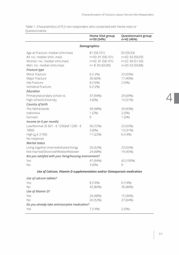
83
Characterization of Fracture Liaison Service Non-Responders
4
Table 1. Characteristics of FLS non-responders who consented with Home visits or Questionnaires.
Home Visit groupn=50 (54%)
Questionnaire groupn=42 (46%)
Demographics
Age at Fracture: median (min,max)All: no.: median (min, max)Women: no.: median (min,max)Men: no.: median (min,max)
81 (58,101)n=50: 81 (58,101)n=42: 81 (58,101)n= 8: 83 (65,89)
63 (50,93)n=42: 63 (50,93)n=22: 60 (51,93)n=20: 63 (50,88)
Fracture typeMinor fracture Major Fracture Hip Fracture Vertebral Fracture
6 (12%)30 (60%8 (16%)6 (12%)
23 (55%)17 (40%)2 (5%)-
Education Primary/secondary school vs. High school/University
47 (94%)3 (6%)
29 (69%)13 (31%)
Country of birthThe NetherlandsIndonesiaSurinam
49 (98%)1 (2%)0
39 (93%)2 (5%)1 (2%)
Income (in € per month)Low/Normal: (€ 601 - € 1200)/(€ 1200 - € 1800)High (> € 2100)No response
36 (72%)3 (6%)11 (22%)
23 (55%)13 (31%)6 (14%)
Marital statusLiving together (married/shared living)Not married/Divorced/Widow/Widower
26 (52%)24 (48%)
23 (55%)19 (45%)
Are you satisfied with your living/housing environment?Yes No
47 (94%)3 (6%)
42 (100%)0
Use of Calcium, Vitamin D supplementation and/or Osteoporosis medication
Use of calcium tablets? YesNo
8 (16%)42 (84%)
6 (14%)36 (86%)
Use of Vitamin D?YesNo
24 (48%)26 (52%)
15 (36%)27 (64%)
Do you already take antiresorptive medication?Yes 7 (14%) 2 (5%)

84
Chapter 4
Home Visit groupn=50 (54%)
Questionnaire groupn=42 (46%)
Patient opinions
What caused your fracture: Osteoporosis (yes/no; %) The fall (yes/no, %) The accident (yes/no, %) Bad physical condition (yes/no, %)Imbalance / dizziness (yes/no, %)
p= .02p= .01NSp= .0001p=.03
8 (16%)48 (96%)35 (70%)18 (36%)12 (24%)
42 (84%)2 (4%)15 (30%)32 (64%)38 (76%)
1 (2%)33 (79%)28 (67%)1 (2%)3 (7%)
41 (98%)9 (21%)14 (33%)41 (98%)39 (93%)
In my opinion subsequent fracture risk is increased after fracture.YesNoDo not know
13 (26%)18 (36%) 19 (38%)
7 (17%)22 (52%) 13 (31%)
Perceived advice to attend the FLSPerceived advice to attend the FLS by letter = YesPerceived advice to attend the FLS by letter = No
34 (68%)
16 (32%)
23 (55%)
19 (45%)
Taking interest in Bone QualityYesNo/Somewhat
22 (44%)28 (56%)
14 (33%)28 (67%)
Various aspects of health and frailty
How healthy is your lifestyle?HealthyNot healthy
31 (62%)19 (38%)
24 (57%)18 (43%)
Self-reported Level of HealthGoodPoor
25 (50%)25 (50%)
32 (76%)10 (24%)
Do you suffer from 2 or more chronic diseases?YesNo
17 (34%)33 (66%)
12 (29%)30 (71%)
Do you take 4 or more tablets every day?YesNo
25 (50%)25 (50%)
7 (17%)35 (83%)
Frailty (Tilburg Frailty Indicator scores)TFI score > 5 (frail)TFI score < 5 (not frail)
23 (46%)27 (54%)
13 (31%)29 (69%)
Parental hip FractureYesNo
6 (12%)44 (88%)
6 (14%)36 (86%)
Legends: Results are presented as median (min, max) or no. (%).

85
Characterization of Fracture Liaison Service Non-Responders
4
Table 2. Significant univariate associations to consent with Home Visit (HV) versus Questionnaire (Q).
Factors OR 95%CI p-valueAge ≥ 70 year 17.3 6.03;49.7 < .001Woman 4.77 1.81;12.6 .001Low Income 6.78 1.74;26.4 .003Self-rated low quality of life after fracture 4.26 1.59;11.4 .003Loss of weight after fracture 3.09 1.09;8.78 .03Difficulty in walking 3.97 1.58;9.99 .003Difficulty maintaining your balance 6.83 2.31;20.2 < .001Poor hearing 7.97 2.16;29.4 < .001Lack of strength in your hands 3.81 1.27;11.5 .013Age ≥ 70 years and no frailty (TFI ≥ 5) 19.2 3.6;102.0 < .001Major fractures (no hip or vertebral fractures) 2.89 1.2;6.94 0.02All Major fractures (all major including hip and vertebral fractures)
4.27 1.88;51.59 0.04
Legends:
OR: Odds Ratio; 95%CI: 95% Confidence Interval for population Odds Ratio, p-value < .05 is considered significant.
The recalls of patient information in HV or Q patients.
None of the HV and Q patients could recall any face-to-face patient information in the
hospital during time of fracture treatment including advice to attend the FLS. Receipt of
an FLS invitation letter was recalled by 68% of HV versus 55% of Q patients (p = .002).
Factors associated with consent for HV or Q
The proportion of patients that perceived a poor physical condition as the main cause
of fracture was significantly higher in the HV compared to the Q group (36% and 2%,
p = 0.0001). The proportion of patients with a “poor” self-reported health was also
significantly higher in HV than Q (50% vs 24%, p = .001).
HV and Q patients considered falls, the accident, bad physical condition and dizziness
or imbalance as main cause of recent fracture. In addition, the main cause of fracture
was differently believed between HV and Q; the fall: 96% versus 79%, p = .02, bad
physical condition: 36% versus 2%, p = .0001, dizziness or imbalance: 24% versus
7%, p = .03, while for osteoporosis it was 16% in HV versus 2% in Q, p = .02 and
the belief that subsequent fracture risk was increased; 26% versus 17%, respectively
(NS). There was no difference in patients` reports about have more than 2 chronic

86
Chapter 4
diseases or using more than 4 tablets per day, see Table 1.
According to univariate analysis HV patients were older (age > 70 years; OR 17.3), with
a higher contribution of women (OR 4.77) and more major fractures (OR 4.27). HV
patients reported lower income more frequently (OR 6.78) as well as quality of life (OR
4.26) and weight loss after fracture (OR 3.09). Finally, more HV patients had walking
difficulties (OR 3.97) and difficulties in maintaining balance (OR 6.83), more frequent
hearing loss (OR 7.97) and loss of grip strength (OR 3.81). In both HV and Q there was
a moderate frailty prevalence and no significant difference between groups according
to TFI (Frailty: ≥ 5). Only significant univariate associations to consent with HV versus
Q are shown in Table 2.
In a multivariate model, three factors remained significantly associated with consent for
HV versus Q: age > 70 years (OR: 16.8), gender (women, OR: 5.3) and major fractures
(OR 6.1); the same three factors were associated with consent for HV versus non-HV|Q:
(OR 2.7, 2.5 and 2.4, respectively). Age < 70 years and minor fractures (OR 3.2 and 5.3,
respectively) were associated to consent with Q versus non-HV|Q (Table 3).
Table 3. Significant multivariate associations to consent with HV versus Q.
Factors - HV (n=50) versus Q (n=42) OR (95%CI) p valueAge ≥ 70 16.8 (5.0; 56.7) < .001Women 5.3 (1.5; 19.2) .008Major Fractures 6.1 (1.7; 22.3) .0045
Legends:
Multivariate associations in Age (dichotomized at ≥ 70 or < 70), Gender and Fracture Type. Factors were first analysed of association with each pair of groups using crosstabulation, using adjusted standardized residuals and Chi-square/Fisher Exact tests p value for significance and Odds Ratio for measure of strength. OR: Logistic Regression Odds Ratio; 95%CI: 95% Confidence Interval for population Odds Ratio; p value < .05 is considered significant.

87
Characterization of Fracture Liaison Service Non-Responders
4
Tabl
e 4.
Dis
trib
utio
n of
Age
, Gen
der a
nd F
ract
ures
in F
LS n
on-r
espo
nder
s an
d at
tend
ers.
FLS
non-
resp
onde
rs/n
on-a
tten
ders
FLS
atte
nder
s
Dem
ogra
phic
s / F
ract
ures
HV
n= 5
0Q
n=
42no
n-H
V|Q
n=
81N
ot re
ache
d n=
16
FLS
Atte
nder
s n=
510
Age
at F
ract
ure
(min
, max
) W
omen
no.
(%)
med
ian
(min
, max
)M
en: n
o. (%
) m
edia
n (m
in, m
ax)
81 (5
8,10
1)n=
42
(84%
) 81
(58,
101)
n= 8
(16%
) 83
(65,
89)
63 (5
0,93
) n=
22
(58%
) 62
(51,
93)
n= 2
0 (4
2%)
64 (5
0,88
)
70 (5
0,92
)n=
53
(65%
) 6
7 (5
0,92
)n=
28
(35%
) 69
(50,
92)
78 (6
7,91
)n=
9 (5
6%)
79 (7
2;84
)n=
7 (4
4%)
77 (6
7;91
)
67 (5
0,95
)n=
363
(71%
) 67
(50,
95)
n= 1
47 (2
9%)
63 (5
0,83
)
Min
or fr
actu
re n
o. (%
)- D
ecea
sed
no.
All M
ajor
Fra
ctur
es n
o. (%
)- D
ecea
sed
no.
6 (1
2%)
1 44 (8
8%)
3
22 (5
2%)
1 20 (4
8%)
1
36 (4
4%)
- 45 (5
6%)
6
- - 16 5
219
(43%
)3 29
1 (5
7%)
5
Maj
or F
ract
ure
- Dec
ease
d no
. H
ip F
ract
ure
- Dec
ease
d no
.Ve
rteb
ral F
ract
ure
- Dec
ease
d no
.
29
3 9 - 6
-
17 1 3 - 0 -
12
2 26
4 7
-
4 2 9 3 3 -
225
2 38
2 28
1
Lege
nds:
Resu
lts a
re p
rese
nted
as
med
ian
(min
, max
) or n
o. D
ecea
sed:
stu
dy p
artic
ipan
ts w
ho re
tros
pect
ivel
y ha
d di
ed w
ithin
1 y
ear a
fter s
tudy
sta
rt a
nd c
losu
re o
f the
stu
dy in
no.
Fr
actu
res
wer
e ca
tego
rized
acc
ordi
ng to
War
riner
[19]
. Abb
r.: H
V= c
onse
nted
with
hom
e vi
sit;
Q=
cons
ente
d to
fill
in a
que
stio
nnai
re; n
on-H
V|Q
= no
con
sent
for H
V or
Q.

88
Chapter 4
Distribution of fractures among FLS non-responders and the patients who attended
the FLS.
Distribution according to fracture type (major fractures including hip and vertebral
fractures versus minor fractures) was compared between FLS attenders and traceable
FLS non-responders (Table 4).
The proportion of patients with major fractures in FLS attenders and traceable non-
responders was similar 291/510 (57%) and 109/173 (63%). The proportion of major
fractures was significantly higher in HV than in Q (p < .0001) and non-HV|Q (p =
.0001). In the latter two groups minor and major fracture proportions were similar.
Deceased patients
Several patients with major fractures died within 12 months after initiation of the
study; 3 patients in HV and 1 in Q), 6 patients in non-HV|Q, 5 were deceased in
not-reached patients and 5 were deceased in FLS attenders. In minor fractures it
was 1 patient in HV and 1 patient in Q and 3 patients after minor fracture in the FLS
attending group. All of the 16 untraceable individuals were elderly patients and 12
patients in this group had died within 12 months post-fracture.
Discussion
This study was specifically designed to characterize patients who did not respond
to FLS invitation after a recent fracture and showed that it is feasible to trace and
contact more than 90% of non-responders by one phone call of whom more than
25% agreed with a home visit, especially women of 70 years or older who sustained
a major fracture. They primarily considered a fall, their bad physical condition,
dizziness or balance problems as the main cause of the recent fracture. Only one
in five believed that subsequent fracture risk was increased, and a small proportion
considered osteoporosis as underlying risk factor for subsequent fractures. As far as
we know the characteristics of FLS non responders have not been studied before by
home visits and questionnaires. Therefore, the current study design differs entirely
from an earlier study by our group comparing personal opinions, drives and motives
of patients who were willing to share details of their decision to attend or not attend
the FLS [7].
In this study, 931 fracture patients were identified of whom 716 were eligible for FLS
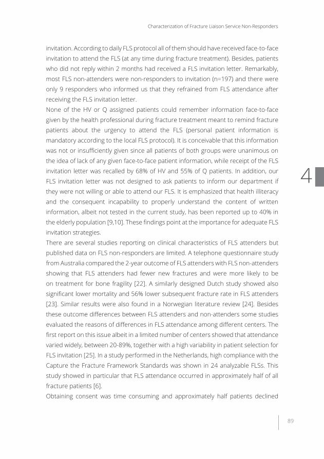
89
Characterization of Fracture Liaison Service Non-Responders
4
invitation. According to daily FLS protocol all of them should have received face-to-face
invitation to attend the FLS (at any time during fracture treatment). Besides, patients
who did not reply within 2 months had received a FLS invitation letter. Remarkably,
most FLS non-attenders were non-responders to invitation (n=197) and there were
only 9 responders who informed us that they refrained from FLS attendance after
receiving the FLS invitation letter.
None of the HV or Q assigned patients could remember information face-to-face
given by the health professional during fracture treatment meant to remind fracture
patients about the urgency to attend the FLS (personal patient information is
mandatory according to the local FLS protocol). It is conceivable that this information
was not or insufficiently given since all patients of both groups were unanimous on
the idea of lack of any given face-to-face patient information, while receipt of the FLS
invitation letter was recalled by 68% of HV and 55% of Q patients. In addition, our
FLS invitation letter was not designed to ask patients to inform our department if
they were not willing or able to attend our FLS. It is emphasized that health illiteracy
and the consequent incapability to properly understand the content of written
information, albeit not tested in the current study, has been reported up to 40% in
the elderly population [9,10]. These findings point at the importance for adequate FLS
invitation strategies.
There are several studies reporting on clinical characteristics of FLS attenders but
published data on FLS non-responders are limited. A telephone questionnaire study
from Australia compared the 2-year outcome of FLS attenders with FLS non-attenders
showing that FLS attenders had fewer new fractures and were more likely to be
on treatment for bone fragility [22]. A similarly designed Dutch study showed also
significant lower mortality and 56% lower subsequent fracture rate in FLS attenders
[23]. Similar results were also found in a Norwegian literature review [24]. Besides
these outcome differences between FLS attenders and non-attenders some studies
evaluated the reasons of differences in FLS attendance among different centers. The
first report on this issue albeit in a limited number of centers showed that attendance
varied widely, between 20-89%, together with a high variability in patient selection for
FLS invitation [25]. In a study performed in the Netherlands, high compliance with the
Capture the Fracture Framework Standards was shown in 24 analyzable FLSs. This
study showed in particular that FLS attendance occurred in approximately half of all
fracture patients [6].
Obtaining consent was time consuming and approximately half patients declined

90
Chapter 4
to consent for participation. Remarkably, most HV consenters hardly needed any
persuasion, while it took eight weeks until all questionnaires were returned by Q
patients. This difference may be explained by stronger concerns about health issues
and fracture risk in the HV group compared to Q. Another study of the FLS found that
older patients reported less mobility and more dependency on their caregivers for
transportation to the hospital [8]. We speculate therefore that older patients consider
a home visit as a solution for their inability to attend the FLS.
There were also similarities between both groups (HV and Q) noteworthy to report. i.e.
a similar low frailty prevalence (according to the TFI, frailty: ≥ 5) and a similar believed
major reasons of fracture i.e. the fall and accident. Another similar characteristic (39%
and 31%, respectively) was the low proportion of knowledge of patients concerning
increased subsequent fracture risk in agreement with previous reports [7,9,15,16].
ASBMR, IOF, EULAR and EUGMS fully endorse the importance of secondary fracture
prevention and FLS care is considered the most optimal approach in this respect
[2-5]. To optimize FLS care according to this vision the issue of attendance needs full
attention.
This study has several weaknesses. 1/ the study is an explorative study and therefore
not based on preliminary determined statistical power. The outcome does not
allow generalization outside this region and to other countries. 2/ Age was
dichotomously analyzed (< or >70 years), threshold proposed by the Dutch VMS
authority (Safety Management System) and commonly used for patient safety issues
in hospital care. [21]. Dichotomizing data can lower power in study outcomes. 3/ we
have no information on the exact number of patients who did or did not receive face-
to-face patient information during fracture treatment encouraging them to attend
the FLS.
In conclusion, a high proportion of FLS non-responders who participated in HV or Q
consider subsequent fracture risk to be low. Nevertheless, 50 patients (about 25%
of FLS non-responders) consented with a home visit after one telephone call, mainly
elderly women with a major fracture. These patients have a high subsequent fracture
risk and are approachable for evaluation. The encountered approachability of FLS
non-responders indicates that innovative strategies in optimizing FLS invitation can
improve secondary fracture prevention care.

91
Characterization of Fracture Liaison Service Non-Responders
4
References1. McLellan AR, Gallacher SJ, Fraser M, McQuillian C. The fracture liaison service: success of a
program for the evaluation and management of patients with osteoporotic fracture. Osteoporos Int. 2003 Dec;14(12):1028-34.
2. Akesson K, Marsh D, Mitchell PJ, McLellan AR, Stenmark J, Pierroz DD, Kyer C, Cooper C (IOF Fracture Working Group). Capture the Fracture®: a Best Practice Framework and global campaign to break the fragility fracture cycle. Osteoporos Int. 2013 24(8):2135-52. doi: 10.1007/s00198-013-2348-z.
3. Lems WF, Dreinhöfer KE, Bischoff-Ferrari H, Blauth M, Czerwinski E, da Silva J, Herrera A, Hoffmeyer P, Kvien T, Maalouf G, Marsh D, Puget J, Puhl W, Poor G, Rasch L, Roux C, Schüler S, Seriolo B, Tarantino U, van Geel T, Woolf A, Wyers C, Geusens P. EULAR/EFORT recommendations for management of patients older than 50 years with a fragility fracture and prevention of subsequent fractures. Ann Rheum Dis. 2017 May;76(5):802-810. doi: 10.1136/annrheumdis-2016-210289.
4. Eisman JA, Bogoch ER, Dell R, Harrington JT, McKinney RE Jr, McLellan A, Mitchell PJ, Silverman S, Singleton R, Siris E. Making the first fracture the last fracture: ASBMR task force report on secondary fracture prevention. J Bone Miner Res. 2012 Oct;27(10):2039-46. doi: 10.1002/jbmr.1698.
5. Blain H, Masud T, Dargent-Molina P, Martin FC, Rosendahl E, van der Velde N, Bousquet J, Benetos A, Cooper C, Kanis JA, Reginster JY, Rizzoli R, Cortet B, Barbagallo M, Dreinhöfer KE, Vellas B, Maggi S, Strandberg T; EUGMS Falls and Fracture Interest Group; European Society for Clinical and Economic Aspects of Osteoporosis and Osteoarthritis (ESCEO), Osteoporosis Research and Information Group (GRIO), and International osteoporosis Foundation (IOF). A Comprehensive Fracture Prevention Strategy in Older Adults: The European Union Geriatric Medicine Society (EUGMS) Statement. J Nutr Health Aging. 2016;20(6):647-52. doi: 10.1007/s12603-016-0741-y.
6. van den Berg P, Schweitzer DH, van Haard PM, van den Bergh JP, Geusens PP. Meeting international standards of secondary fracture prevention: a survey on Fracture Liaison Services in the Netherlands. Osteoporos Int. 2015 Sep;26(9):2257-63. doi: 10.1007/s00198-015-3117-y.
7. van den Berg P, van Haard PMM, Geusens PP, van den Bergh JP, Schweitzer DH. Challenges and opportunities to improve fracture liaison service attendance: fracture registration and patient characteristics and motivations. Osteoporos Int. 2019 May 25. doi: 10.1007/s00198-019-05016-4.
8. Eekman DA, van Helden SH, Huisman AM, Verhaar HJ, Bultink IE, Geusens PP, Lips P, Lems WF. Optimizing fracture prevention: the fracture liaison service, an observational study. Osteoporos Int. 2014 Feb;25(2):701-9. doi: 10.1007/s00198-013-2481-8.
9. Raybould G, Babatunde O, Evans AL, Jordan JL, Paskins Z. Expressed information needs of patients with osteoporosis and/or fragility fractures: a systematic review. Arch Osteoporos. 2018 May 8;13(1):55. doi: 10.1007/s11657-018-0470-4.
10. Heijmans M, Waverijn G, Rademakers J, van der Vaart R, Rijken M. Functional, communicative and critical health literacy of chronic disease patients and their importance for self-management. Patient Educ Couns. 2015;98(1):41-48.
11. Giangregorio L, Thabane L, Cranney A, Adili A, deBeer J, Dolovich L, Adachi JD, Papaioannou A. Osteoporosis knowledge among individuals with recent fragility fracture. Orthop Nurs. 2010 Mar-Apr;29(2):99-107. doi: 10.1097/NOR.0b013e3181d2436c. PMID: 20335769
12. Grover ML, Edwards FD, Chang YH, Cook CB, Behrens MC, Dueck AC. Fracture risk perception study: patient self-perceptions of bone health often disagree with calculated fracture risk. Womens Health Issues. 2014 Jan-Feb;24(1):e69-75. doi: 10.1016/j.whi.2013.11.007. PMID: 24439949
13. Alami S, Hervouet L, Poiraudeau S, Briot K, Roux C. One Barriers to Effective Postmenopausal Osteoporosis Treatment: A Qualitative Study of Patients’ and Practitioners’ Views. PLoS One. 2016

92
Chapter 4
Jun 29;11(6):e0158365. doi: 10.1371/journal.pone.0158365. eCollection 2016. PMID: 27355576
14. Ong T, Tan W, Marshall L, Sahota O. The relationship between socioeconomic status and fracture in a fracture clinic setting: data from the Nottingham Fracture Liaison Service. Injury. 2015 Feb;46(2):366-70. doi: 10.1016/j.injury.2014.10.002.
15. Boudreau DM, Yu O, Balasubramanian A, Wirtz H, Grauer A, Crittenden DB, Scholes D. A Survey of Women’s Awareness of and Reasons for Lack of Postfracture Osteoporotic Care. J Am Geriatr Soc. 2017 Aug;65(8):1829-1835. doi: 10.1111/jgs.14921.
16. Besser SJ, Anderson JE, Weinman J. How do osteoporosis patients perceive their illness and treatment? Implications for clinical practice. Besser SJ, Anderson JE, Weinman J. Arch Osteoporos. 2012;7:115-24. doi: 10.1007/s11657-012-0089-9. PMID: 23225289
17. Dutch Institute for Healthcare Improvement CBO (2011) Richtlijn Osteoporose en Fractuurpreventie. www.diliguide.nl/document/1015/file/pdf/. (Dutch) Assessed 14-02-2018
18. https://chipsoft.com/?Source=https%3A%2F%2Fwww%2Echipsoft%2Enl%2F. Assessed 14-02-2018
19. Warriner A ea. Minor, major, low-trauma, and high-trauma fractures: what are the subsequent fracture risks and how do they vary? Curr. Osteoporos Rep. 2011 Sep;9(3):122-8. doi: 10.1007/s11914-011-0064-1.
20. Gobbens RJ, van Assen MA, Luijkx KG, Wijnen-Sponselee MT, Schols JMJ. The Tilburg Frailty Indicator: psychometric properties. Am Med Dir Assoc. 2010 Jun;11(5):344-55. doi: 10.1016/j.jamda.2009.11.003.
21. https://www.vmszorg.nl/wp-content/uploads/2017/11/web_2009.0104_praktijkgids_kwetsbare_ouderen.pdf (Dutch) assessed 30-12-2018
22. Van der Kallen J, Giles M, Cooper K, Gill K, Parker V, Tembo A, Major G, Ross L, Carter J. A fracture prevention service reduces further fractures two years after incident minimal trauma fracture. Int J Rheum Dis. 2014 Feb;17(2):195-203. doi: 10.1111/1756-185X.12101.
23. Huntjens KM, van Geel TA, van den Bergh JP, van Helden S, Willems P, Winkens B, Eisman JA, Geusens PP, Brink PR. Fracture liaison service: impact on subsequent nonvertebral fracture incidence and mortality. J Bone Joint Surg Am. 2014 Feb 19;96(4):e29. doi: 10.2106/JBJS.L.00223.
24. Andreasen C, Solberg LB, Basso T, Borgen TT, Dahl C, Wisløff T, Hagen G, Apalset EM, Gjertsen JE, Figved W, Hübschle LM, Stutzer JM, Elvenes J, Joakimsen RM, Syversen U, Eriksen EF, Nordsletten L, Frihagen F, Omsland TK, Bjørnerem Å. Effect of a Fracture Liaison Service on the Rate of Subsequent Fracture Among Patients With a Fragility Fracture in the Norwegian Capture the Fracture Initiative (NoFRACT): A Trial Protocol. JAMA Netw Open. 2018 Dec 7;1(8):e185701. doi: 10.1001/jamanetworkopen.2018.5701.
25. Vranken L, Wyers CE, van den Bergh JPW, Geusens PPMM. The Phenotype of Patients with a Recent Fracture: A Literature Survey of the Fracture Liaison Service. Calcif Tissue Int. 2017 Sep;101(3):248-258. doi: 10.1007/s00223-017-0284-1.

93
Characterization of Fracture Liaison Service Non-Responders
4

9494
5

95
Peter van den Berg (1), Paul M.M. van Haard (2), Joop P. van den Bergh (3), Dieu Donné
Niesten (4), Maarten van der Elst (5), Dave H. Schweitzer (6) *.
1. Dept. of Orthopedics and Surgery, Reinier de Graaf Hospital, Delft,
The Netherlands.
2. Dept. Medical Laboratories/Diagnostic Centre SSDZ, Reinier de Graaf Hospital,
Association of Clinical Chemistry, Delft, The Netherlands.
3. Dept. of Internal Medicine, VieCuri Medical Centre Noord-Limburg and
Department of Internal Medicine, Subdivision Rheumatology, Maastricht
University Medical Centre, Maastricht, The Netherlands.
4. Dept. of Orthopaedics, Reinier de Graaf Hospital, Delft, The Netherlands.
5. Dept. of Surgery, Reinier de Graaf Hospital, Delft, The Netherlands.
6. Dept. of Internal Medicine, Reinier de Graaf Hospital, Delft, The Netherlands. *Corresponding author: Dave H. Schweitzer, Dept. of Internal Medicine, Reinier
de Graaf Hospital, Reinier de Graafweg 3–11, 2625AD Delft, The Netherlands;
E-mail: [email protected]; Tel.: +31 703401106; Fax: +31 70 3401103
First Quantifi cation of Calcium Intake from Calcium-Dense Dairy Products in Dutch Fracture Patients (The Delft Cohort Study)
CHAPTER 5
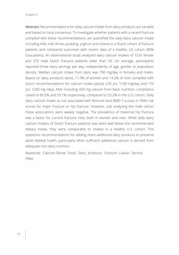
96
Chapter 5
Abstract: Recommendations for daily calcium intake from dairy products are variable
and based on local consensus. To investigate whether patients with a recent fracture
complied with these recommendations, we quantified the daily dairy calcium intake
including milk, milk drinks, pudding, yoghurt and cheese in a Dutch cohort of fracture
patients and compared outcomes with recent data of a healthy US cohort (80%
Caucasians). An observational study analyzed dairy calcium intakes of 1526 female
and 372 male Dutch fracture patients older than 50. On average, participants
reported three dairy servings per day, independently of age, gender or population
density. Median calcium intake from dairy was 790 mg/day in females and males.
Based on dairy products alone, 11.3% of women and 14.2% of men complied with
Dutch recommendations for calcium intake (adults ≤70 yrs: 1100 mg/day and >70
yrs: 1200 mg /day). After including 450 mg calcium from basic nutrition, compliance
raised to 60.5% and 59.1% respectively, compared to 53.2% in the U.S. cohort. Daily
dairy calcium intake as not associated with femoral neck BMD T-scores or FRAX risk
scores for major fracture or hip fracture. However, sub analyzing the male cohort
these associations were weakly negative. The prevalence of maternal hip fracture
was a factor for current fracture risks, both in women and men. While daily dairy
calcium intakes of Dutch fracture patients was were well below the recommended
dietary intake, they were comparable to intakes in a healthy U.S. cohort. This
questions recommendations for adding more additional dairy products to preserve
adult skeletal health, particularly when sufficient additional calcium is derived from
adequate non-dairy nutrition.
Keywords: Calcium-Dense Food; Dairy products; Fracture Liaison Service;
FRAX.

97
Quantification of Calcium Intake from Calcium-Dense Dairy Products
5
Introduction In 1993, the U.S. Food and Drug Administration authorized a health claim for foods and
supplements related to calcium to prevent osteoporosis. In January 2010, the health
claim was expanded for the combination of calcium and vitamin D supplements. (FDA
21CFR101.72) This claim raises questions about the amount of calcium that should
be recommended to the adult population [1]. Today, the Recommended Dietary
Intake (RDI) for calcium, meaning the average daily level of intake sufficient to meet
the nutrient requirements of healthy individuals, is 1200 mg for individuals older than
50 yrs in the US (Food Guide Pyramid FGP, US Department of Agriculture) [2]. In the
Netherlands, RDI for total daily calcium intake is quite similar to that in the US: 1100
mg for the age category 51-69 yrs and 1200 mg for people older than 70 yrs. Based
on the US recommendation, the number of recommendable calcium-dense servings
should be four per day [2,3], which is more than the actual average consumption in
the Netherlands as well as in the USA. While extra intake of dairy servings would seem
the most straightforward way to increase calcium intake, it is questionable whether it
is beneficial for bone health to use more dairy servings than those taken according to
cultural habits and traditions. Over-nutrition of dairy products may cause maldigestion
and malabsorption, which may overrule beneficial effects of calcium intake. Besides, it
is unclear how traditional use of liquid milk and milk products relates to bone mineral
density (BMD) and fracture risk during adulthood.
Public health authorities [4] claim that actual calcium intake is 25 percent lower
in Europe than in the U.S. Although a large part of the agricultural activities in the
Netherlands is dedicated to milk production and processing, the average Dutch
consumption of liquid milk and milk products per capita in kg/year is estimated to be
83.5, while it is 85.1 on average in 27 other European countries and 111.4 in the U.S.
[5]. According to the Dutch National Food Consumption Survey (DNFCS 2007-2010),
the median habitual total calcium intake per day (51-69 yrs; P50) in Dutch men is 1109
mg calcium and in women 985 mg [6].Non-dairy food sources contribute 42% of total
calcium content in daily nutrition [6].
Since dairy products consumption per capita in kg/year in the Netherlands compares
well with the average in North-Western Europe, we were interested in comparing
calcium intake in the Dutch adult population with that of an U.S. cohort of more than
80% Caucasians, which was extracted from the Continuing Survey of Food Intake by
Individuals, 1994-96, 1998, (CSFII) and the National Health and Nutrition Examination

98
Chapter 5
Survey, 1999-2000 (NHANES). We wondered whether the CSFII en NHANES reports
would agree with the number of daily servings taken by Dutch individuals.
In order to answer this question, we conducted the present study at the Fracture
Liaison Service (FLS) of the Reinier de Graaf Group of Hospitals in The Netherlands.
Our primary research goals were to determine the number of daily servings of dairy
products in a Dutch cohort of fracture patients after partitioning for age and gender,
but also for population density; to correlate daily calcium intake from dairy products
with femoral neck BMD T-scores and 10-year probability of major fracture or hip
fracture; and to compare intakes with another, mainly Caucasian cohort in the U.S.,
consisting of patients without reported fracture. We further questioned whether the
total amount of calcium from both daily basic nutrition and daily dairy consumption is
appropriate for fulfilling Dutch recommendations for total calcium intake.
Experimental Section
Patients and methods
This prospective observational study was conducted at the Fracture Liaison Service
(FLS) of the Reinier the Graaf Group of Hospitals, Delft (The Netherlands). The FLS is
organized according to a previously reported concept [7-9]. We included women and
men of 50 yrs and older with a recent fracture. These patients were evaluated by
means of a structured diagnostic work up, including a detailed questionnaire regarding
their daily calcium intake from dairy and a Dual Energy X-Ray Absorptiometry (DXA)
measurement for femoral neck BMD T-scores. Each patient received a scan, with the
exception of those who were already known with osteoporosis evidenced by older
DXA measurements or with prevalent vertebral fractures evidenced by previous spinal
radiographs. Patients using prescribed calcium and/or bisphosphonates were excluded.
Calcium intake was assessed using a validated Dutch questionnaire on Daily Calcium
Intake (DCI-Holland), which was developed by Het Rijksinstituut voor Volksgezondheid
en Milieu (National Institute for Public Health and the Environment, RIVM), Bilthoven,
the Netherlands; see: NEVO-online.rivm.nl [4]. The DCI-Holland calculated the calcium
content of food categories such as meat, fish, poultry, eggs, vegetables, fruits, potatoes,
pasta, and milk products. According to the DCI-Holland, the average calcium content
is 270 mg in one glass of milk (200 ml), 400 mg in one glass of calcium-enriched

99
Quantification of Calcium Intake from Calcium-Dense Dairy Products
5
milk, and 240 mg in one serving of yoghourt or pudding (200 ml). Non-dairy food
contributes (as median) 430 mg calcium in females and 443 mg calcium in males,
based on the habitual intake distribution at P50 of calcium food sources by the Dutch
population aged 51-69 yrs, weighted for socio-demographic factors, season and day
of the week.
Patients were recruited from the Emergency Department or were outpatients of the
departments of Trauma surgery or Orthopaedic surgery. Each patient was asked to fill
in a questionnaire that included questions about calcium intake from dairy nutrition
categorized per product type (milk, calcium-enriched milk, yoghurt, pudding, cheese)
as mentioned in the DCI-Holland. We estimated total daily dairy calcium intake using
the number of glasses of milk or yoghourt/pudding servings. The calcium intake of
cheese products was dichotomized (Yes/No). The intake of calcium from cheese
products was calculated as half the amount in a glass of milk. The questionnaire also
included the WHO Fracture Assessment Tool (FRAX) for evaluating fracture risks [10]
and additional questions regarding age at menopause, daily exercise and sports
habits. Calcium intake from dairy nutrition, per serving, was calculated using the
formula of Fulgoni [2], one serving representing 250 mg calcium, according to the
Dutch Standard (DCI-Holland).
FRAX risk scores were calculated according to the Dutch FRAX algorithm [11].
Osteoporosis was defined as a T-score ≤ -2.5*SD (SD=standard deviation) at either
total hip, femoral neck or lumbar spine, normal BMD as a T-score ≥ -1*SD at all three
locations and osteopenia as a T-score between -1*SD and -2.5*SD.
In patients with a T-score less than -2*SD, additional laboratory assessments and
radiographs of lumbar and thoracic spine were performed, according to protocol.
Vertebral fractures were scored according to the semi-quantitative method of Genant
et al.. A vertebral fracture was defined as an infraction of at least 25% reduction in
anterior, middle and/or posterior height and a reduction in area by 20-40% [12].
Analyses were performed on 5 age decades (50-59, 60-69, 70-79, 80-89 and 90-100
yrs), gender, and on 5 (Dutch) population density categories (1: 21-250, 2: 250-500, 3:
500-1000, 4: 1000-2500 and 5: 2500-5976 inhabitants per square kilometres (source:
Dutch National Atlas of Public Health; see: www.zorgatlas.nl.)[13].
Data were analysed using Statgraphics Centurion XVI software (Version 16.2.4 for MS-
Windows; Statpoint, Inc., VA, USA). Multiple-variable correlation analysis was applied
after partitioning of data by gender. Wilcoxon&Mann&Whitney rank-sum test was

100
Chapter 5
applied to compare medians of numerical variables with respect to gender. Mood’s
Median and Kruskall&Wallis tests were applied to genders in order to compare
numerical variables between the categories of the diagnosis variable (normal,
osteopenia, osteoporosis). Variance Components Analysis was applied, using fracture
frequency as a dependent variable and relevant numerical variables as factors in
order to assess their contribution to total variation in fracture frequency. To estimate
odds ratios in genders, logistic regression with backward factor selection (p-to-enter
.05; p-to-remove .05) was applied, with maternal hip fracture as dependent variable
(Yes=1; No=0) and all numerical and dichotomous categorical variables as factors.
Non-parametric correlations were then assessed again between final numerical
factors of the best logistic regression model. Where applicable, 95% Confidence
intervals were used and a p-value < .05 was considered statistically significant at the
95% confidence level. The study was approved by the Medical Review Board (METC-
Zuid Holland), The Netherlands.
Results From March 2008 to November 2011, we included 1526 women and 372 men
of 50 yrs and older with a recent fracture. All patients were Caucasians. Of these
patients, 1451 women and 349 men received a Dual Energy X-Ray Absorptiometry
(DXA: Hologic QDR 4500 C) of total hip, femoral neck and lumbar spine and had a
consultation at the outpatient clinic with a specialised nurse practitioner. Relevant
complete data were obtained from all 1898 patients who were originally included,
and demographics are listed in Table 1 and 2. Calculated daily dairy calcium intakes in
genders are graphically represented in Figure 1 and the distribution of genders into
age decades is listed in table 3.
Table 1. Demographic data of a Dutch cohort after a recent fracture
Sex Numbers Age % Osteoporosis(T-score<-2.5*SD)
FRAX,%(for major fracture)
FRAX,%(for hip fracture)
Women 1526 66 (50-96) 13.6 11.0 (2.4-90.0) 2.6 (0.0-73.0)
Men 372 65 (50-90) 6.2 7.5 (1.6-41.0) 2.7 (0.1-37.0)
Fracture Assessment Tool (FRAX) was used to estimate FRAX risk scores for major osteoporotic fracture or hip fracture. Data were not normally distributed and are given as Median (Range).

101
Quantification of Calcium Intake from Calcium-Dense Dairy Products
5
Table 2. Variables of a Dutch cohort after a recent fracture
Variables Data
Sex
Women 0
Men 1
Age decades
50-59 yrs 1
60-69 yrs 2
70-79 yrs 3:
80-89 yrs 4:
90-100 yrs 5
Age at menopause age (yrs), women only
Population density
category 1 21-250 inhabitants/km2
category 2 250-500 inhabitants/ km2
category 3 500-1000 inhabitants/ km2
category 4 1000-2500 inhabitants/ km2
category 5 2500-5967 inhabitants/ km2
Dairy calcium intake
per serving milk 270 mg calcium
per serving yoghourt 240 mg calcium
per serving cheese 160 mg calcium
Currently smoking
No 0
Yes 1
Alcohol
≥ 3 servings per day 1
≤ 2 servings per day 0
Current use of corticosteroids
No 0
Yes 1

102
Chapter 5
Variables Data
Rheumatoid arthritis
No 0
Yes 1
Previous fracture
No 0
Yes 1
Hip fracture mother
No 0
Yes 1
Current use of calcium prescribed or as OTC
Excluded
Current use of any drug against osteoporosis #
Excluded
Length cm
Weight kg
BMI Body Mass Index: Weight/(Length*Length)
BMD Bone Mineral Density test, BMD-at-the-femoral neck (T-score)
FRAX 10-year probability of major osteoporotic fracture
FRAX Hip 10-year probability of hip fracture
Secondary causes for osteoporosis known to the patientNo 0
Yes 1
Legends:
OTC: Over-The-Counter available drugs not prescribed by a physician.# Any drug against osteoporosis i.e. bisphosphonates, estrogens or Selective Estrogen Receptor Modulators

103
Quantification of Calcium Intake from Calcium-Dense Dairy Products
5
Fig 1. Distributions of daily calcium intake (mg/day) from dairy products in women and men.
Histogram Females
Dairy Calcium Intake (mg/day)0 1 2 3 4
(X 1000,0)
0
100
200
300
400
freq
uenc
y
Histogram Males
Dairy Calcium Intake (mg/day)0 1 2 3 4
(X 1000,0)
0
20
40
60
80
100
freq
uenc
y
This study included more women than men, but genders were distributed equally over age decades, as is demonstrated in Table 2.

104
Chapter 5
Table 3. Distribution of 1898 patients into 5 age decades (range 50-100 yrs).
Age (yrs) WomenW (N=1526)
MenM (N=372)
Ratio of gendersF/M
50-59 399 (26.1) 113 (30.4) 3.560-69 528 (34.6) 125 (33.6) 4.270-79 428 (28.0) 96 (25.8) 4.580-89 161 (10.6) 37 (10.0) 4.4
90-100 10 (0.7) 1 (0.2) 10.0.
Legends: Data for women and men per age decade are given in numbers of patients and percentage of patients (%).
Overall, we found no significant difference in calcium intakes from dairy food products
between women and men (median 790 (range 0-4360) and 790 (0-3435) mg/day,
respectively, p = .80). The dairy calcium intake was also expressed as numbers of
servings per day, which were similar for each age decade except for those patients
who were older than 80 yrs (Table 4). Numbers of servings did not differ between
genders, nor between female and male cohorts compared for population density
categories (Table 5).
Table 4. Number of daily servings of dairy products in females and males distributed into 5 age decades
Age (yrs)
Women(N=1526)
Servings/day
50-59 399 (26.1) 3 (0-9)60-69 528 (34.6) 3 (0-15)70-79 428 (28.0) 3 (0-13)80-89 161 (10.6) 3 (0-8)90-100 10 (0.7) 3 (2-4)Age (yrs)
Men(N=372)
Servings/day
50-59 113 (30.4) 3 (0-15)60-69 125 (33.6) 3 (0-9)70-79 96 (25.8) 3 (0-8)80-89 37 (10.0) 3 (1-5)90-100 1 (0.2) 1
Legends: Data for women and men per age decade are given in numbers of patients having completed the questionnaire and in percentage of patients (%). Data for number of servings were not normally distributed and are given as median (range).

105
Quantification of Calcium Intake from Calcium-Dense Dairy Products
5
Table 5. Number of daily servings of dairy products in women and men grouped by population density
Population Density Category (Inhabitants per Km2) Women (N=1526) Servings/day2 (250 – 500) 98 (6.4) 3 (1-10)3 (500 – 1000) 5 (0.3) 4 (3-5)4 (1000 – 2500) 565 ( 37.0) 3 (0-9)5 (2500 – 5976) 858 (56.3) 3 (0-15)Population Density Category (Inhabitants per Km2) Men (N=372) Servings/day2 (250 – 500) 19 (5.1) 3 (1-6)3 (500 – 1000) 2 (0.5) 4 (3-5)4 (1000 – 2500) 156 (41.9) 3 (0-15)5 (2500 – 5976) 195 (52.5) 3 (0-9)
Legends:
Inhabitants per Km2 per category was defined according to www.zorgatlas.nl [13]. Category 1 was not represented in the study region. Data for women and men per population density category are given in numbers of patients and in percentage of patients (%).Data for number of servings were not normally distributed and are given as median (range).
Most women and men were osteopenic and used 500 -1000 mg calcium calculated
from total dairy product intake per day. No significant differences in patients in both
genders were found for dairy intake nor for DXA T-scores normal BMD, osteopenia or
osteoporosis (Table 6).
Table 6. Stratified Calcium intakes from dairy products in women and men grouped by Dual Energy X-Ray Absorptiometry (DXA) T-scoresDairy Calcium intake (mg/day) Women (N=1451)
DXA T-score (*SD)≥ -1 -1.0 to -2.5 ≤ -2.5
<500 42 (2.9) 141 (9.7) 49 (3.4)500-1000 182 (12.5) 445 (30.7) 152 (10.5)1000-1500 87 (6.0) 228 (15.7) 59 (4.1)1500-2000 12 (0.8) 34 (2.3) 6 (0.4)≥2000 4 (0.3) 7 (0.5) 3 (0.2)Dairy Calcium intake (mg/day) Men (N=349)
DXA [T-score (*SD)]≥ -1 -1.0 to -2.5 ≤ -2.5
<500 12 (3.4) 37 (10.6) 9 (2.6)500-1000 53 (15.2) 118 (33.8) 14 (4.0)1000-1500 21 (6.0) 62 (17.8) 6 (1.7)1500-2000 2 (0.6) 9 (2.6) 0 (0.0)≥ 2000 0 (0.0) 6 (1.7) 0 (0.0)
Legends: There were no significant differences between calcium intake from dairy per DXA T-score category, neither within gender nor between genders.

106
Chapter 5
Median (range) FRAX 10-year probability for major osteoporotic fracture was
significantly higher in women than in men (11.0% (2.4 - 90.0) and 7.5% (1.6 - 41.0)
respectively, p < .05). 10-year probability of hip fracture (median(range)) was not
significantly different between genders (2.6% (0.0 -73.0) in women and 2.7% (0.1 -
37.0) in men, respectively).
In men (Figure 2), dairy calcium intake showed a weak but significant negative
correlation with the 10-year FRAX probability of major fracture as well as of hip
fracture (Rho: -.13 and -.14, respectively, p < .01), but no significant correlation with
age, height, Body Mass Index (BMI), weight or BMD at the femoral neck. In females
we found no significant correlation between dairy calcium intake and the variables
mentioned, nor with ‘age at menopause’.
Logistic regression analysis disclosed ̀ maternal hip fracture` as a dependent variable.
This was not found for any of the other variables like age, BMI, age at menopause
(women only), population density, dairy calcium intake, more than one fracture prior
to analysis, currently smoking, currently glucocorticoids use, rheumatoid arthritis,
secondary causes for osteoporosis known to the patient, alcohol use (either ≤2 or
≥3 servings), BMD at the femoral neck, 10-year probability of osteoporotic major
fracture, and 10-year probability of hip fracture. In women, logistic regression showed
a weak relationship between `maternal hip fracture` and the independent variables
(factors) BMD-at-the-femoral neck, 10-year probability of osteoporotic major fracture,
10-year probability of hip fracture, BMI, height, weight, Alcohol use=0, currently
smoking=No. In men, logistic regression showed a strong relationship between
`maternal hip fracture` and the independent variables BMD at the femoral neck,
10-year probability of osteoporotic major fracture, 10-year probability of hip fracture,
BMI, and Prednisone=0.

107
Quantification of Calcium Intake from Calcium-Dense Dairy Products
5
Fig 2. Relationship between daily dairy calcium intake and 10-year FRAX probability of osteoporotic major fracture or hip fracture in men.
10-year FR AX probability of os teoporotic major fracture in Men
Dairy C alcium Intake (mg/day)
Maj
or f
ract
ure
risk
(%
)
0 1 2 3 4(X 1000,0)
0
10
20
30
40
50
10-year FR AX probability of hip fracture in Men
Dairy C alcium Intake (mg/day)
Hip
fra
ctur
e ri
sk (
%)
0 1 2 3 4(X 1000,0)
0
10
20
30
40

108
Chapter 5
Comparison between the current Dutch cohort and a cohort from the U.S.
The percentage of patients fulfilling the recommended total calcium intake of
>1100 mg/day (≤69 yrs) or > 1200 mg/day (>70 yrs) depends on the total amount
of calcium in Dutch basic nutrition. In this study, this would mean that focusing on
dairy intake alone, just 11.3% of the women and 14.2% of the men complied with
Dutch recommendation. However, assuming that Dutch basic non-dairy nutrition
provides 42% of the total daily calcium intake (P50= 450 mg) [6], our findings mean
that 60.5% of women and 59.1% of men complied with Dutch recommendations for
nutritional calcium intake. U.S. data from the group of Fulgoni [2] showed a mean
calcium intake of 674±6 mg/day from dairy products for both women and men older
than 50 yrs; 15.2% of both women and men complied with U.S. recommendations for
nutritional calcium intake. Moreover, U.S. non-dairy nutrition was estimated to contain
250-475 mg calcium, and complemented with 2.93 dairy servings this would imply
that 53.2% of this group complied with U.S. recommendations for calcium from
dailynutrition [3].
Discussion The current study shows that calcium intake from dairy products in a Dutch cohort
of Caucasians with a recent fracture was well below the Dutch RDI of 1100 mg for
individuals between 50 and 70 yrs and of 1200 mg for older people. There were no
differences in dairy calcium intake between age decades, genders or population density
categories. In contrast to women in men there was a weak but significantly negative
correlation between dairy calcium intake and 10-year probability of major fracture and
hipfracture. Importantly, the present study showed that prevalence of `maternal hip
fracture` was a factor for current fracture risk, both in women and men.
In spite of the fact that the esteemed calcium intake is lower in most European
countries than in the US [2], there were no significant differences between our cohort
of fracture patients and a comparable non-fracture cohort from the U.S. in terms
of dairy calcium intake (expressed as the consumed amount of calcium or as daily
number of servings).
The current study demonstrated that only 11.3% of women and 14.2% of men comply
with Dutch recommendations for daily calcium intake from dairy products. However,
a different picture emerges after adding 450 mg calcium from basic nutrition [4,6].

109
Quantification of Calcium Intake from Calcium-Dense Dairy Products
5
Nevertheless, even in this scenario, 39.1% of women and 37.4% of men still do not
comply with Dutch recommendations (i.e. consume less calcium than recommended).
We emphasize that our choice for 450 mg calcium from non-dairy nutrition was
based on previously published data by the Dutch National Institute for Public Health
and the Environment (RIVM) [6]. These estimates are clearly higher compared with
those made by Heaney [14] and documented in the Clinician’s Guide to Prevention
and Treatment of Osteoporosis by the National Osteoporosis Foundation (NOF) i.e.
250 mg per day [15].
Whether this non-compliance with Dutch recommendations should be considered
a health risk is debatable, since there is no uniformity amongst countries in the
recommendation of calcium intake from dairy products or supplements. The Dutch
daily recommendations for adults are similar to those in the U.S. i.e. men 50 – 70 years:
1000 mg, women > 50 years and men > 71: 1200 mg calcium per day (NOF Clinician’s
guide). For Australia and New Zealand recommendations are 1300 mg, for the UK 700
mg and for Scandinavian countries 800 mg. What calcium intake is optimal for skeletal
health in adults remains an unresolved issue [16-20] For example, daily calcium intake
in Asian and African countries is about a third of that in Western countries [21], and in
Japan it is no more than 400-500 mg/day [2].
The current study found weak but significant negative correlations between dairy
calcium intake and 10-year probability of major fracture and 10-year probability of
hip fracture in the male cohort. This implies that males with the lowest calcium intake
are at highest risk for fracture.
These data agree with the age-adjusted incidence rates of hip fractures around the
world, which show a negative association with calcium intake by country and hip
fracture rates [17,23,26,27]. However several other cohort studies in middle-aged
and older Caucasian women and men found no proof of beneficial effects of dairy in
reducing hip fractures [21,23-25]. Finally, a recent study by Feskanich et al. has shown
that the highest hip fracture risk in men appeared to be in those individuals who had
consumed most dairy products during their teenage years [22].
The different outcomes of studies with regard to effects of dairy consumption and
osteoporotic fractures are confusing, but they may possibly be explained by dual
mode effects of dairy consumption and the incidence of osteoporotic fractures.
This phenomenon has been previously shown in the prospective longitudinal
mammography cohort from Sweden analysing the cumulative intake of dietary

110
Chapter 5
calcium study amongst 61.433 participants [21]. Primary outcome measures were
incident fractures of any type and hip fracture (6% during 19 years of follow-up). From
these large amount of data it appeared that the risk of low calcium intake in the
lowest quintile was associated with an increased fracture risk (for all as well as hip
fractures). Oppositely, in the highest quintile of calcium intake there was an increased
risk but only for hip fractures. Moreover, the investigators showed a multivariable
adjusted spline curve for relation between cumulative average intake of dietary
calcium and time to first hip fracture suggesting a protective effect for dairy calcium
intake amounting to 800 mg per day, whereas, a fracture-promoting effect for intakes
exceeding 800 mg of dietary calcium. In other words, it seems that the beneficial
effects of dairy calcium intake followed a U-shaped pattern with calcium dose [21].
The variability of consumption of dairy products around the globe can be explained
both by historical/cultural food aspects and biological variations, such as differences
in intestinal lactase expression in populations. Lactase expression at infancy is
universal, but in most mammals its activity declines after the age of weaning. Lactase
activity in humans, however, is variable. Although most populations of the world have
a low prevalence of lactase persistence, Northern European populations tend to have
a high preservation of intestinal lactase expression, while it is least among Asians
[27]. Combined databases of non-cohort studies have shown that lactose intolerance
and malabsorption are clearly less prevalent among individuals of Northern
European descent than in African American, Hispanic, Asian and American Indian
populations [28].
While lactose intolerance can be a barrier to milk consumption, it has been shown
that at least 240 ml of whole or skimmed milk per day (containing appr. 37.5g lactose)
may be consumed by lactose maldigesters without experiencing adverse symptoms,
especially if amounts are divided into smaller doses taken throughout the day [29,30].
Most individuals with lactose intolerance can tolerate up to 12g of lactose in a single
dose, though symptoms will become more prominent at doses above 12g and
appreciably so after 25g of lactose; 50g will induce symptoms in the vast majority of
lactose intolerant individuals [28].
While U.S. authorities support supplemental milk feeding programs [29] it is in this
respect remarkable to note that three servings of dairy products, uniform among
adult populations of North-Western European descent, regardless of age, gender
or population density, the latter at least in the Netherlands. Recommending more

111
Quantification of Calcium Intake from Calcium-Dense Dairy Products
5
than three daily dairy servings to protect adult bone in North-Western European
populations does not seem to have any scientific grounds.
An important finding of the present study is that prevalence of maternal hip fracture
was a outcome variable for current fracture risks, both in men and women. These
findings are in line with the publication and data of Lalmohamed [11] who investigated
and calibrated the Dutch version of the FRAX algorithm. Our calculations were based
on logistic regression models applying numeric as well as dichotomized variables
(i.e. smoking, alcohol use, prednisone use, rheumatoid arthritis) and applying the
FRAX algorithm in a fracture cohort. Our data indicate that genetics may play a more
prominent role in osteoporotic fractures, independently of gender. Moreover, when
calibrating FRAX for our Dutch cohort, we also found that maternal hip fracture
accounted for the greatest increase in 10-year fracture probability.
A possible limitation of our study is that the Dutch data were obtained from patients
who sustained a fracture, while the U.S. data were obtained from healthy individuals.
It may be argued that a recent fracture may bias memorized daily dairy intakes. Another
limitation of our study might be the fact that we did not focus on the vitamin D levels.
These assays were not systematically performed in the included patients.
Conclusions We have shown that Dutch fracture patients are used to taking on average three
servings of dairy products per day, containing approximately 750 mg of calcium, plus
an estimated 450 mg extra calcium from basic nutrition. There were no differences in
daily dairy intake between age cohorts, genders or population densities. While daily
dairy calcium intakes of Dutch fracture patients were well below the recommended
dietary intake, they were comparable to healthy U.S. cohorts composed of mainly
Caucasians. This questions recommendations to use additional amounts of dairy
products to preserve adult skeletal health, particularly when sufficient additional
calcium is derived from adequate non-dairy basic nutrition.

112
Chapter 5
AcknowledgmentsThe authors are grateful to Mrs. Wil Aarssen and Mrs. Sabine ‘t Hart for their excellent
secretarial services, to Mr. Luc Huijskes for support with programming the MS Access
database and to Mrs. Lisette van Hulst for her critical linguistic advice.
Conflicts of InterestThe authors declare no conflict of interest.

113
Quantification of Calcium Intake from Calcium-Dense Dairy Products
5
References1. Cho K, Cederholm T, Lökk J. Calcium intake in elderly patients with hip fractures. Food Nutr Res.
2008; 52. doi: 10.3402/fnr.v52i0.1654
2. Fulgoni VL 3rd, Huth PJ, DiRienzo DB, Miller GD. Determination of the optimal number of dairy servings to ensure a low prevalence of inadequate calcium intake in Americans. J Am Coll Nutr. 2004; 23(6):651-9.
3. Beasley JM, LaCroix AZ, Neuhouser ML, Huang Y, Tinker L, Woods N, Michael Y, Curb JD, Prentice RL. Protein intake and incident frailty in the women’s health initiative observational study. J Am Geriatr Soc. 2010; 58(6):1063-71.
4. National Institute for Public Health and the Environment RIVM. NEVO-tabel: Nederlands Voedingsstoffenbestand 2006/ Stichting Nederlands Voedingsstoffenbestand. 2006. Den Haag: Voedingscentrum. Internet: http://www.rivm.nl/Onderwerpen/Onderwerpen/N/Nederlands_Voedingsstoffenbestand/Publicaties 2006.
5. Dutch Dairy Board. Kerncijfers belangrijkste zuivellanden 2010. Internet: http://www.prodzuivel.nl/pz/productschap/publicaties/Kerncijfers/Kerncijfers_belangrijkste_zuivellanden.htm (2010)
6. National Institute for Public Health and the Environment RIVM. Dutch National Food Consumption Survey 2007-2010: Diet of children and adults aged 7 to 69 years. Internet: http://www.rivm.nl/Bibliotheek/Wetenschappelijk/Rapporten/2011/oktober/Dutch_National_Food_Consumption_Survey_2007_2010_Diet_of_children_and_adults_aged_7_to_69_years
7. Dutch Institute for Healthcare Improvement CBO. Richtlijn Osteoporose en Fractuurpreventie. http://www.cbo.nl/Downloads/1318/Definitieve%20richtlijn%20Osteoporose%2028-04-2011 (2011)
8. McLellan AR, Gallacher SJ, Fraser M, McQuillian C. The fracture liaison service: success of program for the evaluation and management of patients with osteoporotic fracture. Osteoporos Int. 2003; 14(12):1028-34.
9. Hegeman JH, Oskam J, van der Palen J, Ten Duis HJ, Vierhout PA.The distal radial fracture in elderly women and the bone mineral density of the lumbar spine and hip. J Hand Surg Br. 2004; 29(5):473-6.
10. Kanis JA, Johnell O, Oden A, Johansson H, McCloskey E. Frax and the assessment of fracture probability in men and women from the UK. Osteoporos Int.2004; 19(4):385-97.
11. Lalmohamed A, Welsing PMJ, Lems WF, Jacobs JWG, Kanis JA, Johansson H, De Boer A, De Vries F. The Calibration of FRAX ® 3.1 to the Dutch population with data on the epidemiology of hip fractures. Osteoporos Int. 2012 23:861-899 DOI 10.1007/s00198-011-1852-2
12. Genant HK, Wu CY, van Kuijk C, Nevitt MC. Vertebral fracture assessment using a semiquantitive technique. J Bone Miner Res. 1993 ;8(9):1137-48.
13. Dutch National Atlas of Public Health. Bevolkingsdichtheid per gemeente. Internet: http://www.zorgatlas.nl/beinvloedende-factoren/demografie/groei-en-spreiding/bevolkingsdichtheid- per-gemeente#breadcrumb 2010
14. Heaney RP. Calcium, dairy products and osteoporosis. J Am Coll Nutr. 2000 Apr;19(2 Suppl): 83S-99S.
15. National Osteoporosis Foundation. Clinician’s Guide to Prevention and Treatment of Osteoporosis. Washington, DC: National Osteoporosis Foundation; 2014
16. Zhong Y, Okoro CA, Balluz LS Association of Total calcium and dietary protein intakes with fractures risk in postmenopausal women: the 1999-2002 National Health and Nutrition Examination Survey (NHANES). Nutrition. 2009; 25(6):647-54.
17. Prentice A. Diet, Nutrition and the prevention of Osteoporosis. Public Health Nutr. 2004; 7(1A):227-43.
114
Chapter 5
18. Tang BM, Eslick GD, Nowson C, Smith C, Bensoussan A. Use of calcium or calcium in combination with vitamin D supplementation to prevent fractures and bone loss in people aged 50 years or older: a meta-analysis. Lancet. 2007; 370(9588):657-66.
19. Heaney RP. Dairy and bone health. J Am Coll Nutr. 2009; 28 Suppl 1:82S-90S.
20. Surdykowski AK, Kenny AM, Insogna KL, Kerstetter JE. Optimizing bone health in older adults: the importance of dietary protein. Aging health. 2010; 6(3):345-357.21. Warensjö E, Byberg L, Melhus H, Gedeborg R, Mallmin H, Wolk A, Michaëlsson K. Dietary calcium intake and risk of fracture and osteoporosis: prospective longitudinal cohort study. BMJ. 2011; 342:d1473. doi: 10.1136/bmj.d1473.
21. Warensjö E, Byberg L, Melhus H, Gedeborg R, Mallmin H, Wolk A, Michaëlsson K. Dietary calcium intake and risk of fracture and osteoporosis: prospective longitudinal cohort study. BMJ. 2011; 342:d1473. doi: 10.1136/bmj.d1473.23.
22. Feskanich D, Bischoff-Ferrari HA, Frazier AL, Willett WC. Milk consumption during teenage years and risk of hip fractures in older adults. MA Pediatr. 2014 Jan;168(1):54-60. doi: 10.1001/jamapediatrics.2013.3821
23. Bischoff-Ferrari HA, Dawson-Hughes B, Baron JA, Burckhardt P, Li R, Spiegelman D, Specker B, Orav JE, Wong JB, Staehelin HB, ea. Calcium intake and hip fractures in men and women: a meta-analysis of prospective cohort studies and randomised controlled trails. Am J Clin Nutr. 2007; 86(6):1780-90.
24. Feskanich D, Willett WC, Colditz GA. Calcium, Vitamin D, milk consumption, and hip fractures: a prospective study among postmenopausal women. Am J Clin Nutr. 2003; 77(2):504-11.
25. Cheng SY, Levy AR, Lefaivre KA, Guy P, Kuramoto L, Sobolev B. Geographic trends in incidence of hip fractures: a comprehensive literature review. Osteoporos Int. 2011; 22(10):2575-86.
26. Bischoff-Ferrari HA, Dawson-Hughes B, Baron JA, Kanis JA, Orav EJ, Staehelin HB, Kiel DP, Burckhardt P, Henschkowski J, Spiegelman D, ea. Milk intake and risk of hip fracture in men and women: a meta analysis of prospective cohort studies. J Bone Miner Res. 2011; 26(4):833-9. doi: 10.1002/jbmr.279.
27. Wilt TJ, Shaukat A, Shamliyan T, Taylor BC, MacDonald R, Tacklind J, Rutks I, Schwarzenberg SJ, Kane RL, and Levitt M. Lactose Intolerance and Health. No. 192 (Prepared by the Minnesota Evidence-based Practice Center under Contract No. HHSA 290-2007-10064-I.) AHRQ Publication No. 10-E004. Rockville, MD. Agency for Healthcare Research and Quality. February 2010.
28. Swallow DM. Genetics of lactase persistence and lactose intolerance. Annu Rev Genet. 2003; 37:197-219.
29. Scrimshaw NS, Murray EB. The acceptability of milk and milk products in populations with a high prevalence of lactose intolerance. Am J Clin Nutr. 1988 Oct;48(4 Suppl):1079-159. Chapter 5:Experimental and field observations of lactose and milk acceptability. Am J Clin Nutr 1988 48:1110S–1117S
30. Suarez FL, Adshead J, Furne JK, Levitt MD. Lactose maldigestion is not an impediment to the intake of 1500 mg calcium daily as dairy products. Am J Clin Nutr 1998; 68(5):1118-22.

115
Quantification of Calcium Intake from Calcium-Dense Dairy Products
5

116116
6

117
Peter van den Berg (1)*, Dave H. Schweitzer (2), Paul M.M. van Haard (3), Piet P.
Geusens (4), Joop P. van den Bergh (5).
1. Dept. of Orthopedics and Surgery, Fracture Liaison Service, Reinier de Graaf
Hospital, Delft, the Netherlands
2. Dept. of Internal Medicine and Endocrinology, Reinier the Graaf Hospital, Delft,
the Netherlands
3. Dept. of Medical Laboratories, Association of Clinical Chemistry, Reinier the Graaf
Hospital, Delft, the Netherlands
4. Dept. of Internal Medicine, Subdivision Rheumatology, Maastricht University
Medical Center, Maastricht, the Netherlands and Hasselt University, Hasselt,
Belgium
5. Dept. of Internal Medicine, VieCuri Medical Centre Noord-Limburg, Venlo,
the Netherlands.
* Corresponding author E-Mail: [email protected]
Phone: + 31 152604926; Fax: + 31 152605982
The use of Pulse-Echo Ultrasound in women with a recent non-vertebral fracture to identify those without osteoporosis and/or a subclinical vertebral fracture: a pilot study
CHAPTER 6

118
Chapter 6
DisclosuresPeter van den Berg: no disclosures.
Dave H. Schweitzer: no disclosures.
Paul M.M. van Haard: no disclosures.
Piet P. Geusens: no disclosures.
Joop P. van den Bergh: no disclosures.
Conflicts of Interest:Peter van den Berg, Dave Schweitzer Paul van Haard, Piet Geusens, and Joop van den
Bergh declare that they have no conflict of interest.
AcknowledgmentsWe are grateful to Mrs. Jeanette Kat for the P-EU measurements and to Mrs. Wil Aarssen
and Mrs. Maria van Woerden for their excellent secretarial services. We thank Bindex®
(Bone Index Finland Oy, Kuopio, Finland) for making the device available for the study.
ContributionsPVDB, DHS and PVH are responsible for the study design. PVDB and DHS were the
authors, strongly supported by PVH, who also performed all statistical analyses. PG and
JVDB supported the process with important scientific contributions and as co-authors.
Mini abstract:A pilot study on the use of P-EU to identify patients without osteoporosis and/or a
subclinical vertebral fracture after a recently sustained non-vertebral fracture (NVF).
Keywords: DXA/VFA; FLS; Non-vertebral fracture; Osteoporosis; Subclinical Vertebral
Fracture; Pulse-Echo Ultrasound.

119
The use of Pulse-Echo Ultrasound in women with a recent non-vertebral fracture
6
Abstract
Introduction
Screening with portable devices at emergency departments or plaster rooms could be
of interest to limit referrals for Dual X-Ray Absorptiometry (DXA) and Vertebral Fracture
Assessment (VFA). We calculated the number of negative tests for osteoporosis and/
or subclinical vertebral fractures (VFs) using Pulse-Echo Ultrasonometry (P-UE) at
different thresholds.
Patients and Methods
In this cross-sectional study, 209 consecutive women of 50-70 years with a recent
non-vertebral fracture (NVF) were studied at the Fracture Liaison Service (FLS) of one
hospital. All women received DXA/VFA and P-EU (Bindex®) assessments. Various P-EU
thresholds (based on the Density Index (DI, g/cm2)) were analyzed to calculate the
best balance between true negative (indeed no osteoporosis and/or subclinical VF)
and false negative tests (osteoporosis and/or subclinical VF according to DXA/VFA).
Results
83 women had osteoporosis (40%) and 17 women at least one VF (8%). Applying the
manufacturer’s recommended P-EU threshold (DI 0.844g/cm2) being their proposed
cut-off for not having hip osteoporosis resulted in 77 negative tests (37%, 31% true
negative and 6% false negative tests). A DI of 0.896 g/cm2 resulted in 40 negative tests
(19.3%) (38 true negative (18.3%) and 2 false negative tests (1.0%).
Conclusion
The application of P-EU enables the identification of a substantial proportion of
women with recent non-vertebral fractures at the FLS who would not need a DXA/
VFA referral because they had no osteoporosis and/or subclinical vertebral fractures.
The most conservative P-EU threshold resulted in 18.3% true negative tests verified
by DXA/VFA against 1% false negative test results.

120
Chapter 6
IntroductionThe burden of Osteoporotic fractures increases worldwide due to aging and the
contribution of the subsequent in part preventable fractures [1-3]. Osteoporotic
fractures not only represent a large disease burden but it is also associated with
increased mortality [4,5]. Secondary fracture prevention care is considered to be best
organized at a Fracture Liaison Service (FLS) [6] and this is therefore propagated by
the International Osteoporosis Foundation, the ASBMR and EULAR/EFORT [1,3,7].
However, a substantial work-up and treatment gap still remains due to limited or even
low attendance rates [8]. Lack of recalled patient information (extrinsic motivation)
and lack of patient’s interest into personal health issues (intrinsic motivation) have
been shown to be major clues for FLS non-attendance [9-12]. Screening for the
presence of osteoporosis with peripheral devices in patients with a fracture intending
to improve the proportion of patients that get further analyzed and treated with anti
– osteoporosis medication has been previously highlighted, but did not yet result
in guidelines worldwide [12]. Fast track diagnosis and treatment shortly after the
index fracture could be efficacious regarding subsequent fracture risk reduction in
any fracture patient > 50 years [13]. Because of the imminent subsequent fracture
risk [14,15] it could be of added value to use a mobile peripheral screening imaging
technique shortly after index fracture during fracture treatment procedures,
on the one hand to rule out patients with a “negative” screening test for having
osteoporosis or prevalent subclinical VFs in order to reduce the number of DXA and
VFA measurements and, on the other hand to motivate, hence rule in, those with a
“positive” screening test for further assessment at the FLS.
Screening at emergency departments and plaster rooms may be achievable and
advantageous with peripheral portable non-ionizing ultrasound devices. Accuracy
of quantitative ultrasound (QUS) in diagnosing osteoporosis as found by DXA has
been reported in a number of studies [16,17]. In a meta-analysis that explored the
usability of QUS as a pre-screen stratification tool for assessment of osteoporosis
on DXA of the spine and hip showed a wide range of supposed DXA scan savings
ranging from 3 to 69% at the expense of false negative tests ranging from 0 to 12%,
depending on the screening strategy and study characteristics, device, measured
variable and cut-off values [18,19]. In contrast to QUS, P-EU (Bindex®) measurements
are performed at the proximal tibia and measures mainly cortical bone properties.
In earlier studies, the P-EU outcomes showed reproducible a good and significant
correlation coefficient, between r = 0.86 and r = 0.89 with BMD at the femoral neck

121
The use of Pulse-Echo Ultrasound in women with a recent non-vertebral fracture
6
[20,21]. Diagnostic accuracy studies using this device at a single site showed a
specificity of 82% and a sensitivity of 80% for detection of osteoporosis at the femoral
neck or total hip [22,23].
According to our knowledge there are no data published on prescreening with QUS
and/or P-EU in FLS cohorts and there are also no studies prescreening P-EU to predict
subclinical VFs [16-18]. The rationale to use ultrasound to predict BMD - a different
technique - is based on significant fracture prediction for both techniques [20-23].
However, DXA/VFA is the golden standard. Efficient and safe prediction of a negative
test (i.e. low numbers of false negative tests) was held to be feasible in 50-70 year old
women with reference to subclinical vertebral fractures of 11% < 70 years versus 23%
>70 years. [24,25]. Besides, most data reported is in heterogeneous cohorts with or
without previous fractures [20,22].
Recently a handheld CE approved Pulse-Echo Ultrasonometer device (P-EU Bindex
®) that measures cortical thickness (CTh) has been developed. The application of the
P-EU Bindex® is based on the Density Index (DI), a composite value for sonographically
measured cortical thickness (CTh) adjusted for age and body mass index (BMI) [20-
22]. High prediction was reported for a DI threshold of 0.844 g/cm2 to differentiate
BMD femoral neck t-scores below or above - 2.5*SD [22]. The NICE guideline on
osteoporosis care recommends FRAX for fracture risk assessment [26,27]. In case
of a calculated risk above the threshold for intervention a DXA including VFA is
recommended. However, NICE leaves the application of Bindex® optional providing
that it becomes adopted into NHS guidelines. In that scenario Bindex could be used
after FRAX as an adjunct tool to screen before DXA referral [27].
We aimed to study the number of avoidable DXA/VFA referrals applying different P-EU
cut-off levels for DI. The approach was to calculate the proportion of DXA/VFA scan
savings based on the calculated true as well as false negative tests per DI threshold in
parallel. The study was performed in 50-70 year old women who recently sustained
a traumatic or non-traumatic NVF, a group considered to be at relative low risk of
osteoporosis and subclinical VFs. [24,25].
Patients and methodsA total of 591 consecutive women with a recent NVF and between 50 and 70 yr.
were identified between September 1st 2016 and June 30th 2017 at the emergency

122
Chapter 6
department and plaster room of the Reinier de Graaf Hospital, Delft, The Netherlands.
After exclusion of women with toe, finger and skull fractures, all women (except for
women with fractures due to high velocity traumata) were invited to attend the FLS
both after exchanging patient information at the emergency and plaster room or
outpatient clinic and in case of no attendance within 1 month by written invitation.
As a pilot we studied women between 50-70 years after recent fracture expecting
their risk on osteoporosis and non-clinical vertebral fractures to be low [24,25]. For
this study we excluded patients with clinical VFs since treatment is started in these
patients regardless of the DXA outcome, according to the Dutch guideline [28].
DXA and Vertebral Fracture Assessments (VFA) were standard clinical procedures
in all FLS patients. For this study, women were separately asked permission for a
P-EU (Bindex®) measurement. Patients and health care professionals had no insight
in the outcome at the time of this measurement. Anti-osteoporosis treatment was
recommended according to the Dutch guidelines (the presence of osteoporosis and/
or a grade II/III subclinical prevalent VF) [28].
Index fractures were categorized in non-vertebral/non-hip minor, non-vertebral/non-
hip major, hip and VFs according to Warriner [29].
The study was carried out in accordance with Good Clinical Practice (GCP; Medical
Ethical Review Board: METC Zuidwest Holland; approval 16.190).
DXA and VFA
Patients had a DXA/VFA measurement (Hologic Discovery QDR®) at the lumbar spine,
total hip and femoral neck and a VFA. Osteoporosis was defined according to the WHO
as a t-score < -2.5 *SD at the femoral neck, total hip or lumbar spine. Prevalent subclinical
VF severity was graded according the classification of Genant i.e., grade I (mild) > 20% - <
25%, grade II (moderate) > 25% - <40% and grade III (severe) > 40% height loss [30].
P-EU (Bindex®)
P-EU (Bindex®; Bone Index Finland, Kuopio, Finland) was used for this study [20-22].
Cortical Thickness (CTh) was calculated by multiplying the time of flight between the
ultrasound echoes from the periosteal and endosteal surfaces by the speed of sound
(SOS) [31]. According to manufacturers’ recommendation, CTh is measured at one-
third of the proximal tibia, meaning a third of the distance from the knee joint space

123
The use of Pulse-Echo Ultrasound in women with a recent non-vertebral fracture
6
to the medial malleolus. First, the participant was asked to bend the knee in order to
palpate the knee joint. The leg was returned to a flat position while keeping contact
with the medial knee joint gap. The joint was marked using a ball pen. Then the ruler
was held next to the lower leg with the lower end at the most distal side of the medial
malleolus. We used a manufacturer’s ruler to indicate the measuring exactly at one
third of the tibia length. Echo gel was applied at this site and the transducer was
moved over the tibia in a slight angle. This movement was repeated five times to
obtain five valid signals. All measurements were performed by the same operating
technician who was trained by the manufacturer. These 5 results were averaged,
computed further in the software (Bindex® software v2.0) using CTh, age, weight and
height. Density Index (DI) g/cm2) was calculated with Bindex® software [20-22]. After
two days training of the local operating technician by the manufacturer the inter-rater
agreement between the manufacturer’s instructor and the local operator was tested
(Inter Rater Reliability - IRR) was 87% (R^2= 0.8716, CV= 3.2%) measuring 16 patients,
which reflects a high agreement [32].
Aims of the Study
We studied various P-EU cut-off levels to determine the proportion of women with
a true negative P-EU test for having no osteoporosis at femoral neck, total hip or
lumbar spine and/or a subclinical VF with the consequent proportion of women with
a false negative P-UE test i.e. the confirmed presence of osteoporosis or at least one
subclinical prevalent VF according to DXA/VFA. The first step in this research was to
study the manufacturer`s recommended P-EU (Bindex) DI score of > 0.844 g/cm2 that
was originally developed and tested for osteoporosis at the hip and femoral neck. The
second step was to determine the P-EU DI threshold with the highest proportion of
true negative tests at the expense of the lowest proportion of false negative tests.
Statistical methods
Statistical analyses were performed using StatGraphics Centurion software (Version
17.2.05 for MS-Windows; Statpoint, Inc., Warrenton, VA, USA) and R (2018, The R
Foundation for Statistical Computing Platform, version 3.5) [33]. All DXA and VFA scores
were dichotomized being Yes=1 (t-score < -2.5*SD and/or at least one prevalent VF >
25% and No=0 (t-score > -2.5*SD and no prevalent VF > 25%).

124
Chapter 6
The Wilcoxon-Mann-Whitney rank-sum test was used to compare two groups formed
by censoring via DXA Spine, Total Hip and Femoral Neck (osteoporosis and osteopenia/
normal) and dichotomized DI as outcome variable. The Kruskall-Wallis test was used
to compare these groups among the various numerical variables DI and DXA spine,
DXA total hip and femoral neck t-scores. Pearson’s Chi Square test or Fisher’s Exact
Probability test were used to examine the significance of the association between
dichotomized DI and dichotomized DXA total hip, DXA femoral neck, DXA lumbar
spine, DI t-scores and Genant classifications. OR and 95%CI were calculated to assess
the effect size of a significant association. The diagnostic accuracies of varying DI
threshold levels to predict t-score defined osteoporosis and/or subclinical VF grade
II and III were analyzed performing ROC analyses aiming to find DI threshold values
corresponding with AUC > 80%.
Spearman’s rank correlations were calculated between DXA lumbar spine, femoral neck
and total hip, DI t-scores and Genant classification vs. numerical DI as outcome variable
before establishing the most adequate logistic regression model, as well as for the
interpretation of the Inter-rater reliability. Collinearity and interactions were analyzed
using R and judged by interpretation of VIF, Condition Indices and interaction trees and
plots. Receiver Operator Curve (ROC) analyses were performed using library pROC in R
aiming to achieve an Area under the ROC curve (AUC=auroc) > 80% [34].
The best DI threshold (method Youden), all other thresholds and adhering diagnostics
like sensitivity, specificity, NPV, PPV and their respective 95%CI were calculated by
bootstrapping using typically 10,000 replicates to obtain good estimates of statistics
and adequate significant figures [35].
For the calculation of the proportion of correctly and incorrectly non-referred women
for DXA/VFA, we calculated by tabulation the number of women who are above or
below a specific threshold DI (g/cm^2) and had a dichotomous score in DXAVFAALL
and related that number to N (the total number of women). P-value < .05 was
considered statistically significant at the 95 % confidence level.

125
The use of Pulse-Echo Ultrasound in women with a recent non-vertebral fracture
6
ResultsOf a total of 591 consecutive women with a NVF we excluded 122 women with finger,
toe, and skull fractures (fig 1). Therefore, 469 patients were invited at the FLS resulting
in 263 attenders (56%) and 206 FLS non-attenders (44%). After exclusion of 54 women
(18 with a clinical VF, 9 no informed consent, 20 with unreadable or incomplete data,
7 not accessible for P-UE measurement), the study population consisted out of 209
women. Patient characteristics are provided in table 1. Median age was 62.2 ± 6.0
years, 19 women (9%) had a hip fracture, 94 (45%) a non-vertebral/non-hip major
fracture and 96 (46%) a non-vertebral/non-hip minor fracture. Osteoporosis was
present in 83 women (40%) and/or at least one prevalent subclinical VF was present
in 10 women while 7 women had a VF without osteoporosis. 73 women (35%) had
osteoporosis but no prevalent subclinical VFs.
Table 1.
Characteristics of 209 women, 50-70 years old, with a recent Non-Vertebral Fracture at the FLS Number (%) Median Range (min/max)
Age (yr) 209 (100%) 62.2 50 70OsteoporosisOsteopeniaNormal BMD
83 (40%) 92 (44%) 34 (16%)
At least one Prevalent VF (Genant grade II/III)Grade II (> 25% but <40%) 13 (6%)Grade III (> 40%) 4 (2%)Hip fracture* 19 (9%)Major fractures* n= 94 (45%)- Distal radius and lower arm - Sub-capital humerus- Rib- Pelvis and Acetabulum- Tibia proximal / plateau - Calcaneal / tarsal
52 (55%)16 (17%)6 (6%)6 (6%)10 (11%)10 (11%)
Minor fractures* n= 96 (46%) - Clavicle / scapula - Scaphoid - Metacarpal - Radius neck / head - Patella - Ankle / malleolus - Metatarsal
6 (6%)7 (7%)12 (13%)9 (9%)7 (7%)25 (26%)18 (19%)
Age at menopause (self-reported; yr.) 50 41 61DXA femoral neck t-score (SD) -1.9 -4.4 +1.3DXA total hip t-score (SD) -1.6 -5.2 +2.4DXA t-score lumbar spine (SD) -1.6 -5.2 +2.4

126
Chapter 6
Number (%) Median Range (min/max)
Cortical Thickness (CTh) (mm) 2.1 1 2.3Density Index (DI) (g/cm2) 0.82 0.667 1.183Density Index t-score (SD) -1.6 -3.4 +2.7BMI (kg / cm*cm) 21.8 16.1 23.1Weight (kg) 68 45 130Height (cm) 168 149 188Smoking (Yes) 44 (19%)Alcohol (> 2 U/daily) 33 (15%)Previous fracture(s): Yes (self-reported) 53 (23%)Previous fall: Yes (self-reported) 23 (10%)Parental hip fracture (mother: Yes) 23 (10%)Parental hip fracture (father: Yes) 9 (4%)Rheumatoid arthritis (existing) 11 (5%)Diabetes (existing) 9 (4%)Hyperthyroidism (existing) 2 (1%)COPD 16 (7%)
Vitamin D supplementation (Yes). Nr. conc. nmol/l) 105 (46%) 68 18 175
Legends:
* Fractures were categorized according to Warriner [20].
Fig 1. Flowchart of the current study

127
The use of Pulse-Echo Ultrasound in women with a recent non-vertebral fracture
6
Applying the manufacturer`s standard P-EU threshold (DI 0.844g/cm2) resulted in
130 women with a DI ≤ 0.844 g/cm2 (62%) and 79 with women with a DI > 0.844
(38%). Odds Ratios of a DI > 0.844 g/cm2 for not having osteoporosis at the femoral
neck, total hip or lumbar spine and for not having osteoporosis or subclinical VFs,
respectively, are presented in table 2.
Table 2. Odds Ratio’s (OR) using manufacturer’s Density Index (DI) > 0.844 g/cm2 to exclude for osteoporosis and/or subclinical vertebral fractures in 209 women who sustained a recent non-vertebral fracture.
DXA per site p-value 95%CI ORDXA femoral neck < .001 7.46;61.76 21.47DXA total hip < .001 3.43;38.31 11.47DXA spine < .001 3.69;25.14 9.68DXA ALL sites < .001 2.69;10.23 5.25DXA all sites including subclinical vertebral fracturesDXA ALL sites (including subclinical vertebral fractures Grade II and III)
< .001 2.54;9.61 4.94
Legends:
DI measures (g/cm2) in women with and without osteoporosis (dichotomized DI > 0.844: 0 and 1 resp.). DXA measures were dichotomized for osteoporosis (t-score < -2.5*SD: 1) at least at one of the DXA sites (femoral neck, total hip and spine) and/or grade II and III prevalent vertebral fractures. OR: Odds Ratio; 95%CI: 95% Confidence Interval. p-value < .05 is considered significant.
The diagnostic accuracy of DI thresholds for any combined DXA/VFA outcome
according to the ROC analyses are presented in table 3. The ROC diagnostic accuracy
analysis of P-EU (Bindex®) is depicted in figure 2.

128
Chapter 6
Fig 2. ROC curvesFig. A ROC curves: Three DXA-based predictors vs. dichotomized Bindex® Density Index predictors.
Legends:
The area under each ROC curve (i.e. auroc) quantifies the overall ability of each “diagnostic test” to discriminate between women (50-70 yr.) with suspicion of osteoporosis (dichotomized DI < 0.844) and those without osteoporosis (dichotomized DI > 0.844). From the three curves it can be seen that the 3 predictors may be able to discriminate between the two groups of women. The 95% Confidence Intervals of the aurocs are not significantly different between Femoral Neck (auroc: 0.8116, 95% CI: 0.7506-0.8726) and Total hip (auroc: 0.7841, 95% CI: 0.7209-0.8473) and between Total hip and Lumbar Spine (auroc: 0.7337, 95% CI: 0.6656-0.8017). However, aurocs of Femoral Neck and Lumbar spine are significantly different (bootstrap method; p-value .02).
Fig. B ROC curve: Density Index (numerical) vs. dichotomized DXA ALL sites
Fig. C ROC curve: Density Index (numerical) vs. dichotomized DXAVFAALL sites including all subclinical vertebral fractures grade II and III
Legends Fig. B and C:
The area under the ROC curve (i.e. auroc) quantifies the overall ability of the “diagnostic test” DI to discriminate between women (50-70 yr.) with and without osteoporosis and clinical vertebral fractures with DXAVFA ALL sites-based absence of osteoporosis with and without vertebral fractures grade II and III. Each point on the ROC curve represents a sensitivity/specificity pair corresponding to a particular decision threshold of the respective predictor.

129
The use of Pulse-Echo Ultrasound in women with a recent non-vertebral fracture
6
Table 3. Calculated Bindex Density Index (DI) cut-off for achieving the optimal AUCs according to DXA with or without subclinical Grade II and III on VFA, using ROC analyses.
DXA sites DXAVFAALL including VFs Grade II/III
DXA ALL sites
DXA Lumbar Spine (L1-4)
DXA Femoral Neck
DXA Total hip
Optimal DI Cut-off 0.837 0.837 0.822 0.812 0.837
AUC (= auroc) 0.7844 0.7878 0.7337 0.7841 0.8116
Specificity (%) 60.2 60.0 59.4 63.5 59.9
Sensitivity (%) 87.2 88.1 81.5 78.6 92.5
NPV (%) 87.1 88.2 90.2 92.2 93.6
PPV (%) 60.5 59.7 41.1 35.1 55.6
Legends:
Optimal DI cut-offs (numerical, g/cm2) are obtained from Receiver Operator Curve (ROC) analyses. Auroc: Area under the Receiver Operator Curve. NPV: Negative predictive value. PPV: positive predictive value. The calculated optimal DI cut-off agreed with that of the manufacturers recommended cut-off.
Table 4 shows the number and proportion of true negative and false negative tests
for a range of DI thresholds including that formulated by the manufacturer (from
0.844 to 0.896 g/cm2). Use of the manufacturer’s recommended DI cut-off of > 0.844
g/cm2 resulted in 77/209 true negative (37.8%) and 12 (5.8%) false negative tests. The
lowest proportion of false negative tests was achieved at a DI of 0.896 g/cm2 being
true negative in 38 women (18.3%) and false negative in 2 women (1%) in whom one
with a subclinical VF and one women with osteoporosis but no subclinical VF.

130
Chapter 6
Table 4. Proportion of true positive and false negative tests in women 50-70 year for not having osteoporosis and/or prevalent morphometric vertebral fractures according to different Density Index (DI) cut-offs.
True negative and false negative tests Test characteristics.
Measured DI
True negative (nr.)
True negative (%)
False negative (nr.)
False negative (%)
NPV (%) PPV (%)
Specificity (%)
Sensitivity (%)
0.844 65 31.3 12 5.8 84.40 58.80 54.60 86.50
0.846 65 31.3 11 5.3 85.50 59.10 54.60 87.60
0.848 63 30.3 11 5.3 85.10 58.20 52.90 87.60
0.851 60 28.8 11 5.3 84.50 56.90 50.40 87.60
0.854 60 28.8 10 4.8 85.70 57.20 50.40 88.80
0.855 59 28.4 9 4.3 86.80 57.10 49.60 89.90
0.857 58 27.9 9 4.3 86.60 56.70 48.70 89.90
0.860 57 27.4 9 4.3 86.40 56.30 47.90 89.90
0.862 57 27.4 8 3.8 87.70 56.60 47.90 91.00
0.864 56 26.9 7 3.4 88.90 56.60 47.10 92.10
0.866 54 26.0 7 3.4 88.50 55.80 45.40 92.10
0.868 52 25.0 7 3.4 88.10 55.00 43.70 92.10
0.870 51 24.5 7 3.4 87.90 54.70 42.90 92.10
0.872 49 23.6 7 3.4 87.50 53.90 41.20 92.10
0.875 47 22.6 7 3.4 87.00 53.20 39.50 92.10
0.876 46 22.1 7 3.4 86.80 52.90 38.70 92.10
0.879 45 21.6 6 2.9 88.20 52.90 37.80 93.30
0.881 44 21.2 6 2.9 88.00 52.50 37.00 93.30
0.883 43 20.7 5 2.4 89.60 52.50 36.10 94.40
0.884 41 19.7 5 2.4 89.10 51.90 34.50 94.40
0.886 41 19.7 3 1.4 93.20 52.40 34.50 96.60
0.890 40 19.2 3 1.4 93.00 52.10 33.60 96.60
0.893 39 18.8 3 1.4 92.90 51.80 32.80 96.60
0.895 38 18.3 3 1.4 92.70 51.50 31.90 96.60
0.896 38 18.3 2 1.0 95.00 51.80 31.90 97.80
Legends:
True Negative and False Negative tests in women 50-70 year not having osteoporosis and/or grade II or III vertebral fractures by application of various (higher) DI cut-offs. DI cut-offs are obtained from Receiver Operator Curve (ROC) analyses in DXAVFAALL. NPV: negative predictive value. PPV: positive predictive value.

131
The use of Pulse-Echo Ultrasound in women with a recent non-vertebral fracture
6
DiscussionIn this cross-sectional pilot study we showed outcome results of several P-UE
DI thresholds (including the cut-off DI (0.844 g/cm2) as recommended by the
manufacturer. The proportions of true negative and false negative tests were
calculated to get a first notion about potential DXA/VFA non-referrals because of no
suspicion of osteoporosis and/or a subclinical vertebral fracture on VFA in parallel
with proportions of false negative results down to 1%. The results are relevant since
there are no previous studies performed in high risk FLS patients that focused on
screening with peripheral ultrasound technology showing a 18.3% proportion of true
negative tests (indeed no osteoporosis and/or prevalent subclinical VFs grade II/III).
Previous studies were limited in reporting the accuracy of peripheral ultrasound
devices with a more general approach to find optimal cut-off thresholds for
‘osteoporosis’ based on central DXA, but not to additionally diagnose those patients
with a subclinical VF. In this context it has been shown that the diagnostic inaccuracy
of ultrasound techniques mostly using ROCs ranged from 0 to 12.4% with DXA savings
from 3 up to 69% [16-18,20,21].
In this study, Bindex® showed a good diagnostic accuracy for DXA-defined femoral
neck osteoporosis (AUC >0.81) and a fair accuracy for DXA measurements at the
lumbar spine and total hip and for the combination of the three locations and
subclinical VFs >25% (Table 2 and 3, Fig. 2). ROC analysis comparing P-UE versus DXA/
VFA outcomes showed an optimal AUC of 78.4% at a DI of 0.837 g/cm2, which is
almost equivalent to manufacturers’ recommended DI 0.844 g/cm2. However, the aim
of the study was not to test accuracy of P-UE but to identify a specific DI threshold
that covers optimal balance between true negative and false negative test results as
compared to the results generated by DXA/VFA. The lowest DI threshold reported
here was the manufacturers’ recommended DI of 0.844 g/cm2 followed by a range
of higher DI cut-off steps up to 0.896 g/cm2. Using the composite DXA at all sites/
VFA measure (DXAVFAALL; see table 3) a calculated range of true negative and false
negative results is shown in table 4. In this single center FLS cross-sectional study
and according to current definition we found true and false test negative outcomes
to be optimally balanced at a DI cut-off of 0.896g/cm2. By doing so, we found
18.3% true negative and 1% false negative P-UE tests. This higher DI as compared
to recommendation enables safely the identification of a substantial proportion of
women who may be in no need for DXA/VFA referral. However, prospective studies

132
Chapter 6
are further needed. This pilot study was performed in a small FLS group of women
among 50 to 70 years of age, assuming that the prevalence of subclinical VFs was not
very high in this group of women and that it therefore could be of additional value to
perform screening with a peripheral device in this group of women. By doing so, the
prevalence of osteoporosis was 40% and of one or more subclinical VFs 8%, which is
similar to previous Dutch studies and also to a UK cohort [15,24,36].
It is of note that the attendance rate in most FLSs is only around 50% of all patients
with a fracture regardless of face-to-face patient information and of FLS invitations
by letter afterwards [8,36]. Besides the fact that 18.3% of women had a true negative
test based on DI of 0.896 g/cm2 and may be confidently not in need for DXA/VFA,
there may be another advantageous aspect of immediate post-fracture screening
with a mobile device. A ‘positive’ Bindex test could promote the awareness for having
a condition in order to motivate them for further assessment at the FLS. In fact, of the
women with a DI < 0.896 g/cm2, 50% had osteoporosis and/or a VF. Screening may,
therefore, be promising to encourage a proportion of individuals to attend the FLS,
who would have decided otherwise not to attend the FLS [8]. This easy to operate
hand-held non-ionizing tool offers an important opportunity as it can be used during
the early stages of fracture care, being the optimum moment when the patient
realizes that there might be a problem with skeletal health.
This study has several limitations. First, it was conducted in a small group of women of
50-70 years and not among older women and men. FLS attendance was again confirmed
to be low. Therefore, the results cannot be extrapolated to elderly women or men.
Second, the percentage of osteoporosis was higher than expected. Third, the number of
subclinical VFs was smaller than expected, the prevalence of grade II and III VFs increases
with age [24,25]. This low number as found may be a confounder in the interpretation
of the results. However, the higher the prevalence of patients with osteoporosis or
subclinical VFs, the lower will be the proportion of subjects that is correctly excluded for
DXA/VFA referral. Secondly, data were obtained from FLS attenders and not from FLS
non-attenders. FLS attenders who were requested to consent with a scientific study are
probably different from FLS non-attenders [36]. Since this study was post-hoc analyzed
this discrepancy may have caused selection bias. Therefore, further studies in a complete
consecutive cohort of patients of 50 years and older including men with a recent fracture
are needed. In addition, it has to be further studied whether the screening approach
would result in a higher FLS attendance. Finally, this is a single-FLS study; comparisons
with and experiences from other FLS centers are still needed.

133
The use of Pulse-Echo Ultrasound in women with a recent non-vertebral fracture
6
In conclusion, based on specific P-EU thresholds, a substantial proportion of women
between 50 and 70 years with a recent NVF could confidently not be referred for
further assessment with DXA/VFA. Moreover the strategy of immediate screening
during post-fracture care may help to improve FLS attendance and DXA/VFA testing
in patients with a positive screening outcome.

134
Chapter 6
References1. Akesson K, Marsh D, Mitchell PJ, McLellan AR, Stenmark J, Pierroz DD, Kyer C, Cooper C (IOF
Fracture Working Group). Capture the Fracture®: a Best Practice Framework and global campaign to break the fragility fracture cycle. Osteoporos Int. 2013 24(8):2135-52. doi: 10.1007/s00198-013-2348-z.
2. Javaid MK, Kyer C, Mitchell PJ, et al (2015) Effective secondary fracture prevention: implementation of a global benchmarking of clinical quality using the IOF Capture the Fracture® Best Practice Framework tool. Osteoporos Int 26:2573–2578 . doi: 10.1007/s00198-015-3192-0.
3. Eisman JA, Bogoch ER, Dell R, Harrington JT, McKinney RE Jr, McLellan A, Mitchell PJ, Silverman S, Singleton R, Siris E. Making the first fracture the last fracture: ASBMR task force report on secondary fracture prevention. J Bone Miner Res. 2012 Oct;27(10):2039-46. doi: 10.1002/jbmr.1698.
4. Hauger AV, Bergland A, Holvik K, Ståhle A, Emaus N, Strand BH. Osteoporosis and osteopenia in the distal forearm predict all-cause mortality independent of grip strength: 22-year follow-up in the population-based Tromsø Study. Osteoporos Int. 2018 Nov;29(11):2447-2456. doi: 10.1007/s00198-018-4653-z.
5. Bliuc D, Alarkawi D, Nguyen T V, et al. Risk of Subsequent Fractures and Mortality in Elderly Women and Men with Fragility Fractures with and without Osteoporotic Bone Density: The Dubbo Osteoporosis Epidemiology Study. J Bone Miner Res 2015 30:637–646 . doi: 10.1002/jbmr.2393.
6. McLellan AR, Gallacher SJ, Fraser M, McQuillian C. The fracture liaison service: success of a program for the evaluation and management of patients with osteoporotic fracture. Osteoporos Int. 2003 Dec;14(12):1028-34.
7. Lems WF, Dreinhöfer KE, Bischoff-Ferrari H, Blauth M, Czerwinski E, da Silva J, Herrera A, Hoffmeyer P, Kvien T, Maalouf G, Marsh D, Puget J, Puhl W, Poor G, Rasch L, Roux C, Schüler S, Seriolo B, Tarantino U, van Geel T, Woolf A, Wyers C, Geusens P. EULAR/EFORT recommendations for management of patients older than 50 years with a fragility fracture and prevention of subsequent fractures. Ann Rheum Dis. 2017 May;76(5):802-810. doi: 10.1136/annrheumdis-2016-210289.
8. van den Berg P, Schweitzer DH, van Haard PMM, van den Bergh JP, Geusens PP. Meeting international standards of secondary fracture prevention: a survey on Fracture Liaison Services in the Netherlands. Osteoporos Int. 2015 Sep;26(9):2257-63. doi: 10.1007/s00198-015-3117-y.
9. Eekman DA, van Helden SH, Huisman AM, Verhaar HJ, Bultink IE, Geusens PP, Lips P, Lems WF. Optimizing fracture prevention: the fracture liaison service, an observational study. Osteoporos Int. 2014 Feb;25(2):701-9. doi: 10.1007/s00198-013-2481-8.
10. Luc M, Corriveau H, Boire G, Filiatrault J, Beaulieu MC, Gaboury I. Patient-Related Factors Associated with Adherence to Recommendations Made by a Fracture Liaison Service: A Mixed-Method Prospective Study. Int J Environ Res Public Health. 2018 May 9;15(5). pii: E944. doi: 10.3390/ijerph15050944.
11. Giangregorio L, Thabane L, Cranney A, Adili A, deBeer J, Dolovich L, Adachi JD, Papaioannou A. Osteoporosis knowledge among individuals with recent fragility fracture. Orthop Nurs. 2010 Mar-Apr;29(2):99-107. doi: 10.1097/NOR.0b013e3181d2436c. PMID: 20335769.
12. Grover ML, Edwards FD, Chang YH, Cook CB, Behrens MC, Dueck AC. Fracture risk perception study: patient self-perceptions of bone health often disagree with calculated fracture risk. Womens Health Issues. 2014 Jan-Feb;24(1):e69-75. doi: 10.1016/j.whi.2013.11.007. PMID: 24439949.
13. Drew, A. Judge, C. Cooper, M. K. Javaid, A. Farmer, and R. Gooberman-Hill. Secondary prevention of fractures after hip fracture: a qualitative study of effective service delivery. Osteoporos Int.

135
The use of Pulse-Echo Ultrasound in women with a recent non-vertebral fracture
6
2016; 27: 1719–1727. doi: 10.1007/s00198-015-3452-z.
14. Johansson H, Siggeirsdóttir K, Harvey NC, Odén A, Gudnason V, McCloskey E, Sigurdsson G, Kanis JA. Imminent risk of fracture after fracture. Osteoporos Int. 2017 Mar;28(3):775-780. doi: 10.1007/s00198-016-3868-0.
15. van der Velde RY, Wyers CE, Geusens PPMM, van den Bergh JPW, de Vries F, Cooper C, van de Staa TP, Harvey NC. Incidence of subsequent fractures in the UK between 1990 and 2012 among individuals 50 years or older. Osteoporos Int. 2018 Nov;29(11):2469-2475. doi: 10.1007/s00198-018-4636-0.
16. Pisani P, Renna MD, Conversano Casciaro E, Muratore M, Quarta E, Paola MD, Casciaro S. Screening and early diagnosis of osteoporosis through X-ray and ultrasound based techniques. World J Radiol. 2013 Nov 28;5(11):398-410. doi: 10.4329/wjr.v5.i11.398.
17. Holmberg T, Bech M, Gram J, Hermann AP, Rubin KH, Brixen K. Point-of-Care Phalangeal Bone Mineral Density Measurement Can Reduce the Need of Dual-Energy X-Ray Absorptiometry Scanning in Danish Women at Risk of Fracture. Calcif Tissue Int. 2016 Mar;98(3):244-52. doi: 10.1007/s00223-015-0084-4.
18. Thomsen K, Jepsen DB, Matzen L, Hermann AP, Masud T, Ryg J. Is calcaneal quantitative ultrasound useful as a prescreen stratification tool for osteoporosis? Osteoporos Int. 2015 May;26(5):1459-75. doi: 10.1007/s00198-014-3012-y.
19. Hans D, Baim S. Quantitative Ultrasound (QUS) in the Management of Osteoporosis and Assessment of Fracture Risk.J Clin Densitom. 2017 Jul - Sep;20(3):322-333. doi: 10.1016/j.jocd.2017.06.018
20. Karjalainen JP, Riekkinen O, Töyräs J, et al. Multi-site bone ultrasound measurements in elderly women with and without previous hip fractures. Osteoporos Int 2012 23:1287–95 . doi: 10.1007/s00198-011-1682-2.
21. Karjalainen JP, Riekkinen O, Töyräs J, Jurvelin JS, Kröger H. New method for point-of-care osteoporosis screening and diagnostics. Osteoporos Int. 2016 Mar;27(3):971-977. doi: 10.1007/s00198-015-3387-4.
22. Schousboe JT, Riekkinen O, Karjalainen J. Prediction of hip osteoporosis by DXA using a novel pulse-echo ultrasound device. Osteoporos Int 2017 28:85–93 . doi: 10.1007/s00198-016-3722-4.
23. Schneider J, Ramiandrisoa D, Armbrecht G, Ritter Z, Felsenberg D, Raum K, Minonzio JG. In Vivo Measurements of Cortical Thickness and Porosity at the Proximal Third of the Tibia Using Guided Waves: Comparison with Site-Matched Peripheral Quantitative Computed Tomography and Distal High-Resolution Peripheral Quantitative Computed Tomography. Ultrasound Med Biol. 2019 May;45(5):1234-1242. doi: 10.1016/j.ultrasmedbio.2019.01.008.
24. van der Velde RY, Bours SPG, Wyers CE, Lems WF, Geusens PPMM, van den Bergh JPW. Effect of implementation of guidelines on assessment and diagnosis of vertebral fractures in patients older than 50 years with a recent non-vertebral fracture. Osteoporos Int. 2017 Oct;28(10):3017-3022. doi: 10.1007/s00198-017-4147-4.
25. Lötters FJ, van den Bergh JP, de Vries F, Rutten-van Mölken MP. Current and Future Incidence and Costs of Osteoporosis-Related Fractures in The Netherlands: Combining Claims Data with BMD Measurements. Calcif Tissue Int. 2016 Mar;98(3):235-43. doi: 10.1007/s00223-015-0089-z.
26. https://www.sheffield.ac.uk/FRAX/ Assessed 10-11-2019.
27. https://www.nice.org.uk/advice/mib106/resources/bindex-for-investigating-suspected-osteoporosis-pdf-2285963217659077 Assessed 10-11-2019.
28. Dutch Institute for Healthcare Improvement CBO (2011) Richtlijn Osteoporose en Fractuurpreventie. www.diliguide.nl/document/1015/file/pdf/. (Dutch) Assessed 14-02-2019.

136
Chapter 6
29. Warriner A ea. Minor, major, low-trauma, and high-trauma fractures: what are the subsequent fracture risks and how do they vary? Curr. Osteoporos Rep. 2011 Sep;9(3):122-8. doi: 10.1007/s11914-011-0064-1.
30. Genant HK, Jergas M, Palermo L, et al. Comparison of semiquantitative visual and quantitative morphometric assessment of prevalent and incident vertebral fractures in osteoporosis. J Bone Miner Res 2009 11:984–996 . doi: 10.1002/jbmr.5650110716.
31. Eneh CT, Malo MK, Karjalainen JP, Liukkonen J, Töyräs J, Jurvelin JS. Effect of porosity, tissue density, and mechanical properties on radial sound speed in human cortical bone. Med Phys. 2016 May;43(5):2030. doi: 10.1118/1.4942808.
32. Hallgren KA. Computing Inter-Rater Reliability for Observational Data: An Overview and Tutorial. Tutor Quant Methods Psychol. 2012;8(1):23-34.
33. R Development Core Team (version 3.5.2; 2018). R: A language and environment for statistical computing. R Foundation for Statistical Computing, Vienna, Austria. ISBN 3-900051-07-0, URL http://www.R-project.org.
34. DeLong ER, DeLong DM, Clarke-Pearson DL. Comparing the areas under two or more correlated receiver operating characteristic curves: a nonparametric approach. Biometrics (1988) 44, 837–845.
35. Youden, W.J. Index for rating diagnostic tests. Cancer 1950 3: 32–35. doi:10.1002/1097-0142; 3:1<32::aid-cncr2820030106>3.0.co;2-3.
36. van den Berg P, van Haard PMM, Geusens PP, van den Bergh JP, Schweitzer DH. Challenges and opportunities to improve fracture liaison service attendance: fracture registration and patient characteristics and motivations. Osteoporos Int. 2019 May 25. doi: 10.1007/s00198-019-05016-4.

137
The use of Pulse-Echo Ultrasound in women with a recent non-vertebral fracture
6

138138
7

139
Peter van den Berg (1)*, Paul M.M. van Haard (2), Eveline van der Veer (3), Piet P. Geusens (4), Joop P. van den Bergh (5), Dave H. Schweitzer (6).
1. Dept. of Orthopedics and Surgery, Fracture Liaison Service, Reinier de Graaf Hospital, Delft, the Netherlands.
2. Dept. of Medical Laboratories, Association of Clinical Chemistry, Reinier the Graaf Hospital, Delft, the Netherlands
3. Laboratory Medicine, University Medical Centre, Groningen, the Netherlands
4. Dept. of Internal Medicine, Subdivision Rheumatology, Maastricht University Medical Center, Maastricht, the Netherlands and Hasselt University, Hasselt, Belgium
5. Dept. of Internal Medicine, VieCuri Medical Centre Noord-Limburg and Dept. of Internal Medicine, Subdivision Rheumatology, Maastricht University Medical Center, Maastricht, the Netherlands
6. Dept. of Internal Medicine and Endocrinology, Reinier the Graaf Hospital, Delft, the Netherlands.
* Corresponding author E-Mail: [email protected]
Tel.: +31 152604926; Fax: +31 152605982
Keywords: Alendronate, Capture the Fracture® Best Practice Framework, Medication dispensation, s-CTX, P1NP, Pharmacy Deliveries.
A dedicated Fracture Liaison Service telephone program and use of bone turnover markers for evaluating one-year persistence with Oral Bisphosphonates
CHAPTER 7

140
Chapter 7
Mini-abstractTelephone call intervention did not improve alendronate persistence in FLS patients
in this study. A bone turnover marker cut-off point for alendronate persistence is
proposed for individual FLS patients.
Abstract
Introduction
A Fracture Liaison Service (FLS) aims to prevent subsequent fractures, which should
include improving patients’ persistence with prescribed oral bisphosphonates. We
studied the influence of telephone calls and the predictive value of changes in bone
turnover markers (BTMs) for evaluating persistence with alendronate.
Methods
Postmenopausal women with a recent fracture and osteoporosis who started
alendronate were randomized to receive three Phone Calls (PC) (after 1, 4 and 12
months) or No Phone Calls (no PC). S-CTX and PINP were measured at baseline and
after 3, 6, 9 and 12 months. As a reference group, thirty postmenopausal osteopenic
patients with a recent fracture were analyzed as well. Persistence was assessed using
the Dutch National Switch Point Pharmacies-GPs database and cross-referenced with
PC, no PC and BTM changes. Cut-off values of BTMs were calculated based on least
significant change (LSC) and also on underrunning median values of the untreated
osteopenic postmenopausal reference group with a recent fracture.
Results
Out of 119 patients, 93 (78 %) completed 12 months follow-up (45 PC and 48 No
PC). Mean age was 69 years. Persistence was similar in PC and no PC participants.
The cut-off value >29% (<415 ng/L) as LSC of s-CTX and >36% (<53,1 µg/L) as LSC
of PINP was determined optimally showing alendronate persistence after one year
(being 93 and 88% respectively).

141
A dedicated Fracture Liaison Service telephone program and use of bone turnover markers
7
Conclusions
In this context, telephone calls did not improve persistence. In around 90% of
patients, one-year alendronate persistence was confirmed by achieving LSC of s-CTX
and of PINP at 12 months.
Conflicts of Interest.Peter van den Berg, Paul van Haard, Eveline van der Veer, Piet Geusens, Joop van den
Bergh, and Dave Schweitzer declare that they have no conflict of interest.
ACKNOWLEDGMENTSThe authors are grateful to Lisette van Hulst for her critical linguistic advice, to Wil
Aarssen, Erica Smit and Tina Polko for their excellent secretarial services, to Luc
Huijskes for his support with programming the study database and to Hannette
Kamminga at the UMCG Laboratory. This study was funded by an unrestricted grant
of Novartis.
IntroductionThe Fracture Liaison Service (FLS) is advocated as the most appropriate approach for
secondary fracture prevention in patients with osteoporosis [1,2]. Besides successful
prevention of subsequent fractures, FLS activities have been shown to reduce
mortality [3]. The FLS concept and its necessity were first reported by the Glasgow
group [4,5]. The concept includes full diagnostic evaluation with a focus on detecting
underlying disorders and determining the appropriate tailor-made treatment. The
International Osteoporosis Foundation (IOF) introduced the Capture The Fracture®
(CtF) Best Practice Framework (BPF), which consists of 13 standards for evaluating
the performance of any FLS, however not including telephone intervention nor taking
lab samples for BTMs.[6]. The current study pertains to BPF standard 12, i.e. to
ascertaining what processes are in place to ensure that long-term management of
fracture risk is reliably provided [6].
Recently, inventories on CtF criteria analyzed FLS qualities worldwide [7-9], and our
own group reported nationwide on 24,418 Dutch patients [10]. The principal weakness
was an FLS attendance of 49% [10]. Notably, all recent fracture patients older than

142
Chapter 7
50 received invitations for follow-up in line with the Dutch Guideline on Osteoporosis
and Falls [11]. Besides low numbers of patients attending, a second concern is the low
persistence with treatments. In the Netherlands, a nationwide survey of medication
dispensation showed up to 40% persistence for anti-osteoporosis medication during
12 months [12]. These findings were obtained from the medication dispensation
database on osteoporosis medication, which was generated by IMS Health based
on Dutch nationwide data of most pharmacies’ sales to patients. Contrarily, a recent
study indicated a much higher persistence of 75% in patients with a recent fracture
[13]. Therefore, we hypothesized that persistence in recently fractured patients may
be improved by means of telephone calls during the first year of follow-up. Telephone
initiatives to improve persistence with bisphosphonates have been tested previously,
yet not in patients attending FLS [14-26] (Table 1).
In this study, the primary objective was to compare the effect of a dedicated telephone
call intervention with standard FLS care on persistence with bisphosphonates at 12
months. The secondary objective was to analyze if BTMs are markers of persistence
verified by pharmacy deliveries in LSP.

143
A dedicated Fracture Liaison Service telephone program and use of bone turnover markers
7
Tabl
e 1.
Stu
dies
on
inte
rven
tions
with
Pho
ne C
alls
Stud
yD
esig
nCo
untr
ySe
ttin
gN
umbe
r of
Pa
rtic
ipan
tsD
escr
ipti
on
of th
e st
udy
Stud
yflow
Resu
lts
Clow
es
2004
[31]
RCT
UK
Ost
eopo
rosi
s Cl
inic
Inte
rven
tion
Arm
A: 2
4
Arm
B: 2
5
Arm
A: P
atie
nts
met
with
nur
sing
st
aff
at
12,
24,
36
wee
ks
and
part
icip
ated
in
pr
e-de
fined
in
terv
iew
s co
nsis
ting
of
open
qu
estio
ns
rela
ted
to
wel
l-bei
ng
and
med
icat
ion
prob
lem
s.
Arm
B:
Sam
e as
Arm
A e
xcep
t fo
llow
ing
each
inte
rvie
w, p
atie
nts
wer
e al
so p
rese
nted
with
a g
raph
sh
owin
g th
eir r
espo
nse
to th
erap
y ba
sed
on B
TM m
easu
rem
ents
.
Posi
tive.
Sur
viva
l ana
lysi
s sh
owed
th
at
the
mon
itore
d gr
oup
incr
ease
d cu
mul
ativ
e ad
here
nce
to th
erap
y by
57%
com
pare
d w
ith
no m
onito
ring.
Ther
e w
as
a tr
end
for
the
mon
itore
d gr
oup
to p
ersi
st w
ith
ther
apy
for
25%
long
er c
ompa
red
with
no
m
onito
ring.
M
arke
r m
easu
rem
ents
di
d no
t im
prov
e ad
here
nce
or
pers
iste
nce
to
ther
apy
com
pare
d w
ith
nurs
e-m
onito
ring
alon
e.Co
ntro
l24
Usu
al c
are.
Cont
rol
269
Shou
sboe
20
05[3
2]
RCT
US
Clin
ic/
Tele
phon
eIn
terv
entio
n37
Patie
nts
rece
ived
an
oste
opor
osis
in
form
atio
nal b
roch
ure,
BM
D te
st,
and
four
tel
epho
ne c
onsu
ltatio
ns
with
a n
urse
edu
cato
r.
Neg
ativ
e re
sult
nurs
e gr
oup
vers
us u
sual
car
e.
Cont
rol
31Pa
tient
s re
ceiv
ed a
n os
teop
oros
is
info
rmat
iona
l bro
chur
e.Co
oper
20
06[3
3]
RCT
UK
Phys
icia
ns’
office
sIn
terv
entio
n54
7M
onth
ly
iban
dron
ate
tabl
et
(150
mg)
an
d a
patie
nt
supp
ort
prog
ram
that
incl
uded
info
rmat
ion
abou
t os
teop
oros
is;
mon
thly
re
min
der p
hone
cal
ls fr
om n
urse
s w
ho p
rovi
ded
dosi
ng in
stru
ctio
ns
and
oste
opor
osis
info
rmat
ion
and
who
str
esse
d th
e im
port
ance
of
adhe
renc
e; a
nd a
new
slet
ter
at 3
m
onth
s.
Posi
tive.
Th
e PE
RSIS
T st
udy
dem
onst
rate
d th
at
pers
iste
nce
with
tre
atm
ent
was
inc
reas
ed i
n pa
tient
s re
ceiv
ing
once
-mon
thly
ib
andr
onat
e pl
us p
atie
nt s
uppo
rt
com
pare
d w
ith
once
-wee
kly
alen
dron
ate.
Cont
rol
529
Patie
nts
met
with
a p
hysi
cian
(n
o le
aflet
pro
vide
d).

144
Chapter 7
Stud
yD
esig
nCo
untr
ySe
ttin
gN
umbe
r of
Pa
rtic
ipan
tsD
escr
ipti
on
of th
e st
udy
Stud
yflow
Resu
lts
Del
mas
20
07[3
4]
RCT
Mul
ti-na
tiona
lH
ospi
tal-
base
d an
d ac
adem
ic
clin
ics
Inte
rven
tion
1189
Sam
e as
con
trol
exc
ept
patie
nts
also
rec
eive
d fe
edba
ck o
n th
eir
resp
onse
to
th
erap
y ba
sed
on
BTM
mea
sure
men
ts a
t w
eeks
13
and
25.
Neg
ativ
e.
Pers
iste
nce
asse
ssed
w
ith e
lect
roni
c dr
ug m
onito
rs w
as
mea
sure
d.Co
mpa
red
with
an
in
terv
entio
n ba
sed
on a
goo
d BT
Ms
resp
onse
, re
sult
was
as
soci
ated
w
ith
a si
gnifi
cant
im
prov
emen
t in
pe
rsis
tenc
e.
Pers
iste
nce
was
un
chan
ged
or
low
er
whe
n re
info
rcem
ent
was
ba
sed
on
a st
able
or
po
or
BTM
re
spon
se
resp
ectiv
ely.
Cont
rol
1113
Patie
nts
rece
ived
Ca
lciu
m,
Vita
min
D, a
nd r
ised
rona
te (5
mg)
w
ith
inst
ruct
ions
fo
r ta
king
th
e m
edic
atio
n. A
t w
eeks
13
and
25
patie
nts
rece
ived
in
form
atio
n ab
out
the
impo
rtan
ce
of
adhe
renc
e w
ith th
erap
yCo
ok20
07[3
5]
NRN
CTU
SCl
inic
/ Te
leph
one
Inte
rven
tion
188
Patie
nts
rece
ived
ph
one
calls
fr
om
nurs
e ed
ucat
ors
who
pr
ovid
ed
coun
selin
g co
nsis
tent
w
ith
mot
ivat
iona
l in
terv
iew
ing
prin
cipl
es.
Posi
tive.
Si
x-m
onth
ad
here
nce
base
d on
pha
rmac
y an
d cl
inic
al
inte
rvie
w
data
w
as
sign
ifica
ntly
hi
gher
th
an
the
gene
ral
popu
latio
n ra
te.
A co
mpa
rison
w
ith n
onpa
rtic
ipan
ts a
ppro
ache
d si
gnifi
canc
e,
and
ther
e w
as
a si
gnifi
cant
do
se-r
espo
nse
effec
t. Th
e re
sults
su
ppor
t th
e us
e of
ps
ycho
logi
cal
tech
niqu
es
to
impr
ove
adhe
renc
e an
d th
e us
e of
te
lehe
alth
to d
isse
min
ate
evid
ence
-ba
sed
patie
nt c
ouns
elin
g.
Cont
rol
529
(NR)
Nat
iona
l ba
selin
e da
ta
on
oste
opor
osis
m
edic
atio
n ad
here
nce
was
use
d.
Patie
nts
rece
ived
w
eekl
y al
endr
onat
e 70
mg
Shu
2009
[36]
RCT
US
Phys
icia
ns’
office
s/Te
leph
one/
Mai
l
Inte
rven
tion
80Pa
tient
s re
ceiv
ed
a le
tter
with
os
teop
oros
is i
nfor
mat
ion
and
an
auto
mat
ed p
hone
call i
nviti
ng th
em
for
a BM
D t
est.
Thei
r ph
ysic
ians
re
ceiv
ed o
steo
poro
sis
educ
atio
n.
Neg
ativ
e.
The
educ
atio
nal
inte
rven
tion
did
not
sign
ifica
ntly
im
prov
e m
edic
atio
n co
mpl
ianc
e or
per
sist
ence
with
ost
eopo
rosi
s dr
ugs.
Cont
rol
46U
sual
car
e.

145
A dedicated Fracture Liaison Service telephone program and use of bone turnover markers
7
Stud
yD
esig
nCo
untr
ySe
ttin
gN
umbe
r of
Pa
rtic
ipan
tsD
escr
ipti
on
of th
e st
udy
Stud
yflow
Resu
lts
Waa
len
2009
[37]
RCT
USA
Tele
phon
e Ca
llsIn
terv
entio
n10
9W
omen
in
the
tele
phon
e-ba
sed
oste
opor
osis
gro
up w
ere
mon
thly
re
min
ded
by t
elep
hone
to
colle
ct
med
icat
ion.
Posi
tive.
O
f 10
9 w
omen
in
th
e te
leph
one-
base
d os
teop
oros
is
clin
ic g
roup
, 75
(68.
8%) w
ere
usin
g os
teop
oros
is m
edic
atio
n at
1 y
ear
com
pare
d w
ith 4
6 of
102
wom
en
(45.
1%) i
n th
e us
ual c
are
grou
p (p
<.
001)
. Th
e us
e of
ost
eopo
rosi
s m
edic
atio
n m
ay
be
sign
ifica
ntly
im
prov
ed v
ia a
mon
thly
tele
phon
e fo
llow
-up.
Cont
rol
102
Usu
al c
are
Solo
mon
20
10[3
8]
RCT
USA
Mot
ivat
iona
l te
leph
one
coun
selli
ng
Inte
rven
tion
1046
OPT
IMA
stud
y. T
elep
hone
-bas
ed
coun
selli
ng.
Bord
erlin
e po
sitiv
e. N
o st
atis
tical
ly
sign
ifica
nt im
prov
emen
t was
foun
d in
adh
eren
ce t
o an
ost
eopo
rosi
s m
edic
atio
n re
gim
en
usin
g a
tele
phon
e-ba
sed
mot
ivat
iona
l in
terv
iew
ing
inte
rven
tion.
Cont
rol
1041
Usu
al c
are
plus
edu
catio
nal m
ails
Tam
one
2012
[39]
NRC
TIta
lyTe
leph
one
Calls
Inte
rven
tion
382
Patie
nts
star
ting
with
ter
ipar
atid
e re
ceiv
ed o
ne p
hone
cal
l per
wee
k du
ring
the
first
mon
th, t
hen
one
phon
e ca
ll pe
r m
onth
and
per
3
mon
ths
durin
g th
e fo
llow
ing
5 an
d 12
mon
ths.
Bord
erlin
e po
sitiv
e.
The
pers
iste
nce
rate
of
th
e gr
oup
follo
win
g th
e pr
ogra
m w
as 8
5.6%
, 8.
2% h
ighe
r tha
n th
at o
f the
gro
up
not f
ollo
win
g an
y pr
ogra
m (7
7.4%
).D
isco
ntin
uatio
n in
the
fol
low
-up
prog
ram
gro
up o
ccur
red
mai
nly
at
early
sta
ges
of t
he t
reat
men
t du
e to
adv
erse
eve
nts
His
toric
al39
8H
isto
rical
coh
ort

146
Chapter 7
Stud
yD
esig
nCo
untr
ySe
ttin
gN
umbe
r of
Pa
rtic
ipan
tsD
escr
ipti
on
of th
e st
udy
Stud
yflow
Resu
lts
Stuu
rman
-Bi
eze
2014
[40]
RCT
Net
her-
la
nds
Phar
mac
y in
itiat
ive
Inte
rven
tion
495
MeM
O
inte
rven
tion.
Co
ntin
uous
m
onito
ring
of c
hron
ic d
rug
use.
Ta
ilore
d ph
arm
acy
coun
selli
ng
and
inte
rven
tions
.
Posi
tive.
Ph
arm
acis
ts
can
decr
ease
no
nadh
eren
ce
to
oste
opor
osis
m
edic
atio
n,
via
cont
inuo
us
mon
itorin
g an
d ta
ilore
d co
unse
ling
sess
ions
. In
th
e us
ual
care
gro
up,
32.8
% o
f pa
tient
s in
itiat
ing
oste
opor
osis
m
edic
atio
n di
scon
tinue
d or
wer
e no
n-ad
here
nt, c
ompa
red
to 1
9.0%
of
pa
tient
s in
th
e in
terv
entio
n gr
oup.
Cont
rol
442
Usu
al c
are
Bian
chi
2015
[41]
RCT
Italy
Tele
phon
e ca
llsIn
terv
entio
nAr
m 1
: 110
Ar
m 2
: 111
Inte
rven
tion
grou
p Ar
m 1
rece
ived
in
form
atio
n m
ater
ials
an
d In
terv
entio
n gr
oup
Arm
2 re
ceiv
ed
the
sam
e in
form
atio
n pl
us t
hree
-m
onth
ly te
leph
one
calls
.
Neg
ativ
e. A
dditi
onal
inte
rven
tions
du
ring
the
follo
w-u
p,
incl
udin
g co
stly
inte
rven
tions
suc
h as
pho
ne
calls
and
edu
catio
nal m
eetin
gs, d
id
not p
rovi
de s
igni
fican
t adv
anta
ges.
The
spec
ial e
ffort
of
devi
sing
and
pr
ovid
ing
addi
tiona
l rem
inde
rs d
id
not p
rove
effe
ctiv
e.
Cont
rol
113
Usu
al c
are.

147
A dedicated Fracture Liaison Service telephone program and use of bone turnover markers
7
Patients and methods
Study procedure
Consecutive female patients who attended the FLS due to a recent non-vertebral
or clinical vertebral fracture were included if they were 50 years or older. In each
patient, treatment was initiated in line with the Dutch Guideline on Osteoporosis
and Falls [11]. This guideline recommends treatment in case of a T-score of - 2.5*SD
or less, or a T-score between -1.0 and 2.5*SD and prevalent vertebral fractures on
Vertebral Fracture Assessment (VFA). Patients were excluded in case of metabolic
bone disorders. Vitamin D deficiency without secondary hyperparathyroidism was
not an exclusion criterion.
All patients with osteoporosis and osteopenia received FLS standard care, including
lifestyle and nutrition education according to the Dutch National Guideline [11].
Patients with vitamin D deficiency (< 50 nmol/l) were prescribed a daily dose of
calcium (500 mg) and vitamin D3 (800 IU)[11]. Each patient received alendronate 70
mg once weekly.
After obtaining informed consent, patients who agreed to participate in the study
were randomized to either Phone Calls intervention (PC) or no Phone Calls (No PC).
Besides, we selected a reference group of thirty postmenopausal osteopenic patients
with a recent fracture to observe the course of BTMs during fracture repair. In all
patients in the randomized groups and in the reference group, blood was drawn for
BTM assessment at study start and after 3, 6, 9, and 12 months. Patients in the PC
group were called after 1, 4 and 12 months. All telephone calls were made by the
same experienced FLS nurse. These calls were particularly meant to remind patients
not to forget medication and to exchange views about side effects. In case of clear
evidence of drug intolerance, patients were withdrawn from the study and alternative
treatments were offered.

148
Chapter 7
Fig 1. Study Flowchart
Bone density measurement and VFA
A patient was diagnosed with osteoporosis if she fulfi lled the WHO criteria for osteoporosis
(a T-score of ≤ -2.5SD at the total hip, femoral neck or lumbar spine) or if she had had
at least one vertebral fracture and a loss of height of more than 25% on radiography
or of more than 40% on VFA, according to Genant’s classifi cation [27]. Criteria for
osteopenia were a T-score between -1.0 and - 2.5SD at the lumbar spine and/or hip and
no morphometric abnormalities (Hologic DXA equipment, Hologic Discovery QDR Series).
Bone Turnover Markers (BTMs)
P1NP was measured by means of radioimmunoassay (RIA; Orion Diagnostica, Espoo,
Finland), and s-CTX by means of electrochemiluminescence immunoassay (ECLIA; Elecsys
2010 Roche Mannheim, Germany). Fasting serum samples were stored frozen at -20ºC
within one hour after blood sampling until analysis. P1NP and s-CTX were assayed and
expressed in concentrations and in z-scores, using a Dutch Reference Values Group (350
women older than 50) [28]. Blood collection took place at baseline (study start) and after
3, 6, 9 and 12 months, and all samples were fi nally sent off as one batch for analysis at the
laboratory of the University Medical Centre, Groningen, the Netherlands.

149
A dedicated Fracture Liaison Service telephone program and use of bone turnover markers
7
Evaluation of persistence with alendronate based on the Dutch National Exchange
Point Pharmacies-GPs. (LSP)
After study completion, dispensation data of all participants were collected from the
Dutch Landelijk Schakel Punt (LSP), translated in English as National Exchange Point
Pharmacies-GPs.) [29] after verifying whether each patient had consented to the use
of personalized data stored in this database. Since patients in the Netherlands are
encouraged to store their identifiable healthcare and pharmacy data in the LSP, this
database offers accurate information on prescriptions and data on deliveries, including
the name of the pharmacy, the date of prescription, the number of prescribed tablets,
the prescriber and the dosage regimen. Thus far, nationwide more than 11 million
Dutch citizens (which is over 70% of the population) consented to giving access to
LSP. Pharmacy staff are legally bound to request informed consent regarding the
review of individual dispense data.
Statistics
Data were analyzed using Statgraphics Centurion XVII software (Version 17.1.08 for
MS-Windows; Statpoint, Inc., Warrenton, VA, USA).
A power analysis was conducted using G*Power software (Germany, version 3.1) to
determine the number of patients needed in this study. Two groups of women would
have bisphosphonates prescribed. Sample sizes per group were estimated a priori for
two-sided significance level alpha= .05 and Power= 80% using Fisher’s exact test for
unequal proportions in two independent groups. Proportions of patients (= % medication
compliant) with phone calls versus no phone calls were compared in the analysis: the first
proportion was 40% (no phone calls) and the second proportion was 70% (phone calls).
Compliancy was scored by calculating the biologically and statistically significant decrease
(so-called Least Significant Change) in the two serum BTMs. The output indicated that 42
patients would be needed in each group to have a power of 80%. These numbers were
corrected by 15% both for non-parametric statistical tests and Losts-to-Follow-up: the
final numbers of patients estimated were (rounded up) 50 per group.

150
Chapter 7
Comparison of Phone Call (PC) vs. No Phone Call (No PC) intervention effects on
persistence
We used a logistic regression model comparing the binominal variable Intervention
(PC=1; No PC= 0) and persistence with alendronate at 12 months (LSP Yes vs. No) as
a binary outcome variable. Besides least significant change (LSC), other censors were
studied using data from the reference group after showing statistical feasibility of
pooling. The outcomes of LSC and censors applied in our FLS patients were compared
to the LSC criterion proposed by the IOF/ECTS Working Group [30]. BTM levels and
both the age- and gender-adjusted near-Gaussian z-scores were analyzed. BTMs and
LSP results were analyzed separately according to time since fracture, study start
and a fixed time of 180 days (as time outcome variables). For this, linear models were
used analyzing BTMs at various time points with the fracture codes as categorical
factors. Since the outcomes at these various timepoints were similar, we only report
those at 180 days after fracture (see Fig.2). Measuring data of the reference group
were repeated at the various time points and pooled for further analysis. Statistical
changes between the BTM level or z-score at study start before treatment and after
alendronate treatment were analyzed in order to find medication-compliant BTM
measures in accordance with LSP Yes or No. The binominal censors explored were
LSC, underrunning the Median estimates (< Median; abbr. MedREF) from the pooled
BTM levels of the reference Group as previously described [31] and underrunning
the calculated outcome BTM level of the Median after correction for outliers (<
Median Absolute Deviation; abbr. MedMAD) [32]. Since near-Gaussian distribution
was observed and tested for the BTM levels of the reference group at the various
points in time, we applied a consistency factor of 1.4826. For this study, we used
LSCs (95% confidence, 2-sided) as previously reported for another Dutch cohort, i.e.
z-scores of 36% for P1NP and 29% for s-CTX [33]. Logistic regression was applied to
estimate odds ratios and their 95% confidence intervals, using completed medication
dispensation at 12 months (LSP Yes or No) and BTMs and PC and No PC and the
separate persistence parameter for both P1NP and s-CTX Yes=1;No=0) and fractures.
Where applicable, a p-value of p < .05 was considered statistically significant at the
95% confidence level.

151
A dedicated Fracture Liaison Service telephone program and use of bone turnover markers
7
Ethics
The study with number NL 35164.098.11 was approved by the regional Medical
Ethical Review Board (METC Zuidwest Holland) and was carried out in accordance
with the declaration of Helsinki and the guidelines of the International Conference on
Harmonization Good Clinical Practice (GCP). Written informed consent was obtained
from all participants included in the study and in the reference group.
ResultsFrom June 2012 to January 2014, 881 postmenopausal women that attended the
FLS of the Reinier de Graaf Gasthuis, Delft, the Netherlands were evaluated: 350
(40%) with osteoporosis, 399 (45%) with osteopenia and 132 (15%) with a normal
T-score. Of the 350 osteoporotic patients, 119 (34%; mean age 69.5yrs (range 53-
86)) consented to participate in our telephone intervention study. After withdrawal
for several reasons, 45 completed the study in the PC group and 48 in the No PC
group. Obviously, reasons for dropping-out were known in the PC group (12 patients
dropped out for GI reasons and 3 patients dropped out for motivational issues). In
each case, dropping out did occur within the first 4 months of the study based on
self-reporting. Other non-oral osteoporosis treatments were offered and accepted by
6 patients. In the NoPC group dropping out was registered in LSP at 12 months (LSP
does not include documentation about the cases who dropped out from the study).
Subsequently none of these patients received other osteoporosis medication.
Of 30 osteopenic reference group patients, 23 gave the adequate number of blood
samples according to study protocol, see Fig 1. Base-line characteristics of all
participants are listed in Table 2.

152
Chapter 7
Fig. 2 (2.1 – 2.6) P1NP and s-CTX z-scores versus Time Period 180 days post-fracture.
Legenda:
Scatterplots of P1NP and s-CTX z-scores depicted at 180 days post-fracture. Each line represents an individual patient. Data of the PC group and the No PC group on alendronate are depicted in the upper panels (2.1 and 2.2) resp. the middle panels (2.3 and 2.4). Data of the post-menopausal reference group without alendronate therapy are depicted in the lower panels (2.5 and 2.6), Lines at z-Score= 0.0 show the mean of the applied parametric 95% Reference Interval.

153
A dedicated Fracture Liaison Service telephone program and use of bone turnover markers
7
Tabl
e 2.
Bas
elin
e ch
arac
teris
tics
of R
efer
ence
and
Stu
dy g
roup
s
Base
line
Char
acte
rist
ics
N=9
3RC
T G
roup
Ph
one
Calls
Gro
up
No
Phon
e Ca
lls G
roup
Re
fere
nce
Gro
up
Per P
roto
col A
naly
sis
N=9
3n=
45 (4
8%)
n=48
(52%
)n=
23Ag
e: y
rs (m
ean)
D
istr
ibut
ion
69.5
53
-86
67.9
55
-78
69.4
53
-86
60.2
51
-74
Hip
frac
ture
; num
ber (
%)
7 (7
%)
5 (1
1%)
2 (4
%)
1 (4
%)
Vert
ebra
l Fra
ctur
e; n
umbe
r (%
)6
(6%
)2
(4%
)4
(8%
)-
Maj
or F
ract
ure;
num
ber (
%)
41 (4
4%)
21 (4
7%)
20 (4
2%)
6 (2
6%)
Min
or F
ract
ure;
num
ber (
%)
39 (4
3%)
17 (3
8%)
22 (4
6%)
16 (7
0%)
DXA
T-s
core
Hip
: Mea
n (*
SD)
Dis
trib
utio
n (*
SD)
-2.2
- 0
.3 to
- 4.
2-2
.5
- 1.5
to -
4.2
-2.0
- 0
.3 to
- 3.
7-1
.7
- 0.4
to -
2.4
DXA
T-s
core
LS
Mea
n (*
SD)
Dis
trib
utio
n (*
SD)
-2.1
+
0.3
to -
4.3
-2.4
- 0
.5 to
- 4.
2-1
.9
+ 0.
3 to
- 4.
3-1
.3
- 0.3
to -
2.5
VFA:
Num
ber o
f Pre
vale
nt V
erte
bral
lo
ss o
f hei
ght >
40%
(%)
15 (1
6%)
4 (8
%)
11 (2
3%)
-
Lege
nd:
T-hi
p sc
ores
, T-lu
mba
r sp
ine
scor
es a
nd V
FA m
easu
res
from
the
PC a
nd N
oPC
grou
ps d
id n
ot s
how
a n
ear-
Gau
ssia
n di
strib
utio
n. A
fter
chec
king
for
equa
l dis
trib
utio
ns
usin
g th
e Ko
lmog
orov
-Sm
irnov
test
the
med
ians
of t
he v
aria
bles
men
tione
d w
ere
com
pare
d ap
plyi
ng b
oth
the
non-
para
met
ric W
ilcox
on-M
ann-
Whi
tney
test
and
Not
ched
Bo
x-an
d-W
hisk
er p
lots
. We
foun
d no
sig
nific
ant d
iffer
ence
s in
T-h
ip, T
-lum
bar s
pine
and
VFA
bet
wee
n PC
and
NoP
C gr
oups
.

154
Chapter 7
PC versus No PC
Censors indicate significant bisphosphonate-induced lowering of the respective BTMs
and were described as proportions at each point in time using Intention-To-Treat (ITT)
(n= 119) and Per Protocol (PP) (n= 93) analyses. LSP analysis according to ITT revealed:
LSP PC: 71.2% and No PC: 67.9% (p>0.05).
Moreover, the 93 patients who completed the study (PP) also showed no significant
difference in LSP between the PC group and the no PC group (PC: 75% and No PC:
76%, p>0.05).
Bone Turnover Markers and LSP
Logistic regression using the binominal variable intervention PC and No PC disclosed
no significant relationship between intervention and LSP Yes or No, thus allowing data
from both interventions to be combined (n=93). In this group analysis, we found an
LSP Yes in more than 85% (range 85-94) of those patients who had a P1NP decrease
of more than 36% LSC and an LSP Yes in nearly 100% of those who had an s-CTX
decrease of more than 29% LSC. Looking for a single optimal BTM for persistence
prediction, we compared previously reported LSCs and two other censors: Median
of Reference group (MedREF) and Median Absolute Deviation of Reference group
(MedMAD). (see Table 3).

155
A dedicated Fracture Liaison Service telephone program and use of bone turnover markers
7
Tabl
e 3.
BTM
s Co
mpl
ianc
es o
utco
mes
at 3
and
12
mon
ths
3 M
onth
s vs
. Bas
elin
eAv
aila
ble
BTM
s in
86/
93 p
atie
nts
(92%
)n=
7 LS
P=0
(8%
)n=
86 L
SP=1
(92%
)
P1N
P1 LS
C =>
36%
s-CT
X1
LSC
=>29
%P1
NP
< 53
.1 µ
g/L
Med
REF2
s-CT
X<
415
ng/L
M
edRE
F2
P1N
P<
30 µ
g/L
Med
MAD
3
s-CT
X<
233
ng/L
M
edM
AD3
P1N
P4 LS
C=>
38%
(IO
F)
s-CT
X4
LSC
=> 5
6% (I
OF)
Non
-Agr
eem
ent
13 (1
5%)
6 (7
%)
10 (1
2%)
4 (4
%)
50 (5
8%)
22 (2
5%)
16 (1
9%)
23 (2
7%)
Agre
emen
t73
(85%
)80
(93%
)76
(88%
)82
(96%
)36
(42%
)64
(75%
)70
(81%
)63
(73%
)
12 M
onth
s vs
. Bas
elin
eAv
aila
ble
BTM
s in
65/
93 p
atie
nts
(70%
)n=
26 L
SP=0
(28%
) n=
67 L
SP=1
(72%
)
P1N
P1
LSC
=>36
%s-
CTX1
LSC
=>29
%
P1N
P<
53.1
µg/L
M
edRE
F2
s-CT
X<
415
ng/L
M
edRE
F2
P1N
P<
30 µ
g /L
M
edM
AD3
s-CT
X<
233
ng/L
M
edM
AD3
P1N
P4 LS
C=>
38%
(IO
F)
s-CT
X4
LSC
=> 5
6% (I
OF)
Non
-Agr
eem
ent
6 (9
%)
4 (6
%)
2 (3
%)
1 (2
%)
12 (1
9%)
11 (1
7%)
6 (9
%)
15 (2
2%)
Agre
emen
t60
(91%
)60
(94%
)62
(97%
)63
(98%
)52
(81%
)53
(83%
)60
(91%
)52
(78%
)
Lege
nd:
1 :is
defi
ned
as th
e le
ast a
mou
nt o
f cha
nge
betw
een
mea
sure
men
ts o
ver
time
that
mus
t be
exce
eded
bef
ore
a ch
ange
can
be
cons
ider
ed tr
ue (w
ith a
cer
tain
con
fiden
ce
2.77
) in
an in
divi
dual
; 2 : P
1NP
AND
s-C
TX v
alue
s ar
e ca
lcul
ated
bas
ed o
n M
edia
n of
poo
led
Refe
renc
e Po
stm
enop
ausa
l gro
up d
ata
(Med
REF)
; 3 :P
1NP
and
s-CT
X va
lues
are
ca
lcul
ated
bas
ed o
n M
ediu
m o
f poo
led
data
at t
ime
of s
tudy
sta
rt*1
.48
Med
ian
Abso
lute
Dev
iatio
n of
poo
led
data
at t
ime
of s
tudy
sta
rt (M
edM
AD), 4
: IOF
prop
osed
LSC
pe
rcen
tage
s re
sp. P
1NP
38%
and
s-C
TX 5
6% in
coh
orts
with
out p
revi
ous
frac
ture
≤ 3
mon
ths,
5 :Ca
lcul
atio
ns b
ased
on
IOF-
prop
osed
Pre
men
opau
sal R
efer
ence
Val
ues.

156
Chapter 7
Listed data represent optimal LSC cut-off values for alendronate persistence at 12
months according to LSP for P1NP and s-CTX. The outcomes P1NP and s-CTX levels
were expressed as identifiable patient numbers (Yes/No). Censors were expressed
at 3 and 12 months after study start. Besides the data obtained in the current study,
IOF recommended cut-off values were also listed (see Table 3) [30,34]. The results for
PINP and s-CTX at 3,6,9 months are comparable with the 12 month results (data not
shown). Using underrunning median values obtained from the reference group led to
similar results, but only with use of the MedREF censor.
DiscussionIn this study, we found no favorable effects of a dedicated telephone call intervention
on standard care regarding persistence with bisphosphonates at 12 months. Note
that this study was executed and analyzed within an FLS setting, which to our
knowledge has not been studied before. This study as a whole encompasses real-life
FLS practice with or without telephone calls and also BTMs for monitoring medication
persistence per individual patient.
We were able to study persistence with BTMs for the analysis of identifiable pharmacy
deliveries in the early post-fracture phase and at one-year follow-up. BTMs in the first
3 months post-fracture are notoriously hard to interpret because of the bias caused
by fracture repair in this phase. As we are interested in the effects of treatment in
that early post-fracture phase, we studied BTMs in the randomized study groups on
alendronate and as a surplus in a group of postmenopausal osteopenic women (as
a non-treated reference group) reflecting the natural course of BTMs post-fracture.
To substantiate BTMs and the pharmacy deliveries in the analysis of individual
persistence, we cross-referenced the identifiable patients in our study groups to
a Dutch exchange system on pharmacy deliveries (LSP). This system enables the
exchange of healthcare data on pharmacy deliveries among authorized health care
staff. First of all, the LSP system offers an overview of prescribed medication, but
secondly it generates monitoring facilities of persistence, as nationwide more than
11 million Dutch citizens (which is over 70% of the population) consented to giving
healthcare staff access to their personal LSP data.
At the end of this study we found out firstly that telephone support of patients with
an alendronate prescription after a recent fracture is not of importance regarding

157
A dedicated Fracture Liaison Service telephone program and use of bone turnover markers
7
persistence, which was about 75% (after exclusion of 26 patients who stopped taking
bisphosphonates due to GI-related complaints or motivational issues) in both the PC
and the no PC groups at one year. This finding differs entirely from the persistence of
40% that was previously reported by Netelenbos and Geusens [12]. However, their
study was based on the general osteoporotic population-based pharmacy deliveries
of alendronate and is not a reflection of those patients starting alendronate soon
after sustaining a fracture. Moreover, this difference in persistence could also be
explained by the less intensive supervision of patients outside the closely monitored
conditions of an RCT. Our findings are in line with the persistence of 74 to 88% found
in the study on osteoporosis medication and persistence of Klop [13], who provided
more differentiated data on persistence, taking a recent fracture into account as a
discerning variable.
Although telephone interventions did not influence persistence in our study, an
important favorable effect of these phone calls was that patients who stopped taking
alendronate were identified at an early stage. Of the patients who recently started
taking alendronate and then stopped, the majority reported GI side effects as reason
for stopping this medication. Any undetected cessation of alendronate therapy
should be considered an FLS failure, which might be prevented by telephone calls at
an early stage and offering alternatives, such as liquid or non-oral medication.
Alendronate persistence is crucial in the long-term treatment for osteoporosis
patients. Medication persistence in general regarding chronic conditions is reported
to be low, and the World Health Organization (WHO) as well as the International
Osteoporosis Foundation (IOF) declared the matter of persistence a major challenge
to effective long-term management [35]. It is unfortunate that no effects of telephone
intervention were confirmed. Finding no effects is, however, in line with other non-FLS
initiated studies, see Table 1 [14-26,34].
Nevertheless, a number of FLS-initiated actions need to be taken to ensure persistence
[36]. Firstly, personal encouragement of taking medication should continue shortly
after any traumatic and painful event [37]. Secondly, it is important to try and solve the
matter of underestimating the impact of sustaining a fracture and the low attendance
of FLS patients, as we have shown previously in an FLS questionnaire study [11]. This
low attendance at FLSs is a world-wide phenomenon, resulting in a major care gap.
This notion was one of the main starting points for the CtF campaign.
For observing persistence after initiating alendronate, BTMs can be used. However,

158
Chapter 7
any biomarker used to monitor persistence may be influenced by fracture repair
and bone union. Moreover, the effects of fracture repair could last for more than
800 days, as was shown by data of our untreated reference group ((Fig. 2 (2.4 and
2.5)). Therefore, we compared the course of BTMs since fracture, since start of study
and also at 180 days post-fracture, based on data of the minimal wash-out period of
fracture effects [38,39]. Notably, the outcomes at the various timepoints were similar.
Our findings support the results of the previous TRIO study that compared the
persistence with several oral bisphosphonates by assessing P1NP and s-CTX and
found that the use of BTMs is feasible [36]. In addition, we studied the individual
persistence and our study revealed a promising exactness for s-CTX at 3 months.
Compared to s-CTX precision, the reliability of P1NP for individual persistence at 12
months is somewhat lower, as is shown in Table 3.
Notably, data of the current study are based on LSP outcomes per identifiable patient,
while previous data were reported at group level. In general, persistence indicated via
P1NP and s-CTX agreed fairly well for P1NP, i.e. 36% (current study) vs. 38% (TRIO
study), but the LSC cut-off point for s-CTX was clearly low in the current study (29% vs.
56% (TRIO study)). Note the importance of the time point at which the BTM samples
were taken and of the patient’s physical condition at that moment. In our study, blood
samples for BTM analysis were taken in recent fracture patients. Our next step was
to study individual persistence after correction for outlying data due to biological
variations caused by the very long wash-out period of fracture and fracture repair.
Besides the Median of the reference group (MedREF) to calculate underrunning BTMs
on alendronate treatment, we also compared persistence after correction for outliers
as calculated with the MedMAD (see statistical paragraph) with most commonly used
LSCs. Reference group-derived censors were compared to rule out effects for existing
skewness and kurtosis of alendronate BTMs suppression and to avoid statistical bias
of existing outliers and small sample size. However, use of reference-group-derived
censors revealed no improvement in the prediction of alendronate persistence.
Several LSC thresholds have been reported using automated as well as manual
assays. Roche Elecsys as used for this study is a commercially available assay and
is widely used. Previously reported LSC declines were s-CTX lower than 27% and
P1NP lower than 20% [30]. Clearly different LSCs have also been reported for several
bisphosphonates, those for alendronate ranging from 38% to 56% [30,36]. These
variations make it questionable whether the percentages reported for the same

159
A dedicated Fracture Liaison Service telephone program and use of bone turnover markers
7
bisphosphonate can also be ascribed to related factors, for example retrospective
or prospective cohort analysis, ethnicity or time of fracture repair. Moreover, in
previous studies calculations were based on patients with older osteoporotic
fractures [28,30,33,36]. Therefore, we decided to study real-life outcome data from
this RCT describing a prospective Dutch FLS treated group shortly after fracture using
previously reported Dutch LSC cut-off levels.
In our study, the presumed cut-off values reflecting persistence were rather similar to
those reported by Rogers [30], which were 28% for P1NP and 25% for s-CTX versus
36% and 29%, respectively, in our study. Regarding our calculated cut-off values,
assessment of s-CTX at 3 months revealed to be best predictive on BTMs for one-year
alendronate persistence. In more detail, in our study, an LSC of s-CTX lower than 29%
or a level of less than 415 ng/L (MedREF) at 3 months agreed with nearly all except
7 non-delivery cases. By contrast, these s-CTX cut-offs failed in 4 LSP-confirmed
deliveries, see Table 3.
Comparing our results with the IOF-proposed LSCs showed less favorable results on
pharmacy deliveries. The IOF cut-off levels, however, were not based on osteoporotic
women with recent fractures [30,36].
In comparison to the high agreement regarding LSCs for both the time points 3 and
12 months, similarity in results was found in using the reference-group-based censor
MedREF (s-CTX <415 ng/L and P1NP <53 ug/L); at 3 and 12 months, s-CTX was 96%
and 98% respectively, and P1NP was 76% and 97%, respectively.
By contrast, less agreement was found at 3 months using the reference-group-based
censor corrected for outliers MedMAD (s-CTX lower than 233 ng/L and P1NP lower
than 30 µg/L); this was 75% for s-CTX and 42% for P1NP, respectively, although the
difference decreased at 12 months and went up to 83% for s-CTX and 81% for P1NP.
This study has some important limitations. Firstly, the sample size is fairly small but
our results on the absence of effects of telephone calls are in our opinion robust and
clear. A second limitation is the use of the non-treated reference group, which is small
as well, but accepted statistical techniques such as pooling data made the outcome
of the reference group useful and valuable, especially for the calculated MedREF and
MedMAD besides LSC. Thirdly, a limitation worth mentioning may be the introduction
of a potential Hawthorne effect (5 blood drawings) towards persistence.
An important strength of this study was the use of identifiable LSP data, which served
as proxy for persistence with any medication, for example treatment with alendronate.

160
Chapter 7
To conclude, this FLS-initiated study showed an alendronate persistence of 75%
after correction for individuals who had to stop taking bisphosphonates. Telephone
intervention did not have an add-on eff ect to alendronate persistence in this study,
but off ered the advantage of early detection of any reason for stopping medication.
LSC, particularly that of s-CTX (compared to baseline BTMs level) after early drop-out
(1 to 3 months) is a practical measure to be used in an FLS real-life situation to analyze
persistence with alendronate after one year providing use of adequate cut-off points.
The weakness of this study is that outcome results have been reported in a small
number of patients. Therefore, more FLS studies are needed to strengthen LSC data
while comparing outcomes for diff erent treatments and diff erent populations.
Fig 3. Algorithm on Alendronate Persistence Follow-up
Contributions.
PVDB, DHS and PVH are responsible for the study design. EVDV analyzed the BTMs
and provided Reference Intervals and z-scores. PVDB and DHS were the authors,
strongly supported by PVH, who also performed all statistical analyses. PG and JVDB
supported the process with important scientifi c contributions during the analyses.

161
A dedicated Fracture Liaison Service telephone program and use of bone turnover markers
7
References1. Eisman JA, Bogoch ER, Dell R, Harrington JT, McKinney RE Jr, McLellan A, Mitchell PJ, Silverman
S, Singleton R, Siris E. Making the first fracture the last fracture: ASBMR task force report on secondary fracture prevention. J Bone Miner Res. 2012 Oct;27(10):2039-46. doi: 10.1002/jbmr.1698.
2. Lems WF, Dreinhöfer KE, Bischoff-Ferrari H, Blauth M, Czerwinski E, da Silva J, Herrera A, Hoffmeyer P, Kvien T, Maalouf G, Marsh D, Puget J, Puhl W, Poor G, Rasch L, Roux C, Schüler S, Seriolo B, Tarantino U, van Geel T, Woolf , Wyers C, Geusens P. EULAR/EFORT recommendations for management of patients older than 50 years with a fragility fracture and prevention of subsequent fractures. Ann Rheum Dis. 2016 Dec 22. pii: annrheumdis-2016-210289. doi: 10.1136/annrheumdis-2016-210289.
3. Huntjens KM, van Geel TA, van den Bergh JP, van Helden S, Willems P, Winkens B, Eisman JA, Geusens PP, Brink PR. Fracture liaison service: impact on subsequent nonvertebral fracture incidence and mortality. J Bone Joint Surg Am. 2014 19:96(4):e29. doi: 10.2106/JBJS.L.00223.
4. Marsh D, Akesson K, Beaton DE, Bogoch ER, Boonen S, Brandi ML, McLellan AR, Mitchell PJ, Sale JE, Wahl DA; IOF CSA Fracture Working Group. Osteoporos Int. 2011 22(7):2051-65. doi: 10.1007/s00198-011-1642-x.
5. Svedbom A, Hernlund E, Ivergård, M Compston J, Cooper C, Stenmark J, McCloskey EV, Jönsson B, Kanis JA & the EU review panel of the IOF Osteoporosis in the European Union: a compendium of country-specific reports. Arch Osteoporos. 2013 8:137 doi 10.1007/s11657-013-0137-0
6. Akesson K, Marsh D, Mitchell PJ, McLellan AR, Stenmark J, Pierroz DD, Kyer C, Cooper C (IOF Fracture Working Group). Capture the Fracture®: a Best Practice Framework and global campaign to break the fragility fracture cycle. Osteoporos Int. 2013 24(8):2135-52. doi: 10.1007/s00198-013-2348-z.
7. Ganda K, Puech M, Chen JS, Speerin R, Bleasel J, Center JR, Eisman JA, March L, Seibel MJ (2013) Models of care for the secondary prevention of osteoporotic fractures: a systematic review and meta-analysis. Osteoporos Int. 24(2):393-406. doi: 10.1007/s00198-012-2090-y.
8. Mitchell PJ (2013) Best practices in secondary fracture prevention: fracture liaison services. Curr Osteoporos Rep. 11(1):52-60. doi: 10.1007/s11914-012-0130-3
9. Javaid MK, Kyer C, Mitchell PJ, Chana J, Moss C, Edwards MH, McLellan AR, Stenmark J, Pierroz DD, Schneider MC, Kanis JA, Akesson K, Cooper C; IOF Fracture Working Group; EXCO. Effective secondary fracture prevention: implementation of a global benchmarking of clinical quality using the IOF Capture the Fracture® Best Practice Framework tool. Osteoporos Int. 2015 Nov;26(11):2573-8. doi: 10.1007/s00198-015-3192-0.
10. van den Berg P, Schweitzer DH, van Haard PM, van den Bergh JP, Geusens PP. Meeting international standards of secondary fracture prevention: a survey on Fracture Liaison Services in the Netherlands. Osteoporos Int. 2015 Sep;26(9):2257-63. doi: 10.1007/s00198-015-3117-y.
11. Dutch Institute for Healthcare Improvement CBO (2011) Richtlijn Osteoporose en Fractuurpreventie. www.diliguide.nl/document/1015/file/pdf/. (Dutch) Accessed 19 December 2015
12. Netelenbos JC, Geusens PP, Ypma G, Buijs SJ. Adherence and profile of non-persistence in patients treated for osteoporosis - a large-scale, long-term retrospective study in The Netherlands. Osteoporos Int. 2011 May;22(5):1537-46. doi: 10.1007/s00198-010-1372-5.
13. Klop C, Welsing PM, Elders PJ, Overbeek JA, Souverein PC, Burden AM, van Onzenoort HA, Leufkens HG, Bijlsma JW, de Vries F. Long-term persistence with anti-osteoporosis drugs after fracture. Osteoporos Int. 2015 Jun;26(6):1831-40. doi: 10.1007/s00198-015-3084-3.

162
Chapter 7
14. Clowes JA, Peel NF, Eastell R. The impact of monitoring on adherence and persistence with antiresorptive treatment for postmenopausal osteoporosis: a randomised controlled trial. J Clin Endocrinol Metab. 2004;89(3):1117-1123.
15. Schousboe JT, DeBold RC, Kuno LS, Weiss TW, Chen Y-T, Abbott TA III. Education and phone follow-up in postmenopausal women at risk for osteoporosis: effects on calcium intake, exercise frequency, and medication use. Dis Manag Health Outcomes. 2005;13(6):395-404
16. Cooper A, Drake J, Brankin E; PERSIST Investigators. Treatment persistence with once-monthly ibandronate and patient support vs. once-weekly alendronate: results from the PERSIST study. Int J Clin Pract. 2006;60(8):896-905.
17. Delmas PD, Vrijens B, Eastell R, et al; Improving Measurements of Persistence on Actonel Treatment (IMPACT) Investigators. Effect of monitoring bone turnover markers on persistence with risedronate treatment of postmenopausal osteoporosis. J Clin Endocrinol Metab. 2007;92(4):1296-1304.
18. Cook PF, Emiliozzi S, McCabe MM. Telephone counselling to improve osteoporosis treatment adherence: an effectiveness study in community practice settings. Am J Med Qual. 2007;22(6): 445-456.
19. Shu AD1, Stedman MR, Polinski JM, Jan SA, Patel M, Truppo C, Breiner L, Chen YY, Weiss TW, Solomon DH. Adherence to osteoporosis medications after patient and physician brief education: post hoc analysis of a randomized controlled trial. Am J Manag Care. 2009 Jul;15(7):417-24.
20. Waalen J, Bruning AL, Peters MJ, Blau EM. A telephone-based intervention for increasing the use of osteoporosis medication: a randomized controlled trial. Am J Manag Care. 2009;15(8):e60-e70.
21. Solomon DH, Iversen MD, Avorn J, et al. Osteoporosis telephonic intervention to improve medication regimen adherence: a large, pragmatic, randomized controlled trial. Arch Intern Med. 2012;172(6):477-483.
22. Tamone C, Fonte G, Panico A, Molinatti PA, D’Amelio P, Isaia GC. Impact of a phone follow-up program on persistence with teriparatide or PTH(1-84) treatment. Calcif Tissue Int. 2012;90(4): 272-278.
23. Stuurman-Bieze AG, Hiddink EG, van Boven JF, Vegter S. Proactive pharmaceutical care interventions decrease patients’ nonadherence to osteoporosis medication. Osteoporos Int. 2014 Jun;25(6):1807-12. doi: 10.1007/s00198-014-2659-8.
24. Bianchi ML, Duca P, Vai S, Guglielmi G, Viti R, Battista C, Scillitani A, Muscarella S, Luisetto G, Camozzi V, Nuti R, Caffarelli C, Gonnelli S, Albanese C, De Tullio V, Isaia G, D’Amelio P, Broggi F, Croci M. Improving adherence to and persistence with oral therapy of osteoporosis. 2015 May;26(5):1629-38. doi: 10.1007/s00198-015-3038-9.
25. Hiligsmann M, Salas M, Hughes DA, Manias E, Gwadry-Sridhar FH, Linck P, Cowell W. Interventions to improve osteoporosis medication adherence and persistence: a systematic review and literature appraisal by the ISPOR Medication Adherence & Persistence Special Interest Group. Osteoporos Int. 2013 Dec;24(12):2907-18. doi: 10.1007/s00198-013-2364-z.
26. Gleeson T, Iversen MD, Avorn J, et al. Interventions to improve adherence and persistence with osteoporosis medications: a systematic literature review. Osteoporos Int. 2009; 20(12):2127-2134.
27. Genant HK, Wu CY, van Kuijk C, Nevitt MC. Vertebral fracture assessment using a semiquantitative technique. J Bone Miner Res. 1993 Sep;8(9):1137-48.
28. Arends S, Spoorenberg A, Bruyn GA, Houtman PM, Leijsma MK, Kallenberg CG, Brouwer E, van der Veer E. The relation between bone mineral density, bone turnover markers, and vitamin D status in ankylosing spondylitis patients with active disease: a cross-sectional analysis. Osteoporos Int. 2011 May;22(5):1431-9. doi: 10.1007/s00198-010-1338-7.

163
A dedicated Fracture Liaison Service telephone program and use of bone turnover markers
7
29. https://www.vzvz.nl/uploaded/FILES/htmlcontent/Voorlichtingsmateriaal/Folder%20met%20toestemmingsformulier%20-%20Engels.pdf. Assessed February 2017.
30. Diez-Perez A, Naylor KE, Abrahamsen B, Agnusdei D, Brandi ML, Cooper C, Dennison E, Eriksen EF, Gold DT, Guañabens N, Hadji P, Hiligsmann M, Horne R, Josse R, Kanis JA, Obermayer-Pietsch B, Prieto-Alhambra D, Reginster JY, Rizzoli R, Silverman S, Zillikens MC, Eastell R; Adherence Working Group of the International Osteoporosis Foundation and the European Calcified Tissue Society Working Group. Recommendations for the screening of adherence to oral bisphosphonates. Osteoporos Int. 2017 Mar;28(3):767-774. doi: 10.1007/s00198-017-3906-6. Epub 2017 Jan 16.
31. Vasikaran S, Eastell R, Bruyère O, Foldes AJ, Garnero P, Griesmacher A, McClung M, Morris HA, Silverman S, Trenti T, Wahl DA, Cooper C, Kanis JA; IOF-IFCC Bone Marker Standards Working Group. Markers of bone turnover for the prediction of fracture risk and monitoring of osteoporosis treatment: a need for international reference standards. Osteoporos Int. 2011 Feb;22(2):391-420. doi: 10.1007/s00198-010-1501-1.
32. Leys, C., et al., Detecting outliers: Do not use standard deviation around the mean, use absolute deviation around the median, Journal of Experimental Social Psychology, Volume 49, Issue 4, July 2013, pp. 764-766.
33. Eekman DA, Bultink IE, Heijboer AC, Dijkmans BA, Lems WF. Bone turnover is adequately suppressed in osteoporotic patients treated with bisphosphonates in daily practice. BMC Musculoskelet Disord. 2011 Jul 21;12:167. doi: 10.1186/1471-2474-12-167.
34. Naylor KE, Jacques RM, Paggiosi M, Gossiel F, Peel NF, McCloskey EV, Walsh JS, Eastell R. Response of bone turnover markers to three oral bisphosphonate therapies in postmenopausal osteoporosis: the TRIO study. Osteoporos Int. 2016 Jan;27(1):21-31. doi: 10.1007/s00198-015-3145-
35. Sabaté E. Adherence to long-term therapies. Evidence for action. Geneva: World Health Organization, 2003 http://www.who.int/chp/knowledge/publications/adherence_report/en/. Published 2003. Accessed December 2015.
36. Ganda K, Schaffer A, Pearson S, Seibel MJ. Compliance and persistence to oral bisphosphonate therapy following initiation within a secondary fracture prevention program: a randomised controlled trial of specialist vs. non-specialist management. Osteoporos Int. 2014 Apr;25(4):1345-55. doi: 10.1007/s00198-013-2610-4.
37. Jacob L, Dreher M, Kostev K, Hadji P. Increased treatment persistence and its determinants in women with osteoporosis with prior fracture compared to those without fracture. Osteoporos Int. 2016 Mar;27(3):963-9. doi: 10.1007/s00198-015-3378-5.
38. Wölfl C, Schweppenhäuser D, Gühring T, Takur C, Höner B, Kneser U, Grützner PA, Kolios L. Characteristics of bone turnover in the long bone metaphysis fractured patients with normal or low Bone Mineral Density (BMD). PLoS One. 2014 May 1;9(5):e96058. doi: 10.1371/journal.pone.0096058. eCollection 2014.
39. Ivaska KK, Gerdhem P, Akesson K, Garnero P, Obrant KJ. Effect of fracture on bone turnover markers: a longitudinal study comparing marker levels before and after injury in 113 elderly women. J Bone Miner Res. 2007 Aug;22(8):1155-64.

164164
8

165
SUMMARY
CHAPTER 8

166
Chapter 8
In order to strive for standardized and optimal FLS-care, a systematic, preferably
coordinator-based, approach for identification, enrollment, evaluation, treatment
and monitoring of patients with a fracture after the age of 50 years has been
proposed (van den Bergh JP, van Geel TA, Geusens PP. Osteoporosis, frailty and fracture:
implications for case finding and therapy. Nat Rev Rheumatol. 2012). After identification
of patients, a detailed evaluation of medical history, medication use, clinical risk
factors, vitamin D status, dietary calcium intake, known contributors to secondary
osteoporosis and fall risk should then be performed, together with assessment
of BMD and VFA. Next, patients need to be further evaluated for undiagnosed
contributors to secondary osteoporosis and metabolic bone disorders. Then
a multifactorial intervention should follow, including lifestyle management
recommendations, calcium and/or vitamin D supplementation if required, and
treatment of underlying disorders. Specific anti-osteoporotic treatment should be
considered in line with national guidelines with organized follow-up of patients after
3 months and annually, thereafter (Richtlijn Osteoporose en Fractuurpreventie - Dutch
Institute for Healthcare Improvement - CBO 2011).
This dissertation consists of six studies on several aspects of secondary fracture
prevention. We studied the Dutch FLS performance in Chapter 2. The hospital
registration and patient-related factors that were associated with FLS attendance and
non-attendance were studied in chapter 3 and in chapter 4 we further studied the
reasons for non-attendance. In Chapter 5 we assessed if a Pulse Echo Ultrasound
device enables identification of women with recent non-vertebral fractures at the FLS
who would not need a DXA/VFA. In Chapter 6 we analyzed the daily calcium intake in
FLS patients and in Chapter 7 the impact of telephone calls on one-year osteoporosis
medication adherence was studied.
In Chapter 2 we evaluated the implementation of the “Capture the Fracture”
standards proposed by the IOF in non-university hospitals in the Netherlands by
questionnaire to gather information on the selection, evaluation and treatment
of patients older than 50 years with a recent fracture. All 90 Dutch non-university
hospitals received the invitation to participate in this survey which resulted in 24
(27%) full responses, providing data of 24,468 consecutive patients, corresponding
with 25% of fracture patients in the Netherlands in the year 2012. The data showed
that all FLSs in the participating hospitals scored above 90% for the following Best

167
Summary and conclusions
8
Practice Framework standards: identification of patients with a recent fracture in
the hospitals, invitation for FLS, timing of assessment, identification of vertebral
fractures, application of national guidelines, evaluation of secondary osteoporosis,
drug initiation when indicated, communication with the general practitioner and
application of follow-up strategy. Our data suggest that patients attending the
Dutch FLSs were evaluated, treated and followed in high compliance with the IOF
standards but with the major shortcoming that FLS’s attendance rates are low
and that future research should focus on identification of the causes of this low
attendance rate and ways to improve it.
In Chapter 3 we studied hospital registration and patient-related factors that were
associated with attendance or non-attendance to the Fracture Liaison Service (FLS).
During the process of this study, we found that there was an important invitation gap,
indicating that 14% (278 out of 2006 consecutive patients with a fracture) was not
invited at the FLS due to administrative errors. All 1288 patients that were invited at the
FLS received a questionnaire. A total of 745 patients (of whom 537 attended and 208
did not attend the FLS) returned an analyzable questionnaire. Non-attendance was
associated with male gender, frailty, living alone, low education, being not interested
in bone strength and being unaware of increased subsequent fracture risk (with
Odds Ratios (ORs) between 1.62 and 2.08). Attendance was significantly associated
with information perceived by the patient (OR: 3.32). Based on this study, failures
in administrative fracture entry registration as well as frailty, male gender, having
low general education, living alone, and low interest in bone health and subsequent
fracture risk were independent determinants for FLS non-attendance. Adequate
motivation of patients by the healthcare professional shortly after the fracture, or the
lack of it, was the strongest determinant associated with both FLS attendance and
non-attendance. Therefore, adequate registration processes and a more personal
tailored approach with adequate patient information and shared decision making
may improve FLS attendance rates.
In Chapter 4 we further analyzed the characteristics of patients who did not respond
to an invitation for an FLS visit. Non-responders were traced and contacted by phone
to consent with a home visit (HV) and to fill in a questionnaire or, if HV was refused, to
receive a questionnaire by post (Q), in order to gain insight in their believes on fracture

168
Chapter 8
cause and subsequent fracture risk. Of the 197 non-responders, 181 patients were
traced and phoned until 50 consented with HV; 42 declined HV but consented with
Q. Excluded were 8 Q-consenters to whom no choice was offered (either HV or Q)
and 81patients who declined any proposition (non-HV|Q); 62% could recall the initial
FLS invitation letter. Patient believes about the main causes of fracture significantly
differed between HV and Q with regard to a fall (96% vs 79%), bad physical condition
(36% vs 2%), dizziness or imbalance (24% vs Q 7%) and osteoporosis (16% vs 2%). Age
≥70, woman gender and major fracture were significantly associated with consent for
HV compared to Q (OR 2.7, 2.5 and 2.4, respectively) and HV compared to non-HV|Q
(OR 16.8, 5.3 and 6.1). This study showed that FLS non-responders consider their
fracture risk to be low. Note, 50 patients (about 25%) consented with a home visit
after one telephone call, mainly older women with a major fracture. This subgroup
of non-responders with high subsequent fracture risk is therefore presumably still
approachable for secondary fracture prevention.
In Chapter 5 we evaluated whether the application of Pulse-Echo Ultrasound (P-EU)
enables the identification of women with recent non-vertebral fractures at the FLS
who would not need a DXA/VFA referral because they had no osteoporosis and/or
subclinical vertebral fractures. In this cross-sectional study, 209 consecutive women
of 50-70 years with a recent non-vertebral fracture (NVF) were studied at the Fracture
Liaison Service (FLS) of one hospital. All women received DXA/VFA and P-EU (Bindex®)
assessments. Various P-EU thresholds (based on the Density Index (DI, g/cm2)) were
analyzed to calculate the best balance between true negative (indeed no osteoporosis
and/or subclinical VF) and false negative tests (osteoporosis and/or subclinical VF
according to DXA/VFA). 83 women had osteoporosis (40%) and 17 women at least one
VF (8%). Applying the manufacturer’s recommended P-EU threshold (DI 0.844g/cm2)
being their proposed cut-off for not having hip osteoporosis resulted in 77 negative
tests (37%, 31% true negative and 6% false negative tests). A DI of 0.896 g/cm2
resulted in 40 negative tests (19.3%) (38 true negative (18.3%) and 2 false negative
tests (1.0%)). The application of P-EU enabled the identification of a proportion of
women with recent non-vertebral fractures at the FLS who would not need a DXA/
VFA referral because they had no osteoporosis and/or subclinical vertebral fractures.
The most conservative P-EU threshold resulted in 18.3% true negative tests verified
by DXA/VFA against 1% false negative test results.

169
Summary and conclusions
8
In a dairy producing country such as the Netherlands (globally the second provider of
dairy and agricultural products after the USA) milk consumption has been considered
healthy and indispensable in a bone supporting menu mainly for the elderly. In
Chapter 6 we investigated whether FLS patients complied with recommendations for
daily calcium intake, and quantified the daily dairy calcium intake including milk, milk
drinks, pudding, yoghurt, and cheese and compared outcomes with recent data of a
healthy U.S. cohort (80% Caucasians). We collected data of 1526 female and 372 male
FLS patients older than 50 years of age. On average, participants reported three dairy
servings per day, independently of age, gender or population density. Median calcium
intake from dairy was 790 mg/day in women and men. Based on dairy products
alone, 11.3% of women and 14.2% of men complied with Dutch recommendations
for calcium intake (adults ≤ 70 years: 1100 mg/day and >70 years: 1200 mg/day). After
including 450 mg calcium from basic nutrition, compliance with the recommendation
raised to 60.5% and 59.1%, respectively, compared to 53.2% in the U.S. cohort. While
daily dairy calcium intake of Dutch fracture patients was well below the recommended
dietary intake, it was comparable to intakes in a healthy U.S. cohort. These findings
question recommendations for additional dairy products to preserve adult skeletal
health, particularly when sufficient additional calcium is derived from adequate non-
dairy nutrition.
In Chapter 7 we focused on the treatment persistence in FLS patients who started
with alendronate. Postmenopausal women with a recent fracture and osteoporosis
who started alendronate were randomized to receive three phone calls (PC) (after
1, 4, and 12 months) or no phone calls (no PC). s-CTX and P1NP were measured at
baseline and after 3, 6, 9, and 12 months. As a reference group, 30 postmenopausal
osteopenic patients with a recent fracture were analyzed as well. Persistence was
assessed using the Dutch National Switch Point Pharmacies-GPs database and cross-
referenced with PC, no PC, and BTM changes. Cut-off values of BTMs were calculated
based on least significant change (LSC) and also on underrunning median values of
the untreated osteopenic postmenopausal reference group with a recent fracture.
Out of 119 patients, 93 (78%) completed 12 months follow-up (45 PC and 48 no PC).
Mean age was 69 years. Persistence was similar in PC and no PC participants. Using
an optimal cut-off value > 29% (< 415 ng/L) as LSC of s-CTX and > 36% (< 53.1 μg/L)
as LSC of P1NP, alendronate persistence after 1 year was 93 and 88% for PC and
no PC, respectively. It was concluded that in this context, telephone calls did not

170
Chapter 8
improve persistence. In around 90% of patients, the 1-year alendronate persistence
was confirmed by levels beyond the LSC of s-CTX and P1NP at 12 months compared
to baseline values.

171
Summary and conclusions
8

172172
9
173
Discussion and future perspectives
CHAPTER 9

174
Chapter 9
The work presented in this dissertation is focused on the key steps of secondary
fracture prevention at the Fracture Liaison Service (FLS) (Fig.1) [1]. First, in Chapter
2 a nationwide survey was performed on the implementation of quality standards in
Dutch FLSs. Next, in Chapters 3 to 7 specifi c studies addressing FLS attendance and
non-attendance, screening with peripheral ultrasound, evaluation of daily calcium
intake and treatment persistence were performed based on data from the FLS of the
‘Reinier de Graaf Gasthuis’ Delft, in the Netherlands.
Fig 1. The 5- key steps for secondary fracture prevention and the items addressed in this dissertation.
The FLS is supported by international and national scientifi c organizations as the
most appropriate care model for secondary fracture prevention [2-6]. According to
the Dutch guidelines, fracture risk assessment including clinical risk factors, DXA, VFA,
laboratory testing and appropriate anti-osteoporosis treatment with adequate follow-
up is considered as standard service to off er to patients older than 50 years with
a recent fracture [7]. The primary FLS objective is obviously to prevent subsequent
fractures. Published data that originate from FLS initiatives with high quality standards
and excellent rankings have shown favorable outcomes with regard to subsequent
fracture risk and mortality risk after introduction of FLS care [8-11].
According to information from the Dutch nationwide hospital quality indicator sets, in
2016 there were 90 non-university hospitals [12]. Although it is known that almost all
Dutch hospitals deliver FLS care, detailed information about the implementation of
quality standards was lacking. We, therefore performed a nationwide audit of Dutch
FLSs, based on the 13 quality indicators defi ned by the Capture the Fracture® Best
Practice Framework (CtF®-BPF) by the International Osteoporosis Foundation (IOF),
[13-15] as described in Chapter 2 of this dissertation. Our fi ndings suggest that patients
attending Dutch FLSs were evaluated, treated and followed-up with high compliancy
using these IOF standards but with the major shortcoming that FLS attendance rates

175
General discussion and future perspectives
9
were low. Although the outcomes in Chapter 2 are promising since responding FLSs
showed optimal level CtF®-BPF related results, there may be bias to be accounted for
since only 24 out of 90 FLSs responded to the questionnaire. Non-response bias to
questionnaire studies is a well-known phenomenon and often under-estimated [16].
In particular respondents who perform well usually complete questionnaires [17,18].
Additionally non-response bias may also be caused by questionnaires that are too
complex or extensive [17], which may be the case regarding the audit in Chapter
2 since it was based on the rather complex IOF CtF®-BPF questionnaire. Access to
detailed hospital and registration data was a requisite for completing many questions
in this audit and this could have attributed to non-response bias. [18,19]. Finally,
predefined criteria on how to calculate FLS-attendance were not provided.
The number of patients that attended the FLS as numerator in the calculation is
usually well registered. However, for the denominator various numbers can be used
depending on identification and selection of patients (such as the total number of
patients with a fracture or the total number of patients with a fracture minus number
of deceased patients or number of otherwise intentionally not invited for FLS visit for
various reasons or other combinations).
Since 2012, Dutch hospitals have to provide FLS-related data based on “osteoporosis
Quality Indicators” requested by Dutch healthcare authorities wherein the number
of patients with a fracture (based on DBC fracture type codes) and the proportion
of those patients that had a DXA should be reported yearly [12]. In general, this
proportion is reported to be near 33% of all patients, with a range between 5-100%
[12,20]. This Quality Indicator does only provide limited information and is not
consistent with internationally propagated standards of care quality measuring tools.
In addition, as in our audit, response bias is also likely to be present regarding the
yearly reports of this indicator.
Attempting to overcome non-response bias due to complex audit tools and to avoid
low quality audits, a patient-level key performance indicator (KPI) set was developed
on behalf of the IOF [21]. This set of KPIs adds simple numerical data to process-
related scores by mentioning practical and comprehensible issues. For example,
rather than measuring DXA numbers alone, it adds quality issues to FLS’s DXA
performance (i.e. quality of readings, arrival of readings in time, active performance of
fracture and fall risk assessment, consequent initiation of treatment after diagnosis
and follow-up). It is important to note that any KPI-type audit is only feasible in case of

176
Chapter 9
adequate registration of available data, software support and suffi cient personnel to
handle the extra workload [21]. This further emphasizes the importance of integrated
registration and management of hospital data.
Case fi nding
Based on the fi ndings from our national audit as described in Chapter 2 and the
Dutch national osteoporosis Quality Indicator set [12], we studied FLS attendance
and non-attendance in more depth at the FLS in Delft in Chapter 3 and 4 of the
dissertation. In these Chapters, we studied hospital registration and patient-related
factors that were associated with attendance or non-attendance and we found
that 14% of consecutive patients with a fracture was not invited at the FLS due to
administrative errors. We also found that frailty, male gender, having low general
education, living alone, and low interest in bone health and subsequent fracture
risk were independent determinants of FLS non-attendance. Additionally, adequate
patient information provided by the healthcare professional shortly after the fracture,
or the lack of it, was the strongest determinant associated with both FLS attendance
and non-attendance. As mentioned before, there are no uniform criteria for selection
of patients to be invited at the FLS and for calculation of FLS’s attendance rates.
Based on previously reported literature and on the Delft FLS experience described
in Chapter 3 we developed the following fl owchart from fracture registration to
attendance to characterize case fi nding for FLS invitation.

177
General discussion and future perspectives
9
Fig 2. Flow chart for FLS invitation of patients 50 years or older with a recent fracture.
While the IOF/Capture the Fracture® Best Practice Framework (CtF®-BPF) calls to
strive for a 90% FLS attendance [13-15], it appears that in real life FLS attendance
rates in the Netherlands are on average 30% and below 60% at its best [12,20]. We
questioned whether the defi nition of ‘FLS-attendance’ attributes to misinterpretations.
As presented in Fig 2, there can be various reasons that patients are not attending the
FLS (Fig. 2). Subsequently, based on the way FLS attendance is calculated with N as
nominator and a variation of combinations from A, C and F as denominator there is a
need for a clear defi nition of FLS attendance rate.
In the IOF CtF®-BPF Questionnaire, the total number of attenders is divided by the
total number of fracture registrations in patients => 50 years (N/A), while in the Dutch
Quality Indicator, it is the total number of performed DXA scans divided by the total
number of fracture registrations in patients => 50 years (A). In both calculations, a

178
Chapter 9
substantial proportion of registration errors are not included (B), being 14% in our
study (Ch 3). Additionally, a substantial proportion of patients are intentionally not
invited, depending on local FLS protocols (D + E). In our study this was 22% (Ch 3).
Obviously, the number of unintentional exclusions (B) must be kept as low as
possible. Based on our own experience we calculated FLS-attendance based on entry
(emergency dept. medical codes) and exit registrations (hospital financial codes).
Calculations based on financial codes showed 65% FLS attendance (N/F) (see Fig 2.).
However, if predetermined exclusions were not taken into account in the calculation,
this number would drop to 41.9% (N/A).
In our opinion, uniformity in the definition of FLS attendance is needed, and this
can be achieved by implementing a generally accepted formula. We propose to use
the number of FLS attenders as nominator and the number of patients invited as
denominator (N/F). When using this calculation, the number of patients intentionally
not invited for FLS (D+E) also have to be provided.
After reviewing our invitation strategy, our FLS attendance rate was 65% using the
formula N/F, which implied that one-third of patients we invited at the FLS did not
receive FLS care. These patients can be divided in two groups, namely patients who
responded and indicated that they did not feel the need for a FLS visit (G), which was
only 1% of patients in our study (Ch 3). Therefore, almost all FLS non-attenders were
non-responders to FLS invitation.
In Chapter 4 we intended to study the reasons for non-response by questionnaire
and by home visits. We were able to reach 181 (92%) of 197 non-responders of whom
50 patients (28%) consented with a home visit, while 42 patients declined our request
but completed a posted questionnaire. Remarkably, in terms of time effort it was far
easier to collect patients who consented with a home visit than those who refused a
home visit but consented to complete the posted questionnaire. This finding may be
a novel opportunity to be used in future FLS initiated secondary fracture prevention.
The patients that consented with a home visit were mainly elderly women and most
of them had sustained a major fracture. These women considered a fall, their bad
physical condition, dizziness or balance problems to be the main cause of fracture
and underestimated their bone condition as well as subsequent fracture risk. None
of the patients could remember that information was provided face-to-face by the
health professional during fracture treatment, despite the fact this is a standard
procedure according to our local FLS protocol.

179
General discussion and future perspectives
9
Most (88%) of the FLS non-responders that agreed with a home visit had a hip,
vertebral of major fracture (according to Warriner et al.) [22] reflecting index fractures
with high subsequent fracture risk. In spite of this, many of these patients perceived
their subsequent fracture risk being low or even neglectable. The findings in Chapter
4 imply that a substantial proportion of FLS non-responders is approachable for
secondary fracture prevention. Since these are mainly elderly women with perceived
bad physical condition, dizziness or balance problems it may be important to
involve general practitioners in providing optimal care for these patients and to
consider home visits as an extension of the traditionally hospital based FLS care.
Risk Evaluation
The risk of subsequent fractures is particularly high within the first years after a
fracture in patients of 50 years and older [23,24]. This high and immediate risk of
having subsequent fractures is referred to as ‘imminent fracture risk’[25,26].
This high imminent fracture risk stresses the need for evaluation and treatment
decisions shortly after a fracture. Disappointingly, as shown in Chapter 4 there still is
a knowledge gap among patients regarding the imminent subsequent fracture risk.
Ideally, fracture risk evaluation is initiated as soon as possible during initial fracture
treatment, preferably at the emergency or plaster room department.
In Chapter 5 we studied whether the application of a non-ionizing peripheral Pulse
Echo Ultrasound device (P-EU Bindex®) enables identification of women with recent
non-vertebral fractures at the FLS who would not need a DXA/VFA.
There is a wide variability of reports comparing peripheral ultrasound and central
DXA outcomes [27-30], but to the best of our knowledge there are no data comparing
peripheral ultrasound with DXA outcomes (osteoporosis Yes/No) including VFA
(prevalent vertebral fractures Yes/No). Additionally. and in contrast to previous
studies using the same device (P-EU Bindex®), we focused on the identification of
women who would not need a DXA/VFA referral because they had no osteoporosis
and/or subclinical vertebral fractures and not to use the P-EU outcomes for fracture
risk prediction. We performed this study in 50 to 70 years old women with a recent
fracture because we expected a priori low probability for osteoporosis and/or
subsequent vertebral fractures on VFA [31,32]. We found that the most conservative
P-EU threshold resulted in 18.3% true negative DXA/VFA results against 1% false
negative results.

180
Chapter 9
Since this was a study performed at the FLS, in a small group of women of 50–70
years and not among older women and/or men, the results cannot be extrapolated
to the total group of patients with a fracture, especially elderly women or men. Also,
the percentage of osteoporosis was higher and the number of subclinical VFs was
smaller than expected. Therefore, further studies in a larger cohort of patients of
50 years and older including men are needed. In addition, this study was performed
at the FLS, but should be ideally performed in the ER or plaster room setting. This
would also allow to study a potential added value of screening patients in the early
phase of fracture care, namely if a ‘positive’ P-EU test would attribute to a higher FLS
attendance rate. Since “nudging”, meaning influencing people’s behavior is promising
as it usually does not rely on the transfer of often complex health information [33],
it is easier to reach patients with limited health literacy [34]. Therefore, the effect of
“nudging” by a ‘positive’ P-EU test at the plaster room on FLS attendance rates is one
of the future directives of our Delft FLS research planning.
Differential diagnosis
The 3rd of the 5 key steps of secondary fracture prevention (Fig. 1) was not a part of
the dissertation.
Therapy: calcium intake
In the context of the 4th step, we aimed to analyze whether FLS patients complied
with recommendations for daily calcium intake with specific focus on calcium
intake from dairy products and complete nutrition in chapter 6. Today, there is still
debate in the necessary amount of dietary calcium intake i.e. in dairy products and
in non-dairy nutrition to preserve adult skeletal health and advices on intake are
variable and country dependent [35]. Like in many other Western countries, Dutch
recommendations on dairy consumption are based on authority-based opinions [36].
In chapter 6, we found that mean calcium intake from dairy products was 790 mg/day
in both female and male FLS attenders older than 50 years. Based on calcium intake
from dairy products alone, 11.3% of women and 14.2% of men complied with Dutch
recommendations (adults ≤ 70 years: 1100 mg/day and >70 years: 1200 mg/day) [7].
There were no differences between urbanized and countryside living patients. We
calculated the total nutritional calcium intake based on the number of dairy servings

181
General discussion and future perspectives
9
per day plus 450 mg calcium from non-dairy nutrition based on the RIVM “Dutch
National Food Consumption Survey 2007-2010: Diet of children and adults aged 7
to 69 years” (DNFCS)) [37]. When daily calcium intake was calculated based on the
average 450 mg from basic nutrition plus dairy intake, 60.5% of women and 59.1% of
men complied with Dutch recommendations.
Based on the ease to estimate calcium intake from nutrition and our study outcome,
we recommend that FLS nurse specialists (i.e. nurses, nurse practitioners and
physician assistants) implement the assessment of dietary calcium intake of FLS
patients in their daily routine. Additionally, tailor made dietary advices and, if needed
prescribing calcium supplements should be provided in order to strive for a total daily
calcium intake of 1100-1200 mg per day. If there appears to be any food intolerance,
consultation of a dietician and oral calcium supplementation should be considered.
Follow-up
In the 5-step approach, treatment with anti-osteoporosis medication and adequate
follow-up is the last and major pillar of fracture prevention. Initiation of medication
at the FLS is encouraged by international and national experts, however if FLS
attendance remains low, the treatment gap will not be closed. Another important
issue regarding the treatment gap is treatment persistence. In the Netherlands, the
one-year persistence of oral anti-osteoporosis medication was reported to be 45-
50%, and about a third of patients stopped taking anti-osteoporosis medication by 3
months [38]. A higher persistence of 75% was found in FLS patients [39].
The concepts of adherence and persistence are regularly used interchangeably
but basically medication adherence refers to ‘the extent to which a patient acts
in accordance with the prescribed interval and dose of a dosing regimen’ while
persistence refers to ‘the duration of time from initiation to discontinuation of
therapy’. Reasons for medication non-adherence and non-persistence are numerous
and multidimensional and it is mandatory to understand the problem and the extent
in order to identify solutions. Promising tailor-made actions include patient education
with counselling, adherence monitoring with feedback and dose simplification
including flexible dosing regimen [40]. A number of methods to improve medication
persistence have been reported, such as telephone-based interventions [41,42] and
tailored pharmaceutical care interventions including counseling sessions with non-
adherent patients [43]. Since the response of bone turnover markers (BTMs) can be

182
Chapter 9
used to monitor the effect of anti-osteoporosis treatment [44], this response can also
be used to counsel patients during treatment [45].
In a recent systematic review, it was reported that patient education, monitoring
and supervision, changing drugs regimens and interdisciplinary collaboration
had mixed results on persistence and adherence [46]. More positive effects were
found for multicomponent interventions with active patient involvement, including
individualized solutions based on patient and healthcare providers` collaboration,
counseling and shared decision making [46]. In the Netherlands the National Switch
Point infrastructure (Landelijk Schakelpunt (LSP)) can be used to verify the last
medication delivery to patients that consented with registration of their medication
in this system [47].
In Chapter 7, we studied the influence of telephone calls and the predictive value
of changes in bone turnover markers for evaluating persistence with alendronate.
Persistence was objectified by LSP data. In this study, one-year alendronate persistence
was 75% after correction for individuals who had to stop taking bisphosphonates (GI-
effects and motivational issues to take oral bisphosphonates). These findings are in
line with of another Dutch study in FLS patients [39]. We found that telephone calls did
not have an add-on effect to alendronate persistence, but they offered the advantage
of early detection of patients that stopped taking medication and the reason for it.
An important contributive effect was that side effects could be detected at an early
phase of treatment with the opportunity to switch to IV or SC administration of anti-
osteoporosis medication.
A decrease of the BTMs CTX and PINP at 3 months below the cut-off level that
represented the least significant change, predicted nearly all LSP confirmed deliveries.
This finding shows that monitoring of BTMs can be used in clinical practice in FLS
patients using oral alendronate, despite the fact that BTMs are increased shortly
after a fracture and gradually decrease over a period of months [44]. Apparently, the
suppression of BTMs during treatment with alendronate is strong enough to induce
a decrease greater than the least significant change.
According to the Dutch guidelines, a visit or telephone call at 3 months after
treatment initiation is advocated [7]. This study (Chapter 7) showed that a contact at
3 months offered the advantage of early detection of side effects allowing a switch
of medication before patients stop taking medication and early detection of patients
that already stopped taking medication. We believe that it would be of additional

183
General discussion and future perspectives
9
value to evaluate treatment persistence by measuring BTMs at this timepoint in
combination with verification of pharmacy deliveries in patients that gave permission
for this registration (LSP).
Future perspectives.
FLS care is widely available and accessible in the Netherlands. FLSs increase the
likelihood of BMD testing and treatment initiation. In this dissertation we studied
a number of knowledge gaps in case finding strategies and in patient knowledge
about the cause of their fracture and risk of subsequent fracture. Overall there is the
need for efforts to reduce gaps regarding the implementation of case finding and to
improve patients’ knowledge gaps on subsequent fracture risk.
First, in order to further improve FLS care and to evaluate its performance in
approaching patients for secondary fracture prevention in the Netherlands, we will
need a standardized method for calculation of FLS attendance, taking into account
exact and timely fracture registration as well as the in- and exclusion criteria for
invitation. Our proposed FLS-attendance calculation aims at standardization and
therefore adequate comparison of attendance rates between and within FLS facilities
based on adequate reporting.
Second, there is need for evaluation of how information procedures can be improved
towards patients with a recent fracture. There is a need for improving access to early
diagnostic procedures during fracture treatment including immediate evaluation of
clinical risk factors, scheduling immediate appointments for DXA, VFA and lab, and to
intensify collaboration with Geriatric Trauma Units after hip or other major fractures
that require hospitalization. Possibly screening strategies with peripheral devices such
as ultrasound may contribute to further improvement of the diagnostic evaluation.
Initiatives enabling early diagnostic procedures will contribute to the application of
personalized medicine in the FLS. Questions such as in which patients one can omit
diagnostics or start treatment immediately must be answered by scientific evidence.
These questions are still to be studied in the clinic and at patients’ private homes.
Herein, further studies by and with FLS nurse specialists are needed to evaluate its
effects on secondary fracture prevention.
The Multidisciplinary Trauma Unit (MTU) is a mandatory collaboration to be started in
2020 on behalf of the scientific associations of trauma (NVT) and orthopedic surgeons

184
Chapter 9
(NOV). Being in close cooperation with the MTU, it should be evaluated how FLS
nurse specialists can establish and maintain contact with general practitioners and
geriatricians or physicians in nursery homes. This new concept of MTU-care and its
impact on FLS care should be evaluated.
This is the one of the first dissertations on nurse practitioner’s-initiated research at
the FLS. In this dissertation, we intended to contribute to FLS-related research from
a nurse specialist’s perspective. We believe that FLS nurse specialists (nurses, nurse
practitioners and physician assistants) play an important role in secondary fracture
prevention. In total 145 nurse specialists (of whom 66 nurse practitioners and/
or physician assistants) are (part-time) active in 74 FLSs divided amongst 72 non-
academic and 8 academic hospitals in the Netherlands. It is therefore important that
nurse specialists are united in a solid organization focusing on further improvement
of quality and knowledge of FLS care by combining patient care, management and
research. In this context, future research should not only focus on adequate diagnosis,
initiation and follow-up of treatment but also on other outcomes of FLS care such as
improvement of quality of life and the reduction of fracture risk and mortality risk.

185
General discussion and future perspectives
9
References1. van den Bergh JP, van Geel TA, Geusens PP. Osteoporosis, frailty and fracture: implications
for case finding and therapy. Nat Rev Rheumatol. 2012 Jan 17;8(3):163-72. doi: 10.1038/nrrheum.2011.217.
2. Eisman JA, Bogoch ER, Dell R, Harrington JT, McKinney RE Jr, McLellan A, Mitchell PJ, Silverman S, Singleton R, Siris E. Making the first fracture the last fracture: ASBMR task force report on secondary fracture prevention. J Bone Miner Res. 2012 Oct;27(10):2039-46. doi: 10.1002/jbmr.1698.
3. Lems WF, Dreinhöfer KE, Bischoff-Ferrari H, Blauth M, Czerwinski E, da Silva J, Herrera A, Hoffmeyer P, Kvien T, Maalouf G, Marsh D, Puget J, Puhl W, Poor G, Rasch L, Roux C, Schüler S, Seriolo B, Tarantino U, van Geel T, Woolf A, Wyers C, Geusens P. EULAR/EFORT recommendations for management of patients older than 50 years with a fragility fracture and prevention of subsequent fractures. Ann Rheum Dis. 2017 May;76(5):802-810. doi: 10.1136/annrheumdis-2016-210289.
4. Dreinhöfer KE, Mitchell PJ, Bégué T, Cooper C, Costa ML, Falaschi P, Hertz K, Marsh D, Maggi S, Nana A, Palm H, Speerin R, Magaziner J; on behalf of: the Fragility Fracture Network (FFN); European Geriatric Medicine Society (EuGMS); European Federation of National Associations of Orthopaedics and Traumatology (EFORT); International Collaboration of Orthopaedic Nursing (ICON); International Geriatric Fracture Society (IGFS); International Osteoporosis Foundation (IOF). A global call to action to improve the care of people with fragility fractures. Injury. 2018 Aug;49(8):1393-1397. doi: 10.1016/j.injury.2018.06.032.
5. Conley RB, Adib G, Adler RA, Åkesson KE, Alexander IM, Amenta KC, Blank RD, Brox WT, Carmody EE, Chapman-Novakofski K, Clarke BL, Cody KM, Cooper C, Crandall CJ, Dirschl DR, Eagen TJ, Elderkin AL, Fujita M, Greenspan SL, Halbout P, Hochberg MC, Javaid M, Jeray KJ, Kearns AE, King T, Koinis TF, Koontz JS, Kužma M, Lindsey C, Lorentzon M, Lyritis GP, Michaud LB, Miciano A, Morin SN, Mujahid N, Napoli N, Olenginski TP, Puzas JE, Rizou S, Rosen CJ, Saag K, Thompson E, Tosi LL, Tracer H, Khosla S, Kiel DP. Secondary Fracture Prevention: Consensus Clinical Recommendations from a Multistakeholder Coalition. J Bone Miner Res. 2020 Jan;35(1):36-52. doi: 10.1002/jbmr.3877.
6. McLellan AR, Gallacher SJ, Fraser M, McQuillian C (2003). The fracture liaison service: success of program for the evaluation and management of patients with osteoporotic fracture. Osteoporos Int 14:1028–1034.
7. Dutch Institute for Healthcare Improvement CBO (2011) Richtlijn Osteoporose en Fractuurpreventie. www.diliguide.nl/document/1015/file/pdf/. (Dutch) Assessed 14-02-2019.
8. Huntjens KM, van Geel TA, van den Bergh JP, van Helden S, Willems P, Winkens B, Eisman JA, Geusens PP, Brink PR. Fracture liaison service: impact on subsequent non-vertebral fracture incidence and mortality. J Bone Joint Surg Am. 2014 Feb 19;96(4):e29. doi: 10.2106/JBJS.L.00223.
9. Huntjens KM, van Geel TC, Geusens PP, Winkens B, Willems P, van den Bergh J, Brink PR, van Helden S. Impact of guideline implementation by a fracture nurse on subsequent fractures and mortality in patients presenting with non-vertebral fractures. Injury. 2011 Sep;42 Suppl 4:S39-43. doi: 10.1016/S0020-1383(11)70011-0.
10. van Geel, TC, Bliuc D, Geusens, PPM, Center, JR, Dinant, G-J, Thach Tran, T, van den Bergh, JPW, McLellan AR, Eisman JA. Reduced mortality and subsequent fracture risk associated with oral bisphosphonate recommendation in a fracture liaison service setting: A prospective cohort study. PLOS ONE, 13(6), [0198006]. https://doi.org/10.1371/journal.pone.0198006.
11. Geusens P, Bours SPG, Wyers CE, van den Bergh JP. Fracture liaison programs. Best Pract Res Clin Rheumatol. 2019;33(2):278‐289.
12. https://www.zorginzicht.nl/kwaliteitsinstrumenten/osteoporose-indicatoren

186
Chapter 9
13. Akesson K, Marsh D, Mitchell PJ, McLellan AR, Stenmark J, Pierroz DD, Kyer C, Cooper C (IOF Fracture Working Group) (2013). Capture the Fracture®: a Best Practice Framework and global campaign to break the fragility fracture cycle. Osteoporos Int. 24(8):2135-52. doi: 10.1007/s00198-013-2348-z.
14. https://www.capturethefracture.org/ (assessed 25-3-2020)
15. https://www.iofbonehealth.org/ (assessed 25-3-2020 )
16. Catalogue of Bias Collaboration. Turk A, Heneghan C, Nunan D. Non-response bias. In: Catalogue of Bias 2019. https://catalogofbias.org/biases/non-response-bias/
17. Lewis EF, Hardy M, Snaith B. Estimating the effect of nonresponse bias in a survey of hospital organizations. Eval Health Prof. 2013 Sep;36(3):330-51. doi: 10.1177/0163278713496565.
18. Cheung KL, Ten Klooster PM, Smit C, de Vries H, Pieterse ME. The impact of non-response bias due to sampling in public health studies: A comparison of voluntary versus mandatory recruitment in a Dutch national survey on adolescent health. BMC Public Health. 2017 Mar 23;17(1):276. doi: 10.1186/s12889-017-4189-8.
19. F Pouwer, HM van der Ploeg, I Bramsen. Straying in the Methodology II. Bias Introduced by Questionnaires. Ned Tijdschr Geneeskd. 1998 Jul 4;142(27):1556-8. [Article in Dutch]
20. https://opendata.cbs.nl/statline/#/CBS/nl/navigatieScherm/zoeken?searchKeywords= osteoporose. Assessed 25-3-2020.
21. Javaid MK, Sami A, Lems W, Mitchell P, Thomas T, Singer A, Speerin R, Fujita M, Pierroz DD, Akesson K, Halbout P, Ferrari S, Cooper C. A patient-level key performance indicator set to measure the effectiveness of fracture liaison services and guide quality improvement: a position paper of the IOF Capture the Fracture Working Group, National Osteoporosis Foundation and Fragility Fracture Network. Osteoporos Int. 2020 Apr 8. doi: 10.1007/s00198-020-05377-1. [Epub ahead of print].
22. Warriner AH, Patkar NM, Yun H, Delzell E. Minor, Major, Low-Trauma, and high-trauma fractures: What are the subsequent fracture risks and how do they vary? Curr Osteoporos Rep. 2011 Sep;9(3):122–8.
23. van Geel TA, van Helden S, Geusens PP, Winkens B, Dinant GJ. Clinical subsequent fractures cluster in time after first fractures. Ann Rheum Dis. 2009 Jan;68(1):99-102. doi: 10.1136/ard.2008.092775.
24. Balasubramanian A, Zhang J, Chen L, Wenkert D, Daigle SG, Grauer A, Curtis JR. Risk of subsequent fracture after prior fracture among older women. Osteoporos Int. 2019 Jan;30(1):79-92. doi: 10.1007/s00198-018-4732-1.
25. Toth E, Banefelt J, Åkesson K, Spångeus A, Ortsäter G, Libanati C. History of Previous Fracture and Imminent Fracture Risk in Swedish Women Aged 55 to 90 Years Presenting With a Fragility Fracture. J Bone Miner Res. 2020 Jan 8. doi: 10.1002/jbmr.3953. [Epub ahead of print].
26. Johansson H, Siggeirsdóttir K, Harvey NC, Odén A, Gudnason V, McCloskey E, Sigurdsson G, Kanis JA. Imminent risk of fracture after fracture. Osteoporos Int. 2017 Mar;28(3):775-780. doi: 10.1007/s00198-016-3868-0.
27. Hans D, Baim S. Quantitative Ultrasound (QUS) in the Management of Osteoporosis and Assessment of Fracture Risk.J Clin Densitom. 2017 Jul - Sep;20(3):322-333. doi: 10.1016/j.jocd.2017.06.018.
28. Thomsen K, Jepsen DB, Matzen L, Hermann AP, Masud T, Ryg J. Is calcaneal quantitative ultrasound useful as a prescreen stratification tool for osteoporosis? Osteoporos Int. 2015 May;26(5):1459-75. doi: 10.1007/s00198-014-3012-y.
29. Schousboe JT, Riekkinen O, Karjalainen J. Prediction of hip osteoporosis by DXA using a novel pulse-echo ultrasound device. Osteoporos Int 2017 28:85–93 . doi: 10.1007/s00198-016-3722-4.

187
General discussion and future perspectives
9
30. Schneider J, Ramiandrisoa D, Armbrecht G, Ritter Z, Felsenberg D, Raum K, Minonzio JG. In Vivo Measurements of Cortical Thickness and Porosity at the Proximal Third of the Tibia Using Guided Waves: Comparison with Site-Matched Peripheral Quantitative Computed Tomography and Distal High-Resolution Peripheral Quantitative Computed Tomography. Ultrasound Med Biol. 2019 May;45(5):1234-1242. doi: 10.1016/j.ultrasmedbio.2019.01.008.
31. van der Velde RY, Wyers CE, Geusens PPMM, van den Bergh JPW, de Vries F, Cooper C, van de Staa TP, Harvey NC. Incidence of subsequent fractures in the UK between 1990 and 2012 among individuals 50 years or older. Osteoporos Int. 2018 Nov;29(11):2469-2475. doi: 10.1007/s00198-018-4636-0.
32. Lötters FJ, van den Bergh JP, de Vries F, Rutten-van Mölken MP. Current and Future Incidence and Costs of Osteoporosis-Related Fractures in The Netherlands: Combining Claims Data with BMD Measurements. Calcif Tissue Int. 2016 Mar;98(3):235-43. doi: 10.1007/s00223-015-0089-z.
33. Leonard TC, Thaler RH, Cass R. Sunstein CR, Nudge: Improving decisions about health, wealth, and happiness. Const Polit Econ 19, 356–360 (2008). https://doi.org/10.1007/s10602-008-9056-2.
34. https://www.loketgezondleven.nl/vraagstukken/nudging (Dutch, Dutch Health Government) (assessed 15-5-2020)
35. https://www.who.int/dietphysicalactivity/publications/trs916/en/gsfao_osteo.pdf?ua=1 (assessed 28-5-2020)
36. https://www.gezondheidsraad.nl/binaries/gezondheidsraad/documenten/adviezen/2018/09/18/gezondheidsraad-herziet-voedingsnormen-voor-volwassenen/kernadvies+Voedingsnormen+ voor+vitamines+en+mineralen+voor+volwassenen.pdf (Dutch) (assessed 25-5-2020)
37. National Institute for Public Health and the Environment RIVM. Dutch National Food Consumption Survey 2007-2010: Diet of children and adults aged 7 to 69 years.
38. Netelenbos JC, Geusens PP, Ypma G, Buijs SJ. Adherence and profile of non-persistence in patients treated for osteoporosis--a large-scale, long-term retrospective study in The Netherlands. Osteoporos Int. 2011 May;22(5):1537-46. doi: 10.1007/s00198-010-1372-5.
39. Klop C, Welsing PM, Elders PJ, Overbeek JA, Souverein PC, Burden AM, van Onzenoort HA, Leufkens HG, Bijlsma JW, de Vries F. Long-term persistence with anti-osteoporosis drugs after fracture. Osteoporos Int. 2015 Jun;26(6):1831-40. doi: 10.1007/s00198-015-3084-3.
40. M Hiligsmann, D Cornelissen, B Vrijens, B Abrahamsen, N Al-Daghri, E Biver, M L Brandi, O Bruyère, N Burlet, C Cooper, B Cortet, E Dennison, A Diez-Perez, A Gasparik, A Grosso, P Hadji, P Halbout, J A Kanis, J M Kaufman, A Laslop, S Maggi, R Rizzoli, T Thomas, S Tuzun, M Vlaskovska, J Y Reginster. Determinants, Consequences and Potential Solutions to Poor Adherence to Anti-Osteoporosis Treatment: Results of an Expert Group Meeting Organized by the European Society for Clinical and Economic Aspects of Osteoporosis, Osteoarthritis and Musculoskeletal Diseases (ESCEO) and the International Osteoporosis Foundation (IOF) Osteoporos Int. 2019 Nov;30(11):2155-2165. doi: 10.1007/s00198-019-05104-5.
41. Waalen J, Bruning AL, Peters MJ, Blau EM. A telephone-based intervention for increasing the use of osteoporosis medication: a randomized controlled trial. Am J Manag Care. 2009;15(8):e60-e70.
42. Solomon DH, Iversen MD, Avorn J, et al. Osteoporosis telephonic intervention to improve medication regimen adherence: a large, pragmatic, randomized controlled trial. Arch Intern Med. 2012;172(6):477-483.
43. Stuurman-Bieze AG, Hiddink EG, van Boven JF, Vegter S. Proactive pharmaceutical care interventions decrease patients’ nonadherence to osteoporosis medication. Osteoporos Int. 2014 Jun;25(6):1807-12. doi: 10.1007/s00198-014-2659-8.

188
Chapter 9
44. Vasikaran S, Eastell R, Bruyère O, Foldes AJ, Garnero P, Griesmacher A, McClung M, Morris HA, Silverman S, Trenti T, Wahl DA, Cooper C, Kanis JA; IOF-IFCC Bone Marker Standards Working Group. Markers of bone turnover for the prediction of fracture risk and monitoring of osteoporosis treatment: a need for international reference standards. Osteoporos Int. 2011 Feb;22(2):391-420. doi: 10.1007/s00198-010-1501-1.
45. Delmas PD, Vrijens B, Eastell R, et al; Improving Measurements of Persistence on Actonel Treatment (IMPACT) Investigators. Effect of monitoring bone turnover markers on persistence with risedronate treatment of postmenopausal osteoporosis. J Clin Endocrinol Metab. 2007;92(4):1296-1304.
46. Cornelissen D, de Kunder S, Si L, Reginster JY, Evers S, Boonen A, Hiligsmann M; European Society for Clinical and Economic Aspect of Osteoporosis, Osteoarthritis and Musculoskeletal Diseases (ESCEO). Interventions to improve adherence to anti-osteoporosis medications: an updated systematic review. Osteoporos Int. 2020 May 1. doi: 10.1007/s00198-020-05378-0.
47. https://www.volgjezorg.nl/en/lsp (Assessed 25-5-2020)

189
General discussion and future perspectives
9

190190

191
Nederlandse samenvattingValorizationDankwoordCurriculum vitae

192
Samenvatting
SAMENVATTINGMet als doel een gestandaardiseerde en optimale zorg op een Fractuur &
Osteoporose polikliniek (Fracture Liaison Service, FLS) na te streven, wordt een
systematische, en bij voorkeur coördinator gestuurde aanpak voor identificatie, FLS
aanmelding, evaluatie, behandeling en monitoring van patiënten met een recente
fractuur van 50 jaar en ouder geadviseerd*. Na identificatie van de patiënten met een
recente fractuur, wordt aanbevolen een gedetailleerde evaluatie van de medische
voorgeschiedenis, medicatiegebruik, klinische risicofactoren, vitamine D-status,
calciuminname met de voeding, bekende veroorzakers van secundaire osteoporose
en valrisico uit te voeren, gelijktijdig met een beoordeling van botmineraaldichtheid
(BMD) en Vertebral Fracture Assessment (VFA, beeldvorming van de wervelkolom).
Vervolgens wordt aanbevolen dat patiënten verder worden geëvalueerd op niet
eerder gediagnosticeerde oorzaken van secundaire osteoporose en metabole
botaandoeningen. Aansluitend volgt een multifactoriële interventie, inclusief
aanbevelingen voor leefstijl, calcium- en / of vitamine D-suppletie indien vereist en
indien nodig een behandeling van onderliggende aandoeningen. Specifieke anti-
osteoporose behandeling moet worden aangeboden in overeenstemming met
nationale richtlijnen en met een georganiseerde patiënten follow-up na 3 maanden
en daarna jaarlijks (Richtlijn Osteoporose en Fractuurpreventie; Nederlands Instituut
voor Verbetering van de Gezondheidszorg; CBO 2011).
Dit proefschrift bestaat uit zes onderzoeken naar verschillende aspecten van secundaire
fractuurpreventie. We hebben de implementatie van FLS in Nederlandse Fractuur &
Osteoporose poliklinieken bestudeerd in Hoofdstuk 2. De ziekenhuisregistratie en
patiënt-gerelateerde factoren die geassocieerd waren met het al dan niet bezoeken
van de FLS werden bestudeerd in Hoofdstuk 3 en in Hoofdstuk 4 hebben we de
redenen voor het niet bezoeken van de FLS verder onderzocht. In Hoofdstuk 5 hebben
we onderzocht of een Pulse-Echo Ultrasound apparaat de identificatie mogelijk maakt
van vrouwen met een recente niet-wervel fractuur op de FLS die geen DXA/VFA nodig
zouden hebben. In Hoofdstuk 6 hebben we de dagelijkse calciuminname bij FLS
patiënten geanalyseerd en in Hoofdstuk 7 werd de impact van telefoongesprekken op
de therapietrouw van osteoporose medicatie gedurende één jaar bestudeerd.
* van den Bergh JP, van Geel TA, Geusens PP. Osteoporosis, frailty and fracture: implications
for case finding and therapy. Nat Rev Rheumatol. 2012

193
Samenvatting
In Hoofdstuk 2 hebben we de implementatie van de fractuur en osteoporosezorg
in Nederlandse niet-universitaire ziekenhuizen geëvalueerd met gebruik van een
vragenlijst aan de hand van de vastgestelde FLS zorgstandaarden geformuleerd door
de International Osteoporosis Foundation (IOF) in het Capture the Fracture Best
Practice Framework (CtF-BPF) om informatie te verzamelen over de behandelsituatie
in Nederland ten aanzien van casefinding, de risico evaluatie en de ingestelde
behandelingen bij patiënten van 50 jaar en ouder na een recente fractuur. Alle
(toen nog) 90 Nederlandse niet-universitaire ziekenhuizen kregen de uitnodiging om
deel te nemen aan dit onderzoek. Dit resulteerde in 24 (27%) volledig evalueerbare
vragenlijsten, met gegevens van 24.468 opeenvolgende patiënten, wat overeenkomt
met 25% van alle patiënten ouder dan 50 jaar met een fractuur in Nederland, in het
jaar 2012. De gegevens toonden aan dat alle deelnemende ziekenhuizen boven de
90% scoorden voor de volgende CtF-BPF normen: identificatie van patiënten met een
recente fractuur in de ziekenhuizen, uitnodiging voor FLS, timing van beoordeling,
identificatie van wervelfracturen, toepassing van nationale richtlijnen, evaluatie van
secundaire osteoporose, start met medicatie indien geïndiceerd, communicatie
met de huisarts en de toepassing van een vervolgstrategie. Deze bevindingen
suggereren dat het merendeel van de Nederlandse FLS-en die de vragenlijst
invulden voldoen aan de criteria van het `beste FLS-model` (gebaseerd op de CtF-
BPF zorgstandaarden), waaronder de aanwezigheid van een gespecialiseerde FLS
verpleegkundige, een hoogwaardige FLS service en wanneer geïndiceerd de start van
anti-osteoporosemedicatie. Een ander belangrijk onderzoeksresultaat was dat van
alle geïdentificeerde fractuurpatiënten slechts 50% op de uitnodiging inging en de FLS
bezocht. We concludeerden dat patiënten die deze FLS-en bezochten, in hoge mate
werden geëvalueerd, behandeld en opgevolgd volgens de CtF-BPF zorgstandaarden.
Een belangrijke tekortkoming blijkt te zijn dat te weinig uitgenodigde patiënten
daadwerkelijk de FLS bezochten. Toekomstig onderzoek zou zich zou moeten
concentreren op het identificeren van de oorzaken van deze lage komst naar de FLS
en het zoeken naar manieren om het te verbeteren.
In Hoofdstuk 3 hebben we ziekenhuisregistratie en patiënt-gerelateerde factoren
bestudeerd die verband hielden met het al dan niet bezoeken van de FLS door
fractuurpatiënten die waren uitgenodigd in 2016. Tijdens deze studie werd aangetoond
dat 14% van de fractuurpatiënten, 278 van 2006 opeenvolgende patiënten, vanwege
administratieve fouten niet was uitgenodigd de FLS te bezoeken. Alle 1288 patiënten

194
die wèl werden uitgenodigd voor FLS bezoek kregen een vragenlijst toegestuurd. In
totaal stuurden 745 patiënten (van wie 537 wèl de FLS bezochten en 208 de FLS niet
bezochten) een analyseerbare vragenlijst terug. Het niet bezoeken van de FLS kon
worden geassocieerd met mannelijk geslacht, een kwetsbare gezondheid, het alleen
wonen, een laag opleidingsniveau, het niet geïnteresseerd zijn in de kwaliteit van
het bot en het niet op de hoogte zijn van een toegenomen opvolgend fractuurrisico
(met Odds Ratio’s; OR’s) tussen 1.62 en 2.08). Het wèl bezoeken van de FLS was
significant geassocieerd met aan de patiënt verstrekte informatie (OR: 3.32). Uitkomst
van deze studie toonde aan dat uitnodigen op basis van een onvolledige registratie
leidde tot het onterecht niet oproepen van 14% van alle patiënten. Daarnaast
toonde dit onderzoek dat mannelijk geslacht, kwetsbare gezondheid, een lage
algemene opleiding, het alleen-wonen en een lage interesse in botgezondheid en het
opvolgende fractuurrisico onafhankelijke determinanten zijn voor het niet ingaan op
de FLS uitnodiging. Het al dan niet motiveren van patiënten door de behandelende
zorgverlener kort na het doormaken van de fractuur was de sterkste determinant
geassocieerd met het wel of niet bezoeken van de FLS. Het is van belang de DBC
registratie te gebruiken die het ziekenhuis ook gebruikt voor de facturatie voor
het uitnodigingsproces op de FLS en te werken aan een FLS uitnodigingstraject op
maat waarin de zorgverlener met hulp van adequate patiëntinformatie en gedeelde
besluitvorming het FLS bezoek kan verbeteren.
In Hoofdstuk 4 hebben we onderzocht of screening met een draagbaar ultrasound
apparaat interessant zou kunnen zijn om verwijzingen voor Dual X-Ray Absorptiometry
(DXA) en Vertebral Fracture Assessment (VFA) te beperken. We hebben het aantal
negatieve DXA/VFA uitkomsten voor osteoporose en/of subklinische wervelfracturen
(VF’s) berekend en vergeleken met uitkomsten van Pulse-Echo Ultrasonometry (P-UE)
op basis van verschillende drempelwaarden. In deze cross-sectionele studie werden
209 opeenvolgende vrouwen van 50-70 jaar met een recente niet-wervelfractuur (NVF)
onderzocht op de Fracture Liaison Service (FLS) van het Reinier de Graaf gasthuis in
Delft. Deze vrouwen kregen naast DXA / VFA ook een P-EU (Bindex®) -beoordeling.
Verschillende P-EU-drempelwaarden (gebaseerd op de P-EU dichtheidsindex
(DI, g/cm2)) werden geanalyseerd om de beste balans te berekenen tussen echt-
negatief (inderdaad geen osteoporose en/of subklinische VF) en fout-negatieve
tests (osteoporose en/of subklinische VF volgens DXA / VFA). 83 vrouwen hadden
osteoporose (40%) en 17 vrouwen tenminste één VF (8%). Het toepassen van de
Samenvatting

195
door de fabrikant aanbevolen P-EU drempelwaarde (DI 0.844 g/cm2) als voorgestelde
grenswaarde voor het niet hebben van osteoporose in de heup(hals) resulteerde
bij 77 vrouwen in negatieve testen (37%, 31% echt-negatieve en 6% fout-negatieve
tests). Een DI van 0.896 g/cm2 resulteerde bij 40 deelnemende vrouwen in een
negatieve test (19.3%) (38 echt-negatieve (18.3%) en 2 fout-negatieve testen (1.0%).
Uit de onderzoeksresultaten blijkt dat door de toepassing van P-EU een deel van de
deelnemende vrouwelijke fractuurpatiënten tussen 50-70 jaar geen DXA/VFA nodig
zou hebben omdat er geen osteoporose en/of subklinische wervelfracturen werden
aangetoond. De toepassing van de meest conservatieve P-EU drempelwaarde (0.896
g/cm2) resulteerde in 18.3% echt-negatieve testresultaten tegen 1% fout-negatieve
testresultaten geverifieerd door DXA/VFA in de onderzochte groep vrouwen 50-70 na
een recente fractuur.
In Hoofdstuk 5 hebben we de kenmerken van patiënten die niet ingingen op de FLS
uitnodiging verder geanalyseerd door hen tijdens een huisbezoek (HV) door een
FLS medewerkster een vragenlijst in te laten vullen of, als HV werd geweigerd, een
vragenlijst per post toe te sturen (Q) om inzicht te krijgen in hun mening over de
oorzaak van de fractuur en hun mening te vragen over het opvolgende fractuurrisico.
Van deze 197 fractuurpatiënten die niet reageerden op de FLS uitnodiging (non-
responders) werden 181 patiënten opgespoord en gebeld totdat 50 van hen met
HV instemden. 42 patiënten weigerden HV, maar stemden in met Q, 81 patiënten
wezen elk voorstel af (non-HV | Q). Van de HV en Q-patiënten kon 62% zich de eerste
FLS uitnodigingsbrief herinneren. De mening van de patiënt over de belangrijkste
oorzaak van fracturen verschilde tussen HV en Q en was: de val (HV 96% versus Q
79%, p = .02), slechte fysieke conditie (HV 36% versus Q 2%, p = .0001), duizeligheid
of disbalans (HV 24% versus Q 7%, p = .03), osteoporose (HV16% versus Q 2%, p =
.02) en verhoogd fractuurrisico (HV 26% versus Q 17%, NS). Leeftijd ≥70, vrouwelijk
geslacht en een ernstige fractuur waren significant geassocieerd met toestemming
voor HV in vergelijking met Q (respectievelijk OR 2.7, 2.5 en 2.4) alsook HV vergeleken
met non-HV|Q (OR 16.8, 5.3 en 6.1). FLS non-responders achten hun opvolgende
fractuurrisico laag. Toch stemde 25% in met een huisbezoek na één telefoontje,
voornamelijk oudere vrouwen na een ernstige fractuur. Deze subgroep van FLS
non-responders met een hoog daaropvolgend fractuurrisico lijkt dan ook goed
benaderbaar voor secundaire fractuurpreventie.
Samenvatting

196
In Hoofdstuk 6 hebben we onderzocht of patiënten met een recente fractuur voldeden
aan de aanbevelingen voor dagelijkse calciuminname en de gekwantificeerde
dagelijkse calciuminname uit zuivel (melk, melkdranken, pudding, yoghurt en kaas)
in een Nederlands cohort van fractuurpatiënten en de resultaten vergeleken
met recente gegevens van een gezond Amerikaans cohort (80% kaukasisch). Dit
observationele onderzoek analyseerde de calciuminname van 1526 vrouwelijke en
372 mannelijke Nederlandse fractuurpatiënten van 50 jaar en ouder die de FLS van
het Reinier de Graaf Gasthuis bezochten. De deelnemers namen gemiddeld drie
porties melk per dag, onafhankelijk van leeftijd, geslacht of bevolkingsdichtheid. De
mediane calciuminname uit zuivel was 790 mg/dag bij vrouwen en mannen. Op basis
van zuivelproducten alleen voldeed 11,3% van de vrouwen en 14,2% van de mannen
aan de Nederlandse aanbevelingen voor calciuminname (volwassenen ≤ 70 jaar:
1100 mg / dag en >70 jaar: 1200 mg / dag). Met een aanvullende berekening waarbij
uitgegaan werd van 450 mg calcium uit de basisvoeding voldeed respectievelijk
60.5% en 59.1% aan de Nederlandse aanbevelingen vergeleken met 53.2% in
Amerikaanse cohort. Hoewel de dagelijkse zuivelcalciuminname van Nederlandse
fractuurpatiënten ver onder de aanbevolen inname lag, was deze vergelijkbaar met
de inname in een Amerikaans cohort (met/zonder fracturen) uit een studie van
Fulgoni. Tijdens de anamnese naar de consumptie van calcium dient men niet alleen
het aantal zuivelproducten maar ook de overige voeding mee te tellen.
In Hoofdstuk 7 hebben we ons gericht op de medicatie persistentie van patiënten
die na FLS bezoek met orale anti-osteoporose behandeling (alendronaat) waren
begonnen. Postmenopauzale vrouwen na een recente fractuur en osteoporose
die met alendronaat begonnen, werden gerandomiseerd om drie ondersteunende
telefoongesprekken te krijgen (PC) (na 1, 4 en 12 maanden) of geen ondersteunende
telefoongesprekken (no PC) te krijgen. De botombouw merkstoffen (BM) s-CTX en
P1NP werden gemeten bij aanvang van het onderzoek (vóór medicatiestart) en na 3, 6,
9 en 12 maanden. Als referentiegroep werden ook 30 postmenopauzale osteopenie
patiënten met een recente fractuur geanalyseerd. De medicatie persistentie werd
beoordeeld met behulp van het Landelijk Schakelpunt (LSP) waarin de afleverdata
van apotheken door geauthoriseerde zorgverleners kunnen worden ingezien) en
vergeleken met PC, no PC en de veranderingen in de BM. De afkapwaarden van
de BM werden berekend op basis van de “kleinste significante verandering” (Least
Significant Change, LSC) evenals op het onderschrijden van de mediane waarden
Samenvatting

197
van de onbehandelde postmenopauzale osteopenie referentiegroep na een
recente fractuur. De onderzoeksresultaten toonden dat van de 119 patiënten, 93
patiënten (78%), gemiddelde leeftijd 69 jaar, na 12 maanden medicatie persistent
waren (45 PC en 48 no PC). Deze persistentie was vergelijkbaar tussen de PC en
de no PC deelnemers. De BM afkapwaarde voor s-CTX werd bepaald op >29%
(<415 ng/L) als LSC en voor P1NP op >36% (<53,1 μg/L) als LSC. De afkapwaarden
toonden alendronaat persistentie bij respectievelijk 93 en 88 % van de patiënten.
Wij concludeerden dat telefoongesprekken in dit verband de medicatie persistentie
niet verbeterden. Bij ongeveer 90% van de patiënten werd de 1-jaars persistentie
ten aanzien van alendronaat bevestigd door het bereiken van LSC van s-CTX en van
P1NP, waarbij de uitgangswaarden werden vergeleken met waarden na 3,6,9 en 12
maanden.
Samenvatting

198
Valorization
VALORIZATION
Introduction
This chapter describes valorization of a number of our findings in this dissertation,
focusing on secondary fracture prevention and the Fracture Liaison Service (FLS), from
a nurse practitioner`s perspective. The importance of Secondary fracture prevention
in general is recognized and supported by the International Osteoporosis Foundation
(IOF) and by other international and national scientific organizations and authorities.
In view of effectiveness, FLS care has become highly recommended. However, the
low FLS attendance rate of patients with a recent fracture has been recognized as
a huge problem needing further exploration. In this dissertation, we examined the
following topics related to secondary fracture prevention: hospital registration, patient
information and patient knowledge about their subsequent fracture risk, value of a
portable ultrasound device in screening, nutritional calcium intake and medication
adherence.
Valorization
In the Reinier de Graaf Hospital, we discovered that 14% of patients older than
50 years with a fracture were not invited at the FLS, due to hospital registration
inaccuracies. The cause is that FLS invitation strategies are traditionally based
on hospital registrations at entry, not on exit registration and between these time
points a diagnosis of fracture could have been added. Use of financial department
registrations (using exit DBC registrations) is recommended to increase the accuracy
of FLS invitation.
The second topic studied is patient information, which is part of our invitation strategy
to attend the FLS. Surprisingly, our personal information given to the patient was
not recalled by any of our non-attending fracture patients. In contrast, more than
half of FLS attenders could recall the information letter received as a reason to
attend the FLS. We also observed a major knowledge gap regarding the increased
imminent subsequent fracture risk, which was often grossly underestimated. The
main reasons were low interest in bone health and (according to the literature) low
health illiteracy. These findings emphasize the need for a systematic FLS nurse-led
approach to ensure adequate and timely information to patients, preferentially as
soon as possible after the initial fracture, e.g. during the fracture healing phase. This

199
Valorization
practice has been routinely implemented in the Reinier de Graaf Hospital FLS setting.
Patient information is given by our plaster nurses and/or orthopedic and trauma
surgeons, but still needs improvement so that patients recall this information.
Another topic addressed in this dissertation is the reinforcement of screening
procedures. By using a portable ultrasound device, we yielded a 19 percent benefit
by avoiding the performance of DXA and VFA in low risk women of 50-70 years.
In addition, this might be attractive as a moment of persuasion to strengthen the
message to our patients, since a ‘positive’ outcome could motivate patients to agree
with further assessment by DXA and VFA, while a negative outcome could reassure
patients without the need of further assessments. However, it should be further
evaluated whether the same holds for elderly patients.
We also performed an audit in the Netherlands on Dutch FLS quality using the
Capture the Fracture® (CtF®) Best Practice Framework Standards of Care. FLS
attendance reported by 24 Dutch FLS’s was on average 49%, which obviously is too
low. In FLS attenders the FLS’s complied for more than 90% on the 13 CtF® Standards
of care and for more than 70% with fall prevention strategies. Currently there is no
nationwide database on patients evaluated in Dutch FLSs. Based on the findings of
this first audit in the Netherlands, we were able to bring the results to the attention of
the Dutch governmental Health Council (Zorginstituut) focusing on the unacceptably
low FLS attendance rates.
Reasons for ‘non-attendance’ were further assessed in two FLS studies, carried out
in the Reinier de Graaf hospital. Frailty, male gender, having low general education,
living alone, and low interest in bone health and subsequent fracture risk were
independent determinants for FLS non-attendance. Adequate motivation of patients
by the healthcare professional shortly after the fracture, or the lack of it, was the
strongest determinant associated with both FLS attendance and non-attendance.
Remarkably, still 25% of non-responders to FLS invitation consented with a home
visit after one telephone call, and these were mainly older women with a major
fracture, i.e. those who would most benefit from secondary fracture prevention.
These patients could not recall any face-to-face patient information given to them in
the hospital. Still FLS invitation by letter was recalled in more than 60%. FLS nurses
and nurse practitioners could therefore ease the transition from hospital to GP care
in the high-risk group of FLS non-responders/non-attenders to contact the GP for
further evaluation and treatment. For the nurse in charge with FLS logistics it may be

200
Valorization
advisable to contact the non-responding patients and organize further FLS care in
collaboration with GPs. Care for the patient, wherever offered, is part of our mission
and is directly in line with the vision propagated by Value Based Health Care (VBHC).
Our study on dairy calcium consumption among fracture patients turned out to be
very feasible. We found a shortage of daily dairy calcium intake in many fracture
patients, independently of age, gender or residential area (urban or rural). A good
nutritional history and advice is therefore part of the basic package of care that can
specifically be offered by FLS nurses.
Based on well reported follow-up strategies by 24 FLS’s in the Netherlands (as
reported in our Dutch audit), we decided to study our own performance with regard to
oral bisphosphonates-initiation and adherence. One telephone call at three months
after treatment initiation was useful to discuss adherence, eventual side effects or
medication discontinuation. Additional telephone calls during follow-up appeared
not to be efficacious. Corrected bone turnover marker levels (P1NP and s-CTX)
showed high agreements with pharmacies deliveries according to the Dutch Landelijk
Schakelpunt database (LSP) by 12 months. Since LSP is accessible to all health care
professionals, we propose that FLS nurses can use LSP to assure adherence to
oral bisphosphonates at 12 months post-treatment initiation. Thus, one telephone
call is advised for evaluating adherence, side effects and if applicable, reasons for
medication discontinuation. The value of measuring and following bone markers is
more complex, as it needs strict monitoring of timing of blood samples and additional
studies on cost.
The work presented in this dissertation is based on nurse practitioner initiated
studies. This should be a stimulus for nurse colleagues for initiating new FLs studies
on their role in secondary fracture prevention. We foresee a challenging role for NPs
to participate in future initiatives. Therefore, Dutch FLS nurses and nurse practitioners
should further strengthen their vision and experience with implementation in the
national professional association (VF&O) guaranteeing professional information
and training for newcomers. Moreover, next to nurses, nurse practitioners act in a
coordinative role in many FLS’s throughout the country. Their legal authorizations
are not only confined to traditional aspects of nursery care, lifestyle aspects and
nutrition, but also include initiation of medical therapies and consequent medication
adherence.
Finally, dedicated nurses and/or nurse practitioners support the vision of a FLS

201
Valorization
nurse-led model in line with the concepts of Value Based Health Care (VBHC). Our
professional association was privileged to participate in the national working group
for the preparation of a national guideline and working group on standards of
osteoporosis care. In this position, the nurses’ association can propagate their visions
on transition of hospital care in the Zinnige Zorg Trajectory on Osteoporosis (Dutch
governmental Health Council, Gezondheidsraad) and the National Guideline on
Osteoporosis and Fracture prevention.
In summary, this dissertation may serve as a blueprint of our motivation and scientific
interest. I would like to commission my work to all colleague nurses and nurse
practitioners who act according to care, cure and scientific reasoning.

202
Dankwoord
DANKWOORDDit proefschrift zou niet tot stand zijn gekomen als ik daarin niet door collega`s,
vrienden en familie met veel belangstelling zou zijn gesteund: ik ben iedereen
daarvoor zeer dankbaar!
Eigenlijk is dit proefschrift de ontknoping van jarenlange groei door alle werkzame
jaren in de gezondheidszorg, toegespitst op de zorg voor fractuur patiënten. De basis
daarvoor werd gelegd tijdens mijn opleiding tot verpleegkundige in het toenmalige
St. Hippolytus ziekenhuis in Delft en vervolgens tot gipsverbandmeester in het
Academisch Ziekenhuis Leiden, nu LUMC, in veel opzichten een leerzame periode.
Maar pas de afgelopen 15 jaar ben ik gegroeid in het inzicht dat een fractuur op
oudere leeftijd veel meer impliceert dan het gevolg van een trauma en daarmee ook
vraagt om een veel bredere benadering dan vaak gebruikelijk.
Met grote dankbaarheid kijk ik terug op de voorbije jaren in het Delftse Reinier de Graaf
Gasthuis waarin de kwaliteit van zorg altijd uitgangspunt was (en is!): dit topklinische
ziekenhuis met al die prachtige collega`s die mij in staat stelden en motiveerden tot
groei en ontwikkeling. Ik was bevoorrecht authentieke collega`s (verpleegkundigen,
artsen en minstens zo belangrijk: al die andere ziekenhuis collega`s!) te mogen
ontmoeten, van hen te mogen leren en met hen samen te mogen werken. De
pionierende fase na de entree van de verpleegkundig specialisten was boeiend en
heeft tot de huidige implementatie mogen leiden (dank Marieke, Colette, Nel, Sandra,
Yvonne en Miep). Bijzondere dank aan orthopedisch chirurg Bernard Jansen die me
uitdaagde zelfstandig na te denken, keuzes te maken en daar ook naar te handelen
(“als jij je werk niet goed doet zal ik die patiënt toch moeten opereren”). Met wat heimwee
maar ook voldoening zie ik terug op de `domein` discussies. Verpleegkundigen
(en daarmee dus gipsverbandmeesters!) en verpleegkundig specialisten zullen zelf
het domein moeten afbakenen en de transitie vervolgens blijvend en verantwoord
moeten inrichten.
Een aantal mensen komt een bijzonder woord van dank toe.
Prof. dr. J.J.P. van den Berg, beste Joop, je bent gedreven en hebt een enorme parate
kennis. Je overziet als geen ander de consequenties die de details soms kunnen
hebben voor het grotere geheel. Secundaire fractuur preventie in Nederland heeft
je volle aandacht en je ziet kans alle initiatieven met elkaar te verbinden. Je lijkt over

203
Dankwoord
een vrijwel onuitputtelijke energie te beschikken om voortdurend present te zijn je
kennis uit te dragen en daarmee secundaire fractuurpreventie de plaats te geven die
het verdient. Altijd als ik dacht dat een artikel `af` was, had jij wel scherpe suggesties
die opnieuw tot nadenken stemden. Ik ben je dankbaar voor de kansen die ik van je
kreeg om in jouw groep te promoveren.
Prof. dr. P.P. Geusens, beste Piet, we hebben als promotiecommissie wat moeten
wennen aan het moderne promoveren, de afstand en het werken met skype, maar dat
verliep allengs steeds beter. Ik leerde je aanvankelijk kennen als getalenteerd redenaar
op diverse bijeenkomsten. Onze eerste ontmoetingen tijdens en na het FFN congres
in Berlijn illustreerden bij uitstek je creatieve denken over de Fractuurpreventie &
Osteoporosepoliklinieken als “best model of care” voor de kwaliteitsverbetering van
de secundaire fractuurpreventie. Tijdens onze waardevolle skype gesprekken leerde
ik van je om hoofd- en bijzaken te onderscheiden en nam het onderzoek door jouw
creatieve suggesties geregeld onverwachte wendingen.
Dr. D.H. Schweitzer, allerbeste Dave, co-promotor maar vooral vriend: we made it! We
hebben het voor elkaar gekregen, maar zonder jouw motivatie en échte ondersteuning
zou dit proefschrift er nooit zijn gekomen! Jouw creatieve geest is een bruisende bron
van inspiratie en je vrijwel ongelimiteerde inzet bij het nadenken over de aanpak en
interpretatie van een onderzoek is een enorme stimulans voor de promovendus.
Enorm veel dank voor je vele geïnvesteerde uren in het lezen en corrigeren van teksten
waarin naast correcte verslaglegging de hoge kwaliteit van het taalgebruik erg belangrijk
voor je was. Als team vullen we elkaar naadloos aan en zouden we met jouw creativiteit
nog jaren als onderzoekers vooruit kunnen. Veel dank voor alle kennis waarin je me
hebt meegenomen en die je onvermoeibaar met me deelt, maar vooral voor de plaats
die ik als vriend in het leven van jou en Myra mocht gaan innemen.
Dr. P.M.M. van Haard, beste Paul, statistische held, onze eerste ontmoetingen
speelden zich af in jouw kantoor in Delft te midden van stapels papieren. Mijn eerste
kennismaking met de échte statistiek maakte op mij grote indruk. Al snel vormden we
met Dave een hecht team en steeds opnieuw bleek je inzet veel groter dan alleen de
statistische analyse. Tijdens de diners met jou en Hennie bleek ook je grote algemene
kennis en je beeldende culinaire bespiegelingen! Je meedenken bij de interpretatie en
de verwerking van de data in een ongeëvenaard hoog tempo en je grote bijdrage in
de correctie van papers: geweldig. Elke vraag om uitleg leverde mij naast antwoorden
ook weer nieuwe statistische vragen en stapels verduidelijkende artikelen op. Ik heb

204
enorm veel van je geleerd en voel me erg bevoorrecht door je vriendschap.
Veel dank ben ik verschuldigd aan de beoordelingscommissie en hun deskundige en
weloverwogen oordeel: prof. dr. LW. van Rhijn, prof. dr. M.C. Zillikens, prof. dr. M.H.
Emmelot, prof. dr. L.P.S. Stassen en dr. S.P.G. Bours.
Dank aan Marsha van Oostwaard voor alle gesprekken over het gestructureerde
verpleegkundig aandeel in fractuurpreventie en osteoporose zorg. Met de
oprichting van VF&O hebben we dat op gang gebracht en blijvend op de zorgkaart
gezet. En nu ook jouw promotie: houd vol! Je ziet dat het kan, het lukt jou ook!
Dank voor de inspirerende samenwerking met het huidige VF&O bestuur (Jennie,
Hetty, Anja en Ingrid) waar ik opnieuw deel van uit ben gaan maken, nu in de rol
van voorzitter. Mooi dat we met ons aller inzet (en dankzij de meer dan bereidwillige
inzet van docenten) zo`n gave opleiding konden opzetten en dank voor de inzet
van al die bevlogen VF&O verpleegkundigen en verpleegkundig specialisten die
fractuurpreventie als dagelijkse zorg invulling geven.
Hartelijk dank aan de directe collega`s op de Fractuurpreventie & Osteoporose
polikliniek Wil, Sabine, Yvonne en Astrid. Wil, dank aan jou als pionierende bevlogen
kracht achter deze polikliniek in de beginjaren en door de start met onderzoek tegen
wil en dank ook organisator van talloze administratieve zaken. We hebben heel
wat samen moeten uitvinden en wanneer jij dan zorgde voor de uitwerking kwam
het altijd goed. Sabine, dank voor de verdere stroomlijning en uitrol van polikliniek-
taken bij groeiende patiëntenstromen die nieuwe uitdagingen vormden, dank voor
de uitwerking van onderzoeksdata en het organiseren van databases. Dank aan de
huidige collega`s Yvonne en Astrid voor de verdere voortzetting van het werk op het
stevig gelegde fundament door Wil en Sabine.
Een speciaal woord van dank richt ik aan Luc Huijskes. Luc is van meet af aan betrokken
geweest bij de Delftse Fractuurpreventie & Osteoporose polikliniek en bouwde de
basis database zodanig dat deze als managementtool geschikt was voor het doen van
onderzoek maar - minstens zo belangrijk - ook geschikt als ingenieus instrument voor
het monitoren van medicatietrouw.
Dank aan alle gipsverbandmeesters op de mooiste afdeling van het Reinier de Graaf
Gasthuis (en tevens van elk ziekenhuis in het algemeen): de gipskamer, inmiddels
ook al jaren de vaste aanlandplek voor een internist-endocrinoloog. Nicole, Evelien,
Stephanie, Danny, Lisette, Anouk, Han, Christiane, Dirk, Lonneke (met een aantal
van jullie gaat een hechte band al heel lang terug!) en de administratieve talenten
Dankwoord

205
Kim en Amber: heel veel dank voor alle collegialiteit, luisterende oren en jullie
support en de mooie afdeling die we in de afgelopen jaren met z`n allen mochten
opbouwen.
Dank past zeker ook aan de collegiale en betrokken ondersteuning door de vakgroep
nucleair geneeskundigen waar de DXA onderzoeken werden gedaan. Grote dank ben
ik verschuldigd aan de (voormalig) DXA laborante Jeanette van de afdeling Nucleaire
Geneeskunde. Toen alles nog in ontwikkeling was en we de processen nog op elkaar
moesten laten aansluiten was jij de drijvende kracht vanuit de afdeling Nucleaire
Geneeskunde om de flow in dat proces te brengen en het contact tussen de beide
afdelingen te laten opbloeien, Loes heeft het goed van je overgenomen. Helaas zijn
ons in die periode ook voor de afdeling belangrijke mensen ontvallen. Van groot
belang is de inzet van jou en Marc geweest voor de studie naar ultrageluid en DXA:
razend snel was je in staat alle noodzakelijke activiteiten te organiseren. Veel dank,
patiënten waren altijd blij met je levenswijze Twentse aanpak.
Ik ben het bestuur van de Stichting Evidence Based Fracture Care (inmiddels
Stichting Fragility Fracture Care Delft) erkentelijk waarin ik - na oprichting door (de
te vroeg overleden) Ron te Slaa, Maarten van der Elst en ikzelf - nog steeds mag
samenwerken met de bestuursleden Dieu Donné, Maarten en Dave (orthopedisch
chirurg, traumachirurg, internist-endocrinoloog en een verpleegkundig specialist: FLS
team in optima forma!) met inmiddels focus op “fragility fracture care”: geïntegreerde
fractuurbehandeling en secundaire fractuurpreventie door de specialismen heen.
Onze stichting is nauw verbonden met het internationale Fragility Fracture Network
(FFN) waarvan Dieu Donné bestuurslid is. We beogen de komende jaren een verdere
uitbouw van deze internationale samenwerking.
Dank voor de betrokken belangstelling van de zijde van de orthopedisch chirurgen
en traumachirurgen van het Reinier de Graaf Gasthuis. Het zoeken naar verbinding
is uitgangspunt voor alle beleid in het Reinier de Graaf Gasthuis en ik ben er trots
op dat ik zo lang ‘verbindend’ in dit ziekenhuis heb mogen werken. Mede door de
verbindende gedeelde visie op secundaire fractuurpreventie en de consensus daarover
binnen fractuur-behandelende specialismen konden we de Fractuurpreventie &
Osteoporose polikliniek maken tot wat deze vandaag is. In de geest van de Delftse
Evidence Based Fracture Care congressen van indertijd moet dat perspectief bieden
voor de Multidisciplinaire Trauma Unit (MTU) in ons ziekenhuis zoals deze per januari
2021 van start zal gaan.
Dankwoord

206
Dank aan alle vrienden (Bram, Ada, Lex, Arna, Peter, Thuly, Ferry en Carla) die ik geregeld
te kort heb gedaan omdat er weer eens te weinig tijd was of omdat m`n hoofd er
weer niet goed bij was. Ondanks dat bleven jullie me uitnodigen! Dank ook aan al m`n
fietsmaten (Ronald, Astrid, Marcel, Marlies, Frans, Ton, Ward, Lars en natuurlijk Paul†)
voor jullie betrokkenheid en het accepteren van mijn achterblijvende prestaties en het
overslaan van de beurt bij `t kopwerk. We pakken de draad weer op!
De paranimfen Jurriën, mijn zoon, en Peter, één van mijn ‘ouwe trouwe’ vrienden,
beiden ook vaste volgers van het hele promotieproces vanaf het prille begin: dank dat
jullie deze rol op jullie wilden nemen!
Dank aan Sophie, mijn zus en haar echtgenoot Chris, en ook alle schoonzussen en
zwagers en alle andere familieleden voor de niet aflatende belangstelling gedurende
dit hele proces!
Lieve ma, als inmiddels 90 jarige vrouw ben je dapper doorgegaan na het overlijden
van pa, nu 10 jaar geleden. Pa en ma: ik ben er zo trots op jullie zoon te mogen zijn. Ma,
je geest is helder en ik weet dat je je snel bezorgd maakt, de laatste diagnose maakte
dat ook niet beter. Deze ziekte wint het langzaam van je en dat doet me veel verdriet.
Je was tot op het laatst erg betrokken bij je kinderen, maar minstens zo begaan met
en geïnteresseerd in de ontwikkeling van je klein- en achterkleinkinderen. Ook met de
eenzaamheid leerde je omgaan, steeds meer van je leeftijdsgenoten vielen de laatste
jaren weg en nu was zelfs je wekelijkse kerkgang (én het koffiedrinken daarna!) niet
meer mogelijk. Ik houd van je, we houden allemaal van je en bewonderen je sterke
levenshouding.
Lieve Maria, mijn meisje, als laatste maar beslist de allerbelangrijkste ben ik jou
dankbaar dat je me de ruimte hebt gegeven zoveel tijd in de promotie te hebben
mogen investeren. Je accepteerde meestal zonder bezwaren vele uren afwezigheid
(“ga je weer naar boven”) en mijn beperkte beschikbaarheid voor activiteiten samen en
bezoek aan onze vrienden. We zijn al lang samen en gaan dat ook nog een hele tijd
zijn, ik beloof je meer tijd en oprechte aandacht. Dank aan onze prachtige kinderen,
dochter Maaike, haar echtgenoot Fred, zoon Jurriën en vriendin Monique, en zeker
ook aan onze geweldige en sportieve kleinkinderen Noa en Tom als ik weer eens tijd
tekort dacht te hebben om naar jullie sportwedstrijden te komen kijken.
Dankwoord

207
Curriculum Vitae
CURRICULUM VITAEPeter van den Berg was born in Delft, the Netherlands, on May 28th 1955. After
graduating from secondary school (Dr. Kuyper MULO) he served in the Dutch military
and in 1981 he graduated as nurse at the St. Hippolytus Hospital, Delft, The Netherlands.
He also followed two traineeships at the Intensive Care and Coronary Care unit. This
early episode in Delft was followed by a full traineeship at the University Hospital Leiden
for his cast-technician traineeship that ended by 1986. His fully graduated professional
career was continued at the Reinier de Graaf Gasthuis, meanwhile a merged hospital
of three former Delft hospitals, and soon he was elected to become a board member
(secretary) of the Dutch national cast-technicians association (VGN).
He obtained a bachelor degree in Healthcare Management (Leiden Hogeschool 2005)
and the degree of Master of Advanced Nursing Practice (Leiden Hogeschool 2007).
In January 2015, a PhD trajectory started at NUTRIM (Maastricht University Medical
Center), which resulted in the present dissertation (promotores: prof. dr. J.P. van den
Bergh and prof. dr. P.P. Geusens; co-promotor dr. D.H. Schweitzer). The scientific
work described in this dissertation has been published in peer reviewed medical
journals and presented at national and international conferences. He also was co-
founder of VF&O, the Dutch association of FLS nurses and nurse practitioners (2008)
and was elected as president of VF&O. Today, he is head coordinator of the Fracture
Liaison Service (FLS), Reinier de Graaf Gasthuis and supports osteoporosis care in a
number of Dutch organizations. Peter is married to Maria van Woerden, they have
two children Maaike and Jurriën and two grandchildren Noa and Tom.
Copyright © 2022 FDOKUMEN