A bio-orthogonal functionalization strategy for site-specific ...

1 23
Ecotoxicology ISSN 0963-9292Volume 21Number 8 Ecotoxicology (2012) 21:2205-2213DOI 10.1007/s10646-012-0975-3
Surface-functionalization effects on uptakeof fluorescent polystyrene nanoparticles bymodel biofilms
Brian A. Nevius, Yung Pin Chen, JohnL. Ferry & Alan W. Decho
1 23
Your article is protected by copyright and
all rights are held exclusively by Springer
Science+Business Media, LLC. This e-offprint
is for personal use only and shall not be self-
archived in electronic repositories. If you
wish to self-archive your work, please use the
accepted author’s version for posting to your
own website or your institution’s repository.
You may further deposit the accepted author’s
version on a funder’s repository at a funder’s
request, provided it is not made publicly
available until 12 months after publication.

Surface-functionalization effects on uptake of fluorescentpolystyrene nanoparticles by model biofilms
Brian A. Nevius • Yung Pin Chen • John L. Ferry •
Alan W. Decho
Accepted: 4 July 2012 / Published online: 18 July 2012
� Springer Science+Business Media, LLC 2012
Abstract A study was conducted to investigate the role
of nanoparticle (NP) surface functionalization/charge on
their uptake by biofilms. Biofilms, bacterial colonies
attached to surfaces via extracellular polymers, are effec-
tive at removing suspended nanomaterials from the aque-
ous phase. However, the mechanisms regulating particle
uptake are unknown. Here, it was shown that the mecha-
nism was strongly dependent on the nanoparticle surface
ionization, and not the core composition of the NP. Uptake
experiments were conducted using laboratory-cultured
biofilms. The biofilms were incubated in the presence of
fluorescent polystyrene NPs with either negatively-charged
surfaces (i.e. functionalized with sulfated (SO4--NP) or
carboxylated (COO--NP) groups) or positively-charged
surfaces (functionalized with primary amines, Amine-P).
Particles with negatively-charged sulfated surfaces associ-
ated most strongly to biofilms across all experimental
conditions. Associations of positively-charged amine
particles with biofilms were greatest at high ionic condi-
tions resembling those of seawater, but were sensitive to
changes in ionic strength. Sorption of COO--NPs was
lowest, relative to other particle types, and was not sensi-
tive to ionic strength. The results of this study support an
emerging precedent that biofilms may be an effective
player in the binding and sequestration of nanoparticles in
aqueous systems.
Keywords Functionalized-NPs � Bacteria � Biofilms �Sorption � EPS
Introduction
Nanomaterials enter the environment through a manifold of
anthropogenic and natural processes (Farre et al. 2009).
Their effects on human and environmental health are lar-
gely unknown, in part because their attenuation mecha-
nisms and pathways are poorly mapped. In the aquatic
compartment nanoparticle (NP) attenuation is recognized
to be a function of several different parameters, including
ionic strength, dissolved salts and organic matter and the
availability of biologically derived structures that can act as
nanomaterial sinks. Bacteria are an essential component of
natural and many engineered systems, and often the first
organisms to interact with introduced compounds. In the
environment, most bacteria occur as biofilms (Fig. 1)
where cells are attached and surrounded by a secreted
matrix of extracellular polymeric substances (EPS) (Mayer
et al. 1999; Flemming and Wingender 2010). These com-
munities can be strongly cohesive and have been shown to
be an effective matrix for the trapping, sorption and
binding of metal ions, organic molecules and even viruses
Electronic supplementary material The online version of thisarticle (doi:10.1007/s10646-012-0975-3) contains supplementarymaterial, which is available to authorized users.
B. A. Nevius � Y. P. Chen � A. W. Decho (&)
Department of Environmental Health Sciences, Arnold School
of Public Health, University of South Carolina, Columbia,
SC 29208, USA
e-mail: [email protected]
J. L. Ferry
Department of Chemistry and Biochemistry, College of Arts
and Sciences, University of South Carolina, Columbia,
SC 29208, USA
J. L. Ferry � A. W. Decho
Nanocenter at the University of South Carolina,
Columbia, SC 29208, USA
123
Ecotoxicology (2012) 21:2205–2213
DOI 10.1007/s10646-012-0975-3
Author's personal copy
(Mayer et al. 1999; Flemming and Wingender 2010;
Beveridge 1989).
At present little is known concerning interactions of
bacterial biofilms with NPs (Neal 2008). Recent studies
have shown significant accumulations of NPs occurring in
the biofilms of riverine- (Battin et al. 2009) and marine-
(Ferry et al. 2009) mesocosms. Recently, gold (Au) NPs
were added to a marine/estuarine mesocosm ecosystem
containing different biological compartments including
sediments, Spartina, bivalves, shrimp, free-floating plank-
ton and multispecies microbial biofilms. Introduced NPs
rapidly partitioned from the aqueous (introduction) phase
to other phases in the system, with the microbial biofilms
the most significant sink after two weeks of equilibration.
In that study, biofilms accounted for more than 60 % of the
total mass of added NPs. Similar accumulations were found
using riverine mesocosms with titanium dioxide (TiO2)
NPs (Battin et al. 2009). These initial studies suggest an
important role of biofilms for influencing the overall
environmental partitioning of NPs within natural systems.
Accordingly, understanding the factors that influence NP
uptake by these communities is a fundamental step in their
broader environmental fate.
In this paper, it is reported for the first time that NP
surface functional groups, and not core composition,
strongly determine sorption to biofilms. Controlled biofilm
sorption experiments conducted over 36 h time-frames
showed that SO4-- and COO--NP (20 nm), and positively-
charged amine-particles (200 nm) exhibited different
accumulation profiles over a range of ionic conditions.
Materials and methods
Biofilm culture systems
The marine euryhaline bacterium Alteromonas macleodii
(Genbank Accession # 0111138) was used to produce bio-
films. Preliminary experiments showed that this bacterium has
rapid growth and produces repeatable biofilm formation over
a salinity range of 2–50 g l-1. Biofilms were grown on glass
slides within flow-through CDC Biofilm Reactors (CBR)
(BioSurface Technol., Bozeman, MT, USA). CBRs are che-
mostat reactors that allow a gradual but constant exchange of
nutrients in order to facilitate growth of attached biofilm
communities (Donlan et al. 2004). All biofilms were cultured
in 10 % Difco WL Nutrient Broth (Sigma Chem. Co., St.
Louis, MO, USA) supplemented with artificial seawater
(ASW) consisting of the following major ion concentrations
(30 g/l, or 513.4 mM total per liter), expressed as mM per
liter: Cl-, 468 mM; Na?, 401 mM; Mg 2?, 45.68 mM;
SO42?, 24.08 mM; Ca2?, 8.914 mM; K?, 8.545 mM; Br-,
0.711 mM; B(OH)3, 0.394 mM; Sr2? 0.078 mM;
Fl 0.583 mM; HCO3-, 2.01 mM. This was called the ‘High-
Salinity ASW trt’ (hereafter), and was used approximate
conditions representative of the outer estuary. A ‘Low-
Salinity ASW’ trt (hereafter) represented a 910 dilution
(de-ionized H2O) of high-salinity water. Biofilms were grown
(25 �C) with constant stirring (100 rpm; exchange-rate of
0.1 ml/min) on glass microscope slides vertically-oriented on
the inner periphery of the CBR vessels. Under these condi-
tions, biofilms took approximately 6–7 days to allow for
adequate biofilm growth (i.e. to form firm, coatings on glass
slides that were visible to the eye). The biofilms were further
characterized using confocal scanning laser microscopy
(CSLM), fourier-transform infrared spectroscopy (FT-IR),
protein and carbohydrate analyses (see below).
Nanoparticles
Nanoparticles consisted of fluorescent spheres having a con-
stant polystyrene core composition, but varying in the types
of surface functional groups (i.e. carboxyl, sulfate or amine
groups) (Table 1): (a) Carboxylate-nanoparticles (COO--
NPs): Surfaces of COO--NPs (20 ± 4 nm dia.; excit./emiss.,
575/620 nm) are relatively hydrophilic, with each nanoparticle
containing a relatively high density (ca. 3,290 per nanoparticle)
of (pendent) carboxylic acids on their surface. (b) Sulfated-
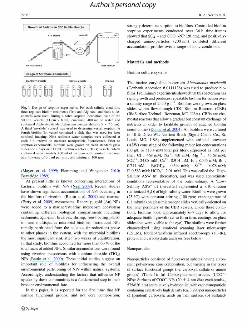
Fig. 1 Design of sorption experiments. For each salinity condition,
three replicate biofilm treatments (Trt), and Alginate- and blank slide-
controls were used. During a batch sorption incubation, each of the
500 ml vessels (11 cm 9 8 cm) contained 400 ml of water and
contained duplicate, standard glass microscope slides (2.5 9 7.5 cm).
A third ‘no-slide’ control was used to determine vessel sorption. A
fourth biofilm Trt vessel contained a slide that was used for later
confocal imaging. Nine replicate water samples were collected at
each 3 h interval to measure nanoparticle fluorescence. Prior to
sorption experiments, biofilms were grown on clean standard glass
slides for 7 days in 1 l CDC biofilm reactors (CBRs) vessels, which
contained approximately 400 ml of medium with constant exchange
at a flow-rate of 0.1 ml per min., and stirring at 100 rpm
2206 B. A. Nevius et al.
123
Author's personal copy

nanoparticles (SO4--NPs): SO4
--nanoparticles (i.e. 20 ± 4
nm dia., 485/528 nm; ca. 2,540 pendant sites per nanoparticle)
are relatively hydrophobic enabling passive sorption of almost
any protein (pKa \ 2). (c) Amine-particles (Amine-Ps): amine-
modified particles are functionalized with amine groups (i.e.
200 ± 11 nm dia.; 580/605 nm; ca. 429,000 pendant sites per
nanoparticle) to provide hydrophilic particles with positively-
charged amine groups (FluoSpheres�, Invitrogen Inc.). Zeta
potentials (mV) of NPs (and washed A. macloedii bacterial
cells) were measured using a ZetaPALS Zeta-Potential Ana-
lyzer (Brookhaven Instr., NY, USA). These nanoparticles have
been used in many previous studies for their efficient fluores-
cence yield, purity, and uniformity of size (Bloem et al. 1995;
Kolodny et al. 2001; Lai et al. 2007).
Experimental approach
Once the biofilms were developed, they were removed
from the CBRs, gently rinsed, to remove excess growth
medium, in water having the same ionic conditions as those
used for growth (but lacking growth medium), then
immediately transferred into previously-prepared batch
sorption vessels (see below) containing nanoparticles, and
water having similar ionic conditions (high- or low-salinity)
as those used for biofilm growth, but with no added growth
medium. An additional treatment, using 513.2 mM NaCl,
termed ‘High-Salinity NaCl’, was used to examine the
effects of univalent cations present in seawater.
To prepare for each batch-sorption experiment, NPs
were sonicated for approximately 3 min prior to addition to
sorption vessels. Sorption vessels consisted of 500 ml glass
beakers containing 400 ml of water with one of three dif-
ferent ionic conditions (Table 2). Then, an approximately
equal concentration of NPs (ca. 5.72 ± 0.38 9 1016;
mean ± SD) was added to each of seven treatment or
control vessels (Fig. 1), and allowed to stir (in absence of
biofilms) for 24 h prior to the start of each experiment.
To commence a sorption experiment, biofilm-coated
slides (and biofilm-free control slides) were suspended
vertically within the vessels containing one of three dif-
ferent types of NP suspensions, and incubated under con-
stant stirring (100 rpms). A sorption equilibrium time-
frame of 36 h, determined from preliminary experiments,
was used for nanoparticle sampling during sorption
experiments. To measure NP sorption versus time, nine
replicate aliquots (0.2 ml) of vessel water were collected
approx. 2 cm below the water surface at time zero (i.e. start
of experiment), and over a series of time points throughout
the 36 h sorption experiment. Samples from each time
point were measured for NP fluorescence (FLx800
Microplate Reader, BioTek Inst.). Means (±SD; n = 9)
were determined for each 3-h interval and correlated with
NP concentrations. The precision of replicate (n = 9)
fluorescence measurements (\0.5 %) indicated that a rel-
atively homogenous suspension of NPs occurred in solu-
tions throughout experiments. Although NP fluorescence
was homogeneous among replicate water samples within
each time point throughout experiments, the degree to
which NPs were individually-dispersed versus aggregated
(within suspensions) could not be determined. The relative
sorption to biofilms at each time point was calculated from
the relative loss of NPs from the aqueous phase correcting
for controls (e.g. vessel sorption). NP measurements were
based on fluorescence emissions for each type of NP, with
their conversion to concentration determined using stan-
dard curves (Supplemental Figure 1). Three replicate
treatment vessels were used for each time-course mea-
surement; a fourth was used for observations of biofilms
using confocal scanning laser microscopy (CSLM, see
below). Replicate high-NaCl treatments were used to test
Table 1 Properties of surface-functionalized nanoparticles
Nanoparticle
type
Product, cat.
no., lot no.aDiameter
(nm)aZeta
potential (mV)
Mean surface
charge (meq/g)
Charged
groups/
particlea
Fluorescence
absorption
(nm)a
Fluorescence
emission
(nm)a
Amine Fluospheres, F-8763; 29965 W 190 ± 11 21.89 ± 0.61 0.1 429,000 575 ± 15 620 ± 40
COO--NPs Fluospheres, F-8782; 538961 24 ± 3 -40.24 ± 1.74 0.7 3,290 575 ± 15 620 ± 40
SO4--NPs Fluospheres, F-8845; 454238 22 ± 3 -36.01 ± 0.59 0.7 2,540 485 ± 20 528 ± 20
A. macleodii cells GenBank no. # 0111138 – -42.10 ± 0.46 – – – –
Nanoparticles used in sorption experiments were surface-functionalized with carboxyl (COO--), sulfated (SO4--) or amine-functional groups
(Fluospheres, Invitrogen Inc.). Zeta potentials of nanoparticles and A. macleodii bacterial cells, measured in deionised water, were expressed in
millivolts (mV). Values represent mean ± SD; n = 3. Mean surface charge is expressed as milliequivalents of exchangeable lights per g
particles. Charged groups per particle represent the total(s) on the particle surfacea Information from manufacturer
Surface-functionalization effects on uptake of fluorescent 2207
123
Author's personal copy

effects of univalent ions on NP binding at high ionic
conditions. Two different types of controls were used: (1)
slide-only controls, consisting of blank slides (free of
biofilms) and (2) no-slide controls, using only vessels, were
used to differentiate non-biofilm sorption during experi-
ments. A total of 63 separate sorption experiments
were conducted with constant pH (8.0 ± 1) maintained
throughout.
Sample measurements
At the conclusion of 36 h incubations, stirring was ceased
and slides were gently removed from each treatment vessel
and NP sorption to biofilms was quantified. All material
(cells, NPs, EPS) attached to each slide was removed by
carefully scraping slides with a sterile, flat razor. Slides
were washed with 0.5 ml sterile deionized-H2O to remove
any additional material. Samples were measured for fluo-
rescence, and converted to (ca.) NP concentration as pre-
viously described (Supplemental Figure 1). Then, cells and
EPS were placed in microcentrifuge tubes, and separated
by repeated (39) centrifugation (10,0009g; 10 min)
(Decho 1990). Supernatants were used for EPS extraction.
Bulk concentration factors (Cf) of nanoparticles on biofilms
were determined under the pH and ionic conditions for
each experimental treatment. Percent recovery of NPs was
calculated as the portion of total-added NPs recovered from
the aqueous phase ? biofilm ? vessel-sorbed (Table 2).
EPS were extracted in cold (-20 �C) ethanol (EtOH;
70 % final conc.) from centrifuged supernatants (Decho
1990). EPS was dried, weighed and later analyzed. Fourier-
transform infrared spectroscopy (FT-IR) was used to
determine the presence of specific functional groups in EPS
(approx 1 mg) using a Nexus 670 spectrometer (Thermo-
Nicolet Inc.) equipped with attenuated total reflectance
(ATR) and multibounce germanium crystal. Absorbance
data were collected between 680 and 4,000 cm-1 (resolu-
tion 8 cm-1, 64 scans co-added and averaged). Confocal
scanning laser microscopy (CSLM) imaging (Decho et al.
2003) was conducted using an SP5 CSLM System (Leica
Microsystems) equipped with four excitation lasers (405,
488, 594, 633 nm). Cells were stained with SYTO 45
(Invitrogen), and EPS with fluorescein isothiocyanate
(FITC)- or Alexa 594-concanavalin A lectin (Bockelmann
et al. 2002).
Statistical analyses
Statistical analyses were performed on semi-log trans-
formed NP fluorescence data using least square linear
regression. P values less than 0.05 were considered sta-
tistically significant. All statistical analyses were
Table 2 Sorption nanoparticles (NPs) to bacterial biofilms incubated under different ionic conditions
NP surface-functional
group
Ionic conc. Slope y-intercept R2 K (#nanos/cm2) Cfa % Recovery t� loss
of NP (h)b
SO4--NPs L-ASW 0.0070 19.193 0.939*** 5.4 9 1017 2.071 9 106 92 ± 15.3 [36
H-ASW 0.0175 16.037 0.979*** 7.8 9 1017 1.735 9 106 91 ± 15.7 12.1
H-NaCl 0.0140 19.158 0.780*** 1.6 9 1017 3.353 9 105 71 ± 10.7 5.3
COO--NPs L-ASW 0.0011 17.974 0.562* 1.2 9 1016 1.192 9 105 67 ± 1.9 [36
H-ASW 0.0017 18.007 0.920*** 1.1 9 1016 1.018 9 105 63 ± 3.0 [36
H-NaCl 0.0018 18.083 0.770*** 3.7 9 1015 nd 53 ± 1.2 [36
Amine-P L-ASW 0.0036 16.476 0.617** 1.6 9 1015 7.826 9 105 92 ± 20.9 [36
H-ASW 0.0080 16.499 0.870*** 1.0 9 1015 6.212 9 105 89 ± 2.6 27.2
H-NaCl 0.0012 16.454 0.963*** 1.5 9 1014 1.698 9 104 58 ± 2.7 [36
Results showing the sorptive accumulation of surface-functionalized NPs to biofilms of the bacterium A. macloedii, incubated under different
ionic conditions. Values for slope, y-intercept, and coefficient of determination (R2) are derived least squares linear regression using semi-
log-transformed data. Significance by regression: * P \ 0.05; ** P \ 0.01; *** P \ 0.001. Concentration factor (Cf) values represent ratios of
concentrations (lg/g) of bound NPs in biofilms over concentrations of unbound NPs present in aqueous phase at experiment conclusion. Ionic
concentrations are expressed as: H-ASW is High-Salinity ASW (513.4 mM per liter); L-ASW is Low-Salinity ASW (51.34 mM/l); H-NaCl is
High Salinity NaCl (513.4 mM). High- and Low-Salinity artificial seawater salts (ASW) were used for High- and Low-Salinity ASW treatments
and included mono- and divalent-cations. A high-salinity sodium chloride treatment (H-NaCl) was used to include only monovalent ions Na?
and Cl-. K is the binding constant, and is based on biofilm area
ND not determineda The concentration factor (Cf) applies to a steady state, and was the ratio of bound/unbound particles at the conclusion of the experiment.
%Recovery represents a mass balance, expressed as a percentage, of the total nanoparticles recovered from all fractions compared to the added
nanoparticlesb The t� represents the half-life (h) for loss of nanoparticles from overlying water during sorption experiments
2208 B. A. Nevius et al.
123
Author's personal copy

performed using statistical analysis systems (SAS) soft-
ware (SAS Inst., Cary, NC, USA).
Results
The results of sorption incubations showed that for the three
types of NPs, over 80 % of the total NP sorption occurred
within 24 h (Fig. 1). The mean percent recoveries of SO4--,
COO-- and amines-NPs after 36 h incubations were
85 ± 13.9, 61 ± 2.03, and 80 ± 8.73 % (mean ± SD;
n = 3), respectively (Table 2). Coefficients of determination
(R2) indicated high precision among values within a given
experimental time point (Table 2). Controls indicated that less
than 3 % of total added NPs were sorbed to glass vessels
(mean ± SD; 104 ± 7.8 9 1015 NPs). No significant dif-
ferences were found for nanoparticles sorbed to vessels
(P = 0.799) among the different treatments and salinities
used.
SO4--NPs showed highest net accumulations at high
ionic concentrations, irrespective of whether mono- (High-
Salinity NaCl) or divalent- (High-Salinity ASW) ions were
present (Table 2). Accumulations of COO--NPs were less
than 15 % across all salinity conditions, while amine-
particles showed greater than 50 % binding in the presence
of divalent ions (i.e. High-Salinity ASW) and less than
10 % binding in the presence of only monovalent ions
present (i.e. High-Salinity NaCl) (Fig. 1).
The highest sorptive concentration to biofilms was
observed with negatively-charged SO4--NPs. Mean bind-
ing constants (K), indicated that SO4--NPs showed the
highest concentration by biofilms under all salinity condi-
tions. Highest K values occurred at relatively high ionic
conditions (e.g. High-Salinity ASW and NaCl), followed
by Low-Salinity ASW (Table 2). Concentration factors
(Cf), a measure of the relative partitioning of NPs between
biofilms and the bulk medium, were 2.1 9 106 and 1.7 9
106, for SO4--NPs at Low-Salinity and High-Salinity ASW
biofilms, respectively.
Sorptive concentration of COO--NPs to biofilms under
all ionic conditions was less than 15 %), and Cf’s of
COO--NPs to biofilms were an order of magnitude lower
(e.g. 1.2 9 105 at Low-Salinity ASW) than SO4--NPs
(Table 2). Regression analysis results showed a significant
(P \ 0.05) R2 and slope, indicating a positive net
accumulation of COO--NPs to biofilms over the 36 h time-
period. Positively-charged amine-particles exhibited high-
est sorption to biofilms at high ASW concentration.
Approximately 30 % of the recovered particles were found
to be associated with biofilms. Mean Cf’s for amine-
particle sorption to biofilms were calculated between
7.8 9 105 and 6.2 9 105, occurring at Low- and High-
Salinity ASW, respectively.
Fourier-transform infrared (FT-IR) spectroscopic anal-
yses of EPS extracted from A. macleodii biofilms (Fig. 2)
showed a range of peaks corresponding to a C=O stretch
(1,419 cm-1), amides (1,621 cm-1), and amines (2,360 cm-1).
Imaging using confocal scanning laser microscopy
(CSLM) indicated fluorescence signatures corresponding to
SO4-- and COO--NPs.
Discussion
Results showed that for the three types of NPs, a relatively
large portion ([80 %) of sorption occurred over 24 h
(Fig. 1) with lesser net accumulation thereafter. Mean
percent recoveries of SO4--, COO-- and amines-NPs after
36 h were 85 ± 13.9, 61 ± 2.03, and 80 ± 8.73 %
(mean ± SD; n = 3) respectively (Table 2). Coefficients
of determination (R2), calculated from analyses of log-
transformed data using least squares linear regression
analyses (Table 2), showed that replicate sorption values
collected within a given experimental time point had high
precision (i.e. low variability among replicates). Controls
indicated that minimal (i.e. typically \3 % of total added)
sorption to glass (vessels) occurred (mean ± SD;
104 ± 7.8 9 1015 NPs). No significant differences were
found for nanoparticles that were sorbed to vessels
(P = 0.799) among the different treatments and salinities
used.
Salinity conditions affected sorption differently depend-
ing on the surface functionalization of the NP. SO4--NPs
exhibited greatest net accumulations at high ionic con-
centrations irrespective of whether mono- (High-Salinity
NaCl) or divalent- (High-Salinity ASW) ions were present
(Table 2). Accumulations of COO--NPs remained rela-
tively minimal (15 %) over all salinity conditions, while
amine-particles showed greatest binding (50 %) in the
presence of divalent ions (i.e. High-Salinity ASW) but
minimal binding (\ 10 %) with only monovalent ions
present (i.e. High-Salinity NaCl). Thus, the three ionic
conditions resulted in very different sorption patterns for
NPs, with the latter depending on their surface function-
alization (Fig. 1).
Largest sorptive concentration to biofilms occurred with
negatively-charged S-NPs. Examinations of mean binding
constants (K), {i.e. a conditional distribution coefficient
since not constant) which represented numbers of bound
NPs per cm2 for a given set of experiment conditions,
showed that S-NPs were concentrated most efficiently by
biofilms under all salinity conditions. Biofilms typically
exhibit much heterogeneity in density and thickness over
small (e.g. um) spatial scales (Flemming and Wingender
2010). Therefore, it was necessary to calculate K based on
biofilm area. Highest K values occurred at relatively high
Surface-functionalization effects on uptake of fluorescent 2209
123
Author's personal copy

ionic conditions (e.g. High-Salinity ASW and NaCl), fol-
lowed by Low-Salinity ASW (Table 2). However, irre-
spective of ionic conditions, SO4--NPs were more
concentrated onto biofilms by an order of magnitude, when
compared to either COO-- or amine-functionalized NPs
under similar conditions. Concentration factors (Cf) were
calculated to determine the relative partitioning of NPs
between biofilms and the bulk medium. The Cf represents
the ratio of the concentration of bound NPs in biofilm
phase (lg/g) over the concentration in aqueous phase
(lg/g) at the termination of experiments. Cf‘s for SO4--
NPs at Low-Salinity and High-Salinity ASW biofilms were
2.1 9 106 and 1.7 9 106, respectively.
Sorptive concentration of COO--NPs to biofilms under
all ionic conditions was minimal (\ 15 %), and Cf‘s were
an order of magnitude lower (e.g. 1.2 9 105 at Low-
Salinity) than SO4--NPs (Table 2). Results of regression
analyses indicated significant (P \ 0.05) R2 and slopes
indicating a net accumulation of COO--NPs to biofilms
over the 36 h time-period. However, accumulations did not
vary as a function of salinity conditions. Positively-charged
amine-particles exhibited highest sorption to biofilms at
Low-Salinity ASW
Time (h)
0 6 12 18 24 30 36
Aq
ueo
us-
Ph
ase
Nan
op
arti
cles
/ N
ano
par
ticl
es a
t T 0
0.0
0.2
0.4
0.6
0.8
1.0
SO4-
COO-
Amine
High-Salinity ASW
Time (h)
0.0
0.2
0.4
0.6
0.8
1.0
SO4-
COO-
Amine
High-Salinity NaCl
Time (h)
0.0
0.2
0.4
0.6
0.8
1.0
SO4-
COO-
Amine
Aq
ueo
us-
Ph
ase
Nan
op
arti
cles
/ N
ano
par
ticl
es a
t T 0
Aq
ueo
us-
Ph
ase
Nan
op
arti
cles
/ N
ano
par
ticl
es a
t T 0
0 6 12 18 24 30 36
0 6 12 18 24 30 36
Fig. 2 Sorption of NPs with
different surface functional
groups to biofilms. Graphs show
results of 36 h time-course
experiments examining sorption
of NPs having different surface
functional groups to biofilms
under different salinity
conditions: Low-salinity
artificial seawater (ASW);
High-salinity ASW; High-
salinity NaCl. NPs had a
polystyrene core and were
surface-functionalized with
either: SO4--; carboxylic acid-;
or amine-functional groups.
Sorption of NPs to biofilms
could not be measured directly
until the conclusion of a given
time course experiment.
Therefore, each line represents
the decrease of NPs versus time,
measured from aqueous phase.
Relative decrease was used to
infer (i.e. by difference) biofilm
sorption, correcting for controls.
Values represent measurements
of nanoparticle disappearance
from the medium. Each time
point value represents the
mean ± SD bars (n = 9).
Y-axes represent NP
concentrations normalized
against the time zero (T0)
concentration. All experiments
were conducted at a constant pH
(8.0 ± 1)
2210 B. A. Nevius et al.
123
Author's personal copy

high ASW concentration. Approximately 30 % of the
recovered particles were found to be associated with bio-
films. Mean Cf‘s for amine-particle sorption to biofilms
ranged between 7.8 9 105 and 6.2 9 105, occurring at
Low- and High-Salinity ASW, respectively. The relatively
high Cf estimates observed for positively-charged particles
in experiments (621 9 104) were within range of those
determined for positively-charged gold nanorods
(Cf = 1.53 9 104) bound to natural marine biofilms (Ferry
et al. 2009). In that study, marine mesocosms contained
seawater having a range of divalent cations, and a wide
range of other natural environmental components including
sediments, seagrass, bivalves, shrimp and plankton. Mass
balance analyses showed that biofilms alone accounted for
approximately 60 % of added NPs; with Cf’s considered to
be conservative estimates (Ferry et al. 2009).
NPs and the EPS matrix
The extracellular polymeric secretions (EPS) of biofilms
(Decho 1990; Flemming and Wingender 2010) represent a
primary site for the binding of charged ions. Binding of
cations and anions to EPS occurs largely through func-
tional groups located on EPS molecules (Decho 1990).
Analyses of EPS extracted from A. macleodii biofilms by
fourier-transform infrared (FT-IR) spectroscopy (Fig. 2)
indicated a range of functional groups that can form
complexes with NPs via cation- or anion-bridging. These
include carboxyls as evidenced by a C=O stretch
(1,419 cm-1), amides (1,621 cm-1), and amines (2,360 cm-1),
and are in agreement with the literature data (Braissant
et al. 2007). Carboxylic acids, for example, impart EPS
molecules with a net negative-charge at neutral pH and
facilitate binding of metal cations (Ferris et al. 1989; Jang
et al. 1990; Foster et al. 2000; Spagnoli et al. 2005).
Hydrophobic or electrostatic forces can also contribute to
binding between the charged NPs and the biofilm matrix.
The polysaccharides (present in natural EPS), specifically
their -CH groups could exhibit hydrophobic interactions in
the complexation of SO4--NPs. Binding may also occur
through transient bidentate bridging involving divalent
cations (e.g. Ca2? and Mg2?) present in ASW, and occurs
between carboxyl groups on uronic acids and NP functional
groups. However, sorption of SO4--NPs occurred in the
presence of High-Salinity NaCl, therefore, not all binding
could be explained by divalent cations. This suggests that
at least some of the binding under the experimental con-
ditions was simply related to an ionic strength effect. It was
not known, however, which functionalities on EPS directly
contributed to binding with NP surface groups. The FT-IR
study was designed to analyze which functional groups
present on EPS might contribute to biofilm binding,
therefore, analyses were not conducted to determine how
nanoparticle exposure affected EPS functional groups.
Also, our study could not differentiate between NPs that
were sorbed to the biofilm surface versus those which had
migrated into the biofilm (Fig. 3).
Currently, several unanswered questions must be
addressed before the mechanisms of NP binding can be
fully understood. It was not known if charged NPs sorb to
the surface of biofilms or, once bound, can migrate into the
biofilm matrix. It was postulated that brownian motion, in
concert with the transient nature of cation bridges, can
allow NPs to migrate deeper into biofilms over time.
However, brownian motion for an entity as large as a
nanoparticle (e.g. 20 nm dia.) could be quite slow. This
would depend, in part, on the relative spacing between
adjacent polymer molecules. In fact, the larger the particle
the more likely that random motion will average to zero
0.000
0.020
0.040
0.060
0.080
0.100
500 1000 1500 2000 2500 3000 3500 4000
Ab
sorb
ance
Wavenumber (cm )-1
EPS
Amine
(a)
(b)
100 nm
-COO
1091.4
-amide
-amine -OH -COOH
1621.3
2360.2 3379.0 1419.4
Fig. 3 Biofilm EPS. a Confocal scanning laser micrograph (CSLM)
of A. macleodii biofilm showing amine-particles bound to EPS
(bacteria not visible). EPS are stained (green) with concanavalin-A
lectin. Individual amine-NPs (red fluorescence; 200 nm dia.) in the
biofilm are barely visible (in image); however groups of particles
brightly fluoresce. b Infrared spectra of EPS extracted from A.macloedii bacterial biofilms (used in experiments) showing several
absorption peaks representing functional groups on EPS that can
potentially bind surface-functionalized NPs. Attenuated total reflec-
tance—fourier-transform infrared (ATR-FTIR) spectroscopy was used
to determine functional groups. Assignment of wavenumber peaks:
1,091 cm-1 = carbohydrate (C–O) stretching vibrations; 1,419 cm-1 =
carboxylic (C=O) stretch; 1,621 cm-1 = amides (can also be attrib-
uted to more C=O bonds typical of carboxyl groups; and 2,360 cm-1
and 3,379 cm-1 represent amines and O–H, respectively. Wavenum-
ber is equal to 1/wavelength. Spectrum values represent means of 64
co-added scans (Color figure online)
Surface-functionalization effects on uptake of fluorescent 2211
123
Author's personal copy

and the particle will not move. Virus particles (40–100 nm),
however, which are larger than the SO4-- and COO--NPs
(20 nm dia.) used in the study, have been shown to effi-
ciently penetrate biofilms (Briandet et al. 2008). Also,
small particles are known to enter and move through water
channels within the EPS matrix, which have been observed
in some biofilms (Stoodley et al. 1994). Finally, there is a
growing realization that functionalized NPs can become
coated with sorbed organic molecules forming a ‘protein
corona’, which coats the NP (Lundqvist et al. 2008). The
corona surrounding the NP then interacts with surfaces or
matrices during binding. In the present study, efforts were
made to minimize organic molecules in the aqueous phase
during sorption experiments. However, there remained the
possibility that some NP binding may have resulted from
molecules sorbed to the NPs. These are important consid-
erations for understanding binding and subsequent pene-
trations of biofilms by nanoparticles.
Biofilms were imaged using confocal scanning laser
microscopy (CSLM), and fluorescence signatures of NPs
were concurrently detected. Individual 20 nm NPs are
outside the resolving power of CSLM (i.e. light
microscopy). However, fluorescence peaks indicative of
SO4-- and COO--NPs were detectable and showed accu-
mulations over 2-dimensional areas of biofilms (data not
shown). Higher magnification imaging was used to illustrate
the binding of much larger 200 nm dia. amine-particles on
EPS (Fig. 2), although these particles are larger than the
typical size range (1–100 nm) of nanoparticles. It is not
known if the increased size of the amine- particles influ-
enced their binding when compared with the 20 nm
nanoparticles.
The results of this study show that biofilms were
effective at removing suspended nanomaterials from the
aqueous phase. While the mechanisms regulating particle
uptake are not yet known, it was shown that the mecha-
nisms are strongly dependent on the surface nanoparticle
ionization. In both natural- and engineered-systems, NP
surface properties will contribute to their dispersion,
sorptive accumulation, and even bioaccumulation in
organisms. While previous studies of NPs and microbial
systems have focused on core composition (e.g. Ag, TiO2)
(Fabrega et al. 2009; Hetrick et al. 2009; Lellouche et al.
2009; Tong et al. 2010), the present work is the first report
that surface charges on NPs, and not their core properties,
strongly affect sorption and bioconcentration by biofilms.
Biofilms are a rapidly-emerging area of importance in
understanding the bioconcentration of NPs, but one that is
only marginally understood. Biofilms and their sorbed
components, such as NPs, represent a fundamental step for
entry into food webs and humans.
Acknowledgments The authors thank Dr. Cathy Murphy (Univer-
sity of Illinois at Urbana-Champaign) for assistance in zeta-potential
measurements. This work was supported by the University of South
Carolina NanoCenter, and the National Science Foundation (BME-
1032579). The authors declare that they have no conflict of interest.
References
Battin TJ, Kammer FVD, Weilhartner A, Ottofuelling S, Hofmann T
(2009) Nanostructured TiO2: transport behavior and effects on
aquatic microbial communities under environmental conditions.
Environ Sci Technol 43:8098–8104
Beveridge TJ (1989) Role of cellular design in bacterial metal
accumulation and mineralization. Annu Rev Microbiol 43:
147–171
Bloem J, Veninga M, Shepherd J (1995) Fully automatic determina-
tion of soil bacterial numbers, cell volumes, and frequency of
dividing cells by confocal laser scanning microscopy and image
analysis. Appl Environ Microbiol 61:926–936
Bockelmann U, Manz W, Neu TR, Szewzyk U (2002) Investigation
of lotic microbial aggregates by a combined technique of
fluorescent in situ hybridization and lectin-binding-analysis.
J Microbiol Meth 49:75–87
Braissant O, Decho AW, Dupraz C, Glunk C, Przekop KM, Visscher
PT (2007) Exopolymeric substances of sulfate-reducing bacteria:
interactions with calcium at alkaline pH and implication for
formation of carbonate minerals. Geobiology 5:401–411
Briandet R, Lacroix-Gueu P, Renault M, Lecart S, Meylheuc T,
Bidnenko E, Steenkeste K, Bellon-Fontaine MN, Fontaine-
Aupart MP (2008) Fluorescence correlation spectroscopy to
study diffusion and reaction of bacteriophages inside biofilms.
Appl Environ Microb 74:2135–2143
Decho AW (1990) Microbial exopolymer secretions in ocean
environments—their role(s) in food webs and marine processes.
Oceanogr Mar Biol Ann Rev 28:73–153
Decho AW, Kawaguchi T, Allison MA, Louchard EM, Reid RP,
Stephens FC, Voss KJ, Wheatcroft RA, Taylor BB (2003) Sediment
properties influencing upwelling spectral reflectance signatures: the
‘‘biofilm gel effect’’. Limnol Oceanogr 48:431–443
Donlan RM, Piede JA, Heyes CD, Sanii L, Murga R, Edmonds P, El-
Sayed I, El-Sayed MA (2004) Model system for growing and
quantifying Streptococcus pneumoniae biofilms in situ and in
real time. Appl Environ Microb 70:4980–4988
Fabrega J, Renshaw JC, Lead JR (2009) Interactions of silver
nanoparticles with Pseudomonas putida biofilms. Environ Sci
Technol 43:9004–9009
Farre M, Gajda-Schrantz K, Kantiani L, Barcelo D (2009) Ecotoxicity
and analysis of nanomaterials in the aquatic environment. Anal
Bioanal Chem 398:81–95
Ferris FG, Schultze S, Witten TC, Fyfe WS, Beveridge TJ (1989)
Metal interactions with microbial biofilms in acidic and neutral
pH environments. Appl Environ Microb 55:1249–1257
Ferry JL, Craig P, Hexel C, Sisco P, Frey R, Pennington PL, Fulton
MH, Scott IG, Decho AW, Kashiwada S, Murphy CJ, Shaw TJ
(2009) Transfer of gold nanoparticles from the water column to
the estuarine food web. Nat Nanotechnol 4:441–444
Flemming HC, Wingender J (2010) The biofilm matrix. Nat Rev
Microbiol 8:623–633
Foster LJR, Moy YP, Rogers PL (2000) Metal binding capabilities of
Rhizobium etli and its extracellular polymeric substances.
Biotechnol Lett 22:1757–1760
2212 B. A. Nevius et al.
123
Author's personal copy

Hetrick EM, Shin JH, Paul HS, Schoenfisch MH (2009) Anti-biofilm
efficacy of nitric oxide-releasing silica nanoparticles. Biomate-
rials 30:2782–2789
Jang LK, Brand W, Resong M, Mainieri W, Geesey GG (1990)
Feasibility of using alginate to absorb dissolved copper from
aqueous-media. Environ Prog 9:269–274
Kolodny LA, Willard DM, Carillo LL, Nelson MW, Van Orden A
(2001) Spatially correlated fluorescence/AFM of individual
nanosized particles and biomolecules. Anal Chem 73:1959–1966
Lai SK, O’Hanlon DE, Harold S, Man ST, Wang YY, Cone R, Hanes
J (2007) Rapid transport of large polymeric nanoparticles in
fresh undiluted human mucus. Proc Natl Acad Sci USA
104:1482–1487
Lellouche J, Kahana E, Elias S, Gedanken A, Banin E (2009)
Antibiofilm activity of nanosized magnesium fluoride. Bioma-
terials 30:5969–5978
Lundqvist M, Stigler J, Elia G, Lynch I, Cedervall T, Dawson KA
(2008) Nanoparticle size and surface properties determine the
protein corona with possible implications for biological impacts.
Proc Natl Acad Sci USA 105:14265–14270
Mayer C, Moritz R, Kirschner C, Borchard W, Maibaum R,
Wingender J, Flemming HC (1999) The role of intermolecular
interactions: studies on model systems for bacterial biofilms. Int
J Biol Macromol 26:3–16
Neal AL (2008) What can be inferred from bacterium-nanoparticle
interactions about the potential consequences of environmental
exposure to NPs? Ecotoxicology 17:362–371
Spagnoli C, Korniakov A, Ulman A, Balazs EA, Lyubchenko YL,
Cowman MK (2005) Hyaluronan conformations on surfaces: effect
of surface charge and hydrophobicity. Carbohydr Res 340:929–941
Stoodley P, Debeer D, Lewandowski Z (1994) Liquid flow in biofilm
systems. Appl Environ Microb 60:2711–2716
Tong MP, Ding JL, Shen Y, Zhu PT (2010) Influence of biofilm on
the transport of fullerene (C-60) NPs in porous media. Water Res
44:1094–1103
Surface-functionalization effects on uptake of fluorescent 2213
123
Author's personal copy
Copyright © 2022 FDOKUMEN