A bio-orthogonal functionalization strategy for site-specific ...
14
Polymer Chemistry PAPER Cite this: Polym. Chem., 2020, 11, 527 Received 30th July 2019, Accepted 14th October 2019 DOI: 10.1039/c9py01136f rsc.li/polymers A bio-orthogonal functionalization strategy for site-specific coupling of antibodies on vesicle surfaces after self-assembly† Meiyu Gai, ‡ a Johanna Simon, ‡ a,b Ingo Lieberwirth, a Volker Mailänder, a,b Svenja Morsbach * a and Katharina Landfester a Attaching targeting ligands on the surface of self-assembled drug delivery systems is the key request for a controlled transport of the drug to a desired location. Most commonly, the amphiphilic molecules (block- copolymers, lipids etc.) are therefore pre-functionalized before the self-assembly takes place. However, this strategy cannot be applied, if it interferes with the self-assembly process, if the introduced functional groups react with loaded cargo or if natural carriers like extracellular vesicles should be functionalized. Here, we present the site-specific coupling of antibodies to the surface of amino group-terminated lipo- somes via bio-orthogonal copper-free click chemistry after liposome formation. The present primary amino groups were functionalized with a linker carrying a strained alkyne group for a bio-orthogonal strain-promoted alkyne–azide cycloaddition (SPAAC) reaction where Cu(I) as a catalyst can be avoided. Antibodies were site-specifically functionalized with azide moieties along the Fc region to avoid inter- ference with the antigen binding sites. The liposome surface functionalization reaction was optimized by precisely analyzing the number of available functional groups (both amine and alkyne), which often rep- resents a challenge for self-assembled systems. By finally confirming the successful antibody coupling, we provide a facile and robust functionalization strategy, which can be applied to a wide range of self- assembled systems and desired targeting antibodies maintaining physiological conditions throughout the procedure. 1. Introduction Self-assembled systems of amphiphilic molecules are nowa- days applied as drug delivery vehicles as they are biocom- patible and can transport hydrophilic as well as hydrophobic cargos. Typically, such self-assembled systems can be categor- ized into the main groups of polymer-based structures, lipid- based structures and more recently introduced natural vesi- cles. 1 Polymer-based self-assembled systems consist of amphi- philic block-copolymers, which typically form double-layers and assemble into polymersomes or micelles. 2–4 Regarding lipid-based structures, liposomes, which are small artificial vesicles consisting of one or more phospholipid bilayers, were first introduced by Bangham et al. in the 1960s. 5 They are still of high interest as their composition is similar to natural cellular membranes and as such have an extraordinary biocompatibility. 6–9 Basically, there are four major types of liposomes: (1) MLVs, multilamellar vesicles with several lamel- lar lipid phase bilayers, (2) SUVs, small unilamellar vesicles with one lipid bilayer, 10–14 (3) LUVs, large unilamellar vesicles, and (4) cochleate vesicles. 15 Compared to these conventional structures, “second-gene- ration” vesicles are chemically modified to obtain specific pro- perties such as a prolonged blood circulation time or targeting capability. More specifically, vesicles are functionalized with various targeting ligands such as antibodies, peptides, pro- teins or aptamers, with a variety of surface engineering techniques. 14,16–18 These ligands are mostly chemically attached to the vesicles through interaction of reactive groups on the liposomes’ or polymersomes’ surface and specific groups present in the ligand. For this purpose, the pre-modifi- cation of various types of block-copolymers, lipids or chole- sterol is often applied to introduce functional groups. 19–24 One example is the commercially available DBCO modified lipo- some formulation (Immunosome®-DBCO). In this case, the functional DBCO groups were directly introduced via the phos- † Electronic supplementary information (ESI) available. See DOI: 10.1039/ c9py01136f ‡ Authors contributed equally. a Max Planck Institute for Polymer Research, Ackermannweg 10, 55128 Mainz, Germany. E-mail: [email protected] b Department of Dermatology, University Medical Center of the Johannes Gutenberg-University Mainz, Langenbeckstrasse 1, 55131 Mainz, Germany This journal is © The Royal Society of Chemistry 2020 Polym. Chem. , 2020, 11, 527–540 | 527 Open Access Article. Published on 14 oktyabr 2019. Downloaded on 11.07.2022 09:08:08. This article is licensed under a Creative Commons Attribution 3.0 Unported Licence. View Article Online View Journal | View Issue
-
Upload
khangminh22 -
Category
Documents
-
view
0 -
download
0
Transcript of A bio-orthogonal functionalization strategy for site-specific ...

PolymerChemistry
PAPER
Cite this: Polym. Chem., 2020, 11,527
Received 30th July 2019,Accepted 14th October 2019
DOI: 10.1039/c9py01136f
rsc.li/polymers
A bio-orthogonal functionalization strategy forsite-specific coupling of antibodies on vesiclesurfaces after self-assembly†
Meiyu Gai, ‡a Johanna Simon, ‡a,b Ingo Lieberwirth, a Volker Mailänder, a,b
Svenja Morsbach *a and Katharina Landfester a
Attaching targeting ligands on the surface of self-assembled drug delivery systems is the key request for a
controlled transport of the drug to a desired location. Most commonly, the amphiphilic molecules (block-
copolymers, lipids etc.) are therefore pre-functionalized before the self-assembly takes place. However,
this strategy cannot be applied, if it interferes with the self-assembly process, if the introduced functional
groups react with loaded cargo or if natural carriers like extracellular vesicles should be functionalized.
Here, we present the site-specific coupling of antibodies to the surface of amino group-terminated lipo-
somes via bio-orthogonal copper-free click chemistry after liposome formation. The present primary
amino groups were functionalized with a linker carrying a strained alkyne group for a bio-orthogonal
strain-promoted alkyne–azide cycloaddition (SPAAC) reaction where Cu(I) as a catalyst can be avoided.
Antibodies were site-specifically functionalized with azide moieties along the Fc region to avoid inter-
ference with the antigen binding sites. The liposome surface functionalization reaction was optimized by
precisely analyzing the number of available functional groups (both amine and alkyne), which often rep-
resents a challenge for self-assembled systems. By finally confirming the successful antibody coupling,
we provide a facile and robust functionalization strategy, which can be applied to a wide range of self-
assembled systems and desired targeting antibodies maintaining physiological conditions throughout the
procedure.
1. Introduction
Self-assembled systems of amphiphilic molecules are nowa-days applied as drug delivery vehicles as they are biocom-patible and can transport hydrophilic as well as hydrophobiccargos. Typically, such self-assembled systems can be categor-ized into the main groups of polymer-based structures, lipid-based structures and more recently introduced natural vesi-cles.1 Polymer-based self-assembled systems consist of amphi-philic block-copolymers, which typically form double-layersand assemble into polymersomes or micelles.2–4 Regardinglipid-based structures, liposomes, which are small artificialvesicles consisting of one or more phospholipid bilayers, werefirst introduced by Bangham et al. in the 1960s.5 They are still
of high interest as their composition is similar to naturalcellular membranes and as such have an extraordinarybiocompatibility.6–9 Basically, there are four major types ofliposomes: (1) MLVs, multilamellar vesicles with several lamel-lar lipid phase bilayers, (2) SUVs, small unilamellar vesicleswith one lipid bilayer,10–14 (3) LUVs, large unilamellar vesicles,and (4) cochleate vesicles.15
Compared to these conventional structures, “second-gene-ration” vesicles are chemically modified to obtain specific pro-perties such as a prolonged blood circulation time or targetingcapability. More specifically, vesicles are functionalized withvarious targeting ligands such as antibodies, peptides, pro-teins or aptamers, with a variety of surface engineeringtechniques.14,16–18 These ligands are mostly chemicallyattached to the vesicles through interaction of reactive groupson the liposomes’ or polymersomes’ surface and specificgroups present in the ligand. For this purpose, the pre-modifi-cation of various types of block-copolymers, lipids or chole-sterol is often applied to introduce functional groups.19–24 Oneexample is the commercially available DBCO modified lipo-some formulation (Immunosome®-DBCO). In this case, thefunctional DBCO groups were directly introduced via the phos-
†Electronic supplementary information (ESI) available. See DOI: 10.1039/c9py01136f‡Authors contributed equally.
aMax Planck Institute for Polymer Research, Ackermannweg 10, 55128 Mainz,
Germany. E-mail: [email protected] of Dermatology, University Medical Center of the Johannes
Gutenberg-University Mainz, Langenbeckstrasse 1, 55131 Mainz, Germany
This journal is © The Royal Society of Chemistry 2020 Polym. Chem., 2020, 11, 527–540 | 527
Ope
n A
cces
s A
rtic
le. P
ublis
hed
on 1
4 ok
tyab
r 20
19. D
ownl
oade
d on
11.
07.2
022
09:0
8:08
. T
his
artic
le is
lice
nsed
und
er a
Cre
ativ
e C
omm
ons
Attr
ibut
ion
3.0
Unp
orte
d L
icen
ce.
View Article OnlineView Journal | View Issue

pholipid before the liposomes’ self-assembly. In a followingreaction, azide containing peptides or proteins can be immobi-lized in the liposomes’ surface.
However, direct lipid or polymer functionalization mightnot be applicable when the self-assembly of the vesicle struc-ture is disturbed by the chemical modification of one com-ponent. Similarly, pre-functionalization is not advisable whenthe functional groups should only be present on the outervesicle surface to avoid reactions with sensitive cargos.Additionally, the functional moieties, which are introducedprior to self-assembly, might partially be buried in the hydro-phobic environment of the formed membrane, as it wasdemonstrated for polymersomes.20 Ultimately, also for pre-assembled natural structures like extracellular vesicles25–27 adifferent functionalization strategy is required. Therefore, arobust way is needed to enable bio-orthogonal and highlyspecific functionalization of the vesicle surfaces after self-assembly. Additionally, the proposed functionalization strategyshould allow for reliable surface characterization and precisereaction control, which is challenging for preformed self-assembled systems.
For a convenient and reliable coupling of two differentfunctional moieties, click chemistry is widely used due to itsunique reaction properties: the procedure is very efficient,mild and can be carried out in aqueous reaction conditions.Click chemistry is rapidly becoming a popular tool to functio-nalize the surface of biomacromolecules, including viruses,DNA, peptides, antibodies, liposomes, micelles, and nano-particles with a wide variety of conjugates.28–30 There are twowell established bio-orthogonal coupling approaches: (1)copper-catalyzed azide–alkyne cycloaddition (CuAAC) “click”reactions, which are attractive as the reaction kinetics are favor-able and azide-containing reagents are widely available11,29,31,32
(2) the strain-promoted alkyne–azide cycloaddition (SPAAC)where the use of Cu(I) as a catalyst is avoided.33 This bio-orthog-onal copper-free click chemistry offers a great advantage regard-ing the known deleterious effects associated with copper in cell-based studies and applications.34
In this work, we describe the formation of liposomes basedon lipid thin-film hydration followed by extrusion and sub-sequent surface functionalization of lipid headgroups(Scheme 1). To enable chemical modification via the SPAAC
Scheme 1 Schematic step-by-step illustration of liposome synthesis, surface functionalization and site-selective antibody attachment.
Paper Polymer Chemistry
528 | Polym. Chem., 2020, 11, 527–540 This journal is © The Royal Society of Chemistry 2020
Ope
n A
cces
s A
rtic
le. P
ublis
hed
on 1
4 ok
tyab
r 20
19. D
ownl
oade
d on
11.
07.2
022
09:0
8:08
. T
his
artic
le is
lice
nsed
und
er a
Cre
ativ
e C
omm
ons
Attr
ibut
ion
3.0
Unp
orte
d L
icen
ce.
View Article Online

approach, the lipid bilayer was composed out of cholesteroland two different phospholipids – 2-dioleoyl-sn-glycero-3-phos-phoethanolamine (DOPE) and L-α-phosphatidylcholine (eggPC) – with a molar ratio of 1 : 1 : 1. The formed liposomes werecharacterized thoroughly to determine the exact number ofavailable surface primary amine groups (–NH2). Amino groupswere further coupled with dibenzylcyclooctyne–PEG4–NHSester to introduce a strained alkyne (DBCO group) to the lipo-somes’ surface under different reaction pH. Successful modifi-cation with DBCO groups was proven by an anthracene azide(Anth-N3) assay which was carried out for a precise DBCOgroup quantification as well as analysis of the stability inthe aqueous solution. With this, we provide a universalfunctionalization strategy for any NH2-terminated nanocarrier.In addition, we give a detailed protocol on methods to monitoreach functionalization step. Next to this, the antibodyCD11c was site-specifically modified with azide groups bytransferase and UDP-N-azidoacetylgalactosamine (UDP-GalNAz).Subsequently, the azide-functionalized antibody was coupledto the DBCO-functionalized liposomes via bio-orthogonalcopper-free click chemistry (SPAAC). The successful couplingwas confirmed by a flow cytometry based binding assay usingfluorescently labeled secondary antibodies.
2. Experimental2.1. Materials
All chemicals and materials were used as received. 1,2-Dioleoyl-sn-glycero-3-phosphoethanolamine (DOPE) with ≥99.0% purity(10 mg mL−1 in CHCl3), L-α-phosphatidylcholine (egg PC) withpurity ≥99.0% (100 mg mL−1 in CHCl3), cholesterol (powder,BioReagent, ≥99%, Germany) and dimethylsulfoxide (DMSO)were purchased from Sigma Aldrich (>99% purity, Germany).The dye DiI Stain (1,1′-dioctadecyl-3,3,3′,3′-tetramethyl-indocarbocyanine perchlorate) for labelling liposomes was pur-chased from Thermo Fisher (catalog number: D282, Germany).Dibenzylcyclooctyne (DBCO)–PEG4–NHS ester (cat. no.CLK-A134-100) was purchased from Jena Bioscience (purity:>95% (HPLC), Germany). Phosphate buffered saline (DPBS-buffer, –Mg2+, –Ca2+) was procured from Sigma-Aldrich (USA),while ethanol (99.5%) was acquired from Carl Roth, Germany.A MilliQ device (Merck Millipore, Germany) was used to obtainthe demineralized water. Demineralized water was used for allexperiments. The antibodies anti-mouse CD11c (clone N418)and the FITC-labeled goat anti-hamster (Armenian, clonePoly4055) was purchased from Biolegend (0.5 mg mL−1). TheSiteClick™ Antibody Azido Modification Kit was obtainedfrom Thermo Fisher (catalog number: S20026, America).
2.2. Liposome preparation
Preparation of the thin film. Amine group-terminated lipo-somes were prepared from cholesterol (Chol) and two differentphospholipids: 1,2-dioleoyl-sn-glycero-3-phosphoethanolamine(DOPE) and L-α-phosphatidylcholine (egg PC) with a molarratio of egg PC : DOPE : Chol = 1 : 1 : 1 by film hydration fol-
lowed by extrusion. Briefly, both lipids and cholesterol stocksolutions in chloroform were prepared at a concentration of10 mg mL−1. Then, 835 µL, 767 µL and 398 µL of the egg PC,DOPE and Chol solutions, respectively, were added into a50 mL round-bottomed flask with an additional 2 mL of amixture of chloroform with 1 vol% EtOH. The mixture wasdried with a rotary evaporator at a reduced pressure of450 mbar and 3 mbar each for half an hour at 42 °C.Subsequently, the flask was placed in a vacuum oven for1 hour to remove organic solvent residues (images of the filmformation procedure, see Fig. S1†).
Hydration of the thin film and extrusion. A volume of 4 mLPBS buffer (0.1 M, pH = 7.4) was added into the flask and themixture was stirred over night at 550 rpm (Fig. S1†). Then themixture was sonicated in a water bath for 20 min. In order toobtain small unilamellar liposomes, extrusion was performed.The liposome solutions were extruded through two polycarbo-nate membranes with pore sizes of 800 nm, 400 nm and200 nm a number of 11 times at each step. Afterwards, theliposome samples were stored at 4 °C until further use.
2.3. Liposome characterization
Dynamic light scattering (DLS). Generally, the average sizeand the size distribution of all liposome samples was deter-mined with diluted dispersions (10 μL sample with a solidcontent of 0.35 wt% were diluted in 200 µL PBS buffer) with aZetasizer Nano S90 (Malvern Panalytical GmbH, Germany) at20 °C. In order to obtain also the angular dependenthydrodynamic radius (Rh), radius of gyration (Rg) and structureparameter of non-functionalized liposomes, multi-angle DLSwas performed with an ALV spectrometer (ALV-GmbH,Germany). The set-up consisted of a goniometer and an ALV/LSE-5004 multiple-tau full-digital correlator with 320 channels.As a light source a He–Ne laser was used at a wavelength of632.8 nm. The samples were filtered through Millex-LCR0.45 μm syringe filters (Merck, Germany) into cylindricalquartz cuvettes (18 mm diameter, Hellma, Germany). The cuv-ettes were cleaned in an acetone fountain prior to usage forremoving dust. Liposome samples were prepared in a concen-tration of 0.003 mg mL−1 in PBS. The CONTIN algorithm35 wasused for data analysis.
Zeta potential. A Zetasizer Nano Z (Malvern PanalyticalGmbH, Germany) with disposable folded capillary cells wasused to determine the zeta-potential (ζ-potential) of thevarious liposome samples. Basically, 10 µL of a dispersion ofeach liposome sample (3.5 µg mL−1) were diluted with 1 mL ofa 1 mM potassium chloride (KCl) solution. The measurementwas performed at 25 °C after 2 min of equilibration. Eachmeasurement was repeated in triplicate and mean values aswell as standard deviations were calculated.
Cryogenic transmission electron microscopy (cryo-TEM). Forthe visualization of the morphology of synthesized liposomes,cryo-TEM measurements were performed. The liposomesample (dispersed in PBS) was placed onto a 400 mesh coppergrid covered with lacey film, which was treated with oxygenplasma to make it hydrophilic, and immobilized using high
Polymer Chemistry Paper
This journal is © The Royal Society of Chemistry 2020 Polym. Chem., 2020, 11, 527–540 | 529
Ope
n A
cces
s A
rtic
le. P
ublis
hed
on 1
4 ok
tyab
r 20
19. D
ownl
oade
d on
11.
07.2
022
09:0
8:08
. T
his
artic
le is
lice
nsed
und
er a
Cre
ativ
e C
omm
ons
Attr
ibut
ion
3.0
Unp
orte
d L
icen
ce.
View Article Online

pressure freezing (Engineering Office M. Wohlwend GmbH,Switzerland). The specimen (sapphire discs with cells) wasenclosed and protected in a small volume between two speci-men carriers and locked inside the specimen pressurechamber by blotting two times for 3 seconds each. Liquidnitrogen was used as cooling medium. After the preparation,the samples were carefully transferred into the liquid nitrogenfor further imaging.
Quantification of primary amine groups on the surface ofliposomes. The amount of NH2 groups present on the lipo-somes’ surface was determined based on a fluorescamineassay (FA assay). Hexylamine, which contains primary aminegroups, was selected as a reference for establishing the stan-dard calibration curve. For the assay, 250 µL fluorescaminestock solution (concentration of 0.3 mg mL−1) and 25 µLsample solution (H2O as the control) as well as 725 µL boratebuffer (0.1 M, pH = 9.5) were added into a 2 mL Eppendorftube. The mixture was vortexed (Heidolph REAX2000 atmaximum speed) for 30 seconds and then immediately ana-lyzed in a plate reader (Tecan AG, Switzerland) at 25 °C byexciting at 410 nm and detecting the fluorescence emission at470 nm. All fluorescence measurements were repeated threetimes (3 × 100 µL in a well of a 96-well-plate).
2.4. Conjugation of DBCO groups to liposome surface viaNHS ester reaction
Firstly, a dibenzylcyclooctyne (DBCO)–PEG4–NHS ester (Mw =388.37 g mol−1) stock solution was prepared by dissolving10 mg DBCO–PEG4–NHS ester in 250 µL of dry DMSO toobtain a final concentration of 40 mg mL−1. Secondly, in orderto compare the DBCO conjugation efficiency, different reactionparameters were under consideration: (1) the molar ratio ofadded DBCO–PEG4–NHS ester to amine groups (–NH2) wasvaried to be 1 : 1, 3 : 1 and 6 : 1; (2) the reaction pH was variedfrom 7.1 to 7.6 to 8.2. To achieve the different coupling ratios,126 µL, 63 µL or 21 µL of DBCO stock solution (concentration:40 mg mL−1) were added into 500 µL of liposome sample(solid content: 0.35 wt%), separately. The different reaction pHwas adjusted by addition of 0.5 M NaOH or 0.1 M HCl solu-tion. All the NHS ester conjugation reactions were carried outby firstly vortexing for 20 s and then stirring (700 rpm) overnight at room temperature. Afterwards, the liposomes werepurified from excess functionalization reagent and organicsolvent by centrifugation at 20 000g and 4 °C for 1 h. The lipo-somes were then redispersed in PBS. This washing procedurewas performed for a total of three times (see Fig. S2†).
2.5. Quantification of the DBCO groups on the liposomes’surface
An anthracene-azide (Anth-N3) assay was performed to deter-mine the DBCO conjugation efficiency. For the Anth-N3 stocksolution (needs to be freshly prepared before the assay):1.8 mg Anth-N3 yellow powder was added into 1.05 mL dryDMSO and covered with aluminum foil. The modified lipo-somes (solid content: 0.35–0.42 wt%, ∼3.5 mg mL−1) were dis-persed in PBS buffer (0.1 M, pH = 7.4). The mixture solutions
were vortexed for 20 s with maximum speed after addition of 5different sample types into 1.5 ml Eppendorf tubes: (a–b)17.3 µL Anth-N3/32.6 µL DMSO; (c–d) 25 µL liposome dis-persion/17.3 µL Anth-N3/7.63 µL DMSO; (e) 25 µL liposomedispersion/25 µL DMSO. All the samples had the same totalvolume of 50 µL, were covered with aluminum foil and placedon the shaker over night at 25 °C. For the fluorescence intensitymeasurement by the plate reader (Ex: 370 nm, Em: 414 nm), allthe mixtures were separated into two groups: 10 times dilutionand 100 times dilution by DMSO. Namely, 10 µL of the sampleswere added into 90 µL DMSO and 1 µL of the samples addedinto 99 µL DMSO. All the measurement data were calculatedaccording to a previously reported procedure.24
2.6. Site-specific, enzymatic modification of antibodies
Anti-mouse CD11c antibodies (clone N418, Biolegend, 0.5 mgmL−1) were modified site-specifically on the heavy chain Fcparts with an azide using the Site Click™ Antibody AzidoModification Kit from Thermo Fisher. In the first step, a bufferexchange was performed. Therefore, 250 µg of the anti-mouseCD11c antibody (500 µL, 0.5 mg mL−1) were loaded onto anantibody concentrator (supplemented with the kit) and centri-fuged for 6 min at 5000g. The flow-through was discarded and450 µL of antibody preparation buffer (supplemented with thekit) were added and centrifuged for 8 min at 5000g. For anti-body recovery, the concentrator was inverted, placed into afresh lo-bind Eppendorf tube (2 mL) and centrifuged (3 min,1000g) to obtain a concentrated antibody solution (80 µL, 2 mgmL−1, 160 µg). Afterwards, galactosidase (supplemented withthe kit, 10 µL) was added to the antibody solution and incu-bated overnight at 37 °C to remove the galactose residues onthe carbohydrate domain at the Fc region. Further, the azidegroup was enzymatically attached to the carbohydrate modi-fied-antibody. Therefore, 75 µL of H2O, 12.5 µL of 20× Trisbuffer (pH = 7), 25 µL of buffer additive and 80 µL of GalT(Y289L) enzyme as well as the prepared carbohydrate modifiedantibody were added to a tube containing UDP-GalNAz (allcomponents supplemented with the kit) and incubated over-night at 30 °C. To remove excess UDP-GalNAz, the azide-modi-fied antibody was purified with a large antibody concentrator(supplemented with the kit). Therefore, the large antibody con-centrator was pre-washed with 1 mL of 1× Tris (pH = 7) via cen-trifugation (10 min, 1200g). Afterwards, the azide-modifiedantibody was loaded onto the large antibody concentrator and1.6 mL of 1× Tris (pH = 7) was added. The concentrator wascentrifuged (6 min, 1200g) and the flow-through was dis-carded. This step was repeated three times. Finally, the con-centrator was inverted, placed into the conical collection tubeand centrifuged for 3 min at 1000g. The azide-modified anti-body (∼40 µL, 2.6 mg mL−1) was recovered, transferred to afresh lo-bind Eppendorf tube (1.5 mL) and stored at 4 °C.
2.7. Liposome surface functionalization with antibody viacopper-free click chemistry
167 µL DBCO-liposome (∼3 mg mL−1 assumption = 500 µg)and 6.7 µL of azide-modified anti-mouse CD11c-antibody
Paper Polymer Chemistry
530 | Polym. Chem., 2020, 11, 527–540 This journal is © The Royal Society of Chemistry 2020
Ope
n A
cces
s A
rtic
le. P
ublis
hed
on 1
4 ok
tyab
r 20
19. D
ownl
oade
d on
11.
07.2
022
09:0
8:08
. T
his
artic
le is
lice
nsed
und
er a
Cre
ativ
e C
omm
ons
Attr
ibut
ion
3.0
Unp
orte
d L
icen
ce.
View Article Online

(2.6 mg mL−1, 17.5 µg) were mixed and incubated overnight atroom temperature. In order to remove unconjugated anti-bodies, the dispersion was centrifuged (20 000g, 15 min, 4 °C)and washed with 500 µL of PBS. This step was repeated threetimes and in the end the liposome-antibody pellet was resus-pended in 80 µL of PBS.
2.8. Detection of antibodies on the liposomes’ surfaces viaflow cytometry
The successful coupling of the anti-mouse CD11c antibodieson the liposomes’ surfaces was confirmed by our previouslyestablished flow cytometry protocol.36 Therefore, liposomeswith DBCO or liposomes with attached antibodies (2 µL, 3 mgmL−1, 6 µg) were incubated with FITC-labeled goat anti-hamster (Armenian, clone Poly4055, Biolegend, 0.5 mg mL−1,2 µL, 1 µg) for 30 min at room temperature in the dark.Afterwards, the mixture was filled up with PBS (1 mL). Flowcytometry measurements were performed on an Attune Nxtflow cytometer (Thermo Fisher Scientific) equipped with fourlasers (405, 488, 561 and 637 nm) and 16 channels (VL1-4,BL1-4, YL1-4, RL1-4). Liposomes were displayed in a logarith-mic scale in a dot plot (SSC vs. YL1). Further, the number ofFITC-labeled liposomes was quantified in a histogram via themedian fluorescent intensity (MFI) and amount of FITC posi-tive liposomes (%).
2.9. Nano differential scanning fluorimetry (nanoDSF)
Measurements were performed with the Prometheus NT.48device from NanoTemper Technologies GmbH (Munich,Germany) using high sensitivity glass capillaries (NanoTemper).The excitation power was set to 50% for all experiments and atemperature ramp from 20 °C to 95 °C with a heat rate of 1 °Cmin−1 was used. The capillary was loaded with 10 µL of theantibody (0.5 mg mL−1). The fluorescence signal at a wave-length of 330 nm and 350 nm was recorded. For data analysis,the ratio of the 350 nm to the 330 nm channel was plottedagainst temperature. The melting point (TM) was determinedfrom the plot of the first derivative of the fluorescence signal.
2.10. SDS-PAGE
The antibody solution (3 µg in a total volume of 26 µL) wasmixed with 4 μL of reducing agent and 10 μL of sample buffer.Samples were loaded onto a NuPAGE Novex 10% bis–tris geland the gel was run for 1 h at 120 V. SeeBlue Plus2 Pre StainedStandard was used as marker. Protein bands were visualizedwith the SilverQuest Silver Staining Kit according to the manu-factures’ instruction. All consumables were obtained fromThermo Fisher Scientific, Waltham, MA.
2.11. Functionalization of azide-modified antibodies withDBCO–PEG5 ka
The azide-modified antibody (0.5 mg mL−1, 5 µL) was incu-bated with DBCO–PEG5 ka dissolved in DMSO (2.5 mg mL−1,1 µL, Iris Biotech GmbH, Germany) overnight at room tem-perature. The resulting PEGylated antibody conjugate wasfurther applied to SDS-PAGE.
2.12. Functionalization of azide-modified antibodies withDIBO–PE
The azide-modified antibody (0.5 mg mL−1, 15 µL) was incu-bated with DIBO–PE (11.25 µL, supplemented with the SiteClick™ Antibody Labeling Kit from Thermo Fisher) overnightat room temperature. The PE-conjugated antibody was purifiedaccording to the manufactures’ instruction.
2.13. DC2.4 cell binding assay
Murine dendritic cells (DC2.4, Merck, Germany) were culturedin Iscove’s Modified Dulbecco’s Medium (IMDM), sup-plemented with 5% fetal bovine serum (FBS), 1% penicillin(100 U mL−1) and streptomycin (100 mg mL−1) as well as 1%2-mercaptoethanol (100×), in an incubator with 37 °C and 5%CO2 humidity (CO2 Incubator C200, Labotect). For the cellbinding assay, 100 000 DC2.4 cells were incubated with the PEmodified anti-mouse CD11c antibody (0.34 mg mL−1, 1 µL) for30 min at 4 °C in a total volume of 100 µL PBS. For the block-ing assay, DC2.4 cells were treated with unlabeled anti-mouseCD11 antibody (2 mg mL−1, 5 µL) for 30 min at 4 °C in a totalvolume of 100 µL PBS. All samples were filled up with 900 µLof PBS and run on an Attune NxT Flow cytometry. The cellpopulation was selected with a FSC/SSC scatter plot, excludingcell debris populations. The gated events were evaluated by thefluorescent signal (YL1 channel for PE) expressed as medianfluorescence intensity (MFI).
3. Results and discussion3.1. Liposome preparation and characterization
The liposomes used for this study were produced by lipid dry-film hydration followed by extrusion with an original lipid con-centration of 5 mg mL−1 (Fig. 1.A). This method is applicableto both lipids and block-copolymers for the formation of self-assembled vesicles. The final dispersion in PBS buffer yieldedliposomes with very narrow size distribution and a hydrodyn-amic radius (Rh) of 97 ± 10 nm (Fig. 1.B, Fig. S3†). For the rehy-dration, also deionized water instead of PBS buffer can beused, which however changes the obtained final size slightlydue to the different ionic strength (see Fig. S4†). In this case,PBS was chosen for conducting all further experiments tomaintain a physiological sample environment. The zeta poten-tial of the liposomes yielded a slightly negative surface chargeof −16 ± 7 mV at pH 7 (zeta potential measurements seeFig. S5†). The final synthesized liposomes were obtained witha solid content of about 0.35–0.38 wt% (∼3.5–3.8 mg mL−1)after extrusion, which corresponds to a concentration of about1.0 ± 0.2 × 10¹³ liposomes mL−1. The morphology of liposomeswas then analyzed by cryo-TEM and the images demonstratedspherical nanosized unilamellar liposomes with a homo-geneous size distribution (Fig. 1.C and D) and a membranethickness of 4.0 ± 0.5 nm. Additionally, the prepared lipo-somes were characterized with regards to their number ofaccessible functional amine groups on the surface, which isextremely important to yield defined conditions for the sub-
Polymer Chemistry Paper
This journal is © The Royal Society of Chemistry 2020 Polym. Chem., 2020, 11, 527–540 | 531
Ope
n A
cces
s A
rtic
le. P
ublis
hed
on 1
4 ok
tyab
r 20
19. D
ownl
oade
d on
11.
07.2
022
09:0
8:08
. T
his
artic
le is
lice
nsed
und
er a
Cre
ativ
e C
omm
ons
Attr
ibut
ion
3.0
Unp
orte
d L
icen
ce.
View Article Online

sequent coupling reactions. According to the fluorescamineassay (reaction scheme see Fig. S6†), the synthesized egg PC/DOPE/Chol liposomes contained (1.68 ± 0.01) × 1018 NH2
groups mL−1 corresponding to (1.6 ± 0.3) × 105 NH2 groups perliposome.
3.2. Conjugation of DBCO groups to liposomes via NHS esterreaction
The DBCO–PEG4–NHS ester was selected for liposome surfacemodification to introduce active alkyne groups, which reactspecifically and efficiently with a primary amine (–NH2) at pH7.0–8.5 to form a covalent bond (see Fig. 2A). This is especiallydesired since amine groups can also easily be introduced intoe.g. polymersomes and are present on other natural carriersystems such as extracellular vesicles, where they mostlyappear in the form of membrane proteins. Like this, thefunctionalization strategy could directly be adapted for allkinds of self-assembled structures. The hydrophilic polyethyl-ene glycol (PEG) spacer arm imparts water solubility and pro-vides a flexible connection that minimizes steric hindranceinvolved with ligation to complementary azide-containingmolecules. This spacer can further be varied in length to laterprovide optimal accessibility of the coupled compound.
At this point, it is worth to point out that the liposomes(including all other self-assembled structures comprised ofamphiphilic molecules) are sensitive towards organic solvents,which can cause liposome aggregation. Thus, the DBCO–PEG4–NHS ester was dissolved in DMSO in a very high concen-tration (40 µg µL−1) so that only a minimal addition of organicsolvent DMSO was required to carry out the functionalization
reaction successfully. With this low amount of organic solvent,no aggregation of liposomes was observed.
The reaction of NHS esters with amines is strongly pH-dependent: on one hand at a low pH, the amino groups on theliposomes’ surface will be protonated and therefore the reactiv-ity is decreased. On the other hand, if the reaction is per-formed at higher pH, hydrolysis of the NHS ester will be veryfast and thus the functionalization yield of liposomes will bereduced. Therefore, optimization of the pH value for this reac-tion is crucial. In our work, different reaction pH values (7.1,7.6 and 8.2) as well as different molar ratios of DBCO/NH2
groups (1 : 1, 3 : 1 and 6 : 1) were compared carefully withregards to the modification efficiency.
3.3. Quantification of DBCO groups via an anthracene-azide(Anth-N3) assay
For further antibody click reaction, it was important to deter-mine the total number of DBCO groups that could participatein a further click reaction. Therefore, a click reaction of Anth-N3 (9-(azidomethyl) anthracene) to the Lipo-DBCO sampleswas performed for the quantification of the surface alkynegroups. This procedure was described in literature by Baieret al.31 The Anth-N3 click reaction mechanism is shown inFig. 2.B. Anth-N3 itself is already fluorescent (I0, in eqn (1))(Ex: 370 nm, Em: 414 nm). However, after the click reactionwith the Lipo-DBCO the quantum yield increases by a factor of48 resulting in an increase of the overall fluorescence intensity(I, in eqn (1)) due to a photoinduced electron transfer (PET)effect. A 48-fold increased quantum yield was used for our cal-culation for the following reason: the quantum yield of theazide is 0.02 and the newly formed triazole has a quantum
Fig. 1 (A) Schematic overview of liposome preparation. (B) Physicochemical characterization of synthesized liposomes. C, (D) cryo-TEM images ofliposomes. From images with high magnification (D) the membrane thickness of the liposomes d was determined.
Paper Polymer Chemistry
532 | Polym. Chem., 2020, 11, 527–540 This journal is © The Royal Society of Chemistry 2020
Ope
n A
cces
s A
rtic
le. P
ublis
hed
on 1
4 ok
tyab
r 20
19. D
ownl
oade
d on
11.
07.2
022
09:0
8:08
. T
his
artic
le is
lice
nsed
und
er a
Cre
ativ
e C
omm
ons
Attr
ibut
ion
3.0
Unp
orte
d L
icen
ce.
View Article Online
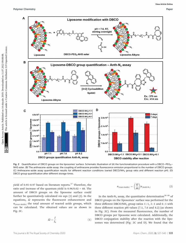
yield of 0.95–0.97 based on literature reports.37 Therefore, theratio and increase of the quantum yield is 0.96/0.02 = 48. Theamount of DBCO groups on the liposome surface couldfurther be quantitatively calculated via eqn (1) and (2). In theequations, Ω represents the fluorescent enhancement andn(react-Azido) the total amount of reacted azide groups, whichcan be calculated. The obtained values are as shown inFig. 2C.
Ω ¼ II0
ð1Þ
nðreact‐AzidoÞ ¼ Ω
48
� �nðAnth‐N3Þ ð2Þ
In the Anth-N3 assay, the quantitative determination38–40 ofDBCO groups on the liposomes’ surface was performed for thethree different DBCO/NH2 group ratios 1 : 1, 3 : 1 and 6 : 1 withthree different reaction pH values (7.1, 7.6 and 8.2) (as shownin Fig. 2C). From the measured fluorescence, the number ofDBCO groups per liposome were calculated. Additionally, theDBCO conjugation stability after the reaction with the lipo-somes was determined (Fig. 2C and D). We found that the
Fig. 2 Quantification of DBCO groups on the liposomes’ surface. Schematic illustration of (A) the functionalization procedure with a DBCO–PEG4–
NHS ester. (B) The anthracene-azide assay: the coupling of anthracene provides fluorescence emission proportional to the number of DBCO groups.(C) Anthracene-azide assay quantification results for different reaction conditions (varied DBCO/NH2 group ratio and different reaction pH). (D)DBCO group quantification after different storage times.
Polymer Chemistry Paper
This journal is © The Royal Society of Chemistry 2020 Polym. Chem., 2020, 11, 527–540 | 533
Ope
n A
cces
s A
rtic
le. P
ublis
hed
on 1
4 ok
tyab
r 20
19. D
ownl
oade
d on
11.
07.2
022
09:0
8:08
. T
his
artic
le is
lice
nsed
und
er a
Cre
ativ
e C
omm
ons
Attr
ibut
ion
3.0
Unp
orte
d L
icen
ce.
View Article Online

highest modification efficiency is observed for a 3-fold molarratio of DBCO to liposomes for the same reaction pH value. Inthis case, the maximum conjugation efficiency is ∼20%. Inaddition, the conjugation efficiency increased slightly alongwith the increasing reaction pH for all reaction molar ratiosbetween DBCO/NH2. Next to this, the stability of the DBCOgroups after the liposome modification was investigated(Fig. 2D). The value of DBCO groups per liposomes decreasedby more than 50% after 7 days compared to the fresh samples.Moreover, some floccules started to appear within the Lipo-DBCO suspension, especially for the molar ratio DBCO/NH2 =6 : 1 after 7 days. This means that further coupling by clickchemistry ideally should be performed within the same day.
Since during this reaction optimization step it was found thatthe ratio of DBCO/NH2 = 6 : 1 did not result in further increaseof the reaction yield, but rather in a significant decrease, sub-sequent coupling of the chosen antibody was only performedfor the 1 : 1 and 3 : 1 ratio conditions, which were coupled at areaction pH of 8.2.
3.4. Antibody functionalization and coupling to liposomesvia copper-free click reaction
Most widely in literature, antibodies are functionalized viaNHS-chemistry.41–43 In this case, the functional group isattached on the lysine residues or the N-termini of the anti-bodies. This functionalization strategy is highly unspecific as
Fig. 3 Antibody functionalization and attachment to surface-modified liposomes. (A) Enzymatic removal of galactose from the Fc-part of theCD11c-antibody with galactosidase. (B) Site-specific, enzymatic attachment of an azide functionality using UDP-N-azidoacetylgalactosamine(UDP-GalNAz) to the sugar residues on the Fc-part of the CD11c-antibody (C) bio-orthogonal copper-free click reaction to attach the azide-modified antibody on the DBCO-modified liposome surface.
Paper Polymer Chemistry
534 | Polym. Chem., 2020, 11, 527–540 This journal is © The Royal Society of Chemistry 2020
Ope
n A
cces
s A
rtic
le. P
ublis
hed
on 1
4 ok
tyab
r 20
19. D
ownl
oade
d on
11.
07.2
022
09:0
8:08
. T
his
artic
le is
lice
nsed
und
er a
Cre
ativ
e C
omm
ons
Attr
ibut
ion
3.0
Unp
orte
d L
icen
ce.
View Article Online

lysine residues are distributed over the whole antibodysequence including the antigen-binding site (Fab region).44
Next to this, the two N-termini of the antibody are also locatedwithin the Fab region. Therefore, introducing functionalgroups along those groups can cause a loss of the antibodyfunctionality and decreased cellular recognition.36 Therefore,we chose a site-selective antibody functionalization strategyto overcome the loss of binding specificity and antibodyfunctionality. Monoclonal antibodies have two conservedN-glycosylation sites on the heavy chain of the fragment crys-tallizable region (Fc-part).45 It is known that N-glycosylationoccurs at the amino acid asparagine (Asn) in the position Asn-297.46 Therefore, here we used a functionalization strategy,which allows the site-selective attachment of a maximum offour azide groups (two per heavy chain) at the Asn-297 positionto the CD11c antibody. It is a two-step protocol, where in thefirst step galactose is enzymatically removed from the sugar-tree and an N-acetylglucosamine residue is exposed (Fig. 3.A).In a second step, an azide-labeled sugar derivate (GalNAz,N-azidoacetylgalactosamine-tetraacylated) is enzymaticallyattached via the galactose-specific transferase (β1,4-galactosyl-tranferase mutant Y289L-Gal-T1) to the glycans of the antibody(Fig. 3.B). This method was first developed for the site-selective
radiolabeling of antibodies.47 In this case, the azide-modifiedantibody was conjugated with a desferrioxamine-modified di-benzocyclooctyne (DIBO) in a copper-free click reaction and89Zr was further added for radiolabeling. The same antibodyfunctionalization strategy was also applied for engineeringsite-specific antibody–drug-conjugates.48,49
To investigate the structural and functional integrity of theazide-modified antibody compared to the native antibody,nanoDSF measurements, SDS-PAGE (Fig. 4) and a cell bindingassay (Fig. S7†) were performed. nanoDSF is a method to deter-mine protein stability based on the intrinsic fluorescenceintensity of a protein upon heating. The characteristic meltingtemperature (TM) is defined at the point, where 50% of theprotein is unfolded. For the native anti-mouse CD11c antibody(blue) and the azide-modified antibody (orange), the thermalbehavior determined by nanoDSF was highly comparable(Fig. 4A). Next to this, the melting temperature before andafter functionalization was about 67 °C, which is in accordancewith literature reports for other antibodies.50 This indicatesthat the azide modification of the antibody had no majorimpact on the structural properties of the antibody.
Next to this, the unmodified (1) and modified (2) antibodywas applied to SDS-PAGE (Fig. 4C) to investigate molecular
Fig. 4 Characterization of the azide-modified antibody. (A) and (B) nanoDSF measurements of the unmodified and azide-modified anti-mouseCD11c antibody (10 µL, ∼0.5 mg mL−1). The antibody was heated up (1 °C min−1) from 20 °C to 95 °C and the intrinsic fluorescence intensity at330 nm and 350 nm was recorded. The ratio of the fluorescence intensity (350/330 nm, A) and the first derivation (B) is plotted. The melting temp-erature TM is determined from the maximum of the 1st derivation of the ratio. (C) SDS-PAGE of unmodified (1), azide modified CD11c (2) andPEGylated CD11c antibody (3). Proteins (3 µg) are reduced, applied to an SDS-PAGE and protein bands are visualized by Silver Staining.
Polymer Chemistry Paper
This journal is © The Royal Society of Chemistry 2020 Polym. Chem., 2020, 11, 527–540 | 535
Ope
n A
cces
s A
rtic
le. P
ublis
hed
on 1
4 ok
tyab
r 20
19. D
ownl
oade
d on
11.
07.2
022
09:0
8:08
. T
his
artic
le is
lice
nsed
und
er a
Cre
ativ
e C
omm
ons
Attr
ibut
ion
3.0
Unp
orte
d L
icen
ce.
View Article Online

weight differences and possible degradation products afterantibody functionalization. We could observe no significantdifference for the azide-modified antibody compared to theunmodified one. To verify the successful azide functionali-zation of the antibody, a model reaction with a DBCO-PEG-linker (5 kDa) was carried out and the resulting conjugatewas applied to SDS-PAGE. In lane 3 (Fig. 4C) there are twoadditional protein bands visualized, which correspond to theheavy chain (HC) fragment of the antibody (50 kDa) modifiedwith one DBCO–PEG5 kDa (+5 kDa) and thus one azide groupor an HC fragment conjugated with two DBCO–PEG5 kDa
(+10 kDa) and thus two azide groups per HC fragment. Thismeans for the full antibody (having two HC fragments) thelabeling efficiency is between 2–4 azides per antibody. This isalso in agreement with literature reports, stating a labelingdegree of 2.7 ± 0.2 GalNAz residues per antibody.47
To determine the functional binding properties of themodified antibody, a DIBO-modified fluorescent dye (PE) wasincubated with the azide-modified anti-mouse CD11c anti-body. Murine dendritic cells (DC2.4), which express CD11cantigen, were incubated with the PE-modified anti-mouseCD11c antibody for 30 min at 4 °C (Fig. S7†). The PE-modifiedanti-mouse CD11c antibody strongly bound to the DC2.4 cells.To confirm the antibody specificity, cells were pre-treated with
free unlabeled anti-mouse CD11c antibody (blocking assay). Inthis case, the PE-modified anti-mouse CD11c antibody was notable to bind to the DC2.4 cells proving the receptor specificityof the PE-modified anti-mouse CD11c antibody.
In a last step, the azide-modified anti-mouse CD11c anti-bodies were incubated with the DBCO-functionalized lipo-somes overnight at room temperature (Fig. 3C). Via the bio-orthogonal copper-free click reaction (SPAAC), it was possibleto covalently immobilize the azide-modified antibodies on theDBCO-functionalized liposomes’ surface.
To prove the successful coupling, flow cytometry measure-ments were performed using our previously established proto-col.36 Here, we chose a fluorescently-labeled secondary anti-body (FITC-anti hamster antibody), which specifically binds tothe immobilized anti-mouse CD11c antibodies (clone N418,host species Armenian Hamster) on the liposomes’ surface byrecognition of the Fc-region (Fig. 5A). As a control, liposomesfunctionalized with DBCO only were incubated with the FITC-labeled secondary antibody.
As a result of the flow cytometry analysis, we detected ahigh amount of FITC-fluorescence positive liposomes for Lipo-CD11c (∼60% to 80%, Fig. 5B). In contrast, there was a verylow unspecific binding of the FITC-labeled secondary antibodyto the Lipo-DBCO samples (<20%) as expected without any
Fig. 5 Verification of antibody presence on the liposomes’ surface. (A) Schematic illustration of the flow cytometry measurements for the antibodyquantification on the liposome surface. (B) and (C) Flow cytometry measurements for liposomes functionalized with anti-mouse-CD11c antibodies.A secondary labeled FITC-anti-hamster antibody was used to detect the antibodies on the liposome surface. Different reaction conditions (ratiobetween DBCO/NH2 groups 1 : 1 or 3 : 1) were investigated. The number of FITC-positive liposomes in % (B) or the median fluorescence intensity(MFI) is shown (C).
Paper Polymer Chemistry
536 | Polym. Chem., 2020, 11, 527–540 This journal is © The Royal Society of Chemistry 2020
Ope
n A
cces
s A
rtic
le. P
ublis
hed
on 1
4 ok
tyab
r 20
19. D
ownl
oade
d on
11.
07.2
022
09:0
8:08
. T
his
artic
le is
lice
nsed
und
er a
Cre
ativ
e C
omm
ons
Attr
ibut
ion
3.0
Unp
orte
d L
icen
ce.
View Article Online

attached proteins being present. Along with the quantificationof the DBCO groups on the liposomes’ surface (Fig. 2B), ahigher amount of antibodies was detected for liposomes modi-fied with the DBCO/NH2 = 3 : 1 ratio (Fig. 5C). This means that,indeed, the number of attached antibodies was also pro-portional to the number of available DBCO groups on the lipo-somes and the proposed coupling strategy provides precisecontrol over the obtained liposomal surface. It has to be noted,however, that the exact number of antibodies per liposomecannot be deducted from the fluorescence intensity, as theunspecific binding also has to be taken into account. Thetheoretical maximum number of antibodies per liposome wascalculated to be 42 (see Table S1†).
Finally, the liposome–antibody conjugates were againcharacterized with regards to their physico-chemical propertiesas shown in Fig. 6. The size and surface charge of the non-functionalized as well as the functionalized liposomes wascompared for both coupling ratios DBCO/NH2 1 : 1 and 3 : 1.
As indicated in Fig. 6.A and C, the size of the liposomesincreased step-wise after functionalization. Coupling of theDBCO groups and the antibody CD11c slightly enlarged theliposomes hydrodynamic diameter from 194 nm up to 293 and325 nm (1 : 1 and 3 : 1 DBCO/NH2 groups, respectively). Thereare mainly two reasons: (1) for the DBCO conjugation/antibody
click reaction, the DBCO-liposomes and the liposome-antibodyconjugates have to be purified from free excess DBCO–PEG4–
NHS ester and further free unreacted antibodies by centrifu-ging 3 times at 20 000g. During these washing steps, smallerliposomes could also be removed. (2) Long chain moleculePEG and DBCO groups as well as the attached antibodiesresult in larger overall liposomes. Furthermore, we observedthat the zeta potential of the functionalized liposomes wasmore negative compared to the non-functionalized surface.After attachment of the CD11c antibodies, the zeta potentialapproached a value of −25 mV (for the 3 : 1 DBCO/NH2 ratio),which is typical for nanocarrier surfaces covered with proteins.
Finally, with the performed characterization approaches, weare confident that we have presented a facile functionalizationapproach for bio-orthogonal and site-specific attachment ofantibodies on a liposome surface. By making use of specificquantification techniques, the available surface functionalgroups can precisely be analyzed so that the click chemistryreaction stoichiometry can be adjusted to yield an optimalefficiency. With the site-specific antibody modification,additionally it can be assured that the final conjugationproduct carries recognizable antibody units, which is of impor-tance for further application. The availability of the coupledantibody could even be increased if necessary upon introduc-
Fig. 6 Physicochemical characterization of liposomes before and after functionalization with DBCO/antibodies. (A) Size of liposomes before/afterDBCO and antibody attachment using 1 : 1 and 3 : 1 DBCO/NH2 ratios. (B) Surface charge before and after liposome functionalization. (C) Overviewof the physicochemical properties of the liposomes for the different reaction conditions.
Polymer Chemistry Paper
This journal is © The Royal Society of Chemistry 2020 Polym. Chem., 2020, 11, 527–540 | 537
Ope
n A
cces
s A
rtic
le. P
ublis
hed
on 1
4 ok
tyab
r 20
19. D
ownl
oade
d on
11.
07.2
022
09:0
8:08
. T
his
artic
le is
lice
nsed
und
er a
Cre
ativ
e C
omm
ons
Attr
ibut
ion
3.0
Unp
orte
d L
icen
ce.
View Article Online

tion of a longer PEG linker, so that e.g. the adsorption of pro-teins would not cover the targeting structures.
4. Conclusion
In this study, we have shown the synthesis of amine group-terminated liposomes via lipid dry film rehydration followedby extrusion. In contrast to common vesicle functionalizationapproaches, which rely on pre-modification of lipids or block-copolymers, we introduced further functional groups and per-formed antibody coupling via bio-orthogonal copper-free clickchemistry after liposome self-assembly. For precise reactioncontrol and tunability, a fluorescamine assay (FA assay) and ananthracen-azide (Anth-N3) assay were utilized to determine thenumber of present NH2 groups as well as DBCO groups afterfunctionalization. The antibody, which was introduced as apossible targeting structure, was modified site specifically atthe Fc fragment and coupled to the liposomes using thestrain-promoted alkyne–azide click (SPAAC) reaction. Thissurface functionalization procedure offers the advantage to betransferred to all self-assembled systems bearing primarysurface amino groups without damaging the assembled struc-ture because no large amounts of organic solvent are requiredand the carriers can be kept in physiological conditions.Amino groups can easily be introduced as end groups of block-copolymers and are already present on natural structures suchas extracellular vesicles (in the form of membrane proteins).Additionally, this type of antibody coupling is applicable forall antibody types without the disadvantage of potentially cov-ering or modifying the antigen binding sites. If needed, theintroduced PEG spacer can also be varied in length or type ofbuilding block to provide additional characteristics likeenhanced stealth properties or better antibody accessibility. Insummary, this versatile vesicle surface functionalization strat-egy is applicable for a wide range of drug delivery systems aswell as straightforward and robust in terms of handling.
Conflicts of interest
The authors declare no conflict of interest.
Acknowledgements
This project has received funding from the European Union’sHorizon 2020 research and innovation program under grantagreement No. 801338 (Ves4Us project). Open Access fundingprovided by the Max Planck Society.
References
1 E. Rideau, R. Dimova, P. Schwille, F. R. Wurm andK. Landfester, Liposomes and polymersomes: a compara-
tive review towards cell mimicking, Chem. Soc. Rev., 2018,47(23), 8572–8610.
2 J. Braun, N. Bruns, T. Pfohl and W. Meier, Phase Behaviorof Vesicle-Forming Block Copolymers in AqueousSolutions, Langmuir, 2007, 23(14), 7484–7490.
3 D. E. Discher and A. Eisenberg, Polymer vesicles, Science,2002, 297(5583), 967–973.
4 R. P. Brinkhuis, F. P. J. T. Rutjes and J. C. M. van Hest,Polymeric vesicles in biomedical applications, Polym.Chem., 2011, 2(7), 1449–1462.
5 A. D. Bangham and R. W. Horne, Negative staining of phos-pholipids and their structural modification by surface-active agents as observed in the electron microscope,J. Mol. Biol., 1964, 8(5), 660–668.
6 J. Li, X. Wang, T. Zhang, C. Wang, Z. Huang, X. Luo andY. Deng, A review on phospholipids and their main appli-cations in drug delivery systems, Asian J. Pharm. Sci., 2015,10(2), 81–98.
7 G. Caracciolo, Clinically approved liposomal nanomedi-cines: lessons learned from the biomolecular corona,Nanoscale, 2018, 10(9), 4167–4172.
8 D. G. Villalva, L. Giansanti, A. Mauceri, F. Ceccacci andG. Mancini, Influence of the state of phase of lipid bilayeron the exposure of glucose residues on the surface of lipo-somes, Colloids Surf., B, 2017, 159, 557–563.
9 N. Monteiro, A. Martins, L. Reis Rui and M. Neves Nuno,Liposomes in tissue engineering and regenerative medi-cine, J. R. Soc., Interface, 2014, 11(101), 20140459.
10 Y.-C. Kuo, I. Y. Chen and R. Rajesh, Use of functionalizedliposomes loaded with antioxidants to permeate the blood–brain barrier and inhibit β-amyloid-induced neurodegenera-tion in the brain, J. Taiwan Inst. Chem. Eng., 2018, 87, 1–14.
11 S. Cavalli, A. R. Tipton, M. Overhand and A. Kros, Thechemical modification of liposome surfaces via a copper-mediated [3 + 2] azide–alkyne cycloaddition monitored by acolorimetric assay, Chem. Commun., 2006, (30), 3193–3195.
12 C. Saraiva, C. Praça, R. Ferreira, T. Santos, L. Ferreira andL. Bernardino, Nanoparticle-mediated brain drug delivery:Overcoming blood–brain barrier to treat neurodegenerativediseases, J. Controlled Release, 2016, 235, 34–47.
13 J. Kreuter, Nanoparticulate systems for brain delivery ofdrugs, Adv. Drug Delivery Rev., 2001, 47(1), 65–81.
14 A. Akbarzadeh, R. Rezaei-Sadabady, S. Davaran, S. W. Joo,N. Zarghami, Y. Hanifehpour, M. Samiei, M. Kouhi andK. Nejati-Koshki, Liposome: classification, preparation,and applications, Nanoscale Res. Lett., 2013, 8(1), 102.
15 B. S. Pattni, V. V. Chupin and V. P. Torchilin, NewDevelopments in Liposomal Drug Delivery, Chem. Rev.,2015, 115(19), 10938–10966.
16 M. K. Riaz, M. A. Riaz, X. Zhang, C. Lin, K. H. Wong, X. Chen,G. Zhang, A. Lu and Z. Yang, Surface Functionalization andTargeting Strategies of Liposomes in Solid Tumor Therapy:A Review, Int. J. Mol. Sci., 2018, 19(1), 195.
17 P. V. Pawar, S. V. Gohil, J. P. Jain and N. Kumar,Functionalized polymersomes for biomedical applications,Polym. Chem., 2013, 4(11), 3160–3176.
Paper Polymer Chemistry
538 | Polym. Chem., 2020, 11, 527–540 This journal is © The Royal Society of Chemistry 2020
Ope
n A
cces
s A
rtic
le. P
ublis
hed
on 1
4 ok
tyab
r 20
19. D
ownl
oade
d on
11.
07.2
022
09:0
8:08
. T
his
artic
le is
lice
nsed
und
er a
Cre
ativ
e C
omm
ons
Attr
ibut
ion
3.0
Unp
orte
d L
icen
ce.
View Article Online

18 S. Egli, H. Schlaad, N. Bruns and W. Meier,Functionalization of Block Copolymer Vesicle Surfaces,Polymers, 2011, 3(1), 252–280.
19 C. Weber, M. Voigt, J. Simon, A.-K. Danner, H. Frey,V. Mailänder, M. Helm, S. Morsbach and K. Landfester,Functionalization of Liposomes with Hydrophilic PolymersResults in Macrophage Uptake Independent of the ProteinCorona, Biomacromolecules, 2019, 20(8), 2989–2999.
20 S. A. Meeuwissen, M. F. Debets and J. C. M. van Hest,Copper-free click chemistry on polymersomes: pre- vs. post-self-assembly functionalisation, Polym. Chem., 2012, 3(7),1783–1795.
21 P. Vabbilisetty and X.-L. Sun, Liposome surface functionali-zation based on different anchoring lipids via Staudingerligation, Org. Biomol. Chem., 2014, 12(8), 1237–1244.
22 Y. Ma, H. Zhang, V. Gruzdys and X.-L. Sun, Azide-ReactiveLiposome for Chemoselective and BiocompatibleLiposomal Surface Functionalization and Glyco-LiposomalMicroarray Fabrication, Langmuir, 2011, 27(21), 13097–13103.
23 S. J. Rijpkema, B. J. Toebes, M. N. Maas, N. R. M. de Klerand D. A. Wilson, Designing Molecular Building Blocks forFunctional Polymersomes, Isr. J. Chem., 2019, 59, 1–18.
24 S. F. M. van Dongen, M. Nallani, S. Schoffelen,J. L. M. Cornelissen, R. J. M. Nolte and J. C. M. van Hest, ABlock Copolymer for Functionalisation of PolymersomeSurfaces, Macromol. Rapid Commun., 2008, 29(4), 321–325.
25 R. C. Lai, R. W. Y. Yeo, K. H. Tan and S. K. Lim, Exosomesfor drug delivery—a novel application for the mesenchymalstem cell, Biotechnol. Adv., 2013, 31(5), 543–551.
26 S. Stremersch, R. E. Vandenbroucke, E. Van Wonterghem,A. Hendrix, S. C. De Smedt and K. Raemdonck, Comparingexosome-like vesicles with liposomes for the functional cel-lular delivery of small RNAs, J. Controlled Release, 2016,232, 51–61.
27 J. G. van den Boorn, J. Daßler, C. Coch, M. Schlee andG. Hartmann, Exosomes as nucleic acid nanocarriers, Adv.Drug Delivery Rev., 2013, 65(3), 331–335.
28 M. A. Bruckman, G. Kaur, L. A. Lee, F. Xie, J. Sepulveda,R. Breitenkamp, X. Zhang, M. Joralemon, T. P. Russell,T. Emrick and Q. Wang, Surface Modification of TobaccoMosaic Virus with “Click” Chemistry, ChemBioChem, 2008,9(4), 519–523.
29 R. K. O’Reilly, M. J. Joralemon, K. L. Wooley andC. J. Hawker, Functionalization of Micelles and Shell Cross-linked Nanoparticles Using Click Chemistry, Chem. Mater.,2005, 17(24), 5976–5988.
30 J. Gierlich, G. A. Burley, P. M. E. Gramlich,D. M. Hammond and T. Carell, Click Chemistry as aReliable Method for the High-Density PostsyntheticFunctionalization of Alkyne-Modified DNA, Org. Lett., 2006,8(17), 3639–3642.
31 G. Baier, J. M. Siebert, K. Landfester and A. Musyanovych,Surface Click Reactions on Polymeric Nanocapsules forVersatile Functionalization, Macromolecules, 2012, 45(8),3419–3427.
32 E. M. Alexandrino, P. Buchold, M. Wagner, A. Fuchs,A. Kreyes, C. K. Weiss, K. Landfester and F. R. Wurm, Amolecular “screw-clamp”: accelerating click reactions inminiemulsions, Chem. Commun., 2014, 50(72), 10495–10498.
33 C. Ornelas, J. Broichhagen and M. Weck, Strain-PromotedAlkyne Azide Cycloaddition for the Functionalization ofPoly(amide)-Based Dendrons and Dendrimers, J. Am. Chem.Soc., 2010, 132(11), 3923–3931.
34 E. Oude Blenke, G. Klaasse, H. Merten, A. Plückthun,E. Mastrobattista and N. I. Martin, Liposome functionali-zation with copper-free “click chemistry”, J. ControlledRelease, 2015, 202, 14–20.
35 S. W. Provencher, Contin - a General-Purpose ConstrainedRegularization Program for Inverting Noisy LinearAlgebraic and Integral-Equations, Comput. Phys. Commun.,1982, 27(3), 229–242.
36 M. Tonigold, J. Simon, D. Estupiñán, M. Kokkinopoulou,J. Reinholz, U. Kintzel, A. Kaltbeitzel, P. Renz,M. P. Domogalla, K. Steinbrink, I. Lieberwirth, D. Crespy,K. Landfester and V. Mailänder, Pre-adsorption of anti-bodies enables targeting of nanocarriers despite a bio-molecular corona, Nat. Nanotechnol., 2018, 13(9), 862–869.
37 F. Xie, K. Sivakumar, Q. Zeng, M. A. Bruckman, B. Hodgesand Q. Wang, A fluorogenic ‘click’reaction of azidoanthra-cene derivatives, Tetrahedron, 2008, 64(13), 2906–2914.
38 I. S. Marks, J. S. Kang, B. T. Jones, K. J. Landmark,A. J. Cleland and T. A. Taton, Strain-promoted “click”chemistry for terminal labeling of DNA, BioconjugateChem., 2011, 22(7), 1259–1263.
39 P. Van Delft, N. J. Meeuwenoord, S. Hoogendoorn,J. Dinkelaar, H. S. Overkleeft, G. A. van der Marel andD. V. Filippov, Synthesis of oligoribonucleic acid conjugatesusing a cyclooctyne phosphoramidite, Org. Lett., 2010,12(23), 5486–5489.
40 A. Jawalekar, S. Malik, J. Verkade, B. Gibson, N. Barta,J. Hodges, A. Rowan and F. van Delft, Oligonucleotidetagging for copper-free click conjugation, Molecules, 2013,18(7), 7346–7363.
41 H. Gong, I. Holcomb, A. Ooi, X. Wang, D. Majonis,M. A. Unger and R. Ramakrishnan, Simple Method ToPrepare Oligonucleotide-Conjugated Antibodies and ItsApplication in Multiplex Protein Detection in Single Cells,Bioconjugate Chem., 2016, 27(1), 217–225.
42 J. A. G. L. van Buggenum, J. P. Gerlach, S. Eising,L. Schoonen, R. A. P. M. van Eijl, S. E. J. Tanis,M. Hogeweg, N. C. Hubner, J. C. van Hest, K. M. Bongerand K. W. Mulder, A covalent and cleavable antibody-DNAconjugation strategy for sensitive protein detection viaimmuno-PCR, Sci. Rep., 2016, 6, 22675.
43 S. L. Filbrun, A. B. Filbrun, F. L. Lovato, S. H. Oh,E. A. Driskell and J. D. Driskell, Chemical modification ofantibodies enables the formation of stable antibody–goldnanoparticle conjugates for biosensing, Analyst, 2017,142(23), 4456–4467.
Polymer Chemistry Paper
This journal is © The Royal Society of Chemistry 2020 Polym. Chem., 2020, 11, 527–540 | 539
Ope
n A
cces
s A
rtic
le. P
ublis
hed
on 1
4 ok
tyab
r 20
19. D
ownl
oade
d on
11.
07.2
022
09:0
8:08
. T
his
artic
le is
lice
nsed
und
er a
Cre
ativ
e C
omm
ons
Attr
ibut
ion
3.0
Unp
orte
d L
icen
ce.
View Article Online

44 H. Yao, F. Jiang, A. Lu and G. Zhang, Methods to Designand Synthesize Antibody-Drug Conjugates (ADCs),Int. J. Mol. Sci., 2016, 17(2), 194.
45 F. Cymer, H. Beck, A. Rohde and D. Reusch, Therapeuticmonoclonal antibody N-glycosylation – Structure,function and therapeutic potential, Biologicals, 2018, 52,1–11.
46 M. Kiyoshi, K. Tsumoto, A. Ishii-Watabe andJ. M. M. Caaveiro, Glycosylation of IgG-Fc: a molecular per-spective, Int. Immunol., 2017, 29(7), 311–317.
47 B. M. Zeglis, C. B. Davis, R. Aggeler, H. C. Kang, A. Chen,B. J. Agnew and J. S. Lewis, Enzyme-Mediated Methodologyfor the Site-Specific Radiolabeling of Antibodies Based onCatalyst-Free Click Chemistry, Bioconjugate Chem., 2013,24(6), 1057–1067.
48 P. Thompson, E. Ezeadi, I. Hutchinson, R. Fleming,B. Bezabeh, J. Lin, S. Mao, C. Chen, L. Masterson,H. Zhong, D. Toader, P. Howard, H. Wu, C. Gao andN. Dimasi, Straightforward Glycoengineering Approach toSite-Specific Antibody–Pyrrolobenzodiazepine Conjugates,ACS Med. Chem. Lett., 2016, 7(11), 1005–1008.
49 X. Cai and K. D. Janda, A chemoenzymatic approachtoward the preparation of site-specific antibody–drug con-jugates, Tetrahedron Lett., 2015, 56(23), 3172–3175.
50 M. Tonigold, J. Simon, D. Estupiñán, M. Kokkinopoulou,J. Reinholz, U. Kintzel, A. Kaltbeitzel, P. Renz,M. P. Domogalla, K. Steinbrink, I. Lieberwirth, D. Crespy,K. Landfester and V. Mailänder, Pre-adsorption of anti-bodies enables targeting of nanocarriers despite a bio-molecular corona, Nat. Nanotechnol., 2018, 1.
Paper Polymer Chemistry
540 | Polym. Chem., 2020, 11, 527–540 This journal is © The Royal Society of Chemistry 2020
Ope
n A
cces
s A
rtic
le. P
ublis
hed
on 1
4 ok
tyab
r 20
19. D
ownl
oade
d on
11.
07.2
022
09:0
8:08
. T
his
artic
le is
lice
nsed
und
er a
Cre
ativ
e C
omm
ons
Attr
ibut
ion
3.0
Unp
orte
d L
icen
ce.
View Article Online