Sunitinib mediates reversal of myeloid-derived suppressor cell accumulation in renal cell carcinoma...
11
2009;15:2148-2157. Published online March 10, 2009. Clin Cancer Res Jennifer S. Ko, Arnold H. Zea, Brian I. Rini, et al. Cell Accumulation in Renal Cell Carcinoma Patients Sunitinib Mediates Reversal of Myeloid-Derived Suppressor Updated Version 10.1158/1078-0432.CCR-08-1332 doi: Access the most recent version of this article at: Material Supplementary C1.html http://clincancerres.aacrjournals.org/content/suppl/2009/03/13/1078-0432.CCR-08-1332.D Access the most recent supplemental material at: Cited Articles http://clincancerres.aacrjournals.org/content/15/6/2148.full.html#ref-list-1 This article cites 48 articles, 27 of which you can access for free at: Citing Articles http://clincancerres.aacrjournals.org/content/15/6/2148.full.html#related-urls This article has been cited by 32 HighWire-hosted articles. Access the articles at: E-mail alerts related to this article or journal. Sign up to receive free email-alerts Subscriptions Reprints and . [email protected] Publications Department at To order reprints of this article or to subscribe to the journal, contact the AACR Permissions . [email protected] Department at To request permission to re-use all or part of this article, contact the AACR Publications American Association for Cancer Research Copyright © 2009 on February 20, 2013 clincancerres.aacrjournals.org Downloaded from DOI:10.1158/1078-0432.CCR-08-1332
-
Upload
independent -
Category
Documents
-
view
2 -
download
0
Transcript of Sunitinib mediates reversal of myeloid-derived suppressor cell accumulation in renal cell carcinoma...
2009;15:2148-2157. Published online March 10, 2009.Clin Cancer Res Jennifer S. Ko, Arnold H. Zea, Brian I. Rini, et al. Cell Accumulation in Renal Cell Carcinoma PatientsSunitinib Mediates Reversal of Myeloid-Derived Suppressor
Updated Version 10.1158/1078-0432.CCR-08-1332doi:
Access the most recent version of this article at:
MaterialSupplementary
C1.htmlhttp://clincancerres.aacrjournals.org/content/suppl/2009/03/13/1078-0432.CCR-08-1332.DAccess the most recent supplemental material at:
Cited Articles http://clincancerres.aacrjournals.org/content/15/6/2148.full.html#ref-list-1
This article cites 48 articles, 27 of which you can access for free at:
Citing Articles http://clincancerres.aacrjournals.org/content/15/6/2148.full.html#related-urls
This article has been cited by 32 HighWire-hosted articles. Access the articles at:
E-mail alerts related to this article or journal.Sign up to receive free email-alerts
SubscriptionsReprints and
[email protected] Department atTo order reprints of this article or to subscribe to the journal, contact the AACR
To request permission to re-use all or part of this article, contact the AACR Publications
American Association for Cancer Research Copyright © 2009 on February 20, 2013clincancerres.aacrjournals.orgDownloaded from
DOI:10.1158/1078-0432.CCR-08-1332

Sunitinib Mediates Reversal of Myeloid-Derived Suppressor CellAccumulation in Renal Cell Carcinoma PatientsJennifer S. Ko,1,2,3 Arnold H. Zea,6 Brian I. Rini,3,4 Joanna L. Ireland,1Paul Elson,5 Peter Cohen,1,3
Ali Golshayan,3 Patricia A. Rayman,1LauraWood,3 Jorge Garcia,3 Robert Dreicer,3,4
Ronald Bukowski,3,4 andJames H. Finke1,2,3,4
Abstract Purpose: Immune dysfunction reported in renal cell carcinoma (RCC) patients may contribute totumor progression. Myeloid-derived suppressor cells (MDSC) represent one mechanism bywhich tumors induceT-cell suppression. Several factors pivotal to the accumulation of MDSCare targeted by the tyrosine kinase inhibitor, sunitinib.The effect of sunitinib on MDSC-mediatedimmunosuppression in RCC patients has been investigated.Experimental Design: Patient peripheral blood levels of MDSC and regulatoryT-cell (Treg) andT-cell production of IFN-g were evaluated before and after sunitinib treatment. Correlationsbetween MDSC andTreg normalization as well asT-cell production of IFN-g were examined.Thein vitro effect of sunitinib on patient MDSC was evaluated.Results: Metastatic RCC patients had elevated levels of CD33+HLA-DR- and CD15+CD14-
MDSC, and these were partially overlapping populations. Treatment with sunitinib resultedin significant reduction in MDSC measured by several criteria. Sunitinib-mediated reduction inMDSC was correlated with reversal of type 1T-cell suppression, an effect that could be repro-ducedby the depletionof MDSC in vitro. MDSC reduction in response to sunitinib correlatedwitha reversal of CD3+CD4+CD25hiFoxp3+ Treg cell elevation. No correlation existed betweena change in tumor burden and a change in MDSC,Treg, orT-cell production of IFN-g. In vitroaddition of sunitinib reduced MDSC viability and suppressive effect when used at z1.0 Ag/mL.Sunitinib did not induce MDSC maturation in vitro.Conclusions: Sunitinib-based therapy has the potential to modulate antitumor immunity byreversing MDSC-mediated tumor-induced immunosuppression.
This year f36,000 Americans will be newly diagnosed withkidney cancer, resulting in 12,890 deaths (1). Poor outcome inrenal cell carcinoma (RCC) is related to its late diseasepresentation, propensity for recurrence, and refractorinessto traditional chemotherapy or radiotherapy. Additionally,treatment with cytokine therapy has resulted in limitedsuccess despite the known immunogenicity of RCC (2–6). The
receptor tyrosine kinase inhibitor sunitinib is an oral medicationbeing used with significant clinical effect in metastatic clear cellRCC (7–10). Sunitinib inhibits signaling through the vascularendothelial growth factor receptors (VEGFR), as well as platelet-derived growth factor receptor, stem cell factor receptor (c-Kit),Flt3, and colony-stimulating factor (CSF)-1 receptor (11).
VEGF signaling plays a prominent role in the pathogenesis ofclear cell RCC, particularly due to the common occurrence ofvon Hippel-Lindau gene inactivation in these tumors (10, 12).VEGF overproduction promotes tumor-associated angiogenesisrequired for tumor growth and metastasis (10, 12). VEGFmay also support tumor growth via negative effects onhost antitumor adaptive immunity, as increased VEGF levelshave been associated with alterations in myeloid cell differen-tiation, which impair competent dendritic cell formation andencourage suppressive myeloid cell formation in cancerpatients (13–18).
Myeloid-derived suppressor cells (MDSC) impair T-celleffector function and represent a heterogeneous population ofcells that accumulate in tumor-bearing hosts as a result oftumor-induced alterations in myelopoiesis (19). MDSC accu-mulating in the tumors and lymphoid organs of tumor-bearingmice are CD11b+Gr1+ and mediate T-cell impairment that isreversed with tumor removal or CD11b+ or Gr1+ cell depletion(18–21). MDSC detected in the peripheral blood of patientsbearing several tumor types (16, 22–27) express the common
Cancer Therapy: Clinical
Authors’ Affiliations: 1Department of Immunology, Cleveland Clinic LernerResearch Institute; 2Department of Pathology, Case Western Reserve University;3Taussig Cancer Institute, 4Glickman Urological Institute, and 5Quantitative HealthSciences Department, Cleveland Clinic, Cleveland, Ohio; and 6Department ofMicrobiology, Stanley S. Scott Cancer Center, Louisiana State University HealthSciences Center, New Orleans, LouisianaReceived 5/23/08; revised 10/6/08; accepted 10/13/08; published OnlineFirst3/10/09.Grant support: Pfizer (J. Finke, B. Rini) and Frank Rudy Fund for Cancer Research(J. Finke, R. Bukowski).The costs of publication of this article were defrayed in part by the payment of pagecharges.This article must therefore be hereby marked advertisement in accordancewith18 U.S.C. Section1734 solely to indicate this fact.Note: Supplementary data for this article are available at Clinical Cancer ResearchOnline (http://clincancerres.aacrjournals.org/).Requests for reprints: James H. Finke, Department of Immunology, LernerResearch Institute, Cleveland Clinic Foundation, NE4-250, 9500 Euclid Avenue,Cleveland, OH 44195. Phone: 216-444-5186; Fax: 216-444-9329; E-mail:[email protected].
F2009 American Association for Cancer Research.doi:10.1158/1078-0432.CCR-08-1332
www.aacrjournals.orgClin Cancer Res 2009;15(6) March15, 2009 2148
American Association for Cancer Research Copyright © 2009 on February 20, 2013clincancerres.aacrjournals.orgDownloaded from
DOI:10.1158/1078-0432.CCR-08-1332
myeloid marker CD33 but lack markers of mature myeloidcells such as the MHC class II molecule HLA-DR. Expression ofthe granulocytic marker CD15 divides patient MDSC into atleast two subsets. The CD15+ subset has been shown tosuppress T-cell function in patients with kidney cancer, amongothers, through an arginase and/or reactive oxygen species-dependent mechanism (23, 24). A CD15- subset of MDSC wasalso shown to suppress T-cell function in patients withsquamous cell carcinoma of the head/neck/lung or adenocar-cinoma of the breast/lung through an unclear mechanism (22).These subsets likely parallel those recently identified in themouse model, where the CD15+ (human) and the Gr1hi
(mouse) MDSC are granulocytic and the CD15- (human) andthe Gr1lo (mouse) MDSC are monocytic (28). New evidence inhepatocellular carcinoma and malignant melanoma patientssuggests that a third, CD14+HLA-DRdim subset of MDSC alsoexists (26, 27).
Because VEGF has been implicated in the generation ofMDSC (29, 30), we have evaluated the effect of sunitinib,which blocks signaling through multiple receptors, includingVEGFRs (11), on MDSC in metastatic RCC (mRCC) patients.MDSC are reported to inhibit T-cell function directly (19) aswell as indirectly via the induction of regulatory T-cell (Treg)formation (27, 31). We have therefore examined patient T-cellproduction of IFN-g, as well as patient Treg levels, before andafter treatment with sunitinib. We observed an elevation inCD15+ and CD15- MDSC in RCC patients compared with age-matched normal (AMN) donors. We show that treatment withsunitinib in mRCC patients reduces the elevated levels of bothMDSC subsets to near normal levels. A reduction in MDSCmeasured by two criteria (CD15+CD14- and CD33+HLA-DR-)was correlated with a subsequent recovery in patient T-celleffector function but not with tumor regression. A reduction inCD15+CD14- MDSC was associated with a reduction in patientTreg levels. mRCC patient MDSC-mediated T-cell suppressionin vitro was reversible with MDSC depletion and modestly withthe addition of excess L-arginine. Sunitinib, when used in vitro,
partially blocked the suppressive function of MDSC possiblydue to its effects on MDSC viability when added at z1.0 Ag/mLrather than its effects on MDSC maturation. Sunitinib-basedcombination immunotherapy may be a promising option forthe future treatment of mRCC.
Materials and Methods
Patient population and treatment. Patients received sunitinibmonotherapy for mRCC (50 mg by mouth daily) for 28 days followedby 14 days of rest, comprising one 6-week cycle. Patients were excludedif they received any anticancer therapy concomitant with sunitinib, ifthey had a diagnosis other than clear cell RCC, or if they did not receiveat least 28 days of sunitinib. Patients underwent disease assessment(computed tomography and bone scans) at baseline and after every 2cycles (every f12 weeks). Objective response according to theResponse Evaluation Criteria in Solid Tumor (32) and tumor burdenshrinkage were determined by investigator assessment of radiographs.Patients were treated until Response Evaluation Criteria in Solid Tumor-defined disease progression or unacceptable toxicity occurred. Doseinterruption and modification was done according to the treatingphysician’s discretion. All patients and healthy volunteer blood donorssigned an institutional review board-approved, written informedconsent for collection of blood samples. AMN donors were healthyvolunteers ages >50 years.Reagents. Human IgG and L-arginine were from Sigma-Aldrich.
Catalase was from Calbiochem. [3H]thymidine was from Amersham-Buchler. Anti-human CD3, CD4, IFN-g, interleukin (IL)-4, CD11c,CD13, CD14, CD15, CD33, HLA-DR, Annexin V, 7-amino-actinomycinD, and the Annexin V staining kit were from BD Biosciences. Anti-human Foxp3, CD11b, CD19, CD40, CD56, CD80, and CD86 werefrom eBioscience. Mouse isotype control antibodies were from BD,eBioscience, or Immunotech. Anti-human CD15 and CD33 antibody-coated magnetic microbeads and LS magnetic columns were fromMiltenyi Biotec. Granulocyte macrophage-CSF (GM-CSF) and IL-4 werefrom R&D Systems. Sunitinib in pure powder form was from Pfizer.Peripheral blood mononuclear cell isolation. Peripheral blood (60
mL) was drawn from mRCC patients before sunitinib treatment (cycle 1day 1) and on day 28 after one cycle of treatment (cycle 1 day 28), andin a subset of patients on day 28 after two cycles of treatment (cycle 2day 28), and from AMN donors. Peripheral blood was drawn inheparin-containing tubes. Peripheral blood mononuclear cells (PBMC)were isolated within 2 h of blood draw and either used fresh or frozenfor later use according to the methods described previously (33). Forphenotypic and functional studies where multiple time points wereavailable, all time points for an individual patient were thawed togetherand used in the same experiment.Fluorescence-activated cell sorting analysis of patient PBMC. Analysis
of MDSC percentages in patient PBMC were done on thawed samples.Cells were stained in fluorescence-activated cell sorting (FACS) buffer(1� PBS with 2% heat-inactivated fetal bovine serum and 0.02%sodium azide). Nonspecific antibody binding was blocked bypretreatment of cells with 10 Ag/mL human IgG for 20 min at roomtemperature. Surface stains were added to cells for 30 min at 4jC. Cellswere stained with anti-human CD11b, CD11c, CD14, CD15, CD33,and HLA-DR. In a subset of patients, cells were also stained with anti-human CD3, CD19, CD56, CD80, and CD86. Cells were washed inbuffer and then fixed in 1% paraformaldehyde and ran for FACS.
Treg were enumerated by FACS on thawed patient PBMC rested incomplete RPMI 1640 overnight (37jC, 5% CO2) and then stainedwith anti-CD3, CD4, CD25 (Stem Cell Technologies), and Foxp3according to the instructions included in the BD intracellular stainingkit. PBMC were incubated overnight to facilitate experiments thatcould examine both Treg and T-cell stimulation from the samesample. Thawed T cells stimulate better after some amount of resting
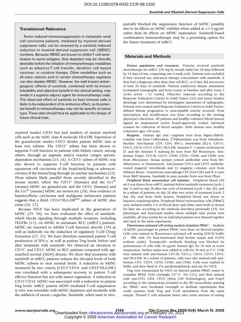
Translational Relevance
Tumor-induced immunosuppression in metastatic renalcell carcinoma patients, mediated by myeloid-derivedsuppressor cells, can be reversed by a sunitinib-inducedreduction in myeloid-derived suppressor cell (MDSC)numbers. Because MDSC are known to inhibitT-cell sensi-tization to tumor antigens, their depletion may be clinicallydesirable before the initiation of immunotherapy modalitiessuch as adoptive T-cell transfer, dendritic cell-basedvaccines, or cytokine therapy. Other modalities such asall-trans-retinoic acid or certain chemotherapy regimenscan also deplete MDSC. However, the well-known antian-giogenic effects of sunitinib, combined with its knowntolerability and objective benefit in the clinical setting, mayrender it a superior adjunct agent for immunotherapy trials.The observed effect of sunitinib on host immune cells islikely tobe independent of its antitumor effect, so itspoten-tial benefit in immunotherapy may not be specific to tumortype.These data should thus be applicable to the design offuture clinical trials.
Sunitinib andMyeloid-Derived Suppressor Cells
www.aacrjournals.org Clin Cancer Res 2009;15(6) March15, 20092149
American Association for Cancer Research Copyright © 2009 on February 20, 2013clincancerres.aacrjournals.orgDownloaded from
DOI:10.1158/1078-0432.CCR-08-1332
period, and multiple past experiments have shown numericallyequivalent results for Treg staining done immediately after thawingsamples or done after an overnight culture. All data were acquiredusing CellQuest on a BD FACSCalibur and analyzed using either FlowJo (Tree Star) or CellQuest software. At least 300,000 live cell eventswere collected for each tube used in analysis.Determination of patient T-cell IFN-g response. Patient PBMC
samples were stimulated with anti-CD3/CD28-bound beads (Dynal)and IL-2 (Chiron) for 72 h. Golgi plug was added to cells for the last 6h, and harvested cells were stained with anti-human CD3, CD4, IFN-g,and IL-4 according to the protocol provided using the BD intracellularstaining kit. Nonstimulated cells from each donor served as a negativecontrol. Additionally, specificity of cytokine staining was confirmed ineach sample via subtraction of any nonspecific staining occurring in
samples pretreated with unlabeled anti-cytokine antibodies before theaddition of fluorochrome-labeled antibodies.MDSC depletion. One half of each patient sample was treated with
anti-CD15 antibody-coated magnetic microbeads. Cells were incubatedat 4jC for 20 min and then washed and resuspended in PBE (PBS withBSA and EDTA). Cells were run over a LS magnetic column for thedepletion of bead-labeled cells as per the manufacturer’s instructions.FACS analysis was done on a small aliquot of cells to assure that MDSChad been effectively depleted. Cells were then resuspended in completeRPMI 1640 and then activated and stained for intracellular cytokines(IFN-g and IL-4) as described above.
In vitro sunitinib culture assays. Fresh patient PBMC wereincubated with a mixture of anti-human CD33 and CD15 antibody-conjugated magnetic beads. CD33+ and CD15+ myeloid cells were
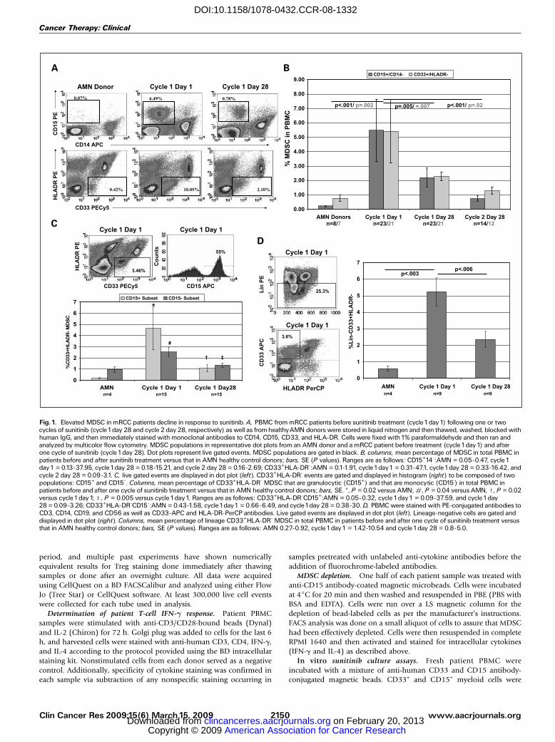
Fig. 1. Elevated MDSC in mRCC patients decline in response to sunitinib. A, PBMC from mRCC patients before sunitinib treatment (cycle1day1) following one or twocycles of sunitinib (cycle1day 28 and cycle 2 day 28, respectively) as well as from healthyAMN donors were stored in liquid nitrogen and then thawed, washed, blocked withhuman IgG, and then immediately stained with monoclonal antibodies to CD14, CD15, CD33, and HLA-DR. Cells were fixed with1% paraformaldehyde and then ran andanalyzed by multicolor flow cytometry. MDSC populations in representative dot plots from an AMN donor and a mRCC patient before treatment (cycle1day1) and afterone cycle of sunitinib (cycle 1day 28). Dot plots represent live gated events. MDSC populations are gated in black. B, columns, mean percentage of MDSC in total PBMC inpatients before and after sunitinib treatment versus that in AMN healthy control donors; bars, SE (P values). Ranges are as follows: CD15+14-:AMN = 0.05-0.47, cycle 1day1 = 0.13-37.95, cycle1day 28 = 0.18-15.21, and cycle 2 day 28 = 0.16-2.69; CD33+HLA-DR-:AMN = 0.1-1.91, cycle1day1 = 0.31-47.1, cycle1day 28 = 0.33-16.42, andcycle 2 day 28 = 0.09-3.1. C, live gated events are displayed in dot plot (left). CD33+HLA-DR- events are gated and displayed in histogram (right) to be composed of twopopulations: CD15+ and CD15-. Columns, mean percentage of CD33+HLA-DR- MDSC that are granulocytic (CD15+) and that are monocytic (CD15-) in total PBMC inpatients before and after one cycle of sunitinib treatment versus that in AMN healthy control donors; bars, SE. *, P = 0.02 versus AMN; #, P = 0.04 versus AMN, c, P = 0.02versus cycle1day1; b, P = 0.005 versus cycle1day1. Ranges are as follows: CD33+HLA-DR-CD15+:AMN = 0.05-0.32, cycle1day1 = 0.09-37.59, and cycle1day28 = 0.09-3.26; CD33+HLA-DR-CD15-:AMN = 0.43-1.58, cycle1day1 = 0.66-6.49, and cycle1day 28 = 0.38-30. D, PBMC were stained with PE-conjugated antibodies toCD3, CD14, CD19, and CD56 as well as CD33-APC and HLA-DR-PerCP antibodies. Live gated events are displayed in dot plot (left). Lineage-negative cells are gated anddisplayed in dot plot (right). Columns, mean percentage of lineage CD33+HLA-DR- MDSC in total PBMC in patients before and after one cycle of sunitinib treatment versusthat in AMN healthy control donors; bars, SE (P values). Ranges are as follows: AMN 0.27-0.92, cycle 1day1 = 1.42-10.54 and cycle 1day 28 = 0.8-5.0.
Cancer Therapy: Clinical
www.aacrjournals.orgClin Cancer Res 2009;15(6) March15, 2009 2150
American Association for Cancer Research Copyright © 2009 on February 20, 2013clincancerres.aacrjournals.orgDownloaded from
DOI:10.1158/1078-0432.CCR-08-1332

positively selected with a LS column. Cells that flowed through thecolumns were at least 80% CD3+ T cells, whereas positively selectedmyeloid cells were all CD11b+, and on average, 40% were negative forHLA-DR and thus represented MDSC.
For studies evaluating the in vitro effect of sunitinib on MDSC, aminimal concentration of 0.1 Ag/mL was used, as this is equivalent tothe levels detected in human plasma (34), and sunitinib was tittered upto 10- to 50-fold from there. To measure the effect of sunitinib onMDSC viability, myeloid cells containing MDSC were incubated in
complete RPMI 1640 with 20% SK-RC26B (gift from Dr. Neil Bander,Cornell Medical Center) RCC cell line-conditioned medium (tumorconditioned medium) and 50 ng/mL GM-CSF to support cell viabilityover 48 h (observed and published findings; ref. 22). Sunitinibsuspended in plain RPMI 1640 was added at 0.1, 1.0, and 5.0 Ag/mLto three groups of cells. After 48 h, cells were harvested, and surfacestained for HLA-DR and CD33, for 15 min at 4jC in FACS buffer andthen stained for Annexin V and 7-amino-actinomycin D per BDprovided protocol for 15 min and then ran for FACS.
Fig. 2. Sunitinib-mediated normalization of MDSC is associated with sunitinib-mediated enhancement inT-cell production of IFN-g in mRCC patients. A, patient or controldonor PBMC were stored in liquid nitrogen and thawed, and a portion of PBMC was analyzed immediately for MDSC as described previously.The other portion of PBMCwas rested overnight in complete medium, and equal numbers of nonadherent cells were stimulated polyclonally the following day with anti-CD3/CD28 antibody-coatedbeads and IL-2. Columns, median percentage of CD3+ cells staining positive for IFN-g. Squares, median percentage of MDSC in PBMC. Ranges for IFN-g production: normaldonors = 11.07-31.52, pretreatment = 2.25-38.31, cycle 1day 28 = 2.23-21.29, and cycle 2 day 28 = 5.09-34.08. Mean F SE and P values are as follows: AMN donors =18.40 F 1.78%, cycle1day1 = 11.07 F 2.13% (P = 0.008 forAMN versus cycle1day1), cycle1day 28 = 11.93 F 1.19% (P = 0.26 for cycle1day1versus cycle1day 28), andcycle 2 day 28 = 15.98 F 2.34% (P = 0.21for cycle 1day 28 versus cycle 2 day 28). B, changes in patientT-cell production of IFN-g in response to sunitinib werecompared with changes in their own levels of MDSC in response treatment with sunitinib. Each dot represents a single patient (r = -0.66; P = 0.03).
Fig. 3. In vitro depletionof MDSC restores patientT-cell productionof IFN-g.A, pretreatment patient PBMC samples were thawed and immediately stained for the expressionof myeloid cell surface markers with or without the removal of MDSC by anti-CD15 magnetic microbeads and magnetic column. Representative dot plots from live gatedcells in patients without (mRCC) and with (mRCC-MDSC) MDSC depletion. Plots shown after MDSC depletion contain less acquired events. Pretreatment patient PBMCsamples were thawed and polyclonally activated as described previously with or without the prior removal of MDSC. After 72 h, IFN-g and IL-4 were detected in CD3+ cells byFACS. Representative dot plots looking atT-cell IFN-g production in a patient without (mRCC) and with (mRCC-CD15) MDSC depletion. B, columns, mean percentage ofCD3+ cells staining positive for IFN-g or IL-4; bars, SE (P values). Ranges are as follows: AMN = 13.4-35.4, mRCC = 2.4-17.6, and mRCC-MDSC = 6-39.9.
Sunitinib andMyeloid-Derived Suppressor Cells
www.aacrjournals.org Clin Cancer Res 2009;15(6) March15, 20092151
American Association for Cancer Research Copyright © 2009 on February 20, 2013clincancerres.aacrjournals.orgDownloaded from
DOI:10.1158/1078-0432.CCR-08-1332

For studies examining the effect of sunitinib on MDSC differentia-tion, patient myeloid cells were isolated and incubated in GM-CSF andtumor conditioned medium-containing cultures as above, with theaddition of 50 ng/mL IL-4, with or without sunitinib at either 0.1 or1.0 Ag/mL. Half the medium was replaced after 3 days, and after 6 days,the cells were harvested and analyzed for the expression of HLA-DR,CD40, CD80, and CD86 by FACS. Remaining cells were irradiated at3,000 rad and used as stimulators in mixed lymphocyte reactions withnormal donor, allogeneic, fresh T cells. T-cell proliferation wasdetermined after 6 days by the incorporation of tritiated thymidine.
Finally, for studies examining the mechanism of MDSC-mediatedT-cell suppression, and the effect of sunitinib on this, patient T cells andMDSC-containing myeloid cells isolated as before were cocultured 1:1in the presence of 200 units/mL catalase, 2 mmol/L L-arginine, or 0.1 or1.0 Ag/mL sunitinib, and T cells were stimulated with anti-CD3/CD28-coated beads for 72 h with intracellular cytokine production beingevaluated as described above.Statistical analysis. Wilcoxon rank-sum test was used to compare
mRCC patients and healthy donors with respect to MDSC, intracellularIFN-g, and Treg and to compare these parameters in patients whoachieved a partial response by Response Evaluation Criteria in SolidTumor versus patients whose best response was stable disease orprogression. Spearman rank correlations were used to assess associa-tions between immune parameters and associations with changes intumor burden. The t test was used to compare results of in vitroexperiments. All statistical tests were two-sided and all analyses wereconducted using SAS version 9.1 (SAS Institute).
Results
Patient characteristics and clinical response to sunitinib. Datafrom 23 mRCC patients treated with sunitinib monotherapybetween August 2005 and August 2007 were available foranalysis. Patient characteristics are summarized in Supplemen-tary Table S1. Overall, 67% of patients were male, median agewas 57 years (range, 41-80), and most patients (86%) hadEastern Cooperative Oncology Group performance status 0 or1. Eighty-one percent of patients had prior nephrectomy, 43%had received prior systemic therapy (primarily sorafenib,
thalidomide, IFN-a, and/or IL-2), and one patient hadreceived prior radiotherapy. Using the Memorial Sloan-Kettering Cancer Center criteria for previously untreatedpatients (35), 29% of patients were considered to have afavorable risk profile, 62% were considered intermediate, and10% were considered poor risk. Forty-three percent of patientsachieved a partial response by Response Evaluation Criteria inSolid Tumor; the median change in tumor burden was a22.5% decrease (range, 60% decrease to 50% increase). Sevenpatients have progressed and 5 patients have died. Medianfollow-up for the patients still being followed is 4.4 months(range, 3.0-16.6).Elevated MDSC in mRCC patients decline in response to
sunitinib. mRCC patient PBMC were analyzed before the startof treatment (cycle 1 day 1) and after one or two cycles oftreatment with sunitinib (cycle 1 day 28 and cycle 2 day 28,respectively) for MDSC, and their levels were compared withthose in AMN donors. MDSC previously described in RCCpatients, CD14-CD15+ MDSC as well as CD33+HLA-DR- MDSC(24, 25), were quantified as shown (Fig. 1A). These cells werealso confirmed to be positive for myeloid markers CD11b,CD11c, and CD13 in a subset of patients (data not shown).When each of the MDSC populations were calculated as apercentage of total PBMC, a highly significant increase in thenumber of both circulating CD14-CD15+ MDSC andCD33+HLA-DR- MDSC were seen in mRCC patients (mean,5.49% and 5.42%, respectively) when compared with healthyAMN donors (mean, 0.23% and 0.76%; P < 0.001 andP = 0.002, respectively; Fig. 1B). MDSC by both criteriasignificantly declined after one cycle of treatment (mean, 2.21%and 2.28%; P = 0.005 and 0.007, respectively; Fig. 1B). In thesubset of patients available who were treated with two cycles ofsunitinib, MDSC continued to decline with an additional cycleof therapy (mean, 0.75% and 1.29%; P < 0.001 and P = 0.02,respectively; Fig. 1B).
To confirm our suspicion that some degree of overlap existedbetween the two populations of MDSC as well as to assure thatCD15- cells were also a target of sunitinib, a subset of 15 patientswas analyzed by four-color FACS whereby anti-CD11c, CD33,HLA-DR, and CD15 were all in the same acquisition tube. Arepresentative dot plot and histogram detail the analysis under-taken (Fig. 1C), which allowed for the quantification of bothCD33+HLA-DR-CD15- immature myeloid cells, which arelikely to be more monocytic in nature (22), and CD33+HLA-DR-CD15+ immature myeloid cells, which are likely to be moregranulocytic in nature (23, 24). Both populations of MDSC,which are likely similar to those recently characterized in themouse tumor model (28), declined in response to treatmentwith sunitinib (CD15+ P = 0.02 and CD15- P = 0.005; Fig. 1C).MDSC declining in response to sunitinib were confirmed tobe lineage negative in a subset of patients (P < 0.006; Fig. 1D).Sunitinib suppresses bone marrow production of myeloid cells
but enhances lymphoid cell production. Because sunitinibinduced marked changes in MDSC, and because some ofthe receptors targeted by sunitinib influence hematopoiesis,we analyzed 20 patient complete blood counts with WBCdifferentials that have been reported at the appropriate timerelative to sunitinib treatment (Supplementary Table S2). TotalWBC counts declined with treatment from a median pretreat-ment amount of 7.7 to 4.1 K/AL (pretreatment to cycle 1 day28; P < 0.001) but stayed within the range of normal for
Fig. 4. Effect of sunitinib on MDSC suppressive function in vitro. RCC patientmyeloid cells (f30-50% HLA-DR- MDSC) were positively selected withanti-CD33/CD15 magnetic beads and added or not to patient lymphoid cells(f80% Tcells) at a1:1ratio in the presence or absence of sunitinib 0.1or1.0 Ag/mL,200 units/mL catalase, or 2 mmol/L L-arginine.Tcells were stimulated withanti-CD3/CD28 for 72 h and intracellular IFN-g production in CD3+ cells wasmeasured by FACS analysis. Pooled results are from three experiments. Mean, SE,and P values are shown.
Cancer Therapy: Clinical
www.aacrjournals.orgClin Cancer Res 2009;15(6) March15, 2009 2152
American Association for Cancer Research Copyright © 2009 on February 20, 2013clincancerres.aacrjournals.orgDownloaded from
DOI:10.1158/1078-0432.CCR-08-1332

most patients. The percentage of neutrophils significantlydecreased but stayed within the range of normal for mostpatients (71% to 63%; P < 0.001). In contrast, a decline inmonocyte percentage was slight (8% to 7%), whereasthe decline in absolute monocyte counts was significant(0.689-0.281; P < 0.001). Meanwhile, the percentage oflymphocytes significantly increased (16-27%; P < 0.001) intothe normal range after treatment in most patients. Thus,sunitinib may have a myelospecific effect on bone marrowfunction.Declines in MDSC are associated with increases in
IFN-g–producing T cells after sunitinib therapy. In a relatedstudy, we have evaluated CD3+ T-cell production of IFN-g inmRCC patients before and after treatment with sunitinib andfound that mRCC patients have significantly reduced amountsof IFN-g production and that this type 1 response increasesafter sunitinib treatment (P V 0.001; n = 38; ref. 33). Weobserved similar results in this study, which included a smallersubset of patients. Pretreatment patient T-cell production ofIFN-g was reduced (median, 7.71%) when compared withAMN donors (median, 16.43%; P = 0.008). Treatment withsunitinib increased the amount of IFN-g-producing T cells inmRCC patients after one cycle (median, 13.81%) and twocycles of therapy (median, 15.93%), although this did notreach statistical significance in this subset (Fig. 2A). Type 1T-cell IFN-g response normalization following sunitinib treat-ment was seen to coincide with normalizing numbers ofMDSC, suggesting that MDSC levels may need to be reducedbelow a certain threshold before adaptive T-cell immunity canbe recovered. Indeed, as seen in Fig. 2B, reductions in MDSCafter two cycles of therapy were directly correlated with anoverall increase in patient T-cell IFN-g production frombaseline (r = -0.66; P = 0.03). Additionally, mRCC patientswith relatively larger numbers of persisting MDSC aftersunitinib treatment had relatively lower amounts of plasmaIFN-g (r = -0.81; P = 0.02; n = 8; data not shown).In vitro depletion of patient MDSC partially restores patient
T-cell production of IFN-g. Because of the negative associationbetween mRCC patient MDSC and T-cell IFN-g production, wesought to determine whether removal of MDSC in vitro couldrender patient T cells capable of a type 1 response. SelectedPBMC samples taken from patients with high levels of MDSCbefore the initiation of therapy were chosen and half of eachsample was depleted of MDSC with anti-CD15 magnetic beads(Fig. 3A), and both conditions were stimulated with anti-CD3/CD28 for 72 h as described previously. CD3+ cells wereanalyzed for IFN-g and IL-4 production (Fig. 3A), and meanlevels of these cytokines in normal donor T cells or patientT cells with or without the removal of MDSC were calculated.MDSC depletion improved the ability of mRCC patient T cellsto produce IFN-g (P < 0.05; Fig. 3B). In all groups, there werelow levels of IL-4 production seen at baseline that did notchange with MDSC depletion.In vitro effect of sunitinib on MDSC-mediated T-cell suppres-
sion. MDSC characterized previously in RCC patients havebeen shown to inhibit T-cell function in an arginase-dependentmanner (24). In addition, CD15+ and CD33+ MDSC were alsosuggested to inhibit T cells via the production of reactive oxygenspecies (23, 36). We therefore wanted to compare the abilityof sunitinib to reverse patient MDSC-mediated T-cell suppres-sion in vitro to the ability of 2 mmol/L L-arginine (24) and
200 units/mL catalase (23) to do the same. To ensure thatboth MDSC subtypes would be included in our experiments,we positively selected for RCC patient myeloid cells with acombination of anti-CD33 and anti-CD15 magnetic beads.Positively selected MDSC were added to patient T-cell culturesin the presence or absence of the various potential MDSCinhibitors, and T-cell production of IFN-g at 72 h followingpolyclonal stimulation was compared with cultures where noMDSC were added. MDSC isolated from patients were highlysuppressive of patient T-cell function, and the addition ofL-arginine to cultures resulted in significant, although modest,reversal of T-cell suppression (Fig. 4). Sunitinib used at0.1 Ag/mL, a level equivalent to that detected in patient plasma(36), induced a trend toward normalization of T-cell function,which was equivalent to that seen with the addition of catalase,although it did not reach significance. Finally, in the presenceof increased concentrations of sunitinib (1.0 Ag/mL), thereappeared to be significant, but modest, reversal of MDSC-mediated T-cell suppression (Fig. 4).
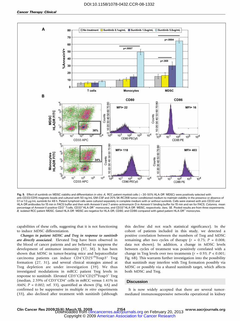
In vitro effect of sunitinib on MDSC viability and differ-entiation. We next asked whether the sunitinib-induceddepression of MDSC accumulation observed in patients couldbe related to sunitinib-mediated MDSC apoptosis or matura-tion. It was necessary to add patient MDSC to culturescontaining 20% SK-RC26B cell line tumor conditionedmedium and/or 50 ng/mL GM-CSF to support long-termMDSC viability.7 To assess the effect of sunitinib on MDSCapoptosis, patient myeloid cells were treated or not withsunitinib at 0.1, 1.0, or 5.0 Ag/mL for 48 h and then analyzedby FACS for Annexin V staining in both the CD33+HLA-DR-
MDSC and the CD33+HLA-DR+ monocytes/dendritic cells.Patient T cells were also separately cultured for 48 h incomplete RPMI 1640 with or without the same amounts ofsunitinib and assessed for viability. We found that, relative tolymphocytes, patient myeloid cells displayed an increasedsensitivity to sunitinib-induced cell death (Fig. 5A). PatientMDSC seemed also to be somewhat more sensitive tosunitinib in vitro when compared with patient monocytes.
To assess patient MDSC differentiation in response tosunitinib, isolated myeloid cells were cultured with 50 ng/mL GM-CSF and IL-4 to stimulate dendritic cell maturation aswell as 20% tumor conditioned medium to prevent indis-criminant maturation. Sunitinib was added or not to culturesat 0.1 or 1.0 Ag/mL. Figure 5B shows that patient CD33+HLA-DR- myeloid cells added to cultures were negative for theexpression of CD80 and CD86 compared with CD33+HLA-DR+ cells. After 6 days in culture, cells were harvested and theirexpression of dendritic cell maturity markers was compared bysingle-color FACS. Remaining cells were used in mixedlymphocyte reactions to stimulate allogeneic T-cell prolifera-tion. Comparison of the expression of HLA-DR, CD40, CD80,and CD86, as well as the ability to stimulate T-cellproliferation in an mixed lymphocyte reaction, shows thatsunitinib-treated and untreated cells became essentially equiv-alent dendritic cells (Fig. 5C). Sunitinib did not increase MHCclass II and costimulatory marker expression on patientmyeloid cells, nor did it increase the T-cell-stimulating
7 Unpublished results.
Sunitinib andMyeloid-Derived Suppressor Cells
www.aacrjournals.org Clin Cancer Res 2009;15(6) March15, 20092153
American Association for Cancer Research Copyright © 2009 on February 20, 2013clincancerres.aacrjournals.orgDownloaded from
DOI:10.1158/1078-0432.CCR-08-1332
capabilities of these cells, suggesting that it is not functioningto induce MDSC differentiation.Changes in patient MDSC and Treg in response to sunitinib
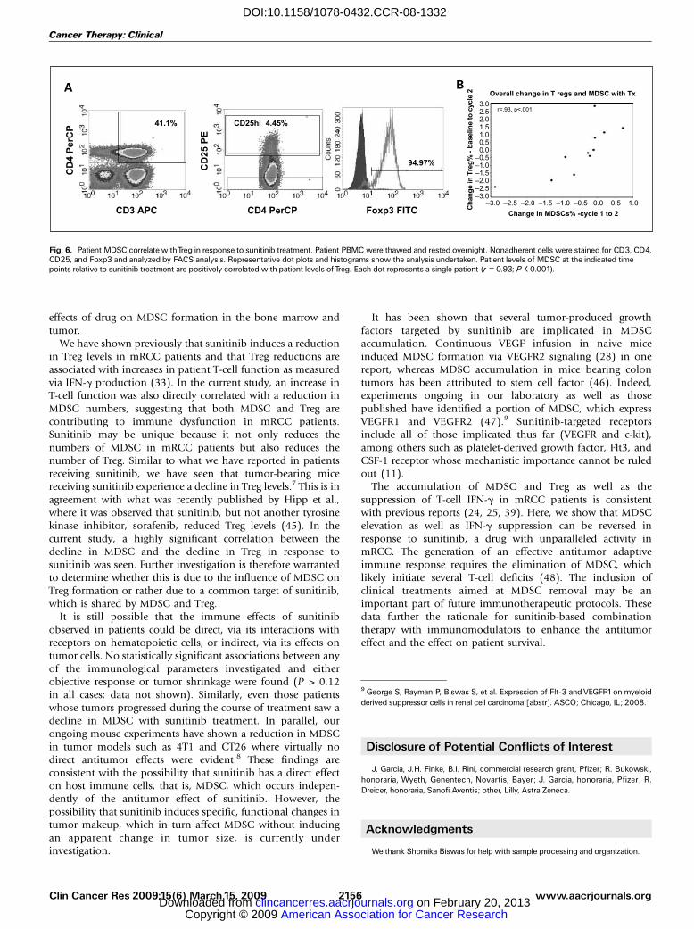
are directly associated. Elevated Treg have been observed inthe blood of cancer patients and are believed to suppress thedevelopment of antitumor immunity (37, 38). It has beenshown that MDSC in tumor-bearing mice and hepatocellularcarcinoma patients can induce CD4+CD25+hiFoxp3+ Tregformation (27, 31), and several clinical strategies aimed atTreg depletion are under investigation (39). We thusinvestigated modulations in mRCC patient Treg levels inresponse to sunitinib. Elevated CD3+CD4+CD25hiFoxp3+ Treg(median, 2.59% of CD3+CD4+ cells in mRCC versus 1.41% inAMN; P = 0.002; ref. 33), quantified as shown (Fig. 6A) andconfirmed to be suppressive in multiple in vitro experiments(33), also declined after treatment with sunitinib (although
this decline did not reach statistical significance). In thecohort of patients included in this study, we detected apositive correlation between the numbers of Treg and MDSCremaining after two cycles of therapy (r = 0.75; P = 0.008;data not shown). In addition, a change in MDSC levelsbetween cycles of treatment was positively correlated with achange in Treg levels over two treatments (r = 0.93; P < 0.001;Fig. 6B). This warrants further investigation into the possibilitythat sunitinib may interfere with Treg formation possibly viaMDSC or possibly via a shared sunitinib target, which affectsboth MDSC and Treg.
Discussion
It is now widely accepted that there are several tumor-mediated immunosuppressive networks operational in kidney
Fig. 5. Effect of sunitinib on MDSC viability and differentiation in vitro. A, RCC patient myeloid cells (f30-50% HLA-DR- MDSC) were positively selected withanti-CD33/CD15 magnetic beads and cultured with 50 ng/mL GM-CSF and 20% SK-RC26B tumor conditioned medium to maintain viability in the presence or absence of0.1or1.0 Ag/mL sunitinib for 48 h. Patient lymphoid cells were cultured separately in complete medium with or without sunitinib. Cells were stained with anti-CD33 andHLA-DR antibodies for15 min in FACS buffer and then with AnnexinVand 7-amino-actinomycin D in AnnexinV binding buffer for15 min and ran for FACS. Columns, meanpercentage of AnnexinV-positive CD3+ Tcells, CD33+HLA-DR+ monocytes, and CD33+HLA-DR- MDSC, respectively; bars, SE. Pooled results are from three experiments.B, isolated RCC patient MDSC. Gated HLA-DR- MDSC are negative for HLA-DR, CD80, and CD86 compared with gated patient HLA-DR+ monocytes.
Cancer Therapy: Clinical
www.aacrjournals.orgClin Cancer Res 2009;15(6) March15, 2009 2154
American Association for Cancer Research Copyright © 2009 on February 20, 2013clincancerres.aacrjournals.orgDownloaded from
DOI:10.1158/1078-0432.CCR-08-1332

cancer that impede the success of immune-based therapies(3, 4, 40, 41). One of these networks involves the tumor-induced accumulation of MDSC (42–44). Elevated levels ofperipheral blood MDSC in mRCC patients have been shownto decline after treatment with all-trans-retinoic acid presum-ably due to all-trans-retinoic acid-induced maturation ofthese cells (25). Here, we report that sunitinib monotherapy,a frontline treatment for patients with mRCC, inducesa significant reduction in circulating MDSC in mRCC patients.In addition, this reduction was associated with an improvementin effector T-cell function, and direct correlations were observedbetween the drug-mediated reduction in MDSC numbersand an improvement in T-cell IFN-g production as well as adecline in Treg numbers. Interestingly, we did not see acorrelation between changes in any of the immune parameterstested and changes in tumor burden, response to treatment, orsurvival.
The mechanism by which sunitinib improves type 1 T-cellfunction in mRCC patients is currently under investigation.It is likely to be in part due to the reduction of MDSC, whichinhibit effector T-cell function directly. Results from in vitroexperiments indicate that a portion of MDSC-mediated T-cellsuppression in patients was mediated by L-arginine depletionand perhaps ROS production; however, further investigation iscurrently under way to identify additional mechanisms thatmay be relatively active in all MDSC or MDSC subsets.Importantly, patient T cells can be rendered functional whenMDSC are depleted. Indeed, removal of MDSC from the PBMCof selected patients before the stimulation of their T cells
resulted in a significant improvement in the ability of thoseT cells to produce IFN-g.
Our in vitro results support the notion that patient T cellsare viable and functional even in the presence of concen-trations of sunitinib that depress MDSC viability and function.Exposure to 1 Ag/mL sunitinib induced 30% of patient MDSCto undergo apoptosis, and >60% of MDSC were killed with5.0 Ag/mL sunitinib over 48 h, although no effect on patientT-cell viability was seen. Thus, it is likely that the recovery ofIFN-g production by patient T cells that were cocultured withtheir own MDSC in the presence of 1.0 Ag/mL sunitinib was aresult of sunitinib-induced MDSC apoptosis. Modest recoveryin T-cell IFN-g production in cocultures exposed to 0.1 Ag/mLsunitinib may relate to sunitinib-mediated inhibition ofMDSC suppressive function. Sunitinib-mediated improve-ments in patient T-cell function likely occur via effects onMDSC that are exerted by multiple mechanisms including aneffect on MDSC viability and function. Because it is difficult todirectly recalculate the in vitro and in vivo doses of sunitinib,we have begun parallel experiments in several mouse tumormodels where we have also observed a dramatic suppressionof MDSC in response to sunitinib.8 We plan to evaluate theeffects of sunitinib on MDSC function and viability in tumor-bearing mice receiving sunitinib and also to determine the
Fig. 5 Continued. C, RCC patient myeloid cells were isolated and cultured as above (GM-CSF and tumor conditioned medium) with the addition of IL-4 at 50 ng/mL to aid inthe potential differentiation of myeloid cells into dendritic cells in the presence or absence of sunitinib. After 6 d, cells were harvested and a portion was stained for dendriticcell maturation markers as seen in four histograms: purple, untreated cells; green, 0.1 Ag/mL sunitinib-treated cells; pink, 1.0 Ag/mL sunitinib-treated cells. Another portionof cells was used in mixed lymphocyte reaction with freshly isolated, normal donor, allogeneicTcells.T-cell proliferation was measured after 6 d in culture by tritiated thymidineincorporation. Representative experiment from a total of three experiments.
8 Cohen et al., in preparation.
Sunitinib andMyeloid-Derived Suppressor Cells
www.aacrjournals.org Clin Cancer Res 2009;15(6) March15, 20092155
American Association for Cancer Research Copyright © 2009 on February 20, 2013clincancerres.aacrjournals.orgDownloaded from
DOI:10.1158/1078-0432.CCR-08-1332
effects of drug on MDSC formation in the bone marrow andtumor.
We have shown previously that sunitinib induces a reductionin Treg levels in mRCC patients and that Treg reductions areassociated with increases in patient T-cell function as measuredvia IFN-g production (33). In the current study, an increase inT-cell function was also directly correlated with a reduction inMDSC numbers, suggesting that both MDSC and Treg arecontributing to immune dysfunction in mRCC patients.Sunitinib may be unique because it not only reduces thenumbers of MDSC in mRCC patients but also reduces thenumber of Treg. Similar to what we have reported in patientsreceiving sunitinib, we have seen that tumor-bearing micereceiving sunitinib experience a decline in Treg levels.7 This is inagreement with what was recently published by Hipp et al.,where it was observed that sunitinib, but not another tyrosinekinase inhibitor, sorafenib, reduced Treg levels (45). In thecurrent study, a highly significant correlation between thedecline in MDSC and the decline in Treg in response tosunitinib was seen. Further investigation is therefore warrantedto determine whether this is due to the influence of MDSC onTreg formation or rather due to a common target of sunitinib,which is shared by MDSC and Treg.
It is still possible that the immune effects of sunitinibobserved in patients could be direct, via its interactions withreceptors on hematopoietic cells, or indirect, via its effects ontumor cells. No statistically significant associations between anyof the immunological parameters investigated and eitherobjective response or tumor shrinkage were found (P > 0.12in all cases; data not shown). Similarly, even those patientswhose tumors progressed during the course of treatment saw adecline in MDSC with sunitinib treatment. In parallel, ourongoing mouse experiments have shown a reduction in MDSCin tumor models such as 4T1 and CT26 where virtually nodirect antitumor effects were evident.8 These findings areconsistent with the possibility that sunitinib has a direct effecton host immune cells, that is, MDSC, which occurs indepen-dently of the antitumor effect of sunitinib. However, thepossibility that sunitinib induces specific, functional changes intumor makeup, which in turn affect MDSC without inducingan apparent change in tumor size, is currently underinvestigation.
It has been shown that several tumor-produced growthfactors targeted by sunitinib are implicated in MDSCaccumulation. Continuous VEGF infusion in naive miceinduced MDSC formation via VEGFR2 signaling (28) in onereport, whereas MDSC accumulation in mice bearing colontumors has been attributed to stem cell factor (46). Indeed,experiments ongoing in our laboratory as well as thosepublished have identified a portion of MDSC, which expressVEGFR1 and VEGFR2 (47).9 Sunitinib-targeted receptorsinclude all of those implicated thus far (VEGFR and c-kit),among others such as platelet-derived growth factor, Flt3, andCSF-1 receptor whose mechanistic importance cannot be ruledout (11).
The accumulation of MDSC and Treg as well as thesuppression of T-cell IFN-g in mRCC patients is consistentwith previous reports (24, 25, 39). Here, we show that MDSCelevation as well as IFN-g suppression can be reversed inresponse to sunitinib, a drug with unparalleled activity inmRCC. The generation of an effective antitumor adaptiveimmune response requires the elimination of MDSC, whichlikely initiate several T-cell deficits (48). The inclusion ofclinical treatments aimed at MDSC removal may be animportant part of future immunotherapeutic protocols. Thesedata further the rationale for sunitinib-based combinationtherapy with immunomodulators to enhance the antitumoreffect and the effect on patient survival.
Disclosure of Potential Conflicts of Interest
J. Garcia, J.H. Finke, B.I. Rini, commercial research grant, Pfizer; R. Bukowski,honoraria, Wyeth, Genentech, Novartis, Bayer; J. Garcia, honoraria, Pfizer; R.Dreicer, honoraria, Sanofi Aventis; other, Lilly, Astra Zeneca.
Acknowledgments
We thank Shomika Biswas for help with sample processing and organization.
Fig. 6. Patient MDSC correlate withTreg in response to sunitinib treatment. Patient PBMC were thawed and rested overnight. Nonadherent cells were stained for CD3, CD4,CD25, and Foxp3 and analyzed by FACS analysis. Representative dot plots and histograms show the analysis undertaken. Patient levels of MDSC at the indicated timepoints relative to sunitinib treatment are positively correlated with patient levels ofTreg. Each dot represents a single patient (r = 0.93; P < 0.001).
9 George S, Rayman P, Biswas S, et al. Expression of Flt-3 andVEGFR1on myeloidderived suppressor cells in renal cell carcinoma [abstr]. ASCO; Chicago, IL; 2008.
Cancer Therapy: Clinical
www.aacrjournals.orgClin Cancer Res 2009;15(6) March15, 2009 2156
American Association for Cancer Research Copyright © 2009 on February 20, 2013clincancerres.aacrjournals.orgDownloaded from
DOI:10.1158/1078-0432.CCR-08-1332

References1. Jemal A, Siegel R,Ward E, MurrayT, Xu J,Thun MJ.
Cancer statistics, 2007. CA Cancer J Clin 2007;57:43^66.
2. Yang JC, Childs R. Immunotherapy for renal cell can-cer. JClin Oncol 2006;24:5576^83.
3.TatsumiT, Kierstead LS, Ranieri E, et al. Disease-asso-ciated bias inT helper type1 (Th1)/Th2 CD4(+) Tcellresponses against MAGE-6 in HLA-DRB10401(+)patients with renal cell carcinoma or melanoma. JExpMed 2002;196:619^28.
4.TatsumiT, Herrem CJ, OlsonWC, et al. Disease stagevariation in CD4+ and CD8+ T-cell reactivity to the re-ceptor tyrosine kinase EphA2 in patients with renalcell carcinoma. Cancer Res 2003;63:4481^9.
5. Finke JH, Rayman P, Hart L, et al. Characterizationof tumor-infiltrating lymphocyte subsets from hu-man renal cell carcinoma: specific reactivity definedby cytotoxicity, interferon-g secretion, and prolifera-tion. J Immunother Emphasis Tumor Immunol 1994;15:91^104.
6. Finke JH, Rayman P, Edinger M, et al. Characteriza-tion of a human renal cell carcinoma specific cytotoxicCD8+ Tcell line. J Immunother 1992;11:1^11.
7. Brugarolas J. Renal-cell carcinomaPmolecular path-ways and therapies. NEngl JMed 2007;356:185^7.
8. Motzer RJ, HutsonTE,Tomczak P, et al. Sunitinib ver-sus interferon a in metastatic renal-cell carcinoma. NEngl JMed 2007;356:115^24.
9. Motzer RJ, Rini BI, Bukowski RM, et al. Sunitinib inpatients with metastatic renal cell carcinoma. JAMA2006;295:2516^24.
10. Garcia JA, Rini BI. Recent progress in the manage-ment of advanced renal cell carcinoma. CA Cancer JClin 2007;57:112^25.
11. Roskoski R, Jr. Sunitinib: aVEGF and PDGF receptorprotein kinase and angiogenesis inhibitor. BiochemBiophys Res Commun 2007;356:323^8.
12. Kaelin WG, Jr. The von Hippel-Lindau tumor sup-pressor protein and clear cell renal carcinoma. ClinCancer Res 2007;13:680^4s.
13. Gabrilovich DI, IshidaT, Nadaf S, Ohm JE, CarboneDP. Antibodies to vascular endothelial growth factorenhance the efficacy of cancer immunotherapy by im-proving endogenous dendritic cell function. Clin Can-cer Res1999;5:2963^70.
14. Ohm JE, Shurin MR, Esche C, Lotze MT, CarboneDP, Gabrilovich DI. Effect of vascular endothelialgrowth factor and FLT3 ligand on dendritic cell gener-ation in vivo. J Immunol 1999;163:3260^8.
15. Gabrilovich DI, Chen HL, Girgis KR, et al. Productionof vascular endothelial growth factor by humantumors inhibits the functional maturation of dendriticcells. Nat Med1996;2:1096^103.
16. Almand B, ResserJR, Lindman B, et al. Clinical sig-nificance of defective dendritic cell differentiation incancer. Clin Cancer Res 2000;6:1755^66.
17. Saito H,Tsujitani S, Ikeguchi M, Maeta M, Kaibara N.Relationship between the expression of vascular en-dothelial growth factor and the density of dendriticcells in gastric adenocarcinoma tissue. Br J Cancer1998;78:1573^7.
18. Gabrilovich D. Mechanisms and functional signifi-cance of tumour-induced dendritic-cell defects. NatRev Immunol 2004;4:941^52.
19. Kusmartsev S, Gabrilovich DI. Role of immature my-eloid cells in mechanisms of immune evasion in can-cer. Cancer Immunol Immunother 2006;55:237^45.
20. Gabrilovich DI, Velders MP, Sotomayor EM, KastWM. Mechanism of immune dysfunction in cancermediated by immature Gr-1+ myeloid cells. J Immunol2001;166:5398^406.
21. Kusmartsev SA, Li Y, Chen SH. Gr-1+ myeloid cellsderived from tumor-bearing mice inhibit primaryTcellactivation induced through CD3/CD28 costimulation.J Immunol 2000;165:779^85.
22. Almand B, Clark JI, Nikitina E, et al. Increased pro-duction of immature myeloid cells in cancer patients: amechanism of immunosuppression in cancer. J Immu-nol 2001;166:678^89.
23. Schmielau J, Finn OJ. Activated granulocytes andgranulocyte-derivedhydrogen peroxide are theunder-lying mechanism of suppression of T-cell function inadvanced cancer patients. Cancer Res 2001;61:4756^60.
24. Zea AH, Rodriguez PC, Atkins MB, et al. Arginase-producing myeloid suppressor cells in renal cell carci-noma patients: a mechanism of tumor evasion. CancerRes 2005;65:3044^8.
25. Mirza N, Fishman M, Fricke I, et al. All-trans-retinoicacid improves differentiation of myeloid cells and im-mune response in cancer patients. Cancer Res 2006;66:9299^307.
26. Filipazzi P,Valenti R, HuberV, et al. Identification of anew subset of myeloid suppressor cells in peripheralblood of melanoma patients with modulation by agranulocyte-macrophage colony-stimulation factor-based antitumor vaccine. J Clin Oncol 2007;25:2546^53.
27. Hoechst B, Ormandy LA, Ballmaier M, et al. Anew population of myeloid-derived suppressor cellsin hepatocellular carcinoma patients inducesCD4(+)CD25(+)Foxp3(+) T cells. Gastroenterolo-gy 2008;135:234^43.
28. Movahedi K, Guilliams M, Van den Bossche J,et al. Identification of discrete tumor-induced mye-loid-derived suppressor cell subpopulations withdistinct T cell-suppressive activity. Blood 2008;111:4233^44.
29. Gabrilovich D, IshidaT, OyamaT, et al.Vascular en-dothelial growth factor inhibits the development ofdendritic cells and dramatically affects the differentia-tion of multiple hematopoietic lineages in vivo. Blood1998;92:4150^66.
30. HuangY, Chen X, Dikov MM, et al. Distinct roles ofVEGFR-1and VEGFR-2 in the aberrant hematopoiesisassociated with elevated levels of VEGF. Blood 2007;110:624^31.
31. Huang B, Pan PY, Li Q, et al. Gr-1+CD115+ imma-ture myeloid suppressor cells mediate the develop-ment of tumor-induced T regulatory cells and T-cellanergy in tumor-bearing host. Cancer Res 2006;66:1123^31.
32. Therasse P, Arbuck SG, Eisenhauer EA, et al. Euro-pean Organization for Research andTreatment ofCan-cer, National Cancer Institute of the United States,National Cancer Institute of Canada. New guidelinesto evaluate the response to treatment in solid tumors.JNatl Cancer Inst 2000;92:205^16.
33. Finke JH, Rini B, Ireland J, et al. Sunitinib reversestype-1immune suppressionanddecreasesT regulatorycells in renal cell carcinoma patients. Clin Cancer Res2008;14:6674^82.
34. Mendel DB, Laird AD, Xin X, et al. In vivo antitumoractivity of SU11248, a novel tyrosine kinase inhibitortargeting vascular endothelial growth factor and plate-let-derived growth factor receptors: determination ofa pharmacokinetic/pharmacodynamic relationship.Clin Cancer Res 2003;9:327^37.
35. Motzer RJ, Bacik J, Murphy BA, Russo P, Mazum-dar M. Interferon-a as a comparative treatment forclinical trials of new therapies against advanced renalcell carcinoma. JClin Oncol 2002;20:289^96.
36. NefedovaY, Fishman M, Sherman S, et al. Mecha-nism of all-trans retinoic effect on tumor-associatedmyeloid-derived suppressor cells. Cancer Res 2007;67:11021^8.
37. Curiel TJ. Tregs and rethinking cancer immunother-apy. JClin Invest 2007;117:1167^74.
38. Cesana GC, DeRaffele G, Cohen S, et al. Character-ization of CD4+CD25+ regulatory T cells in patientstreated with high-dose interleukin-2 for metastaticmelanoma or renal cell carcinoma. J Clin Oncol 2006;24:1169^77.
39. Dannull J, Su Z, Rizzieri D, et al. Enhancement ofvaccine-mediated antitumor immunity in cancerpatients after depletion of regulatoryTcells. J Clin In-vest 2005;115:3623^33.
40.Tatsumi T, Kierstead LS, Ranieri E, et al. MAGE-6encodes HLA-DRh1*0401-presented epitopes rec-ognized by CD4+ T cells from patients with melano-ma or renal cell carcinoma. Clin Cancer Res 2003;9:947^54.
41.Vieweg J, Su Z, Dahm P, Kusmartsev S. Reversal oftumor-mediated immunosuppression. Clin Cancer Res2007;13:727^32s.
42. Ochoa AC, Zea AH, Hernandez C, Rodriguez PC.Arginase, prostaglandins, and myeloid-derivedsuppressor cells in renal cell carcinoma. Clin CancerRes 2007;13:721^6s.
43. Serafini P, De Santo C, Marigo I, et al. Derangementof immune responses by myeloid suppressor cells.Cancer Immunol Immunother 2004;53:64^72.
44.Young M, Newby M,Wepsic HT. Hematopoiesis andsuppressor bone marrow cells in mice bearing largemetastatic Lewis lung carcinoma tumors. Cancer Res1987;47:100^5.
45. Hipp M, Hilf N, Walter S, et al. Sorafenib, but notsunitinib, affects function of dendritic cells and induc-tion of primary immune responses. Blood 2008;111:5610^20.
46. Pan PY,Wang GX,Yin B, et al. Reversion of immunetolerance in advanced malignancy: modulation of my-eloid-derived suppressor cell development by block-ade of stem-cell factor function. Blood 2008;111:219^28.
47. Kusmartsev S, Eruslanov E, Kubler H, et al. Oxidativestress regulates expression ofVEGFR1in myeloid cells:link to tumor-induced immune suppression in renal cellcarcinoma. Cancer Res 2008;181:346^53.
48. Rabinovich G, Gabrilovich D, Sotomayor E. Immu-nosuppressive strategies that are mediated by tumorcells. Annu Rev Immunol 2007;25:267^96.
Sunitinib andMyeloid-Derived Suppressor Cells
www.aacrjournals.org Clin Cancer Res 2009;15(6) March15, 20092157
American Association for Cancer Research Copyright © 2009 on February 20, 2013clincancerres.aacrjournals.orgDownloaded from
DOI:10.1158/1078-0432.CCR-08-1332